Embed Size (px)
Citation preview

Biochimica et Biophysica Acta 1809 (2011) 327–336
Contents lists available at ScienceDirect
Biochimica et Biophysica Acta
j ourna l homepage: www.e lsev ie r.com/ locate /bbagrm
Changes in gene expression induced by Sp1 knockdown differ from those caused bychallenging Sp1 binding to gene promoters
Sylvia Mansilla a, Waldemar Priebe b, José Portugal a,⁎a Instituto de Biología Molecular de Barcelona, CSIC, Parc Cientific de Barcelona, E-08028 Barcelona, Spainb The University of Texas M. D. Anderson Cancer Center, Houston, TX 77030, USA
⁎ Corresponding author at: Instituto de Biologia MoleCientific de Barcelona, Baldiri Reixac, 10, E-08028 Barce4959; fax: +34 93 403 4979.
E-mail address: [email protected] (J. Portugal).
1874-9399/$ – see front matter © 2011 Elsevier B.V. Aldoi:10.1016/j.bbagrm.2011.06.003
a b s t r a c t
a r t i c l e i n f oArticle history:Received 27 January 2011Received in revised form 20 April 2011Accepted 3 June 2011Available online 13 June 2011
Keywords:Gene expressionMDA-MB231 cellsshRNASp1 knockdownWP631
C/G-rich DNA regions, which include those recognized by the Sp1 transcription factor in several genepromoters, also encompass potential binding sites for the DNA-intercalating anthracyclines doxorubicin andWP631. We explored the differences between changes in gene expression caused by the ability of these drugsto compete with Sp1 for binding to DNA and those produced by Sp1 knockdown. By quantitative RT-PCR ofaround 100 genes, most of them involved in control of cell cycle progression, we found that the treatment ofhuman MDA-MB231 breast carcinoma cells with bis-anthracycline WP631 for 24 h produced a profile of genedown-regulation markedly different from the profile caused by doxorubicin treatment or by stable Sp1knockdown. These observations are rationalized by considering a near-specific effect of WP631 on Sp1interaction with several gene promoters, thus representing potential therapeutic targets for WP631, incontrast to a less specific effect of reducing the availability of Sp1 through RNA interference. Genes down-regulated upon each treatment were mapped to their molecular and biological functions, which documentedthe down-regulation, among other things, of genes involved in mRNA transcription regulation, granting usinsights into the effects of challenging the transactivation of gene expression by Sp1.
cular de Barcelona, CSIC, Parclona, Spain. Tel.: +34 93 403
l rights reserved.
© 2011 Elsevier B.V. All rights reserved.
1. Introduction
Understanding of the mechanisms that control gene expressioncan be used to design drugs that affect tumor transcriptional patternsspecifically [1–3]. In this context exploring the mechanisms ofcompetition between transcription factors and small ligands fortheir putative DNA-binding sequences should contribute to improvingchemotherapy.
DNA-intercalators and some minor-groove binders can inhibittranscription by impeding the binding of transcription factors tocertain promoters [1–5]. The presence of C/G-rich tracts specificallyrecognized by Sp1 in several promoters suggest that DNA-Sp1complexes are potential specific targets for some small drugs thatalso recognize these sequences and may impair Sp1-activated genetranscription in vivo [2,4].
Sp1 is a ubiquitously expressed zinc-finger DNA-binding protein,which can activate or repress transcription in response to physiologicand pathological stimuli [6–9]. Sp1 binds to consensus C/G-rich tractswidely present in gene promoters, where it regulates the expression ofboth TATA-less and canonical promoters [7–9] by means of protein–
protein interactions, or interplaywith other transcription factors and/orcomponents in the basal transcriptional machinery [9,10]. It regulatesthe expression of thousands of genes involved in the control of diversecellularprocesses suchas cell growth, angiogenesis and apoptosis [9,11].Sp1 knockout in mice results in retarded development and embryonicdeath [12].
Sp1 expression is altered during some events associated withtumor development [13,14], and both pro-apoptotic and anti-apoptotic factors have been found among Sp1-targeted genes [15].Regulation of Sp1-dependent transcription may be affected bychanges in Sp1 abundance [9,11,16] and Sp1 expression increasesduring some events associated with tumor development. For a varietyof cancers, Sp1 overexpression is considered a negative prognosticfactor for survival andmight contribute tometastasis [13]. Sp1 proteinlevels are raised in most of the human breast carcinomas, but not inbenign lesions [17].
Sp1 can transactivate gene transcription via a single Sp1-bindingsite and can transactivate synergistically with other Sp1 moleculesthrough two ormore Sp1-binding siteswithout cooperative binding toDNA [9]. It has been reported that phosphorylation of Sp1 isfundamental for Sp1-dependent activation of some genes [13,18],while other modifications may inhibit its transcriptional activity [19].Anothermechanism in the regulation of gene expression by Sp1 arisesfrom Sp1 being a member of a multigene family consisting of Sp1, Sp2,Sp3 and Sp4 [8,20,21]. All four proteins have very similar structural

328 S. Mansilla et al. / Biochimica et Biophysica Acta 1809 (2011) 327–336
features, and molecular, genetic, and biochemical analyses havedemonstrated that Sp2, Sp3, and Sp4 are not simply functionalequivalents of Sp1. Both Sp1 and Sp3 are ubiquitously expressed inhuman cells, competing for common target sequences [8,21], with theSp1 to Sp3 ratio playing an important role in gene regulation [9,21].There is experimental evidence that Sp1, Sp3 and Sp4 are over-expressed in tumor cells, in which they contribute to the proliferativeand angiogenic phenotypes [22]. Sp1 and Sp3 have very similar, if notidentical, DNA-binding specificities and affinities so that they competein binding to the same C/G-rich boxes. Overall, Sp3 represses the Sp1-mediated transactivation of promoterswith two ormore Sp1 sites, buthas little effect on the Sp1-mediated transactivation of promotersbearing one Sp1 site only [9,20]. Hence, when Sp3 displaces Sp1 frompromoters with at least two Sp1 sites [23], it results in a net repressionof initial Sp1-mediated transactivation. Moreover, Sp1 can interactwith other transcription factors bound to DNA [7,8].
Bis-anthracycline WP631 bis-intercalates into C/G-rich regions inDNA with a binding constant close to that of various transcriptionfactors [1]. It is a potent inhibitor of transcription through directcompetition with the Sp1 transcription factor [24–26]. The in vivoeffects ofWP631 are not easy to separate, though the direct effects dueto inhibition of the Sp1-binding to DNA should be distinguished fromthe more indirect effects due to the network of pathways relating tothe control of progression through the cell cycle, which may decreasetranscription levels without direct drug interference with Sp1-DNAinteractions [27]. For the easy of comparison between the effects ofdoxorubicin orWP631 and of the knockdown of Sp1-family members,we interfered Sp1 exclusively because this protein acts as a geneactivator, and thus its down-regulation can result in effects that couldbe equivalent to the down-regulation by known gene repressors suchas the two antitumor agents. The different isoforms of Sp3 can eitheractivate or repress gene expression [28] and thus their knockdownmay, at first glance, provide with gene expression effects that aredifficult to compare with those of the small molecules. In this contextwe have demonstrated elsewhere that the effect of WP631 on Sp1-transactivated gene expression does not require the binding of Sp3to the consensus binding sites [26].
We used human MDA-MB231 breast carcinoma cells, which carrymutant p53 at both alleles [29], to examinewhether the known abilityof the anthracyclines doxorubicin and WP631 to interfere with genetranscription induces changes in the cell transcriptome that areequivalent to reducing Sp1 levels by Sp1 knockdown, since in bothcircumstances it is expected that the amount of active Sp1 availablefor transactivating gene transcription will be reduced. For the smalldrugs, this should be a consequence of their competing for the bindingto putative Sp1-binding sites in promoters, whereas when interfer-ence RNA is used a shortage in the Sp1 protein levels is expected. Inaddition, variations in transcription of particular genes may provideus with insights into the mechanisms of drug sensitization andresistance.
Fig. 1. DNA sequences designed to be homologous to Sp1-encoding RNA (named as A-Sp1 ancomplementary domains highlighted in bold. The oligonucleotides were annealed and ligat
2. Materials and methods
2.1. Design of shRNA template oligonucleotides and construction of thepSUPER expression vector
Two potential shRNA target sequences for Sp1 RNA (named asA-Sp1 and B-Sp1) were designed to be homologous to different zonesof the Sp1 cDNA sequence (NCBI nucleotide cDNA sequence:BC043224). For the unrelated P. pyralis luciferase, the gene sequence(GeneBank accession number: M15077) was used to design theshRNA. Complementary oligonucleotides encoding hairpin structureswith a 19-mer stem derived from the mRNA target site were designedonline at JURA web site (http://jura.wi.mit.edu/bioc/siRNAext), and a19-bp loop sequence separated the two complementary domains(Fig. 1). For each shRNA, the 5′ end of two oligonucleotides containedBglII and HindIII restriction site overhangs. The oligonucleotides wereannealed and ligated into the BglII and HindIII sites of linearizedpSUPER.retro.puro system (OligoEngine), following the supplier'sprotocols, to obtain shRNA expression plasmids. The resultingplasmids were named as pSUPER.A-Sp1shRNA, pSUPER.B-Sp1shRNAand pSUPER.LucshRNA respectively.
2.2. Cell culture
Human MDA-MB231 breast carcinoma cells were maintained inRPMI 1640 medium (Invitrogen) supplemented with 10% fetal bovineserum (Invitrogen) 100 U/ml penicillin, 100 μg/ml streptomycin,2 mM sodium pyruvate (Invitrogen), and 2 mM HEPES (pH 7.4), at37 °C in a humidified atmosphere with 5% CO2.
2.3. Transfection with shRNA expression vector and drug treatments
MDA-MB231 cells in the exponential phase of growthwere seededin 6-well plates at 2×10⁵ cells per well, grown for 24 h, and thentransfected with shRNA expression plasmids specific to Sp1 preparedas described above. Control cells were transfected with pSUPERplasmid without inserts, or with pSUPER plasmid encoding RNA forluciferase (pSUPER.LucshRNA, Fig. 1). Cells were also co-transfectedwith pCMV-Luc plasmid (Stratagene) expressing luciferase. MDA-MB231 cells stably expressing shRNA were established by selection incell culture medium containing 1 μg/ml puromycin (Sigma).
Stock solutions containing 500 μM doxorubicin (Sigma) or WP631(synthesized as described elsewhere [30]) were prepared with sterile150 mM NaCl, maintained at −20 °C, and brought to the finalconcentration with RPMI 1640 medium (Invitrogen) just before use.Exponentially growing cells subcultured at a density of 5×10⁴ cells/mlwere incubated with 300 nM doxorubicin or WP631 at 37 °C for 4 and24 h.
d B-Sp1) and to the unrelated luciferase RNA. A 19-bp loop sequences separated the twoed into an shRNA expression vector as detailed in the main text.

329S. Mansilla et al. / Biochimica et Biophysica Acta 1809 (2011) 327–336
2.4. Testing Sp1 knockdown by Western blotting
To analyze whether the shRNAs described above were able tospecifically knockdown Sp1 levels, MDA-MB231 cells transfected withboth A-Sp1 shRNA or B-Sp1 shRNA, or with both shRNA constructstogether, or with Luc shRNA (always co-transfected with the pCMV-Lucplasmid) were used to obtain cell extracts with a lysis buffer consistingof 50 mM Tris–HCl (pH 8), 150 mM NaCl, 5 mM EDTA, 0.5% NonidetP-40 and 0.1 mM phenylmethylsulfonyl fluoride, containing 2 μg/mlaprotinin and 0.1 μg/ml leupeptin. Equal amounts of protein (50 μg)were separated on 10% SDS-polyacrylamide gels and transfected toOptitran BA-S85 membranes (Schleicher and Schuell). The blots wereincubated with either rabbit polyclonal antibodies against Sp1, Sp3 orSp4 (all from Santa Cruz Biotechnology) and β-tubulin (ChemiconInternational).Membraneswerewashed and incubatedwith secondaryperoxide-conjugated anti-(rabbit IgG) antibody, and the bound anti-bodywasdetectedby chemiluminescenceusing luminol (Sigma). Signalintensities were quantified in a Bio-Rad GS-800 densitometer, and usedto determine the relative Sp1 contents in the knockdown experiments.Moreover, to examine the specificity of Sp1 depletion, changes inducedby RNA interference to the unrelated luciferase activity were deter-mined in a Sirius luminometer (Berthold Detection Systems).
2.5. RNA extraction and Quantitative real Time RT-PCR assays
The experiments were designed and carried out in accordancewith the MIQE guidelines [31]. Details are provided in the followingparagraphs and in Results.
Total RNA was isolated from control cells (those to which noshRNA or drug was added) and from cells stably transfected withASp1-shRNA or treated with either 300 nM doxorubicin or 300 nMWP631 for 4 h or 24 h. The UltraspecRNA isolation reagent (Biotecx)was used following the procedure provided by the vendor. RNA wasdigested with RNAse-free DNAse I (Roche) in the presence of RNAseinhibitors (RNasin, Promega), phenol extracted and precipitated. Thepellet was dissolved in RNAse-free water. The yield and purity of totalRNA were assessed spectrophotometrically, and RNA integrityexamined in an Agilent 2100 Bioanalyzer.
For quantitative real time PCR (qRT-PCR), first strand cDNAs weresynthesized from 4 μg of DNAse I-treated total RNA, obtained of twobiological replicates, in a 20 μl reaction volume using the Transcriptionfirst strand cDNA synthesis kit (Roche) following manufacturer'sinstructions. The Human Cell Cycle RT2 Profiler PCR Array (SABios-ciences) was used to analyze the expression of 89 genes mainlyinvolved in cell cycle regulation. Moreover, other 11 genes known torespond to Sp1-transactivation, including Sp1 and Sp3, were alsoincluded in the quantitative analysis. For them, gene-specific primerswere designed using the Primer Express software (Applied Biosystems).Supplementary Table S1 shows a list of these genes and the primers(purchased from Sigma) used in the qRT-PCR experiments. qRT-PCRreactionswere performed in triplicate in a Roche LightCycler 480, usingthe SYBR-Green PCR Master Mix (Roche). PCR conditions included aninitial denaturation step at 95 °C for 10 min, followed by 45 cycles of adenaturation step at 95 °C for 15 s and an annealing/extension step at60 °C for 1 min. A final dissociation curvewas generated to verify that asingle productwas amplified. Series of 10-fold dilutions of genomicDNAcovering a total dilution range from 1 to 10−6 was used to generate thestandard curves to calculate the efficiency for the genes that were notcontained within the commercial array. Reactions in the absence oftemplate and in the absence of enzyme were also performed for eachprimer pair as negative controls.
2.6. Analysis and clustering of gene expression
The qRT-PCR raw data were analyzed using the PCR Array Dataanalysis template (SABiosciences) and the software available online at the
SABioesciences website: www.sabiosciences.com/pcr/arrayanalysis.php,which provided a statistical analysis of data. Relative expression values ofthe different genes were calculated from the threshold cycle (Ct)following the ΔΔCt method [32] using GAPDH as internal housekeepingcontrol.
Logarithmic (log2) values of the normalized expression ratios(treated versus untreated cells) were hierarchically clustered usingthe TIGR-MeV program [33]. Clustering was based on the Pearsoncorrelations, and the average linkagemethodwas used with leaf orderoptimization of both samples (treatments) and genes.
Functional classification of genes down-regulated upon treat-ments with ASp1-shRNA or 300 nM WP631 was performed inBiological Process and Molecular Function categories by comparisonwith the list of all the genes using PANTHER 6.2 [34] at http://panther6.ai.sri.com/tools, which uses the binomial test for assessingstatistical significance.
2.7. Computational analysis of putative Sp1-binding sites in thepromoters of down-regulated genes
The proximal−80 to+1 gene promoter sequences were retrievedfrom the Transcriptional Regulatory Element database (TRED) at http://rulai.cshl.edu/cgi-bin/TRED. Presence of putative Sp1-binding sites ingene promoters was searched online using AliBaba 2.1 (http://www.gene-regulation.com/pub/programs/alibaba2/index.html), using thestandard setting except for the ‘minimal mat. conservation’ that wasfixed to the highest (80%) value.
2.8. Changes in protein levels after treatment with WP631
To explore whether the changes in gene transcription induced byWP631 were also observable at the protein level, we analyzed theeffect of different concentrations of this drug on Sp1, Sp3 and CCNB1(Cyclin B1) by Western blot, using the antibodies and experimentaldetails described in Section 2.4, as well as an antibody against Cyclin B(Santa Cruz Biotechnology). Western blots were quantified in a Bio-Rad GS-800 densitometer.
3. Results
As an experimental model, we used two different shRNAs specificto Sp1-RNA (named as A-Sp1 and B-Sp1; Fig. 1) to knockdown Sp1levels in human MDA-MB231 breast carcinoma cells. Fig. 2A showsdirect confirmation, by use of antibodies, that both shRNAs causeddecrease in the levels of Sp1 protein in stably transfected cells, thoughthe highest effect (69% knockdown) was observed with A-Sp1 shRNA.Indeed, a stronger effect was observed in cells transfected with bothshRNA together, but this co-transfection resulted in toxicity (cellmortality) that was too high upon transfection, which is why all thefollowing experiments were undertaken in MDA-MB231 cells thatstably expressed A-Sp1 shRNA. Sp1 knockdown resulted in a decreasein the amounts of the three forms of the related Sp3 protein (Fig. 2B),although a lower effect was observed on the larger ‘activating’isoform, about 30% decrease, than in the truncated isoforms (42–48%decrease) that might act as gene repressors. Besides, Sp1 knockdownhad a slight effect on Sp4 protein levels (Fig. 2B). It is worth notingthat changes in Sp3 and Sp4 protein levels would be produced by thereduction of Sp1 protein, instead of being a consequence of a non-specific effect of Sp1 shRNA against Sp3 and Sp4 expression, since Sp1is involved in the regulation of those genes [9]. Fig. 2C documents howwhile the levels of luciferase were reduced by an shRNA specific toluciferase (Luc shRNA), the intracellular levels of luciferase remainedunaltered in the presence of A-Sp1 shRNA, consistent with thespecificity of this shRNA. The effects of interfering RNA on Sp1transcription were also evaluated by quantitative qRT-PCR (seebelow) which showed a down-regulation of its transcription, while

Fig. 2. Interfering Sp1 RNA (shRNA) decreased Sp1 protein levels in human MDA-MB231 breast carcinoma cells. (A) Changes in Sp1 protein levels induced by two different shRNA(B-Sp1 and A-Sp1) after transfection and by the co-transfection of both shRNAs (A+B-Sp1), while and shRNA specific to luciferase did not affect Sp1 levels. β-tubulin was used as agel loading control used to normalize the protein levels. (B) Effect of Sp1 knockdown on Sp3 and Sp4 levels. The effect on the different isoforms of Sp3 is uneven. On average,about 40% decrease in Sp3 protein levels was observed, while the levels of Sp4 remained almost unaltered. (C) Changes in the relative luciferase activity measured by luminometry(mean±SD; for three experiments) were used to document that A-Sp1 shRNA did not interfere with the expression of the unrelated luciferase gene, which in turn was down-regulated by the presence of an shRNA specific to luciferase (*pb0.01; unpaired Student's t-test). The effect of A-Sp1 shRNA on Sp1 and Sp3 transcriptional levels was alsodocumented by qRT-PCR (see Fig. 4 and Table S2).Interfering Sp1 RNA (shRNA) decreased Sp1 protein levels in human MDA-MB231 breast carcinoma cells. (A) Changes in Sp1protein levels induced by two different shRNA (B-Sp1 and A-Sp1) after transfection and by the co-transfection of both shRNAs (A+B-Sp1), while and shRNA specific to luciferase didnot affect Sp1 levels. β-tubulin was used as a gel loading control used to normalize the protein levels. (B) Effect of Sp1 knockdown on Sp3 and Sp4 levels. The effect on the differentisoforms of Sp3 is uneven. On average, about 40% decrease in Sp3 protein levels was observed, while the levels of Sp4 remained almost unaltered. (C) Changes in the relativeluciferase activity measured by luminometry (mean±SD; for three experiments) were used to document that A-Sp1 shRNA did not interfere with the expression of the unrelatedluciferase gene, which in turn was down-regulated by the presence of an shRNA specific to luciferase (*pb0.01; unpaired Student's t-test). The effect of A-Sp1 shRNA on Sp1 and Sp3transcriptional levels was also documented by qRT-PCR (see Fig. 4 and Table S2).
330 S. Mansilla et al. / Biochimica et Biophysica Acta 1809 (2011) 327–336
the transcription of the unrelated luciferase was not affected, whichdemonstrates the specificity of Sp1 knockdown.
We explored by qRT-PCR the effects of 4-h and 24-h treatmentswith either 300 nMdoxorubicin or 300 nMWP631 on gene expressionin MDA-MB231 breast carcinoma cells, and as a consequence of stableSp1 knockdown. For each experimental condition, altered genetranscription patterns were observed with down-regulation of somegenes and up-regulation of others. Fig. 3 shows scatter plots of thecorrected levels of gene expression upon treatment compared tountreated MDA-MB231 cells used as control. A comparison of thepanels in this figure shows that the different treatments enhanced orinhibited the expression of distinct sets of genes. In general, changes ingene expression observed after 4-h treatmentwith each anthracyclinewere similar to those obtained after 24 h, though in 24-h experimentsthe expression fold change (treated versus untreated) was, in general,larger (Supplementary Table S2).
Among the 100 genes examined by qRT-PCR, which included thehousekeeping GAPDH used for data normalization, treatment with300 nM WP631 for 24 h altered significantly (≥2-fold; pb0.05) the
expression of 62 genes (29 genes (47%) down-regulated), while 24-htreatment of MDA-MB231 cells with 300 nM doxorubicin altered theexpression of 72 genes (4 genes (6%) down-regulated). Stable Sp1knockdown resulted in the altered expression of 68 genes (7 genes(10%) down-regulated). A complete data list of the effects of alltreatment conditions on gene expression is given in supplementaryTable S2. This table includes the average Ct values obtained in theqRT-PCR assays performed in triplicate. Notably, there are only twogenes whose expression was down-regulated significantly by all thetreatments: Sp1 and DHFR, the later a paradigm of TATA-lesspromoters. CDKN2A (p16), which participates in cell cycle checkpointcontrols, was down-regulated upon 24-h treatments with theanthracyclines, but not by Sp1 knockdown.
A hierarchical clustering analysis of the transcripts affected by thedifferent treatments, based on the Pearson correlation between thenormalized fold changes in gene expression (treated versusuntreated) is shown in Fig. 4A. Average linkage clustering wasoptimized for both gene expression and response to treatments. Inthis way, information generated on drug activity in MDA-MB231 cells

Fig. 3. Distribution of the treatment-induced expression profiles in MDA-MB231 breast carcinoma cells. Bivariate scatter plots display a comparison of the expression profilesuntreated, control cells, versus the transcription profiles induced by stable Sp1 knockdown (A-Sp1 shRNA), WP631 (treatments with 300 nM drug for 4 h or 24 h) and doxorubicin(treatments with 300 nM for 4 h or 24 h). Data are corrected average Ct values obtained from qRT-PCR experiments performed in triplicate, which are plotted in normalized 0 to 1scales for the sake of comparison.
331S. Mansilla et al. / Biochimica et Biophysica Acta 1809 (2011) 327–336
could be compared with the effect of stable Sp1 knockdown on geneexpression. The average linkage dendrograms, shown on the left sideof the cluster (Fig. 4A), contain four large coherent clusters, one ofwhich corresponds to genes down-regulated by all treatments, andanother corresponds to genes down-regulated by treatments withWP631, but up-regulated by doxorubicin and Sp1 knockdown.Dendrograms showing the hierarchical clustering of the differenttreatments are shown at the top of Fig. 4A. Treatments of MDA-MB231cells with WP631 for 4 and 24 h clustered nearby, and the sameoccurred with the two treatments with doxorubicin, which clusteredcloser to Sp1. More striking is that Sp1 knockdown and WP631clustered at greater distances.
Venn diagrams were used for their ease of comparison betweenthe different experimental conditions. They disclosed the relationsbetween the differences in gene expression produced by everytreatment in gene transcription levels. Fig. 4B compares globaltranscription effects of ASp1 versus those produced by WP631, aswell as ASp1 versus doxorubicin andWP631 versus doxorubicin (24-htreatments with the drugs).
Among the genes down-regulated by WP631, there was relative“enrichment” of genes belonging to pathways related to cell cycleprogression and entry into mitosis, for example, MYC, NRAS andCDKN2A (p16), as well as Sp1 and Sp3 (Figs. 4 and S1). A Venn diagramof Sp1 knockdown (ASp1) versus WP631 treatment for 24 h revealedthat one gene (EGR1) showed attenuated transcription due to theshRNA expression, while it was not affected by the treatment withWP631 (supplementary Fig. S1A). In this Venn diagram it was clearthat the number of genes altered by Sp1 and WP631 differedmarkedly. However, 6 genes were down-regulated by both treat-ments (Sp1, DHFR, ANAPC2, RAD1, SERTAD1 and TFDP2). Down-regulation of these genes was greater in the presence of WP631(Table S2). In fact, as mentioned above, Sp1 and DHFRwere repressedby all treatments. Fig. S1B shows Venn diagrams of the genes thatwere up-regulated upon treatment with the drug or by stable Sp1knockdown.
Table 1 lists geneswhose expressionwas down-regulated byWP631but not by Sp1knockdown(N2-fold; pb0.01). This set of genesmight beseen as potential therapeutic targets for the bis-anthracycline. Among

Fig. 4. Changes in the expression of 100 genes examined in MDA-MB231 breast carcinoma cells by qRT-PCR upon treatment with 300 nM WP631 or 300 nM doxorubicin for 4 h or24 h, and in cells expressing a stable shRNA specific to Sp1 (A-Sp1 shRNA). (A) Hierarchical clustering based on the Pearson correlation of corrected induction of repression fold foreach treatment, normalized for the expression of the housekeeping GAPDH gene, compared to the gene expression in untreated cells. (B) Venn diagrams representing genes affectedby Sp1 knockdown, or by 24-h treatments with WP631 or doxorubicin (≥2.0 fold change; pb0.05). Numbers inside the intersections correspond to genes influenced by bothtreatments.
332 S. Mansilla et al. / Biochimica et Biophysica Acta 1809 (2011) 327–336
these is BRCA1 (breast cancer 1, early onset), which is considered apotential target in the treatmentof breastmalignancies [35]. TheWP631effects on gene transactivation might be enhanced by the direct down-regulation of the Sp3 gene, which is not observed with doxorubicin orSp1 knockdown (Tables 1 and S2).
A functional classification of genes down-regulated by either Sp1knockdown or WP631 (24-h treatments) was undertaken with thePanther tool [34], which also provided us with a statistical estimate ofthe degree of over-representation of a given functional category.Table 2 lists molecular functions and biological processes of over-
represented genes among the down-regulated ones. Although theenrichment in certain genes in some of the classes was close torandom occurrence (pN0.05; binomial test) –which to some extent isa likely consequence of the relatively small size of the experimentaldata set, consisting of 100 genes characterized by qRT-PCR – thecoincidences and differences among the genes listed for eithertreatment in Table 2 were significant for the most widely representedcategories (pb0.01).
To corroborate whether changes in gene expression observed upontreatment with bis-anthracycline WP631 were consistent with a

Table 1Genes down-regulated N2-fold in human MDA-MB231 breast carcinoma cells by 24-htreatment with 300 nM WP631, but whose transcription levels were not significantlyreduced by stable Sp1 knockdown.a
Gene Fold repression p-Valueb Gene Fold repression p-Valueb
SP3 14.1 2.00E-06 CKS2 14.4 4.88E-04MYC 15.3 0.00E+00 CUL1 6.3 1.00E-06SRC 3.7 1.78E-03 GTF2H1 4.1 3.00E-06NRAS 2.1 5.50E-04 GTSE1 3.8 0.00E+00CCNA2 7.0 1.93E-04 MAD2L2 2.2 5.10E-05ABL1 3.7 6.40E-05 MCM3 3.5 0.00E+00BRCA1 2.3 1.04E-04 MCM5 2.2 1.85E-04CCNB1 3.0 4.00E-06 MKI67 4.4 6.29E-04CCNB2 3.2 5.00E-06 RAD17 2.7 2.51E-02CCNF 5.7 3.10E-05 RAD9A 4.1 0.00E+00CDK5RAP1 2.5 5.50E-05CDK7 3.3 2.10E-05CDKN2A 4.0 6.00E-06
a These genes might be down-regulated by the interfering of bis-anthracyclineWP631 with Sp1 binding to their promoters, thus representing potential therapeutictargets.
b Student's t-test p value (treated versus untreated cells) from experimentsperformed in triplicate.
333S. Mansilla et al. / Biochimica et Biophysica Acta 1809 (2011) 327–336
preferential effect of WP631 on Sp1-binding sites, we performed apredictive ‘in silico’ analysis of Sp1-binding sites in the proximalpromoter (−80 to +1) regions of the genes that were N2.5-fold(pb0.01) down-regulated by WP631. Among them, four promoters(CUL1, RAD17, SRC and TFDP2) lacked the putative Sp1-binding in thisproximal region (Fig. 5). For SRC, Sp1-binding sites have been,nevertheless, characterized upstream from the proximal region shownin Fig. 5 [36]. The effects of 24-h treatments with WP631 on genes likeMYC or CKS2 were more pronounced in keeping with the presence ofSp1-putative binding sites that also encompass C/G-rich sequencespreferentially recognized by the drug (cf. Fig. 5 and Ref. [37]). Fig. 6presents some examples that illustrate that down-regulation of genetranscription by the bis-anthracycline WP631 through the Sp1-bindingregions in gene promoters can end in the reduction of protein levels.
Table 2Genes differentially down-regulated in human MDA-MB231 breast cancinoma cells by stamapped to Panther molecular function and biological process categories.
Sp1 knockdown
Molecular functionBasal transcription factor and other transcription factors SERTAD1; TFDP2Basic helix-loop-helix transcription factorKinase activatorKinase inhibitorKRAB box transcription factor EGR1Non-receptor Tyrosine protein kinaseNucleic acid binding Sp1; EGR1; RAD1; TFDP2Reductase DHFR; EGR1Transcription cofactor and zinc finger transcription factor Sp1; EGR1Transcription factor Sp1; SERTAD1; EGR1; TFDP2
Biological processApoptosis and induction of apoptosis TFDP2Cell cycle control ANAPC2; RAD1
Cell proliferation and differentiation Sp1; TFDP2DNA repair RAD1DNA replicationMitosis ANAPC2mRNA transcription Sp1; EGR1; TFDP2mRNA transcription initiationmRNA transcription regulation EGR1; TFDP2; Sp1Oncogene or oncogenesis
Tumor suppressorNucleotide, nucleoside metabolism Sp1; DHFR; RAD1; EGR1; TF
4. Discussion
In this study, we found that treatment of MDA-MB231 humanbreast carcinoma cells with the antitumor anthracyclines doxorubicinor WP631, down-regulated the expression of Sp1 and of several genesthat can be transactivated by Sp1. Both the number of affected genes,with the larger set of them altered by WP631, and their involvementin several pathways that control cell cycle function differed from thegenes that became down-regulated by stable Sp1 knockdown attainedby RNA interference using a specific shRNA. The treatments of MDA-MB231 cells with WP631 for 4 or 24 h clustered at a short distance inhierarchical dendrograms (Fig. 4A), and at a large distance from Sp1knockdown and the two treatments with doxorubicin, which, in turn,clustered next to Sp1. These observations indicate that in MDA-MB231 cells the effect of Sp1 knockdown on gene expression is moresimilar to the treatment with doxorubicin than with WP631. Thedistance (in terms of Pearson correlation) between WP631 and Sp1knockdown corroborates that they have dissimilar effects on geneexpression. In addition, the mapping of down-regulated genes upontreatment with WP631 indicates a relative enrichment in genesrelated to the regulation of mRNA synthesis and processing (Table 2).
Although doxorubicin, as well as the structurally related dauno-rubicin, bind to C/G-rich regions in DNA, their binding site is shorterthan that of bis-anthracycline WP631 [24,30,37], which results inhigher affinity of the latter for binding to 6-pb long DNA regionscontaining, in general, CpG steps [30,37]. Among these is the putativeSp1-binding sequence found in various gene promoters (Fig. 5 and[24,26]). We have demonstrated elsewhere that the capacity forinhibiting transcription byWP631 is stronger in promoters containingSp1-binding sites than in promoters lacking the protein-binding site,and clearly superior to the effect of either daunorubicin ordoxorubicin [24,27], see also Fig. 6. The observation that Sp3 genewas only down-regulated by WP631 (Tables 1 and S2) may proverelevant to the in vivo strong effect of this compound on Sp1-dependent transcription [26], since Sp3 can compete with Sp1 forbinding to promoters [23].
ble RNA interference (Sp1 knockdown), or by 24-h treatment with 300 nM WP631,
WP631
GTF2H1; SERTAD1; TFDP2MYCCCNB1 (Cyclin B1); CCNB2 (Cyclin B2); RAD9A; CCNA (Cyclin A2)CDKN2A (p16)
SRC; CDK7; ABL1 (v-abl)Sp1; Sp3; MYC; MCM3;MCM5; RAD1DHFRSp1; Sp3; BRCA1Sp1; SERTAD1; Sp3; MYC; GTF2H1; TFDP2
TFDP2, MYC; ABL1(v-abl); CUL1ANAPC2; CCNB1 (Cyclin B1); CDKN2A (p16); CCNB2 (Cyclin B2); MYC; RAD1;RAD17; RAD 9A; BRCA1;SRC; CCNA (Cyclin A2); CDK7; ABL1 (v-abl); CUL1Sp1; Sp3; TFDP2; GTSE1; SRC; NRAS (v-ras)RAD1; RAD17; RAD9A; BRCA1MCM3; MCM5ANAPC2; CDK7Sp1; Sp3; MYC; GTF2H1; BRCA1GTF2H1MYC; BRCA1; Sp1MYC; SRC; CDKN2A (p16); CCNA (Cyclin A2); NRAS (v-ras); BRCA1;CUL1; ABL1 (v-abl)CDKN2A (p16); BRCA1
DP2 Sp1; Sp3; BRCA1; DHFR; MYC; RAD17; MCM3; RAD1; RAD9A; GTF2H1; MCM5
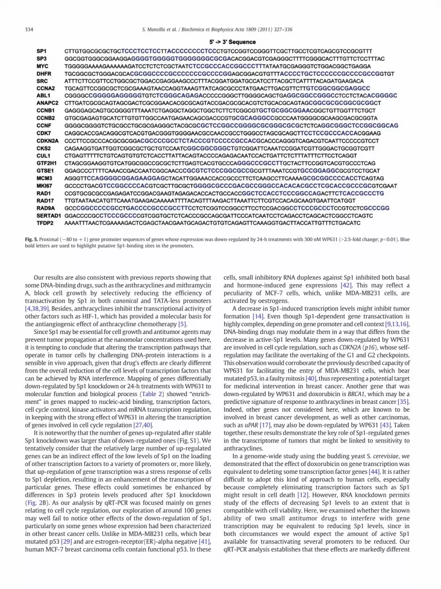
Fig. 5. Proximal (−80 to +1) gene promoter sequences of genes whose expression was down-regulated by 24-h treatments with 300 nMWP631 (N2.5-fold change; pb0.01). Bluebold letters are used to highlight putative Sp1-binding sites in the promoters.
334 S. Mansilla et al. / Biochimica et Biophysica Acta 1809 (2011) 327–336
Our results are also consistent with previous reports showing thatsomeDNA-binding drugs, such as the anthracyclines andmithramycinA, block cell growth by selectively reducing the efficiency oftransactivation by Sp1 in both canonical and TATA-less promoters[4,38,39]. Besides, anthracyclines inhibit the transcriptional activity ofother factors such as HIF-1, which has provided a molecular basis forthe antiangiogenic effect of anthracycline chemotherapy [5].
Since Sp1may be essential for cell growth and antitumor agentsmayprevent tumor propagation at the nanomolar concentrations used here,it is tempting to conclude that altering the transcription pathways thatoperate in tumor cells by challenging DNA-protein interactions is asensible in vivo approach, given that drug's effects are clearly differentfrom the overall reduction of the cell levels of transcription factors thatcan be achieved by RNA interference. Mapping of genes differentiallydown-regulated by Sp1 knockdown or 24-h treatments withWP631 tomolecular function and biological process (Table 2) showed “enrich-ment” in genes mapped to nucleic-acid binding, transcription factors,cell cycle control, kinase activators and mRNA transcription regulation,in keeping with the strong effect of WP631 in altering the transcriptionof genes involved in cell cycle regulation [27,40].
It is noteworthy that the number of genes up-regulated after stableSp1 knockdownwas larger than of down-regulated ones (Fig. S1). Wetentatively consider that the relatively large number of up-regulatedgenes can be an indirect effect of the low levels of Sp1 on the loadingof other transcription factors to a variety of promoters or, more likely,that up-regulation of gene transcription was a stress response of cellsto Sp1 depletion, resulting in an enhancement of the transcription ofparticular genes. These effects could sometimes be enhanced bydifferences in Sp3 protein levels produced after Sp1 knockdown(Fig. 2B). As our analysis by qRT-PCR was focused mainly on genesrelating to cell cycle regulation, our exploration of around 100 genesmay well fail to notice other effects of the down-regulation of Sp1,particularly on some genes whose expression had been characterizedin other breast cancer cells. Unlike in MDA-MB231 cells, which bearmutated p53 [29] and are estrogen-receptor(ER)-alpha negative [41],human MCF-7 breast carcinoma cells contain functional p53. In these
cells, small inhibitory RNA duplexes against Sp1 inhibited both basaland hormone-induced gene expressions [42]. This may reflect apeculiarity of MCF-7 cells, which, unlike MDA-MB231 cells, areactivated by oestrogens.
A decrease in Sp1-induced transcription levels might inhibit tumorformation [14]. Even though Sp1-dependent gene transactivation ishighly complex, depending on gene promoter and cell context [9,13,16],DNA-binding drugs may modulate them in a way that differs from thedecrease in active-Sp1 levels. Many genes down-regulated by WP631are involved in cell cycle regulation, such as CDKN2A (p16), whose self-regulation may facilitate the overtaking of the G1 and G2 checkpoints.This observationwould corroborate the previously described capacity ofWP631 for facilitating the entry of MDA-MB231 cells, which bearmutatedp53, in a faultymitosis [40], thus representing apotential targetfor medicinal intervention in breast cancer. Another gene that wasdown-regulated by WP631 and doxorubicin is BRCA1, which may be apredictive signature of response to anthracyclines in breast cancer [35].Indeed, other genes not considered here, which are known to beinvolved in breast cancer development, as well as other carcinomas,such as uPAR [17], may also be down-regulated by WP631 [43]. Takentogether, these results demonstrate the key role of Sp1-regulated genesin the transcriptome of tumors that might be linked to sensitivity toanthracyclines.
In a genome-wide study using the budding yeast S. cerevisiae, wedemonstrated that the effect of doxorubicin on gene transcriptionwasequivalent to deleting some transcription factor genes [44]. It is ratherdifficult to adopt this kind of approach to human cells, especiallybecause completely eliminating transcription factors such as Sp1might result in cell death [12]. However, RNA knockdown permitsstudy of the effects of decreasing Sp1 levels to an extent that iscompatible with cell viability. Here, we examined whether the knownability of two small antitumor drugs to interfere with genetranscription may be equivalent to reducing Sp1 levels, since inboth circumstances we would expect the amount of active Sp1available for transactivating several promoters to be reduced. OurqRT-PCR analysis establishes that these effects are markedly different

Fig. 6. (A) A representative Western blot showing concentration-dependent changesinduced byWP631 on Sp1, Sp3 and cyclin B levels. MDA-MB231 cells were treated withthe concentrations of WP631 indicated in the figure for 24 h before protein extraction.(B) Quantitative representation of changes in protein levels induced by WP631. Valuesare arithmetic means of two independent experiments with similar results. Relativeprotein expression was normalized to β-tubulin, and the protein levels in untreatedcells were set as 100%.
335S. Mansilla et al. / Biochimica et Biophysica Acta 1809 (2011) 327–336
(Figs. 3 and 4), in keeping with previous reports on doxorubicin andWP631 direct effects on gene transcription [4,27,45]. Althoughdoxorubicin down-regulates several genes in MDA-MB231 cellsupon treatment, the number of affected genes was clearly lowerthan upon treatment with WP631 (Figs. 4B and S1). Furthermore, asuperior effect of WP631 was observed, consistent with its strongerbinding to Sp1-binding sites [24].
Understanding why certain genes can be down-regulated by someantitumor drugs at the transcriptional level and how this is aconsequence of interference with DNA-protein interactions mighthelp us in the process of drug discovery. The effects of several smallmolecules, other than those described here, have been also documen-ted [46–48], while other strategies have been explored so far to reduceSp1 activity, which include using ‘decoy’ oligonucleotides and RNAinterference to produce cell growth inhibition in several cancermodels[14,42]. Small drugs can be easier to exploit clinically because theirpharmacokinetics and delivery in vivo can present advantages overRNA interference approaches. Nevertheless, the differences we haveseen between using small antitumor drugs and RNA interferencepresents an opportunity to develop newdrugs using RNA interference.
Supplementarymaterials related to this article can be found onlineat doi:10.1016/j.bbagrm.2011.06.003.
Acknowledgements
This work was supported by grant BFU2010-15518 from theSpanishMinistry of Science and Innovation, and the FEDER program ofthe European Community, and it was performed within theframework of the “Xarxa de Referencia en Biotecnologia” of theGeneralitat de Catalunya.
References
[1] W. Priebe, I. Fokt, T. Przewloka, J.B. Chaires, J. Portugal, J.O. Trent, Explotinganthracycline scaffold for designing DNA-targeting agents, Methods Enzymol. 340(2001) 529–555.
[2] M. Gniazdowski, W.A. Denny, S.M. Nelson, M. Czyz, Effects of anticancer drugs ontranscription factor-DNA interactions, Expert Opin. Ther. Targets 9 (2005) 471–489.
[3] K.A. Muzikar, N.G. Nickols, P.B. Dervan, Repression of DNA-binding dependentglucocorticoid receptor-mediated gene expression, Proc. Natl. Acad. Sci. U. S. A.106 (2009) 16598–16603.
[4] S. Mansilla, J. Portugal, Sp1 transcription factor as a target for anthracyclines:effects on gene transcription, Biochimie 90 (2008) 976–987.
[5] K. Lee, D.Z. Qian, S. Rey, H.Wei, J.O. Liu, G.L. Semenza, Anthracycline chemotherapyinhibits HIF-1 transcriptional activity and tumor-induced mobilization ofcirculating angiogenic cells, Proc. Natl. Acad. Sci. U. S. A. 106 (2009) 2353–2358.
[6] J.T. Kadonaga, K.A. Jones, R. Tjian, Promoter-specific activation of RNA polymeraseII transcription by Sp1, Trends Biochem. Sci. 11 (1986) 20–23.
[7] J.C. Azizkhan, D.E. Jensen, A.J. Pierce, M. Wade, Transcription from TATA-lesspromoters: dihydrofolate reductase as a model, Crit. Rev. Eukaryot. Gene Expr. 3(1993) 229–254.
[8] G. Suske, The Sp-family of transcription factors, Gene 238 (1999) 291–300.[9] I. Wierstra, Sp1: emerging roles—beyond constitutive activation of TATA-less
housekeeping genes, Biochem. Biophys. Res. Commun. 372 (2008) 1–13.[10] A. Tapias, C.J. Ciudad, I.B. Roninson, V. Noé, Regulation of Sp1 by cell cycle related
proteins, Cell Cycle 7 (2008) 2856–2867.[11] E. Deniaud, J. Baguet, R. Chalard, B. Blanquier, L. Brinza, J. Meunier, M.C. Michallet,
A. Laugraud, C. Ah-Soon, A. Wierinckx, M. Castellazzi, J. Lachuer, C. Gautier, J.Marvel, Y. Leverrier, Overexpression of transcription factor Sp1 leads to geneexpression perturbations and cell cycle inhibition, PLoS One 4 (2009) e7035.
[12] I. Krüger, M. Vollmer, D.G. Simmons, H.P. Elsasser, S. Philipsen, G. Suske, Sp1/Sp3compound heterozygous mice are not viable: impaired erythropoiesis and severeplacental defects, Dev. Dyn. 236 (2007) 2235–2244.
[13] S. Safe, M. Abdelrahim, Sp transcription factor family and its role in cancer, Eur. J.Cancer 41 (2005) 2438–2448.
[14] Z. Lou, S. O'Reilly, H. Liang, V.M. Maher, S.D. Sleight, J.J. McCormick, Down-regulation of overexpressed Sp1 protein in human fibrosarcoma cell lines inhibitstumor formation, Cancer Res. 65 (2005) 1007–1017.
[15] E. Deniaud, J. Baguet, A.L. Mathieu, G. Pages, J. Marvel, Y. Leverrier, Overexpressionof Sp1 transcription factor induces apoptosis, Oncogene 25 (2006) 7096–7105.
[16] A.O. Williams, R.J. Isaacs, K.M. Stowell, Down-regulation of human topoisomeraseIIalpha expression correlates with relative amounts of specificity factors Sp1 andSp3 bound at proximal and distal promoter regions, BMC Mol. Biol. 8 (2007) 36.
[17] A. Zannetti, S. Del Vecchio, M.V. Carriero, R. Fonti, P. Franco, G. Botti, G. D'Aiuto,M.P. Stoppelli, M. Salvatore, Coordinate up-regulation of Sp1 DNA-bindingactivity andurokinase receptor expression in breast carcinoma, Cancer Res. 60 (2000)1546–1551.
[18] P. Fojas-de-Borja, N.K. Collins, P. Du, J. Azizkhan-Clifford, M. Mudryj, Cyclin A-CDKphosphorylates Sp1 and enhances Sp1-mediated transcription, EMBO J. 20 (2001)5737–5747.
[19] X. Yang, K. Su, M.D. Roos, Q. Chang, A.J. Paterson, J.E. Kudlow, O-linkage of N-acetylglucosamine to Sp1 activation domain inhibits its transcriptional capability,Proc. Natl. Acad. Sci. U. S. A. 98 (2001) 6611–6616.
[20] B. Majello, P. De Luca, G. Hagen, G. Suske, L. Lania, Different members of the Sp1multigene family exert opposite transcriptional regulation of the long terminalrepeat of HIV-1, Nucleic Acids Res. 22 (1994) 4914–4921.
[21] L. Li, S. He, J.M. Sun, J.R. Davie, Gene regulation by Sp1 and Sp3, Biochem. Cell Biol.82 (2004) 460–471.
[22] S.U. Mertens-Talcott, S. Chintharlapalli, X. Li, S. Safe, The oncogenic microRNA-27atargets genes that regulate specificity protein transcription factors and the G2-Mcheckpoint in MDA-MB-231 breast cancer cells, Cancer Res. 67 (2007)11001–11011.
[23] B. Yu, P.K. Datta, S. Bagchi, Stability of the Sp3-DNA complex is promoter-specific:Sp3 efficiently competes with Sp1 for binding to promoters containing multipleSp-sites, Nucleic Acids Res. 31 (2003) 5368–5376.
[24] B. Martín, A. Vaquero, W. Priebe, J. Portugal, Bisanthracycline WP631 inhibitsbasal and Sp1-activated transcription initiation in vitro, Nucleic Acids Res. 27(1999) 3402–3409.
[25] T.H. Inge, L.K. Casson, W. Priebe, J.O. Trent, K.E. Georgeson, D.M. Miller, P.J. Bates,Importance of Sp1 consensus motifs in the MYCN promoter, Surgery 132 (2002)232–238.
[26] S. Mansilla, W. Priebe, J. Portugal, Sp1-targeted inhibition of gene transcription byWP631 in transfected lymphocytes, Biochemistry 43 (2004) 7584–7592.
[27] S. Mansilla, W. Priebe, J. Portugal, Transcriptional changes facilitate mitoticcatastrophe in tumour cells that contain functional p53, Eur. J. Pharmacol. 540(2006) 34–45.

336 S. Mansilla et al. / Biochimica et Biophysica Acta 1809 (2011) 327–336
[28] S.B. Kennett, A.J. Udvadia, J.M. Horowitz, Sp3 encodes multiple proteins that differin their capacity to stimulate or repress transcription, Nucleic Acids Res. 25 (1997)3110–3117.
[29] A.B. D'Assoro, R. Busby, K. Suino, E. Delva, G.J. Almodovar-Mercado, H. Johnson, C.Folk, D.J. Farrugia, V. Vasile, F. Stivala, J.L. Salisbury, Genotoxic stress leads tocentrosome amplification in breast cancer cell lines that have an inactive G1/S cellcycle checkpoint, Oncogene 23 (2004) 4068–4075.
[30] J.B. Chaires, F.F. Leng, T. Przewloka, I. Fokt, Y.H. Ling, R. Perez-Soler, W. Priebe,Structure-based design of a new bisintercalating anthracycline antibiotic, J. Med.Chem. 40 (1997) 261–266.
[31] S.A. Bustin, V. Benes, J.A. Garson, J. Hellemans, J. Huggett, M. Kubista, R. Mueller, T.Nolan, M.W. Pfaffl, G.L. Shipley, J. Vandesompele, C.T. Wittwer, The MIQEguidelines: minimum information for publication of quantitative real-time PCRexperiments, Clin. Chem. 55 (2009) 611–622.
[32] M.W. Pfaffl, A new mathematical model for relative quantification in real-timeRT-PCR, Nucleic Acids Res. 29 (2001) e45.
[33] A.I. Saeed, N.K. Bhagabati, J.C. Braisted, W. Liang, V. Sharov, E.A. Howe, J. Li, M.Thiagarajan, J.A. White, J. Quackenbush, TM4 microarray software suite, MethodsEnzymol. 411 (2006) 134–193.
[34] H. Mi, N. Guo, A. Kejariwal, P.D. Thomas, PANTHER version 6: protein sequenceand function evolution data with expanded representation of biological pathways,Nucleic Acids Res. 35 (2007) D247–D252.
[35] A.F. Munro, D.A. Cameron, J.M. Bartlett, Targeting anthracyclines in early breastcancer: new candidate predictive biomarkers emerge, Oncogene 29 (2010)5231–5240.
[36] S. Ritchie, F.M. Boyd, J. Wong, K. Bonham, Transcription of the human c-Srcpromoter is dependent on Sp1, a novel pyrimidine binding factor SPy, and can beinhibited by triplex-forming oligonucleotides, J. Biol. Chem. 275 (2000) 847–854.
[37] K.R. Fox, R. Webster, R.J. Phelps, I. Fokt, W. Priebe, Sequence selective binding ofbis-daunorubicin WP631 to DNA, Eur. J. Biochem. 271 (2004) 3556–3566.
[38] S.W. Blume, R.C. Snyder, R. Ray, S. Thomas, C.A. Koller, D.M. Miller, Mithramycininhibits Sp1 binding and selectively inhibits transcriptional activity of the dihydro-folate reductase gene in vitro and in vivo, J. Clin. Invest. 88 (1991) 1613–1621.
[39] Z. Jia, Y. Gao, L. Wang, Q. Li, J. Zhang, X. Le, D. Wei, J.C. Yao, D.Z. Chang, S. Huang, K.Xie, Combined treatment of pancreatic cancer with mithramycin A and tolfenamicacid promotes Sp1 degradation and synergistic antitumor activity, Cancer Res. 70(2010) 1111–1119.
[40] S. Mansilla, W. Priebe, J. Portugal, Mitotic catastrophe results in cell death bycaspase-dependent and caspase-independent mechanisms, Cell Cycle 5 (2006)53–60.
[41] S. Nomoto, Y. Arao, H. Horiguchi, K. Ikeda, F. Kayama, Oestrogen causes G2/M arrestand apoptosis in breast cancer cells MDA-MB-231, Oncol. Rep. 9 (2002) 773–776.
[42] M. Abdelrahim, I. Samudio, R. Smith III, R. Burghardt, S. Safe, Small inhibitory RNAduplexes for Sp1 mRNA block basal and estrogen-induced gene expression andcell cycle progression in MCF-7 breast cancer cells, J. Biol. Chem. 277 (2002)28815–28822.
[43] R.R. Nair, H. Wang, M.S. Jamaluddin, I. Fokt, W. Priebe, D.D. Boyd, Abisanthracycline (WP631) represses uPAR gene expression and cell migrationof RKO colon cancer cells by interfering with transcription factor binding to achromatin-accessible –148/–124 promoter region, Oncol. Res. 15 (2005)265–279.
[44] M. Rojas, M. Casado, J. Portugal, B. Piña, Selective inhibition of yeast regulons bydaunorubicin: a transcriptome-wide analysis, BMC Genomics 9 (2008) 358.
[45] S. Villamarín, N. Ferrer-Miralles, S. Mansilla, W. Priebe, J. Portugal, Induction ofG2/M arrest and inhibition of c-myc and p53 transcription by WP631 in Jurkat Tcells, Biochem. Pharmacol. 63 (2002) 1251–1258.
[46] I. Jutooru, G. Chadalapaka, S. Sreevalsan, P. Lei, R. Barhoumi, R. Burghardt, S. Safe,Arsenic trioxide downregulates specificity protein (Sp) transcription factors andinhibits bladder cancer cell and tumor growth, Exp. Cell Res. 316 (2010) 2174–2188.
[47] I. Jutooru, G. Chadalapaka, P. Lei, S. Safe, Inhibition of NFkappaB and pancreaticcancer cell and tumor growth by curcumin is dependent on specificity proteindown-regulation, J. Biol. Chem. 285 (2010) 25332–25344.
[48] I. Jutooru, G. Chadalapaka, M. Abdelrahim, M.R. Basha, I. Samudio, M. Konopleva, M.Andreeff, S. Safe, Methyl 2-cyano-3,12-dioxooleana-1,9-dien-28-oate decreasesspecificity protein transcription factors and inhibits pancreatic tumor growth: roleof microRNA-27a, Mol. Pharmacol. 78 (2010) 226–236.


