Embed Size (px)
Citation preview
Chemical activity of anticancer compounds : computational studies onthe mechanism of bleomycin and the recognition of flavonoidsKarawajczyk, A.
CitationKarawajczyk, A. (2007, October 31). Chemical activity of anticancer compounds :computational studies on the mechanism of bleomycin and the recognition of flavonoids.Retrieved from https://hdl.handle.net/1887/12409 Version: Corrected Publisher’s Version
License: Licence agreement concerning inclusion of doctoral thesis in theInstitutional Repository of the University of Leiden
Downloaded from: https://hdl.handle.net/1887/12409 Note: To cite this publication please use the final published version (if applicable).
Chemical Activity of
Anticancer Compounds
Computational studies on the mechanism of
bleomycin and the recognition of flavonoids
Proefschrift
ter verkrijging van
de graad van Doctor aan de Universiteit Leiden,
op gezag van Rector Magnificus prof. mr. P.F. van der Heijden,
volgens besluit van het College voor Promoties
te verdedigen op woensdag 31 oktober 2007
klokke 15:00 uur
door
Anna Karawajczyk geboren te Legnica, Polen
in 1977
Promotiecommissie
Promotor:
Prof. dr. H. J. M. de Groot
Copromotor:
Dr. F. Buda
Referent:
Prof. dr. E. J. Baerends
Overige leden:
Prof. dr. J. Brouwer
Prof. dr. M. C. van Hemert
Prof. dr. G. W. Canters
2
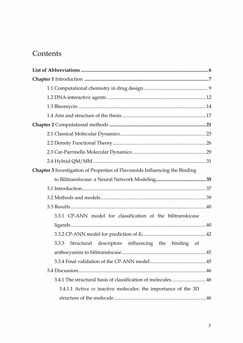
Contents List of Abbreviations ........................................................................................................6
Chapter 1 Introduction .....................................................................................................7
1.1 Computational chemistry in drug design.....................................................9
1.2 DNA-interactive agents .................................................................................12
1.3 Bleomycin ........................................................................................................14
1.4 Aim and structure of the thesis ....................................................................17
Chapter 2 Computational methods ...............................................................................21
2.1 Classical Molecular Dynamics......................................................................23
2.2 Density Functional Theory............................................................................26
2.3 Car-Parrinello Molecular Dynamics ............................................................29
2.4 Hybrid QM/MM ............................................................................................31
Chapter 3 Investigation of Properties of Flavonoids Influencing the Binding
to Bilitranslocase: a Neural Network Modeling.........................................35
3.1 Introduction.....................................................................................................37
3.2 Methods and models......................................................................................39
3.3 Results ..............................................................................................................40
3.3.1 CP-ANN model for classification of the bilitranslocase
ligands ..............................................................................................................40
3.3.2 CP-ANN model for prediction of KI ...................................................42
3.3.3 Structural descriptors influencing the binding of
anthocyanins to bilitranslocase.....................................................................45
3.3.4 Final validation of the CP-ANN model..............................................45
3.4 Discussion........................................................................................................46
3.4.1 The structural basis of classification of molecules............................46
3.4.1.1 Active vs inactive molecules: the importance of the 3D
structure of the molecule ...........................................................................46
3
3.4.1.2 Competitive vs non-competitive inhibition by aglycones:
the importance of hydroxylation of B ring .............................................48
3.4.1.3 Pure competitive vs mixed-type inhibition: the possible
role of tautomerism of ring B....................................................................48
3.4.2 The structural basis of the activity of competitive inhibitors..........50
3.4.2.1 Flavonoid aglycones: the importance of hydroxylation of
the B ring......................................................................................................51
3.4.2.2 Flavonoid aglycones: the importance of steric hindrance
and charge distribution caused by the carbonyl group of the C
ring................................................................................................................51
3.4.2.3 Anthocyanidin mono- and diglucosides.....................................52
3.4.3 A hypothesis for the structural basis of the activity of ID 18..........53
3.5 Conclusions .....................................................................................................54
3.6 Appendix .........................................................................................................55
A.1 Counterpropagation Artificial Neural Network.................................55
A.2 Genetic Algorithm...................................................................................57
A.3 Structural descriptors..............................................................................58
Chapter 4 The Metal Bonding Domain of the Antitumor Drug Fe(II)-
bleomycin: a DFT Investigation....................................................................65
4.1 Introduction.....................................................................................................67
4.2 Models and computational details...............................................................70
4.3 Results and discussion...................................................................................72
4.3.1 Complexes A and AH ...........................................................................73
4.3.2 Complexes B and BH ............................................................................75
4.3.3 Complexes C and D...............................................................................76
4.3.4 Comparison with experiment ..............................................................77
4.4 Conclusions .....................................................................................................79
Chapter 5 The Mechanism of the Bleomycin Suicide: A Car-Parrinello
Molecular Dynamics Investigation ..............................................................81
5.1 Introduction.....................................................................................................83
5.2 Models and computational methods ...........................................................86
4
5.3 Results ..............................................................................................................88
5.3.1 Test calculations.....................................................................................88
5.3.2 ABLM in vacuum ..................................................................................90
5.3.3 ABLM in H2O .........................................................................................92
5.4. Conclusions and discussion .........................................................................96
Chapter 6 The mechanism of the bleomycin action: A Car-Parrinello
molecular dynamics investigation ...............................................................99
6.1 Introduction...................................................................................................101
6.2 Models and computational details.............................................................103
6.3 Results and discussion.................................................................................106
6.3.1 ABLM with deoxyribose sugar in vacuum......................................106
6.3.2 Molecular orbitals analysis of the BLM-Fe(IV)=O complex..........107
6.3.3 Selectivity of the hydrogen atom abstraction..................................108
6.3.4 ABLM with DNA: QM/MM study...................................................110
6.4 Conclusions and future outlook.................................................................113
References .......................................................................................................................115
Summary. ........................................................................................................................125
Samenvatting..................................................................................................................129
Podsumowanie...............................................................................................................133
Publications ....................................................................................................................137
Curriculum Vitae ...........................................................................................................138
Nawoord. .........................................................................................................................139
5
List of Abbreviations aALA Aminoalanine ABLM Activated bleomycin ADF Amsterdam density functional BLM Bleomycin BLYP Becke, Lee, Yang, Parr BP Becke, Perdew CP-ANN Counterpropagation artificial neural network CPMD Car-Parrinello molecular dynamics DFT Density functional theory DNA Deoxyribonucleic acid EPR Electron paramagnetic resonance Eq. Equation FPMD First principles molecular dynamic GA Genetic algorithm GGA Generalized gradient approximation H-bond Hydrogen bond HIS Histidine HOMO Highest occupied molecular orbital KI Inhibition constant LDA Local density approximation MD Molecular dynamics MM Molecular mechanics NMR Nuclear magnetic resonance PW Plane waves PYR Pyrimidine QM Quantum mechanics QM/MM Quantum mechanics/Molecular mechanics RMSD Root mean square deviation RMSE Root mean square error Ry Rydbergs 2D Two-dimensional 3D Three-dimensional
6

Chapter one
INTRODUCTION

8
ABSTRACT
In this introductory chapter the work on the bleomycin anticancer drug is put in
the broader context of the drug design process. We discuss the contribution of
different computational methods into this field emphasizing the growing role
played by quantum mechanical methods.
Introduction
1.1 Computational chemistry in drug design
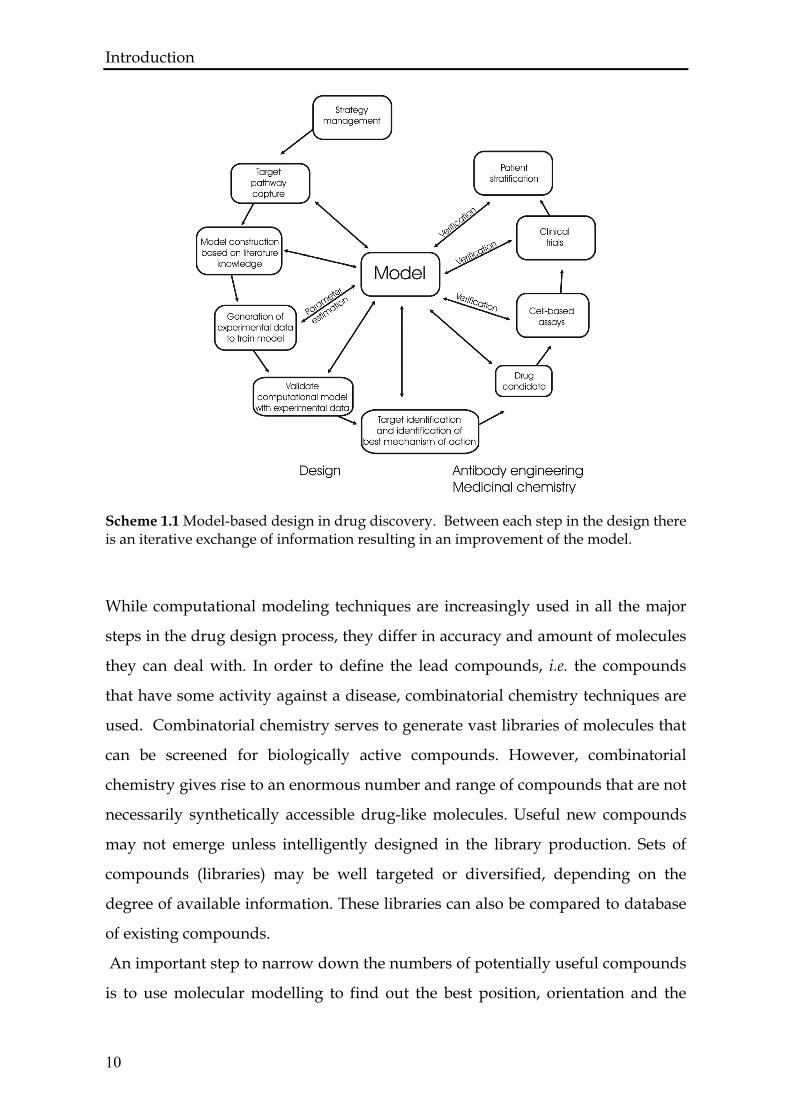
The design of pharmaceuticals is an extremely complex process as it is shown on
the scheme 1.1 [6]. Computational chemists combine their knowledge of
molecular interactions and drug activity, together with visualization techniques,
detailed energy calculations, geometric considerations, and data filtered out of
huge databases, in an effort to narrow down the search for effective drugs.
A fundamental assumption for rational drug design is that drug activity is
obtained through the molecular binding of one molecule, the ligand, to the pocket
of another and usually larger molecule, the receptor. In their active or binding
conformations, the molecules exhibit geometric and chemical complementarity,
both of which are essential for successful drug activity [6, 7]. By binding to
macromolecules, drugs may modulate signal pathways, for example, by altering
sensitivity to hormonal action, or by altering metabolism, or by interfering with
the catalytic activity of the enzyme. Most commonly, this is achieved by binding
in a specific cavity of the enzyme, the active site, which catalyzes the reaction,
thus preventing access of the natural substrate.
Computer-aided drug design will be a significant component of future rational
drug design strategies, and is becoming more relevant as the understanding of
molecular activity improves and the amount of available experimental data that
requires processing increases [8]. The role of quantum mechanical methods has
been until now very limited in the drug design process, mostly because of the
high computational demands involved that allowed to deal only with small
molecules [9]. The recent progress in first-principles electronic structure
calculations together with the steady increase in computational power have
considerably broadened the range and scope of application of these theoretical
methods. Of particular interest are density functional theory calculations that
have been proven to be a powerful tool for studying a large variety of problems in
chemistry and more recently in highly complex systems of biophysics and
biochemistry.
9
Introduction
10
Scheme 1.1 Model-based design in drug discovery. Between each step in the design there is an iterative exchange of information resulting in an improvement of the model.
While computational modeling techniques are increasingly used in all the major
steps in the drug design process, they differ in accuracy and amount of molecules
they can deal with. In order to define the lead compounds, i.e. the compounds
that have some activity against a disease, combinatorial chemistry techniques are
used. Combinatorial chemistry serves to generate vast libraries of molecules that
can be screened for biologically active compounds. However, combinatorial
chemistry gives rise to an enormous number and range of compounds that are not
necessarily synthetically accessible drug-like molecules. Useful new compounds
may not emerge unless intelligently designed in the library production. Sets of
compounds (libraries) may be well targeted or diversified, depending on the
degree of available information. These libraries can also be compared to database
of existing compounds.
An important step to narrow down the numbers of potentially useful compounds
is to use molecular modelling to find out the best position, orientation and the
Introduction
most favorable conformation of a compound based on energy considerations. In
addition, the active-analog approach assists the design of a ligand based on
similarities to a set of compounds known to possess the desired activity. The
affinity score is calculated to select candidate compounds with strong binding to
the target site. In addition, when the target site is not known for a ligand, various
programs allow to characterize likely sites through computational approaches for
functional site mapping. This involves repeatedly placing small functional groups
into the possible site to approximate the shape of the binding region. Likely sites
may also be inferred from similarity to known site structures. In this step several
different computational techniques are used like docking, structure based design
or molecular modeling where steric and electrostatic interactions are taken into
account.
Another essential step in the process of drug design is to refine the drug activity.
For instance, statistical techniques such as QSAR (quantitative structure activity
relationship) analysis may be used in order to choose targeted compounds with
required features. In QSAR, or QSPR (quantitative structure property
relationship), statistical correlation is explored between an activity or a property
and geometric or chemical characteristics (pharmacophores) of the molecule. It is
often used to analyse the effect of a particular substructure on the activities or
properties of compounds. The attributes of the compound being analysed such as
the activity, property, or structure are referred to as a descriptor.
Ideally there is a continuous exchange of information between the researchers
doing QSAR studies, synthesis and testing. These techniques are frequently used
and often very successful since they do not rely on knowing the biological basis of
the disease which can be very difficult to determine. However, they are not able to
investigate directly the chemical activity of a lead compound or drug interacting
with a specific target. Here quantum mechanics based computational tools may
become essential for validation of a small number of potential drugs before going
into the expensive and time consuming clinical stage.
11
Introduction
1.2 DNA-interactive agents
Deoxyribonucleic acid or DNA, the polynucleotide that carries the genetic
information in cells, is also one of the receptors with which drugs can interact.
Because this receptor is so vital to human functioning, and since from the
perspective of a medicinal chemist the overall shape and chemical structure of
DNA found in normal and abnormal cells is nearly indistinguishable, DNA-
interactive drugs that interact with this receptor are generally very toxic to normal
cells. Therefore, these drugs are reserved only for life-threatening diseases such as
cancers. There is little information that can guide the design of selective agents
against abnormal DNA. One feature differentiating cancer cells from most normal
cells is that the cancer cells undergo a rapid, abnormal, and uncontrolled cell
division. Genes coding for differentiation in cancer cells appear to be shut off or
inadequately expressed, while genes coding for cell proliferation are expressed
when they should not be. Because the cells are continuously undergoing mitosis,
there is a constant need for rapid production of DNA. Because of the similarity of
normal and abnormal DNA, a compound that reacts with a cancer cell will react
with a normal cell as well. However, because of the rapid cell division, cancer cell
mitosis can be halted more easily than in normal cells where there is sufficient
time for repair mechanisms to act. Hence, anticancer drugs are most effective
against malignant tumors with a large proportion of rapidly dividing cells, such
as leukemias and lymphomas. In addition, DNA damage in a cell is sensed by
several as yet poorly defined mechanisms involving a number of proteins. Tumor
cells, however, are defective in their ability to undergo cell cycle arrest or
apoptosis in response to DNA damage [10]. Cancer cells that cannot undergo cell
cycle arrest are sensitive to DNA damaging agents.
There are three major classes of clinically important DNA-interactive drugs: (1)
reversible binders, which interact with DNA through the reversible formation of
noncovalent interaction; (2) alkylators, which react covalently with DNA bases;
(3) DNA strand breakers, which generate reactive radicals that produce cleavage
of the polynucleotide strands.
12
Introduction
The reversible binders interfere with the interaction of nucleic acids with a variety
of small molecules, including water, metal cations, small organic molecules, and
proteins, all of which are essential for stabilization of the nucleic acid structure
inside the cell [11]. This interference can disrupt the DNA structure. There are
three important ways small molecules can reversibly bind to duplex DNA and
interfere with DNA function: (1a) by electrostatic binding along the exterior of the
helix, (1b) by interaction with the edges of the base pairs in either the major or
minor groove and (1c) by intercalation between the base pairs. The electrostatic
interactions are generally not dependent on the DNA sequence and they are
possible due to the negatively charged sugar phosphate backbone. Groove binders
can be elongated to extend the interaction within the groove, which leads to
highly sequence-specific recognition by these molecules. The major and minor
grooves have significant differences in their electrostatic potential, hydrogen
bonding characteristics, steric effects, and degree of hydration. Proteins exhibit
binding specificity primarily through major groove interactions, but small
molecules prefer minor groove binding. Finally, flat, generally aromatic or
heteroaromatic molecules bind to DNA by intercalating between the base pairs of
the double helix. The principal driving forces for intercalation are stacking and
charge-transfer interactions, while hydrogen bonding and electrostatic forces also
play a role in stabilization [12]. Intercalation, first described in 1961 by Lerman
[13], is a noncovalent interaction in which the drug is held rigidly perpendicular
to the helix axis. This causes the base pairs to separate vertically, thereby
distorting the sugar-phosphate backbone and decreasing the pitch of the helix.
Intercalation is an energetically favorable process. Presumably, the van der Waals
forces that hold the intercalated molecules to the pairs are stronger than the forces
stabilizing the stacked pairs.
The last, third, class of DNA-interactive drugs is DNA strand breakers. They
initially intercalate into DNA, and can react in such a way as to generate radicals
depending on the local environmental and cellular metabolism. These radicals
typically abstract hydrogen atoms from the DNA sugar-phosphate backbone or
from the DNA bases, leading to DNA strand scission. Therefore, these DNA-
13
Introduction interactive compounds are metabolically activated radical generators. Examples
of drugs that operate with this mechanism are the anthracycline antitumor
antibiotics, tirapazamine and the enediyne antitumor antibiotics, as well as the
glycopeptyde antibiotic bleomycin, which is the topic of this thesis.
1.3 Bleomycin
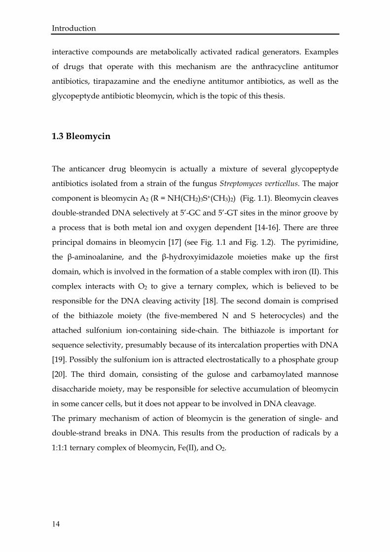
The anticancer drug bleomycin is actually a mixture of several glycopeptyde
antibiotics isolated from a strain of the fungus Streptomyces verticellus. The major
component is bleomycin A2 (R = NH(CH2)3S+(CH3)2) (Fig. 1.1). Bleomycin cleaves
double-stranded DNA selectively at 5’-GC and 5’-GT sites in the minor groove by
a process that is both metal ion and oxygen dependent [14-16]. There are three
principal domains in bleomycin [17] (see Fig. 1.1 and Fig. 1.2). The pyrimidine,
the β-aminoalanine, and the β-hydroxyimidazole moieties make up the first
domain, which is involved in the formation of a stable complex with iron (II). This
complex interacts with O2 to give a ternary complex, which is believed to be
responsible for the DNA cleaving activity [18]. The second domain is comprised
of the bithiazole moiety (the five-membered N and S heterocycles) and the
attached sulfonium ion-containing side-chain. The bithiazole is important for
sequence selectivity, presumably because of its intercalation properties with DNA
[19]. Possibly the sulfonium ion is attracted electrostatically to a phosphate group
[20]. The third domain, consisting of the gulose and carbamoylated mannose
disaccharide moiety, may be responsible for selective accumulation of bleomycin
in some cancer cells, but it does not appear to be involved in DNA cleavage.
The primary mechanism of action of bleomycin is the generation of single- and
double-strand breaks in DNA. This results from the production of radicals by a
1:1:1 ternary complex of bleomycin, Fe(II), and O2.
14
Introduction
N N
H
H2N
CH3 HN
O
NH
O
CH3H
H HN
O
NH
O
NH2O
HN NH2
O
H NH2
HO
H CH3
HO CH3H
H
O
O
OH
HO
OHO
OOH
OH
OH O
O
NH2
H
HNH
N
N
S
S
NR
O
Bithiazole (BITH)
Mannose (MAN)
Threonine (THR)
Methylvalerate (mVAL)
Aminoalanine (aALA)
Histidine (HIS)
Gulose (GUL)
12
4
3
5
Figure 1.1 The chemical structure of bleomycin A2 (R = NH(CH2)3S+(CH3)2), B2 (R =NH(CH2)4NHC(NH)(NH2)), and pepleomycin (R = NH(CH2)3NHCH(CH3)Ph). The numbers from 1 – 5 indicate the coordination sites to the metal.
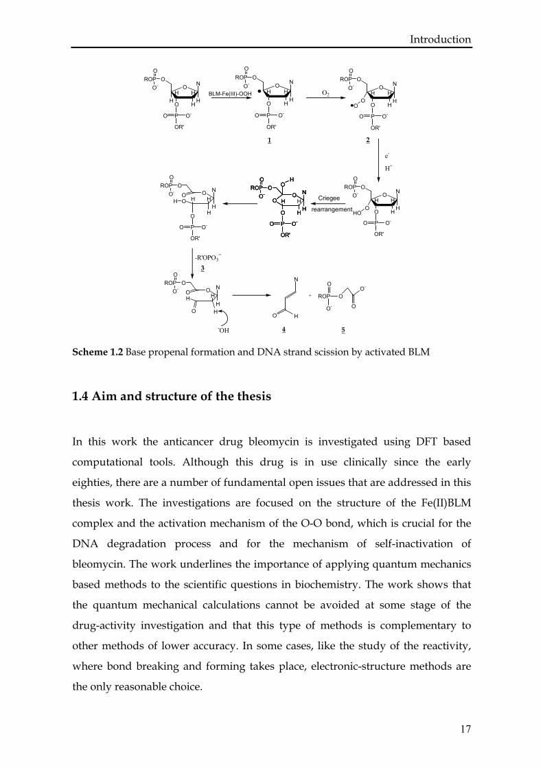
This ternary complex may be self-activated by the transfer of an electron from a
second unit of the ternary complex or activation may be initiated by a microsomal
NAD(P)H-cytochrome P450 reductase-catalyzed reduction [21, 22]. The activated
bleomycin, the peroxide iron (III) bleomycin complex (BLM-Fe(III)-OOH) binds
tightly to guanine bases in DNA, principally via the amino-terminal tripeptide
containing the bithiazole unit [23]. The two major monomeric products formed
when activated bleomycin reacts with DNA are nucleic base propenals (U4U in
Scheme 1.2) and nucleic acid bases. Base propenal formation consumes an
equivalent of O2 in addition to that required for bleomycin activation and is
accompanied by DNA strand scission with the production of 3’-phosphoglycolate
(U5U in Scheme 1.2) and 5’-phosphate-modified DNA fragments (U3U in Scheme 1.2).
DNA base formation does not require additional O2 and results in destabilization
of the DNA sugar-phosphate backbone. Evidence for the 4’C radical (U1U in Scheme
1.2 ) and the peroxy radical (U2U in Scheme 1.2) come from the model studies of
Giese and coworkers who used chemical methods to generate a 4’C radical in a
single-stranded oligonucleotide [24, 25]. They detected the 4’C radical
15
Introduction
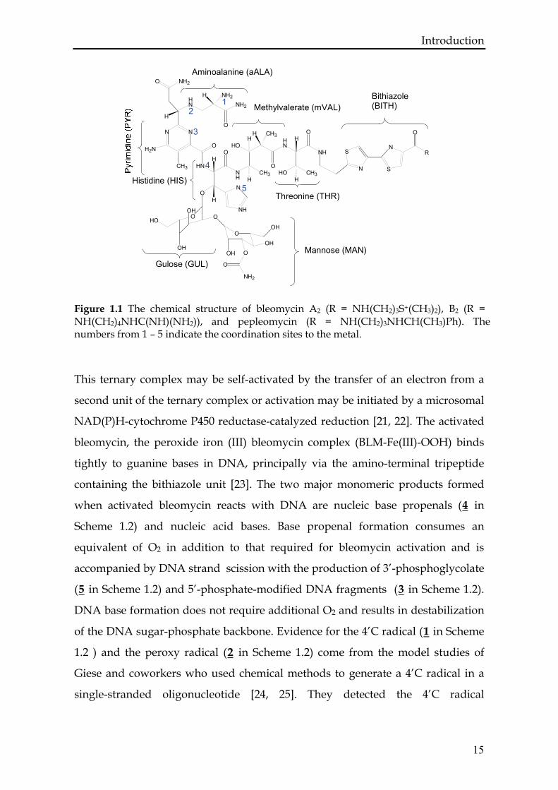
Figure 1.2 The NMR structure of BLM-Co(III)-OOH bound to the fragment of DNA (D(GGAAGCTTCC)) [4]. The structure of the bleomycin complex is represented in color following standard atomic color definition. The three parts of bleomycin are indicated: (i) the metal bonding domain represented in sticks and balls; (ii) the bithiazole tail that is inserted between two pairs of DNA; (iii) the sugar moiety with the carbamoyl group. The deoxyribose sugar of DNA, indicated in cyan in the figure, is the one attacked by activated bleomycin in the first step of DNA degradation.
and the peroxy radical in line with the products that are detected in the reaction
of activated bleomycin with DNA.
DNA strand scission is sequence selective, occurring most frequently at 5’-GC-3’
and 5’-GT-3’ sequence [26]. The specificity for DNA cleavage at a residue located
at the 3’ side of G appears to be absolute. Preferences for cleavage at 5’-GC and
5’-GT instead of corresponding 5’-AC or 5’-AT sites can be attributed to reduced
binding affinity of bleomycin, since guanine can engage in an additional
hydrogen bond compared with adenine [27, 28].
16
Introduction
17
O
HO
HHHH
ON
ROPO
O-
PO O-
OR'
O
HO
HHH
ON
ROPO
O-
PO O-
OR'
O
HO
HHH
ON
ROPO
O-
PO O-
OR'
O
HO
HHH
ON
ROPO
O-
PO O-
OR'
OO
OHO
O
HOH
ON
ROPO
O-
PO O-
OR'
O
OH
HO
HOH
ON
ROPO
O-
PO O-
OR'
O
OH
HO
HO
HH
ON
ROPO
O-
PO O-
OR'
O
OH
HO
HO
HH
ON
ROPO
O-
PO O-
OR'
O HHO
O
HO
HH
ON
ROPO
O-
HO
N
O H
O
ROP
O-
OO-
O
+
-R'OPO3=
Criegee
rearrangement
e-
H+
O2
-OH
1 2
3
4 5
BLM-Fe(III)-OOH
Scheme 1.2 Base propenal formation and DNA strand scission by activated BLM
1.4 Aim and structure of the thesis
In this work the anticancer drug bleomycin is investigated using DFT based
computational tools. Although this drug is in use clinically since the early
eighties, there are a number of fundamental open issues that are addressed in this
thesis work. The investigations are focused on the structure of the Fe(II)BLM
complex and the activation mechanism of the O-O bond, which is crucial for the
DNA degradation process and for the mechanism of self-inactivation of
bleomycin. The work underlines the importance of applying quantum mechanics
based methods to the scientific questions in biochemistry. The work shows that
the quantum mechanical calculations cannot be avoided at some stage of the
drug-activity investigation and that this type of methods is complementary to
other methods of lower accuracy. In some cases, like the study of the reactivity,
where bond breaking and forming takes place, electronic-structure methods are
the only reasonable choice.
Introduction In traditional quantum chemistry, highly accurate calculations are carried out for
small molecules in vacuum at zero temperature, and molecular properties are
deduced from these models. Multidimensional potential energy surfaces can be
constructed for very small molecules or for larger systems by considering only a
limited set of degrees of freedom, which can be chosen a priori by chemical
intuition. However, the recent progress in first-principles electronic structure
calculations together with a steady increase in computational power have
considerably broadened the range and scope of application of quantum chemistry
methods. In particular, DFT provides a versatile tool for the study of medium
sized to large molecules with a good accuracy (see Section 2.3). On the other hand,
the most appropriate method to study reaction pathways is first principles
molecular dynamics. Within this approach the system is allowed to evolve at a
finite temperature and can possibly cross barriers between minima on the
potential energy surface without any a priori assumption on the reaction path. An
elegant way of carrying out first principles molecular dynamics based on DFT is
the Car-Parrinello approach, where the dynamics of the nuclei as well as the
adiabatically evolving electronic wave function are described by Newtonian
equations of motion (see Section 2.3). When dealing with very large biomolecules
it is necessary to use a hybrid quantum mechanics – molecular mechanics
approach. Here, I use a recently developed hybrid QM/MM Car-Parrinello
scheme [29, 30]. This approach enables efficient and robust hybrid Car-Parrinello
simulations of extended systems with the chemically relevant part treated on the
quantum mechanical level while the remainder of the system is described with
less accuracy in order to simulate the effects due to the environment at a
satisfactory level.
The thesis is organized as follows: In Chapter 2, the theoretical foundations of the
applied computational methods are introduced. Chapter 3 presents a classical
approach to the search for potential drug-like molecules. This chapter provides an
example of currently used computational methods in the drug design process and
underlines the need of cooperation between experimentalists and theoreticians. In
18
Introduction
19
Chapter 4, I describe results obtained by static DFT calculations applied to the
structure of the Fe(II) bleomycin complex. The Car-Parrinello molecular dynamics
is used in Chapter 5 for modeling the activation of the O-O bond in the activated
bleomycin. In this chapter the QM/MM approach is introduced to study the
bleomycin case. Chapter 6 is dedicated to the investigation of the reaction
mechanism of activated bleomycin with the deoxyribose sugar since it is known
from experiment that the degradation of DNA starts by forming a radical at the
4’C position of a deoxyribose sugar.


Chapter two
COMPUTATIONAL METHODS

ABSTRACT First principles molecular dynamics (Car-Parrinello) simulations based on density
functional theory have emerged as a powerful tool for studying physical,
chemical and biological systems. Its implementation into a QM/MM approach is
especially attractive for the in situ investigation of chemical reactions that occur in
a complex and heterogeneous environment. Hereby, the theoretical backgrounds
of all the computational techniques applied in the investigation are presented.
22
Computational Methods
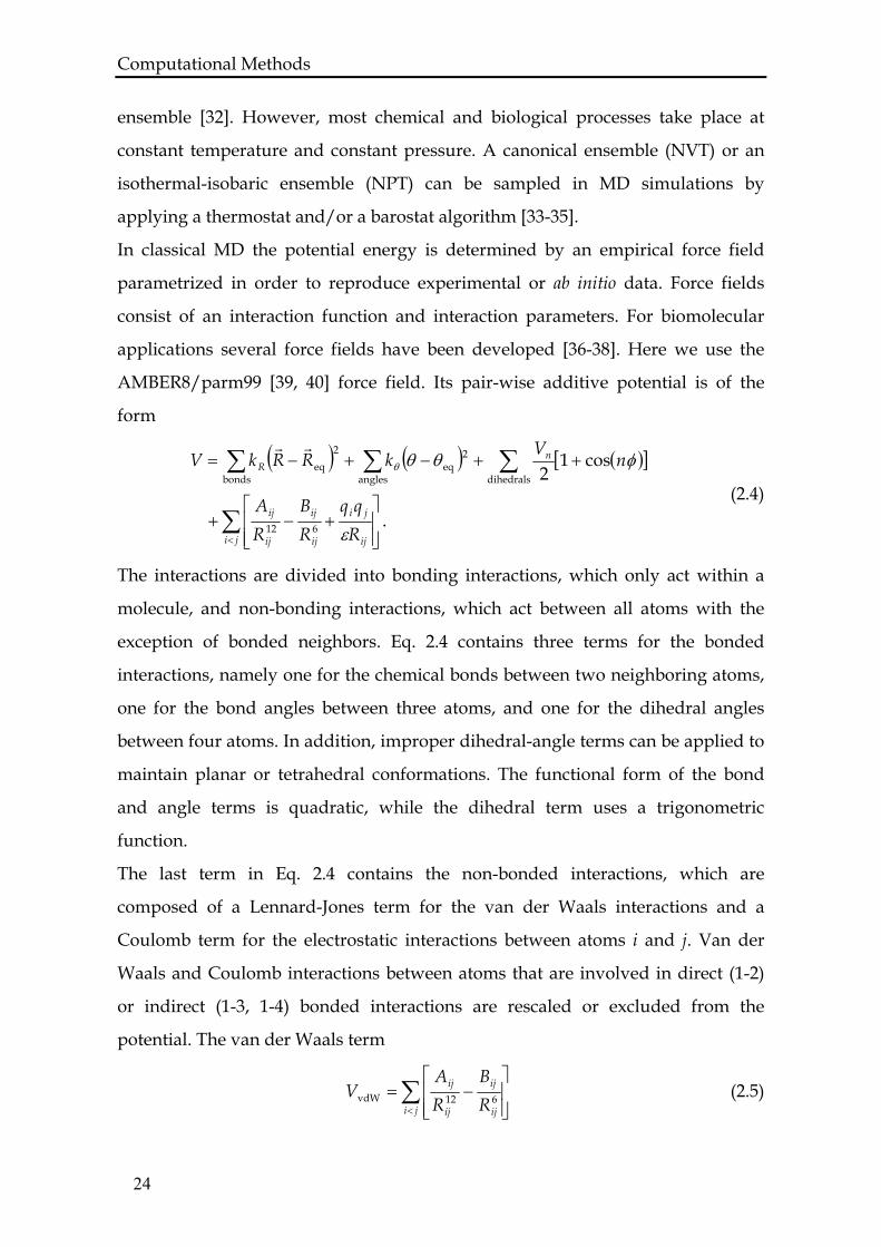
2.1 Classical Molecular Dynamics
Molecular dynamics simulations yield an atomistic time-dependent description of
particles in a system and hence provide an insight into its dynamic and
thermodynamic properties. From the trajectory, a number of properties such as
free-energy differences, reaction rates, and different space and time correlation
functions can be calculated.
To describe the time evolution of a molecular system by MD simulations,
Newton’s equation of motion
2
2
dtRdM
RVF i
ii
i
r
rr
=∂∂
−= (2.1)
has to be integrated. is the force acting on atom i with position and mass Mi,
and V is the potential energy of the system.
iFr
iRr
In general, there is no analytical solution for the integration of Eq. 2.1, and
numerical algorithms based on time discretization have to be used. The size of the
time step depends on the characteristic dynamical time scale of the system and for
classical MD it is typically between 1–2 fs. A commonly used integration
algorithm is the velocity-Verlet algorithm [31], which employs a Taylor expansion
truncated beyond the quadratic term for the coordinates
( ) ( ) ( ) ( ) .2
2tMtFttvtRttR Δ+Δ+=Δ+
rrrr
(2.2)
The update for the velocities is given by
( ) ( ) ( ).
2)( t
MtFttFtvttv Δ
+Δ++=Δ+
rrrr (2.3)
The thermodynamic state of a system is defined in terms of macroscopic
parameters that are constant during a MD simulation. If the number of particles
N, the volume V, and the energy E are fixed, then a constant energy ensemble is
sampled. Under the ergodic hypothesis, i.e., the assumption that a system will
sample the whole phase space given an infinite amount of time, the time averages
over an infinite trajectory correspond to averages over a microcanonical (NVE)
23
Computational Methods ensemble [32]. However, most chemical and biological processes take place at
constant temperature and constant pressure. A canonical ensemble (NVT) or an
isothermal-isobaric ensemble (NPT) can be sampled in MD simulations by
applying a thermostat and/or a barostat algorithm [33-35].
In classical MD the potential energy is determined by an empirical force field
parametrized in order to reproduce experimental or ab initio data. Force fields
consist of an interaction function and interaction parameters. For biomolecular
applications several force fields have been developed [36-38]. Here we use the
AMBER8/parm99 [39, 40] force field. Its pair-wise additive potential is of the
form
( ) ( ) ( )[ ]
.
cos12
612
dihedrals
2eq
angles
2
bondseq
∑
∑∑∑
< ⎥⎥⎦
⎤
⎢⎢⎣
⎡+−+
++−+−=
ji ij
ji
ij
ij
ij
ij
nR
Rqq
RB
RA
nVkRRkV
ε
φθθθ
rr
(2.4)
The interactions are divided into bonding interactions, which only act within a
molecule, and non-bonding interactions, which act between all atoms with the
exception of bonded neighbors. Eq. 2.4 contains three terms for the bonded
interactions, namely one for the chemical bonds between two neighboring atoms,
one for the bond angles between three atoms, and one for the dihedral angles
between four atoms. In addition, improper dihedral-angle terms can be applied to
maintain planar or tetrahedral conformations. The functional form of the bond
and angle terms is quadratic, while the dihedral term uses a trigonometric
function.
The last term in Eq. 2.4 contains the non-bonded interactions, which are
composed of a Lennard-Jones term for the van der Waals interactions and a
Coulomb term for the electrostatic interactions between atoms i and j. Van der
Waals and Coulomb interactions between atoms that are involved in direct (1-2)
or indirect (1-3, 1-4) bonded interactions are rescaled or excluded from the
potential. The van der Waals term
∑< ⎥
⎥⎦
⎤
⎢⎢⎣
⎡−=
ji ij
ij
ij
ij
RB
RA
V 612vdW (2.5)
24
Computational Methods describes the repulsive forces at small interatomic distances due to the Pauli
repulsion between electrons (decaying exponentially, modeled with ), and the
attractive forces at intermediate distances due to instantaneous dipole-induced
dipole interactions (decaying with ).
12−ijR
6−ijR
Finally, the Coulomb term
∑< ⎥
⎥⎦
⎤
⎢⎢⎣
⎡=
ji ij
ji
Rqq
Vεel (2.6)
takes into account the electrostatic interactions between charged particles. In the
AMBER8/parm99 force field, the charge distribution in a molecule is reproduced
by atom-centered point charges derived from the electrostatic potential [41-43].
The Coulomb interaction is a long-range interaction and the sum in Eq. 2.6
converges very slowly. Therefore different algorithms have been developed for a
fast and accurate treatment of electrostatic interactions, based on Ewald
summations. Particle-mesh Ewald [44] and particle-particle/particle-mesh Ewald
[45] algorithms are widely used in classical MD programs.
In classical MD, the size of the system can be of the order of hundred thousand
atoms and the total time of simulation is in the nanoseconds range. Since
nevertheless one cannot reach macroscopic system sizes for simulations in the
condensed phase, usually periodic boundary conditions are used to prevent
surface artifacts. The central simulation box is periodically surrounded by images
of itself. Care has to be taken to avoid the effects of artificial periodicity, such as
the interaction of a molecule with its image in the neighboring box.
The most obvious limitation of empirical force fields is their inability to describe
reactive events. Since they make use of fixed parameters, e.g., for bond distances,
they cannot adapt to different electronic situations. Additionally, most of the
standard force fields employ fixed point charges and do not contain terms that
allow for an explicit polarization of the atoms. To be able to describe chemical
reactions one has to go beyond an empirical force field description by including
explicitly the quantum mechanical description of the electronic structure.
25
Computational Methods
2.2 Density Functional Theory
Among all quantum chemistry techniques, Density Functional Theory is often the
method of choice because of its good compromise between accuracy and
computational cost. In a few words, DFT provides a way to obtain the electron
density and the ground state energy of a polyatomic system given its atomic
coordinates [46]. Most programs based on DFT are capable to search for energy
minima and compute several molecular properties such as atomic charges,
multipole moments, vibrational frequencies, and spectroscopic constants. DFT is
also the basis of first principles molecular dynamics techniques such as the Car–
Parrinello method, in which the molecules evolve in real time and finite
temperature under the forces derived from the instantaneous ground state of the
electron cloud [47]. Since the electronic density changes during the simulation,
polarization effects are described in a natural way, as well as changes in the
bonding pattern of the atoms (e.g., bond breaking and bond-forming processes).
The development of DFT in the area of computational chemistry dates from the
mid 1960s when Hohenberg and Kohn [48] demonstrated that the ground-state
energy of a system of interacting electrons subject to an external potential ( )rV r is
a unique functional of the electron density ( )[ ]rEE rρ= , and it can be obtained by
minimizing the energy functional with respect to the density,
( )( )[ ].minDFT rEE
r
rr ρ
ρ= (2.7)
Later, Kohn and Sham [49] demonstrated that there is equivalence between the
electronic density of the real system and a model comprising noninteracting
electrons that are subject to an effective potential, Veff. This provides a way to
solve the problem of finding the density of the many-electron interacting system,
via obtaining the electron density of the noninteracting system. This density can
be expressed in terms of single-electron orbitals ( )rir
ψ , known as Kohn–Sham (KS)
orbitals,
( ) ( ) ,22occ
∑=i
i rr rrψρ (2.8)
26
Computational Methods where the sum extends over the occupied single-particle orbitals. Eq. 2.8 describes
the simplest situation in which all orbitals are doubly occupied and is easily
generalized to spin polarized systems [50].
Because of the relation in Eq. 2.8, the energy functional can be either expressed in
terms of the density (Eq. 2.7) or using the single-electron orbitals,
{ }( ){ } { }[ ],,min KSDFT
Ni RrEEi
rrψ
ψ= (2.9)
where { }NRr
are the nuclear coordinates fixed within the Born-Oppenheimer
approximation. The energy functional (Eq. 2.7) can be written in atomic units as:
( ) ( ) ( ) ( )
( ) ( ) ( )[ ] .''
'21
22
2
XC
2*KS
∑∫
∫∑∫
> −++
−+
+⎟⎟⎠
⎞⎜⎜⎝
⎛ ∇−=
JI JI
JI
ii
RR
eZZrErdrd
rrrr
rdrrVrdrrE
rrrrr
rr
rr
rrrrrr
ρρρ
ρψψ
(2.10)
The first term on the right-hand side of this expression is the kinetic energy of the
noninteracting electrons. The second term corresponds to the interaction of the
electrons with the nuclear charges and ( )rV r is the potential resulting from the
nuclei. In case only valence electrons are explicitly considered in the calculation,
would be a pseudopotential. The third term corresponds to the classical
Coulomb interaction of a density distribution
( )rV r
( )rrρ . The fourth term, ( )[ rE r ]ρXC , is a
functional of the density that accounts for the remaining contributions to the
electron–electron interaction. The last term accounts for the nucleus-nucleus
electrostatic repulsion, which is a constant in the Born-Oppenheimer
approximation since the nuclear coordinates are fixed.
The only unknown quantity in Eq. 2.10 is Exc, which contains the exchange and
correlation energies. In principle DFT in the Kohn-Sham formulation is an exact
theory, but in practice approximations have to be made for the unknown
exchange and correlation functional.
In the local density approximation [49], which is based on the homogeneous
electron gas approximation, Exc is defined as
[ ] ( ) ( ) ,XCLDAXC rdrE rv ρερρ ∫= (2.11)
27
Computational Methods where εxc(ρ) is the sum of the exchange and correlation energy per electron of a
homogeneous electron gas of density ρ:
( ) ( ) ( ) ,CXXC ρερερε += (2.12)
with
( ) ( ) 3131
X3
43 ρ
πρε ×⎟
⎠⎞
⎜⎝⎛−= (2.13)
and
( ) ( ).VWNCC ρερε = (2.14)
For the correlation term εc, no analytical expression is available, but accurate
values have been obtained from quantum Monte Carlo [51] calculations and
analytic forms of εc have been parametrized with these results by Vosko, Wilk,
and Nusair [52]. LDA yields good results for solid state systems, but the accuracy
provided by this approximation is not enough for most applications in chemistry
and biology. One of the main drawbacks of LDA is that van der Waals
interactions, which originate from correlated motions of electrons caused by
Coulomb interactions between distant atoms, cannot be properly described and
also bond distances and binding energies can have large errors that appear in a
nonsystematic way.
Better results are obtained with functionals that do not only depend on the local
density ( )rrρ , but also on the local gradient ( )rrρ∇ of the density. Many functionals
have been developed in the framework of the generalized gradient
approximation. Among the most popular GGA exchange and correlation
functionals used in biological applications are the ones denoted as BP with the
exchange part by Becke [53] and the correlation according to Perdew [54], BLYP, a
combination of Becke exchange and correlation developed by Lee, Yang and Parr
[55], PBE developed by Perdew, Burke and Ernzerhof [56]. The use of the GGA
approximation improves considerably the description of bonding, in particularly
hydrogen bonding, with respect to pure LDA with a very low additional
computational cost. The description of weak van der Waals interactions, however,
remains problematic. Therefore, special care should be taken when addressing
problems in which van der Waals interactions might play a relevant role, such as
28
Computational Methods stacking interactions between π-systems and the diffusion of ligands in purely
hydrophobic cavities [57]. The introduction of hybrid functionals, which contain a
certain amount of exact non-local exchange from Hartree-Fock theory, has been
an important step towards a higher accuracy of DFT calculations for molecules.
The most popular hybrid functional is the three-parameter functional from Becke
(B3LYP) [58]. However, the high computational cost of calculating the two-
electron integral of non-local exchange within the plane-wave basis set employed
in the Car-Parrinello MD code hampers the use of hybrid functionals for FPMD.
2.3 Car-Parrinello Molecular Dynamics
The basic idea of the Car-Parrinello approach [47] is to treat the electronic degrees
of freedom { }iψ as fictitious classical dynamical variables, exploiting the timescale
separation between the fast electronic and the slow ionic motion to avoid energy
exchange between the two subsystems. The Car-Parrinello Lagrangian
,enCP UTTL −+=
or explicitly
{ } { }[ ] ( )ijjiji
ijIii
iIII
RErdRML δψψλψψμ −+−+= ∑∑ ∫∑••
,
KS2
2CP ,
21 rrr (2.15)
contains the kinetic energy Tn of the nuclei, the fictitious kinetic energy Te of the
electrons, the potential energy U, and the last term is the constraint ensuring the
orthonormality of orbitals. The potential energy term U is given by the Kohn-
Sham energy density functional { } { }[ ]Ii REr
,KS ψ (Eq. 2.10). The electronic
Hamiltonian contains the coulomb interaction between the nuclei. A fictitious
mass or inertia parameter µ is assigned to the orbital degrees of freedom and can
be tuned to ensure adiabaticity. The Newtonian equations of motion are obtained
from the associated Euler-Lagrange equations
II
RL
R
Ldtd
rr ∂
∂=
∂
∂•
CPCP (2.16)
and
29
Computational Methods
ii
LLdtd
ψδδ
ψδ
δ CPCP =•
(2.17)
and the corresponding Car-Parrinello equations of motions are found to be of the
form
[ ]ψ,KS RERM III
rr−∇=
••
(2.18)
and
( ).,
KS
∑ −∂
∂+−=
••
jiijji
iii
E δψψψψδ
δψμ (2.19)
They can be solved numerically using, for example, the Verlet algorithm [59].
The constant of motion is
{ }[ ].,21 KS2
consN
iiiIII
RERMErr
ψψψμ ++= ∑∑•••
(2.20)
The parameter µ has to be chosen in a way that ensures (i) that the lowest
electronic frequency is larger than the highest frequency of the nuclei mineω max
Iω
( Ie )ωω >> , in order to avoid energy transfer, and (ii) that the highest electronic
frequency is compatible with the chosen time step ∆t. Typical values are µ =
400 − 800 a.u. in combination with a time step ∆t = 4 − 6 a.u. (0.096 – 0.144 fs). The
time step in CPMD is smaller than for classical MD because of the fast electronic
motions. Since the electronic degrees of freedom are explicitly included, the size
of a system that can be treated with FPMD is of the order of 100 – 1000 atoms with
a total simulation time in the range of 1 – 10 ps.
maxeω
The original Car-Parrinello method imposes periodic boundary conditions and
expands the wave function in plane waves. Plane waves are defined as
( ) [ ],exp1
cell
PWG rGirf rrr
⋅Ω
= (2.21)
with the reciprocal space vector G and the cell volume Ωcell. Plane waves form a
complete and orthonormal basis, and the Kohn-Sham orbitals can be written in
the form
( ) ( ) [ ].exp1
cell
rGiGcri
iirrrr
⋅Ω
= ∑ψ (2.22)
30
Computational Methods
The expansion has to be truncated at an energy cutoff 2maxcut 2
1 GE = , which
determines the number of plane waves
23cutcell2PW 2
1 EN Ω=π
(2.23)
and therefore the accuracy of the calculation. The advantage of a plane-wave basis
set is that all terms of the Car-Parrinello equations can conveniently be solved
either in real or in reciprocal space, making use of fast Fourier transformation
algorithms. In addition, the Pulay forces are zero, because the basis set does not
depend on the atomic positions, which makes evaluation of nuclear forces easier.
However, a large number of plane waves would be needed for the description of
the highly localized core electrons that are chemically inactive. This problem has
prompted the development of pseudopotentials for the description of core
electrons. Norm-conserving pseudopotentials [60] depend on the angular-
momentum and correctly represent the long-range interactions of the core
electrons as well as the full wave function outside the core radius.
2.4 Hybrid QM/MM
The computer simulations of chemical reactions in a realistic environment is a
particularly challenging task. Chemical bonds are broken and formed during this
process, which implies the use of quantum mechanical methods that can take into
account instantaneous changes in the electronic structure explicitly. On the other
hand, the systems of interest in computational biology are quite complex with
many thousands of atoms. In spite of the considerable progress that was achieved
in the development of DFT approaches, it is clear that, in order to treat complex
biological systems, we still need to be able to combine various computational
chemistry methodologies with different accuracies and cost of calculations. One
possible solution for modeling such systems is the choice of a hierarchical hybrid
approach in which the whole system is partitioned into a localized chemically
active region treated with a quantum mechanical method and the environment,
31
Computational Methods treated with empirical potentials. In this quantum mechanical/molecular
mechanics method the computational effort can be concentrated on the part of the
system where it is most needed while the effects of the surroundings, such as
mechanical constraints, electrostatic perturbations and dielectric screening, are
taken into account with a more expedient model [61]. The idea of a QM/MM
scheme is not new and the first published example appeared already thirty years
ago [62]. However, in the last few years this subject has developed very rapidly
and QM/MM approaches have been implemented in the most commonly used
computational packages.
The particular QM/MM Car-Parrinello method [30] that has been used in this
work is based on a mixed Hamiltonian of the form
,QM/MMMMQM HHHH ++= (2.24)
in which the quantum part HQM is described with the extended Car-Parrinello
Lagrangian (Eq. 2.15). Since this QM/MM Car-Parrinello implementation
establishes an interface between the Car-Parrinello code CPMD [63] and the
classical force fields GROMOS96 [38] and AMBER [39], the classical part HMM
follows the formalism used in these packages according to Eq. 2.4.
The intricacies of QM/MM methods are in the challenge of finding an appropriate
treatment for the coupling between the QM and MM regions as described by the
interaction Hamiltonian HQM/MM. Special care has to be taken that the QM/MM
interface is treated in an accurate and consistent way, in particular in combination
with a plane-wave-based Car-Parrinello scheme. If the QM/MM boundary cuts
through a covalent bond, care has to be taken to saturate the valence orbitals of
the QM system. In the present implementation [30], this can be done by “capping”
the QM site with a hydrogen atom or an empirically parametrized
pseudopotential (“dummy atom”) and such a bond is treated on the QM level.
The remaining bonding interactions of the interface region, i.e. angle bending and
dihedral distortions, are described within the classical force field. The same holds
for the van der Waals interaction between QM and MM parts of the system.
32
Computational Methods
33
The electrostatic effects of the classical environment, on the other hand, are taken
into account in the quantum mechanical description as an additional contribution
to the external field of the quantum system,
( ) ( ),elQM/MM ii
MMii rrvrrdqH rrrr
−= ∫∑∈
ρ (2.25)
where qi is the classical point charge located at ri and ( )ii rrv rr− is a Coulombic
interaction potential modified at short range in such a way as to avoid spill-out of
the electron density to nearby positively charged classical point charges [29]. In
the context of a plane-wave-based Car-Parrinello scheme, a direct evaluation of
Eq. 2.25 is prohibitive, because it involves on the order of NrNMM operations,
where Nr is the number of real space grid points, typically ∼1003, and NMM is the
number of classical atoms, usually of the order 10 000 or more in system of
biochemical relevance. Therefore, the term in Eq. 2.25 is included exactly only for
a set of MM atoms in the vicinity of the QM system. The electrostatic interaction
between the classical point charges of the more distant MM atoms and the QM
system is calculated by a multipolar expansion of the full interaction given in Eq.
2.25. In this way, efficient and consistent QM/MM Car-Parrinello simulations of
complex extended systems can be performed. The steric and electrostatic effects of
the surroundings can be taken into account explicitly while the total energy of the
coupled QM/MM system is conserved during the dynamics.

Chapter three
INVESTIGATION OF PROPERTIES OF FLAVONOIDS INFLUENCING THE BINDING TO BILITRANSLOCASE:
A NEURAL NETWORK MODELING
This chapter is based on the publication by A. Karawajczyk, V. Drgan, N. Medic,
G. Oboh, S. Passamonti and M. Novic, Properties of flavonoids influencing the binding
to bilitranslocase investigated by neural network modelling. Biochemical
Pharmacology, 73, 308 (2007)
ABSTRACT
Bilitranslocase is a plasma membrane carrier. This work is aimed at characterizing
the interaction of bilitranslocase with flavonols, a flavonoid sub-class. The results
obtained show that, contrary to anthocyanins, flavonol glycosides do not interact
with the carrier, whereas just some of the corresponding aglycones act as
relatively poor ligands to bilitranslocase. These data point to a clear-cut
discrimination between anthocyanins and flavonols occurring at the level of the
bilitranslocase transport site. A quantitative structure-activity relationship based
on counter propagation artificial neural network modelling was undertaken in
order to shed light on the nature of flavonoid interaction with bilitranslocase. It
was found that binding relies on the ability to establish hydrogen bonds, ruling
out the involvement of charge interactions. This requisite might be at the basis of
the discrimination between anthocyanins and flavonols by bilitranslocase and
could lie behind some aspects of the distinct pharmacokinetic properties of
anthocyanins and flavonols in mammals.
36
Neural Network Modeling
3.1 Introduction
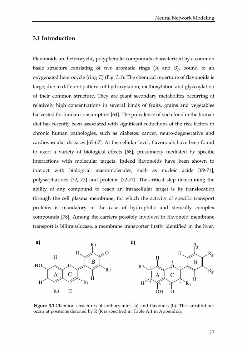
Flavonoids are heterocyclic, polyphenolic compounds characterized by a common
basic structure consisting of two aromatic rings (A and B), bound to an
oxygenated heterocycle (ring C) (Fig. 3.1). The chemical repertoire of flavonoids is
large, due to different patterns of hydroxylation, methoxylation and glycosylation
of their common structure. They are plant secondary metabolites occurring at
relatively high concentrations in several kinds of fruits, grains and vegetables
harvested for human consumption [64]. The prevalence of such food in the human
diet has recently been associated with significant reductions of the risk factors in
chronic human pathologies, such as diabetes, cancer, neuro-degenerative and
cardiovascular diseases [65-67]. At the cellular level, flavonoids have been found
to exert a variety of biological effects [68], presumably mediated by specific
interactions with molecular targets. Indeed flavonoids have been shown to
interact with biological macromolecules, such as nucleic acids [69-71],
polysaccharides [72, 73] and proteins [72-77]. The critical step determining the
ability of any compound to reach an intracellular target is its translocation
through the cell plasma membrane, for which the activity of specific transport
proteins is mandatory in the case of hydrophilic and sterically complex
compounds [78]. Among the carriers possibly involved in flavonoid membrane
transport is bilitranslocase, a membrane transporter firstly identified in the liver,
7
6 35 4
21
A C
BB
CA
3'
5'R7
4'
O
R
H
O H O
R 3H
R
R
HH
R1
3
R5
O
O
HO
H
H
RH
R 2
HH
Figure 3.1 Chemical structures of anthocyanins (a) and flavonols (b). The substitutionsoccur at positions denoted by R (R is specified in 67BTable A.1 in Appendix).
a) b)
37
Neural Network Modeling where it is expressed on the sinusoidal domain of the plasma membrane [79, 80].
At this level, its physiological function is to mediate the diffusion of organic
anions from the blood into the liver, thus playing a role in the hepatic
detoxification pathway(s) of endo- and xenobiotics. Its established substrates are
bilirubin [81, 82] and nicotinic acid [81] with dissociation constants Kd = 2 nM and
Kd = 11 nM, respectively, sulfobromophtalein with Kd = 5 μM) [83, 84] and
anthocyanins with Kd = 1.5-22 μM [85]. The interaction mechanism of
bilitranslocase binders has not yet been established, since the secondary structure
of bilitranslocase is not known.
This investigation focuses on the structural properties of the ligands in order to
infer the mechanisms of their interaction with the transporter. Thus, we study the
nature of the interactions between bilitranslocase and flavonoid ligands, both
anthocyanins and flavonols, by the counter propagation artificial neural network
method, one of the computational approaches already validated as a proper tool
in the investigation of ligand activity [86]. We have used this approach first to
classify the tested molecules into three categories, according to their effect on
bilitranslocase transport activity: i) competitive inhibitors (C), ii) noncompetitive
inhibitors (N), iii) inactive molecules (I). With CP-ANN modeling the inhibition
constant KI of competitive inhibitors can be predicted. Special attention is
dedicated to the examination of the kind of molecular descriptors needed to create
the model. The results of this work show that, contrary to dietary anthocyanins,
most of dietary flavonols do not interact with bilitranslocase, while some flavonol
aglycones act as poor ligands of that carrier [87]. A quantitative analysis of the
structure-activity relationship leads to the identification of parts of ligands
potentially involved in the binding to bilitranslocase, along with an inference on
the kind of interaction between the ligand and the target.
38
Neural Network Modeling
3.2 Methods and models
A detailed description of the counterpropagation artificial neural network
architecture and its learning strategy are given in many articles and text books
[88, 89]. In Appendix A.1 a short description of the method is given in reference to
the specific application that is presented here.
The experimental data for the molecules listed in Table A.1 were obtained by
studying in vitro bilitranslocase transport activity [87]. The models for 22
anthocyanins and 21 flavonols were built up and a structural optimisation was
performed for each of them. The semiempirical AM1 method within MOPAC
packages [90] was used to obtain the equilibrium structures. Next, the CODESSA
program was used to calculate the descriptors on the basis of optimised
geometrical parameters [91]. We obtained 353 descriptors for each molecule as
CODESSA output. Structural descriptors are illustrated in Appendix A.3.
The crucial point in chemometrics is to obtain a proper set of descriptors, which
very often means a reduction in the number of the originally calculated
descriptors. They need to be carefully chosen in order to obtain the best
distribution of molecules in the top-map. Visual inspection of the top-map gives
us information on clusters as structural similarity relationships between
molecules. Descriptors can provide the knowledge about molecular features,
which make the selected molecule, for instance a good competitive inhibitor.
Based on such a study we are able to draw some hypothesis of the potential
nature of the ligand – target interaction. The selection of relevant descriptors is
done for two independent purposes. First we wanted to classify the molecules
according to their effect on bilitranslocase transport activity (I, C, N). To achieve
this aim the average of the absolute deviation was calculated and all descriptors
with values smaller than a threshold of 0.8 were removed. In this way the non-
discriminative variables that are similar for all molecules are effectively
eliminated. The models were tested with the leave-one-out method [89]. The
correlation coefficient of a leave-one-out test was used as a criterion to estimate
39
Neural Network Modeling the quality of the classification models. After following these procedures the
initial number of descriptors is reduced to 207 descriptors.
Second, we wanted to predict the KI value of competitive inhibitors. For this
purpose we used only 18 molecules from the data set, those that show the
competitive inhibition of bilitranslocase. With these molecules we started the
selection procedure once again with the whole set of descriptors. The initial
selection was done using the transposed data matrix, in which one row is an
object representing one descriptor and contains the values of that particular
descriptor for all compounds. The dimension of the input vectors is now equal to
the number of molecules in the data set. After inputting the transposed data
matrix into the input layer of CP-ANN, we obtained the Kohonen map of
descriptors. Some of them either occupied the same neuron or formed clusters.
The descriptors, which occupied one single neuron were selected as specific and
chosen to be submitted to the next selection method. In the next step we applied
the genetic algorithm [92] (see also Appendix A.2) that generates the lowest
possible number of descriptors, thus yielding a satisfactory prediction for KI for
each individual inhibitor in the data set. Once again the leave-one-out cross-
validation method for the training set data was used for verifying the models with
different numbers of descriptors. After the iterative procedure the best results
were obtained for a set of 155 variables and these results are presented in the
following section. The set of these 155 descriptors is listed in the appendix in
Table A.2.
3.3 Results
3.3.1 CP-ANN model for classification of the bilitranslocase ligands
The set of 43 molecules was studied for the classification purpose. In this study
the model adaptive parameters are: number of training epochs, dimension of
network (nx, ny) and maximal correction factor. We divided the molecules into the
training and test sets on the basis of clusters formed on the Kohonen map.
40
Neural Network Modeling
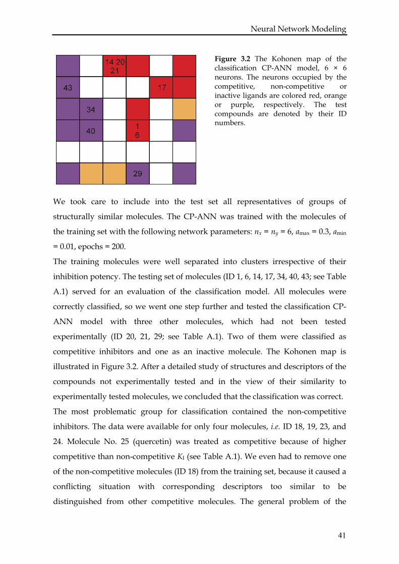
Figure 3.2 The Kohonen map of the classification CP-ANN model, 6 × 6 neurons. The neurons occupied by the competitive, non-competitive or inactive ligands are colored red, orange or purple, respectively. The test compounds are denoted by their ID numbers.
We took care to include into the test set all representatives of groups of
structurally similar molecules. The CP-ANN was trained with the molecules of
the training set with the following network parameters: nx = ny = 6, amax = 0.3, amin
= 0.01, epochs = 200.
The training molecules were well separated into clusters irrespective of their
inhibition potency. The testing set of molecules (ID 1, 6, 14, 17, 34, 40, 43; see Table
A.1) served for an evaluation of the classification model. All molecules were
correctly classified, so we went one step further and tested the classification CP-
ANN model with three other molecules, which had not been tested
experimentally (ID 20, 21, 29; see Table A.1). Two of them were classified as
competitive inhibitors and one as an inactive molecule. The Kohonen map is
illustrated in Figure 3.2. After a detailed study of structures and descriptors of the
compounds not experimentally tested and in the view of their similarity to
experimentally tested molecules, we concluded that the classification was correct.
The most problematic group for classification contained the non-competitive
inhibitors. The data were available for only four molecules, i.e. ID 18, 19, 23, and
24. Molecule No. 25 (quercetin) was treated as competitive because of higher
competitive than non-competitive KI (see Table A.1). We even had to remove one
of the non-competitive molecules (ID 18) from the training set, because it caused a
conflicting situation with corresponding descriptors too similar to be
distinguished from other competitive molecules. The general problem of the
41
Neural Network Modeling investigated molecules is that slight changes in the structure cause big changes in
the activity. This was particularly evident with compounds ID 17 (Cyanidin 3-O-
α-L-arabinopyranoside, competitive inhibitor) and ID 18 (Cyanidin 3-O-β-D-
galactopyranoside, non-competitive inhibitor). In the final classification model
only three non-competitive inhibitors were included (ID 19, 23, 24). A set of three
compounds is obviously not enough for extracting the proper statistical
evaluation of the predictions obtained by an experience-based model as CP-ANN
is. We can only say that the three non-competitive inhibitors, with the exception
of ID 18 that was discarded, were separated from the other compounds (see Fig.
3.2, orange squares). In the absence of test compounds, no prediction was
possible.
3.3.2 CP-ANN model for prediction of KI
The prediction of KI is an essential step to validate the CP-ANN analysis of
experimental data. To study the prediction of KI value, a set of 18 molecules was
used, which experimentally show competitive activity. The molecules with ID
numbers from 1 to 17 and quercetin (ID 25, see Table A.1)
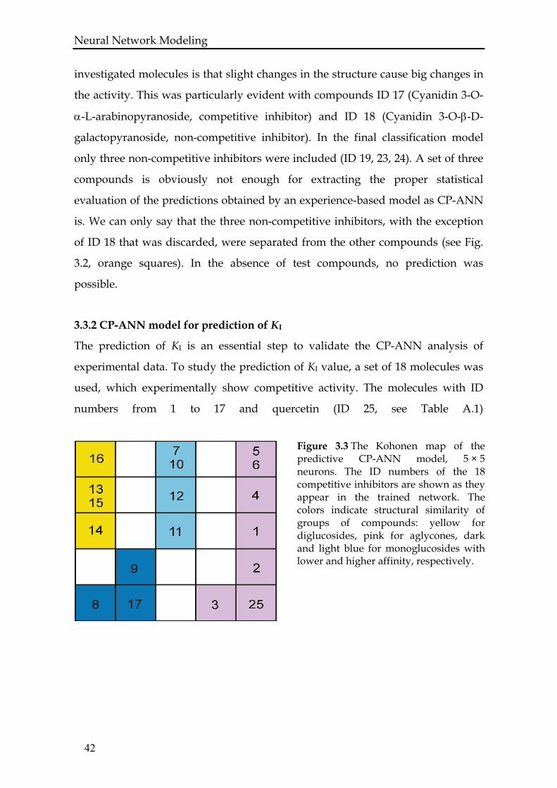
Figure 3.3 The Kohonen map of the predictive CP-ANN model, 5 × 5 neurons. The ID numbers of the 18 competitive inhibitors are shown as they appear in the trained network. The colors indicate structural similarity of groups of compounds: yellow for diglucosides, pink for aglycones, dark and light blue for monoglucosides with lower and higher affinity, respectively.
42
Neural Network Modeling
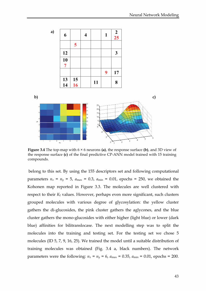
a) 6 4 1 2
25 5
12 3 10 7
9 17 13 14
15 16 11 8
c) b)
Figure 3.4 The top-map with 6 × 6 neurons (a), the response surface (b), and 3D view of the response surface (c) of the final predictive CP-ANN model trained with 15 training compounds.
belong to this set. By using the 155 descriptors set and following computational
parameters nx = ny = 5, amax = 0.3, amin = 0.01, epochs = 250, we obtained the
Kohonen map reported in Figure 3.3. The molecules are well clustered with
respect to their KI values. However, perhaps even more significant, such clusters
grouped molecules with various degree of glycosylation: the yellow cluster
gathers the di-glucosides, the pink cluster gathers the aglycones, and the blue
cluster gathers the mono-glucosides with either higher (light blue) or lower (dark
blue) affinities for bilitranslocase. The next modelling step was to split the
molecules into the training and testing set. For the testing set we chose 5
molecules (ID 5, 7, 9, 16, 25). We trained the model until a suitable distribution of
training molecules was obtained (Fig. 3.4 a, black numbers). The network
parameters were the following: nx = ny = 6, amax = 0.35, amin = 0.01, epochs = 200.
43
Neural Network Modeling We tested the model with the molecules from the test set and, as a result, all the
molecules (Fig. 3.4 a, red numbers) were placed in the correct clusters of trained
molecules.
The predictions of the trained CP-ANN model are stored in the output layer
shown in Figures 3.4 b and c. From Figure 3.4 b it can be seen that we obtain 6 × 6
points of the KI response surface for 6 × 6 neurons in particular network
architecture. The colour bar is given to associate the individual colour of the
square with the KI value; the range 2 to 22 corresponds to the range of KI values of
the training compounds. The prediction of a test compound is obtained by
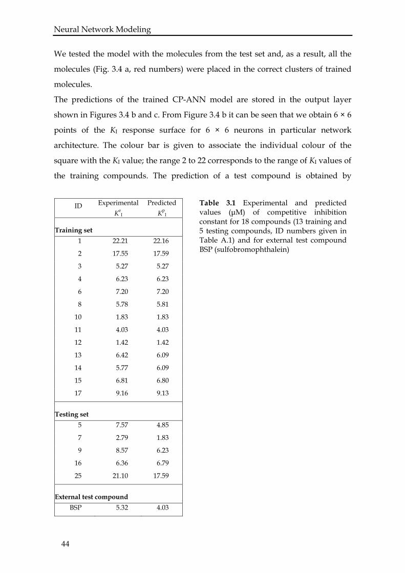
Table 3.1 Experimental and predicted values (μM) of competitive inhibition constant for 18 compounds (13 training and 5 testing compounds, ID numbers given in Table A.1) and for external test compound BSP (sulfobromophthalein)
ID Experimental Ke
I
Predicted Kp
I
Training set 1 22.21 22.16
2 17.55 17.59
3 5.27 5.27
4 6.23 6.23
6 7.20 7.20
8 5.78 5.81
10 1.83 1.83
11 4.03 4.03
12 1.42 1.42
13 6.42 6.09
14 5.77 6.09
15 6.81 6.80
17 9.16 9.13
Testing set 5 7.57 4.85
7 2.79 1.83
9 8.57 6.23
16 6.36 6.79
25 21.10 17.59
External test compound BSP 5.32 4.03
44
Neural Network Modeling pinpointing a co-ordinate on the output surface. In Figure 3.4 c, a 3D view of the
output surface with larger resolution (3 × 3 squares per neuron) is given. The
colour bar is the same as in Figure 3.4 b.
The RMSE of the predicted KI values is equal to 2.2. For a small dataset such as
that investigated in the current study this error is expected. The KI values of the
new molecules predicted by this model could be a good approximation of their
actual values. We are especially satisfied with the results, because the tested
molecules can be correctly placed in clusters of the training model. That result
proves that the set of descriptors is properly chosen for prediction of the KI value.
Table 3.1 reports the experimental and predicted KI values.
3.3.3 Structural descriptors influencing the binding of anthocyanins to
bilitranslocase
The detailed investigations of the 155 descriptors used to classify the compounds
lead us to conclude that the ligand ability to act as H-bond donors and/or
acceptors is very important for establishing an interaction with the host molecule.
Descriptors directly defining the ability for creating this kind of bonds were
indicated by an automated genetic-algorithm-based variable selection procedure.
Only the selected descriptors enabled the CP-ANN training procedure to cluster
the molecules according to their affinity to the target.
3.3.4 Final validation of the CP-ANN model
The final validation of the CP-ANN model was performed by testing the
inhibition activity of BSP, which is an established substrate of bilitranslocase with
the KI = 5.32 ± 0.63 μM [83, 84]. The 3D structure was prepared in the same way as
for all other molecules in the study. Once structural descriptors were obtained,
only those selected in the model building and variable reduction procedure were
accepted, i.e. 155 descriptors. The predicted KI = 4.03 μM (see Table 3.1). The
deviation μM 1.29pI
eI =− KK is twice as large as the established experimental
error of ± 0.63. However, it is within range of the obtained model error (RMSE =
2.2) for the testing set, which validate our CP-ANN model.
45
Neural Network Modeling
3.4 Discussion
The main result of this work is in the identification of the nature of molecular
interactions between the flavonoids and bilitranslocase. This enables a structure-
based classification of molecules according to their activity. Taking into account
the structural features of the molecules belonging to a given cluster and focusing
on the nature of molecular interactions between the carrier and the ligands, a
system of deductions about the structural requirements for the activity of
bilitranslocase ligands is proposed below.
3.4.1 The structural basis of classification of molecules
To understand the structural basis for the partition of molecules in well separated
clusters of inactive and active (competitive and non-competitive) molecules, a
comparative analysis of structural and functional features of anthocyanins vs
flavonols was particularly helpful.
3.4.1.1 Active vs inactive molecules: the importance of the 3D structure of the
molecule
The role played by the 3D structure of the ligands was noticed for the compounds
2 (cyanidin, an anthocyanidin) and 25 (quercetin, a flavonol) and their analogues
8 and 32, substituted with the sugar at position R3 (2→8 and 25→32). Substitution
on cyanidin (ID 2→8) made it a better inhibitor, since its KI decreased from 17.55
to 5.78 μM. On the contrary, substitution on quercetin (ID 25→32) made it an
inactive molecule. We compared the optimised 3D structures of these molecules,
which are shown in Figure 3.5, and we noticed that they differ mostly in the
orientation of the sugar moiety. The sugar in ID 8 lies in the same plane as the
planar aromatic rings. In this way, the hydrogen atoms of the
46

Neural Network Modeling
a) b)
c)
Figure 3.5 3D structures of (a) cyanidin 3-glucoside (ID = 8), (b) quercetin 3-glucoside(ID = 32) and (c) cyanidin 3,5-diglucoside (ID = 14).
sugar OH groups point to the same direction as the substitutes of B ring, ready to
create hydrogen bond(s) with the target. On the contrary, the same sugar moiety
bound to an equivalent position in quercetin is located perpendicularly to the
planar condensed rings of the molecule. This position is forced by the presence of
the oxygen atom in ring C. That may be a good reason for the molecule ID 32 to be
inactive even in a protein environment – it simply does not fit the binding pocket
of the host molecule. The energy needed to flip down the sugar moiety could be
too high, especially if the carbonyl group of ring C acted as an H-bond acceptor
from the carrier active site. We looked at the structure of ID 14 (cyanidin 3,5-di-O-
β-Dglucopyranoside), in order to evaluate the 3D structure of the two sugar
substitutions. We found that both are almost co-planar with rings A and C. From
47
Neural Network Modeling these observations it can be deduced that the ligands of bilitranslocase, whether
acting as competitive or non competitive inhibitors, have to be planar. This
conclusion is in agreement with the original experimental observation that the
only phthalein tautomer transported by bilitranslocase is the planar, quinoidal
one [93].
3.4.1.2 Competitive vs non-competitive inhibition by aglycones: the importance
of hydroxylation of B ring
As a step further, we tried to understand the structural basis underlying the
property of bilitranslocase inhibitors to act either competitively or non-
competitively. Some hints were provided by the behaviour of the three active
flavonols (ID 23-25). Galangin (ID 23) is the only aglycone displaying pure non-
competitive activity. Unique among all flavonoid aglycones tested (both
anthocyanins and flavonols), its ring B has no oxygen atom at position R4’. We
deduced that its interaction with the target must involve the condensed rings A
and C, the only moieties suitable for H-bond formation. By contrast, the
introduction of an OH group in the 4’ position of the B ring of flavonols (as in ID
24 and 25) confers them the further property to interact at the level of the
competitive site, like most of the anthocyanins (ID 1-19).
3.4.1.3 Pure competitive vs mixed-type inhibition: the possible role of
tautomerism of ring B
To understand the nature of mixed-type inhibition, typical of two flavonols (ID
24-25), we started from the evidence that the CP-ANN model clustered
competitive molecules separately from non-competitive ones, reflecting two well
distinct modes of interaction of the same molecule with the carrier. Given the
importance of hydroxylation of the B ring in determining the type of inhibition
(and its strength as well, as discussed below), we speculated that mixed-type
inhibition is compatible with the binding of two alternative structures of ID 24-25
to the carrier, arising from pH-dependent phenolic-quinoidal tautomerism of the
48
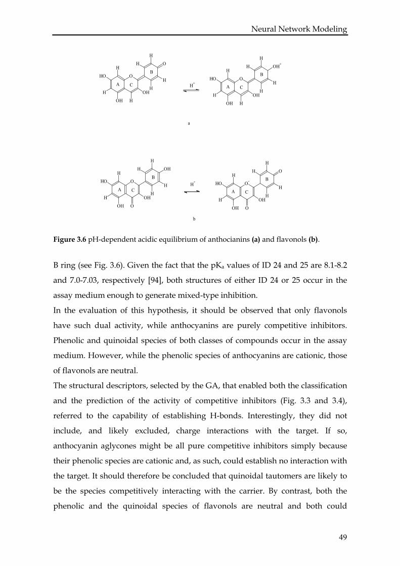
Neural Network Modeling
O
O
H
H
H
HH
HO
H
OH H
OH
B ring (see Fig. 3.6). Given the fact that the pKa values of ID 24 and 25 are 8.1-8.2
and 7.0-7.03, respectively [94], both structures of either ID 24 or 25 occur in the
assay medium enough to generate mixed-type inhibition.
In the evaluation of this hypothesis, it should be observed that only flavonols
have such dual activity, while anthocyanins are purely competitive inhibitors.
Phenolic and quinoidal species of both classes of compounds occur in the assay
medium. However, while the phenolic species of anthocyanins are cationic, those
of flavonols are neutral.
The structural descriptors, selected by the GA, that enabled both the classification
and the prediction of the activity of competitive inhibitors (Fig. 3.3 and 3.4),
referred to the capability of establishing H-bonds. Interestingly, they did not
include, and likely excluded, charge interactions with the target. If so,
anthocyanin aglycones might be all pure competitive inhibitors simply because
their phenolic species are cationic and, as such, could establish no interaction with
the target. It should therefore be concluded that quinoidal tautomers are likely to
be the species competitively interacting with the carrier. By contrast, both the
phenolic and the quinoidal species of flavonols are neutral and both could
Figure 3.6 pH-dependent acidic equilibrium of anthocianins (a) and flavonols (b).
H+
O-
O
H
H
H
HH
HO
H
OH O
OH
a
O
OH+H
H
H
HH
HO
H
OH H
OH
H+
CA
B
CA
B
O
OH
H
H
H
HH
HO
H
OH O
OH
b
B
A C CA
B
49
Neural Network Modeling therefore bind the carrier. However, while quinoidal species of flavonols would
act as competitive inhibitors, in analogy with anthocyanins, phenolic species
would act as non-competitive inhibitors, in analogy with galangin (ID 23) whose
B ring cannot originate a quinoidal tautomer.
The concept that para-quinoidal species of flavonoids competitively interact with
bilitranslocase is supported by the experimental evidence that the pH-indicator
phthaleins are transported only as para-quinoidal species [93]; it is quite realistic
that competitive inhibition of phthalein transport should be exerted by the
structurally closest tautomer of flavonoids. Another point in favour of this
concept is that only the para-quinoidal species of flavonols definitely set the B ring
in the same plane of the rest of the molecule, a structural requirement for
phthaleins to be transported and for glycosylated flavonoids to be active.
Yet, what would prevent phenolic species, freely rotating around C1, from
binding to the competitive site? With respect to the formation of a network of H-
bonds, we have to deduce that the main, obvious difference between para-
phenolic and para-quinoidal species is that the former are H-bond donors whereas
the latter are H-bond acceptors. This would suggest that a decisive structural
feature for a molecule to act as a competitive inhibitor of bilitranslocase transport
activity is the availability of atoms acting as H-bond acceptors from
corresponding H-bond donors of the active site of the carrier. Conversely, the H-
bond formation by donating a hydrogen by a para-phenolic tautomer to the active
site of the carrier might involve a configuration of the flavonol B ring compatible
with non-competitive inhibition.
3.4.2 The structural basis of the activity of competitive inhibitors
The visual inspection of the Kohonen map shown in Figure 3.3 highlights the
relationship between the structure and the activity of investigated compounds.
Clusters are formed according to the degree of glycosylation. The best competitive
inhibitors are anthocyanin mono-glucosides, separated in two sub-classes
grouping molecules with KI < 4 μM (ID 7, 10-12; light blue) and KI > 5.8 μM (ID 8,
9, 17, dark blue) respectively. The di-glucosides form the next group, if ranged by
50
Neural Network Modeling affinity and the last are the aglycones, with the lowest affinity (in Fig. 3.3 shown
as yellow and pink squares, respectively). This map indicates that the carrier
active site somehow “reads” the glycosylation state of anthocyanins, thus
implying not only that it is a complex and multi-compliant structure, but also that
it provides a polar environment for the interaction with polar moieties of the
substrates.
3.4.2.1 Flavonoid aglycones: the importance of hydroxylation of the B ring
Among competitive inhibitors, the least polar anthocyanidin (pelargonidin, ID 1)
is the least active one (KI = 22.21 μM). Similarly, the least polar flavonol
(kaempferol, ID 24) has a very poor competitive inhibition activity (KI = 131.6 μM).
The presence of a second hydroxyl group on ring B, as in either cyanindin (ID 2,
an anthocyanin) or in quercetin (ID 25, a flavonol), enhances the competitive
inhibition of both compounds. While among anthocyanins the gain in activity is
just above the statistical significance: KI = 22.2±1.65 (ID 1) vs KI = 17.5±1.68 μM (ID
2), the effect is marked among the flavonols, as the KI drops from 131.6±3.8 (ID 24)
to 21.1±1.7 μM (ID 25). Interestingly, the difference in KI of either cyanidin (ID 2)
or quercetin (ID 25) is not significant, suggesting that both compounds interact
with the target through the moiety they have in common, i.e. the ring B. Thus, the
number of H-bonds established at the level of the B ring is indeed very important
in shaping the strength of interaction of the aglycones. In more detail, the B ring
interactions are based on the presence of two vicinal oxygen atoms, possibly
acting as a couple of H bond acceptor (the para-quinoid carbonyl) and H-bond
donor (the R3’ hydroxyl group). At the same time, the steric hindrance exerted by
the carbonyl group of the C ring, a specific feature of the flavonols, appears to be
successfully overcome if the B ring interactions prevail over the others.
3.4.2.2 Flavonoid aglycones: the importance of steric hindrance and charge
distribution caused by the carbonyl group of the C ring
The carbonyl group of ring C in flavonols appears to heavily influence the
magnitudes of the competitive KI values. That was realised by comparing
51
Neural Network Modeling pelargonidin (ID 1 an anthocyanin) with kaempferol (ID 24, a flavonol). The
anthocyanin is nearly 6-fold more active than the flavonol (22.2 vs 131.6 μM). The
flavonol appears to be sterically hindered by the carbonyl group of the C ring. It
can be also considered that the carbonyl group on the C ring favours the
establishment of intramolecular H bonds with the OH groups in R3 or in R5 [35],
thus possibly limiting the overall tendency of flavonols to interact with the carrier
and therefore increasing their apparent competitive KI.
In flavonols, the presence of a hydroxyl group in R5’ (ID 26 vs ID 23, 24, 25) or, as
predicted by the CP-ANN (Fig. 3.2), O-methylation of the B ring (ID 29 vs ID 25)
results in the total loss of activity. In none of the anthocyanins were these all-or-
none effects observed on activity (ID 3 vs ID 1, 2 and ID 4 vs ID 2). On the
contrary, triple hydroxylation of the B ring of anthocyanins is better than double
hydroxylation (ID 3 vs ID 2) and O-methylation is also well accepted, apparently
because the anthocyanin aglycone can interact with the target also through the A
ring, as previously proposed [85]. In flavonols, the functional groups introduced
in the B ring, as in ID 26 and ID 29, appear to be equally effective in preventing
their interaction with the carrier, presumably by steric hindrance. It results
therefore that the carbonyl group of the C ring in flavonols acts as constraint for
their ability to take different orientations at the level of the transport site(s) of the
carrier. Thus, in interacting with the carrier, flavonols might be seen as rather stiff
molecules, with a limited repertoire of interactions, whereas anthocyanins appear
to be more flexible and thus better at fitting the chemical environment of
bilitranslocase active site.
3.4.2.3 Anthocyanidin mono- and diglucosides
The distinctive feature for clustering mono-glucosides in two groups according to
their activity (Fig. 3.3, light and dark blue) is the OH group at position R1 of the B
ring, which occurs in the low-affinity molecules (KI > 5.8 μM, ID 8, 9, 17) and is
missing in the high-affinity ones(KI < 4 μM ID 7, 10-12). In the case of mono-
glucosides, an increase in the polarity of B ring (see Table A.1) lowers the affinity
52
Neural Network Modeling for the target (ID 7-9, 10-12). While this finding emphasises one more time the
critical role of this moiety in the interaction with the carrier, it also reflects the
existence of a specific binding pocket for the glycosyl moiety attached to C3,
improving the binding affinity to such an extent that the interaction with the B
ring is devalued. Any changes in the sugar moiety result in the decrease of
affinity to the target (e.g. ID 8 vs ID 17) or even in a change of the type of
inhibition (e.g. ID 8 vs ID 18 and ID 19, Table A.1). This may be an indication that
the ligand does interact with the target at two sites, i.e. at one site through the
sugar moiety and at the other through the B ring.
In contrast, for diglucosides, changes at substituents of ring B don’t influence the
KI value so much. That can suggest that the diglucoside-target interactions occur
either through both sugars or through ring A and its glucosyl moiety, as proposed
earlier [85]. It appears therefore that the target has a structurally complex active
site, providing specific, hydrogen bond-based interactions for a potentially wide
spectrum of structurally unrelated ligands.
3.4.3 A hypothesis for the structural basis of the activity of ID 18
A glycosyl moiety in C3 improves the interaction of anthocyanins with the carrier,
because an additional set of H bonds can be established. Cyanidin 3-O-β-D-
galactopyranoside (ID 18) clustered with competitive inhibitors, without being
one. This would indicate that ID 18 might indeed bind to the carrier at the same
level as the other mono-glucosides, although with lower affinity (KI = 35.2 μM vs
KI = 1.42-9.16 μM). This concept is in apparent conflict with the tenet that non-
competitive inhibitors bind to a site that is close to, but does not coincide with an
enzyme active site. However, binding of the galactosyl moiety to the sugar-
specific pocket of the carrier might affect the conformation of the active site of the
carrier, thus perturbing the kinetics of BSP transport. Two possibilities are equally
applicable, i.e. ID 18 either induces an alternative, catalytically less efficient,
conformational modification of the transport site or it causes a blockade of a
conformational modification essential for transport catalysis.
53
Neural Network Modeling
54
3.5 Conclusions
It can be concluded that the requirement for a flavonoid molecule to interact with
the target is the overall planarity. Then, to interact in a competitive way, and thus
to be presumably transported, it has to have a p-quinoid group on the ring B, in
order to be a hydrogen bond acceptor.
If it has a p-hydroxyl group on the ring B, it is a non-competitive inhibitor. The
OH group may therefore play a dual role, either as a proton donor, or, in the
deprotonated form, as a strong hydrogen bond acceptor. It would remain a
challenge to verify the above statements experimentally or using other
computational tools, such as quantum mechanics based method.
It cannot be stated yet that inhibitors of electrogenic BSP transport activity are
indeed transported, unless they are tested directly as transport substrates.
However, the dependence of the interaction of flavonoids with bilitranslocase on
H bonds shows that those molecules interact without charge compensation, an
essential requirement to be transported electrogenically. By an independent
experimental approach, transport of malvidin 3-glucoside in liver cells has been
shown to be mediated by bilitranslocase [95], suggesting that, at least for
competitive inhibitors, this could be the case.
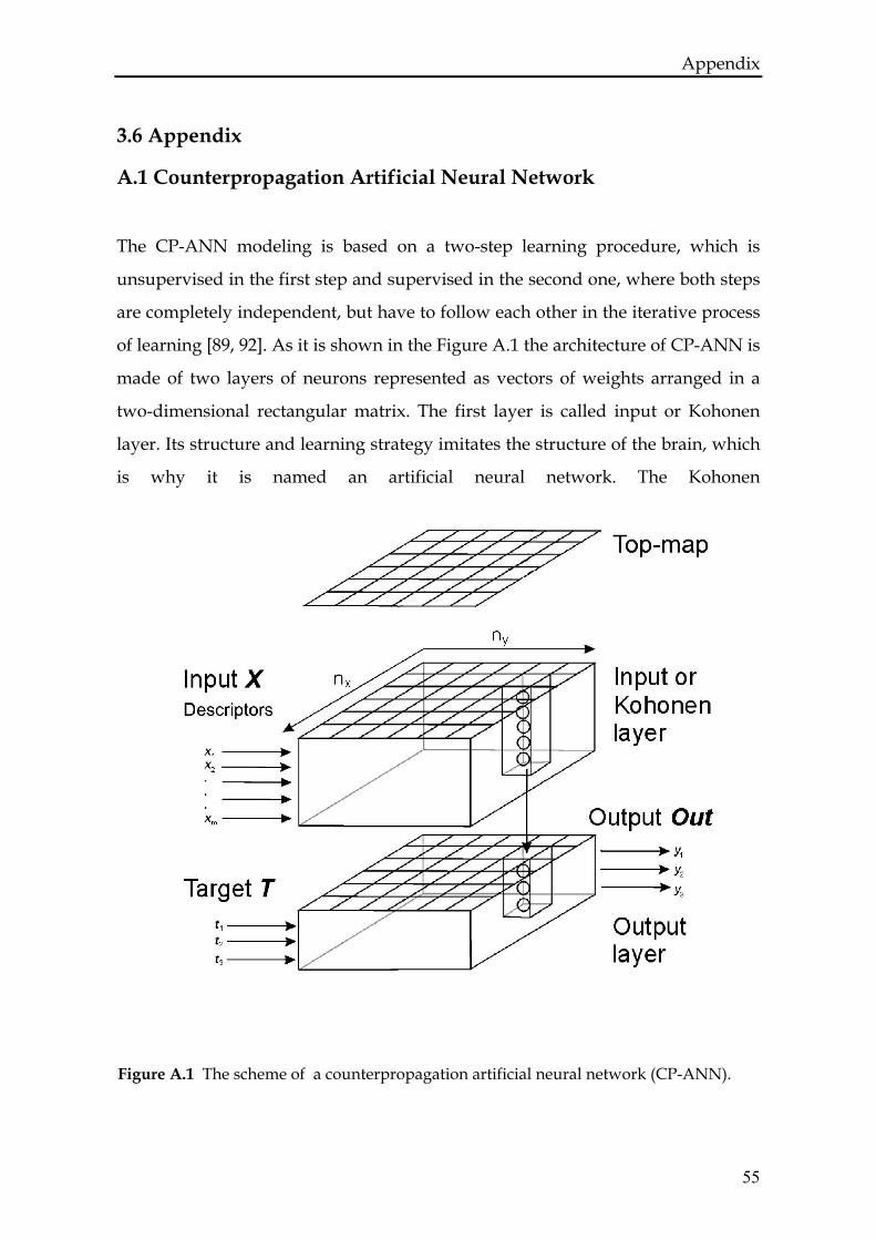
Appendix
3.6 Appendix
A.1 Counterpropagation Artificial Neural Network
The CP-ANN modeling is based on a two-step learning procedure, which is
unsupervised in the first step and supervised in the second one, where both steps
are completely independent, but have to follow each other in the iterative process
of learning [89, 92]. As it is shown in the Figure A.1 the architecture of CP-ANN is
made of two layers of neurons represented as vectors of weights arranged in a
two-dimensional rectangular matrix. The first layer is called input or Kohonen
layer. Its structure and learning strategy imitates the structure of the brain, which
is why it is named an artificial neural network. The Kohonen
Figure A.1 The scheme of a counterpropagation artificial neural network (CP-ANN).
55
Appendix layer is represented as a tensor in three dimensions nx × ny × m. nx, ny are the
number of neurons represented in the X and Y dimensions, while m is the number
of descriptors that spans the Z axis (Fig. A.1). The number of neurons N = nx × ny
is a computational parameter related to the number of molecules in the training
set. The Kohonen layer has a well-defined topology of neurons with defined
lattice structure. The first step, the unsupervised learning procedure is
determined by descriptors X = x1, … xk, … xm. During the training molecules with
similar descriptors are placed close to each other and form clusters that can be
observed in the top-map or Kohonen-map of the CP-ANN (see Fig. A.1). It is
important that only the descriptors guide the determination of the position of the
molecules in the top-map. This top-map contains the ID number of molecules that
were placed at particular positions in the Kohonen layer after learning, or a blank,
if the corresponding neuron in the input layer is empty. When we plot the top-
map, as it is done in Figures 3.2, 3.3, 3.4, we can see which positions are occupied
by individual molecules at the end of the training. The Kohonen layer contains the
"weights" produced in the training procedure. The weights wi,j,k are real numbers,
one for each element i,j,k in the three-dimensional tensor (nx × ny × m).
The second, supervised step of the learning procedure involves training the
output layer, guided by the input layer (see Fig. A.1). The position of molecules
stored in the Kohonen layer is imposed on the output layer in each iteration. As a
result of the second step in the learning procedure each neuron in the output layer
provides an output vector Out for each molecule as close as possible to the initial
target vector T. In the application of CP-ANN for the classification of flavonoid
molecules, the target T is a three-component binary vector (t1, t2, t3, as shown in
Fig. A.1) representing one of three possible classes of bilitranslocase substrates,
with for the competitive inhibitors, )0,0,1(C =T )0,1,0(N =T for non-competitive
inhibitors and for the inactive molecules. The artificial neuron network
trained in such a way can be used to classify new molecules by first situating
them in the Kohonen layer on the neurons with the most similar weights. These
positions are then projected to the output layer, which provides the output class
C, N or I of substrate.
)1,0,0(=IT
56
Appendix In the second application of CP-ANN for the prediction of KI, the target T is the
experimental value of KI and the output layer is a plane, since it has only one
value at each position. The neural network again has to be trained in two steps,
but now with respect to predicting a single scalar value, the KI, for the competitive
inhibitors.
The computational parameters of the neural network construction can influence
the results and should be tested and optimized. In particular the number of
training epochs, the dimension of the network (nx, ny), the maximal correction
factor (amax), the minimal correction factor (amin) [89].
A.2 Genetic Algorithm
A genetic algorithm can be applied to address problems in which optimal values
of a set of parameters or an optimal reduced set of parameters, resulting from a
combination of original parameters, is sought. In the present application the
method was used to reduce the number of descriptors needed for proper
clustering of the training set of molecules. The origin of its name comes from the
resemblance with the biological process: the GA consists of three basic processes
mimicking Darwinian evolution: crossover, mutation and survival of the fittest. In
the crossover step, new chromosomes are generated by mixing fractions of old
individuals, so called parent chromosomes. Mutation is introduced to randomly
change individual bits of the chromosome. In the last step the survivals are
chosen for the next generation, i.e. the cycle of the GA. The survivals are the
chromosomes having the best criterion value determined by the fitness function.
This last step should yield the lowest possible number of descriptors which gives
a satisfactory prediction for KI for each individual inhibitor in the data set. The
length of the chromosome, i.e. the number of genes or bits, is determined by the
number of input parameters. A full description of the GA can be found in Ref. [88]
57
Appendix
A.3 Structural descriptors
Descriptors are used to characterize mathematically the molecules. The set of
descriptors in the CODESSA software is split into five categories: (i)
constitutional descriptors, depending on the number and type of atoms, bonds,
and functional groups; (ii) geometrical descriptors, which give molecular surface
area, projections, and gravitational indices; (iii) topological descriptors, which are
molecular connectivity indices, related to the degree of branching in the
compounds; (iv) electrostatic descriptors, such as partial atomic charges and
other parameters describing the possibility for some sites in the molecule to form
hydrogen bonds; (v) quantum-chemical descriptors, i.e. total energy of molecule,
the energies of the lowest unoccupied and highest occupied orbital, ionization
potential, etc. All the descriptors have to be normalized, maintaining the original
distribution, using a range scaling procedure according to:
( ) ,k
kknormk xs
xxx −=
where is the normalized value of the descriptor xk, the normkx kx is the average
value and s is a standard deviation
( ),
1)(
2
−
−= ∑
nxx
xs kkk
with n the number of molecules.
58
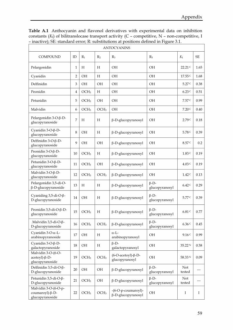
Appendix
Table A.1 Anthocyanin and flavonol derivatives with experimental data on inhibition constants (KI) of bilitranslocase transport activity (C – competitive, N – non-competitive, I – inactive); SE: standard error; R: substitutions at positions defined in Figure 3.1.
ANTOCYANINS
COMPOUND ID R1 R2 R3 R5 KI SE
Pelargonidin 1 H H OH OH 22.21 C 1.65
Cyanidin 2 OH H OH OH 17.55 C 1.68
Delfinidin 3 OH OH OH OH 5.27 C 0.38
Peonidin 4 OCH3 H OH OH 6.23 C 0.51
Petunidin 5 OCH3 OH OH OH 7.57 C 0.99
Malvidin 6 OCH3 OCH3 OH OH 7.20 C 0.40
Pelargonidin 3-O-β-D-glucopyranoside 7 H H β-D-glucopyranosyl OH 2.79 C 0.18
Cyanidin 3-O-β-D-glucopyranoside 8 OH H β-D-glucopyranosyl OH 5.78 C 0.39
Delfinidin 3-O-β-D-glucopyranoside 9 OH OH β-D-glucopyranosyl OH 8.57 C 0.2
Peonidin 3-O-β-D-glucopyranoside 10 OCH3 H β-D-glucopyranosyl OH 1.83 C 0.19
Petunidin 3-O-β-D-glucopyranoside 11 OCH3 OH β-D-glucopyranosyl OH 4.03 C 0.19
Malvidin 3-O-β-D-glucopyranoside 12 OCH3 OCH3 β-D-glucopyranosyl OH 1.42 C 0.13
Pelargonidin 3,5-di-O-β-D-glucopyranoside 13 H H β-D-glucopyranosyl β-D-
glucopyranosyl 6.42 C 0.29
Cyaniding 3,5-di-O-β-D-glucopyranoside 14 OH H β-D-glucopyranosyl β-D-
glucopyranosyl 5.77 C 0.39
Peonidin 3,5-di-O-β-D-glucopyranoside 15 OCH3 H β-D-glucopyranosyl β-D-
glucopyranosyl 6.81 C 0.77
71B69BMalvidin 3,5-di-O-β-D-glucopyranoside 16 OCH3 OCH3 β-D-glucopyranosyl β-D-
glucopyranosyl 6.36 C 0.45
Cyanidin 3-O-α-L-arabinopyranoside 17 OH H α-L-
arabinopyranosyl OH 9.16 C 0.99
Cyanidin 3-O-β-D-galactopyranoside 18 OH H β-D-
galactopyranosyl OH 35.22 N 0.58
Malvidin 3-O-(6-O-acetoyl)-β-D-glucopyranoside
19 OCH3 OCH3 (6-O-acetoyl)-β-D-glucopyranosyl OH 58.33 N 0.09
Delfinidin 3,5-di-O-β-D-glucopyranoside 20 OH OH β-D-glucopyranosyl β-D-
glucopyranosyl Not
tested ---
Petunidin 3,5-di-O-β-D-glucopyranoside 21 OCH3 OH β-D-glucopyranosyl β-D-
glucopyranosyl Not
tested ---
Malvidin 3-O-(6-O-p-coumaroyl)-β-D-glucopyranoside
22 -(6-O-p-coumaroyl)-β-D-glucopyranosyl OCH3 OCH3 OH I I
59
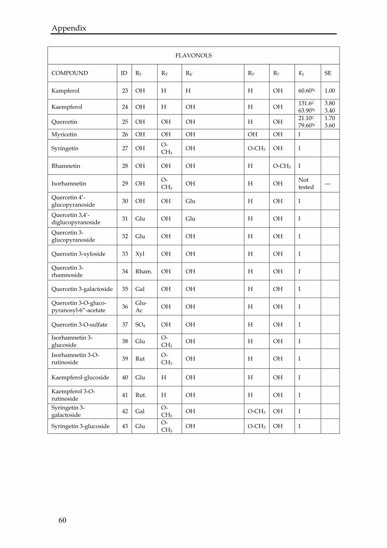
Appendix
60
FLAVONOLS
COMPOUND ID R3 R3’ R4’ R5’ R7 KI SE
Kampferol 23 OH H H H OH 60.60N 1.00
Kaempferol 24 OH H OH H OH 131.6C
63.90N 3.80 3.40
Quercetin 25 OH OH OH H OH 21.10C
79.60N 1.70 3.60
Myricetin 26 OH OH OH OH OH I
Syringetin 27 OH O-CH3
OH O-CH3 OH I
Rhamnetin 28 OH OH OH H O-CH3 I
Isorhamnetin 29 OH O-CH3
OH H OH Not tested ---
Quercetin 4’-glucopyranoside 30 OH OH Glu H OH I
Quercetin 3,4’-diglucopyranoside 31 Glu OH Glu H OH I
Quercetin 3-glucopyranoside 32 Glu OH OH H OH I
Quercetin 3-xyloside 33 Xyl OH OH H OH I
Quercetin 3-rhamnoside 34 Rham. OH OH H OH I
Quercetin 3-galactoside 35 Gal OH OH H OH I
Quercetin 3-O-gluco-pyranosyl-6”-acetate 36 Glu-
Ac OH OH H OH I
Quercetin 3-O-sulfate 37 SO4 OH OH H OH I
Isorhamnetin 3-glucoside 38 Glu O-
CH3 OH H OH I
Isorhamnetin 3-O-rutinoside 39 Rut O-
CH3 OH H OH I
Kaempferol-glucoside 40 Glu H OH H OH I
Kaempferol 3-O-rutinoside 41 Rut. H OH H OH I
Syringetin 3-galactoside 42 Gal O-
CH3 OH O-CH3 OH I
Syringetin 3-glucoside 43 Glu O-CH3
OH O-CH3 OH I
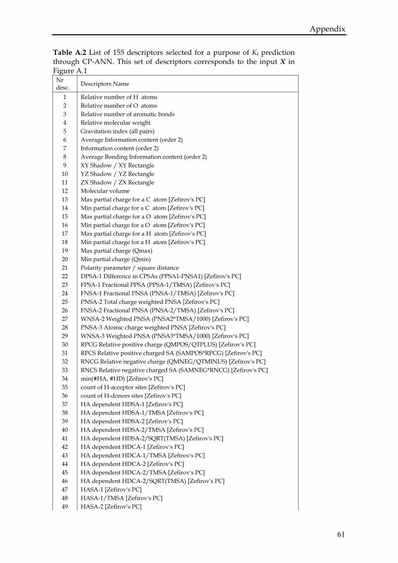
Appendix Table A.2 List of 155 descriptors selected for a purpose of KI prediction through CP-ANN. This set of descriptors corresponds to the input X in Figure A.1
Nr desc. Descriptors Name
1 Relative number of H atoms 2 Relative number of O atoms 3 Relative number of aromatic bonds 4 Relative molecular weight 5 Gravitation index (all pairs) 6 Average Information content (order 2) 7 Information content (order 2) 8 Average Bonding Information content (order 2) 9 XY Shadow / XY Rectangle
10 YZ Shadow / YZ Rectangle 11 ZX Shadow / ZX Rectangle 12 Molecular volume 13 Max partial charge for a C atom [Zefirov's PC] 14 Min partial charge for a C atom [Zefirov's PC] 15 Max partial charge for a O atom [Zefirov's PC] 16 Min partial charge for a O atom [Zefirov's PC] 17 Max partial charge for a H atom [Zefirov's PC] 18 Min partial charge for a H atom [Zefirov's PC] 19 Max partial charge (Qmax) 20 Min partial charge (Qmin) 21 Polarity parameter / square distance 22 DPSA-1 Difference in CPSAs (PPSA1-PNSA1) [Zefirov's PC] 23 FPSA-1 Fractional PPSA (PPSA-1/TMSA) [Zefirov's PC] 24 FNSA-1 Fractional PNSA (PNSA-1/TMSA) [Zefirov's PC] 25 PNSA-2 Total charge weighted PNSA [Zefirov's PC] 26 FNSA-2 Fractional PNSA (PNSA-2/TMSA) [Zefirov's PC] 27 WNSA-2 Weighted PNSA (PNSA2*TMSA/1000) [Zefirov's PC] 28 PNSA-3 Atomic charge weighted PNSA [Zefirov's PC] 29 WNSA-3 Weighted PNSA (PNSA3*TMSA/1000) [Zefirov's PC] 30 RPCG Relative positive charge (QMPOS/QTPLUS) [Zefirov's PC] 31 RPCS Relative positive charged SA (SAMPOS*RPCG) [Zefirov's PC] 32 RNCG Relative negative charge (QMNEG/QTMINUS) [Zefirov's PC] 33 RNCS Relative negative charged SA (SAMNEG*RNCG) [Zefirov's PC] 34 min(#HA, #HD) [Zefirov's PC] 35 count of H-acceptor sites [Zefirov's PC] 36 count of H-donors sites [Zefirov's PC] 37 HA dependent HDSA-1 [Zefirov's PC] 38 HA dependent HDSA-1/TMSA [Zefirov's PC] 39 HA dependent HDSA-2 [Zefirov's PC] 40 HA dependent HDSA-2/TMSA [Zefirov's PC] 41 HA dependent HDSA-2/SQRT(TMSA) [Zefirov's PC] 42 HA dependent HDCA-1 [Zefirov's PC] 43 HA dependent HDCA-1/TMSA [Zefirov's PC] 44 HA dependent HDCA-2 [Zefirov's PC] 45 HA dependent HDCA-2/TMSA [Zefirov's PC] 46 HA dependent HDCA-2/SQRT(TMSA) [Zefirov's PC] 47 HASA-1 [Zefirov's PC] 48 HASA-1/TMSA [Zefirov's PC] 49 HASA-2 [Zefirov's PC]
61
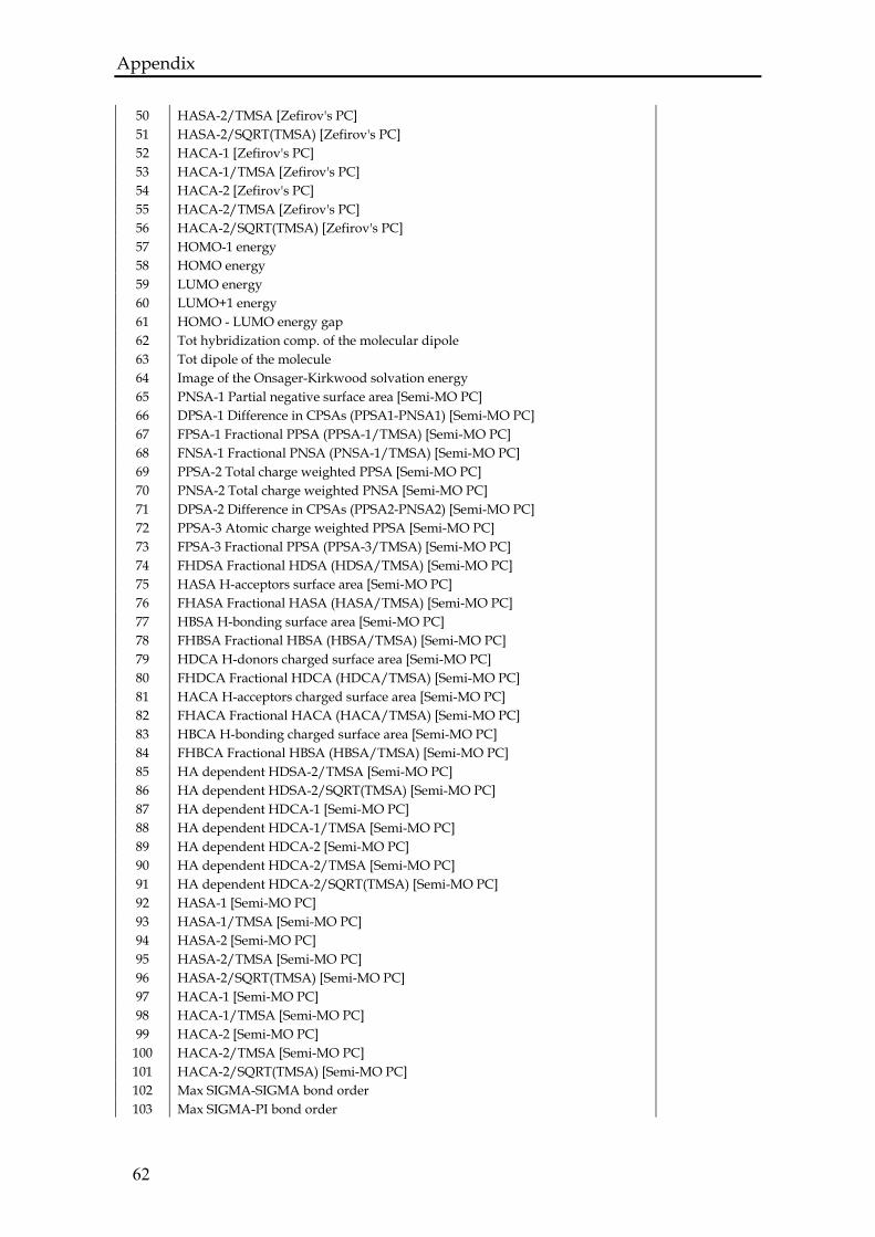
Appendix
50 HASA-2/TMSA [Zefirov's PC] 51 HASA-2/SQRT(TMSA) [Zefirov's PC] 52 HACA-1 [Zefirov's PC] 53 HACA-1/TMSA [Zefirov's PC] 54 HACA-2 [Zefirov's PC] 55 HACA-2/TMSA [Zefirov's PC] 56 HACA-2/SQRT(TMSA) [Zefirov's PC] 57 HOMO-1 energy 58 HOMO energy 59 LUMO energy 60 LUMO+1 energy 61 HOMO - LUMO energy gap 62 Tot hybridization comp. of the molecular dipole 63 Tot dipole of the molecule 64 Image of the Onsager-Kirkwood solvation energy 65 PNSA-1 Partial negative surface area [Semi-MO PC] 66 DPSA-1 Difference in CPSAs (PPSA1-PNSA1) [Semi-MO PC] 67 FPSA-1 Fractional PPSA (PPSA-1/TMSA) [Semi-MO PC] 68 FNSA-1 Fractional PNSA (PNSA-1/TMSA) [Semi-MO PC] 69 PPSA-2 Total charge weighted PPSA [Semi-MO PC] 70 PNSA-2 Total charge weighted PNSA [Semi-MO PC] 71 DPSA-2 Difference in CPSAs (PPSA2-PNSA2) [Semi-MO PC] 72 PPSA-3 Atomic charge weighted PPSA [Semi-MO PC] 73 FPSA-3 Fractional PPSA (PPSA-3/TMSA) [Semi-MO PC] 74 FHDSA Fractional HDSA (HDSA/TMSA) [Semi-MO PC] 75 HASA H-acceptors surface area [Semi-MO PC] 76 FHASA Fractional HASA (HASA/TMSA) [Semi-MO PC] 77 HBSA H-bonding surface area [Semi-MO PC] 78 FHBSA Fractional HBSA (HBSA/TMSA) [Semi-MO PC] 79 HDCA H-donors charged surface area [Semi-MO PC] 80 FHDCA Fractional HDCA (HDCA/TMSA) [Semi-MO PC] 81 HACA H-acceptors charged surface area [Semi-MO PC] 82 FHACA Fractional HACA (HACA/TMSA) [Semi-MO PC] 83 HBCA H-bonding charged surface area [Semi-MO PC] 84 FHBCA Fractional HBSA (HBSA/TMSA) [Semi-MO PC] 85 HA dependent HDSA-2/TMSA [Semi-MO PC] 86 HA dependent HDSA-2/SQRT(TMSA) [Semi-MO PC] 87 HA dependent HDCA-1 [Semi-MO PC] 88 HA dependent HDCA-1/TMSA [Semi-MO PC] 89 HA dependent HDCA-2 [Semi-MO PC] 90 HA dependent HDCA-2/TMSA [Semi-MO PC] 91 HA dependent HDCA-2/SQRT(TMSA) [Semi-MO PC] 92 HASA-1 [Semi-MO PC] 93 HASA-1/TMSA [Semi-MO PC] 94 HASA-2 [Semi-MO PC] 95 HASA-2/TMSA [Semi-MO PC] 96 HASA-2/SQRT(TMSA) [Semi-MO PC] 97 HACA-1 [Semi-MO PC] 98 HACA-1/TMSA [Semi-MO PC] 99 HACA-2 [Semi-MO PC] 100 HACA-2/TMSA [Semi-MO PC] 101 HACA-2/SQRT(TMSA) [Semi-MO PC] 102 Max SIGMA-SIGMA bond order 103 Max SIGMA-PI bond order
62
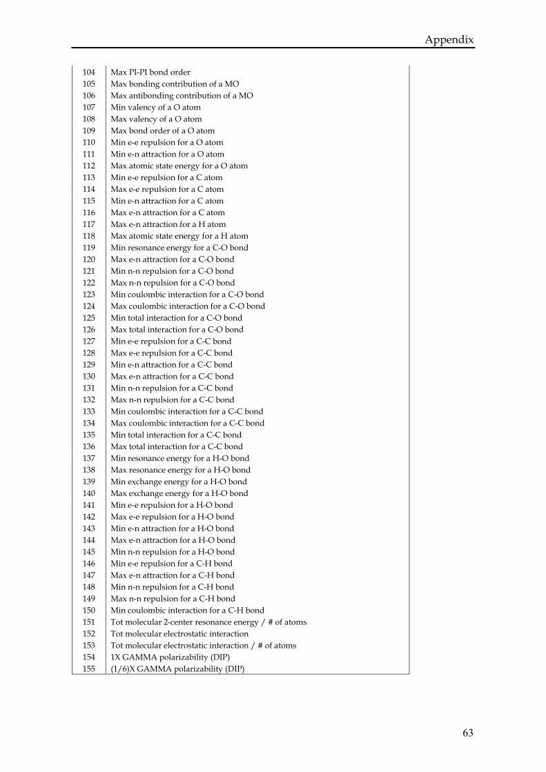
Appendix
63
104 Max PI-PI bond order 105 Max bonding contribution of a MO 106 Max antibonding contribution of a MO 107 Min valency of a O atom 108 Max valency of a O atom 109 Max bond order of a O atom 110 Min e-e repulsion for a O atom 111 Min e-n attraction for a O atom 112 Max atomic state energy for a O atom 113 Min e-e repulsion for a C atom 114 Max e-e repulsion for a C atom 115 Min e-n attraction for a C atom 116 Max e-n attraction for a C atom 117 Max e-n attraction for a H atom 118 Max atomic state energy for a H atom 119 Min resonance energy for a C-O bond 120 Max e-n attraction for a C-O bond 121 Min n-n repulsion for a C-O bond 122 Max n-n repulsion for a C-O bond 123 Min coulombic interaction for a C-O bond 124 Max coulombic interaction for a C-O bond 125 Min total interaction for a C-O bond 126 Max total interaction for a C-O bond 127 Min e-e repulsion for a C-C bond 128 Max e-e repulsion for a C-C bond 129 Min e-n attraction for a C-C bond 130 Max e-n attraction for a C-C bond 131 Min n-n repulsion for a C-C bond 132 Max n-n repulsion for a C-C bond 133 Min coulombic interaction for a C-C bond 134 Max coulombic interaction for a C-C bond 135 Min total interaction for a C-C bond 136 Max total interaction for a C-C bond 137 Min resonance energy for a H-O bond 138 Max resonance energy for a H-O bond 139 Min exchange energy for a H-O bond 140 Max exchange energy for a H-O bond 141 Min e-e repulsion for a H-O bond 142 Max e-e repulsion for a H-O bond 143 Min e-n attraction for a H-O bond 144 Max e-n attraction for a H-O bond 145 Min n-n repulsion for a H-O bond 146 Min e-e repulsion for a C-H bond 147 Max e-n attraction for a C-H bond 148 Min n-n repulsion for a C-H bond 149 Max n-n repulsion for a C-H bond 150 Min coulombic interaction for a C-H bond 151 Tot molecular 2-center resonance energy / # of atoms 152 Tot molecular electrostatic interaction 153 Tot molecular electrostatic interaction / # of atoms 154 1X GAMMA polarizability (DIP) 155 (1/6)X GAMMA polarizability (DIP)

Chapter four
THE METAL BONDING DOMAIN
OF THE ANTITUMOR DRUG FE(II)-BLEOMYCIN:
A DFT INVESTIGATION
This chapter has been published as: A. Karawajczyk and F. Buda, The metal bonding domain of the antitumor drug Fe(II)-bleomycin: a DFT investigation. Journal of Biological Inorganic Chemistry, 10, 33 (2005)
ABSTRACT
The geometric and electronic structure of ferrous complexes of bleomycin
(Fe(II)BLM) has been investigated by means of Density Functional Theory
calculations. The active site of this antitumor drug is a highly distorted octahedral
complex with the coordination sphere completed by the five known endogenous
ligands including pyrimidine, imidazole, deprotonated amide, and secondary and
primary amine. We have addressed the controversial issue of the nature of the
sixth axial ligand that we have identified as the oxygen of the carbamoyl group.
Our conclusions are further validated by a comparison with structural data
derived from NMR experiments. Moreover, because of the high sensitivity of
structural data on the pH of the environment, we have investigated the effect of a
different protonation state of the histidine amide on the geometric structure of the
Fe(II)BLM complex. The extensive model of the active site of bleomycin
considered in this work allows us to check the limitations of previous
investigations based on simplified models.
66
Structure of Fe(II)-bleomycin
4.1 Introduction
Bleomycin is a DNA-cleaving antibiotic, which is isolated as a copper complex
from the culture medium of Streptomyces verticillis and administrated to patients
in its metal-free form. BLM is important for clinical use in the treatment of head
and neck cancer, certain lymphomas and testicular cancer [96]. Interest in the
chemical activity of bleomycin began with the discovery that the presence of
either reducing agents or hydrogen peroxide permits bleomycin to cleave DNA
into simple sets of DNA degradation products [17]. Indications that metals and
oxygen species were also required led to the demonstration that the presence of
Fe(II) and O2 was sufficient to allow BLM to degrade DNA. The first step is the
formation of a high-spin Fe(II)BLM complex which reacts with O2 [1]. In the
second step, the so-called activated bleomycin (ABLM) – a peroxide-Fe(III)BLM
complex – is produced and this then cleaves DNA. The products of DNA
degradation are well known [17], but major gaps in our understanding remain
where the drug activation and DNA degradation pathways intersect. A precise
knowledge of the geometrical and electronic structure of the Fe(II)BLM complex is
a prerequisite to understanding the activity of this drug in DNA degradation.
Unfortunately, no crystal structure is yet available for Fe(II)BLM. There are
several different synthesized models mimicking BLM as a ligand and crystallized
with various metals, for example copper [3, 97], cobalt [98], zinc [99] and even
iron[100]. However, all models thus far lack both the bithiazole and the sugar
residues. For example, the PMAH ligand has five nitrogen donor centers located
in the primary and secondary amines, pyrimidine and imidazole rings, and the
amide moiety (see Fig. 4.1). The ligand synthesized for iron is only tridentate,
since the β-aminoalanine fragment is also not present. Therefore these
crystallographic studies could not give a definitive answer as to whether the
bleomycin is a five- or six-coordinated ligand and, if the sixth ligand is indeed
present, whether it is endo- or exogenous. The exact coordination of the ligand
has thus been a matter of dispute for several years.
67
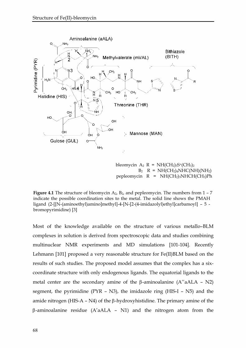
Structure of Fe(II)-bleomycin
Most of the knowledge available on the structure of various metallo–BLM
complexes in solution is derived from spectroscopic data and studies combining
multinuclear NMR experiments and MD simulations [101-104]. Recently
Lehmann [101] proposed a very reasonable structure for Fe(II)BLM based on the
results of such studies. The proposed model assumes that the complex has a six-
coordinate structure with only endogenous ligands. The equatorial ligands to the
metal center are the secondary amine of the β-aminoalanine (A’’aALA – N2)
segment, the pyrimidine (PYR – N3), the imidazole ring (HIS-I – N5) and the
amide nitrogen (HIS-A – N4) of the β-hydroxyhistidine. The primary amine of the
β-aminoalanine residue (A’aALA – N1) and the nitrogen atom from the
Figure 4.1 The structure of bleomycin A2, B2, and pepleomycin. The numbers from 1 – 7 indicate the possible coordination sites to the metal. The solid line shows the PMAH ligand (2-[[N-(aminoethyl)amino]methyl]-4-[N-[2-(4-imidazolyl)ethyl]carbamoyl] – 5 -bromopyrimidine) [3]
68
bleomycin A2 R = NH(CH2)3S+(CH3)2 B2 R = NH(CH2)4NHC(NH)(NH2) pepleomycin R = NH(CH2)3NHCH(CH3)Ph
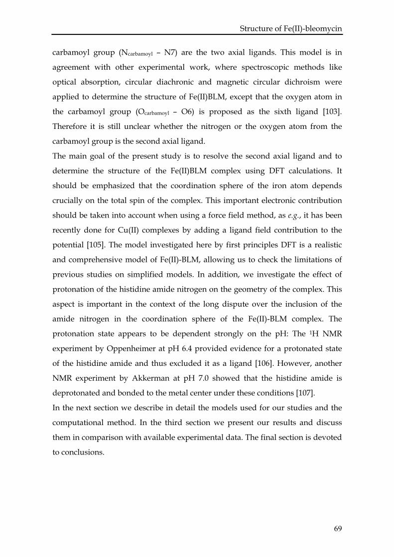
Structure of Fe(II)-bleomycin carbamoyl group (Ncarbamoyl – N7) are the two axial ligands. This model is in
agreement with other experimental work, where spectroscopic methods like
optical absorption, circular diachronic and magnetic circular dichroism were
applied to determine the structure of Fe(II)BLM, except that the oxygen atom in
the carbamoyl group (Ocarbamoyl – O6) is proposed as the sixth ligand [103].
Therefore it is still unclear whether the nitrogen or the oxygen atom from the
carbamoyl group is the second axial ligand.
The main goal of the present study is to resolve the second axial ligand and to
determine the structure of the Fe(II)BLM complex using DFT calculations. It
should be emphasized that the coordination sphere of the iron atom depends
crucially on the total spin of the complex. This important electronic contribution
should be taken into account when using a force field method, as e.g., it has been
recently done for Cu(II) complexes by adding a ligand field contribution to the
potential [105]. The model investigated here by first principles DFT is a realistic
and comprehensive model of Fe(II)-BLM, allowing us to check the limitations of
previous studies on simplified models. In addition, we investigate the effect of
protonation of the histidine amide nitrogen on the geometry of the complex. This
aspect is important in the context of the long dispute over the inclusion of the
amide nitrogen in the coordination sphere of the Fe(II)-BLM complex. The
protonation state appears to be dependent strongly on the pH: The 1H NMR
experiment by Oppenheimer at pH 6.4 provided evidence for a protonated state
of the histidine amide and thus excluded it as a ligand [106]. However, another
NMR experiment by Akkerman at pH 7.0 showed that the histidine amide is
deprotonated and bonded to the metal center under these conditions [107].
In the next section we describe in detail the models used for our studies and the
computational method. In the third section we present our results and discuss
them in comparison with available experimental data. The final section is devoted
to conclusions.
69
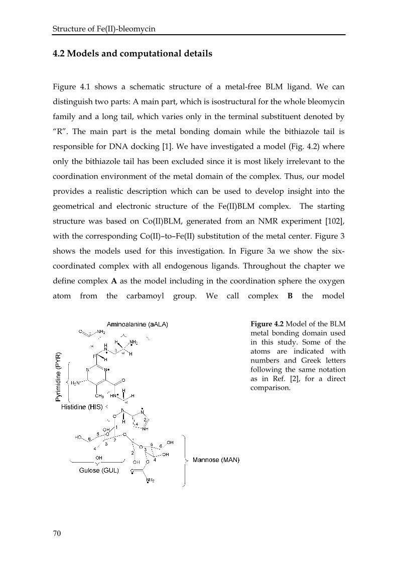
Structure of Fe(II)-bleomycin
4.2 Models and computational details
Figure 4.1 shows a schematic structure of a metal-free BLM ligand. We can
distinguish two parts: A main part, which is isostructural for the whole bleomycin
family and a long tail, which varies only in the terminal substituent denoted by
“R”. The main part is the metal bonding domain while the bithiazole tail is
responsible for DNA docking [1]. We have investigated a model (Fig. 4.2) where
only the bithiazole tail has been excluded since it is most likely irrelevant to the
coordination environment of the metal domain of the complex. Thus, our model
provides a realistic description which can be used to develop insight into the
geometrical and electronic structure of the Fe(II)BLM complex. The starting
structure was based on Co(II)BLM, generated from an NMR experiment [102],
with the corresponding Co(II)–to–Fe(II) substitution of the metal center. Figure 3
shows the models used for this investigation. In Figure 3a we show the six-
coordinated complex with all endogenous ligands. Throughout the chapter we
define complex A as the model including in the coordination sphere the oxygen
atom from the carbamoyl group. We call complex B the model
Figure 4.2 Model of the BLM metal bonding domain used in this study. Some of the atoms are indicated with numbers and Greek letters following the same notation as in Ref. [2], for a direct comparison.
70
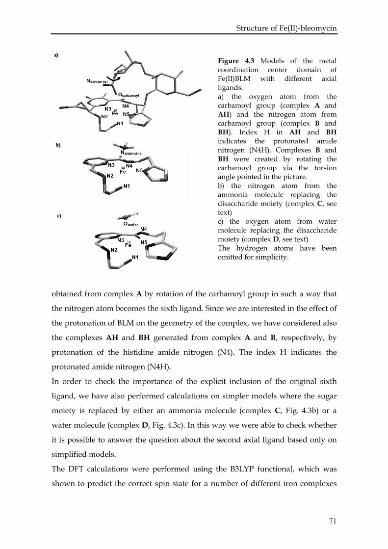
Structure of Fe(II)-bleomycin
obtained from complex A by rotation of the carbamoyl group in such a way that
the nitrogen atom becomes the sixth ligand. Since we are interested in the effect of
the protonation of BLM on the geometry of the complex, we have considered also
the complexes AH and BH generated from complex A and B, respectively, by
protonation of the histidine amide nitrogen (N4). The index H indicates the
protonated amide nitrogen (N4H).
Figure 4.3 Models of the metal coordination center domain of Fe(II)BLM with different axial ligands: a) the oxygen atom from the carbamoyl group (complex A and AH) and the nitrogen atom from carbamoyl group (complex B and BH). Index H in AH and BH indicates the protonated amide nitrogen (N4H). Complexes B and BH were created by rotating the carbamoyl group via the torsion angle pointed in the picture. b) the nitrogen atom from the ammonia molecule replacing the disaccharide moiety (complex C, see text) c) the oxygen atom from water molecule replacing the disaccharide moiety (complex D, see text) The hydrogen atoms have been omitted for simplicity.
In order to check the importance of the explicit inclusion of the original sixth
ligand, we have also performed calculations on simpler models where the sugar
moiety is replaced by either an ammonia molecule (complex C, Fig. 4.3b) or a
water molecule (complex D, Fig. 4.3c). In this way we were able to check whether
it is possible to answer the question about the second axial ligand based only on
simplified models.
The DFT calculations were performed using the B3LYP functional, which was
shown to predict the correct spin state for a number of different iron complexes
71
Structure of Fe(II)-bleomycin [108, 109]. We used the effective core potential basis set LanL2DZ [110, 111] for
the iron atom and the 6-31G basis set for the other atoms as implemented in the
program Gaussian03 [112]. This choice of the basis set is supported by the results
of test calculations performed by Lehnert et al. on similar model systems [5]. Their
conclusion is that the effective core potential basis set LanL2DZ is good enough to
describe the geometry of ABLM and that a triple-ζ all electron basis set gives a
comparable description.
When comparing our results with structural information derived by NMR data in
solution, one should take into account that we are neglecting solvent effects in our
model. Although we can expect small changes in the geometrical parameters due
to the solvent, we think that our conclusions on the coordination shell will not be
affected. Indeed, it is now established in the literature that solvent molecules are
not participating in the coordination sphere of the Fe(II)-BLM complex [101, 113].
4.3 Results and discussion
For each system under consideration full geometry optimizations assuming low,
intermediate or high spin states have been carried out. The final geometric
parameters and electronic energies corresponding to singlet, triplet and quintet
states are summarized in Table 4.1 and Table 4.2 for protonated and deprotonated
complexes, respectively. Complex AH was found to have the lowest energy in its
quintet electronic state and this energy is taken as the reference energy for other
spin states of complexes AH and BH. The same spin order is observed for
deprotonated models and the energy of complex A in its quintet electronic state is
taken as the reference energy for other spin states of complexes A and B. Our
investigation shows that the geometry of the drug complex is highly dependent
on the environment of the iron ion and on its spin state.
72
Structure of Fe(II)-bleomycin 4.3.1 Complexes A and AH
For complexes A and AH the Fe–Ocarbamoyl distance changes as a function of the
spin state, reaching a maximum value for S = 1 (see Table 4.1 and 4.2). In the
complex A, this value is equal to 3.26 Å for S = 1 indicating that the bond is
broken. For S = 2 the Fe–Ocarbamoyl bond is again present with a length of 2.29 Å.
Generally, the bond length between the iron center and its axial ligands (Ocarbamoyl
and N1) in complex A are longer than in complex AH for each spin state (see
Tables 4.1 and 4.2). This can be interpreted as a consequence of the deprotonation
of the equatorial nitrogen donor ligand (N4) and the related shortening of the Fe–
N4 bond length. It is known from ligand field theory, that strong equatorial
ligands cause a tetragonal distortion, which corresponds to an extension along the
z axis and compression on the x and y axes with a stabilizing effect on the dz2
orbital. A Natural Bond Orbital analysis shows that the dz2 orbital is already lower
in energy than the dx2-y2 orbital for the protonated complex. Deprotonation of the
amide nitrogen (N4) causes further lowering of the energy of the dz2 orbital.
Table 4.1 Relative energies and optimized geometric parameters of complexes AH, BH.
Complex AH Complex BH Total spin 0 1 2 0 1 2 11.48 15.12 0.00 36.50 31.41 16.92 ΔE[kcal/mol]a Fe-Xb 2.12 2.25 2.12 2.37 3.70 4.21 Fe-N1 2.04 2.20 2.20 2.05 2.23 2.18 Fe-N2 2.10 2.24 2.26 2.11 2.13 2.28 Fe-N3 1.93 1.94 2.14 1.91 1.92 2.07 Fe-N4 2.10 2.17 2.59 2.11 2.12 2.43 Fe-N5 2.02 2.01 2.22 2.04 2.04 2.16 X-Fe-N1 176.9 171.6 159.4 173.9 173.3 166.1 2-3-4-5 -1.6 -3.7 3.8 0.8 -3.2 -1.8
a For complexes AH and BH the reference energy is the ground quintet electronic state energy of the complex AH b X = Ocarbamoyl, Ncarbamoyl All distances are in Å and angles in degrees
73
Structure of Fe(II)-bleomycin As the spin increases, each metal – equatorial ligand bond length generally in-
creases and, in the particular case of N4 (complex AH), the distance increases up
to 2.59 Å, while the average for the other equatorial ligands is 2.2 Å. Although this
distance seems long, one can find in the Cambridge Structural Database structures
of iron (II) complexes, where the distances between iron(II) and its nitrogen donor
are 2.50 Å [114], 2.54 Å [115] and even 2.65 Å [116]. In additional, the analysis of
the HOMO orbital for β electrons shows that some weak bonding interaction is
present between these two atoms as indicated by overlap between the dz2 orbital
on iron and the lone pair on the N4 nitrogen atom. The same bond in complex A
is less sensitive to a change in the spin (see Table 4.1). These results explain why it
has been difficult to establish which atoms participate to the iron coordination
shell. Hilbers et al. [107, 117] have proposed that BLM binds the metal through
positions N2, N3, N4, N5, N7 while Openheimer and Hecht [106] proposed N1,
N2, N3, N5, N7 thus excluding the N4 atom. These two studies used different
experimental conditions including a different pH = 7.0 and pH = 6.4, respectively.
Our study clearly demonstrates that the problem of the ligand assignment can be
related to the very weak bonding interaction between the N4 atom and the metal
Complex A Complex B Complex C Complex D
Table 4.2 Relative energies and optimized geometry parameters of complexes A, B, C, D.
Total spin 0 1 2 0 1 2 0 1 2 0 1 2
ΔE [kcal/mol]a 12.29 6.96 0.0 32.83 26.10 23.78 5.34 8.00 00.0 4.05 7.17 0.00
Fe-Xb 2.17 3.26 2.29 2.29 3.93 4.02 2.07 2.28 2.26 2.07 2.32 2.22
Fe-N1 2.03 2.26 2.22 2.04 2.27 2.19 2.06 2.29 2.26 2.03 2.31 2.25
Fe-N2 2.16 2.13 2.40 2.14 2.15 2.30 2.17 2.35 2.48 2.15 2.25 2.42
Fe-N3 1.92 1.92 2.13 1.90 1.91 2.07 1.91 1.93 2.14 1.91 1.91 2.13
Fe-N4 1.97 1.94 2.09 1.98 1.94 2.03 1.99 1.99 2.07 1.99 1.97 2.06
Fe-N5 2.01 2.03 2.22 2.01 2.03 2.18 2.03 2.01 2.22 2.03 2.02 2.20
X-Fe-N1 170.3 126.2 140.8 176.8 168.4 166.2 179.3 165.6 161.9 177.9 157.8 155.3
2-3-4-5 7.0 9.4 14.7 5.9 7.0 9.4 5.5 5.3 4.5 -5.1 -5.6 -8.8 a For complexes A and B the reference energy is the ground quintet electronic state energy of the complex A, which has the value Eref = -2968.6627 a.u. For complexes C and D the reference energies are the quintet electronic ground state energies, which have the values Eref = -1144.3408 a.u. and Eref = -1164.1943 a.u., respectively. b X = Ocarbamoyl, Ncarbamoyl, Nammonia, Owater for complex A, B, C, D, respectively. All distances are in Å and angles in degrees
74
Structure of Fe(II)-bleomycin center when the pH is too low for the histidine amide to be deprotonated.
In the original model of the Co(II)BLM complex arising from NMR experiments
combined with force field molecular dynamics simulation, the equatorial ligands
are lying almost in the same plane [102]. We observe similar small distortions
from planarity only for complex AH (see 2-3-4-5 dihedral angle in Table 4.1). The
largest distortion is observed for the deprotonated complex A in its quintet
ground state, with a dihedral angle N2–N3–N4–N5 of 14.7º. The smallest
distortion occurs for S = 0 (see Tables 4.1 and 4.2). Thus we conclude that the
degree of distortion is highly dependent on the spin state of the complexes.
We also observe a large change in the valence angle of Ocarbamoyl–Fe–N1 for
different spin states (Tables 4.1 and 4.2). For complex A, the angle changes from
170.3º for S = 0 to 140.8º for S = 2. This angle distortion together with the
distortion from planarity discussed above brings the complex into a highly
distorted octahedral geometry. Because of these modifications, the Fe–Ocarbamoyl
bond becomes weaker and more susceptible to exchange with an oxygen molecule
to produce the Fe(II)O2BLM complex. This complex then accepts an additional
electron to form the activated BLM species. In previously proposed models for
activated bleomycin based on force field methods, the Fe–N1 bond was not
present and the oxygen molecule entered the coordination sphere from the β-
aminoalanine site [102, 118]. However the crystal structure for copper - BLM [119]
and some NMR studies for other metallo–BLM [4], suggest a different structure in
agreement with our results. Our results strongly support the picture in which the
atoms N1, N2, N3, N4, N5 remain in the coordination sphere of the metal center
and the flexible sugar moiety drifts away making space for an exogenous ligand.
4.3.2 Complexes B and BH
Complex BH also has the lowest energy for total spin S = 2, but this energy is still
higher than the energy of complex AH for all spin states and particularly it is
about 17 kcal/mol higher than the reference energy, which is the ground state of
complex AH for S = 2. For complex BH the length of the Fe–Ncarbamoyl bond also
increases with increasing total spin (see Table 4.1). Specifically, for S = 2 the
75
Structure of Fe(II)-bleomycin distance becomes 4.21 Å. This leads us to conclude that the Ncarbamoyl atom moves
out of the coordination sphere of the iron atom. We notice that the breaking of the
Fe–Ncarbamoyl bond also occurs in the B complex with the anionic ligand. Therefore,
the behavior of the Fe–Ncarbamoyl bond is not related to the specific protonation
state of the ligand, but rather to steric interaction. For S = 0 the distance between
the Ocarbamoyl atom and the Cγ atom from the pyrimidine residue (CγPYR) is equal
to 2.89 Å, while the sum of the van der Waals radii of these atoms is 3.10 Å. When
the Fe–Ncarbamoyl distance increases, the Ocarbamoyl–CγPYR distance increases as
well, minimizing in this way the steric interaction. However, the presence of the
proton affects the Fe–N4 bond. In particular for S = 2 the Fe–N4 distance changes
from 2.43 Å in BH to 2.03 Å in the complex B. Interestingly, the geometries of
complexes A and B are very similar for the lowest spin state S = 0, both having a
six-fold coordination shell. It is only when we go to the high spin state S = 2 that is
the experimentally observed we see major changes in the coordination shell. This
strong dependence of the final geometry on the spin state of the complexes is an
important finding that should be taken into account when describing these
systems in terms of force field modeling [105].
4.3.3 Complexes C and D
We have also investigated simplified models using an ammonia molecule
(complex C) and a water molecule (complex D) to represent the nitrogen and the
oxygen atoms of the carbamoyl group, respectively, to validate the importance of
the inclusion of the big sugar moiety explicitly in the model. We find that the
quintet electronic spin state is the ground state for both complexes C and D,
which is in agreement with the experimentally observed spin state for the
Fe(II)BLM complex. However, the differences between the energies of the
different spin states and the reference energies are only a few kcal/mol (see Table
4.2) and we cannot directly compare energies of complex C and D. Both
complexes form a distorted octahedral geometry, providing no grounds to
distinguish between them. The Fe–Nammonia bond length increases slightly with
increasing spin, but we have not observed the bond breaking that occurs when the
76
Structure of Fe(II)-bleomycin actual sugar moiety is included, in the model complex B (Table 4.2). By comparing
the ground state structures of complex C and D we do not observe a large
difference in the X–Fe–N1 covalent angle, in contrast with the complexes A and B.
This suggests that simplified models are not appropriate for resolving the issue of
the sixth ligand of the Fe(II)BLM. Moreover, it also indicates that the disaccharide
moiety plays a very important role in the metal bonding domain of BLM, showing
that performing DFT calculations with the more realistic model is crucial.
4.3.4 Comparison with experiment
A quintet electronic ground state for Fe(II)BLM complex was established many
years ago using EPR and Mösbauer spectroscopy [120-122]. Our B3LYP - DFT
calculation is able to reproduce this spin state correctly. This is important since
previous DFT calculations could not reproduce the proper experimental spin state
[123]. In Ref. [123] the calculation were carried out using B3LYP functional, as we
have done here, but on a simpler model of bleomycin with only a pentadonor
ligand, and with a doubly deprotonated ligand at the histidine amide N4 and the
N2 of the secondary amine of the β-aminoalanine. The concordance between the
observed spin state and the one computed here emphasizes again the importance
of using a realistic model including all six potential donor sites.
We wish now to compare our optimized geometries with the available NMR data.
Table 4.3 summarizes the comparison between the iron – proton DFT optimized
distances for AH, A, BH and B complexes in their electronic ground state with the
Fe–H distances based on 2D NMR studies of the paramagnetic complex Fe(II)BLM
[2]. The experimental iron – proton distances were obtained from relaxation times
of the paramagnetically shifted protons. The calculated RMSD between
experimental and theoretical data is 0.8 Å for both protonated models and 0.6 Å
and 0.5 Å for deprotonated complexes A and B, respectively. However, if we
consider only the mannose sugar moiety that is directly connected to the
carbamoyl group, the calculated partial RMSD for complex AH is 0.8 Å while for
BH it is equal to 1.2 Å. For their deprotonated analogues these values are 0.5 Å
and 0.8 Å, respectively. This implies that the structures of complexes AH and A
77
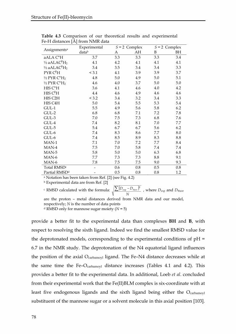
Structure of Fe(II)-bleomycin
provide a better fit to the experimental data than complexes BH and B, with
respect to resolving the sixth ligand. Indeed we find the smallest RMSD value for
the deprotonated models, corresponding to the experimental conditions of pH =
6.7 in the NMR study. The deprotonation of the N4 equatorial ligand influences
the position of the axial Ocarbamoyl ligand. The Fe–N4 distance decreases while at
the same time the Fe–Ocarbamoyl distance increases (Tables 4.1 and 4.2). This
provides a better fit to the experimental data. In additional, Loeb et al. concluded
from their experimental work that the Fe(II)BLM complex is six-coordinate with at
least five endogenous ligands and the sixth ligand being either the Ocarbamoyl
substituent of the mannose sugar or a solvent molecule in this axial position [103].
Assignmentsa Experimental datab
S = 2 Complex A AH
S = 2 Complex B BH
Table 4.3 Comparison of our theoretical results and experimental Fe-H distances [Å] from NMR data
aALA CαH 3.7 3.3 3.3 3.3 3.4 ½ aALACβH2 4.1 4.2 4.1 4.1 4.1 ½ aALACβH2 3.5 3.4 3.4 3.3 3.4 PYR CβH < 3.1 4.1 3.9 3.9 3.7 ½ PYR CαH2 5.0 5.1 4.8 5.0 4.9 ½ PYR CαH2 5.0 5.0 4.6 4.0 3.7 HIS CαH 4.0 4.2 3.6 4.1 4.6 HIS CβH 4.6 4.6 4.4 4.6 4.9 HIS C2H < 3.2 3.4 3.2 3.4 3.3 HIS C4H 5.0 5.4 5.5 5.3 5.4 GUL-1 5.5 4.9 5.6 5.8 6.2 GUL-2 6.8 6.8 7.1 7.2 7.8 GUL-3 7.0 7.5 7.3 6.8 7.6 GUL-4 7.4 8.2 8.1 7.0 7.7 GUL-5 5.4 6.7 6.7 5.6 6.2 GUL-6 7.4 8.3 8.6 7.7 8.0 GUL-6 7.4 8.5 8.9 8.3 8.8 MAN-1 7.1 7.0 7.2 7.7 8.4 MAN-4 7.5 7.0 5.8 7.4 7.4 MAN-5 5.8 5.0 5.0 6.3 6.8 MAN-6 7.7 7.3 7.3 8.8 9.1 MAN-6 7.8 7.5 7.5 9.0 9.3 Total RMSDc - 0.6 0.8 0.5 0.8 Partial RMSDd - 0.5 0.8 0.8 1.2 a Notation has been taken from Ref. [2] (see Fig. 4.2) b Experimental data are from Ref. [2]
( )N
DD theor∑ − 2expc RMSD calculated with the formula: , where Dexp and Dtheor
are the proton – metal distances derived from NMR data and our model, respectively; N is the number of data points d RMSD only for mannose sugar moiety (N = 5)
78
Structure of Fe(II)-bleomycin Their study did not determine whether the Ocarbamoyl atom is directly coordinated
to the metal or connected through H–bonding. Based on our results and the
information from experimental work of Lehmann we can now say that the
Ocarbamoyl atom is directly connected to the iron center, though in a highly
distorted octahedral configuration.
Our findings are also in agreement with very recent data on Fe(II)-bleomycin
obtained by X-ray absorption near edge structure spectroscopy [113]. In this work
the best agreement between theoretical and experimental spectra is achieved for
the bleomycin model complex with the primary amine and oxygen of the
mannose sugar occupying the axial positions. They also observed that the
coordination environment is characterized by serious distortions of the iron
octahedron.
4.4 Conclusions
The DFT calculations have been performed on an extensive model of the
Fe(II)BLM complex based on available NMR data [2, 102]. By comparing the
model complex A and B, distinguished by having the oxygen or the nitrogen
atom from the carbamoyl group as the sixth ligand, respectively, we can conclude
that the oxygen atom is coordinated to the metal center. We find that complex A
besides being energetically more favorable also gives a better fit to the
experimental data [2, 102, 103, 120]. We observe that in complex B the carbamoyl
group tends to move out of the coordination shell in the high spin configuration
contrary to experimental evidence. We suggest that this behavior can be explained
in terms of steric hindrance of the carbamoyl oxygen with the pyrimidine Cγ. The
energetically most stable complex A in the high spin configuration shows a highly
distorted octahedral conformation, with an angle between axial ligands of 140.8º.
This fact should facilitate the exchange of the Ocarbamoyl ligand with the oxygen
molecule during the formation of ABLM.
79
Structure of Fe(II)-bleomycin
80
We have investigated the effect of a different protonation state of the histidine
amide on the geometric structure of the Fe(II)BLM complex. We observe that
deprotonation of that equatorial ligand mainly influences the Fe–N4 distance,
which is very long in the high spin configuration of complex AH and BH. It also
affects the positions of axial ligands, e.g. the Fe–Ocarbamoyl distance increases from
2.12 Å in complex AH to 2.29 Å in complex A resulting in a better fit to
experimental NMR distance estimates. The protonation state has no effect on the
energetic spin order of our model complexes, nor does it influence the behavior of
the carbamoyl group in complexes B and BH.
The study carried out with the simplified models shows that they are not
appropriate for resolving the issue of the sixth ligand of the Fe(II)BLM. The results
also indicate that the disaccharide moiety plays a very important role in the final
structure of the metal bonding domain of BLM, showing that the use of a more
realistic model in our DFT calculations is crucial. We also underline the
importance of a theoretical method which directly takes into account the total spin
of the system. This work provides a good starting point for further studies of the
reaction mechanism of ABLM. One interesting issue is the self-inactivation of the
drug in the absence of DNA. The activation of the O-O bond of the peroxide – the
sixth, external ligand of ABLM – plays the main role in that process.
Chapter five
THE MECHANISM OF THE BLEOMYCIN
SUICIDE: A CAR-PARRINELLO MOLECULAR
DYNAMICS INVESTIGATION
This chapter has been published as: A. Karawajczyk, C. Gossens, U. Rothlisberger, F. Buda, The mechanism of the bleomycin suicide: a Car-Parrinello molecular dynamics Journal of Physical Chemistry B 110, 21245 (2006) ABSTRACT Using first-principles molecular dynamics simulations (Car-Parrinello method)
we investigated the possible reaction pathways for decay of the active bleomycin-
Fe(III)-OOH complex, the so-called bleomycin suicide. The theoretical model of
activated bleomycin contains the whole metal bonding domain of the bleomycin
ligand. Simulations performed both in vacuum and in water solution show that a
facile decaying process involves a homolytic O-O bond cleavage with an almost
simultaneous hydrogen atom abstraction. The formation of an intra- or
intermolecular hydrogen bond appears to be crucial for the decay of the activated
bleomycin. We did not observe any evidence of heterolytic cleavage of the O-O
bond of the Fe(III)-OOH species.
82
Bleomycin Suicide
5.1 Introduction
Bleomycin is a sixendogenous ligand, which is used as antitumor drug
(Blenoxane) in the treatment of several types of cancer since the early eighties [17].
This drug is administrated to the patient in the metal free form. A species called
activated bleomycin is formed in the body and it is identified as a low spin BLM-
Fe(III)-OOH complex by electrospray ionization mass spectroscopy (ESI-MS)
[124], extended X-ray absorption fine structure (EXAFS) spectroscopy [125], EPR
spectroscopy [121], and Mössbauer spectroscopy [120]. The iron atom is
coordinated by five nitrogen atoms coming from the bleomycin ligand (the
secondary amine of the β-aminoalanine (A’’aALA – N2) segment, the pyrimidine
(PYR – N3), the imidazole ring (HIS-I – N5), the amide nitrogen (HIS-A – N4) of
the β - hydroxyhistidine, and the primary amine of the β - aminoalanine residue
(A’aALA – N1) as the axial ligand; see Fig. 1.1) and one end-on peroxide anion
(OOH-) as the second axial ligand. The activation of oxygen by transition-metal
oxides is a fundamental mechanism that is of importance in organic synthesis,
catalysis and biochemistry. ABLM is one natural example of such activation,
similar to cytochrome P450 or methane monooxygenase [126]. The drug acts
through peroxide group cleavage, which is believed to be the first step driving the
DNA double strand degradation and ipso facto the disruption of the cancer cell.
Figure 5.1 illustrates a well accepted cycle of BLM activation and deactivation
which is broadly discussed in the review paper by Burger [1, 17]. It is interesting
that the same products can be formed from DNA when BLM is activated with
Fe2+ + O2 or Fe3+ + H2O2. That is because both sets of activation cofactors yield the
same active state BLM-Fe(III)-OOH as it is shown in Figure 5.1. As it appears from
the presented cycle, ABLM turns into Fe(III)BLM after causing the oxidative
damage of DNA and the Fe(III)BLM complex can be transformed into Fe(II)BLM
and later ABLM in the presence of a reducer and oxygen. This reaction pathway
has been verified experimentally [127]. However, the initial step in the conversion
of ABLM into Fe(III)BLM can proceed both in the presence or absence of DNA
83
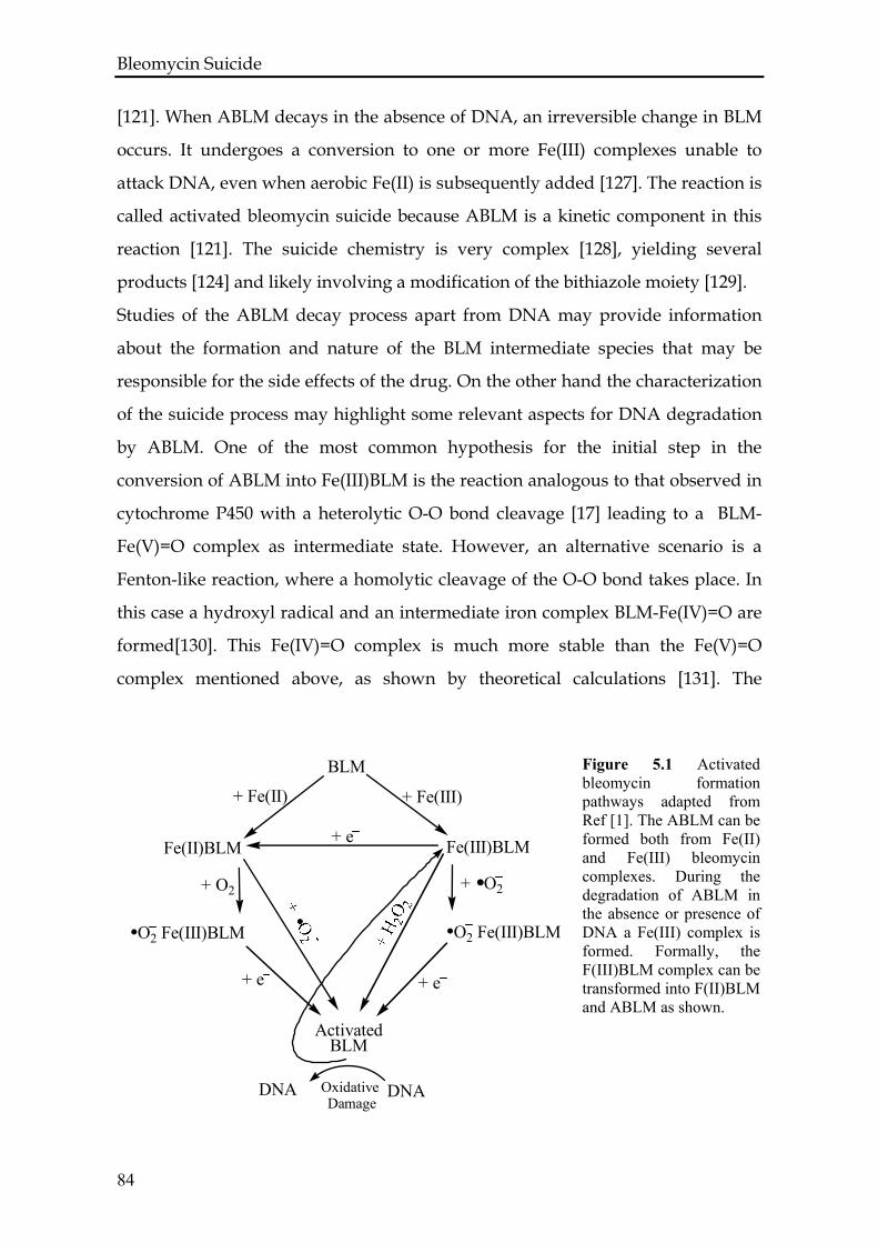
Bleomycin Suicide [121]. When ABLM decays in the absence of DNA, an irreversible change in BLM
occurs. It undergoes a conversion to one or more Fe(III) complexes unable to
attack DNA, even when aerobic Fe(II) is subsequently added [127]. The reaction is
called activated bleomycin suicide because ABLM is a kinetic component in this
reaction [121]. The suicide chemistry is very complex [128], yielding several
products [124] and likely involving a modification of the bithiazole moiety [129]. Studies of the ABLM decay process apart from DNA may provide information
about the formation and nature of the BLM intermediate species that may be
responsible for the side effects of the drug. On the other hand the characterization
of the suicide process may highlight some relevant aspects for DNA degradation
by ABLM. One of the most common hypothesis for the initial step in the
conversion of ABLM into Fe(III)BLM is the reaction analogous to that observed in
cytochrome P450 with a heterolytic O-O bond cleavage [17] leading to a BLM-
Fe(V)=O complex as intermediate state. However, an alternative scenario is a
Fenton-like reaction, where a homolytic cleavage of the O-O bond takes place. In
this case a hydroxyl radical and an intermediate iron complex BLM-Fe(IV)=O are
formed[130]. This Fe(IV)=O complex is much more stable than the Fe(V)=O
complex mentioned above, as shown by theoretical calculations [131]. The
Figure 5.1 Activated bleomycin formation pathways adapted from Ref [1]. The ABLM can be formed both from Fe(II) and Fe(III) bleomycin complexes. During the degradation of ABLM in the absence or presence of DNA a Fe(III) complex is formed. Formally, the F(III)BLM complex can be transformed into F(II)BLM and ABLM as shown.
O2 Fe(III)BLM
BLM
Fe(III)BLM
+ Fe(III)
Fe(II)BLM
O2 Fe(III)BLM
ActivatedBLM
+ Fe(II)
+ O2
+ e
+ O2
+ e
+ e
DNA DNAOxidative Damage
84
Bleomycin Suicide question we address here is which activation mechanism of the O-O bond plays a
role in the self-inactivation of bleomycin. A similar problem is related to the
mechanism of action of ABLM on DNA molecule that is subject of the study
presented in the next chapter of the thesis.
Some experimental studies on the decay of ABLM in the absence of DNA suggest
a heterolytic cleavage of the Fe(III)O-OH bond [132, 133]. In one of these studies a
synthetic model for the metal bonding site of BLM has been treated with Fe(III)-
H2O2 or Fe(II)-O2 aiming to resolve the mechanism of the self-deactivation of
ABLM. A breakdown at the N2 of the amino acid side chain of the metal binding
site has been demonstrated [132]. Another study for the same purpose but with
N-[bis(2-pyridylmethyl)aminoethyl]pyridine-2-carboxamide (HPaPy3) as the
ligand modeled BLM, demonstrated a modification at the carbon Cα to the amide
(N1 in Fig. 5.2). The modification at this site was suggested to be the result of a
two-electron oxidation of the ligand following the heterolytic cleavage of the O-O
bond of the Fe(III)-OOH species [133]. However, studies performed by Padbury et
al. [134] involving the interaction of 10 – hydroperoxy – 8,12 – ocatadecadienoic
acid with Fe(II)BLM suggest that the homolytic O-O bond cleavage of the
peroxide intermediate of ABLM can produce one equivalent of a hydroxyl radical
in addition to the (•OH)-Fe(III)-BLM, which may be the DNA damaging species.
Some other investigators in the field showed that a hydroxyl radical can be
generated by Fe(II)BLM and proposed that these active radical species may be
responsible for degradation of both ABLM and DNA [135, 136]. The Fe(II)-BLM
mediated production of a hydroxyl radical was based on ESR spin-trapping
techniques. The experimental models are therefore to some extent contradictory
and do not provide a clear answer to how the O-O bond is activated in the process
of BLM self-destruction.
85
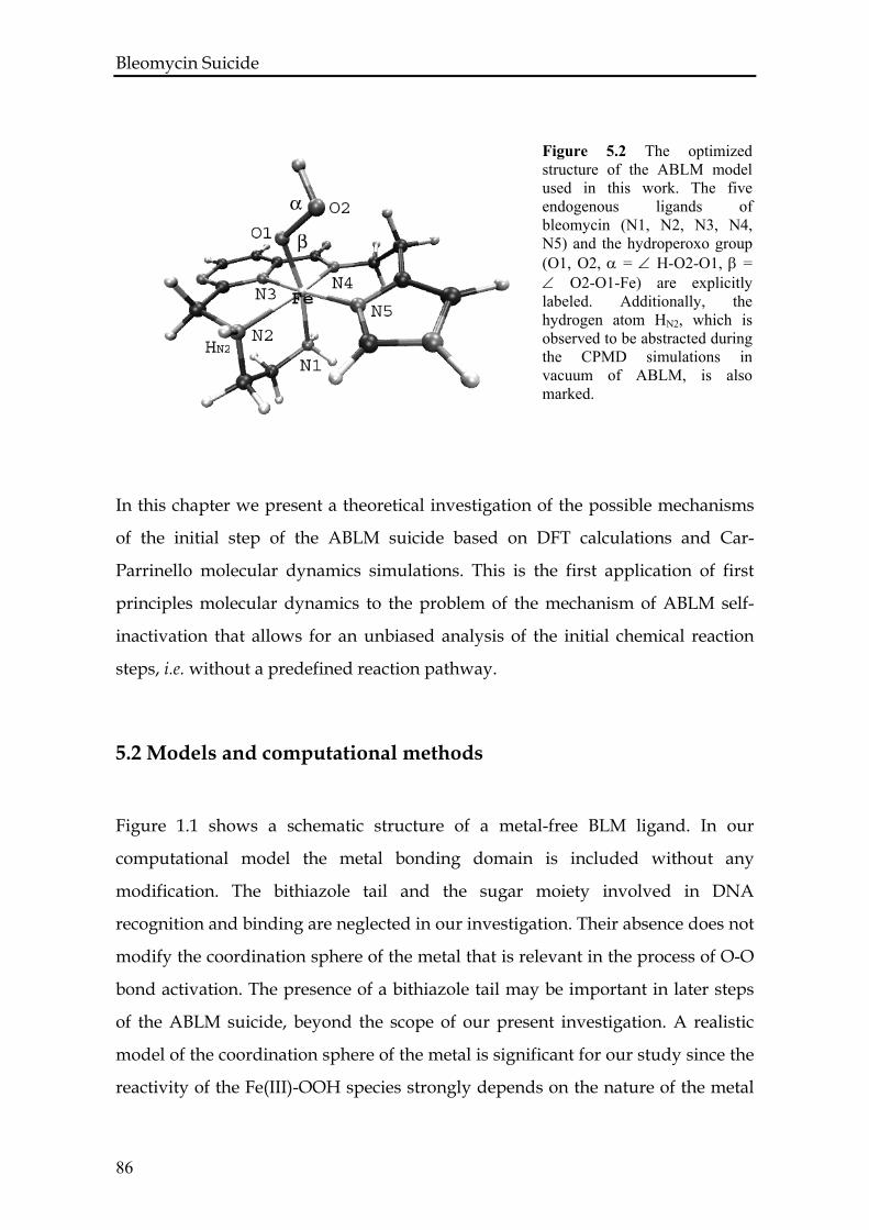
Bleomycin Suicide
In this chapter we present a theoretical investigation of the possible mechanisms
of the initial step of the ABLM suicide based on DFT calculations and Car-
Parrinello molecular dynamics simulations. This is the first application of first
principles molecular dynamics to the problem of the mechanism of ABLM self-
inactivation that allows for an unbiased analysis of the initial chemical reaction
steps, i.e. without a predefined reaction pathway.
Figure 5.2 The optimized structure of the ABLM model used in this work. The five endogenous ligands of bleomycin (N1, N2, N3, N4, N5) and the hydroperoxo group (O1, O2, α = ∠ H-O2-O1, β = ∠ O2-O1-Fe) are explicitly labeled. Additionally, the hydrogen atom HN2, which is observed to be abstracted during the CPMD simulations in vacuum of ABLM, is also marked.
5.2 Models and computational methods
Figure 1.1 shows a schematic structure of a metal-free BLM ligand. In our
computational model the metal bonding domain is included without any
modification. The bithiazole tail and the sugar moiety involved in DNA
recognition and binding are neglected in our investigation. Their absence does not
modify the coordination sphere of the metal that is relevant in the process of O-O
bond activation. The presence of a bithiazole tail may be important in later steps
of the ABLM suicide, beyond the scope of our present investigation. A realistic
model of the coordination sphere of the metal is significant for our study since the
reactivity of the Fe(III)-OOH species strongly depends on the nature of the metal
86
Bleomycin Suicide center ligands. For instance, in the case of the porphyrin ring ligand the reaction
of the O-O bond breaking has a heterolytic character and the ligand stabilizes the
peroxidase compound I [137, 138]. On the other hand, previous CPMD
simulations on the FeIII(H2O)5-OOH complex have shown that the complex is very
stable and no O-O bond cleavage was observed [130]. Static theoretical
calculations on the thermodynamics of the O-O bond homolytic cleavage also
show that the energy barrier drops from 50 kcal/mol for the (NH3)5-Fe(III)-OOH
complex to about 30 kcal/mol for the (NH3)2(Py)Fe(III)-OOH complex [5]. Our
previous work on the structure determination of Fe(II)BLM [139] also highlights
the importance of an accurate model in the theoretical investigation of bleomycin
complexes.
The starting structure was based on NMR data for the CoIIIBLM-OOH complex
[4], with the corresponding Co(III) – to – Fe(III) substitution of the metal center.
The model shown in Figure 5.2 was studied both in vacuum and in a water
solvent. The calculations in vacuum allow to check the stability of the system and
to observe the initial step in the ABLM suicide without any external perturbation
or reactant.
The comparison between the simulations in vacuum and in water enabled us to
check the influence of the water solvent in stabilizing the O-O bond of ABLM and
to check the possible differences in the mechanism of bond cleavage in the initial
step of ABLM self degradation in the gas phase and in aqueous environments.
The calculations were performed using the Car-Parrinello molecular dynamics
method [47] as implemented in the CPMD code [63]. The Kohn-Sham orbitals are
expanded in a PW basis set with a kinetic energy cutoff of 80 Ry. The test
calculations described in detail in the next section, show that this cutoff is
sufficient for achieving a good convergence of energies and structural properties
for the considered systems. We employed ab initio norm-conserving
pseudopotentials, generated within the Troullier-Martins scheme [140]. A
generalized gradient approximation was used for the exchange-correlation
functional, following the prescription of Becke and Perdew [53, 54]. All
simulations are spin polarized with a total spin S = 1/2. Room temperature Car-
87
Bleomycin Suicide Parrinello MD simulations were performed by using a time step of 0.09 fs and a
value of 400 a.u. for the fictitious electronic mass in the Car-Parrinello Lagrangian.
For the simulation in vacuum we have used an isolated cubic supercell of size 17 x
17 x 17 Å3. The simulation in water was performed using a hybrid quantum
mechanics – molecular mechanics approach [29, 30]. In particular, the hybrid
QMMM scheme used here is based on the CPMD method for the quantum part
and on the Gromos96 MD program [38] for the classical molecular mechanics
part. The quantum mechanics part includes the model of ABLM and 6 water
molecules in a cubic box of 15 x 15 x 15 Å3. The excess water molecules were
described using the SPC model [141] with the force field implemented in the
Gromos96 program. To prepare the initial configuration of ABLM in water we
have performed a classical MD simulation using the Gromos96 program, in which
the coordinates of the solute molecule were fixed while the 308 water molecules in
the box have been relaxed. First a constant pressure simulation at 300 K and 1 bar
was performed for 2 ns and then a constant volume simulation was performed for
1 ns. After relaxation of the solvent molecules, 6 waters were found within a
distance of 3 Å from the –OOH ligand. These molecules were included in the
quantum part of the system.
Both simulations in vacuum and in water start with 1000 steps of relaxation of the
system at 100 K following by a slow temperature increase to 300 K.
5.3 Results
5.3.1 Test calculations
To validate the choice of the density functional, the kinetic energy cutoff of PW,
and the pseudopotentials, a series of test geometry optimizations was performed
with different programs using localized basis sets. In particular, we used the
Amsterdam Density Functional code [142], in which the Kohn-Sham orbitals are
expanded in a Slater-type orbital basis set, and the Gaussian03 code [112] using
Gaussian-type orbitals. The model of ABLM shown in Figure 5.2 was optimized
by: i) CPMD with BP functional and 80 Ry cutoff; ii) ADF with BLYP and BP
88
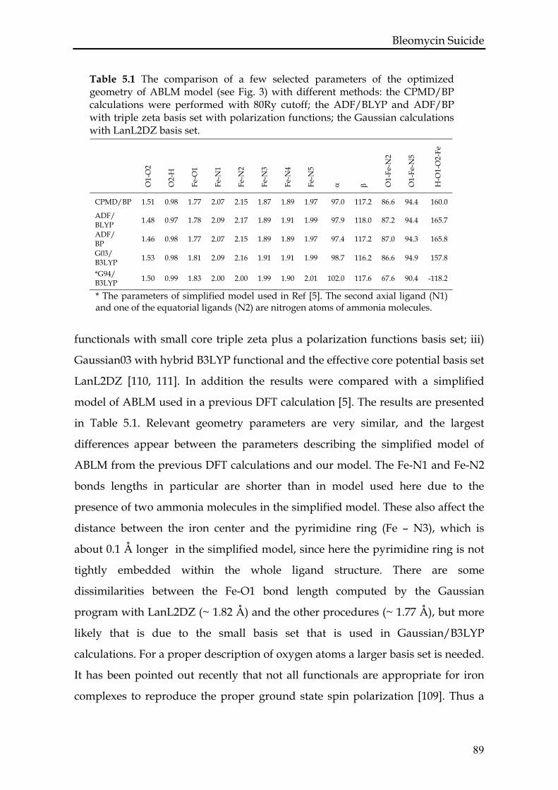
Bleomycin Suicide
Table 5.1 The comparison of a few selected parameters of the optimized geometry of ABLM model (see Fig. 3) with different methods: the CPMD/BP calculations were performed with 80Ry cutoff; the ADF/BLYP and ADF/BP with triple zeta basis set with polarization functions; the Gaussian calculations with LanL2DZ basis set.
H-O
1-O
2-Fe
O1-
Fe-N
2
O1-
Fe-N
5
O1-
O2
Fe-N
1
Fe-N
2
Fe-N
3
Fe-N
4
Fe-N
5
Fe-O
1
O2-
H
α
β
CPMD/BP 1.51 0.98 1.77 2.07 2.15 1.87 1.89 1.97 97.0 117.2 86.6 94.4 160.0
ADF/ 1.48 0.97 1.78 2.09 2.17 1.89 1.91 1.99 97.9 118.0 87.2 94.4 165.7 BLYP ADF/ 1.46 0.98 1.77 2.07 2.15 1.89 1.89 1.97 97.4 117.2 87.0 94.3 165.8 BP G03/ 1.53 0.98 1.81 2.09 2.16 1.91 1.91 1.99 98.7 116.2 86.6 94.9 157.8 B3LYP *G94/ 1.50 0.99 1.83 2.00 2.00 1.99 1.90 2.01 102.0 117.6 67.6 90.4 -118.2 B3LYP
* The parameters of simplified model used in Ref [5]. The second axial ligand (N1) and one of the equatorial ligands (N2) are nitrogen atoms of ammonia molecules.
functionals with small core triple zeta plus a polarization functions basis set; iii)
Gaussian03 with hybrid B3LYP functional and the effective core potential basis set
LanL2DZ [110, 111]. In addition the results were compared with a simplified
model of ABLM used in a previous DFT calculation [5]. The results are presented
in Table 5.1. Relevant geometry parameters are very similar, and the largest
differences appear between the parameters describing the simplified model of
ABLM from the previous DFT calculations and our model. The Fe-N1 and Fe-N2
bonds lengths in particular are shorter than in model used here due to the
presence of two ammonia molecules in the simplified model. These also affect the
distance between the iron center and the pyrimidine ring (Fe – N3), which is
about 0.1 Å longer in the simplified model, since here the pyrimidine ring is not
tightly embedded within the whole ligand structure. There are some
dissimilarities between the Fe-O1 bond length computed by the Gaussian
program with LanL2DZ (~ 1.82 Å) and the other procedures (~ 1.77 Å), but more
likely that is due to the small basis set that is used in Gaussian/B3LYP
calculations. For a proper description of oxygen atoms a larger basis set is needed.
It has been pointed out recently that not all functionals are appropriate for iron
complexes to reproduce the proper ground state spin polarization [109]. Thus a
89
Bleomycin Suicide set of test calculations was performed to verify the accuracy of the BP functional
for our systems. In all tested compounds (FeII(BLM)-O2, S = 0; FeIV(BLM)=O, S = 1;
FeIII(BLM)-OOH, S = 1/2) the calculated spin ground state was in agreement with
experimental data present in literature [17].
We have also checked the influence of the value of the PW energy cutoff on our
system. The test calculations were made for the FeIII(NH3)5-OOH complex, the
HOOH molecule and the OOH anion changing the cutoff value from 70 Ry up to
100 Ry in steps of 10 Ry. The changes of the structural parameters when the
energy cutoff is increased from 70 to 80 Ry are significant, in particular for the Fe-
O and O-O bonds. With 70 Ry the O-O bond is 1.60 Å in the FeIII(NH3)5-OOH
complex and 1.56 Å in the OOH− anion, which is too long and makes the bond
weaker and predisposed to scission. Increasing the cutoff from 90 Ry to 100 Ry
does not change the O-O distance, which converges to 1.50 Å and 1.47 Å for the
FeIII(NH3)5-OOH complex and the OOH− anion, respectively. These results justify
the choice of computational parameters for the CPMD simulations.
5.3.2 ABLM in vacuum
After the initial relaxation of the system the trajectory was followed for a total
time of 290 fs at room temperature. Figure 5.3 shows the behavior of a few
selected bond distances (Fe-O1, O1-O2, O2-HN2) along the dynamics. By analyzing
the O2 – HN2 distance (dashed line), three parts in the dynamics can be identified.
In the first part (I) the –OOH group is rotating around the Fe-O1 axis. The
distance between the O2 and the hydrogen atom bound to the secondary amine of
the β-aminoalanine A’’aALA – N2 is decreasing, while the O1-O2 bond is stable
with an average bond length of 1.57 Å. After 100 fs the O2 oxygen atom forms a
hydrogen bond with the HN2. In the second part of the dynamics the
intramolecular H-bond facilitates the O1-O2 bond cleavage of the hydroperoxo
group (II in Fig. 5.3). After 50 fs from the moment when the H-bond was created,
the O1-O2 bond starts increasing from the initial value of 1.58 Å and reaches a
value of ≈2.00 Å at 200 fs. At the same time the Fe-O1 distance drops to 1.60 Å and
oscillates around this shorter value. The just formed OH radical abstracts almost
90
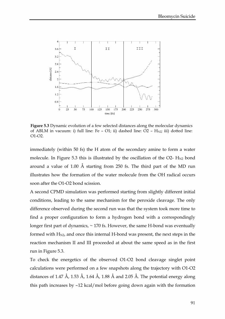
Bleomycin Suicide
Figure 5.3 Dynamic evolution of a few selected distances along the molecular dynamics of ABLM in vacuum: i) full line: Fe – O1; ii) dashed line: O2 – HN2; iii) dotted line: O1-O2.
immediately (within 50 fs) the H atom of the secondary amine to form a water
molecule. In Figure 5.3 this is illustrated by the oscillation of the O2- HN2 bond
around a value of 1.00 Å starting from 250 fs. The third part of the MD run
illustrates how the formation of the water molecule from the OH radical occurs
soon after the O1-O2 bond scission.
A second CPMD simulation was performed starting from slightly different initial
conditions, leading to the same mechanism for the peroxide cleavage. The only
difference observed during the second run was that the system took more time to
find a proper configuration to form a hydrogen bond with a correspondingly
longer first part of dynamics, ~ 170 fs. However, the same H-bond was eventually
formed with HN2, and once this internal H-bond was present, the next steps in the
reaction mechanism II and III proceeded at about the same speed as in the first
run in Figure 5.3.
To check the energetics of the observed O1-O2 bond cleavage singlet point
calculations were performed on a few snapshots along the trajectory with O1-O2
distances of 1.47 Å, 1.53 Å, 1.64 Å, 1.88 Å and 2.05 Å. The potential energy along
this path increases by ∼12 kcal/mol before going down again with the formation
91
Bleomycin Suicide of the water molecule. This shows that the thermal energy is enough to overcome
an energy barrier of this size. The observed energy maximum corresponds to the
snapshot in the trajectory where the H-bond between O2 and HN2 is formed (dO1-
O2 = 1.64 Å).
To identify the products of the O-O bond scission we took a snapshot at ~ 225 fs
where the O1-O2 distance is 2.15 Å and using the ADF program we performed a
single point calculation. The charge on the formed OH species was slightly
negative with a value of -0.266 electronic equivalent according to the Mulliken
analysis and the spin distribution analysis indicated the localization of -0.5 spin
on the oxygen atom of OH species. Therefore we concluded that the OH species
can be identified as an OH radical rather than an OH anion.
For comparison we calculate the energy profile of O1-O2 bond scission in vacuum
at 0 K without creation of any H-bond using the geometry obtained for our ABLM
model from the Gaussian calculation. The O1-O2 bond was elongated with steps
of 0.05 Å from 1.50 Å till 2.4 Å. The structures were fully optimized with
Gaussian03 as described in section Methods. We have noticed that the homolytic
O1-O2 bond scission is an endothermic reaction with activation energy equal to 17
kcal/mol. The results suggest that the reaction is still possible, however the
spontaneous creation of a H-bond in the molecular dynamics simulations
facilitates the scission.
5.3.3 ABLM in H2O
We now investigate the reaction mechanism of the ABLM suicide in water. This
simulation will enable us to check the influence of the water solvent on stabilizing
the O-O bond of ABLM. After the relaxation procedure, we have followed the
trajectory for about 1 ps. We have illustrated the simulation in Figure 5.4, where
the Fe-O1, O1-O2, and O2-HW1 distances are plotted. In this simulation we
observed also the formation of a H-bond after about 500 fs, but in this case the H-
bond was formed between the OOH ligand and one of the water molecules
indicated as W1 in Figure 5.5. The following observed reaction steps were similar
to that described for the simulation in vacuum. Simultaneously with the
92
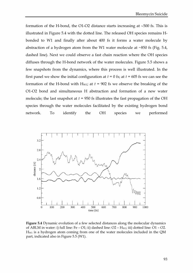
Bleomycin Suicide formation of the H-bond, the O1-O2 distance starts increasing at ∼500 fs. This is
illustrated in Figure 5.4 with the dotted line. The released OH species remains H-
bonded to W1 and finally after about 400 fs it forms a water molecule by
abstraction of a hydrogen atom from the W1 water molecule at ~850 fs (Fig. 5.4,
dashed line). Next we could observe a fast chain reaction where the OH species
diffuses through the H-bond network of the water molecules. Figure 5.5 shows a
few snapshots from the dynamics, where this process is well illustrated. In the
first panel we show the initial configuration at t = 0 fs; at t = 605 fs we can see the
formation of the H-bond with HW1; at t = 902 fs we observe the breaking of the
O1-O2 bond and simultaneous H abstraction and formation of a new water
molecule; the last snapshot at t = 950 fs illustrates the fast propagation of the OH
species through the water molecules facilitated by the existing hydrogen bond
network. To identify the OH species we performed
Figure 5.4 Dynamic evolution of a few selected distances along the molecular dynamics of ABLM in water: i) full line: Fe – O1; ii) dashed line: O2 – HW1; iii) dotted line: O1 – O2. HW1 is a hydrogen atom coming from one of the water molecules included in the QM part, indicated also in Figure 5.5 (W1).
93
Bleomycin Suicide an ADF single point calculation for the configuration at 650 fs. As in the case of
the vacuum simulations, the OH species was identified as a hydroxyl radical with
the spin density localized on the oxygen atom and with a small negative charge.
For a complete picture of the observed reaction mechanism and identification of
the products, we have also performed a single point calculation for the same
configuration of the system at t = 650 fs but with a total spin of 3/2. The total
bonding energy was about 10 kcal/mol higher than for the system with total spin
equal to 1/2. This result shows that no spin flip takes place and our implicit
assumption about the conservation of the total spin during the reaction was
correct. In the equation below we have also explicitly indicated the total spin of
the particles involved to illustrate that the number of unpaired spins can change
during the reaction although the total spin is conserved:
[FeIII(BLM)-OOH]+ → [FeIV(BLM)=O]+ + OH• ↑ ↑↑ ↓
In both single point calculations with different total spin we observed the spin
density localized mostly on the iron atom, the oxygen atom of the complex and on
the oxygen atom of the OH species. The only difference is that the spin density
located on the OH radical is β spin for S = 1/2 while it is α spin for S = 3/2. We do
not see any significant spin density on the equatorial ligands of the iron center
thus ruling out the presence of cytochrome P450 compound I-like [FeV(BLM)=O]+
complex. Importantly, this high-valent species, like cytochrome P450 compound
I, is better described as an Fe(IV)=O unit magnetically coupled to a ligand radical
with the ligand hole essentially localized on the deprotonated amide. A detailed
study of the electronic structure of this Fe(V) compound was performed by static
DFT calculations in Ref. [131].
94
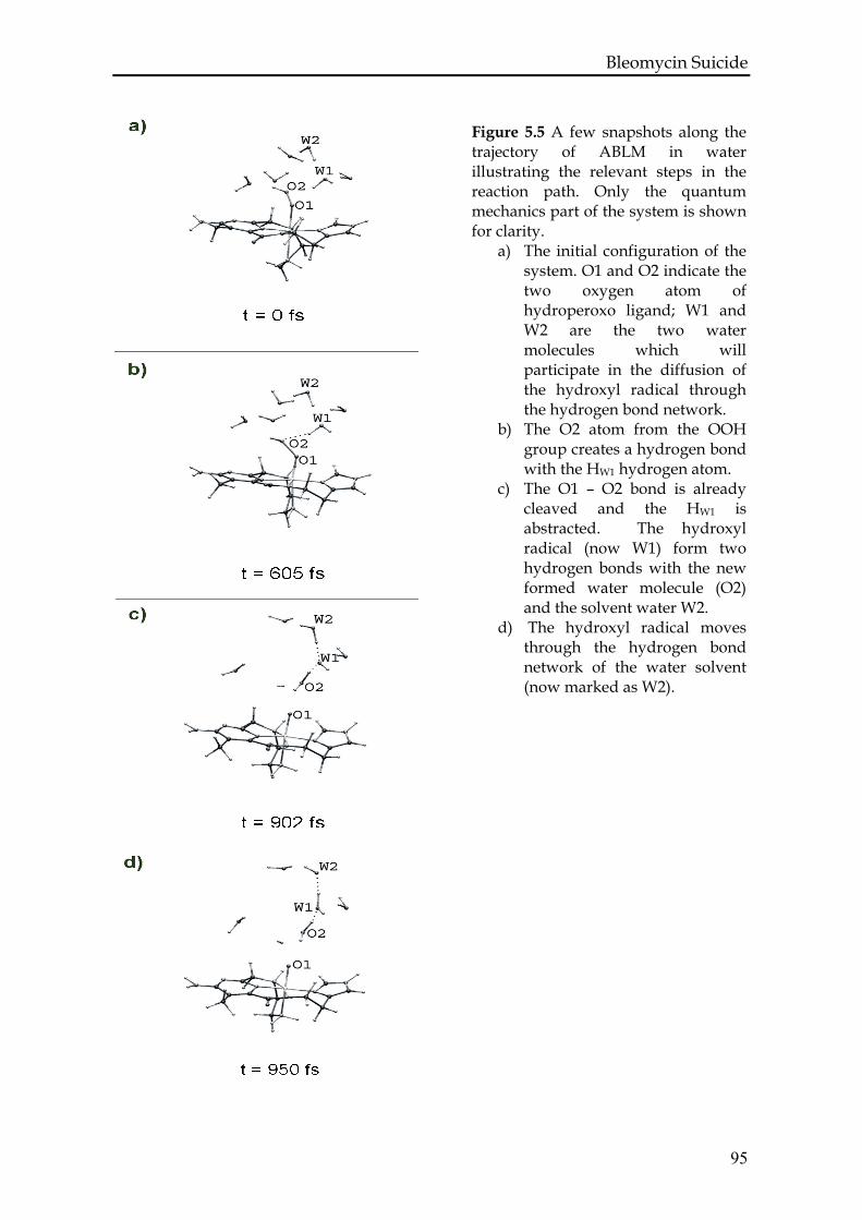
Bleomycin Suicide
Figure 5.5 A few snapshots along the trajectory of ABLM in water illustrating the relevant steps in the reaction path. Only the quantum mechanics part of the system is shown for clarity.
a) The initial configuration of the system. O1 and O2 indicate the two oxygen atom of hydroperoxo ligand; W1 and W2 are the two water molecules which will participate in the diffusion of the hydroxyl radical through the hydrogen bond network.
b) The O2 atom from the OOH group creates a hydrogen bond with the HW1 hydrogen atom.
c) The O1 – O2 bond is already cleaved and the HW1 is abstracted. The hydroxyl radical (now W1) form two hydrogen bonds with the new formed water molecule (O2) and the solvent water W2.
d) The hydroxyl radical moves through the hydrogen bond network of the water solvent (now marked as W2).
95
Bleomycin Suicide
5.4. Conclusions and discussion
In this work we presented a theoretical study of the mechanism of the first
reaction step in the bleomycin suicide based on Car-Parrinello molecular
dynamics simulations. We studied an extensive and realistic model of ABLM both
in vacuum and in water solvent. We point out that a proper description of the
ligand plays a key role in studying the chemistry of ABLM. Our energy profile for
the homolytic O-O bond cleavage calculated in vacuum gives a reaction energy of
17 kcal/mol which is about 13 kcal/mol lower than reported previously [5]. The
only difference between these two calculations is in the ligand model. As already
mentioned in section 3.1, the previous model was simplified by introducing two
ammonia molecules into the coordination sphere of the iron center.
During our simulations in vacuum and in water we observed a hydrogen bond
formation prior to O-O bond scission. In vacuum an intramolecular H-bond is
formed between the OOH group and the hydrogen atom belonging to the
secondary amine of the β-aminoalanine (HN2), while in the presence of water it is
more likely to observe an intermolecular H-bond with a water molecule.
However, in both cases the formation of the H-bond appears to be a necessary
step to facilitate the O-O bond scission by lowering the reaction energy barrier.
We have identified the OH species formed in that process as a short-lived
hydroxyl radical. This radical immediately abstracts the hydrogen atom
participating in the H-bond. The second product of the reaction was identified as
the BLM-Fe(IV)=O complex (S = 1). Thus we can conclude that in the ABLM
suicide process the O-O bond cleavage is homolytic. Furthermore, since the
process of the OH radical formation and the H abstraction proceed in a concerted
fashion, it is more appropriate to classify the whole mechanism as a direct
hydrogen abstraction.
The degradation process of ABLM in the absence of the DNA is rather quick
according to our simulations, being observed spontaneously in the ps time-scale
at 300 K. Experimental data give a half-life time of ABLM between 2 min (6°C, pH
= 7.0) [121] or 1.5 min (0°C, pH = 7.2) and < 15 s at pH = 5.8 [143]. The
96
Bleomycin Suicide
97
experimental results clearly show that the half-life time of ABLM highly depends
on the temperature and pH and possibly also other conditions. Even taking into
account the somehow higher temperature of our simulations, our reaction rate is
definitely much higher than expected. This is likely due to an underestimation of
the energy barrier within the approximated exchange-correlation functional used
here. It has been suggested that self-interaction corrections may improve the
accuracy of chemical reactions involving radical species [144, 145] . Nevertheless,
we think that the mechanism of self-deactivation observed here is realistic
although accelerated.
In this first reaction step we observed the formation of two active species that can
further react with a substrate, indicating that the whole process of the Bleomycin
suicide is very complex. In the context of the observed homolytic cleavage of the
O-O bond, one may easily explain the modification of the bithiazole moiety of the
bleomycin [129]. Some of the drug side effects may also be associated with the
formation of the active species during the degradation process.
A direct hydrogen abstraction was also suggested by static calculations [131] as
the most likely mechanism for the radical formation at the C4’ position of the
DNA ribose sugar. In fact the reaction with participation of free hydroxyl radical
is very unselective and this is why the direct hydrogen abstraction is much easier
to accept. Our study shows that the border between homolytic cleavage and direct
hydrogen atom abstraction is very thin. Moreover, we cannot exclude that the
Fe(IV)=O complex has a direct role in the DNA attack and is not only an
intermediate for regenerating the Fe(III)BLM complex. The BLM-Fe(IV)=O
complex seems to be more stable than BLM-Fe(III)–OOH and it would have a
chance to dock to the DNA molecule. However, at this point we cannot make a
straight analogy to the reaction of ABLM with DNA and this issue still requires
further investigations.

Chapter six
THE MECHANISM OF
THE BLEOMYCIN ACTION: A CAR-PARRINELLO MOLECULAR
DYNAMICS INVESTIGATION
Part of this chapter has been published as: A. Karawajczyk and F. Buda, The anticancer drug bleomycin investigated by density functional theory. Molecular Simulation, 32, 1233 (2006) ABSTRACT Using first-principles molecular dynamics simulations with the Car-Parrinello
method, the possible reaction pathways of the active bleomycin-Fe(III)-OOH
complex with the deoxyribose sugar of DNA were investigated. The theoretical
model of activated bleomycin contains the whole metal bonding domain of the
bleomycin ligand. Simulations were performed in a vacuum and in a natural
environment by explicitly including a DNA fragment within a QM/MM
approach. The simulations show that a facile decaying process involves a
homolytic O-O bond cleavage with an almost simultaneous hydrogen atom
abstraction. The formation of a hydrogen bond appears to be crucial for the O-O
bond cleavage in the Fe(III)-OOH species. The inclusion of a realistic environment
is therefore essential to study the selectivity of the mechanism of bleomycin
action.
100
Bleomycin with Deoxyribose Sugar
6.1 Introduction
Bleomycin is a drug that has been commonly used in chemotherapy since the
early eighties against several types of cancer, including cervix and uterus cancer,
head and neck cancer, testicle and penile cancer, and certain types of lymphoma
[1]. The precise mechanism of action of bleomycin is not known. The drug is
administrated to patients in its metal free form. In vitro studies suggest that it
forms an Fe(II)BLM complex with an iron atom in the body and then binds an
oxygen molecule to generate the O2-Fe(II)-BLM complex. This complex is
converted into activated bleomycin (BLM-Fe(III)-OOH) [120, 121, 124, 125] and is
then bound selectively to DNA at the position 5’-GC-3’ or 5’-GT-3’. Bleomycin
kills cells by binding with and then degrading genetic material. It carries out this
process in the dividing phase of the cell's life cycle and so prevents the cell from
growing. It is known from experiment that the degradation of DNA starts by
forming a radical at 4’C position of a deoxyribose sugar [17]. Why the reaction
occurs selectively at this position is however an open issue, especially given that
the mentioned deoxyribose sugar has three hydrogens available for abstraction,
i.e. 1’H, 2’H and 4’H. Moreover, the first step of the reaction cascade which
involves the activation of the O-O bond is not known. Three scenarios are
possible: (i) heterolytic cleavage of the O-O bond that would lead to a high-valent
intermediate, formally BLM-Fe(V)=O that attacks DNA; (ii) homolytic cleavage of
the O-O bond that would lead to a hydroxyl radical and a high-valent
intermediate, formally BLM-Fe(IV)=O, both of which could attack DNA; (iii)
direct H abstraction of the low-spin BLM-Fe(III)-OOH species with DNA to give a
DNA radical, H2O and a BLM-Fe(IV)=O product. The formally Fe(V)=O species,
formed by heterolytic cleavage of the O-O bond, has been observed in heme
systems, where it is stabilized by the oxidation of the porphyrin ligand (POR),
leading to a (POR+•)Fe(IV)=O intermediate known as compound I. By analogy to
the heme systems, the heterolysis of the O-O bond in ABLM has been accepted as
the most likely first step in DNA degradation [17]. However, in the case of a non-
101
Bleomycin with Deoxyribose Sugar heme environment, the ligands are much more difficult to oxidize, and therefore
not providing sufficient stabilization for the high-valent Fe(V)=O species. Indeed,
theoretical calculations show that BLM-Fe(V)=O complex would be energetically
unfavorable, given that the electron hole is localized on the deprotonated amide
[126]. Thus the homolytic cleavage of the O-O bond started to be considered as a
possible mechanism for the O-O bond activation, especially given that O-O
homolysis has been shown to occur for low-spin analogues of ABLM [146-148].
On the other hand, this mechanism appears to be unlikely for bleomycin action
since it is non-selective. The reactive hydroxyl radical could damage any part of
DNA or ABLM itself. It is known that the DNA is not only the target but also
protects ABLM from permanent self-destruction, as discussed in Chapter 5. The
direct H-atom abstraction from C-H bond of the DNA sugar is closely related to
the O-O homolysis, having identical overall reaction energy but a different
reaction pathway. It remains to be elucidated whether and how DNA affects
ABLM reactivity, what the intermediate reaction products are and why the
selectivity occurs at 4’C position of the deoxyribose sugar. These issues are
addressed in the present study.
In this chapter CPMD simulations are presented giving insight into the reaction of
ABLM with deoxyribose sugar, which is attacked in the first step of the DNA
degradation. Since the reaction involves bonds breaking and forming, the obvious
choice of the computational method is a quantum mechanical one such as DFT.
CPMD is one of the best methods to investigate the reaction pathways without a
priori selection of the reaction coordinates. Another advantage of CPMD is that
the thermal fluctuations are taken into account by performing the simulation at
room temperature. Therefore application of the CPMD method to this study is
especially important for solving the issue of the selectivity of the ABLM action.
Since the role of DNA is questioned, the model should also include the DNA
fragment explicitly. This is done here within a QM/MM framework.
102
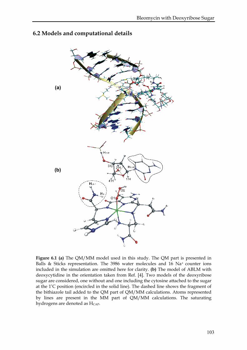
Bleomycin with Deoxyribose Sugar
6.2 Models and computational details
(a)
(b)
Figure 6.1 (a) The QM/MM model used in this study. The QM part is presented in Balls & Sticks representation. The 3986 water molecules and 16 Na+ counter ions included in the simulation are omitted here for clarity. (b) The model of ABLM with deoxycytidine in the orientation taken from Ref. [4]. Two models of the deoxyribose sugar are considered, one without and one including the cytosine attached to the sugar at the 1’C position (encircled in the solid line). The dashed line shows the fragment of the bithiazole tail added to the QM part of QM/MM calculations. Atoms represented by lines are present in the MM part of QM/MM calculations. The saturating hydrogens are denoted as HCAP.
103
Bleomycin with Deoxyribose Sugar The models used in the present study are based on the NMR data for a BLM-
Co(III)-OOH complex bonded to a fragment of DNA (D(GGAAGCTTCC)) [4]. The
NMR structure has been already presented in Figure 1.2. In the bleomycin ligand
we can distinguish three main parts: the metal bonding domain, the sugar moiety,
and the bithiazole tail. While the sugar moiety and the bithiazole tail are
responsible for recognition and docking to DNA, the metal bonding domain plays
a main role in the metal coordination and the O-O bond activation. Thus, for
studying the reaction mechanisms we have chosen the metal bonding domain
(represented by stick and balls in Fig.1.2) to be described by quantum methods
without further simplifications. The equatorial ligands are: the secondary amine
of the β-aminoalanine (A’’aALA – N2) segment, the pyrimidine (PYR – N3), the
imidazole ring (HIS-I – N5) and the amide nitrogen (HIS-A – N4) of the β -
hydroxyhistidine. The primary amine of the β - aminoalanine residue (A’aALA –
N1) and the oxygen atom of the peroxy group are the two axial ligands.
The deoxyribose sugar (indicated in cyan in Fig 1.2) was included in the model to
study the reaction of ABLM with the sugar in the position shown in Figure 1.2.
The positions of the oxygen atoms bonded originally to phosphor were fixed
during the simulations in vacuum. Two models of the deoxyribose sugar are
considered, one without and one including the cytosine attached to the sugar at
the 1’C position to check its effect on the selectivity of the reaction (see also Fig.
6.1) The positions of the atoms forming the aromatic ring of cytosine were fixed
during the simulations in vacuum. In the QM/MM simulations the entire
structure of the BLM-Fe(III)-OOH complex bonded to a fragment of DNA
(D(GGAAGCTTCC)) was used (Fig. 6.1a). The complex of ABLM and DNA was
placed in a box with 3986 water molecules and the total charge of the system was
neutralized by 16 Na+ counter ions. The system was first equilibrated under
constant pressure at 300 K for 1 ns and then for another nanosecond at 300 K with
constant volume with a box of size 49.88 x 44.17 x 59.22 Å3. During the
equilibration procedure the bleomycin was kept fixed.
Analogous to the simulations performed in vacuum there are two set-ups for the
QM/MM simulations that differ in the size of the QM part. The first set-up
104
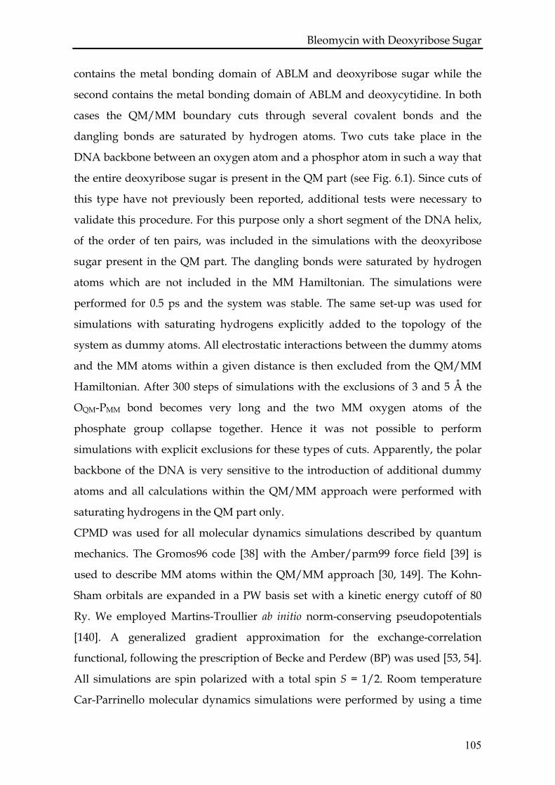
Bleomycin with Deoxyribose Sugar contains the metal bonding domain of ABLM and deoxyribose sugar while the
second contains the metal bonding domain of ABLM and deoxycytidine. In both
cases the QM/MM boundary cuts through several covalent bonds and the
dangling bonds are saturated by hydrogen atoms. Two cuts take place in the
DNA backbone between an oxygen atom and a phosphor atom in such a way that
the entire deoxyribose sugar is present in the QM part (see Fig. 6.1). Since cuts of
this type have not previously been reported, additional tests were necessary to
validate this procedure. For this purpose only a short segment of the DNA helix,
of the order of ten pairs, was included in the simulations with the deoxyribose
sugar present in the QM part. The dangling bonds were saturated by hydrogen
atoms which are not included in the MM Hamiltonian. The simulations were
performed for 0.5 ps and the system was stable. The same set-up was used for
simulations with saturating hydrogens explicitly added to the topology of the
system as dummy atoms. All electrostatic interactions between the dummy atoms
and the MM atoms within a given distance is then excluded from the QM/MM
Hamiltonian. After 300 steps of simulations with the exclusions of 3 and 5 Å the
OQM-PMM bond becomes very long and the two MM oxygen atoms of the
phosphate group collapse together. Hence it was not possible to perform
simulations with explicit exclusions for these types of cuts. Apparently, the polar
backbone of the DNA is very sensitive to the introduction of additional dummy
atoms and all calculations within the QM/MM approach were performed with
saturating hydrogens in the QM part only.
CPMD was used for all molecular dynamics simulations described by quantum
mechanics. The Gromos96 code [38] with the Amber/parm99 force field [39] is
used to describe MM atoms within the QM/MM approach [30, 149]. The Kohn-
Sham orbitals are expanded in a PW basis set with a kinetic energy cutoff of 80
Ry. We employed Martins-Troullier ab initio norm-conserving pseudopotentials
[140]. A generalized gradient approximation for the exchange-correlation
functional, following the prescription of Becke and Perdew (BP) was used [53, 54].
All simulations are spin polarized with a total spin S = 1/2. Room temperature
Car-Parrinello molecular dynamics simulations were performed by using a time
105
Bleomycin with Deoxyribose Sugar step of 0.09 fs and a value of 400 a.u. for the fictitious electronic mass in the Car-
Parrinello Lagrangian. For the simulation including the sugar in vacuum an
isolated cubic supercell of size 18 x 18 x 18 Å3 was used.
37B6.3 Results and discussion
6.3.1 ABLM with deoxyribose sugar in vacuum
This simulation was started at 20 K and then the temperature was slowly
increased to 300 K. As mentioned in the Introduction the sugar molecule has three
hydrogen atoms available for abstraction (1’H, 2’H, 4’H). During the simulation
the formation of H-bonds is observed between the O2 atom of the –OOH group
and all three hydrogens at different time intervals (see Fig. 6.2). We notice
however that only the formation of a H-bond with 4’H has an influence on the
amplitude of the O1-O2 bond length oscillation. Figure 6.2 shows that when the
O2-1’H and O2-2’H distances are shorter than O2-4’H, the O1-O2 bond remains
stable, oscillating with a small amplitude around an average value of 1.5 Å. After
750 fs the O2-4’H distance becomes the shortest and the O1-O2 bond
Figure 6.2 Dynamical evolutions of a few distances along the trajectory of the CPMD simulation of ABLM with deoxyribose sugar in vacuum.
106
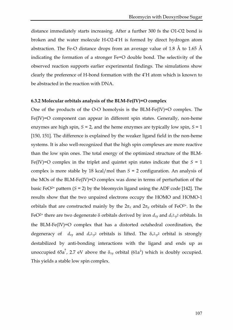
Bleomycin with Deoxyribose Sugar distance immediately starts increasing. After a further 300 fs the O1-O2 bond is
broken and the water molecule H-O2-4’H is formed by direct hydrogen atom
abstraction. The Fe-O distance drops from an average value of 1.8 Å to 1.65 Å
indicating the formation of a stronger Fe=O double bond. The selectivity of the
observed reaction supports earlier experimental findings. The simulations show
clearly the preference of H-bond formation with the 4’H atom which is known to
be abstracted in the reaction with DNA.
6.3.2 Molecular orbitals analysis of the BLM-Fe(IV)=O complex
One of the products of the O-O homolysis is the BLM-Fe(IV)=O complex. The
Fe(IV)=O component can appear in different spin states. Generally, non-heme
enzymes are high spin, S = 2, and the heme enzymes are typically low spin, S = 1
[150, 151]. The difference is explained by the weaker ligand field in the non-heme
systems. It is also well-recognized that the high spin complexes are more reactive
than the low spin ones. The total energy of the optimized structure of the BLM-
Fe(IV)=O complex in the triplet and quintet spin states indicate that the S = 1
complex is more stable by 18 kcal/mol than S = 2 configuration. An analysis of
the MOs of the BLM-Fe(IV)=O complex was done in terms of perturbation of the
basic FeO2+ pattern (S = 2) by the bleomycin ligand using the ADF code [142]. The
results show that the two unpaired electrons occupy the HOMO and HOMO-1
orbitals that are constructed mainly by the 2πx and 2πy orbitals of FeO2+. In the
FeO2+ there are two degenerate δ orbitals derived by iron dxy and dx2-y2 orbitals. In
the BLM-Fe(IV)=O complex that has a distorted octahedral coordination, the
degeneracy of dxy and dx2-y2 orbitals is lifted. The δx2-y2 orbital is strongly
destabilized by anti-bonding interactions with the ligand and ends up as
unoccupied 65a↑, 2.7 eV above the δxy orbital (61a↓) which is doubly occupied.
This yields a stable low spin complex.
107
Bleomycin with Deoxyribose Sugar
BLM-Fe(IV)=O S = 1
FeO2+
S = 2
2πx, 2πyδxy, δx2-y2
1πx, 1πy2σ
3σ
59a (50% δxy, 14% 53a)
59a (51% 53a, 25% 55a)
60a (39% 53a, 22% 55a)
61a (40% 55a, 28% δxy)
60a (70% 55a, 10% 1πy)
62a (92% 2πx)
61a (75% δxy, 8% 55a)63a (65% 2πy, 22% 55a)
62a (70% 2πx, 15% 57a)63a (56% 2πy, 14% 2πx)64a (84% 57a)65a (52% δx2-y2, 30% 56a)64a (65% 57a, 20% 2πy )
66a (44% δx2-y2 )
Figure 6.3 Molecular orbital diagram illustrating the perturbation of the FeO2+ molecular orbitals (right) to create the BLM-Fe(IV)=O complex (left). The percentage composition given in brackets is coming from the molecular orbitals of the BLM ligand and FeO2+ fragment.
6.3.3 Selectivity of the hydrogen atom abstraction
The stability of the three possible radicals of the deoxyribose sugar was checked
to validate the observed selectivity of the hydrogen abstraction. A full energy
minimization was therefore performed for each possible radical. It was observed
that the most stable radical is indeed the 4’C radical, which is lower in energy by
2.9 kcal/mol and 8.4 kcal/mol than the 1’C and 2’C radicals, respectively. This
result is in line with the observed selectivity during the dynamical simulations.
On the other hand, in the natural system the 1’C atom is connected to the cytosine.
The calculated energies for the radicals 1’C and 4’C for deoxycytidine indicate
that the presence of the aromatic ring in that position stabilizes the 1’C radical.
108
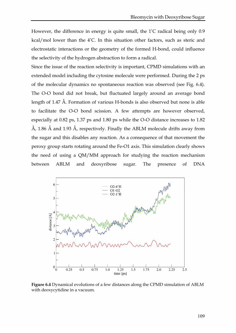
Bleomycin with Deoxyribose Sugar However, the difference in energy is quite small, the 1’C radical being only 0.9
kcal/mol lower than the 4’C. In this situation other factors, such as steric and
electrostatic interactions or the geometry of the formed H-bond, could influence
the selectivity of the hydrogen abstraction to form a radical.
Since the issue of the reaction selectivity is important, CPMD simulations with an
extended model including the cytosine molecule were performed. During the 2 ps
of the molecular dynamics no spontaneous reaction was observed (see Fig. 6.4).
The O-O bond did not break, but fluctuated largely around an average bond
length of 1.47 Å. Formation of various H-bonds is also observed but none is able
to facilitate the O-O bond scission. A few attempts are however observed,
especially at 0.82 ps, 1.37 ps and 1.80 ps while the O-O distance increases to 1.82
Å, 1.86 Å and 1.93 Å, respectively. Finally the ABLM molecule drifts away from
the sugar and this disables any reaction. As a consequence of that movement the
peroxy group starts rotating around the Fe-O1 axis. This simulation clearly shows
the need of using a QM/MM approach for studying the reaction mechanism
between ABLM and deoxyribose sugar. The presence of DNA
Figure 6.4 Dynamical evolutions of a few distances along the CPMD simulation of ABLM with deoxycytidine in a vacuum.
109
Bleomycin with Deoxyribose Sugar prevents the molecules from separation. In a natural setting DNA and ABLM
create a well-defined reaction environment where not only the steric interactions
are taken into account but also all the electrostatic interactions that could
influence the selectivity.
6.3.4 ABLM with DNA: QM/MM study
To gain insight into the selectivity of the reaction of ABLM with DNA, an
extensive model was set up within the QM/MM approach. The QM part was
increased by adding atoms belonging to the bithiazole tail in order to realistically
represent the active site and explore all possible scenarios of the H atom
abstraction. In the active site there are 4 hydrogens available for abstraction: 1’H,
2’H, 4’H of deoxyribose sugar and HN of the bithiazole tail. The dynamics has
been followed for ~2.5 ps and relevant variables are plotted in Figure 6.5. As
illustrated in the graph 6.5 c the temperature was increased during the run from
300 K up to 400 K in order to accelerate the reaction. A spontaneous O-O bond
scission is observed during the simulation after increasing the temperature. Soon
after the O-O bond breaking a water molecule is formed and the deoxyribose
sugar is destabilized. The scission is facilitated by the formation of a H-bond
between O2 and HN with an average distance dO2-HN = 1.8 Å and an angle ∠N-
O2-HN oscillating between 140 and 180 degree. These values provide evidence for
a strong H-bond. It can be noted that formation of the O2…HN H-bond affects the
amplitude of the O-O bond oscillation already at 300 K as illustrated in Figure 6.5
at 0.5 ps and 1.1 ps (red line). Finally at 1.8 ps the O-O bond is broken and the OH
species is formed. The newly formed fragment stays hydrogen bonded to the HN
atom of the bithiazole tail and the 4’H of the sugar molecule. The strong H-bonds
are represented in Figure 6.5 by short distances 4’H-O2 and O2-HN (in green and
brown, respectively) and H-bond angles oscillating around 160 degree. A
visualization of the total spin distribution for snapshots at 1.0 ps and 1.85 ps is
presented in Figure 6.6. The first snapshot is representative of the system during
the first part of the simulations from 0 up to 1.7 ps. As shown in figure 6.6a the
unpaired α electron is situated on the iron-peroxy center of the ABLM.
110
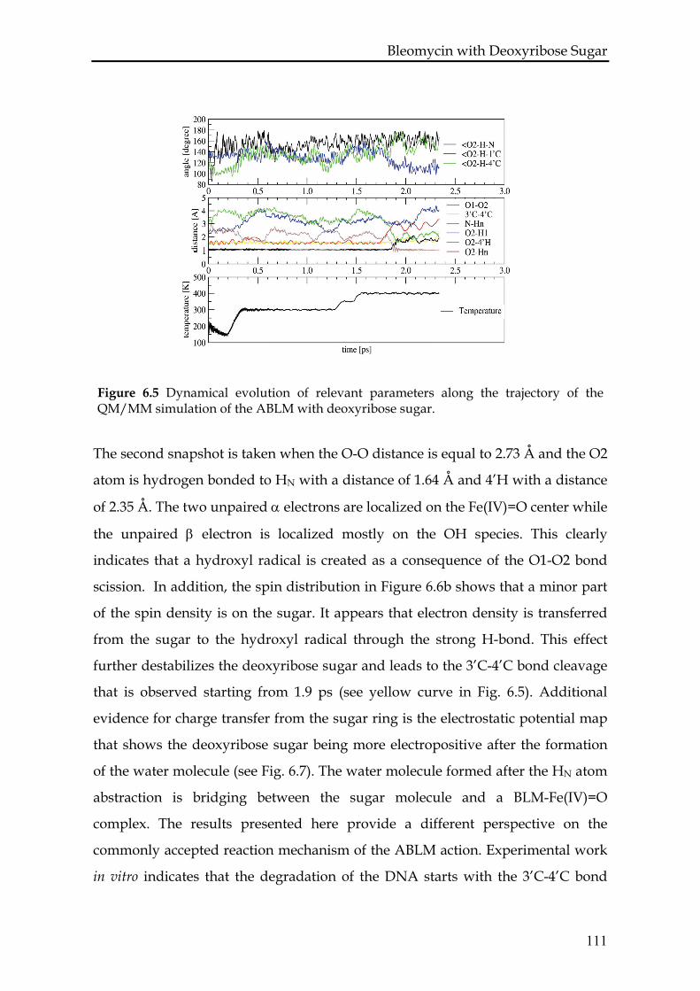
Bleomycin with Deoxyribose Sugar
Figure 6.5 Dynamical evolution of relevant parameters along the trajectory of the QM/MM simulation of the ABLM with deoxyribose sugar.
The second snapshot is taken when the O-O distance is equal to 2.73 Å and the O2
atom is hydrogen bonded to HN with a distance of 1.64 Å and 4’H with a distance
of 2.35 Å. The two unpaired α electrons are localized on the Fe(IV)=O center while
the unpaired β electron is localized mostly on the OH species. This clearly
indicates that a hydroxyl radical is created as a consequence of the O1-O2 bond
scission. In addition, the spin distribution in Figure 6.6b shows that a minor part
of the spin density is on the sugar. It appears that electron density is transferred
from the sugar to the hydroxyl radical through the strong H-bond. This effect
further destabilizes the deoxyribose sugar and leads to the 3’C-4’C bond cleavage
that is observed starting from 1.9 ps (see yellow curve in Fig. 6.5). Additional
evidence for charge transfer from the sugar ring is the electrostatic potential map
that shows the deoxyribose sugar being more electropositive after the formation
of the water molecule (see Fig. 6.7). The water molecule formed after the HN atom
abstraction is bridging between the sugar molecule and a BLM-Fe(IV)=O
complex. The results presented here provide a different perspective on the
commonly accepted reaction mechanism of the ABLM action. Experimental work
in vitro indicates that the degradation of the DNA starts with the 3’C-4’C bond
111
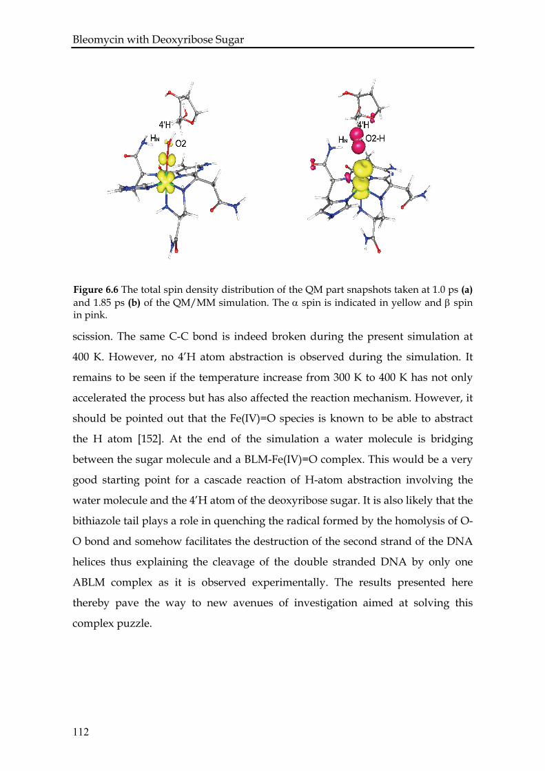
Bleomycin with Deoxyribose Sugar
a) b)
Figure 6.6 The total spin density distribution of the QM part snapshots taken at 1.0 ps (a) and 1.85 ps (b) of the QM/MM simulation. The α spin is indicated in yellow and β spin in pink.
scission. The same C-C bond is indeed broken during the present simulation at
400 K. However, no 4’H atom abstraction is observed during the simulation. It
remains to be seen if the temperature increase from 300 K to 400 K has not only
accelerated the process but has also affected the reaction mechanism. However, it
should be pointed out that the Fe(IV)=O species is known to be able to abstract
the H atom [152]. At the end of the simulation a water molecule is bridging
between the sugar molecule and a BLM-Fe(IV)=O complex. This would be a very
good starting point for a cascade reaction of H-atom abstraction involving the
water molecule and the 4’H atom of the deoxyribose sugar. It is also likely that the
bithiazole tail plays a role in quenching the radical formed by the homolysis of O-
O bond and somehow facilitates the destruction of the second strand of the DNA
helices thus explaining the cleavage of the double stranded DNA by only one
ABLM complex as it is observed experimentally. The results presented here
thereby pave the way to new avenues of investigation aimed at solving this
complex puzzle.
112
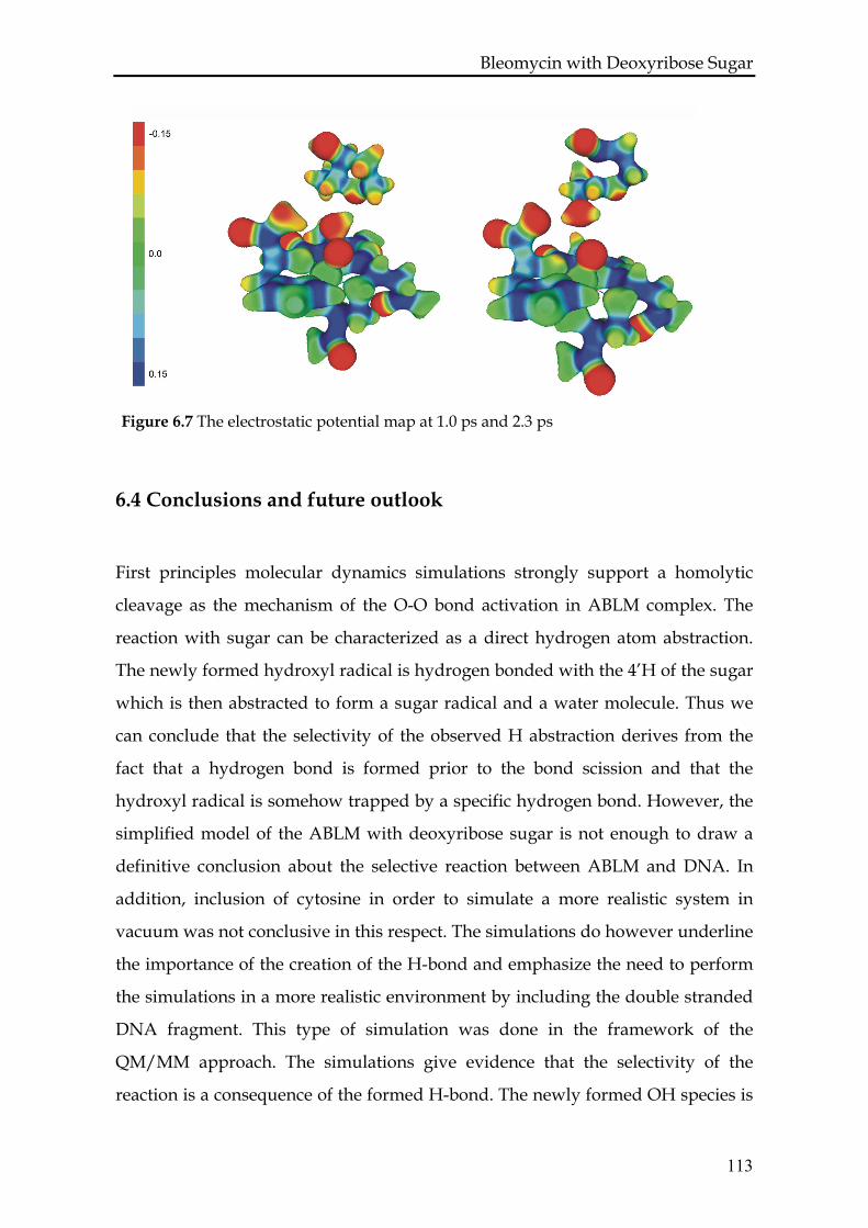
Bleomycin with Deoxyribose Sugar
Figure 6.7 The electrostatic potential map at 1.0 ps and 2.3 ps
6.4 Conclusions and future outlook
First principles molecular dynamics simulations strongly support a homolytic
cleavage as the mechanism of the O-O bond activation in ABLM complex. The
reaction with sugar can be characterized as a direct hydrogen atom abstraction.
The newly formed hydroxyl radical is hydrogen bonded with the 4’H of the sugar
which is then abstracted to form a sugar radical and a water molecule. Thus we
can conclude that the selectivity of the observed H abstraction derives from the
fact that a hydrogen bond is formed prior to the bond scission and that the
hydroxyl radical is somehow trapped by a specific hydrogen bond. However, the
simplified model of the ABLM with deoxyribose sugar is not enough to draw a
definitive conclusion about the selective reaction between ABLM and DNA. In
addition, inclusion of cytosine in order to simulate a more realistic system in
vacuum was not conclusive in this respect. The simulations do however underline
the importance of the creation of the H-bond and emphasize the need to perform
the simulations in a more realistic environment by including the double stranded
DNA fragment. This type of simulation was done in the framework of the
QM/MM approach. The simulations give evidence that the selectivity of the
reaction is a consequence of the formed H-bond. The newly formed OH species is
113
Bleomycin with Deoxyribose Sugar
114
H-bonded to HN and 4’H. The analysis of the electron spin density reveals that the
observed OH species is a hydroxyl radical. This radical is able to destabilize the
3’C-4’C bond of the deoxyribose sugar causing its cleavage and to abstract the
hydrogen from the bithiazole tail of BLM to form a water molecule.
The FPMD method is clearly a powerful approach to study the reaction
mechanism. The presented investigations are able to answer some of the initial
questions and, as it is often the case, they also raise new issues that deserve
further detailed analysis. One appealing question concerns the free energy profile
of the reaction where the hydrogen atom is abstracted by ABLM. Recently
metadynamics method has been developed which allows calculating the free
energy [153, 154]. This method allows exploring the free energy surface in the
space defined by a few collective variables. The collective variables important for
the problem under investigation would be the parameters of the created H-bonds.
The FPMD simulations suggest that the H-atom abstraction mechanism is not
more selective than the homolytic O-O bond cleavage, in contrast with references
from previous studies [5, 126, 131]. The key to understand the cleavage of the
double stranded DNA by only one ABLM complex may be the creation of the
active BLM-Fe(IV)=O complex. This active species is formed during all presented
simulations. The QM/MM simulation shows additionally that the created water
molecule may be used as a bridge to facilitate the cascade reaction of H-atom
abstraction. Unfortunately our simulations are too short to give a strong evidence
for further steps of the reaction and longer QM/MM simulations with extended
QM part of the cytosine have to be performed at 300K to draw stronger
conclusions about the mechanism of ABLM action. Further studies are in progress
to provide complementary knowledge for a complete picture of the ABLM action.
References
REFERENCES 1. R. M. Burger, Nature of activated bleomycin. Metal-Oxo and Metal-Peroxo Species in
Catalytic Oxidations, 97, 287 (2000) 2. T. E. Lehmann, L. J. Ming, M. E. Rosen and L. Que, NMR studies of the paramagnetic
complex Fe(II)-bleomycin. Biochemistry, 36, 2807 (1997) 3. S. J. Brown, P. K. Mascharak and D. W. Stephan, Characterization of a crystalline
synthetic analog of copper(II) bleomycin. Journal of the American Chemical Society, 110, 1996 (1988)
4. C. Q. Zhao, C. W. Xia, Q. K. Mao, H. Forsterling, E. DeRose, W. E. Antholine, W. K. Subczynski and D. H. Petering, Structures of HO2-Co(III)bleomycin A(2) bound to d(GAGCTC)(2) and d(GGAAGCTTCC)(2): structure-reactivity relationships of Co and Fe bleomycins. Journal of Inorganic Biochemistry, 91, 259 (2002)
5. N. Lehnert, F. Neese, R. Y. N. Ho, L. Que and E. I. Solomon, Electronic structure and reactivity of low-spin Fe(III)-hydroperoxo complexes: Comparison to activated bleomycin. Journal of the American Chemical Society, 124, 10810 (2002)
6. L. Balbes, S. Mascarella and D. B. Boyd, A perspective of modern methods in computer-aided drug design. Reviews in Computational Chemistry, 5, 337–370 (1994)
7. T. Lengauer, Algorithmic research problems in molecular bioinformatics. Proceedings of the 2nd Israeli Symposium on the Theory of Computing and Systems, (1993)
8. D. B. Boyd, Successes of computer-assisted molecular design. Reviews in Computational Chemistry, 1, 355–371 (1990)
9. P. W. Finn and L. E. Kavraki, Computational Approaches to drug design. Algorithmica, 25, 347–371 (1999)
10. L. M. Brown, A. M. Malkinson, D. E. Rannels and S. R. Rannels, Compensatory lung growth after partial pneumonectomy enhances lung tumorigenesis induced by 3-methylcholanthrene. Cancer Research, 59, 5089 (1999)
11. I. Haq, Thermodynamics of drug-DNA interactions. Archives of Biochemistry and Biophysics, 403, 1 (2002)
12. S. Neidle and Z. Abraham, Structural and sequence-dependent aspects of drug intercalation into nucleic-acids. Crc Critical Reviews In Biochemistry, 17, 73 (1984)
13. L. S. Lerman, Structural considerations in interaction of DNA and acridines. Journal of Molecular Biology, 3, 18 (1961)
14. L. F. Povirk, Y. H. Han and R. J. Steighner, Structure of bleomycin-induced DNA double-strand breaks - Predominance of blunt ends and single-base 5' extensions. Biochemistry, 28, 5808 (1989)
15. R. J. Steighner and L. F. Povirk, Bleomycin-induced DNA lesions at mutational hot-spots - Implications for the mechanism of double-strand cleavage. Proceedings of the National Academy of Sciences of the United States of America, 87, 8350 (1990)
115
References 16. J. Stubbe, J. W. Kozarich, W. Wu and D. E. Vanderwall, Bleomycins: A structural
model for specificity, binding, and double strand cleavage. Accounts of Chemical Research, 29, 322 (1996)
17. R. M. Burger, Cleavage of nucleic acids by bleomycin. Chemical Reviews, 98, 1153 (1998)
18. E. A. Sausville, R. W. Stein, J. Peisach and S. B. Horwitz, Properties and products of degradation of DNA by Bleomycin and iron(II). Biochemistry, 17, 2746 (1978)
19. G. H. McGall, L. E. Rabow, G. W. Ashley, S. H. Wu, J. W. Kozarich and J. Stubbe, New insight into the mechanism of base propenal formation during bleomycin-mediated DNA-degradation. Journal of the American Chemical Society, 114, 4958 (1992)
20. L. M. Fisher, R. Kuroda and T. T. Sakai, Interaction of Bleomycin A2 with deoxyribonucleic-acid - DNA unwinding and inhibition of bleomycin-induced DNA breakage by cationic thiazole amides related to bleomycin A2. Biochemistry, 24, 3199 (1985)
21. M. R. Ciriolo, R. S. Magliozzo and J. Peisach, Microsome-stimulated activation of ferrous bleomycin in the presence of DNA. Journal of Biological Chemistry, 262, 6290 (1987)
22. I. Mahmutoglu and H. Kappus, Redox cycling of Bleomycin-Fe(III) by an Nadh-dependent enzyme, and DNA damage in isolated rat-liver nuclei. Biochemical Pharmacology, 36, 3677 (1987)
23. H. Umezawa, T. Takita, Y. Sugiura, M. Otsuka, S. Kobayashi and M. Ohno, DNA-bleomycin interaction - Nucleotide sequence-specific binding and cleavage of DNA by bleomycin. Tetrahedron, 40, 501 (1984)
24. B. Giese, X. Beyrichgraf, P. Erdmann, M. Petretta and U. Schwitter, The chemistry of single-stranded 4'-DNA radicals - Influence of the radical precursor on anaerobic and aerobic strand cleavage. Chemistry & Biology, 2, 367 (1995)
25. B. Giese, P. Erdmann, L. Giraud, T. Gobel, M. Petretta, T. Schafer and M. Vonraumer, Heterolytic C-O bond cleavage of 4'-nucleotide radicals. Tetrahedron Letters, 35, 2683 (1994)
26. A. D. Dandrea and W. A. Haseltine, Sequence specific cleavage of DNA by anti-tumor antibiotics neocarzinostatin and bleomycin. Proceedings of the National Academy of Sciences of the United States of America, 75, 3608 (1978)
27. C. Bailly and M. J. Waring, The purine 2-amino group as a critical recognition element for specific DNA cleavage by bleomycin and calicheamicin. Journal of the American Chemical Society, 117, 7311 (1995)
28. D. L. Boger, T. M. Ramsey, H. Cai, S. T. Hoehn and J. Stubbe, Definition of the effect and role of the bleomycin A(2) valerate substituents: Preorganization of a rigid, compact conformation implicated in sequence-selective DNA cleavage. Journal of the American Chemical Society, 120, 9149 (1998)
29. A. Laio, J. VandeVondele and U. Rothlisberger, D-RESP: Dynamically generated electrostatic potential derived charges from quantum mechanics/molecular mechanics simulations. Journal of Physical Chemistry B, 106, 7300 (2002)
30. A. Laio, J. VandeVondele and U. Rothlisberger, A Hamiltonian electrostatic coupling scheme for hybrid Car-Parrinello molecular dynamics simulations. Journal of Chemical Physics, 116, 6941 (2002)
31. W. C. Swope, H. C. Andersen, P. H. Berens and K. R. Wilson, A computer-simulation method for the calculation of equilibrium-constants for the formation of physical clusters of molecules - Application to small water clusters. Journal of Chemical Physics, 76, 637 (1982)
32. D. Frenkel and B. Smit, Understanding molecular smulations. Academic Press, (2002)
116
References
33. H. C. Andersen, Molecular-dynamics simulations at constant pressure and/or temperature. Journal of Chemical Physics, 72, 2384 (1980)
34. W. G. Hoover, Canonical dynamics - equilibrium phase-space distributions. Physical Review A, 31, 1695 (1985)
35. S. Nose, A molecular-dynamics method for simulations in the canonical ensemble. Molecular Physics, 52, 255 (1984)
36. B. R. Brooks, R. E. Bruccoleri, B. D. Olafson, D. J. States, S. Swaminathan and M. Karplus, CHARMM - A program for macromolecular energy, minimization, and dynamics calculations. Journal of Computational Chemistry, 4, 187 (1983)
37. W. L. Jorgensen and J. Tiradorives, The Opls potential functions for proteins - energy minimizations for crystals of cyclic-peptides and crambin. Journal of the American Chemical Society, 110, 1657 (1988)
38. W. F. van Gunsteren, S. R. Billeter, A. A. Eising, P. H. Huenenberger, P. Krueger, A. E. Mark, W. R. P. Scott and I. G. Tironi, Biomolecular simulation: The Gromos96 manual and user guide. Hochschulverlag an der ETH Zurich, (1996)
39. D. A. Case, T. E. Cheatham, T. Darden, H. Gohlke, R. Luo, K. M. Merz, A. Onufriev, C. Simmerling, B. Wang and R. J. Woods, The Amber biomolecular simulation programs. Journal of Computational Chemistry, 26, 1668 (2005)
40. J. W. Ponder and D. A. Case, Force fields for protein simulations. 66 (2003) 41. J. M. Wang, P. Cieplak and P. A. Kollman, How well does a restrained electrostatic
potential (RESP) model perform in calculating conformational energies of organic and biological molecules? Journal of Computational Chemistry, 21, 1049 (2000)
42. C. I. Bayly, P. Cieplak, W. D. Cornell and P. A. Kollman, A Well-behaved electrostatic potential based method using charge restraints for deriving atomic charges - The RESP model. Journal of Physical Chemistry, 97, 10269 (1993)
43. W. D. Cornell, P. Cieplak, C. I. Bayly and P. A. Kollman, Application of RESP charges to calculate conformational energies, hydrogen-bond energies, and free-energies of sSolvation. Journal of the American Chemical Society, 115, 9620 (1993)
44. D. M. York, T. A. Darden and L. G. Pedersen, The effect of long-range electrostatic interactions in simulations of macromolecular crystals - A comparison of the Ewald and truncated list methods. Journal of Chemical Physics, 99, 8345 (1993)
45. J. W. Eastwood and R. W. Hockney, Shaping force law in 2-dimensional particle-mesh models. Journal of Comparative Physiology, 16, 342 (1974)
46. W. Koch and H. C.Holthausen, A chemist's guide to DFT. VCH, (2000) 47. R. Car and M. Parrinello, Unified approach for molecular-dynamics and density-
functional theory. Physical Review Letters, 55, 2471 (1985) 48. P. Hohenberg and W. Kohn, Inhomogeneous electron gas. Physical Review B, 136,
B864 (1964) 49. W. Kohn and L. J. Sham, Self-consistent equations including exchange and correlation
effects. Physical Review, 140, 1133 (1965) 50. R. G. Parr and W. T. Yang, Density functional theory of atoms and molecules. Oxford
University Press, (1986) 51. D. M. Ceperley and B. J. Alder, Ground-state of the electron-gas by a stochastic method.
Physical Review Letters, 45, 566 (1980) 52. S. H. Vosko, L. Wilk and M. Nusair, Accurate spin-dependent electron liquid
correlation energies for local spin-density calculations - A critical analysis. Canadian Journal of Physics, 58, 1200 (1980)
53. A. D. Becke, Density functional calculations of molecular-bond energies. Journal of Chemical Physics, 84, 4524 (1986)
54. J. P. Perdew, Density-functional approximation for the correlation-energy of the inhomogeneous electron-gas. Physical Review B, 33, 8822 (1986)
117
References 55. C. T. Lee, W. T. Yang and R. G. Parr, Development of the Colle-Salvetti correlation-
energy formula into a functional of the electron-density. Physical Review B, 37, 785 (1988)
56. J. P. Perdew, K. Burke and M. Ernzerhof, Generalized gradient approximation made simple. Physical Review Letters, 77, 3865 (1996)
57. J. M. Kriegl, K. Nienhaus, P. C. Deng, J. Fuchs and G. U. Nienhaus, Ligand dynamics in a protein internal cavity. Proceedings of the National Academy of Sciences of the United States of America, 100, 7069 (2003)
58. A. D. Becke, A new mixing of Hartree-Fock and local density-functional theories. Journal of Chemical Physics, 98, 1372 (1993)
59. D. Marx and J. Hutter, Ab initio molecular dynamics: Theory and implementation. Modern Methods and Algorithms of Quantum Chemistry, 1, 301 (2000)
60. D. R. Hamann, M. Schluter and C. Chiang, Norm-conserving pseudopotentials. Physical Review Letters, 43, 1494 (1979)
61. T. Z. Mordasini and W. Thiel, Combined quantum mechanical and molecular mechanical approaches. Chimia, 52, 288 (1998)
62. A. Warshel and M. Levitt, Theoretical studies of enzymic reactions - Dielectric, electrostatic and steric stabilization of carbonium-ion in reaction of lysozyme. Journal of Molecular Biology, 103, 227 (1976)
63. J. Hutter, A. Alavi, T. Deutsch, M. Bernasconi, S. T. Goedecker, D. Marx, M. Tuckerman and M. Parrinello, CPMD, Zurich (2000)
64. C. Manach, A. Scalbert, C. Morand, C. Remesy and L. Jimenez, Polyphenols: Food sources and bioavailability. American Journal of Clinical Nutrition, 79, 727 (2004)
65. J. A. Luchsinger and R. Mayeux, Dietary factors and Alzheimer's disease. The Lancet Neurology, 3, 579 (2004)
66. B. J. Morris, A forkhead in the road to longevity: The molecular basis of lifespan becomes clearer. Journal of Hypertension, 23, 1285 (2005)
67. J. A. Vita, Polyphenols and cardiovascular disease: effects on endothelial and platelet function. The American Journal of Clinical Nutrition, 81, 292S (2005)
68. E. Middleton, Jr., C. Kandaswami and T. C. Theoharides, The effects of plant flavonoids on mammalian cells: implications for inflammation, heart disease, and cancer. Pharmacological Reviews, 52, 673 (2000)
69. C. D. Kanakis, P. A. Tarantilis, M. G. Polissiou, S. Diamantoglou and H. A. Tajmir-Riahi, DNA interaction with naturally occurring antioxidant flavonoids quercetin, kaempferol, and delphinidin. Journal of Biomolecular Structure & Dynamics, 22, 719 (2005)
70. A. Rahman, F. Fazal, J. Greensill, K. Ainley, J. H. Parish and S. M. Hadi, Strand scission in DNA induced by dietary flavonoids: role of Cu(I) and oxygen free radicals and biological consequences of scission. Molecular and Cellular Biochemistry, 111, 3 (1992)
71. R. Solimani, The flavonols quercetin, rutin and morin in DNA solution: UV-vis dichroic (and mid-infrared) analysis explain the possible association between the biopolymer and a nucleophilic vegetable-dye. Biochimica et Biophysica Acta (BBA) - General Subjects, 1336, 281 (1997)
72. M. L. Calabro, S. Tommasini, P. Donato, D. Raneri, R. Stancanelli, P. Ficarra, R. Ficarra, C. Costa, S. Catania, C. Rustichelli and G. Gamberini, Effects of [alpha]- and [beta]-cyclodextrin complexation on the physico-chemical properties and antioxidant activity of some 3-hydroxyflavones. Journal of Pharmaceutical and Biomedical Analysis, 35, 365 (2004)
118
References
73. Y. Zheng, I. S. Haworth, Z. Zuo, M. S. S. Chow and A. H. L. Chow, Physicochemical and structural characterization of quercetin-beta-cyclodextrin complexes. Journal of Pharmaceutical Sciences, 94, 1079 (2005)
74. A. Di Pietro, G. Conseil, J. M. Perez-Victoria, G. Dayan, H. Baubichon-Cortay, D. Trompier, E. Steinfels, J. M. Jault, H. de Wet and a. Maitrejean et, Modulation by flavonoids of cell multidrug resistance mediated by P-glycoprotein and related ABC transporters. Cellular and Molecular Life Sciences: CMLS, 59, 307 (2002)
75. F. Hollosy and G. Keri, Plant-derived protein tyrosine kinase inhibitors as anticancer agents. Current Medicinal Chemistry. Anti-Cancer Agents, 4, 173 (2004)
76. M. N. Jacobs and D. F. V. Lewis, Steroid hormone receptors and dietary ligands: a selected review. The Proceedings of The Nutrition Society, 61, 105 (2002)
77. R. J. Williams, J. P. E. Spencer and C. Rice-Evans, Flavonoids: antioxidants or signalling molecules? Free Radical Biology and Medicine, 36, 838 (2004)
78. K. Ito, H. Suzuki, T. Horie and Y. Sugiyama, Apical/basolateral surface expression of drug transporters and its role in vectorial drug transport. Pharmaceutical Research, 22, 1559 (2005)
79. M. H. Saier, Jr., A functional-phylogenetic classification system for transmembrane solute transporters. Microbiology and Molecular Biology Reviews: MMBR, 64, 354 (2000)
80. G. L. Sottocasa, G. C. Lunazzi, C. Tiribelli and F. Sidney Fleischer Isolation of bilitranslocase, the anion transporter from liver plasma membrane for bilirubin and other organic anions. in Methods in Enzymology, 174, 50 (1989)
81. L. Battiston, S. Passamonti, A. Macagno and G. L. Sottocasa, The Bilirubin-Binding Motif of Bilitranslocase and Its Relation to Conserved Motifs in Ancient Biliproteins. Biochemical and Biophysical Research Communications, 247, 687 (1998)
82. S. Passamonti, M. Terdoslavich, A. Margon, A. Cocolo, N. Medic, F. Micali, G. Decorti and M. Franko, Uptake of bilirubin into HepG2 cells assayed by thermal lens spectroscopy. Function of bilitranslocase. The FEBS Journal, 272, 5522 (2005)
83. S. Passamonti, L. Battiston and G. L. Sottocasa, Bilitranslocase can exist in two metastable forms with different affinities for the substrates--evidence from cysteine and arginine modification. European Journal of Biochemistry / FEBS, 253, 84 (1998)
84. G. L. Sottocasa, G. Baldini, G. Sandri, G. Lunazzi and C. Tiribelli, Reconstitution in vitro of sulfobromophthalein transport by bilitranslocase. Biochimica et Biophysica Acta (BBA) - Biomembranes, 685, 123 (1982)
85. S. Passamonti, U. Vrhovsek and F. Mattivi, The interaction of anthocyanins with bilitranslocase. Biochemical and Biophysical Research Communications, 296, 631 (2002)
86. M. Novic and M. Vracko, Comparison of spectrum-like representation of 3D chemical structure with other representations when used for modelling biological activity. Chemometrics and Intelligent Laboratory Systems, 59, 33 (2001)
87. A. Karawajczyk, V. Drgan, N. Medic, G. Oboh, S. Passamonti and M. Novic, Properties of flavonoids influencing the binding to bilitranslocase investigated by neural network modelling. Biochemical Pharmacology, 73, 308 (2007)
88. J. Dayhof, Neural network architectures, An Introduction. Van Nostrand Reinhold, (1990)
89. J. Zupan, M. Novic and I. Ruisanchez, Kohonen and counterpropagation artificial neural networks in analytical chemistry. Chemometrics and Intelligent Laboratory Systems, 38, 1 (1997)
90. M. J. S. Dewar, E. G. Zoebisch, E. F. Healy and J. P. Stewart, AM1: A new general purpose quantum mechanical molecular model. Journal of the American Chemical Society, 107, 3902 (1985)
119
References 91. A. R. Katritzy, V. S. Lobanov and M. Karelnos, CODESSA 2.0, University of
Florida U.S.A. (1990) 92. J. Zupan and M. Novic, Optimisation of structure representation for QSAR studies.
Analytica Chimica Acta, 388, 243 (1999) 93. S. Passamonti and G. L. Sottocasa, The quinoid structure is the molecular requirement
for recognition of phthaleins by the organic anion carrier at the sinusoidal plasma membrane level in the liver. Biochimica et Biophysica Acta (BBA) - Biomembranes, 943, 119 (1988)
94. K. Lemanska, H. Szymusiak, B. Tyrakowska, R. Zielinski, A. E. M. F. Soffers and I. M. C. M. Rietjens, The influence of pH on antioxidant properties and the mechanism of antioxidant action of hydroxyflavones. Free Radical Biology and Medicine, 31, 869 (2001)
95. S. Passamonti, A. Vanzo, U. Vrhovsek, M. Terdoslavich, A. Cocolo, G. Decorti and F. Mattivi, Hepatic uptake of grape anthocyanins and the role of bilitranslocase. Food Research International, 38, 953 (2005)
96. L. R. Huang, Y. Xie and J. W. Lown, Bleomycin antibiotics and their role in cancer chemotherapy. Expert Opinion on Therapeutic Patents, 6, 893 (1996)
97. S. J. Brown, S. E. Hudson, D. W. Stephan and P. K. Mascharak, Syntheses, structures, and spectral properties of a synthetic analog of copper(II)-bleomycin and an intermediate in the process of its formation. Inorganic Chemistry, 28, 468 (1989)
98. J. D. Tan, S. E. Hudson, S. J. Brown, M. M. Olmstead and P. K. Mascharak, Syntheses, structures, and reactivities of synthetic analogs of the 3 forms of Co(III)-bleomycin - Proposed mode of light-induced DNA damage by the Co(III) chelate of the drug. Journal of the American Chemical Society, 114, 3841 (1992)
99. H. Kurosaki, K. Hayashi, Y. Ishikawa and M. Goto, The first trigonal-bipyramidal structure of zinc(II) complex of a bleomycin model. Chemistry Letters, 8, 691 (1995)
100. S. J. Brown, M. M. Olmstead and P. K. Mascharak, Iron(II) and iron(III) complexes of N-(2-(4- imidazolyl)ethyl)pyrimidine-4-carboxamide, a ligand resembling part of the metal-binding domain of bleomycin. Inorganic Chemistry, 29, 3229 (1990)
101. T. E. Lehmann, Molecular modeling of the three-dimensional structure of Fe(II)-bleomycin: are the Co(II) and Fe(II) adducts isostructural? Journal of Biological Inorganic Chemistry, 7, 305 (2002)
102. T. E. Lehmann, M. L. Serrano and L. Que, Coordination chemistry of Co(II)-bleomycin: Its investigation through NMR and molecular dynamics. Biochemistry, 39, 3886 (2000)
103. K. E. Loeb, J. M. Zaleski, C. D. Hess, S. M. Hecht and E. I. Solomon, Spectroscopic investigation of the metal ligation and reactivity of the ferrous active sites of bleomycin and bleomycin derivatives. Journal of the American Chemical Society, 120, 1249 (1998)
104. K. E. Loeb, J. M. Zaleski, T. E. Westre, R. J. Guajardo, P. K. Mascharak, B. Hedman, K. O. Hodgson and E. I. Solomon, Spectroscopic definition of the geometric and electronic- structure of the nonheme iron active-site in iron(II) bleomycin - correlation with oxygen reactivity. Journal of the American Chemical Society, 117, 4545 (1995)
105. J. P. Piquemal, B. Williams-Hubbard, N. Fey, R. J. Deeth, N. Gresh and C. Giessner-Prettre, Inclusion of the ligand field contribution in a polarizable molecular mechanics: SIBFA-LF (vol 24, pg 1963, 2003). Journal of Computational Chemistry, 25, 308 (2004)
106. N. Oppenheimer, L. Rodriguez and S. Hecht, Structural studies of "active complex" of bleomycin: Assignment of ligands to the ferrous ion in a ferrous-bleomycin-carbon monoxide complex. Proc. Natl. Acad. Sci. USA, 76, 5616 (1979)
120
References
107. M. A. J. Akkerman, E. Neijman, S. S. Wijmenga, C. W. Hilbers and W. Bermel, Studies of the solution structure of the bleomycin-A2 iron(II) carbon-monoxide complex by means of 2-dimensional NMR- spectroscopy and distance geometry calculations. Journal of the American Chemical Society, 112, 7462 (1990)
108. A. Fouqueau, S. Mer, M. E. Casida, L. M. L. Daku, A. Hauser, T. Mineva and F. Neese, Comparison of density functionals for energy and structural differences between the high-[T-5(2g):(t(2g))(4)(e(g))(2)] and low-[(1)A(1g):(t(2g))(6)(e(g))(0)] spin states of the hexaquoferrous cation [Fe(H2O)(6)](2+). Journal of Chemical Physics, 120, 9473 (2004)
109. M. Swart, A. R. Groenhof, A. W. Ehlers and K. Lammertsma, Validation of exchange - correlation functionals for spin states of iron complexes. Journal of Physical Chemistry A, 108, 5479 (2004)
110. P. J. Hay and W. R. Wadt, Ab initio effective core potentials for molecular calculations. Potentials for the transition metal atoms Sc to Hg. Journal of Chemical Physics, 82, 270 (1985)
111. T. H. D. Jr and P. J. Hay, Methods of Electronic Structure Theory. Modern Theoretical Chemistry, 3, 1 (1976)
112. M. J. Frisch, G. W. Trucks, H. B. Schlegel, G. E. Scuseria, M. A. Robb, J. R. Cheeseman, V. G. Zakrzewski, J. A. Montgomery Jr., R. E. Stratmann, J. C. Burant, S. Dapprich, J. M. Millam, A.D. Daniels, K. N. Kudin, M. C. Strain, Ö. Farkas, J. Tomasi, V. Barone, M. Cossi, R. Cammi, B. Mennucci, C. Pomelli, C. Adamo, S. Clifford, J. Ochterski, G. A. Petersson, P. Y. Ayala, Q. Cui, K. Morokuma, P. Salvador, J. J. Dannenberg, D. K. Malick, A. D. Rabuck, K. Raghavachari, J. B. Foresman, J. Cioslowski, J. V. Ortiz, A. G. Baboul, B. B. Stefanov, G. Liu, A. Liashenko, P. Piskorz, I. Komáromi, R. Gomperts, R. L. Martin, D. J. Fox, T. Keith, M. A. Al-Laham, C. Y. Peng, A. Nanayakkara, M. Challacombe, M. W. Gill, B. Johnson, W. Chen, M. W. Wong, J. L. Andres, C. Gonzalez, M. Head-Gordon, E. S. Replogle and J. A. Pople, Gaussian 03, Pittsburgh (2003)
113. G. Smolentsev, A. V. Soldatov, E. C. Wasinger and E. I. Solomon, Axial ligation of Fe(II)-bleomycin probed by XANES spectroscopy. Inorganic Chemistry, 43, 1825 (2004)
114. I. Morgenstern-Badarau, F. Lambert, J. Philippe Renault, M. Cesario, J.-D. Marechal and F. Maseras, Amine conformational change and spin conversion induced by metal-assisted ligand oxidation: from the seven-coordinate iron(II)-TPAA complex to the two oxidized iron(II)-(py)3tren isomers. Characterization, crystal structures, and density functional study. Inorganica Chimica Acta, 297, 338 (2000)
115. D. G. Lonnon, D. C. Craig and S. B. Colbran, Monomeric and dimeric metal complexes of a simply prepared and versatile pentapyridyldiamine. Inorganic Chemistry Communications, 5, 958 (2002)
116. L. Sacconi and M. d. Vaira, Correlation between spin state and nature of donor atoms for five-coordinate iron(II) complexes. X-ray structure determination of two iron(II) complexes with tris(2-diphenylphosphinoethyl)amine and tris(2-diphenylphosphinoethyl)phosphine. Inorganic Chemistry, 17, 810 (1978)
117. M. A. J. Akkerman, C. A. G. Haasnoot, U. K. Pandit and C. W. Hilbers, Complete assignment of the C-13 NMR-spectra of bleomycin-A2 and its zinc complex by means of two-dimensional NMR-Spectroscopy. Magnetic Resonance in Chemistry, 26, 793 (1988)
118. F. Fedeles and M. Zimmer, Conformational/configurational analysis of all the binding geometries of cobalt(III) bleomycin. Inorganic Chemistry, 40, 1557 (2001)
119. M. Sugiyama, T. Kumagai, M. Hayashida, M. Maruyama and Y. Matoba, The 1.6-A crystal structure of the copper(II)-bound bleomycin complexed with the bleomycin-binding
121
References
protein from bleomycin-producing Streptomyces verticillus. Journal of Biological Chemistry, 277, 2311 (2002)
120. R. M. Burger, T. A. Kent, S. B. Horwitz, E. Munck and J. Peisach, Mossbauer study of iron bleomycin and its activation intermediates. Journal of Biological Chemistry, 258, 1559 (1983)
121. R. M. Burger, J. Peisach and S. B. Horwitz, Activated bleomycin - A transient complex of drug, iron, and oxygen that degrades DNA. Journal of Biological Chemistry, 256, 1636 (1981)
122. J. Peisach, R. M. Burger, S. Band Horwitz, T. A. Kent and E. Munck, The elactronic structure of iron complexes of bleomycin. Inorganica Chimica Acta, 79, 33 (1983)
123. M. Freindorf and P. M. Kozlowski, DFT study of the metal coordination center domain of Fe(II)- bleomycin. Journal of Physical Chemistry A, 105, 7267 (2001)
124. J. W. Sam, X. J. Tang and J. Peisach, Electrospray mass-spectrometry of iron bleomycin - Demonstration that activated bleomycin is a ferric peroxide complex. Journal of the American Chemical Society, 116, 5250 (1994)
125. T. E. Westre, K. E. Loeb, J. M. Zaleski, B. Hedman, K. O. Hodgson and E. I. Solomon, Determination of the geometric and electronic-structure of activated bleomycin using X-Ray-absorption spectroscopy. Journal of the American Chemical Society, 117, 1309 (1995)
126. E. I. Solomon, T. C. Brunold, M. I. Davis, J. N. Kemsley, S. K. Lee, N. Lehnert, F. Neese, A. J. Skulan, Y. S. Yang and J. Zhou, Geometric and electronic structure/function correlations in non-heme iron enzymes. Chemical Reviews, 100, 235 (2000)
127. R. M. Burger, J. Peisach, W. E. Blumberg and S. B. Horwitz, Iron-bleomycin interactions with oxygen and oxygen analogs effects on spectra and drug activity. Journal of Biological Chemistry, 254, 906 (1979)
128. C. C. Cheng, J. G. Goll, G. A. Neyhart, T. W. Welch, P. Singh and H. H. Thorp, Relative rates and potentials of competing redox processes during DNA cleavage - Oxidation mechanisms and sequence-specific catalysis of the self-inactivation of oxometal oxidants by DNA. Journal of the American Chemical Society, 117, 2970 (1995)
129. M. Nakamura and J. Peisach, Self-inactivation of Fe(II)-bleomycin. Journal of Antibiotics, 41, 638 (1988)
130. B. Ensing, F. Buda and E. J. Baerends, Fenton-like chemistry in water: Oxidation catalysis by Fe(III) and H2O2. Journal of Physical Chemistry A, 107, 5722 (2003)
131. F. Neese, J. M. Zaleski, K. L. Zaleski and E. I. Solomon, Electronic structure of activated bleomycin: Oxygen intermediates in heme versus non-heme iron. Journal of the American Chemical Society, 122, 11703 (2000)
132. T. Owa, T. Sugiyama, M. Otsuka, M. Ohno and K. Maeda, Synthetic studies on antitumor antibiotic bleomycin. A model study on the mechanism of the autoxidation of bleomycin. Tetrahedron Letters, 31, 6063 (1990)
133. M. R. Bukowski, S. R. Zhu, K. D. Koehntop, W. W. Brennessel and L. Que, Characterization of an Fe-III-OOH species and its decomposition product in a bleomycin model system. Journal of Biological Inorganic Chemistry, 9, 39 (2004)
134. G. Padbury, S. G. Sligar, R. Labeque and L. J. Marnett, Ferric bleomycin catalyzed reduction of 10-hydroperoxy-8,12-octadecadienoic acid - Evidence for homolytic O-O bond scission. Biochemistry, 27, 7846 (1988)
135. L. W. Oberley and G. R. Buettner, Production of hydroxyl radical by bleomycin and iron(II). FEBS Letters, 97, 47 (1979)
136. Y. Sugiura and T. Kikuchi, Formation of superoxide and hydroxy radicals in iron(II)-bleomycin-oxygen system - Electron-spin resonance detection by spin trapping. Journal of Antibiotics, 31, 1310 (1978)
122
References
137. A. J. Allentoff, J. L. Bolton, A. Wilks, J. A. Thompson and P. R. O. Demontellano, Heterolytic versus homolytic peroxide bond-cleavage by sperm whale myoglobin and myoglobin mutants. Journal of the American Chemical Society, 114, 9744 (1992)
138. J. N. Rodriguez-Lopez, D. J. Lowe, J. Hernandez-Ruiz, A. N. P. Hiner, F. Garcia-Canovas and R. N. F. Thorneley, Mechanism of reaction of hydrogen peroxide with horseradish peroxidase: Identification of intermediates in the catalytic cycle. Journal of the American Chemical Society, 123, 11838 (2001)
139. A. Karawajczyk and F. Buda, The metal bonding domain of the antitumor drug Fe(II)-bleomycin: a DFT investigation. Journal of Biological Inorganic Chemistry, 10, 33 (2005)
140. N. Troullier and J. L. Martins, Efficient pseudopotentials for plane-wave calculations. Physical Review B, 43, 1993 (1991)
141. H. J. C. Berendsen, G. Postma, W. F. van Gunsteren and J. Hermans, Interactions models for water in relation to protein hydratation. in Intermolecular Forces Journal, 331, Dodrecht (1981)
142. G. Velde te, F. M. Bickelhaupt, E. J. Baerends, C. Fonseca Guerra, S. J. A. Gisbergen van, J. G. Snijders and T. Ziegler, Chemistry with ADF. Journal of Computational Chemistry, 22, 931 (2001)
143. A. Natrajan, S. M. Hecht, G. A. Vandermarel and J. H. Vanboom, A study of O2-supported versus H2O2-supported activation of Fe-bleomycin. Journal of the American Chemical Society, 112, 3997 (1990)
144. O. A. Vydrov and G. E. Scuseria, A simple method to selectively scale down the self-interaction correction. Journal of Chemical Physics, 124, (2006)
145. J. VandeVondele and M. Sprik, A molecular dynamics study of the hydroxyl radical in solution applying self-interaction-corrected density functional methods. Physical Chemistry Chemical Physics, 7, 1363 (2005)
146. I. Arends, K. U. Ingold and D. D. M. Wayner, A mechanistic probe for oxygen activation by metal-complexes and hydroperoxides and its application to alkane functionalization by [Fe(III)Cl(2)Tris(2-Pyridinylmethyl)Amine](+)Bf4. Journal of the American Chemical Society, 117, 4710 (1995)
147. P. A. MacFaul, K. U. Ingold, D. D. M. Wayner and L. Que, A putative monooxygenase mimic which functions via well-disguised free radical chemistry. Journal of the American Chemical Society, 119, 10594 (1997)
148. C. Nguyen, R. J. Guajardo and P. K. Mascharak, [Fe-III(PMA)](2+): A mononuclear non-heme iron complex that catalyzes alkane oxidation. Inorganic Chemistry, 35, 6273 (1996)
149. A. Laio, J. VandeVondele and U. Rothlisberger, D-RESP: Dynamically Generated Electrostatic Potential Derived Charges from Quantum Mechanics/Molecular Mechanics Simulations. J. Phys. Chem. B, 106, 7300 (2002)
150. A. Bassan, M. R. A. Blomberg, T. Borowski and P. E. M. Siegbahn, Theoretical studies of enzyme mechanisms involving high-valent iron intermediates. Journal of Inorganic Biochemistry, 100, 727 (2006)
151. L. Bernasconi, M. J. Louwerse and E. J. Baerends, The role of equatorial and axial ligands in promoting the activity of non-heme oxidoiron(IV) catalysts in alkane hydroxylation. European Journal of Inorganic Chemistry, in press (2007)
152. B. Ensing, F. Buda, M. C. M. Gribnau and E. J. Baerends, Methane-to-methanol oxidation by the hydrated iron(IV) oxo species in aqueous solution: A combined DFT and Car-Parrinello molecular dynamics study. Journal of the American Chemical Society, 126, 4355 (2004)
153. C. Michel, A. Laio, F. Mohamed, M. Krack, M. Parrinello and A. Milet, Free energy ab initio metadynamics: A new tool for the theoretical study of organometallic reactivity?
123

References
124
Example of the C-C and C-H reductive eliminations from platinum(IV) complexes. Organometallics, 26, 1241 (2007)
154. A. Laio and M. Parrinello, Escaping free-energy minima. Proceedings of the National Academy of Sciences of the United States of America, 99, 12562 (2002)
Summary
SUMMARY
Bleomycin is a DNA-cleaving antibiotic successfully used in the chemotherapy
against several types of cancer like head and neck cancer or certain lymphomas
and testicular cancer. Although it has been in use for more than two decades, the
mechanism of its action is not known. Thus the harmful side effects are difficult to
eliminate. On the other hand the process of design or improvement of
pharmaceuticals is extremely complex and expensive. Moreover, the chemical
drug discovery is often associated with the synthesis of an excessive number of
chemical compounds and tests on animals. In the era of human genome being
deciphered and with 3-dimensional structures of proteins available in an
increasing number, drug companies will have to increase the utilization of
information technology and computer science within the traditional drug design
process to improve quality and efficiency of their output. A new trend within
drug discovery is emerging with the application of clean chemistry, by
performing molecular modeling of new compounds and by running virtual tests
to assess their suitability before an expensive synthesis attempt is made. Virtual
experiments can help scientists to eliminate compounds that would not perform
the required function. It is now very difficult to imagine how a new drug could be
designed in the 21st century with absolutely no contribution from computer aided
drug design, whether reported or not. Computer aided drug design should be
further integrated with traditional medicinal and synthetic chemistry, as well as
biology and pharmacology to maximize its impact.
In the introductory chapter the work on the bleomycin anticancer drug is put in
the broader context of the drug design process. Here I discuss the contribution of
125
Summary different computational methods into this field, emphasizing the growing role
played by quantum mechanical methods. Special attention is given to first
principles molecular dynamics (Car-Parrinello) simulations based on density
functional theory that have emerged as a powerful tool for studying physical,
chemical and biological systems. Its implementation into a QM/MM approach is
especially attractive for the in situ investigation of chemical reactions that occur in
a complex and heterogeneous environment. In Chapter 2 the theoretical
backgrounds of all the computational techniques applied in the studies of the
mechanism of BLM action, are presented.
Chapter 3 contains a complete study done by chemometrics, one of the most
common tools in the drug design process. The system under investigation is the
membrane protein bilitranslocase and its ligands. This chapter shows for a set of
compounds how, by using a powerful method known as counter propagation
artificial neural network, one can determine relevant features that correlate with
their activity against bilitranslocase. As discussed in the introductory chapter a
quantum mechanics based investigation will be a natural further step to verify the
conclusions and analyse the activity of potential drugs.
The following chapters in the thesis focus on the application of quantum
mechanical methods to the anticancer drug bleomycin. At first the investigation of
the geometric and electronic structure of ferrous complexes of bleomycin
(Fe(II)BLM) by means of density functional theory calculations is presented in
Chapter 4. Because of the presence of the transition metal center in the complex, a
simple force-field molecular mechanics approach is not accurate enough to
capture the structural details dictated by different spin states of the complex. The
active site of this antitumor drug turns out to be a highly distorted octahedral
complex with the coordination sphere completed by the five known endogenous
ligands including pyrimidine, imidazole, deprotonated amide, and secondary and
primary amine. The controversial issue of the nature of the sixth axial ligand has
been addressed in this chapter and it has been identified as the oxygen of the
carbamoyl group. In addition it is discussed here how the structure of the
Fe(II)BLM complex depends on the total spin of the complex and the advantage of
126
Summary
127
using DFT calculations in this investigation. The calculations provide evidence for
an easy substitution of the oxygen axial ligand due to the specific geometry of the
complex. This leads to the formation of the activated bleomycin complex that is
believed to be an active species in DNA degradation. The second axial ligand is
substituted first by an oxygen molecule and then the BLM-Fe(III)-OOH complex is
formed.
The active form of bleomycin is unstable and subject to self-inactivation.
Therefore the possible reaction pathways for the decay of the active bleomycin-
Fe(III)-OOH complex, the bleomycin suicide, were investigated using first-
principles molecular dynamics simulations (Car-Parrinello method). The outcome
is presented in Chapter 5. The theoretical model of activated bleomycin contains
the whole metal bonding domain of the bleomycin ligand. Simulations performed
both in a vacuum and in water solution show that a facile decaying process
involves a homolytic O-O bond cleavage with an almost simultaneous hydrogen
atom abstraction. The formation of an intra- or intermolecular hydrogen bond
appears to be crucial for the decay of the activated bleomycin. No evidence of
heterolytic cleavage of the O-O bond of the Fe(III)-OOH species has been
observed, contrary to previously proposed models.
In Chapter 6 the reaction between activated BLM and the deoxyribose sugar in
DNA was investigated in various environments by systematically extending the
model. The simulations performed in vacuum and within a QM/MM approach
where the DNA and aqueous solution are explicitly included in the model
confirm the mechanism for O-O bond activation observed already in the ABLM
suicide study. The important finding here is that the selectivity of the H-atom
abstraction depends directly on the pre-formed hydrogen bond facilitating the
homolytic O-O bond cleavage. Finally, new insights into the mechanism of double
stranded DNA degradations are presented in Chapter 6 where it is shown how
quantum mechanical tools may provide an answer on how one ABLM molecule
destroys both strands of DNA.

Samenvatting
SAMENVATTING
Bleomycine is een DNA-splitsend antibioticum dat met succes wordt gebruikt in
de chemotherapie tegen verschillende typen van kanker zoals hoofd- en nek-
kanker, bepaalde lymfomen en teelbalkanker. Hoewel het al een twintigtal jaar in
gebruik is, is het reactiemechanisme nog steeds onopgehelderd. Het is daarom
moeilijk schadelijke neveneffecten te elimineren. Daarnaast is het ontwikkelen en
optimaliseren van medicijnen een buitengewoon complex en duur proces.
Medicijnontwikkeling gaat veelal gepaard met de synthese van een buitensporig
aantal van chemische verbindingen en dierproeven. In het tijdperk van de
oplossing van het humane genoom en het in toenemende mate beschikbaar
komen van 3-dimensionale eiwitstructuren, worden farmaceutische bedrijven
gedwongen meer informatietechnologie en computerwetenschappen in te zetten
bij het traditionele medicijnontwikkelingsproces om zo tot een verbeterde
kwaliteit en efficiëntie van de productie te komen. Een nieuwe trend binnen de
medicijnontwikkeling is de toepassing van zogenaamde schone chemie, door het
gebruik van molecular modeling en het doen van virtuele tests voor het bepalen
van de geschiktheid van nieuwe verbindingen alvorens een poging tot synthese te
ondernemen. Virtuele experimenten kunnen behulpzaam zijn bij het elimineren
van verbindingen die de vereiste werking ontberen. Het is inmiddels moeilijk
voor te stellen hoe nieuwe medicijnen in de eenentwintigste eeuw ontwikkeld
zouden kunnen worden zonder de inzet van computerondersteunde medicijn
ontwikkeling (computer aided drug design). Computer aided drug design zou
verder geïntegreerd moeten worden met de traditionele medicinale en
129
Samenvatting synthetische chemie, als ook biologie en farmacologie ten einde zijn volle invloed
te laten gelden.
In het inleidende hoofdstuk, wordt het werk aan bleomycine (BLM), een medicijn
tegen kanker, gezet in de bredere context van het medicijnontwikkelinsproces.
Hier worden de bijdragen besproken van verschillende rekenmethoden aan dit
veld, waarbij de nadruk wordt gelegd op de groeiende rol die
kwantummechanische methoden spelen. Speciale aandacht wordt besteed aan de
moleculaire dynamica simulaties gebaseerd op ‘first principles’ density functional
theory (Car-Parrinello) welke hun nut hebben bewezen als krachtig gereedschap
voor het bestuderen van fysische, chemische en biologische systemen. De
implementatie in de QM/MM benadering is in het bijzonder aantrekkelijk voor
het in situ bestuderen van chemische reacties die optreden in een complexe en
heterogene omgeving. In het tweede hoofdstuk wordt de theoretische
achtergrond gepresenteerd van alle bij de bestudering van het
werkingsmechanisme van BLM toegepaste rekentechnieken.
Hoofdstuk 3 bevat een volledige studie met behulp van chemometrie, een van de
gangbare tools in het medicijnontwikkelingsproces. Het bestudeerde systeem is
het membraaneiwit bilitranslocase en haar liganden. Dit hoofdstuk laat voor een
set verbindingen zien hoe bepaald wordt welke van hun eigenschappen
correleren met hun biologische activiteit tegen bilitranslocase door het gebruik
van de krachtige methode als een counter propagation artificial neural network.
Zoals besproken in het inleidende hoofdstuk is de bestudering met een
kwantummechanische methode de volgende logische stap bij de verificatie van de
conclusies en bij de analyse van de werkzaamheid van potentiële medicijnen.
De volgende hoofdstukken in het proefschrift besteden aandacht aan de
toepassing van kwantummechanische methoden op het kankermedicijn
bleomycine. Eerst wordt het onderzoek naar de geometrische en elektronische
structuur van IJzer(II) complexen van bleomycine (Fe(II)BLM) door middel van
de density functional theory berekeningen gepresenteerd in Hoofdstuk 4. Door de
aanwezigheid van een overgangsmetaal centrum in het complex, is de aanpak
met eenvoudige force-field moleculaire mechanica niet nauwkeurig genoeg om de
130
Samenvatting
invloed van verschillende spintoestanden op de structuur van het complex te
beschrijven. De active site van deze antitumordrug blijkt een sterk vervormd
octahedraal complex te zijn waarbij de coördinatieplaatsen bezet worden door de
vijf bekende endogene liganden waaronder pyrimidine, imidazole,
gedeprotoneerd amide, secondaire en primaire amine. Het controversiële punt
van de identiteit van het zesde axiale ligand wordt in dit hoofdstuk behandeld en
wordt geïdentificeerd als de zuurstof van de carbamoyl groep. Tevens wordt hier
besproken hoe de structuur van het Fe(II)BLM complex afhangt van de totale spin
van het complex en het voordeel van het gebruik van DFT berekeningen in dit
onderzoek. De berekeningen ondersteunen een makkelijke vervanging van het
axiale zuurstofligand ten gevolge van de specifieke geometrie van het complex.
Dit leidt tot de vorming van een geactiveerd bleomycine complex dat
verondersteld wordt een actieve rol te spelen bij de DNA degradatie. Het tweede
axiale ligand wordt eerst vervangen door een zuurstofmolecuul en daarna wordt
het BLM-Fe(III)-OOH complex gevormd.
De actieve vorm van bleomycine is instabiel en onderhevig aan zelfinactivatie.
Daarom werden de mogelijke reactiepaden voor het verval van het actieve
bleomycine-Fe(III)-OOH complex, de bleomycine zelfmoord, onderzocht met
behulp van first-principles moleculaire dynamica simulaties (Car-Parrinello
methode). Het resultaat wordt gepresenteerd in Hoofdstuk 5. Het theoretische
model van geactiveerd bleomycine bevat het gehele metaal bindend domein van
het bleomycineligand. Simulaties uitgevoerd zowel in vacuüm als in
wateroplossing laten een gemakkelijk vervalproces zien waarbij een O-O binding
homolytisch wordt verbroken gepaard gaande met een vrijwel onmiddellijke
waterstofatoomabstractie. De vorming van een intra- of inter- moleculaire
waterstofbrug blijkt cruciaal te zijn voor het verval van het geactiveerd
bleomycine. Er is geen bewijs gevonden van een heterolytische verbreken van de
O-O binding van het Fe(III)-OOH complex, in tegenstelling tot eerder
voorgestelde modellen.
In Hoofdstuk 6 wordt de reactie tussen geactiveerd BLM en de deoxyribose suiker
in DNA onderzocht in verschillende omgevingen door systematische uitbreiding
131
Samenvatting
132
van het model. De simulaties uitgevoerd in vacuüm en binnen een QM/MM
benadering, waarbij het DNA en de oplossing in water expliciet worden
meegenomen in het model, bevestigen het mechanisme voor O-O
bindingsactivering dat al eerder is vastgesteld in the studie van de ABLM-
zelfmoord. De belangrijke bevinding hier is dat de selectiviteit van de H-atoom
abstractie direct afhangt van de eerder gevormde waterstofbrug die vervolgens de
homolytische splitsing van de O-O binding mogelijk maakt. Ten slotte worden
nieuwe inzichten in het mechanisme van de degradatie van double stranded
DNA gepresenteerd in Hoofdstuk 6 waar behandeld wordt hoe
kwantummechanische tools een antwoord kunnen leveren op de vraag hoe een
ABLM-molecuul beide strengen van het DNA kan afbreken.
Podsumowanie
PODSUMOWANIE
Bleomycyna jest antybiotykiem z powodzeniem stosowanym w chemoterapii
przeciwko różnego rodzaju chorobom nowotworowym takim jak nowotwory
w obrębie głowy i karku, ziarnica złośliwa czy też chłoniaki nieziarnicze. Chociaż
jest stosowana od 1980 roku, mechanizm jej działania nie jest do końca
wyjaśniony. Tym samym efekty uboczne są trudne do wyeliminowania. Z drugiej
strony, proces opracowywania nowego leku lub ulepszenia istniejącego już na
rynku jest bardzo skomplikowany i kosztowny. Chemiczny proces
opracowywania leków jest kojarzony z syntezą niezliczonej liczby związków i
doświadczeniami przeprowadzanymi na zwierzętach. Współcześnie obraz ten nie
odpowiada jednak rzeczywistości. W wieku, kiedy ludzki genom został
zmapowany, a ilość dostępnych 3-D struktur białek wciąż rośnie, firmy
farmaceutyczne zwiększają wykorzystywanie komputerów i technologii
informatycznych w procesie opracowywania nowych leków, aby zwiększyć
jakość i wydajność procesu. Ten nowy trend w dziedzinie opracowywania
nowych farmaceutyków jest związany z zastosowaniem tak zwanej czystej
chemii, czyli modelowania molekularnego nowych związków i ich właściwości
zanim jeszcze zostaną podjęte próby drogiej eksperymentalnej syntezy. Wirtualne
eksperymenty mogą pomóc naukowcom w wyeliminowaniu związków, które nie
spełniałyby stawianych wymagań, we wczesnym stadium procesu. Trudno jest
sobie wyobrazić w jaki sposób nowy lek mógłby być wynaleziony i
zoptymalizowany w XXI wieku bez udziału modelowania komputerowego.
Komputerowo wspomagany proces opracowywania nowych leków powinien być
133
Podsumowanie ściśle zintegrowany z tradycyjną medyczną i syntetyczną chemią jak również z
biologią i farmakologią, aby zmaksymalizować jego użyteczność.
W rozdziale wprowadzającym praca dotycząca przeciwnowotworowego leku
bleomycyny została włączona w szerszy kontekst procesu opracowywania
nowego leku. Tutaj to dyskutowany jest wkład różnych metod obliczeniowych
używanych w tej dziedzinie, podkreślając rosnącą role odgrywaną przez metody
kwantowomechaniczne. Szczególna uwaga skierowana jest na dynamikę
molekularną ab initio (Car-Parrinello) bazującą na teorii funkcjonałów gęstości,
które, ostatnimi czasy, stają się potężnym narzędziem do studiowania fizycznych,
chemicznych i biologicznych układów. Uczynienie tej metody częścią hybrydy
łączącej mechanikę kwantową z mechaniką molekularną (QM/MM) jest
wyjątkowo atrakcyjne dla badania in situ reakcji chemicznych, które mają miejsce
w kompleksowym i heterogenicznym środowisku. W kolejnym rozdziale pracy
zaprezentowane są podstawy teoretyczne wszystkich obliczeniowych technik
wykorzystywanych do przeprowadzenia badań nad mechanizmem działania
bleomycyny.
Rozdział trzeci to kompletne studium wykonane przy użyciu chemometrii, jednej
z najpowszechniej wykorzystywanych metod w procesie opracowywania nowych
leków. Układ poddany badaniom to bilitranslokaza i jej ligandy. Rozdział ten
ukazuje dla serii związków, jak przy użyciu skutecznej metody znanej jako
counter propagation artificial neural network, można ustalić ważne właściwości,
które mają odniesienie do aktywności wykazywanej w stosunku do bitranslokazy.
Jak zostało to przedstawione we wprowadzeniu, badania bazujące na mechanice
kwantowej byłyby naturalną kontynuacją tego projektu. Dzięki nim można
byłoby zweryfikować konkluzje i zanalizować aktywność potencjalnych leków.
Następne trzy rozdziały pracy skupiają się na wykorzystywaniu metod
kwantowochemicznych do badań nad przeciwnowotworowym lekiem
bleomycyną. Na początek badania geometrycznej i elektronowej struktury
kompleksu Fe(II)BLM przedstawione są w rozdziale czwartym. Ze względu na
obecność metalu przejściowego w kompleksie, proste podejście bazujące na polu
siłowym nie jest wystarczająco dokładne, aby uchwycić szczegóły strukturalne
134
Podsumowanie zależne od stanu spinowego kompleksu, dlatego też obliczenia przeprowadzono
głównie za pomocą metod bazujących na teorii funkcjonałów gęstości. Aktywna
forma tego przeciwnowotworowego leku okazała się być wysoce
zdeformowanym oktaedrycznym kompleksem ze sferą koordynacyjną
zawierającą 5 endogenous ligandów w tym pirimidinę, imidazol, deprotonowany
amid, oraz drugo- i pierwszorzędową aminę. W tym rozdziale dyskutowana jest
kontrowersyjna kwestia o naturę szóstego liganda, który ostatecznie został
zidentyfikowany jako atom tlenu pochodzący od karbomylowj grupy.
Dodatkowo jest tutaj przedyskutowana kwestia, w jaki sposób struktura
kompleksu Fe(II)BLM zależy od całkowitego spinu kompleksu oraz korzyści
płynące z użycia DFT obliczeń do prezentowanych badań. Obliczenia dostarczyły
dowodów na łatwe zastąpienie aksjalnego liganda tlenu pochodzącego od
karbomylowej grupy przez cząsteczkę tlenu dzięki specyficznej geometrii
kompleksu. Prowadzi to do powstania aktywnego kompleksu bleomycyny, która
z kolei jest odpowiedzialna za degradację DNA.
Aktywna forma bleomycyny jest niestabilna i predysponowana do samo-
inaktywacji. Dlatego też możliwe mechanizmy reakcji na inaktywowanie
aktywnego kompleksu bleomycyna-Fe(III)-OOH, tak zwanego samobójstwa
bleomycyny, były badane przy użyciu dynamiki molekularnej ab initio (metoda
Car-Parrinello). Wyniki tych symulacji komputerowych są zaprezentowane
w rozdziale piątym. Model aktywnej bleomycyny zawiera całą domenę
odpowiedzialną za wiązanie metalu. Symulacje wykonane zarówno w próżni jak i
w wodzie wykazały ze łatwy proces degradacji związany jest z homolitycznym
rozpadem wiązania O-O wraz z prawie spontaniczną abstrakcją atomu wodoru.
Formacja intra- lub intermolekularnego wiązania wodorowego okazała się być
istotna dla rozpadu aktywnego kompleksu bleomycyny. Nie zostały
zaobserwowane żadne przesłanki dla heterolitycznego rozpadu wiązania O-O we
fragmencie Fe(III)-OOH, w przeciwieństwie do wcześniej proponowanych
modeli.
W rozdziale szóstym przedstawione są badania reakcji między aktywnym
kompleksem bleomycyny i deoksyrybozy cukrem DNA w różnych środowiskach.
135
Podsumowanie
136
Symulacje wykonane w próżni i przy użyciu QM/MM, gdzie DNA i cząsteczki
wody są bezpośrednio włączone do modelu, potwierdzają mechanizm aktywacji
wiązania O-O obserwowany już wcześniej w badaniach nad samoinaktywacją
ABLM. Ważne odkrycie stanowi fakt, że selektywność abstrakcji atomu wodoru
zależy od selektywności utworzonego wcześniej wiązania wodorowego, które
umożliwia homolityczny rozpad wiązania O-O. Ostatecznie, nowe spojrzenie na
mechanizm zniszczenia obu nici DNA jest zaprezentowany w rozdziale szóstym,
gdzie ukazana jest również rola kwantowomechanicznych obliczeń
w sformułowaniu hipotezy o tym jak jedna cząsteczka ABLM może zniszczyć
obie nici DNA.
Publications
PUBLICATIONS
1. A. Karawajczyk & F. Buda, (2005) The metal bonding domain of the antitumor drug
Fe(II)-bleomycin: a DFT investigation, Journal of Biological Inorganic Chemistry
10(1): 33-40.
2. A. Karawajczyk, et al. (2005) Properties of flavonoids influencing the binding to
bilitranslocase investigated by neural network modelling, Biochemical Pharmacology
73(2): 308-320
3. A. Karawajczyk, C. Gossens, U. Roethlisberger & F. Buda (2006), The
mechanism of the bleomycin suicide: a Car-Parrinello molecular dynamics study, J. Phys.
Chem. B, 110(42): 21245-21250.
4. A. Karawajczyk, & F. Buda, (2006), The anticancer drug bleomycin investigated by
density functional theory, Molecular Simulation. 32(15): 1233-1239.
5. A. Karawajczyk & F. Buda, The mechanism of DNA degradation by activated
bleomycin, manuscript in preparation
137
Curriculum Vitae
CURRICULUM VITAE
Since October 1996 I was a Chemistry student at the University of Wroclaw. As
one of the best chemistry students I followed the Individual Track of Study since
2000. At the same time I joined the research group of the Department of
Theoretical Modeling of Chemical Processes and started the master project under
the supervision of Prof. dr. Zdzislaw Latajka. The subject of my master work was:
“The theoretical studies of interaction between copper and ammonia molecules as
a simple model of hemocyanine”. During my study I was awarded a special
studentship of the Foundation of University of Wroclaw for the best chemistry
student. In June 2002, I received the masters degree in General Chemistry.
In January 2003, I started my PhD research at the Leiden Institute of Chemistry
under the supervision of Dr. Francesco Buda and Prof. dr. Huub J. M. de Groot.
The research described in this thesis was presented at various meetings and
conferences. This includes “Molecular Modelling 2006” (Australia, 2006); “Multi-
scale modelling: Electrons, Molecules and (Bio)Materials”,(Royal Netherlands
Academy of Arts and Sciences, Amsterdam 2006); “Holland Research School of
Molecular Chemistry Symposium” (Amsterdam, 2005); CPMD 2005 Workshop
"Ab-initio Molecular Dynamics Simulations - from Solid State Physics to
Chemistry and Biology" and the Symposium in Honor of Professor Michele
Parrinello on the Occasion of his 60th Birthday, (Switzerland, 2005). During the
PhD study I was granted an EMBO fellowship at the EPFL (2004, Switzerland) in
the group of Prof. U. Rothlisberger and a Marie Curie fellowship at the Host
Training Site no. HPMT-CT-2001-00240 of the National Institute of Chemistry,
Ljubljana, Slovenia.
138
Nawoord
NAWOORD
A PhD project is like exploring the landscape of a free energy surface. It is not
trivial to find an optimal pathway. Fortunately, during that time I met many
precious people, who made this journey exiting and interesting. No
acknowledgment is necessary if you have no friend but the point is that I have
many of them and everyone has had a small or big contribution to my graduation.
First, my husband Tomek should be mentioned who not only is beside me in
every moment of my life and scientific work but also took care of the graphics in
this thesis. Thank you, Tomek, for believing that everything is possible. Without
your acceptance, patience and dedication the thesis wouldn’t exist.
An educative experience for me was the short visit to the Prof. Ursula
Röthlisberger’s group at the EPFL in Lausanne. Thank you, Ursula, for giving me
the opportunity to learn from you and to experience a real team work atmosphere
in your group. Ute Rörig and Christofer Gossen are people who will remain in my
memory and heart for a long time. Thank you for your time, help and friendship.
Dr. Marjana Nović introduced me to another dimension of modeling in a very
efficient way. I very much appreciate the time I spent under your supervision,
Marjana. I also need to say that Slovenia is the most beautiful and friendly
country I know.
As a theoretician I was a bit like an island in the ssNMR group but the social
atmosphere among the AIOs created a nice environment for a scientific work.
Anna, Shipra, Esha, Niels, Piotr, Eugenio, Karthick and many other thank you
very much. I enjoyed the time we spent together playing board games, chatting
139

Nawoord
140
and organizing BBQs. Special acknowledgments go to Alia and Jörg. It is a real
pleasure to be your friend.
To work on the PhD project I left many people I love in Poland and here I want to
thank them for their support and understanding. Mamo, Babciu, Marto, Łukaszu,
Agnieszko jesteście i będziecie dla mnie przystanią i opoką. Bez Was mój świat
byłby płytszy. Dziękuję Wam za modlitwę i za to że we mnie wierzycie i nie
pozwalacie się poddawać. Dziekuję za Waszą pomoc. Kocham Was bardzo.
Arnhem, 24th of June Anna Karawajczyk