Embed Size (px)
Citation preview

Characterization of Chromosome AberrationsAssociated With Soft-Tissue Leiomyosarcomas byTwenty-Four-Color Karyotyping and ComparativeGenomic Hybridization AnalysisRubin Wang,1,2 Yong-Jie Lu,1 Cyril Fisher,3 Julia A. Bridge,4 and Janet Shipley1*1Molecular Cytogenetics Team, Section of Molecular Carcinogenesis, Institute of Cancer Research, Sutton, England2Peking University Health Center, Beijing, China3Department of Histopathology, Royal Marsden Hospital, London, England4Department of Pathology, University of Nebraska Medical Center, Omaha, Nebraska
Data on the chromosome aberrations associated with leiomyosarcomas of soft tissues are limited, complex, and incomplete.The aim of this study was to characterize genetic aberrations associated with this tumor group, to identify consistent regionsof involvement and to determine correlations with clinical outcome. Chromosomes were prepared from 10 primarysoft-tissue leiomyosarcoma samples, and preparations from four of them, plus the cell line SK-LMS-1, were suitable for analysisusing 24-color karyotyping by multifluor fluorescence in situ hybridization. This method allowed rearranged chromosomes tobe characterized, which would not have been possible by banding analysis alone. The remaining six chromosome preparationswere analyzed using standard Giemsa banding. The chromosome imbalances associated with all the samples were determinedby comparative genomic hybridization analysis. Taken together, the results show both intra- and intertumor heterogeneity andconsiderable complexity. Although no highly consistent rearrangements were found, some regions of the genome frequentlywere involved, including 1q21, 5p14-pter, and 20q13, which likely harbor genes that play a role in the pathogenesis ofsoft-tissue leiomyosarcomas. There were no obvious correlations between the chromosomal changes identified and availableclinical details. © 2001 Wiley-Liss, Inc.
INTRODUCTION
Leiomyosarcomas (LMSs) of soft tissues are ma-lignant tumors that have the phenotypic features ofsmooth-muscle differentiation. They develop prin-cipally in adults and in a range of body sites, par-ticularly the retroperitoneum, superficial soft tis-sues, and deep compartments of the extremities(Enzinger and Weiss, 1995). They are an aggressivegroup of tumors with a high incidence of recur-rence and metastasis, which has been related totumor size and grade. The accurate diagnosis ofsoft-tissue LMS has improved, in part owing to theuse of desmin and smooth-muscle actin markers,and some pleomorphic sarcomas previously termedmalignant fibrous histiocytomas now are being di-agnosed as LMS. Recently, smooth-muscle tumorshave been reported in children and adults withimmune disorders, notably transplant recipientsand AIDS patients; in some of these patients, theEpstein-Barr virus genome has been found. Thesefactors have led to an apparent increase in theincidence of LMS to the level of about 30% of alladult soft-tissue sarcomas (Fletcher, 1992; Lee etal., 1995; McClain et al., 1995; Mitelman, 1998;Somers et al., 1998; Fletcher et al., 1999). Therehas not been, however, a concomitant increase in
genetic information or in the understanding of themolecular biology associated with this tumor group.
To date, karyotypes of 92 LMSs of various typeshave been reported, and they show a diverse pat-tern with no single change common to all or most(Mertens et al., 1998; Mitelman, 1998; Schneideret al., 1998; http://cgap.nci.nih.gov/Chromosomes/Mitelman). Nonrandom structural aberrations havebeen noted, including rearrangements involving1p36, 1p32, 1p13, 1q32, 7p11.1-p21, 7q32, 10q22,13q14, and 14p11. Numerical changes, predomi-nantly loss of chromosomes 4, 9, 14, 15, 16, 18, 21,and 22, have been found, as well as evidence ofamplification in the form of homogeneously stain-ing regions and double minutes. Most karyotypesreported, however, are complex and incomplete.Comparative genomic hybridization (CGH) hasbeen applied to identifying chromosomal imbal-ances and regions of amplification (Forus et al.,
Supported by: Cancer Research Campaign; Grant number:SP2328/03101.
*Correspondence to: J. Shipley, Molecular Cytogenetics, Instituteof Cancer Research, Haddow Laboratories, 15 Cotswold Road, Sut-ton, Surrey, SM2 5NG, England. E-mail: [email protected]
Received 18 July 2000; Accepted 31 October 2000Published online 7 March 2001.
GENES, CHROMOSOMES & CANCER 31:54–64 (2001)
© 2001 Wiley-Liss, Inc.
1995; El-Rifai et al., 1998; Otano-Joos et al., 1998;Parente et al., 1999). An increase in copy numberchanges has been associated with tumor size, andthe involvement of specific genomic regions withtumor progression has been indicated (El-Rifai etal., 1998). Mutations of TP53 and amplification ofMDM2 have been associated with a more advancedclinicopathologic stage (Patterson et al., 1994).Moreover, investigation of these and other genes,such as K-RAS and RB1, have suggested their in-volvement (Stratton et al., 1989; Patterson et al.,1994; Dei Tos et al., 1996; Hall et al., 1997; Yoo etal., 1997).
The aim of the current study was twofold. First,we sought to determine whether more completecytogenetic analysis than was hitherto possiblewould identify regions of the genome consistentlyinvolved and therefore the location of genes thatplay a part in the development/progression of soft-tissue LMS. Second, we investigated whether anyapparent differences in clinical outcomes were as-sociated with the chromosome aberrations identi-fied. This was achieved by applying complemen-tary molecular cytogenetic approaches to soft-tissue LMSs (mainly of a similar large size and highgrade) that had different clinical outcomes. Theapproaches used were 24-color karyotyping(Schrock et al., 1996; Speicher et al., 1996), specif-ically the multifluor fluorescence in situ hybridiza-tion (M-FISH) filter-based technique (Speicher etal., 1996), which we have determined to be capableof accurate karyotypic definition (Lu et al., 2000),banding analysis, and CGH.
MATERIALS AND METHODS
Tumor Material
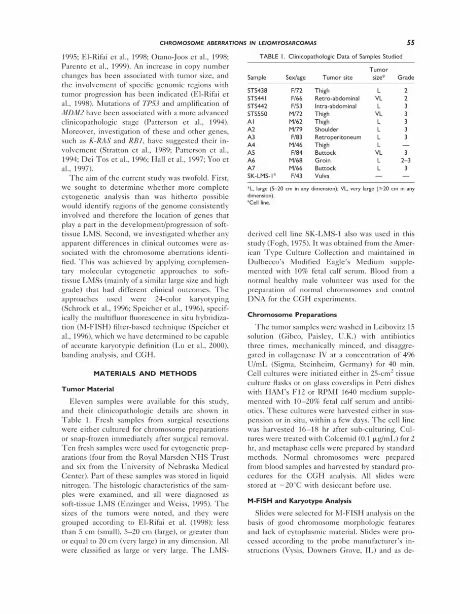
Eleven samples were available for this study,and their clinicopathologic details are shown inTable 1. Fresh samples from surgical resectionswere either cultured for chromosome preparationsor snap-frozen immediately after surgical removal.Ten fresh samples were used for cytogenetic prep-arations (four from the Royal Marsden NHS Trustand six from the University of Nebraska MedicalCenter). Part of these samples was stored in liquidnitrogen. The histologic characteristics of the sam-ples were examined, and all were diagnosed assoft-tissue LMS (Enzinger and Weiss, 1995). Thesizes of the tumors were noted, and they weregrouped according to El-Rifai et al. (1998): lessthan 5 cm (small), 5–20 cm (large), or greater thanor equal to 20 cm (very large) in any dimension. Allwere classified as large or very large. The LMS-
derived cell line SK-LMS-1 also was used in thisstudy (Fogh, 1975). It was obtained from the Amer-ican Type Culture Collection and maintained inDulbecco’s Modified Eagle’s Medium supple-mented with 10% fetal calf serum. Blood from anormal healthy male volunteer was used for thepreparation of normal chromosomes and controlDNA for the CGH experiments.
Chromosome Preparations
The tumor samples were washed in Leibovitz 15solution (Gibco, Paisley, U.K.) with antibioticsthree times, mechanically minced, and disaggre-gated in collagenase IV at a concentration of 496U/mL (Sigma, Steinheim, Germany) for 40 min.Cell cultures were initiated either in 25-cm2 tissueculture flasks or on glass coverslips in Petri disheswith HAM’s F12 or RPMI 1640 medium supple-mented with 10–20% fetal calf serum and antibi-otics. These cultures were harvested either in sus-pension or in situ, within a few days. The cell linewas harvested 16–18 hr after sub-culturing. Cul-tures were treated with Colcemid (0.1 mg/mL) for 2hr, and metaphase cells were prepared by standardmethods. Normal chromosomes were preparedfrom blood samples and harvested by standard pro-cedures for the CGH analysis. All slides werestored at 220°C with desiccant before use.
M-FISH and Karyotype Analysis
Slides were selected for M-FISH analysis on thebasis of good chromosome morphologic featuresand lack of cytoplasmic material. Slides were pro-cessed according to the probe manufacturer’s in-structions (Vysis, Downers Grove, IL) and as de-
TABLE 1. Clinicopathologic Data of Samples Studied
Sample Sex/age Tumor siteTumorsize* Grade
STS438 F/72 Thigh L 2STS441 F/66 Retro-abdominal VL 2STS442 F/53 Intra-abdominal L 3STS550 M/72 Thigh VL 3A1 M/62 Thigh L 3A2 M/79 Shoulder L 3A3 F/83 Retroperitoneum L 3A4 M/46 Thigh L —A5 F/84 Buttock VL 3A6 M/68 Groin L 2–3A7 M/66 Buttock L 3SK-LMS-1a F/43 Vulva — —
*L, large (5–20 cm in any dimension); VL, very large ($20 cm in anydimension).aCell line.
55CHROMOSOME ABERRATIONS IN LEIOMYOSARCOMAS

scribed previously (Lu et al., 2000). Slides wereaged in an oven at 65°C for 2.5 hr and then treatedwith RNase at 100 mg/mL in a humidified chamberat 37°C for 30 min. After rinsing in 23 SSC, theslides were treated with pepsin solution (3.5%) at37°C for 5 min and washed two times for 5 mineach in phosphate-buffered saline at ambient tem-perature. The slides were dehydrated and thendenatured in 70% formamide/23 SSC at 72°C for 2min and dehydrated again. Spectra Vysion (Vysis)consists of a mixture of all the chromosome paintsfor the human complement, each uniquely labeledusing specific combinations of five fluorophores.Ten microliters of the probe mixture was dena-tured at 73°C for 5 min and then applied to thetarget slide and incubated in a humidified box at37°C for 18 hr. Slides were washed in 0.43 SSC/0.3% NP240 at 72°C for 2 min and then 23 SSC/0.1% NP-40 at ambient temperature for 30 sec.After air drying, the target areas were counter-stained and mounted with 10 mL DAPI III (Vysis).Images were captured and analyzed using a ZeissAxioplan microscope with appropriate filters, digi-tal camera, and the Spectra-Vision Imaging System(Vysis). The assignment of chromosomal materialwas made on the basis of the relative intensities ofthe specific fluorochromes and examined in con-junction with the inverted DAPI-banded chromo-somes. All the aberrations were described accord-ing to the International System for ChromosomeNomenclature (1995). Selected confirmation of theM-FISH analysis was undertaken using two-colorexperiments with whole-chromosome-specificpaints, as previously described (Shipley et al.,1996). These included chromosome paints forchromosomes X and 1 (fluorescein isothiocyanatelabeled); chromososmes 7 and 22 (Cy3 labeled)hybridized to chromosome preparations from SK-LMS-1. Some preparations were karyotyped afterstandard trypsin-Giemsa banding only.
CGH Analysis
CGH analysis was done according to the protocoldescribed by Kallioniemi et al. (1993), with somemodifications and as previously described (Weber-Hall et al., 1996). Briefly, tumor DNA was ex-tracted from frozen tumor tissues and labeled bynick translation with rhodamine-11-dUTP. La-beled DNA fragments were in the size range of 600to 2,000 bp. Normal sex-matched reference DNAsamples were extracted from the blood of normalhealthy persons, labeled with fluorescein-11-dUTP, and co-hybridized with the labeled tumorDNA to normal male chromosome preparations.
For the CGH analysis, control hybridizations usingdifferentially labeled normal DNA were per-formed, and the average normalized fluorescenceintensity ratios and their standard deviations didnot exceed 0.2 greater or less than 1. Chromosomalregions were interpreted as underrepresentedwhen the average ratio was less than 0.8; they wereconsidered overrepresented when the average ratioexceeded 1.2 and amplified when the averageratio exceeded 1.5. Slides were analyzed usingQuipstCGH/karyotyper and QuipstInterpreter(Vysis), and for each tumor sample between six and10 good-quality metaphases were analyzed.
RESULTS
Representative images from the M-FISH analy-sis are shown in Figures 1 and 2. After assignmentof the chromosomal material in rearranged chromo-somes, it was possible in most cases to determinethe region involved from the inverted DAPI band-ing pattern. The exceptions were where small re-gions of a chromosome were involved and where nodistinctive banding pattern could be recognized.Chromosome painting confirmed the abnormali-ties, and representative examples are shown in Fig-ure 2D. The M-FISH analysis allowed detailedcharacterization of the aberrations in each cell, anda composite karyotype was produced, which isshown in Table 2. A summary of all the clonalbreakpoints identified is shown in Figure 3. Onesample had an apparently normal karyotype, an-other was near diploid of intermediate complexity,and the remainder were near triploid or tetraploidand showed a very high level of complexity. Theresults of the G-banding analysis of another sixcases are shown in Table 3. One of these cases hadan apparently normal karyotype, and the remainderwere abnormal. In contrast to the M-FISH analysis,it was not possible to characterize the karyotypes ofthese cases completely.
The results of the CGH analysis are summarizedin Figure 4 and Table 4. Regions of imbalancewere identified, though the average intensity ratioswere generally just greater or less than the cutofflimits (data not shown). This was attributed topotentially high ploidy status and heterogeneity.The chromosome arms most frequently involvedwere 1q, 5p, and 20q. Gain of chromosomal mate-rial was found in all cases and in the cell line(except A7), and amplification was evident in fivesamples (45%). Amplicons were identified on 5p,13q, and 20q (each one in one case) and 12q (twocases) (Fig. 4). Common overlapping regions of
56 WANG ET AL.

Figure 1. Example of multifluor fluorescence in situ hybridization karyotypes showing clarified com-posite color chromosomes and their corresponding inverted DAPI-banded images from soft-tissue leiomy-osarcoma samples: A: Case STS550. B: The cell line SK-LMS-1.
57CHROMOSOME ABERRATIONS IN LEIOMYOSARCOMAS

Figure 2. Multifluor fluorescence in situ hybridization (M-FISH)analysis of the cases indicated, showing chromosomal regions frequentlyinvolved in rearrangements of the soft-tissue leiomyosarcoma studiedand an example of confirmatory analysis. The profiles are the relativefluorescence intensities of different fluorochromes (relative to the mostintense) along the length of each rearranged chromosome. For chro-mosomal assignment, the intensities of the fluorochromes to the rightof the gray cutoff line indicate positivity for a particular fluorochrome inthe region, whereas the intensities to the left of this line were taken asnegative. Adjacent to the profiles are the corresponding composite
color images, pseudo-color images for some cases, and inverted DAPI-banded chromosomes. The corresponding normal chromosomes areshown for some cases for comparison. The rearrangements shown arethese: rearrangements involving 9q and breakpoints at the centromericregion (A), rearrangements involving chromosome 10 and breakpointsat 10q22 (B), and rearrangements involving chromosome 20 and break-points at 20q13.1 (C), M-FISH analysis of a der(17) and a der(X)chromosome in the cell line SK-LMS-1 and confirmatory analysis usingpaints for chromosomes 1 (green) and 17 (red) and X (green) and 22(red), respectively (D). (Reagents and software are from Vysis.)

gain included 1q21 (five cases), 5p14-pter (fivecases), and 20q13.1 (six cases). Loss of chromo-somal material was found in 64% of cases. There
were no obvious correlations between the numbersor types of aberrations found and the outcomes ofthe patients.
TABLE 2. Cytogenetic Findings in Four Leiomyosarcomas and One Cell Line by Multifluor Florescence In Situ Hybridization
Case no. andcell line Karoytype
STS438 46,XX [cp8]STS441 34–76,XX,del(X)(q22),1der(X;5)(p10;p10),del(1)(q32),1del(1)(q32),der(1)t(1;1)(p36;q32),del(2)(p11),del(2)
(q11),1del(2)(q11),del(2)(q21),del(3)(q11),der(3)ins(3;14)(q25;?),dup(4)(?p14p15),1dup(4)(?p14p15),1der(4)t(4;12)(q31;q15),der(4)t(4;14)(q27;q11),1der(4)t(4;14)(q27;q11),1der(5)t(X;5)(q22;q11),der(6)t(6;19)(q21;q11),del(7)(q22),der(7)t(7;14)(q11.2;q11)del(14)(q22),1der(7)t(7;14)(q11.2;q11)del(14)(q22),der(7)t(12;19)(p11.2;?)t(7;19)(p11;?)t(7;14)(q21;?)t(12;14)(?;?),1der(8)t(8;12)(q11;p12),1der(8)t(8;14)(p11;q24)del(8)(q24),der(9;12)(q10;q10),1der(9;12)(q10;q10),der(9;17)(p10;q10),der(9;17)(q10;p10),der(9)t(9;17)(q34;q11),1der(9)t(9;17)(q34;q11),der(9;18)(q10;p10),del(10)(q22),del(11)(q21),1der(11)t(11;21)(p15:q?)del(11)(q11),der(12)t(7;12)(q22;q24),der(12)t(?7;12)(?q22;q24),der(12;13)(q10;q10),der(12)t(12;19)(q24;?),der(13)t(13;19)(p11;?)t(19;19)(?;?),214,der(14)t(10;14)(q22;p11),216,del(16)(q11),der(16)t(16;17)(p13;p11),der(16;22)(q10;q10),217,der(17)t(9;17)(q22;q25),der(17)t(4;9)(?;?)t(9;18)(?;q12)t(17;18)(p11.2;q23),der(17)del(17)(p13p11)ins(17;22)(q11;q11q13),218,der(18;19)(q10;p10),der(18)t(18;19)(p11;?)t(6;18)(q15;q23),del(19)(?q11),der(19)t(12;19)(q21;q13),1der(19)t(1;12)(?;q13)t(12;12)(q24;?)t(12;19)(?;p13)ins(19;12)(q13;?)del(19)(?q13.3q13.4)trp(12)(?q14q21),der(19)t(6;19)(p2?3;?)t(6;19)(q2?3;?),1der(19)t(7;19)(?;?q11)t(7;12)(?;q13)trp(12)(q13q22),1der(19)t(7;19)(?;?q11)t(7;12)(?;?)t(12;12)(?;q13)del(12)(q22),der(19)t(12;19)(?;?p13.1)t(12;19)(q21;?q13.3),der(20)t(1;20)(?;q13.1),1der(20)t(1;20)(?;q13.1),der(20)t(3;20)(q21;q13.1),der(20)t(7;20)(q22;p13),1der(20)t(12;20)(?;q13.1),1der(20)t(12;20)(q12;q13.1),221,222,der(22)t(1;22)(?q42;?)t(1;5)(?q25;?q33),der(22)t(18;22)(q?;q13)t(12;18)(q15;q?)[cp8]
STS442 39–46,XX,t(1;14)(q21;q24),t(2;6;9;11)(p15q21q31q37;p11.2;p22;p15),t(7;20;17)(p11.2;q13.1p11.2;q23),t(10;18)(q22;q21),t(18;19)(q10;p10),min(wcp91)[cp13]
STS550 63–203,X,der(X)t(X;3)(p22;p25),1der(X)t(X;3)(p22;p25),1der(X)t(X;3)(q22;p25),1der(X;4)(q10;p10),2Y,2Y,21,del(1)(p21),del(1)(q11),del(1)(q32),del(1)(q32),der(1)t(X;1)(?;q11)t(X;16)(?;p11),der(1)t(1;10)(p32;q22),der(1)t(1;13)(q11;?)t(13;16)(?;p11),22,der(2)t(2;4)(p11;q27),der(2)t(2;15)(q23;?)t(3;15)(q21;?),der(2)t(2;15)(q23;?)t(3;15)(q21;?),der(2)t(3;8)(p21;p23)t(2;8)(p11;p11),del(3)(p11),del(3)(p11),del(3)(q11),del(3)(q11),del(3)(p12q13),der(3)t(3;12)(q21;?)t(6;12)(?;?),der(3)del(3)(q24q25)ins(3;12)(q24;?),24,del(4)(q11),del(4)(q13),del(4)(q22),del(4)(q26),del(5)(p11),1del(5)(p11),del(5)(q11),del(5)(q13),1del(5)(q13),1der(5)t(5;10)(q31;q21),1der(5;22)(q10;q10),der(5)t(5;22)(q33;q11),1der(5)t(5;22)(q33;q11),1der(5)del(9)(q21)t(4;9)(?p16;?q33)t(4;5)(?p12;p13),26,del(6)(p11),del(6)(q11),del(6)(q21),der(6)t(2;20)(p15;?)t(6;20)(p11;?),der(6)t(6;20)(p23;?)t(20;20)(?;q11),der(6)t(6;20)(p23;?)t(20;20)(?;q11)del(6)(q23),1der(6)del(6)(p11)t(6;18)(q11;q11),27,der(7;12)(q10;q10),der(7;14)(p10;q10),1der(7;14)(p10;q10),der(7;14)(p10;q10),1der(7;14)(q10;q10),28,28,del(8)(p11),der(8;10)(q10;p10),29,29,i(9)(q10),der(9)t(9;18)(q21;q?11)t(18;22)(q?23;q11),210,210,del(10)(q11),del(10)(q11),der(10)t(2;6)(p21;q27)t(6;10)(q13;p11),der(10)t(2;9)(?;q?13)ins(2;6)(?;q27q13)t(2;10)(?;p11),der(10)t(20;20)(q11;?)t(6;20)(?q27;?)t(6;18)(?q21;q22)t(10;18)(p11;q11),211,211,del(11)(q11),der(11)t(11;14)(q23,q24),der(11)del(11)(p11)t(6;11)(?q23;q14)t(2;6)(p21;?q27),del(12)(p11),del(12)(q15),der(12)t(6;12)(q21;q22),1der(6;12)t(q21;q22),der(12;14)(q10;q10),1der(12;14)(q10;q10),der(12;19)(p10;p10),1der(12;22)(p10;q10),1der(12)del(12)(p11)t(12;19)(q13;?)t(7;19)(?;?)t(7;22)(?;q11),213,213,213,213,213,214,214,214,214,215,215,215,del(15)(q22),216,der(16;19)(q10;p10),117,1del(17)(p11),del(17)(q11),1del(17)(q11),der(17)t(3;17)(q13.3;q12),218,del(18)(p11q12),del(18)(q11),del(18)(q21),der(18)t(X;18)(q27;q11),der(18)t(6;18)(?;q11)t(3;6)(q21;?),119,der(19)t(10;18)(q11.2;?)t(18;20)(?;?)t(19;20)(?p13;?),120,120,del(20)(p11),1der(20)t(1;20)(?;?q11),1der(20)t(1;20)(?;?q11),1der(20)t(1;20)(?;q13.1),1der(20)t(1;20)(?;q13.1),der(20)t(11;20)(q11;q11)t(6;11)(?;q21)t(2;6)(q21;?),221,221,221,221,der(21)t(3;21)(q13;q?22),222,der(22)t(1;3)(?;q11)t(1;22)(?;p11),der(22)t(1;3)(?;q11)t(1;22)(?;p11),dmin(wcp31),min(wcp31),min(wcp41),min(wcp101),min(wcp181)[cp10]
SK-LMS-1 56–122,2X,2X,der(X)t(X;22)(q13;q12)t(15;22)(q26;q?)t(X;15)(p11;q11),der(X)t(X;22)(q13;q12)t(15;22)(q26;q?)t(X;15)(p11;q11),21,dup(1)(p13p36),dup(1)(p13p36),22,13,24,dic(4;17)t(4;18)(p11;p11)t(4;17)(q28;q22)in(17;4)(p12q22;?),der(4)t(4;17)(q34;q25)del(17)(q11),der(4)t(4;17)(q34;q25)del(17)(q11),der(4;17)(p10;p10),der(5)t(X;5)(p11;p15),der(5)t(X;5)(p11;p15),1der(5)t(X;5)(p11;p15),16,16,17,17,18,der(8)t(3;8)(p13;p12)t(3;8)(q21;q13),der(9;12)(q10;q10),1der(9;12)(q10;q10),110,der(10;14)(q10;q10),111,111,212,213,213,der(14)t(2;14)(p23;p11),1der(14)t(2;14)(p23;p11),der(14)t(6;14)(?;p11),215,215,der(15)t(15;17)(p11;?),der(15)t(2;19)(?;?)t(2;15)(?;p11),216,del(16)(q11),der(16;17)(p10;p10),217,der(17)t(1;17)(p11;p11)t(11;17)(q13;q25),der(17)t(1;17)(p13;p11)t(3;17)(p21;q25),118,119,1del(19)(p11),120,120,120,1der(20)t(14;20)(q24;q13.1),221,222,der(22)ins(22;1)(q?11;?)[cp10]
59CHROMOSOME ABERRATIONS IN LEIOMYOSARCOMAS

DISCUSSION
The chromosomes from four tumors plus onecell line carrying a diagnosis of soft-tissue LMSwere characterized in detail using 24-color karyo-typing by applying the approach of M-FISH(Speicher et al., 1996). We previously showed ac-curate assignment of chromosomal material andcharacterization of rearrangements using the M-FISH approach (Lu et al., 2000). This assessmentwas achieved by comparing the results of M-FISHanalysis of a breast cancer cell line with reverseFISH analysis, which relies on sorting individual
chromosomes and painting them back onto normalchromosomes (Morris et al., 1997). Identification ofsmall deletions or inversions is dependent on theanalysis of chromosomal bands and, for this reason,could have been missed in the M-FISH analysis ofthe LMS. Some limited confirmation of the M-FISH analysis of the LMS cell line was carried out,and the results were concordant (Fig. 2D). There-fore, the results represented here in Table 2 andFigures 1 and 2 probably represent the most accu-rate characterization of chromosomes from soft-tis-sue LMS to date. The results contrast with the
Figure 3. Position of the breakpoints in clonal structural rearrangements identified in multifluorfluorescence in situ hybridization (smaller circles) and G-banding analyses (larger circles) of the leiomyo-sarcoma-derived samples and one cell line (total: nine).
60 WANG ET AL.

majority of karyotypes previously described(Mertens et al., 1998; Mitelman, 1998; Schneider etal., 1998) and the G-banding analysis carried outhere (Table 3), where marker chromosomes andadditional material remain uncharacterized.
The karyotypes determined using M-FISH anal-ysis showed varying levels of complexity, with ex-tensive numerical and structural aberrations. Con-siderable intratumor heterogeneity was evident inthe karyotypes by the varying number of timesparticular clonal aberrations were identified (Table2). No common consistent translocations werefound, and there were no rearrangements in com-mon with published data (Sreekantaiah et al., 1993;Mertens et al., 1998; Mitelman, 1998). One possi-ble exception is the finding of balanced transloca-tions involving chromosomes 10 and 18 in two neardiploid karyotypes described as t(10;18)(q22;q21)(case STS442) and t(10;18)(q22;q13) in a gastricLMS (Sreekantaiah et al., 1993). Collating the po-sition of all the breakpoints defined in clonal rear-rangements, however, shows that some regions ofthe genome are preferentially involved (Fig. 3).They include 1q21, 2q21, 3q21, 9q, 10q22, 11p15,11q21, 14q24, and 20q13.1, often in unbalancedtranslocations. Their significance is unclear, and itremains to be determined whether specific break-points or resultant chromosomal imbalances areimportant.
Two of the karyotypes appeared to be normal,but CGH analysis from the same cases showedimbalances, suggesting that the chromosomescould be derived from dividing normal cells. Inaddition, comparing the CGH results with thekaryotypes of individual cases showed that the di-viding cell population in vitro may not be repre-sentative of the tumor mass. The comparison of the
CGH and M-FISH analysis was closest for the cellline, but even in this case heterogeneity was ap-parent. The average ratios of fluorescence intensi-ties were generally just outside the thresholds.This finding can be explained by heterogeneityand also single copy number changes against abackground of high and variable ploidy. In addi-tion, the unrepresentative nature of the chromo-some preparation has been documented in previ-ous cytogenetic and fluorescence-activated cell-sorting analyses of LMS samples (Orndal et al.,1994).
The overlapping region of loss on 13 likelyencompasses 13q14 and the RB1 gene locus,which has been implicated in a number of can-cers, including sarcomas and LMS (Stratton etal., 1989). Furthermore, loss of critical genes on10q may include the PTEN and MX1 genes lo-cated at 10q23 and 10q24-q25, respectively(Wechsler et al., 1996; Li et al., 1997). It ispossible that the overlapping regions of loss at1q32, 2q32, 4q32-q33, 10q21, 11q22-q23, and13q14-q21 identified in this study are associatedwith tumor suppressor genes that may play a rolein tumor progression.
Five of the 11 samples (45%) studied by CGHshowed evidence of genomic amplification of vari-ous regions. In addition to amplification, gain ofparticular chromosome material was noted, whichfor some regions overlapped with the amplicons.Gain of 1q material, and particularly the 1q21 re-gion, was a common finding here; this material alsois gained and amplified in a number of sarcomasand other tumor types (Forus et al., 1995; Mitel-man, 1998). The only region amplified more thanonce was 12q13-15, which is involved in a numberof sarcoma types. This region encompasses a num-
TABLE 3. Cytogenetic Findings in Six Leiomyosarcomas
Caseno. Karyotype
A1 83–88,XXX,add(X)(p22.3),del(1)(q12)x2,del(1)(q25q41),23,24,25,25,add(5)(p14),add(7)(p22),add(9)(q34),210,210,211,del(12)(p11.1)x2,add(12)(p13),add(13)(q34),216,216,217,217,219,219,120,120,1mar1,1mar2x2,1mar3,1mar4[cp6]/154-176,idemx2[9]
A2 58–63,XX,2Y,der(1)t(1;1)(q21;p12),22,25,28,28,29,210,210,211,212,add(12)(p13)x2,213,der(13)t(10;13)(q11.2;p11),add(15)(p11.2),216,217,218,219,der(19)t(2;19)(q11.2;q13.1),add(20)(q13.3),221,i(21)(q10),222,1mar1,1mar2,1mar3,1mar4,1mar5,1mar6[cp6]/45,X,2Y[5]/46,XY[9]
A3 46,XX[20]A4 46,XY,1der(1;9)(q10;p10),t(5;10)(p14;q21),29,t(15;22)(q24;q13),add(18)(p11),add(19)(q13),13-42dmin[19]/92,idemx2[14]A6 68–77,XYY,add(1)(p13),1add(1)(p22),del(1)(q11),del(3)(p11),24,25,del(7)(q22)x2,del(11)(q21)x2,add(12)(q24.3),
add(13)(p11),add(14)(q32),add(19)(q13.4),add(20)(q13.3),1mar1,1mar2,1mar3,1mar4,1mar5,115-28mar[cp10]A7 49,XY,dic(3;13)(q21;p11),add(7)(p11),add(11)(p15),der(7;12)(p10;p10),der(14)t(1;14)(q21;p13),217,add(19)(p13),1mar1,
1mar2,1mars3,1mar4[cp18]/46,XY[2]
61CHROMOSOME ABERRATIONS IN LEIOMYOSARCOMAS

ber of critical genes, such as GLI1, SAS, GAD153,MDM2, and CDK4, with variable and complex pat-terns of involvement (Roberts et al., 1989; Forus et
al., 1993). Amplification at 13q31-34 was found inone case, and 13q31 is a region that we have iden-tified as amplified and overrepresented in about
Figure 4. Summary of chromosomal imbalances identified by comparative genomic hybridization analysisof the 10 samples of soft-tissue leiomyosarcoma plus the cell line SK-LMS-1. Lines to the left of chromo-somes represent losses, and lines to the right of chromosomes represent gains. Thicker lines to the rightof chromosomes represent regions of genomic amplification. The numbers above each line indicate thecase/cell line in which the change was identified.
62 WANG ET AL.

30% of alveolar rhabdomyosarcomas (Gordon et al.,2000).
Amplification at this region has been describedas the most common amplification (four cases froma series of 58) in malignant fibrous histiocytoma ofsoft tissue. It also was found in a single case of 26malignant fibrous histiocytomas of bone (Larra-mendy et al., 1997; Knuutila et al., 1998). Atpresent, there is no indication of the gene or genesthat might play a part. Amplification and increasedcopy number were indicated at 5p14-pter and20q12-qter. These regions are likely locations forproto-oncogenes, which influence the behavior ofLMS. A previous study has shown a correlationbetween the size of tumors and the number andtype of chromosomal imbalances identified byCGH in LMS (El-Rifai et al., 1998). All of thetumors investigated here were classified as large orvery large; therefore, such comparisons cannot bemade. No obvious correlations were identified be-tween the number and type of chromosome aber-rations and the clinical outcome.
ACKNOWLEDGMENTS
The authors thank the Cancer Research Cam-paign for its support and Omar Al Muderis forcollection of clinical data. They also thank Profes-sor Hongti Jia for his helpful comments and sup-port.
REFERENCES
Dei Tos A, Maestro R, Doglioni C, Piccinin S, Libera D, BoiocchiM, Fletcher C. 1996. Tumor supressor genes and related mole-cules in leiomyosarcoma. Am J Pathol 148:1037–1045.
El-Rifai W, Sarlomo-Rikala M, Knuutila S, Miettinen M. 1998.DNA copy number changes in development and progression inleiomyosarcomas of soft tissues. Am J Pathol 153:985–990.
Enzinger FM, Weiss SW. 1995. Soft tissue tumors, third edition. St.Louis: Mosby Year-Book Inc. p 491–508.
Fletcher C. 1992. Pleomorphic malignant fibrous histiocytoma: factor fiction? A critical reappraisal based on 159 tumors diagnosed aspleomorphic sarcoma. Am J Surg Pathol 16:213–228.
Fletcher C, Dal Cin P, de Wever I, Mandahl N, Mertens F, Miltel-man F, Rosai J, Rydholm A, Sciot R, Tallini G, van den Berghe H,Vanni R, Willen H. 1999. Correlation between clinicopathologicalfeatures and karyotype in spindle cell sarcomas: a report of 130cases from the CHAMP study group. Am J Pathol 154:1841–1847.
Fogh J. 1975. Human tumor cells in vitro. New York: Plenum Press.p.115–159.
Forus A, Florenes V, Maelandsmo G, Meltzer P, Fodstad O, Myke-bost O. 1993. Mapping of amplification units in the q13-q14region of chromosome 12 in human sarcomas: some amplica donot include MDM2. Cell Growth Differ 4:1065–1070.
Forus A, Weghuis D, Smeets D, Fodstad O, Myklebost O, vanKessel A. 1995. Comparative genomic hybridization analysis ofhuman sarcomas. I. Occurrence of genomic imbalances and iden-tification of a novel major amplicon at 1q21-q22 in soft tissuesarcomas. Genes Chromosomes Cancer 14:8–14.
Gordon A, Brinkschmidt C, Anderson J, Coleman N, Dockhorn-Dworniczak B, Pritchard-Jones K, Shipley J. 2000. A novel andconsistent amplicon at 13q31 associated with alveolar rhabdomyo-sarcoma. Genes Chromosomes Cancer 28:220–226.
Hall K, Teneriello M, Taylor R, Lemon S, Ebina M, Linnoila R,Norris J, Park R, Birrer M. 1997. Analysis of Ki-ras, p53 andMDM2 genes in uterine leiomyomas and leiomyosarcomas. Gy-necol Oncol 65:330–335.
Kallioniemi O, Kallioniemi A, Sudar D, Rutovitz D, Gray J, Wald-man F, Pinkel D. 1993. Comparative genomic hybridization: arapid new method for detecting and mapping DNA amplificationin tumors. Semin Cancer Biol 4:41–46.
Knuutila S, Bjorkqvist A-M, Autio K, Tarkkanen M, Wolf M, MonniO, Szymanska J, Larramendy M, Tapper J, Pere H, El-Rifai W,Hemmer S, Wasenius V-M, Vidgren V, Zhu Y. 1998. DNA copynumber amplifications in human neoplasms. Am J Pathol 152:1107–1123.
Larramendy M, Tarkkanen M, Blomqvist C, Virolainen M, WiklundT, Asko-Seljavaara S, Elomaa I, Knuutila S. 1997. Comparativegenomic hybridization of malignant fibrous histiocytoma reveals anovel prognostic marker. Am J Pathol 151:1153–1161.
Lee E, Locker J, Nalesnik M, Reyes J, Jaffe R, Alashari M, Nour B,Tzakis A, Dickman P. 1995. The association of Epstein-Barr viruswith smooth-muscle tumors occurring after organ transplantation.N Engl J Med 332:19–25.
TABLE 4. Comparative Genomic Hybridization Results With Clinical Follow-up Data
Sample
Chromosome armsinvolved in gains and
amplification (bold)Chromosome armsinvolved in losses
No. ofaberrations
Monthsto met
Monthsto last
follow up Status*
STS438 1,4q,5p,6p,7q,8q,10q,17,20q 6q,8p,9p,10p,11q,12p,18q,20p,Xq
18 32.9 33.6 AWD
STS441 4p,7p,12q,17q,20p 18p 6 24.7 36.7 ANEDSTS442 1q,13q,18q 1q,2q,6,8q,10q,12q,
20p,Xp11 28.7 63.5 DOD
STS550 4,5,20 2q,4q,9p,11q,13q,21q 9 5.2 11.2 AWDA1 1q,4p,5p,6p,7p,9q,13q,14q,
17q,19q,20q,22q1q,2q,4q,5q,7q,10,
12p,13q20 — 18 ANED
A2 1p,3q,5p,6,7,8,9q,10p,14q 5q,10q,12 12 0 43 ANEDA3 5p,8p 2 24 84 —A4 1,12q,15q,20q 4 68 99 DODA5 1,5p,6p,7p,17p,20q 6 1 3 DODA6 3p,7q,8,9,17p,20q,22q,Xp 4q,11q 10 — 28 ANEDA7 None None 0 4 6 DODSK-LMS-1a 6p,11q,14q,17q,19q,20p 1q,4q,13,X 10 — — —
*ANED, alive with no evidence of disease; DOD, dead of disease; AWD, alive with disease.aCell line.
63CHROMOSOME ABERRATIONS IN LEIOMYOSARCOMAS
Li J, Liaw D, Podsypanina K, Bose S, Wang S, Puc J, Miliaresis C,Rodgers L, McCombie R, Bigner S, Gioanella B, Ittmann M,Tycko B, Hibshoosh H, Wigler M, Parsons R. 1997. PTEN, aputative protein tyrosine kinase phosphatase gene mutated inhuman brain, breast and prostate cancer. Science 275:1943–1947.
Lu Y-J, Morris JS, Edwards PA, Shipley J. 2000. Evaluation of 24color multifluor-fluorescence in situ hybridization (M-FISH)karyotyping by comparison with reverse chromosome painting ofthe human breast cancer cell line T-47D. Chromosome Res8:127–132.
McClain K, Leach C, Jenson H, Joshi V, Pollock B, Parmley R,DiCarlo F, Chadwick E, Murphy S. 1995. Association of Epstein-Barr virus with leiomyosarcomas in children with AIDS. N EnglJ Med 332:12–18.
Mertens F, Fletcher C, Dal Cin P, De Wever I, Mandahl N,Mitelman F, Rosai J, Rydholm A, Sciot R, Tallini G, Van denBerghe H, Vanni R, Willen H. 1998. Cytogenetic analysis of 46pleomorphic soft tissue sarcomas and correlation with morpho-logic and clinical features: a report of the CHAMP study group.Chromosomes and morphology. Genes Chromosomes Cancer 22:16–25.
Mitelman database of chromosome aberrations in cancer. www.cgap.hci.nih.gov/Chromosomes/Mitelman.
Mitelman F. 1995. An international system for human cytogeneticnomenclature. Cytogenet Cell Genet.
Mitelman F. 1998. Catalog of chromosome aberrations in cancer ’98,version 1. New York: Wiley-Liss.
Morris J, Carter N, Ferguson-Smith M, Edwards P. 1997. Cytoge-netic analysis of three breast carcinoma cell lines using reversechromosome painting. Genes Chromosomes Cancer 20:120–139.
Orndal C, Rydholm A, Willen H, Mitelman F, Mandahl N. 1994.Cytogenetic intratumor heterogeneity in soft tissue tumors. CanerGenet Cytogenet 78:127–137.
Otano-Joos M, Mechtersheimer G, Ohl S, Lehnert T, Willeke F,Moller P, Otto H, Lichter P, Joos S. 1998. Analysis of chromosomecopy number changes in leiomyosarcoma through molecular cy-togenetic methods. Verh Dtsch Ges Pathol 82:207–209.
Parente F, Grosgeorge J, Coindre J-M, Terrier P, Vilain O, Turc-Carel C. 1999. Comparative genomic hybridization reveals novelchromosome deletions in 90 primary soft tissue tumors. CancerGenet Cytogenet 115:89–95.
Patterson H, Gill S, Fisher C, Law MG, Jayatilake H, Fletcher CD,Thomas M, Grimer R, Gusterson BA, Cooper CS.1994. Abnor-malities of the p53 MDM2 and DCC genes in human leiomyo-sarcomas. Br J Cancer 69:1052–1058.
Roberts M, Douglass E, Peiper S, Houghton P, Look A. 1989.Amplification of the gli gene in childhood sarcomas. Cancer Res49:5407–5413.
Schneider B, Lovell M, Golder W. 1998. Cytogenetic abnormalitiesin primary bronchopulmonary leiomyosarcoma of childhood. Can-cer Genet Cytogenet 105:145–151.
Schrock E, du Manoir S, Veldman T, Schoell B, Wienberg J, Fer-guson-Smith M, Ning Y, Ledbetter D, Bar-Am I, Soenksen D,Garini Y, Ried T. 1996. Multicolor spectral karotyping of humanchromosomes. Science 273:494–497.
Shipley J, Crew J, Birdsall S, Gill S, Clark J, Fisher C, Kelsey A,Nojima T, Sonobe H, Cooper C, Gusterson B. 1996. Interphasefluorescence in situ hybridization and reverse transcription poly-merase chain reaction as a diagnostic aid for synovial sarcoma.Am J Pathol 148:559–567.
Somers G, Tesoriero A, Hartland E, Robertson C, Robinson P,Venter D, Chow C. 1998. Multiple leiomyosarcomas of both donorand recipient origin arising in a heart-lung transplant patient. Am JSurg Pathol 22:1423–1428.
Speicher M, Ballard S, Ward D. 1996. Karyotyping human chromo-somes by combinatorial multi-fluor FISH. Nat Genet 12:368–375.
Sreekantaiah C, Davis JR, Sandberg AA. 1993. Chromosomal abnor-malities in leiomyosarcoma. Am J Pathol 142:293–305.
Stratton M, Williams S, Fisher C, Ball A, Westbury G, Gusterson B,Fletcher C, Knight J, Fung Y, Reeves B. 1989. Structural alter-ations of the RB1 gene in human soft tissue tumours. Br J Cancer60:202–205.
Weber-Hall S, Anderson J, McManus A, Abe S, Nojima T, Pinker-ton R, Pritchard-Jones K, Shipley J. 1996. Gains, losses, andamplification of genomic material in rhabdomyosarcoma analyzedby comparative genomic hybridization. Cancer Res 56:3220–3224.
Wechsler D, Shelly C, Dang C. 1996. Genomic organization ofhuman MX1, a putative tumor supressor gene. Genomics 32:466–470.
Yoo J, Lee H, Kang C, Park W, Lee Y, Shim S. 1997. p53 genemutations and p53 protein expression in human soft tissue sarco-mas. Arch Pathol Lab Med 121:395–399.
64 WANG ET AL.


