Embed Size (px)
Citation preview

Characterization of inositol lipids from Leishmania donovani promastigotes: identification of an inositol sphingophospholipid
Edna S. Kaneshiro,* Koka Jayasimhulu,** and Robert L. Lester?
Department of Biological Sciences, University of Cincinnati,* Cincinnati, OH 4522 1-0006, Department of Environmental Health, University of Cincinnati Medical College,** Cincinnati, OH 45267, and Department of Biochemistry, University of Kentucky,? Lexington, KY 40506
Abstract Inositol lipids account for 15% of the total cellular phospholipids of Leishmania donovani promastigotes. Four major inositol lipids were identified and characterized: phosphatidyl- inositol (PI), phosphatidylinositol phosphate (PI-P), phosphati- dylinositol diphosphate (PI-Ps), and an inositol sphingophospho- lipid (InSL). Diacyl and alkyl acyl PI were identified. The major esterified fatty acids of PI, PI-P, and PI-P2 were similar and unlike those of mammalian inositol glycerolipids. Leishmania inositol glycerolipids contained only trace amounts of arachidonic acid; the major species were c16 and C18 acids. The InSL com- prised about 40% of the inositol lipids. The amide-linked fatty acids of InSL were mainly c16 and C18 acids. Differential hy- drolysis and nuclear magnetic resonance spectrometry indicated that the InSL had a phosphoryl bond. The major long chain bases of the InSL were identified by gas-liquid chromatography and high resolution mass spectrometry as straight chain C16 and C18 sphing0sines.l The finding of InSL in Leishmania is of in- terest because InSL have previously been found only in plants and fungi. Metabolic radiolabeling experiments suggest that this lipid may be a precursor of an antigenic cell surface membrane lipophosphoglycan which is shed into the culture medium by the organism.-Kaneshiro, E. S., K. Jayasimhulu, and R. L. Lester. Characterization of inositol lipids from Leishmania donovani pro- mastigotes: identification of an inositol sphingophospholipid. J. Lipid Res. 1986. 27: 1294-1303.
Supplementary key words fatty acids glucose long chain bases membrane lipophosphoglycan polyphosphoinositides
Leishmania donovani, the causative protozoan agent of the human disease kala azar, or visceral leishmaniasis, can be grown axenically as the flagellated promastigote form. T h e major lipid classes and fatty acids of several Leishmania species grown in mass cultures have been characterized (1-3). Detailed analyses of the inositol lipids, however, have not yet been described. T h e present study was con- ducted to examine these lipids in promastigote cells of L. donovani grown in a chemically defined medium (4, 5). Inositol glycerolipids and an inositol sphingophospholipid (InSL) were identified and characterized.
Previous work on a membrane antigen that is released
into the culture medium by L. donovani was described and was shown to exist as an integral component of the cell’s surface membrane (4). This cell surface component ex- hibits immunochemical identity with excreted factor (4) and appears to have chemical properties similar to the L. major glycoconjugate receptor for macrophages described by Handman and Golding (6). In this report the results of metabolic radiolabeling experiments are described that suggest that this antigenic substance is a lipophospho- glycan that contains an InSL domain.
MATERIALS AND METHODS
Culture Promastigotes of Leishmania donovani, MHOM/SD/
62/1S (obtained from Dennis M. Dwyer, National Insti- tutes of Health, Bethesda, MD) were grown in a modified (4) defined medium of Steiger and Steiger ( 5 ) lacking bo- vine serum albumin (RE I11 - BSA). Cultures were in- oculated with 1 / 10 vol of day 4 cultures and were grown to late log or early stationary phase (4 days) at 25-26°C. In some experiments cells were radiolabeled by the ad- dition of 3-67 nCi of [U-’4C]myo-inositol per ml, 67 nCi of [U-’4C]D-glucose per ml, 33 nCi of [ l-’4C]deoxyglucose per ml, 50 nCi of [l-’4C]stearic acid per ml, or 0.1-1.0 PCi of [32P]HsP04 (New England Nuclear, Boston, MA,
Abbreviations: BSTFA, N,O-bis(trimethylsily1)trifluoroacetamide; DS, DS-55 data system; EDTA, ethylenediaminetetraacetate; FAB, fast atom bombardment; FAME, fatty acid methyl ester; GLC, gas-liquid chromatography; IEP, immunoelectrophoresis; IgG, immunoglobulin G; InSL, inositol sphingophospholipid; LCB, long chain base; LPG, lipophosphonoglycan; LPPG, lipopeptidophosphoglycan; MeOH, methanol; MS, mass spectrometry; NMR, nuclear magnetic resonance; PCSP, promastigote cell surface preparation; PI, phosphatidylinositol; PI-P, phosphatidylinositol phosphate; PI-Ps , phosphatidylinositol di- phosphate; TEAE, triethylaminoethyl; TLC, thin-layer chromatog raphy.
1294 Journal of Lipid Research Volume 27, 1986
by guest, on Novem
ber 3, 2015w
ww
.jlr.orgD
ownloaded from

or Amersham Corp., Arlington Heights, IL) to the culture medium. Cells were concentrated by centrifugation at 5,000 g for 30 min at 4°C. Lipids were extracted from cell pellets either after centrifugation, after freezing at -7OoC, or after lyophilization.
Extraction and fractionation of lipids Total lipids were extracted with CHCI,-methanol
(Me0H)-conc. HCl 100:200: 1 (v/v/v) acidic Bligh and Dyer (7), then purified by partitioning according to Folch, Lees, and Sloane Stanley (8). Alternatively, lyophilized promastigotes were extracted with CHCI3-MeOH 2: 1 (v/ v). Lipids were fractionated by triethylaminoethyl (TEAE) cellulose column chromatography (9) and then the last two fractions were pooled and the salts were removed (8). This was designated the inositol lipid fraction. That all inositol lipids were in these two TEAE column fractions was verified by autoradiography of one-dimensional (1 - D) thin-layer chromatograms (TLC) of each fraction off TEAE columns that were used to separate lipids from cells grown with ‘4Cinositol (see below).
Inositol lipid classes Total lipids or the inositol lipid fraction off TEAE col-
umns were analyzed by one- or two-dimensional (2-D) chromatography employing silica gel-impregnated paper (Whatman SG 8 1) pretreated with ethylenediaminetet- raacetate (EDTA) according to Steiner and Lester (1 0). Radiolabeled lipids separated by this procedure were sub- jected to autoradiography (NS-2T X-ray film, Kodak, Rochester, NY). The individual inositol lipids corre- sponding to spots on autoradiograms were cut out of the paper and the material was eluted with CHCl,-MeOH 2: 1 (v/v). Lipids were also analyzed by 1- or 2-D TLC using silica gel H or G and the solvent systems of Turner and Rouser (9). The TLC plates were exposed to 12 vapor, or sprayed with “Phospray” (Supelco, Inc., Bellefonte, PA) for detection of P, and a-naphthol, diphenylamine (Su- pelco), or anthrone (1 1) for sugars. Purified phosphati- dylinositol (PI) and the inositol sphingophospholipid (InSL) were isolated from the inositol lipid fraction off TEAE columns by preparative 1-D TLC on silica gel H or G with CHCls-MeOH-28-30% NH40H-H20 25:20: 1.6:3.9 (v/v/v/v). Lipids were visualized by water or l2 vapor, the bands were scraped off the plates with a razor blade, and the material was eluted from the silica gel with CHC1,-MeOH 1:2 (v/v).
Lipids labeled with [ ‘‘C]inositol were subjected to mild alkali hydrolysis according to the procedures of Smith and Lester (1 2); they were then analyzed by 2-D SG 8 1 chromatography (1 0) and autoradiography. These auto- radiograms were compared with those of unhydrolyzed material.
Diacyl and alkyl acyl species in the PI fraction obtained by neutral CHCI,-MeOH extraction were analyzed after phospholipase C digestion (1 3) followed by acetylation (1 4) and separation by TLC on silica gel H plates (1 5). The BaciElus cereus enzyme (Sigma Chemical Go., St. Louis, MO) was used by procedures previously described (1 3). Complete digestion of PI by this enzyme was monitored by using Leishmania PI labeled with [ “Clinositol. Within 30 min almost all of the radioactivity was rendered water- soluble (Fig. 1). The relative amounts of diglyceride ac- etates were quantified by densitometry of plates after staining with phosphomolybdate (1 6). To obtain esterified fatty acids, diglyceride acetates were eluted from the silica gel that was scraped off unstained plates. Alkenyl acyl glycerides from plasmalogens were not detected by these methods, and aldehydes that would have been released from plasmalogens were similarly not detected by gas- liquid chromatography (GLC) in material eluted from re- gions of TLC plates where aldehydes were expected to be present (1 5).
Fatty acids Phosphatidylinositol phosphate (PI-P) and phosphati-
dylinositol diphosphate (PI-P2) were isolated by 1-D sep- arations employing silica gel-impregnated paper (CHCIS- MeOH-4 N NH40H 9:7:2, v/v/v)(9). The bands con- taining these lipids were cut and subjected directly to al- kaline hydrolysis by the micromethod of MacGee and Al- len (1 7). The fatty acid methyl esters (FAME) produced
I . . . . . . . 0 1 2 3
T I M E ( H I
Fig. 1. Leishmania donmani PI metabolically prelabeled with [U- “C]inositol was subjected to Bacillus cereus phospholipase C digestion. Samples were removed at various times and radioactivity in the organic (X) and the aqueous (0) phases was determined. Hydrolysis was com- plete within 30 min as indicated by the appearance of radioactivity in the water-soluble polar head group product.
Kaneshiro, Jayasimhulu, and Lester Leishmania inositol lipids 1295
by guest, on Novem
ber 3, 2015w
ww
.jlr.orgD
ownloaded from

were quantified by GLC on a 10% EGSS-X column as previously described (1 8).
Amide-linked fatty acids, long chain bases, and water- soluble products from the InSL were released by acid hydrolysis using conc. HCI-MeOH 1:5 (v/v) in sealed ampoules for 48 hr or in MeOH-H,O-HCl 11:2.6:1 (v/ v/v) overnight at 80°C as described by Carter and Gaver (19). Sphingolipid fatty acids were quantified by GLC as above. Alternatively, fatty acid methyl esters (FAME) were identified and quantified by capillary column GLC-mass spectrometry analyses. A Carlo Erba series 4160 Frac- tovap gas chromatograph fitted with a fused silica column (60 m X 0.32 mm id.) coated with SE-54 (SPB-5, Supelco) and a cooled on-column injection system was used to sep- arate the FAME. Material from the GLC system was in- troduced into a high resolution mass spectrometer (MS) system (Kratos, MS-80, Manchester, England) through a jet separator. Samples were injected into the gas chro- matograph at 60°C. Initial oven temperature was main- tained for 2 min at 150°C to permit venting of the solvent, after which the valve to the mass spectrometer was opened. The oven temperature was increased by 6"C/ min to 280°C. Analyses were started 1 min after the opening of the valve allowing material from the gas chro- matograph to enter the MS. Electron impact mass spectra data were continuously collected and processed on a Data General NOVA/4C computer with a DS-55 data sys- tem (DS).
Long chain base The long chain bases (LCB) released by acid hydrolysis
of the InSL were converted to their N-trifluoroacetyl de- rivatives by the addition of trifluoroacetylmethyl ester and adjustment of the pH to 8.4 with triethylamine (20). The
reaction was allowed to proceed overnight at room tem- perature. The solvent was then evaporated under a stream of N2 and the samples were then placed under a vacuum for 2-3 min to ensure complete removal of triethylamine. N,O-bis(trimethylsily1)trifluoroacetamide (BSTFA) was then added to the N-trifluoroacetyl LCB and the mixture was heated for 30 min at 60°C. Samples were dried under a stream of N2, then dissolved in chloroform before anal- yses by GLC-MS-DS.
Samples were injected into the GLC at 60°C. After 1 min the oven temperature was raised to 220°C, then pro- grammed to increase by 2"C/min to 260°C. The GLC- MS data on the LCB derivatives were continuously col- lected 3 min after sample injection and 1 min after open- ing of the gas chromatograph to the MS valve.
Fast atom bombardment MS (FABMS) analyses were done on free ceramides released from the InSL by phos- pholipase C digestion. Sorbitol was applied to the tip of the FAB probe as described previously (2 1, 22). One to two MI of the solution of ceramides dissolved in chloroform was mixed in the sorbitol matrix. The FAB probe was introduced through the direct inlet system. The FAB gun and power supply were from Phrasor (Phrasor Scientific Inc., Duarte, CA).
Polar head groups
Polar head groups were isolated by subjecting individ- ually isolated inositol lipids, prelabeled with ['4C]glucose, [32P]H3P04, or [ ''C]inositol, to phospholipase C digestion (see above). Water-soluble head groups were subjected to 1-D TLC on either cellulose plates (Eastman 13255, East- man Chemicals, Rochester, NY) or SG 8 1 paper (see above) using the solvent system phenol saturated with water- ethanol-acetic acid 50:5:6 (v/v/v) (1 1). The plates were
TABLE 1 . Inositol phospholipid fatty acids of Leishmania donmani promastigotes
Fatty acid PI Diacyl PI Alkyl Acyl PI PI-P PI-Pz lnSL
14:O 15:O 16:O 16:l 17:O 18:O 18:l 18:2 20:2 Othersn No. preparations
4.0 f 0.5
5.3 f 1.6 0.5 f 0.3
trace 31.8 f 0.9 45 .8k 1 . 1
6.8 f 0.5 1.5 f 0.3 6.3 f 2.4 4
trace 0.8f 0.3 0 . 3 f 0.1 7 . 7 f 5.2 0 . 9 f 0.6 0 . 2 f 0.1
17.1 f 12.9 44 .1k 5.1
8 . 6 f 3.5 2 . 6 f 0.5
17 .8 f 4.2 36
weight 7c rf. SEM
0.7 6.3 f 1.4 0.3 3.1 f 1.3 6.4 12.0 f 2.5 0.8 1 . 9 f 0 . 2 0.4 1 . 1 f 0 . 4
41.8 25.1 f 1.9 35.3 29.7 f 3.6
3.7 6 . 0 f 1.3 1.5 2.1 f 0.4 8.9 12.6 f 2.3 1 C 9
6.3 f 0.9 1 . 7 f 0 . 3
25.0 f 5.1 6.7 f 2.6 1.7 f 0.7
23.0 2 3.2 24.0 f 5.1
3.0 f 1.0 1.3 f 0.3 7.3 f 0.3 3
0.0 f 0.0 0.0 f 0.0 6.1 f 1.4 0.0 f 0.0 0.0 f 0.0
86.5 f 4.8 7.0 f 3.6 0.0 f 0.0 0.0 f 0.0 0.0 f 0.0 4
Others are the sums of FAME present in <0.5%. One preparation was a pooled sample. A single determination was done on a pooled sample.
1296 Journal of Lipid Research Volume 27, 1986
by guest, on Novem
ber 3, 2015w
ww
.jlr.orgD
ownloaded from

then exposed to X-ray film and the migrations of the ra- Cell surface membrane and shed antigens diolabeled head groups, as visualized on autoradiograms, Cell-free culture media in which Leishmania had been were compared. grown in the presence of a variety of radiolabeled pre-
cursors (see above) were used as a source of "shed" an- Phosphorus tigens (4). Culture media were concentrated and analyzed
Phosphoryl and phosphonyl linkages in inositol lipids were determined by I) the differential hydrolysis proce- dures of Ferguson et al. (23) or 2) "P nuclear magnetic resonance (NMR). "P NMR spectra were recorded at 121.5 MHz on a Bruker WM 300 NMR spectrometer (Bruker Instruments, Inc., Billerica, MA) using phos- phoric acid as the reference standard.
Fig. 2. Autoradiograms of twdirnmsional chromatographic analyses of total lipids from Lrichmnnio daovani metabolically prelabeled with [U-"C]inositol before (A) and after (B) alkaline hydrolysis. After hy- drolysis, only one detectable radioactive component. InSL, remained. Chromatography was done on silica gel-impregnated paper pretreated with EDTA. The solvent system used for separations in the first di- mension was CHCI,-MeOH-28-30% NH,OH-H,O 25:201.6:3.9 (v/v/v/v). Separations in the second dimension employed CHCIS- MeOH-glacial acetic acid-H,O 60:24:16:6.4 (v/v/v/v).
by immunoelectrophoresis (IEP) as previously described (4). The immunoglobulin G ( I g G ) fraction from rabbit polyvalent antisera used in these analyses was directed against promastigote cell surface preparations (PCSP) (4). Antigens were extracted from radiolabeled promastigotes by first extracting lipids with CHCI,-MeOH 2:l (v/v) and then with acetone. The antigens were then extracted by the "water saturated with butanol" method described for the isolation of cell surface membrane lipophosphon- oglycans from Acanthamoeba by Korn, Dearborn, and Wright (24). Immunoelectrophoresis gels were stained with Coomassie brilliant blue (4) and subjected to auto- radiography.
RESULTS
Inositol lipid classes Inositol lipids from promastigotes were quantified by
"P incorporated into phospholipids under isotope equil- ibration conditions by growth of the cells for 4 days with the radioisotope. Radioactivity in these lipids relative to that in total phospholipids was compared. The radioac- tivity in PI-P was divided by two and the radioactivity in P1-P' was divided by three. Total inositol lipids comprised 15% of the cellular phospholipids.
Cells grown with [14C]inositol retained 4.3% of the in- corporated radiolabel after the lipids were extracted. When the extracted lipids were purified by solvent par- titioning, 89.4% of the radiolabel incorporated into cells was found in the lower organic phase and 6.2% in the upper phase. Of the inositol lipids, five classes were iden- tified and quantified by radioactivity incorporated from ['4C]inositol into Leishmania lipids, which were subse- quently purified by TEAE column chromatography and TLC. The inositol lipid composition of L. dmouani, 1s. was PI, 45.1%; InSL, 37.9%; PI-P, 13.0%; PI-Pp, 2.2%; and lyso PI, 2.6% (n = 3).
Phosphatidylinositol contained both diacyl (90%) and alkyl acyl (1 0%) molecular species. There was no evidence for the presence of plasmalogens (alkenyl glycerols) in PI asjudged by the TLC or GLC methods used in this study.
Inositol lipid head groups Water-soluble polar head groups were released by
phospholipase C digestion of I ) total cellular lipids and
Kaneshiro, Jayan'mhulu, and Lester Leishmania inositol lipids 1497
by guest, on Novem
ber 3, 2015w
ww
.jlr.orgD
ownloaded from

isolated InSL and PI metabolically labeled with [14C]inositol, and 2) isolated InSL and PI metabolically labeled with [32P]H3P04. Analyses by cellulose TLC and autoradiography showed that all head groups had Rf val- ues of 0.23-0.27. Only one radioactive band was detected in the sample of [14C]inositol-labeled total lipids. Thus all inositol lipids had similar polar head groups.
When the defined medium for Leishmania was for- mulated (5), it was not determined whether or not inositol was an absolute growth requirement of the organism. In the earlier nutritional studies, glucose was found not to be an absolute requirement (5 ) . In the present studies, inositol was not shown to be required for growth of Leish- mania donovani, lS, cultures. After six subcultures in RE 111-BSA that lacked inositol, there was no decrease in the density of cultures. Unexpectedly, glucose was found to be an absolute requirement for growth of this strain of L. donovani. By two or three subcultures, after 5 days, all cells were dead in the absence of added glucose, or in the absence of both glucose and inositol. Addition of 1OX or lOOX inositol, in the absence of added glucose, did not prevent cultures from dying by day 5 of the third sub- culture.
[ U-14C]Glucose was added to promastigote cultures and PI and InSL were isolated from the cells. These inositol lipids were hydrolyzed and their water-soluble head groups were found to be radioactive as determined by TLC and autoradiography. Therefore, glucose can serve as a precursor for synthesis of inositol lipid head groups. These results indicate that this strain of Leishmania ap-
parently has changed since the earlier nutritional studies were conducted (5); it now has a stringent glucose re- quirement for growth.
Analyses of P bonds established that Leishmania PI and InSL were in phosphoryl linkages. Differential hydrolysis followed by Pi assays indicated that 93% of PI and 11 3% of InSL were phospholipids (means of three or four rep- licate samples). That InSL contained only phosphoryl P was further confirmed by 31P-NMR. A single peak cor- responding to that of the phosphoric acid reference stan- dard was present in the spectrum. No peak was detected at ca. -23 ppm, the chemical shift characteristic of phos- phony1 bonds.
Fatty acyl groups
The fatty acyl groups of Leishmania inositol glycero- lipids were not like those commonly found in most mam- malian cells, Le., 18:O at C-1 and 20:4 at (2-2. In contrast, the Leishmania inositol lipids contained only trace amounts of arachidonate although stearate was found in high con- centrations (Table 1). Although the major fatty acids in PI, PI-P, and PI-Pp were the same, there were relative increases in C16 acids and relative decreases in CIS acids with increases in phosphate groups of these lipids.
The fatty acyl groups of .diacyl PI and alkyl acyl PI were analyzed after digestion with phospholipase C (Fig. 1) and separation by TLC. Their compositions were not dramatically different from those observed in the mixture when total PI fatty acids were analyzed (Table 1).
A
10BY=3190272
~
7. . . , . . . . . , , . . . . m r-rrm 2 0 1 558 $Ob -' ''5Cb" 488 '450' " ' " $Fib' " " . s 5 0 " ' -
Scan Number j80
Fig. 3. Tracing of the total ion chromatograph of the LCB derivatives of the InSL from Leishmania promastigotes separated by capillary column GLC. Peaks 1 and 2 are CLB sphingosines, peak 3 is CI6 dihydrosphingosine, peaks 4 and 5 are C17 sphingosines, peaks 7 and 8 are CIS sphingosines, peak 9 is CIB dihydrosphingosine, peaks 10 and 1 1 are C19 sphingosines, peak 12 is CI9 dihydrosphingosine, peaks 14 and 15 are Cpo sphingosines, and peak 16 is Cpo dihydrosphingosine. Peaks 6 and 13 were not identified in this study.
1298 Journal of Lipid Research Volume 27, 1986
by guest, on Novem
ber 3, 2015w
ww
.jlr.orgD
ownloaded from

101 I I
I88
88
"$ 38 r
D
33
108
90
08
7 8
68
68
48
38 11(
E 1 1
434
F 13
103 129 (Y - 15 1' 626
(Y-15)' 20
436 178 :;wbA 100 1 E O 280 L5B 400 450 88 0 I00 150 08 58 300 50 408 460 00
m / z Fig. 4. Mass spectra of the six major LCB in Leishmanla dunovani InSL. A and B are spectra of CIS sphingosines (peaks 1 and 2 in Fig. 3); C is C16 dihydrosphingosine (peak 3 in Fig. 3); D and E are C18 sphingosines (peaks 7 and 8 in Fig. 3); F is C18 dihydrosphingosine (peak 9 in Fig. 3). Peaks indicated by (M-15)+ represent the loss of a methyl group from the molecular ions which is commonly observed in silylated derivatives.
The inositol phosphosphingolipid (B) alkaline hydrolysis. After hydrolysis, only the InSL was left intact and it migrated to the same distance in 2- D chromatograms. The InSL was negative for sugars as tested by staining of TLC plates with a-naphthol, an- throne, or diphenylamine. ['4C]Deoxyglucose was not in- corporated in detectable amounts into InSL.
The InSL was identified as a sphingolipid by its resis- tance to alkaline hydrolysis. Fig. 2 shows autoradiograms of total cellular lipids extracted from cells grown with [14C]inositol and separated by TLC, before (A) and after
Kaneshiro, Jayasimhulu, and Lester Leishmania inositol lipids 1299
by guest, on Novem
ber 3, 2015w
ww
.jlr.orgD
ownloaded from
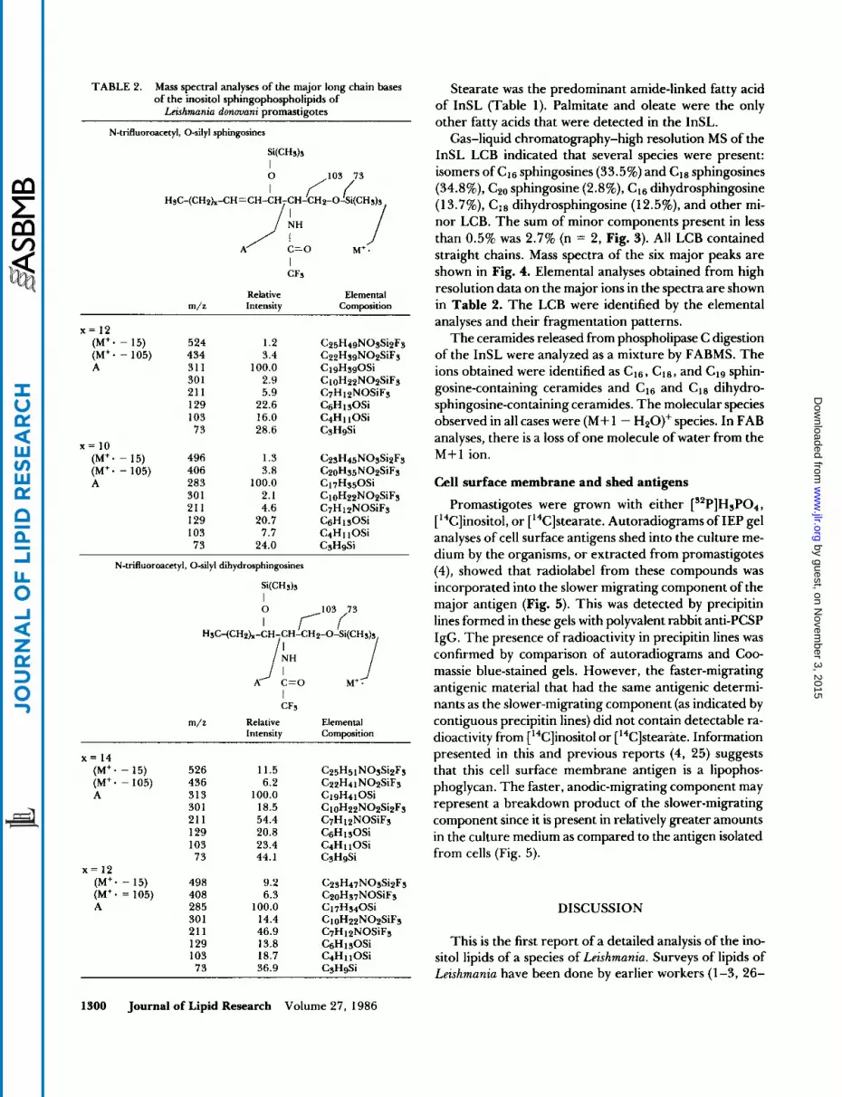
TABLE 2. Mass spectral analyses of the major long chain bases of the inositol sphingophospholipids of Leishmania dmovani promastigotes
Stearate was the predominant amide-linked fatty acid of InSL (Table 1). Palmitate and oleate were the only other fatty acids that were detected in the InSL.
Gas-liquid chromatography-high resolution MS of the InSL LCB indicated that several species were present: isomers of c16 sphingosines (33.5%) and cl8 sphingosines (34.8%), C20 sphingosine (2.8%), c16 dihydrosphingosine ( 1 3.7%), c18 dihydrosphingosine (12.5%), and other mi- nor LCB. The sum of minor components present in less than 0.5% was 2.7% (n = 2, Fig. 3). All LCB contained straight chains. Mass spectra of the six major peaks are shown in Fig. 4. Elemental analyses obtained from high resolution data on the major ions in the spectra are shown
m/z Intensity Composition in Table 2. The LCB were identified by the elemental analyses and their fragmentation patterns.
The ceramides released from phospholipase C digestion (M+* - 15) 524 1.2 C Z ~ H ~ ~ N O S S ~ ~ F S (M+. - 105) 434 3.4 C22HsgNOzSiFs of the InSL were analyzed as a mixture by FABMS. The A 31 1 100.0 ClgHsgOSi ions obtained were identified as c16, C18, and C19 sphin-
30 1 2.9 C 1 oH22N02SiF~ 21 1 5.9 C7H 1 pNOSiFs gosine-containing ceramides and c16 and cl8 dihydro- 129 22.6 C6HlsOSi sphingosine-containing ceramides. The molecular species 103 16.0 C4H110Si observed in all cases were (M+1 - H20)' species. In FAB
analyses, there is a loss of one molecule of water from the
N-trifluoroacetyl, 0-silyl sphingosines
W H d s I
HsC-(CHph-CH =CH-CH-CH-CH~-O-SI(CH~)~ 7 / ' 0 3 i 3
A /" c=o M+ 1 *
I CFs
Relative Elemental
x = 12
73 28.6 CSHgSi x = 10
(M+* - 15) 496 1.3 C2~H45NOsSizFs M + l ion. (M+- - 105) 406 3.8 C20Hs5N02SiFs A 283 100.0 C17Hs50Si Cell surface membrane and shed antigens
30 1 2.1 CloH22N02SiF~ 21 1 4.6 C7H 12NOSiFs Promastigotes were grown with either [s2P]H3P04, 129 20.7 CsHlsOSi [ ''C]inositol, or [14C]stearate. Autoradiograms of IEP gel 103 7.7 C4H11OSi analyses of cell surface antigens shed into the culture me-
dium by the organisms, or extracted from promastigotes (4), showed that radiolabel from these compounds was incorporated into the slower migrating component of the major antigen (Fig. 5). This was detected by precipitin lines formed in these gels with polyvalent rabbit anti-PCSP IgG. The presence of radioactivity in precipitin lines was confirmed by comparison of autoradiograms and Coo- massie blue-stained gels. However, the faster-migrating antigenic material that had the same antigenic determi- nants as the slower-migrating component (as indicated by
m/z Relative Elemental contiguous precipitin lines) did not contain detectable ra- Intensity Composition dioactivity from [14C]inositol or [14C]stearate. Information
presented in this and previous reports (4, 25) suggests (M+* - 15) 526 11.5 C25H51NOsSizFs that this cell surface membrane antigen is a lipophos- (M+. - 105) 436 6.2 C22H41N02SiF3 phoglycan. The faster, anodic-migrating component may A 313 100.0 ClgH41 OSi
301 18.5 c ~ ~ H ~ ~ N O ~ S ~ ~ F ~ represent a breakdown product of the slower-migrating 21 1 54.4 C7H 12NOSiFS component since it is present in relatively greater amounts 129 20.8 C&il~Osi in the culture medium as compared to the antigen isolated 103 23.4 C4H 1 1 OSi
73 24.0 CsHgSi
N-trifluoroacetyl. 0-silyl dihydrosphingosines
x = 14
73 44.1 CsHgSi from cells (Fig. 5) . x = 12 (M+* - 15) 498 9.2 C ~ S H ~ ~ N O S S ~ ~ F S (M+* = 105) 408 6.3 C20Hs7NOSiFs A 285 100.0 Ci 7Hs40Si DISCUSSION
30 1 14.4 C10H22N02SiF~ 211 46.9 C7H 12NOSiFs 129 13.8 CsHlsOSi This is the first report of a detailed analysis of the ino- 103 18.7 C4H 1 1 OSi sitol lipids of a species of Leishmania. Surveys of lipids of
Leishmania have been done by earlier workers (I-3,26- 73 36.9 CgHgSi
1300 Journal of Lipid Research Volume 27, 1986
by guest, on Novem
ber 3, 2015w
ww
.jlr.orgD
ownloaded from

1 2 3 4 1 2 3 4 1 2 3 4 Fig. 5. Immunoelectrophoretic analyses of Leishmania donovani antigens isolated from cells (lanes 1) and in culture media (lanes 2). Antigens were metabolically prelabeled with ["P]HSP04 (A), [U-"C]inositol (B), or [ I-"C]stearic acid (C). The sample origin wells for electrophoresis are at the bottom of the figure. lmmunoprecipitation was accomplished by filling side troughs with rabbit polyvalent anti-promastigote cell surface preparations of IgG. Gels were stained with Coomasie brilliant blue (lanes 1 and 2) then the radioactivity present in the immunoprecipitin lines was detected by autoradiography (lanes 3 and 4). The slower-migrating component had radioactivity when cells were grown with either of the three radioisotopes. The faster-migrating component contained '*P (A) but did not contain detectable radioactivity when cells were grown with either ['4C]inositol or ["Clstearic acid.
30). Thorough characterizations of PI in a variety of Leishmania species were done by Beach, Holz, and Anekwe (2), but PI-P, PI-P2, and InSL were not studied. The fatty acid composition of L. dmmani PI determined in this study agreed with that of the earlier report (2). Phosphatidyl- inositol was identified in the kinetoplastid flagellate, Ty panosoma cruzi, the causative agent of Chagas' disease (3 1). Polyphosphoinositides and PI were identified in another kinetoplastid flagellate, Crithidia (32-35), a parasite of in- sects. In those studies, InSL was not detected in the lipids of those protozoans. Inositol sphingolipids are abundant in some yeast cells where they are usually present as gly- cosphingolipids (1 0, 12). In the present study, InSL of Leishmania was found not to be glycosylated, however, preliminary observations of [ ''C]inositol-labeled lipids suggest that there are minor lipids present that may be similar to the mannose-containing inositol sphingolipids from yeast (1 0, 12). Radioactive spots, as well as diphe- nylamine-positive material on two-dimensional chro- matograms with low R, values like those of inositol gly- cosphingolipids with a hydroxy fatty acid and phyto- sphingosine, were detected in the present study but they were not further characterized because they represented only a small percentage of the total radioactive material present.
Earlier studies have shown that Leishmania lipids, in- cluding PI, contain alkyl acyl as well as alkenyl acyl lipids (1-3,26-30). Also, metabolic studies on the relative rates of incorporation of radiolabeled precursors such as ace- tate, octadecanol, and l-o-octadecyl-sn-glycerol into the alkenyl, alkyl, and acyl chains of PI and other glycerolipids have been reported (27-30); however, definitive chemical
structures of the alkyl chains present were not established. Beach et al. (2) have identified l-hexadec-9-enyl and 1- octadec-9-enyl as the major glyceryl ethers in the phos- phatidylethanolamines of eight Leishmania species. Alkyl acyl PI was only partially characterized in this study; glyc- eryl ethers were not identified.
The previous finding of inositol, ceramides, fatty acids, and phosphorus in major cell membrane components of two protozoans may be relevant to the identification of InSL in the lipids of Leishmania. A lipopeptidophospho- glycan (LPPG) with a glycophosphoceramide structure was isolated from T. cruzi (36-38). The plasma membrane localization of LPPG was also identified by studies on iso- lated cell membranes from this kinetoplastid flagellate (39). A lipophosphonoglycan (LPG) from the soil amoeba, Acanthamoeba castellanii was also established as forming an integral part of the cell surface membrane (23, 40-44). Unlike the LPPG from T. cruzi, the LPG from Acantham- oeba did not contain a peptide moiety and the P was in phosphonyl rather than phosphoryl linkages (24). Our observations on a major cell surface antigen ofLeishmania suggest that it contains sugars, P, fatty acids, and inositol. After the chemical compositions and structures of the Leishmania lipophosphoglycan and the other cell surface components of the other species that contain inositol lipid moieties have been elucidated, comparisons should reveal the similarities and differences. Such comparisons would provide information on the topological relationship of these molecules with the surface membrane of the cells.l This investigation received financial support from the UNDP/ World Bank/WHO Programme for Research and Training in Tropical Diseases, and N.S.F. grant, PCM 8020505. These
1301 Kaneshiro, Jayasimhulu, and Lester Leishmania inositol lipids
by guest, on Novem
ber 3, 2015w
ww
.jlr.orgD
ownloaded from

studies were initiated while the senior author served as an In- teragency Personnel Act Research Microbiologist in the Labo- ratory of Parasitic Diseases, National Institute of Allergy and Infectious Diseases. We thank Dr. D. Dwyer, Dr. T. Nash, and the other members of L.P.D. for their help and for making equipment and facilities available for parts of these studies. Manuscript received 16 April 1986.
REFERENCES
1 .
2.
3.
4.
5.
6.
7.
8.
9.
10.
1 1 .
12.
13.
Gercken, G., U. Hintze, and K. Ahrens. 1976. Analyse der Lipid- und Fettdurezusammensetzung von Leishmania do- nmani. Behring Inst. Mitt. 6 0 58-64. Beach, D. H., G. G. Holz, Jr., and G. E. Anekwe. 1979. Lipids of Leishmania promastigotes. J. Parasitol. 65: 203- 216. Wassef, M. K., T. B. Fioretti, and D. M. Dwyer. 1985. Lipid analyses of isolated surface membranes of Leishmania do- novani promastigotes. Lipids. 20: 108-1 14. Kaneshiro, E. S., M. Gottlieb, and D. M. Dwyer. 1982. Cell surface origin of antigens shed by Leishmania donmani during growth in axenic culture. Infect. Immun. 37: 558-567. Steiger, R. F., and E. Steiger. 1977. Cultivation ofLeishmania donmani and Leishmania braziliensis in defined media: nu- tritional requirements. J. Protomol. 24: 437-441. Handman, E., and J. W. Golding. 1985. The Leishmania receptor for macrophages is a lipid-containing glycoconju- gate. EMBO J. 4 329-336. Bligh, E. G., and W. J. Dyer. 1959. A rapid method of total lipid extraction and purification. Can. J. Biochem. Physiol. 37: 911-917. Folch, J., M. Lees, and G. H. Sloane Stanley. 1957. A simple method for the isolation and purification of total lipides from animal tissues.]. Biol. Chem. 226: 497-509. Turner, J. D., and G. Rouser. 1970. Precise quantitative determination of human blood lipids by thin-layer and tri- ethylaminoethyl cellulose column chromatography. I. Erythrocyte lipids. Anal. Bwchem. 38: 423-436. Steiner, S., and R. L. Lester. 1972. Metabolism of diphos- phoinositide and triphosphoinositide in Saccharomyces cer- evisiae. Biochim. Biophys. Acta. 260 82-87. Kates, M. 1972. Techniques in lipidology: isolation, analyses and identification of lipids. In Laboratory Techniques in Biochemistry and Molecular Biology, 3 (11). T. Work and E. Work, editors. North-Holland, New York. 275-610. Smith, S. W., and R. L. Lester. 1974. Inositol phosphoryl- ceramide, a novel substance and the chief member of a major group of yeast sphingolipids containing a single inositol phosphate. J. Biol. Chm. 249: 3395-3405. Kaneshiro, E. S. 1980. Positional distribution of fatty acids in the major glycerophospholipids of Paramecium tetraurelia. I. Libid Res. 21: 559-570.
14. kenkonen, 0. 1965. Individual molecular species of differ- ent phospholipid classes. Part 11. A method of analysis. J . Am. Oil Chem. SOC. 42: 298-304.
15. Viswanathan, C. V., F. Phillips, and W. D. Lundberg. 1968. Two-dimensional reaction thin-layer chromatography in the analysis of mixtures of alkenyl acyl-, alkyl acyl- and diacyl- choline phosphatides. J . Chromatogr. 3 8 267-273.
16. Beesley, T. 1976. Charring techniques. In Kontes Quant Notes. J. Sherma, editor. Kontes, Vineland, NJ.
17. MacGee, J., and K. Allen. 1974. Preparation of methyl esters
from the saponifiable fatty acids in small biological specimens for gas-liquid chromatographic analyses. J. Chromatogr. 100
18. Kaneshiro, E. S., L. S. Beischel, S. J. Merkel, and D. E. Rhoads. 1979. The fatty acid composition of Paramecium aurelia cells and cilia: changes with culture age. J. Protomol. 26: 147-158.
19. Carter, E. E., and C. Gaver. 1967. Branched-chain sphin- gosines from Tetrahymena pyriformis. Biochem. Biophys. Res. Commun. 2 9 886-891.
20. Nau, H., H-J. Forster, J. A. Kelley, and K. Biemann. 1975. Polypeptide sequencing by a gas chromatograph mass spec- trometer computer system. 11. Characterization of complex mixtures of oligopeptides as trimethylsilylated polyamino alcohols. Bwmed. Mass Spectrom. 2: 326-339.
21. Jayasimhulu, K., R. A. Day, C. J. Metral, J. Wermeling, and M. Memadis. 1985. Orthoquinone sequential degradation of peptides: mass spectral leucine/isoleucine differentiation at internal positions of peptides. 33rd Annual Conference on Mass Spectrometry and Allied Topics. 510.
22. Metral, C. J., and R. A. Day. 1986. Exact mass FAB and EI/FAB MS with peptides using a direct insertion FAB probe. Anal. Lett. 1 9 217-227.
23. Ferguson, K. A., F. M. Davis, R. L. Conner, J. R. Landrey, and F. B. Mallory. 1975. Effect of sterol replacement in vivo on the fatty acid composition of Tetrahymena. J. Bid. Chem. 250: 6998-7005.
24. Korn, E. D., D. G. Dearborn, and P. L. Wright. 1974. Li- pophosphonoglycan of the plasma membrane of Acantham- oeba casteltanii. Isolation from whole amoebae and identifi- cation of the water-soluble products of acid hydrolysis. J . Biol. Chem. 249: 3335-3341.
25. Kaneshiro, E. S., M. Gottlieb, and D. M. Dwyer. 1982. Par- tial characterization of the major shed membrane antigens (SMA-) from Leishmania donouani promastigote cultures. J . Protozool. 2 9 47 1 .
26. Hack, M. H., R. G. Yaeger, and T. D. McCaffery. 1962. Comparative lipid biochemistry. 11. Lipids of plant and an- imal flagellates, a non-motile alga, an ameba and a ciliate. Comp. Biochem. Physiol. 6: 247-252.
27. Herrmann, H., and G. Gercken. 1980. Incorporation of [ 1- ''C]octadecanol into the lipids of Leishmania donovani. Lipids. 1 5 179-185.
28. Herrmann, H., and G. Gercken. 1980. Synthesis of phos- pholipids in Leishmania donovani. Hoppe-Seyler's Z. Physiol. Chem. 361: 1735-1742.
29. Herrmann, H., and G. Gercken. 1982. Incorporation of [ 1- "Clacetate into fatty acids and aliphatic moieties of glyc- erolipids in Leishmania donouani promastigotes. Comp. Bwchem. Physiol. 7 3 B 367-373.
30. Jacobs, G., H. Herrmann, and G. Gercken. 1982. Metab- olism of 1 -0-[ l'-'4C]octadecyl-sn-glycerol in Leishmania do- nouani promastigotes. Ether lipid synthesis and degradation of the ether bond. Molec. Biochem. Parasitol. 5: 65-76.
3 1 . Da Silveira, J. F., and W. Colli. 198 1 . Chemical composition of the plasma membrane from epimastigote forms of Try panosoma cmzi. Biochim. Bwphys. Acta. 644: 341-350.
32. Palmer, F. B. St. C. 1973. LipidsofCrithidiafasciculata. The occurrence and turnover of phosphoinositides. Biochim. Bio-
33. Palmer, F. B. St. C. 1973. Triphosphoinositide phospho- diesterase in a protozoan-Crithidia fasciculata. Bwchim. Bio-
34. Palmer, F. B. St. C. 1976. Hydrolysisoftriphosphoinositide
35-42.
phys. Acta. 316: 296-304.
phys. Acta. 326: 194-200.
1302 Journal of Lipid Research Volume 27, 1986
by guest, on Novem
ber 3, 2015w
ww
.jlr.orgD
ownloaded from

by a soluble fraction of Crithidia fasciculata. Bwchim. Bwphys. Acta. 441: 477-487.
35. Daniels, C. J., and F. B. St. C. Palmer. 1980. Biosynthesis of phosphatidylinositol in Crithidia fasciculata. Biochim. Bw-
36. Lederkremer, R. M., M. J. M. Alves, G. C. Fonseca, and W. Colli. 1977. A lipopeptidophosphoglycan from Trypa- nosoma cruzi (epimastigota). Isolation, purification and car- bohydrate composition. Bwchim. Bwphys. Acta. 444: 85-96.
37. Lederkremer, R. M., C. T. Tanaka, M. J. M. Alves, and W. Colli. 1977. Lipopeptidophosphoglycan from Trypanosoma cruzi. Amide and ester-linked fatty acids. Eur. J . Bwchem.
38. Lederkremer, R. M., 0. L. C a d , C. T. Tanaka, and W. Colli. 1978. Ceramide and inositol content of the lipopep- tidophosphoglycan from Trypanosoma crud. Biochem. Bwphys. Res. Commun. 85: 1268-1274.
39. Alves, M. J. M., J. F. da Silveira, C. H. R. De Paiva, C. T. Tanaka, and W. Colli. 1979. Evidence for the plasma mem-
phys. Acta. 6 1 8 263-272.
74: 263-267.
brane localization of carbohydratecontaining macromole- cules from epimastigote forms of Trypanosoma crud. FEBS Lett. 9 9 81-85.
40. Korn, E. D., and P. L. Wright. 1973. Macromolecular com- position of an amoeba plasma membrane. J . Bid. C h i . 248: 439-447.
41. Dearborn, D. G., and E. D. Kom. 1974. Lipophosphono- glycan of the plasma membrane of Acanthamoeba castellanii. Fatty acid composition. J . Bwl. Chem. 249 3342-3346.
42. Dearborn, D. G., S. Smith, and E. D. Kom. 1976. Lipo- phosphonoglycan of the plasma membrane of Acanthamoeba castellanii. Inositol and phytosphingosine content and general structural features. J . Bwl. Chem. 251: 2976-2982.
43. Bowers, B., and E. D. Korn. 1974. Localization of l i p phosphonoglycan on both sides of Acanthamoeba plasma membrane. J . Cell Bwl. 62: 533-540.
44. Bailey, C. F., and B. Bowers. 1981. Localization of lipo- phosphonoglycan in membranes of Acanthamoeba by using specific antibodies. Molec. Cell. Bwl. 1: 358-369.
Kaneshiro, Jayusimhulu, and Lester Leishmania inositol lipids 1303
by guest, on Novem
ber 3, 2015w
ww
.jlr.orgD
ownloaded from


