Embed Size (px)
Citation preview
Volume 7 Number 7 1979 Nucleic Acids Research
Characterization of the major altered leader sequence of late mRNA induced by SV40 deletionmutant dl-1811
Guy Haegeman, Dirk Iserentant, Dirk Gheysen and Walter Fiers
Laboratory of Molecular Biology, State University of Ghent, Ledeganckstraat, 35 Ghent, Belgium
Received 8 October 1979
ABSTRACT
dl-1811 is a viable SV40 mutant with a 40 base pair dele-tion that includes the major wild-type capping site of late mRNAat map position 0.72. The late viral mRNAs induced by dl-1811have now been further characterized by inversed Sl-mapping ana-lysis. The Sl-resistant, 32P-labeled RNA fragments derived fromthe leader region were examined by fingerprinting and by analy-sis of RNase T2-generated 5'-terminal cap structures. The resultsshow that most if not all of the mutant leader fragments analyzedhave their 5' terminus to the left of the dl-1811 deletion site,i.e., closer to the origin of DNA replication. The major alteredleader fragment is a continuous transcript from the DNA in theregion 0.716 to 0.761 map unit and its 5' terminus has been pre-cisely mapped at nucleotide L290. The observation that the capstructure of the major altered leader is only a minor cap speciesin wild-type late RNA suggests regulation in the use of differentcapping sites in SV40.
INTRODUCTION
During the late stage of SV40 infection of monkey kidney
cells, i.e., after the onset of viral DNA replication, two cyto-
plasmic late mRNA species are synthesized sedimenting at 16S and
19S (1, 2). Both species have been mapped on the genome (3, 4),and more recently it has been found that these viral mRNA mole-
cules are composite structures comprising an untranslated "leader"
segment and a coding sequence or "body". The sequence is not co-
linear with the DNA: each mRNA has been formed by at least one
splicing event (5-8). The most abundant leader fragment is a se-
quence of 202(±l) nucleotides that is transcribed from the DNA
region between 0.72 and 0.76 map unit and which is subsequentlyspliced 42(±1) nucleotides before the body of the VP1 gene (9-
12; the uncertainty is because the exact position of splicingis ambiguous). Alternative leader fragments may also occur in
C Information Retrieval Limited 1 Falconberg Court London Wl V 5FG England 1 799

Nucleic Acids Research
late viral RNA and these actually appear to constitute the most
abundant leader segments in the 19S species (7, 13, 14).
Procedures have been described for creating viable SV40
deletion mutants in the late region of the genome (15-18). The
deletions have been characterized by restriction mapping analysis;
in some cases the exact size and location of the deletion has
been precisely determined by nucleotide sequence analysis (18-20).
It is remarkable that in some of these mutants the major late
capping site, which maps at position 0.722 (21), is included in
the deletion (18, 20). Although in some other systems cap forma-
tion seems to be directly linked with initiation of RNA trans-
cription (22-26), deletion of the major capping site in SV40
does not result in loss of viability but at most only results in
a decreased plaque size (18, 27).
Previously we have shown that deletion of the major capping
site in the mutant dl-1811 results in the appearance of a variety
of alternative capped 5' termini in the corresponding late mRNA
(27). This suggests the presence of a number of alternative lea-
der segments. Indeed, altered 16S and 19S mRNAs have been obser-
ved after infection with mutants that lack DNA segments of diffe-rent size and at slightly different positions compared to the
dl-1811 deletion (28). In this paper we report a further charac-
terization of the dl-1811 late mRNA both by fingerprint analysis
(9, 29) and by the S1 mapping procedure (30), followed again by
fingerprinting and by cap analysis of the Sl-resistant fragments.
This enabled us to determine at the nucleotide level the major
alternative leader segment present in the dl-1811 late mRNA and
to identify the two related cap structures 7mGpppmAmpG and
7mGpppmAmpGmpA. These major terminal structures constitute about
40% of the total dl-1811 late mRNA cap content (27). Furthermore,the latter terminal trinucleotide sequence A-G-A permitted us
to pinpoint unambiguously the 5' end of the alternative leader
segment.
METHODS
Isolation and purification of 32P-labeled cytoplasmic late
viral mRNA from CV-1 monkey kidney cells infected by either wild-
type (WT) SV40 or by dl-1811, fingerprint analysis of the puri-
1800

Nucleic Acids Research
fied RNA, and isolation and structure determination of the appro-
priate 5'-terminal capped structures have been described in de-
tail previously (9, 27, 31). The Berk and Sharp technique (30)
for mapping mRNA sequences on a genome, for pinpointing the 5'
end of leader fragments and for positioning spliced-out regions
has been applied in these studies in an inversed way, i.e., by
using 3 P-labeled RNA and unlabeled DNA. The Sl-resistant DNA-RNA
hybrids were isolated by polyacrylamide gel electrophoresis and
used for further characterization of the mRNA leader fragments
by fingerprinting techniques.
Poly(A)-containing viral RNA was selected by a single hy-
bridization step onto Sepharose-bound SV40 DNA, precipitated in
the presence of carrier yeast ribosomal RNA, and dissolved in
80% deionized formamide, 0.4 M NaCl, 0.001 M EDTA and 0.04 M
Pipes buffer, pH 6.4, along with the appropriate SV40 DNA frag-
ment. The latter was obtained by digestion on a preparative scale
of viral DNA isolated by Hirt extraction of infected cells, or
was derived from plasmids consisting of segments of SV40 DNA in-
tegrated in pBR322 (details given below). In a typical experiment
this solution (final vol. 60 pl) was heated for 15 min. at 85°C
to completely denature the DNA and was then kept at 52C for 3 h
to allow formation of DNA-RNA hybrids. After this hybridization
step the solution was diluted in 900 pl ice-cold Sl buffer con-
taining 0.025 M NaAc, 0.25 M NaCl and 0.0045 M ZnAc, pH 4.4.
150 p1 Sl nuclease (1 unit/pl) was added and the digestion was
carried out for 30 min. at 37°C. The mixture was extracted with
phenol, and the Sl-resistant hybrids were precipitated, and frac-
tionated on a polyacrylamide gel run in 0.09 M Tris-borate,0.0025 M EDTA, pH 8.4. The gel bands containing the DNA-RNA hy-
brids were visualized by autoradiography and were eluted with
1 M NaCl. Each fragment was redissolved in 5 p1 0.02 M Tris-Cl,
0.002 M EDTA, pH 7.4, heated for 10 min. at 85°C and quicklychilled on ice to keep the hybrids denatured. 1 pl of Tl ribo-
nuclease (10 units/pl) was added and the solution was incubated
for 1 h at 370C. The digest was subsequently analyzed by minifin-
gerprinting (29). To isolate the 5'-terminal cap structures of
Sl-resistant RNA fragments, DNA-RNA hybrids were eluted and de-
natured as described above and digested with ribonuclease T2 and
1801

Nucleic Acids Research
bacterial alkaline phosphatase as reported previously (31).
RESULTS
Characterization of dl-1811 late mRNA by minifingerprinting
Twice-hybridized 32P-labeled dl-1811 late mRNA was digested
with Tl ribonuclease and a two-dimensional fingerprint was made.
The dl-1811-specific Tl pattern is shown in Figure 1. The pro-
file is homologous to the WT RNA fingerprint except for only one
additional spot which occurs in about molar amount in the dl-1811
-1Elec pH 3.5
4IIl
0
E0
X_J
._~~~~~~~~~~~~~~~~~~~~~~~~~~~~~~~~~~~~~~~~~~~~~~~~~~~~~~~-R_-
..._ _ __~~~Ag&,___~~~AMW1
.4,
Fig. 1 : Tl-ribonuclease fingerprint of dl-1811 late mRNA.Two-dimensional £ractionation on PEI-cellulose thin-layerplates (20 cm x 20 cm) was carried out according to the pro-cedure of Volckaert et al. (29) of a Tl hydrolysate of totalcytoplasmic late mRNA of dl-1811. The arrow indicates theadditional spot (Tl oligonucleotide 243) present in the dl-1811pattern which represents the unique product with composition(U2,9C2 ,A-C,A-A-C)G.
1802
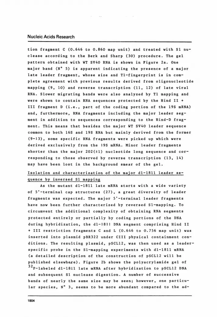
Nucleic Acids Research
pattern. This additional product (Ti oligonucleotide 243; cf.
legend to Figure 5) was analyzed with ribonuclease A (not shown)
and its double-digestion products (U ,C ,A-C,A-A-C)G correspond
to the sequence U-A-C-C-U-A-A-C-C-G, which is the predicted Tl
oligonucleotide on the basis of the SV40 DNA nucleotide sequence
(32) and the extent of the dl-1811 deletion (27). The first Up
residue of this oligonucleotide (at L296) is part of the KpnI
restriction site at map position 0.716 while the pG residue is
derived from beyond the deletion (at L345) and corresponds to the
position next to the HpaII site at 0.726 map unit. The presence
of this oligonucleotide in about molar quantity (the resolution
was not sufficient to allow rigorous quantitation) suggests that
most if not all of the dl-1811 leader fragments extend into the
5' side of the deletion site and that the variety of cap struc-
tures associated with dl-1811 late mRNA (27) is predominantly
derived from an area of DNA preceding the position of the major
WT capping site (i.e., from a region closer to the origin of DNA
replication around 0.67 map unit). The preceding characteristic
Tl oligonucleotides, UUCUUUCCG (N° 530) and CCUCAG (No 131) (cf.
Figure 5), were not revealed as independent spots in the Tl fin-
gerprint of total dl-1811 late mRNA, as they were only partially
resolved from related sequence isomers. However, their presence,
although in variable amounts, was revealed by pancreatic analysis
of the appropriate mixed Tl-oligonucleotide spots. Other unique
Tl oligonucleotides located more upstream (i.e. towards map posi-
tion 0.67) were not readily detectable in the fingerprint of
total dl-1811 mRNA and were considered to be appreciably submolar
compared to the relative abundance of the normal WT leader oligo-
nucleotide sequences.
Isolation of the major WT leader segment by inversed Sl mapping
In order to specifically isolate the leader fragments of the
viral RNA, an inversed Sl-mapping procedure was used (see Methods
section). Total poly(A)-containing RNA was first hybridized to
immobilized SV40 DNA in order to decrease the background of the
gel pattern; this step is almost essential when subsequent mapping
of the oligonucleotides of the protected RNA fragment is intended.
The SV40-specific RNA was then hybridized to the Hind III restric-
1803
Nucleic Acids Research
tion fragment C (0.646 to 0.860 map unit) and treated with Si nu-
clease according to the Berk and Sharp (30) procedure. The gel
pattern obtained with WT SV40 RNA is shown in Figure 2a. One
major band (N° 5) is apparent indicating the presence of a major
late leader fragment, whose size and Tl-fingerprint is in com-
plete agreement with previous results derived from oligonucleotide
mapping (9, 10) and reverse transcription (11, 12) of late viral
RNA. Slower migrating bands were also analyzed by Tl mapping and
were shown to contain RNA sequences protected by the Hind II +
III fragment D (i.e., part of the coding portion of the 19S mRNA)
and, furthermore, RNA fragments including the major leader seg-
ment in addition to sequences corresponding to the Hind-D frag-
ment. This means that besides the major WT SV40 leader sequence
common to both 16S and 19S RNA but mainly derived from the former
(9-13), some specific RNA fragments were picked up which were
derived exclusively from the 19S mRNA. Minor leader fragments
shorter than the major 202(±1) nucleotide long sequence and cor-
responding to those observed by reverse transcription (13, 14)
may have been lost in the background smear of the gel.
Isolation and characterization of the major dl-1811 leader se-
quence by inversed Sl mapping
As the mutant dl-1811 late mRNA starts with a wide variety
of 5'-terminal cap structures (27), a great diversity of leader
fragments was expected. The major 5'-terminal leader fragments
have now been further characterized by reversed Sl-mapping. To
circumvent the additional complexity of obtaining RNA segments
protected entirely or partially by coding portions of the DNA
during hybridization, the dl-1811 DNA segment comprising Hind II
+ III restriction fragments C and L (0.646 to 0.756 map unit) was
inserted into plasmid pBR322 under CIII physical containment con-
ditions. The resulting plasmid, pSCL12, was then used as a leader-
specific probe in the Sl-mapping experiments with dl-1811 mRNA
(a detailed description of the construction of pSCL12 will be
published elsewhexe). Figure 2b shows the polyacrylamide gel of
32P-labeled dl-1811 late mRNA after hybridization to pSCL12 DNA
and subsequent S1 nuclease digestion. A number of successive
bands of nearly the same size may be seen; however, one particu-lar species, N° 3, seems to be more abundant compared to the ad-
1804
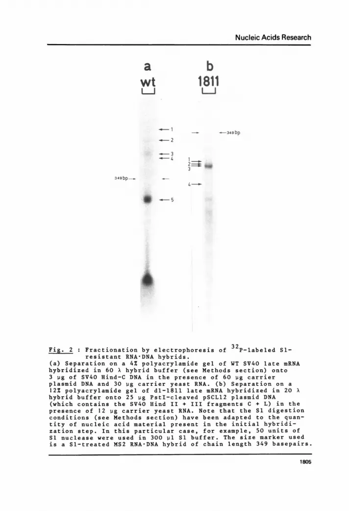
Nucleic Acids Research
awt
-6-1c- 2
3-4- 4
349bp_
b1811
_-349 bp
1 _
3
4 -0
- 5
Fig. 2 : Fractionation by electrophoresis of 32P-labeled S1-resistant RNA-DNA hybrids.
(a) Separation on a 4% polyacrylamide gel of WT SV40 late mRNAhybridized in 60 X hybrid buffer (see Methods section) onto3 pg of SV40 Hind-C DNA in the presence of 60 wg carrierplasmid DNA and 30 wg carrier yeast RNA. (b) Separation on a12% polyacrylamide gel of dl-1811 late mRNA hybridized in 20 Xhybrid buffer onto 25 pg PstI-cleaved pSCL12 plasmid DNA(which contains the SV40 Hind II + III fragments C + L) in thepresence of 12 ug carrier yeast RNA. Note that the S1 digestionconditions (see Methods section) have been adapted to the quan-tity of nucleic acid material present in the initial hybridi-zation step. In this particular case, for example, 50 units ofS1 nuclease were used in 300 p1 S1 buffer. The size marker usedis a Sl-treated MS2 RNA-DNA hybrid of chain length 349 basepairs.
1805

Nucleic Acids Research
jacent compounds. All the bands were eluted and characterized by
Ti fingerprinting. Several of them (N° 1, 2 and 3) were also
analyzed with respect to cap content. Figure 3a and 3b shows the
fingerprint and corresponding oligonucleotide identification,
respectively, of the major band N° 3. Beginning at the 3' termi-
nus of the leader fragment and going in the 5' direction (see
Figure 5), all expected oligonucleotides, including the afore-
mentioned Tl oligonucleotide 243 which spans the deletion site,
are present on the fingerprint. However, oligonucleotide 131,
which precedes oligonucleotide 243 by only 3 nucleotides, was
definitely absent from band N° 3, as were all the remaining Tl
products extending further in the 5' direction. These results
allow the localization of the 5' end of the major mutant leader
to a rather narrow area neighboring the KpnI restriction site at
position 0.716. Total degradation of another aliquot of this
leader segment RNA with T2 ribonuclease and bacterial alkaline
phosphatase (Figure 4) revealed two prominent cap structures
identified by position as 7mGpppmAmpG and 7mGpppmAmpGmpA. This
r~Elec pH3.5 2 3 037 -1 EieccpH .5
,G, c MaD o2 z~~~~a3 1 s'}d
.r F. L=
a 0,2C I
30C 00n
a bFig. 3 Characterization of the major leader fragment of dl-1811
late mRNA.(a) A portion of the Sl-resistant RNADNA hybrid band No 3 inFigure 2b was digested with Tl ribonuclease (see Methods section)and fractionated in two dimensions on PEI-cellulose thin-layerplates (20 cm x 20 cm). (b) Schematic diagram identifying theoligonucleotide spots in (a). A standard nomenclature of threedigits indicating the number of uridine, cytidine and adenosineresidues, respectively, is used for identifying the Tl-spots (38).
1806

Nucleic Acids Research
^~~~at
I *~~~~~~~~~~~~~~
a.
(L
Elec p H 3.5
Fig. 4 :Isolation and identification of the cap structures fromthe major dl-1811 leader fragment.
Another aliquot of the Sl-resistant band N° 3 (see Figure 2b)was treated with T2 ribonuclease and bacterial alkaline phos-phatase and separated by electrophoresis at pH 3.4 in the firstdimension and chromatography in 1 M pyridine-formic acid (pH4.3) in the second dimension (31). B denotes the position ofthe blue dye marker xylene cyanol FF. The identity of the capped
prodcts7mGppmAmpG (cap I) and 7mGppp AmpGmpA (cap II)wadeduced from their specific position on the map relative to theblue dye marker; these spots correspond to spots 5 and 7 inFigure 3 of ref. 27, which describes the characterization ofall the different dl-1811 capped termini.
couple of cap I and corresponding cap II structures is indeed
identical to the major 5' cap terminus of total dl-1811 late
mRNA as previously characterized (27).Based on the trinucleotide sequence A-G-A, we concluded from
these results that the capped nucleotide of the major dl-1811
leader fragment must correspond to position L290 of the late
region of the genome (Figure 5; nucleotides are numbered as des-
cribed in ref. 20). L290 lies upstream from the major WT capping
1807

Nucleic Acids Research
310 320 330 340A A G T T C C T C T T T C A G A G G T TIA T T T C A G G C C A T G G T G C T G CTT CA AG GAG A A AG T CT CC A A TA A AG T CC G G TAG C A C GACGI
...260 270 280 290 300A C A C A T T C C A C A G C T G G T T C T T T C C G C C T C A G A A G G T A C C T A A C C G C C G GTGTGTAAGGTIGTCGAC CAAGAAAGGCGGAGTCTT CCATGGATTGGCGGCC
(0.709) PvuII KpnI HpaIImGpppmAGmAmA G G U A C C U A A C C G C C G G
243 020350 360 370 380 390CUGUCACGCCAGGCCUCCGUUAAGGUUCGUAGGUCAUGGACUGfUAAGUAAVJ~\ \ \ .A J 7JLJ \Z J "__Jl_110 121 021 140 202 210 101 211 111 003400 410 420 430 440
AAAACAG\CUAAC/\CUUUG7JUUUGUUUUAAGCUSUUUGUGCXAAU118 132 520 100 300 401 001 410 110
450 460 470 480 490UUUGUGAAGGGGAAGAUACUGUUGACGGGAAACGCAAAAAACCAGAAAGG_J\J\\JL\JWJ AIL~A412 100 002 002 212 200 011 013 037 003
500 J 510 520UUAACUGAAAAACCAGAAAGUUAACUG...
Fig. 5 : Nucleotide sequence of part of the Hind II + III restric-tion fragment C from the late region of dl-1811 DNA (27)and of the corresponding 5'-terminal sequence of themajor altered leader fragment of the viral late mRNA.
Nucleotide numbers are based on a total WT SV40 sequence of 5243basepairs according to the system of Fiers et al. (32) subse-quently corrected for the late region (20). The recognitionsequences of relevant restriction endonucleases are enclosed inbrackets. The 40 basepair dl-1811 deletion is shown in the boxand the site of the deletion, between nucleotides L304 and L344,is indicated (20, 27). The position of the major WT capping site(21) is marked with an asterisk.
The major leader fragment of dl-1811 late mRNA starts at WTposition L290 and is colinear with the DNA sequence up to nucleo-tide L526(±l)(map position 0.761), where a donor splicing signalis encountered. The Tl oligonucleotides are indicated below theRNA sequence and they are named in a code which gives the basecomposition of the respective products (see legend to Figure 3).These oligonucleotides may be seen on the Tl fingerprint in Fig-ure 3. The arrow marks the end of the DNA fragment used for hy-bridization, i.e., the Hind II cutting site at position 0.756.
.Vsite, and the mutant leader sequence is colinear with the mutant
DNA sequence (27) from L290 up to the position where splicingoccurs (at 0.761 map unit, i.e., nucleotide L526 ± 1). This means
that the length of the major mutant leader sequence (Figure 2b,
1808

Nucleic Acids Research
band 3) amounts to 197(±l) nucleotides.
Analysis of the other bands revealed the presence of addi-
tional A-cap structures characterized previously (cf. ref. 27)
and the occurrence of specific Tl oligonucleotides derived from
the DNA region preceding the aforementioned major dl-1811 capping
site (evidence not shown). However, as there was too little radio-
activity present in these minor Sl-resistant bands, unambiguous
characterization proved difficult and only visual inspection of
the two-dimensional fractionation patterns was possible. From
these data we nevertheless were able to conclude that all the
alternative leader fragments analysed (5 bands) extend further in
the 5' direction than the major mutant leader species (i.e., the
5' terminus was located closer to the origin of DNA replication).However, considering the variety of trinucleotides which could
correspond to the 5'-cap sequences (27), we were not able to pin-
point the 5' terminus of these additional leader segments.
DISCUSSION
The most prominent leader sequence in dl-1811 late mRNA
was characterized both by fingerprinting and by cap structure
analysis of the material purified by the Berk and Sharp Sl-
procedure (30). Under the experimental conditions used, the
RNA-DNA hybrids were found to be fairly stable and showed little
if any breathing at the ends. Indeed, even after extensive di-
gestion with Sl nuclease the 3'-terminal Tl products were pre-
sent in molar amount on the fingerprint. The same holds for the
5'-terminal cap structures, which did not disturb hybridizationand in fact allowed isolation of capped termini from Sl-digestedRNA-DNA hybrids. Indeed, we found that the major leader fragment
in the mutant mRNA starts at nucleotide L290 (map position 0.716)and is colinear with the dl-1811 DNA sequence up to the majordonor splicing site of WT RNA at nucleotide L526(±l) (map posi-tion 0.761). One consequence of the deletion in dl-1811 is that
the coding potential of the open reading frame present in the
WT leader fragment is lost: the presumptive initiation codon of
this "agnogene" is comprised within the dl-1811 deletion (cf.Figure 5) and the reading frame is not restored in the mutant
1809
Nucleic Acids Research
leader segments by any preceding initiation codon. It follows
that the potential product of the agnogene, if it is indeed pro-
duced by the WT virus, is not essential for viability under
tissue-culture conditions. Indeed, the non-essential nature of
this region of the genome is well known (15-18). The slower growthrate of dl-1811, as manifested by the smaller size of the plaques(27), may perhaps be due to a slightly decreased level of late
mRNA synthesis and/or its different 5'-terminal structure. The
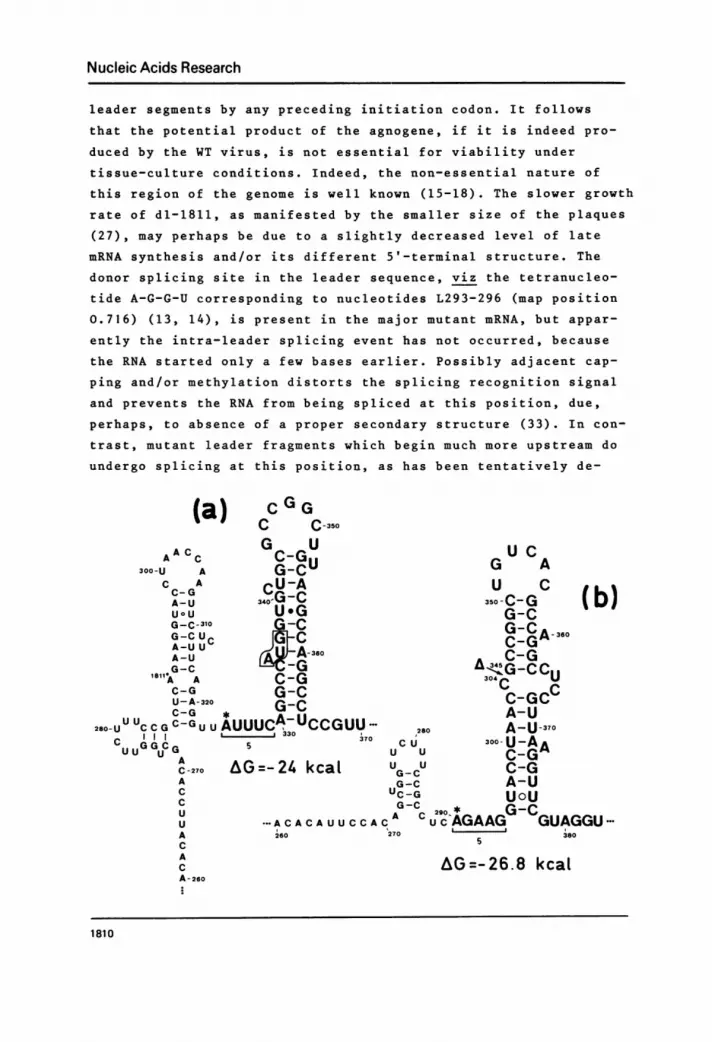
donor splicing site in the leader sequence, viz the tetranucleo-
tide A-G-G-U corresponding to nucleotides L293-296 (map position0.716) (13, 14), is present in the major mutant mRNA, but appar-
ently the intra-leader splicing event has not occurred, because
the RNA started only a few bases earlier. Possibly adjacent cap-
ping and/or methylation distorts the splicing recognition signaland prevents the RNA from being spliced at this position, due,perhaps, to absence of a proper secondary structure (33). In con-
trast, mutant leader fragments which begin much more upstream do
undergo splicing at this position, as has been tentatively de-
(a)AA Cc
300-U AC AC- GA-UU ° UG-C-310G-C UA-U UA-UG-CA AC-GU- A-320C-G *
280-U UUCCGC-GUUAUUtC G G C G 5U U U
A
C-270 AGACCUUACAC
A 6.
C G GC ,-350
G Ur_n.- U C%.*%.Mu ~G AG-CU CCU-A U C ,340G - C 3s~50CG ID)U.G G-cG-C G-C
C C-GCA 360A-3u0 C-G
-JAGO A<G..CCUC-G 3 c uG-C C-GCCG-C A-U
jC7UCCGUU 0 A-U30330 370
c8 300-U-A7
U U C-GA=-24 kcal U UCGG-CCGG-C A-UUC-G UoUG-C * GC
...AACACUUCCAC C-AGAAG GUAGGU'260 27038
AG=-26.8 kcal
1810

Nucleic Acids Research
CUGUGA UG (C)U G240
230-U GA AA GA A G CC
G-CA-UG-C
220-GG-c-250C-G
UoCG-CAG-CA-U AG=-14.8 kcalA-UUoCUOU-260oUeG
AU-AU-A* C-G
-GACAUUUU CUGAUCAAGUAAGU--200' 5 270
7
8
Fig. 6 Proposed secondary structure models for the RNA se-quence corresponding to the 5'-terminal major leader fragment(s)and the preceding, possibly transcribed, nucleotide sequence of(a) WT SV40 late mRNA, (b) dl-1811 late mRNA, and (c) polyomavirus late mRNA. The SV40 nucleotides are numbered as in Figure 5and hence are identical for both WT and dl-1811 DNA (and cor-responding RNA). The 40 nucleotide deletion is indicated by anarrow in (b). Polyoma nucleotide numbers were taken from Flavellet al. (35). Free energies of the hairpin structures were calcu-lated according to the stability rules of Tinoco et al. (39).The small contribution made by a G'U basepair to the stability ofa helix is indicated by a dot. The symbol 0 between two basesindicates that they occur opposite each Dther in the helix butthat the union does not add to the stability of the structure(40). The positions of the major capping sites are marked by anasterisk.Note that in all the examples the major capping site precedesa stable (G-C)-rich hairpin structure by exactly 5 nucleotides.However, in the WT RNA sequence, the major dl-1811 capping site(marked by 1811 ) is part of the stem of a hairpin loop. Theboxed AUG sequence in (a) is the initiation codon of the conjec-tural agnogene.
1811

Nucleic Acids Research
duced from visual interpretation of Ti fingerprints of the alter-
native dl-1811 minor leader segments.
It is not unexpected that the most prominent cap I and II
structure found on dl-1811 late mRNA corresponds to the 5' termini
of the fragment found in the major band of Sl-resistant leader
RNA. It may be noted that this particular capped end is only a
minor component in the diversity of WT SV40 cap structures (13,
34, and our unpublished data) and more particularly it seems that
only one 19S leader species but no 16S species starts at this
position (13). Favoring of the capping site at nucleotide L290
in dl-1811 as compared to the relative use of this position in
WT SV40 suggests that the appearance of different caps is subject
to regulation or depends on local configuration of the mRNA.
Indeed, the relative abundance of different caps may be directly
dependent on structural or functional requirements. A secondary
structure model for part of the WT SV40 leader sequence (Figure 6)
shows that the major WT capping site is located at the base of a
stable hairpin, whereas nucleotide L290 is fully involved in base-
pairing in the stem of the preceding loop. Also, some minor WT
capping sites may be found in single-stranded regions precedingsimilar (G.C)-rich loop structures (our unpublished results).
Remarkably the structure model for the dl-1811 RNA sequence shows
that now the position of the major mutant capping site at nucleo-
tide L290 lies free and at exactly the same distance (5 nucleo-
tides) from an analogous (G*C)-rich hairpin. In the case of
polyoma virus, which is distantly related to SV40, several cap
structures in the late mRNA corresponding to consecutive purinepositions in the DNA sequence have been identified (35). These
may conceivably be derived by a stuttering effect of the polymer-
ase II during initiation of transcription (35). The variety of
capping sites in SV40 late RNA and especially their relative
broad distribution over the DNA sequence suggests a more compli-cated mechanism for their formation than simple stuttering. Pos-
sibly, the RNA polymerase starts at many different positions ina defined region and only those RNA molecules with a hairpin near
their 5' end become stabilized against rapid degradation and are
conserved. Alternatively, initiation of RNA synthesis may occur
more proximal to the origin of DNA synthesis and processing
1812

Nucleic Acids Research
enzymes may subsequently recognize the proposed hairpin loops
(Figure 6), whereupon caps may be formed after further phosphory-
lation at the processed 5' ends, analogous to, e.g., the capping
system of vaccinia virus (36, 37). It is of interest that also in
polyoma virus mRNA a loop structure following (and specifying ?)
the capping site can be drawn. However, this structure is less
stable (and less G.C-rich) than the proposed SV40 hairpins and
may perhaps be responsible for rather imprecise processing,
thereby giving rise to multiple caps around the same position
(35). More direct experimental approaches are needed to dis-
tinguish unambiguously between these two possibilities for
generating the diversity of capped 5'-termini in the papova
virus late mRNAs.
ACKNOWLEDGEMENTS
We thank Jose Van der Heyden for culturing cells and virus
stocks, Dr. A. Van de Voorde for many discussions, and J. Van
Herreweghe for assistance. The research was supported by grants
from the Kankerfonds of the Algemene Spaar- en Lijfrentekas
(ASLK) and from the Geconcerteerde Akties of the Ministry of
Science of Belgium. G.H. holds a fellowship from the Nationaal
Fonds voor Wetenschappelijk Onderzoek.
REFERENCES
1. Weinberg, R., Warnaar, 0. and Winocour E. (1972) J. Virol.10, 193-201.
2. Fareed, G. and Davoli, D. (1977) Annual Rev. Biochern. 46,471 -522.
3. May, E., Kopecka, H. and May, P. (1975) Nucl. Acids Res.2, 1995-2005.
4. Khoury, G., Carter, B., Ferdinand, F., Howley, P., Brown,M. and Martin, M. (1976) J. Virol. 17, 832-840.
5. Aloni, Y., Dhar, R., Laub, O., Horowitz, M. and Khoury, G.
(1977) Proc. Nat. Acad. Sci. U.S.A. 74, 3686-3690.6. Hsu, M. and Ford, J. (1977) Proc. Nat. Acad. Sci. U.S.A.
74, 4982-4985.7. Lai, C., Dhar, R. and Khoury, G. (1978) Cell 14, 971-982.8. Lavi, S. and Groner, Y. (1977) Proc. Nat. Acad. Sci. U.S.A.
74, 5323-5327.9. Haegeman, G. and Fiers, W. (1978) Nature 273, 70-73.10. Celma, M., Dhar, R., Pan, J. and Weissman, S. (1977) Nucl.
Acids Res. 4, 2549-2559.11. Bina-Stein, M., Thoren, M., Salzman, N. and Thompson, J.
(1979) Proc. Nat. Acad. Sci. U.S.A. 76, 731-735.
1813

Nucleic Acids Research
12. Ghosh, P., Reddy, V., Swinscoe, J., Choudary, P., Lebowitz,P. and Weissman, S. (1978) J. Biol. Chem. 253, 3643-3647.
13. Ghosh, P., Reddy, V., Scinscoe, J., Lebowitz, P. andWeissman, S. (1978) J. Mol. Biol. 126, 813-846.
14. Reddy, V., Ghosh, P., Lebowitz, P. and Weissman, S. (1978)Nucl. Acids Res. 5, 4195-4213.
15. Mertz, J. and Berg, P. (1974) Proc. Nat. Acad. Sci. U.S.A.71, 4879-4883.
16. Shenk, T., Carbon, J. and Berg, P. (1976) J. Virol. 18,664-671.
17. Cole, C., Landers, T., Goff, S., Manteuil-Brutlag, S. andBerg, P. (1977) Virology 24, 277-294.
18. Subramanian, K. (1979) Proc. Nat. Acad. Sci. U.S.A. 76,2556-2560.
19. Contreras, R., Cole, C., Berg, P. and Fiers, W. (1979)J. Virol. 29, 789-793.
20. Van Heuverswyn, H. and Fiers, W. (1979) Eur. J. Biochem.,in press.
21. Haegeman, G. and Fiers, W. (1978) Nucl. Acids Res. 5, 2359-2371.
22. Furiuchi, Y., Muthukrishnan, S., Tomasz, J. and Shatkin, A.(1976) J. Biol. Chem. 251, 5043-5053.
23. Furiuchi, Y. (1978) Proc. Nat. Acad. Sci. U.S.A. 75, 1086-1090.
24. Groner, Y. and Hurwitz, J. (1975) Proc. Nat. Acad. Sci.U.S.A., 72, 2930-2934.
25. Wei, C. and Moss, B. (1977) Proc. Nat. Acad. Sci. U.S.A.74, 3758-3761.
26. Winicov, I. and Perry, R. (1976) Biochemistry 15, 5039-5046.
27. Haegeman, G., Van Heuverswyn, H., Gheysen, D. and Fiers, W.(1979) J. Virol. 31, 484-493.
28. Villareal, L., White, R. and Berg, P. (1979) J. Virol. 29,209-219.
29. Volckaert, G., Min Jou, W. and Fiers, W. (1976) Anal. Bio-chem. 72, 433-446.
30. Berk, A. and Sharp, P. (1977) Cell, 12, 721-732.31. Haegeman, G. and Fiers, W. (1978) J. Virol. 25, 824-830.32. Fiers, W., Contreras, R., Haegeman, G., Rogiers, R., Van de
Voorde, A., Van Heuverswyn, H., Van Herreweghe, J.,Volckaert, G. and Ysebaert, M. (1978) Nature 273, 113-120.
33. Iserentant, D. and Fiers, W. (1979) manuscript in prepara-tion.
34. Canaani, D., Kahana, C., Mukamel, A. and Groner, Y. (1979)Proc. Nat. Acad. Sci. U.S.A. 76, 3078-3082.
35. Flavell, A., Cowie, A., Arrand, J. and Kamen, R. (1979)J. Virol. in press.
36. Spencer, E., Loring, D., Hurwitz, J. and Monroy, G. (1978)Proc. Nat. Acad. Sci. U.S.A. 75, 4793-4797.
37. Moss, B., Gershowitz, A., Wei, C. and Boone, R. (1976)Virology 72, 341-351.
38. De Wachter, R., Merregaert, J., Vandenberghe, A., Contreras,R. and Fiers, W. (1971) Eur. J. Biochem. 22, 400-414.
39. Tinoco, I., Borer, P., Dengler, B., Levine, M., Uhlenbeck,0., Crothers, D. and Gralla, J. (1973) Nature New Biol.246, 40-41.
40. Iserentant, D. and Fiers, W. (1979) Eur. J.Biochem. inpress.
1814


