Embed Size (px)
Citation preview

SV40 T antigen interacts with Nbs1to disrupt DNA replication controlXiaohua Wu,1,2,4 Dror Avni,1 Takuya Chiba,2 Feng Yan,2 Qiping Zhao,2 Yafang Lin,1 Henry Heng,3
and David Livingston1,5
1Dana Farber Cancer Institute and Harvard Medical School, Boston, Massachusetts 02115, USA; 2The Scripps ResearchInstitute, Department of Molecular and Experimental Medicine, La Jolla, California 92037, USA; 3Center for MolecularMedicine and Genetics, Wayne State University School of Medicine, Detroit, Michigan 48202, USA
Nijmegen breakage syndrome (NBS) is characterized by radiation hypersensitivity, chromosomal instability,and predisposition to cancer. Nbs1, the NBS protein, forms a tight complex with Mre11 and Rad50, and theseinteractions contribute to proper double-strand break repair. The simian virus 40 (SV40) oncoprotein, large Tantigen (T), also interacts with Nbs1, and T-containing cells experience chromosomal hyperreplication in amanner dependent on T/Nbs1 complex formation. A substantial fraction of NBS-deficient fibroblasts reinitiateDNA replication in discrete regions, and wild-type Nbs1 corrects this defect. Similarly, synthesis of anN-terminal Nbs1 fragment induced DNA rereplication and tetraploidy, in NBS-deficient but notNBS-proficient cells. Moreover, SV40 origin-containing DNA hyperreplicated in T-containing NBS-deficientcells by comparison with T-containing, Nbs1-reconstituted derivatives. Thus, Nbs1 suppresses rereplication ofcellular DNA and SV40 origin-containing replicons, and T targets Nbs1, thereby enhancing the yield of newSV40 genomes during viral DNA replication.
[Keywords: Nbs1; SV40 T; SV40 origin; DNA replication; endoreduplication; mammalian cells]
Received December 30, 2003; revised version accepted April 12, 2004.
To maintain genome stability, the extent of DNA repli-cation is tightly controlled by a mechanism that ensuresthat chromosomal DNA is replicated once and only onceduring S phase. Regulation is achieved, in part, by licens-ing DNA replication initiation from each origin onlyonce in a given S phase (Diffley 1996, 2001). When SV40infects mammalian cells, it stimulates cellular DNAsynthesis while amplifying its own genome (Hatanakaand Dulbecco 1966). Replication of SV40 DNA requires asingle viral protein, SV40 large T antigen (T), and thecellular DNA replication machinery (Challberg andKelly 1989; Stillman 1989; Hurwitz et al. 1990). Al-though the replication initiation function of T is par-tially regulated by cell-cycle-dependent phosphorylation(McVey et al. 1989; Prives 1990), SV40-infected cells es-cape the strict cellular control of one round of DNA rep-lication per cell cycle and massively amplify the viralgenome (Tegtmeyer 1972; Chou et al. 1974; Botchan etal. 1979). Meanwhile, SV40 induces more than one roundof cellular DNA replication within a given cell cycle andregularly induces endoreduplication and hyperploidy. Tis necessary for host chromosome endoreduplication(Friedrich et al. 1992, 1994; Perry and Lehman 1998),
suggesting that it functions to override the normal cellcycle controls that prevent reinitiation of DNA replica-tion within a given S phase. Although this observationwas made more than 10 yr ago, the mechanisms thatunderlie T-mediated endoreduplication have remained amystery.
DNA replication initiation is controlled during cellcycle progression. When the accuracy of DNA replica-tion is challenged by DNA damage or other S-phase-per-turbing events, checkpoints are activated to slow downDNA replication, in part by inhibiting late replicationorigin firing (Paulovich and Hartwell 1995; Santocanaleand Diffley 1998; Shirahige et al. 1998). Moreover, unre-paired lesions not only lead to incorporation of incorrectnucleotides, they often translate into double-strandbreaks when a replication fork passes through an unre-paired area, causing its subsequent collapse (Zou andRothstein 1997; Seigneur et al. 2000). The intra-S-phasecheckpoint senses DNA damage during S phase andslows down DNA replication in an effort to coordinatecell cycle progression with DNA repair. In mammaliancells, Nbs1 plays an essential role in the enactment ofthis checkpoint (Shiloh 1997; Carney et al. 1998; Varonet al. 1998).
Nijmegen breakage syndrome (NBS) is characterizedby radiation sensitivity, chromosomal instability, andpredisposition to cancer (Shiloh 1997; Featherstone andJackson 1998). Cells from NBS patients exhibit radiore-sistant DNA synthesis, suggesting a loss of intra-S-phasecheckpoint control. Both the ATM gene product and
Corresponding author.4E-MAIL [email protected]; FAX (858) 784–79785E-MAIL [email protected]; FAX (617) 632-4381.Article and publication are at http://www.genesdev.org/cgi/doi/10.1101/gad.1182804.
GENES & DEVELOPMENT 18:1305–1316 © 2004 by Cold Spring Harbor Laboratory Press ISSN 0890-9369/04; www.genesdev.org 1305

Nbs1 are active in this process. Nbs1 is phosphorylatedby ATM at several serine residues after S-phase ionizingradiation, and these ATM-mediated phosphorylationevents are essential for activating the intra-S-phasecheckpoint in response to DNA damage (Gatei et al.2000; Lim et al. 2000; Wu et al. 2000; Zhao et al. 2000).Nbs1 forms a tight complex with two repair proteins,Mre11 and Rad50 (Carney et al. 1998). This complexforms nuclear foci at sites of DNA damage and disap-pears from these sites after damage is repaired, suggest-ing a role for this complex in sensing DNA damage and/or in its repair (Carney et al. 1998; Nelms et al. 1998). Inthis regard, experimental depletion of Mre11 leads to theaccumulation of DSBs during DNA replication (Cos-tanzo et al. 2001; Mirzoeva and Petrini 2001). In addition,loss of Nbs1, Mre11, or Rad50 function in mice or inchicken cells is a lethal event (Xiao and Weaver 1997; Luoet al. 1999; Yamaguchi-Iwai et al. 1999; Zhu et al. 2001).Taken together, these findings suggest that one role ofNbs1 is to coordinate DNA replication with DNA repair.
One mechanism that slows down S-phase progressionin response to DNA damage is inhibition of the firing oflate replication origins (Santocanale and Diffley 1998). InSaccharomyces cerevisiae, this inhibition depends onthe checkpoint genes Mec1 and Rad53 (Santocanale andDiffley 1998; Shirahige et al. 1998). The mechanismwhereby Nbs1 participates in the S-phase checkpoint re-mains unknown. However, because S-phase checkpointaction requires specific modulation of DNA replicationinitiation, it is conceivable that one or more cellularcomponents dedicated to the control of replication ini-tiation is active in the checkpoint mechanism. In thisregard, we have detected a heretofore unappreciated invivo interaction between Nbs1 and T and observed thatcomplex formation leads both to enhancement of SV40DNA replication initiation and to endoreduplication ofnonviral chromosomal DNA.
Results
Nbs1 interacts with SV40 large T antigen
We have detected an interaction between the Nbs1/Mre11/Rad50 complex and T in multiple cell lines. Celllysates were prepared from both 293T and 293 cells, andimmunoprecipitation (IP) was performed with antibodiesto Nbs1 (D29) and Mre11 (D27). Immunoblotting of
these precipitates, using antibody to SV40 T (pAB419),revealed that T coimmunoprecipitated with both Nbs1and Mre11 in 293T cell extracts (Fig. 1A). Negative re-sults were obtained with 293 cells that lack T. Similarly,in anti-T IPs of 293T but not of 293 extracts, Nbs1,Mre11, and Rad50 all coimmunoprecipitated with T (Fig.1B). Similar results were obtained when U20S cells thatproduce T were analyzed (data not shown).
T is composed of multiple functional domains (Ali andDeCaprio 2001). It interacts with p53 in its C-terminalregion and binds to Rb through its LxCxE motif. TheN-terminal J domain cooperates with the LxCxE motif toinactivate the function of the Rb family members, in-cluding p107 and p130, and also possesses pocket proteinbinding-independent transformation cooperation func-tion (Lin and DeCaprio 2003). The interaction of Nbs1and SV40 T appears to proceed in the absence of the p53,the Rb binding, and the J domain. Specifically, the Tmutants T�434–T�444 (a p53 binding-defective mu-tant), K1 (a pocket protein binding-defective mutantmapping to the LXCXE), and L19FP28S and D44N (J do-main mutants; Peden et al. 1990; Kierstead and Tevethia1993; Campbell et al. 1997), all bound to Nbs1 at thesame input concentrations as wild-type T (Fig. 2A).
To search for a specific Nbs1-binding domain in T, aseries of C-terminal dl T mutants were coexpressed withmyc-tagged Nbs1 in U2OS cells (Fig. 2B). T fragments,containing as little sequence as the N-terminal 167 resi-dues, readily bound to Nbs1, whereas the 1–157 mutantdemonstrated decreased binding. No binding was evi-dent to the 1–135 and 1–147 fragments. Thus, residueslocated between amino acids 147 and 167 appear to con-tribute to T/Nbs1 complex formation. In this regard, in-ternal deletion mutants of T were also analyzed (Fig. 2B).Full-length T lacking residues 147–201 bound Nbs1 effi-ciently, unlike full-length T lacking residues 147–259.Collectively, this suggests that the T/Nbs1 core bindingunit is composed of redundant sequences located be-tween residues 147 and 259.
SV40 T binds specifically to SV40 replication originsas well as nonspecifically to both single- and double-stranded DNA. Its DNA binding function is a product ofits DNA-binding domain, which extends from residues137 to 246 (Kalderon and Smith 1984; Simmons et al.1990; Wun-Kim et al. 1993). The Nbs1-binding unit of T,thus, overlaps part of the DNA-binding domain. BecauseNbs1 also binds to DNA, we asked whether the T/Nbs1
Figure 1. T interacts with the Nbs1/Mre11/Rad50 complex. (A) Cell lysates were pre-pared from 293T cells or 293 cells (same cellline lacking T). Polyclonal antibodies D27(anti-Mre11) and D29 (anti-Nbs1) along withtheir preimmune sera were used to performimmunoprecipitation (IP) assays. Precipitateswere immunoblotted with the anti-T mono-clonal antibody, pAB419. We estimate that, inthis protocol, ∼20%–30% of Nbs1 and Mre11were coimmunoprecipitated with T. (B) Im-munoprecipitation of 293T and 293 cell lysates with pAB416 and pAB419 (anti-T) and 9E10 (anti-myc), which served as a negativecontrol. The Abs, EE15 (anti-Nbs1), D27 (anti-Mre11), and anti-Rad50 were used for immunoblotting.
Wu et al.
1306 GENES & DEVELOPMENT

interaction is mediated by nonspecific DNA binding.Specifically, immunoprecipitation of SV40 T or Nbs1was performed in extracts of 293T cells in the presenceof 400 µM ethidium bromide. Although DNA/proteininteractions can be selectively inhibited by ethidiumbromide (Lai and Herr 1992), the association of T and theNbs1/Mre11/Rad50 complex was unaffected by thisagent (data not shown). Moreover, we consistently foundthat the T mutant W128 (Leu156Phe; Kalderon andSmith 1984; Simmons et al. 1990), although defective inDNA binding, bound to Nbs1 like wild-type (wt) T (Fig.2A). This implies that the T/Nbs1 interaction is not me-diated by an associated DNA molecule(s).
The T-binding site on Nbs1 was also mapped andfound to be located within the N-terminal region ofNbs1. Two Myc-tagged N-terminal Nbs1 fragments(amino acids 1–357 and 1–478) interacted with T wheneach was coexpressed with T in U2OS cells (Fig. 2C). Incontrast, the Nbs1 C-terminal fragments (amino acids474–754 and 343–754) failed to co-IP with T (Fig. 2C).
The association between SV40 T and Nbs1 does notinfluence Nbs1 function in response to DNA damage
The Nbs1/Mre11/Rad50 complex plays an essential rolein the response to DNA damage. Nbs1 is phosphorylated
Figure 2. Mutation analysis of T and Nbs1 for their interactions. (A) Analyzing the interaction of Nbs1 with wild-type and mutantT. U20S cells were transfected with myc-tagged Nbs1 and wild-type T or T mutants. Immunoprecipitation was performed with anti-TAb (pAB419), and the precipitates were blotted with anti-myc Ab (9E10, top segment). (Bottom segment) The expression of myc-Nbs1,T, and T mutants was examined by Western blot analysis (anti-T and anti-myc) performed on transfected cell lysate. The mutantsstudied were T(434–444�), a p53-binding mutant; T(K1), an Rb-binding mutant; T(W128), an origin-binding mutant; and T(L19F,P28S),a J domain mutant. (B) T interacts with Nbs1 through a region overlapping its viral origin binding domain. Wild-type T(1–708) and aseries of C-terminal and internal deletion mutants were cotransfected with myc-tagged Nbs1 into U2OS cells. Interactions of T andmyc-Nbs1 were studied by immunoprecipitation of T using pAB419 and immunoblotting using 9E10 (anti-myc) as probe. The mutants,T(147–259�) and T(147–201�), have lost residues 147–259 and 147–201, respectively. In the lane labeled mock, T(1–708) was trans-fected in the absence of myc-Nbs1. The expression of the various T fragments is at similar levels in each transfection (data not shown).(C) The interaction of Nbs1 and T is mediated by the N-terminal region of Nbs1. U2OS cells were cotransfected with wild-type T andwith each of a series of myc-tagged Nbs1 alleles, subcloned in the mammalian expression vector pCDNA3�. The sequence compo-sition of each Nbs1 fragment is shown. T-Nbs1 complex formation was monitored by IP using Ab recognizing myc (9E10) followed byimmunoblotting with pAB419 (anti-T) as probe. The expression of myc-tagged Nbs1 fragments and T was examined by Western blotanalysis (anti-myc and anti-T) performed on transfected cell lysate (Lysate). (FHA) The forkhead-associated domain; (BRCT) the breastcancer C-terminal domain (Carney et al. 1998; Varon et al. 1998).
The SV40 T and Nbs1 complex
GENES & DEVELOPMENT 1307

by ATM after DNA damage, and this phosphorylation isimportant for certain subsequent cellular responses,such as S-phase checkpoint control and cellular radiationsensitivity (Gatei et al. 2000; Lim et al. 2000; Wu et al.2000; Zhao et al. 2000). To test whether the presence ofT impairs DNA damage-driven Nbs1 phosphorylation,we compared SDS-gel mobility of Nbs1 before and afterionizing radiation (IR) of IMR 90 primary fibroblasts anda derivative that synthesizes T [IMR90(T)]. IR-inducedNbs1 phosphorylation led to a gel mobility shift (Fig. 3A)that was unaffected by T. Similar results were obtainedwith 293T and 293 cells. The results of exogenous phos-phatase treatment confirmed that the shift was causedby IR-induced phosphorylation (Fig. 3A). We also immu-noprecipitated SV40 T from 293T cells before and afterIR. The same quantity of under- and hyperphosphory-lated Nbs1 was coprecipitated with T before and afterdamage (Fig. 3B). This suggests that T/Nbs1 complex for-mation does not interfere with proper Nbs1 phosphory-lation after DNA damage. Moreover, the formation ofthe Nbs1/Mre11/Rad50 complex was not perturbed by T(Fig. 3C), nor was IR-induced Nbs1, Mre11, and Rad50nuclear focus formation (Fig. 3D). The intra-S-phasecheckpoint response, represented by down-regulation ofDNA synthesis in response to IR, was also unaffected in
multiple T-containing cell species, including IMR90(data not shown). These data imply that T does not nega-tively affect the above-noted Nbs1 functions.
Targeting Nbs1 by SV40 large T antigen is essentialfor SV40 T-mediated endoreduplication
Expression of T in permissive cells, such as CV-1 inducesreinitiation of DNA synthesis within a single cell cycle,resulting in the production of cells with >8N DNA con-tent (Friedrich et al. 1992, 1994; Perry and Lehman 1998).We have found that, in semipermissive human cells,such as IMR90 primary human fibroblasts, this T-depen-dent cellular DNA endoreduplicative response is moreapparent when cells become arrested at G2/M after ion-izing radiation (IR; Fig. 4A). To test whether T/Nbs1complex formation contributes to T-induced endoredu-plication, we infected IMR 90 cells with retroviruses en-coding a NeoR marker and either wild-type T or T(�147–259 amino acids). Cells were selected for G418resistance. Flow cytometry was used to measure DNAcontent before and 48 or 72 h after IR. A significant per-centage of T-producing cells (>12% at 72 h after IR) un-derwent a second round of DNA replication without mi-
Figure 3. The interaction between T and Nbs1 doesnot influence post-IR Nbs1 phosphorylation and focusformation. (A) A primary human fibroblast cell line,IMR 90, and its derivative IMR 90 (T), synthesizingwild-type T were lysed before or 1 h after IR (2 krads),and lysates were fractionated by electrophoresis in 8%SDS gels. Gels were immunoblotted, using EE15 (anti-Nbs1) as probe. A similar experiment was performed in293 and 293T cells. The lysates of 293 and 293T cellswere also treated with 500 U of � phosphastase for 30min at 30°C before loading onto SDS gels. (B) The Nbs1in complex with T is phosphorylated after IR. 293T celllysates were prepared before and 1 h after IR (1 krad or2 krads). pAB419 was used to IP T, and the gel wasimmunoblotted using EE15 as probe. (C) The Nbs1/Mre11/Rad50 complex formation is not disturbed by T.Nbs1 was immunoprecipitated from 293T and 293 cellsusing antibody D29. Mre11 and Rad50 associated withNbs1 were detected by Western blot analysis. Rabbitanti-mouse (R�M) antibody was used as a negative im-munoprecipitation control. (D) IR-induced Nbs1nuclear focus formation is not disturbed by T synthesis.IMR90 cells infected with retroviruses encoding T orempty vector were fixed before and after IR (1 krad).Immunostaining was performed using antibodies recog-nizing Nbs1 (Oncogene) and T (pAB419). DAPI stainingwas shown. (Red) Nbs1; (green) T.
Wu et al.
1308 GENES & DEVELOPMENT
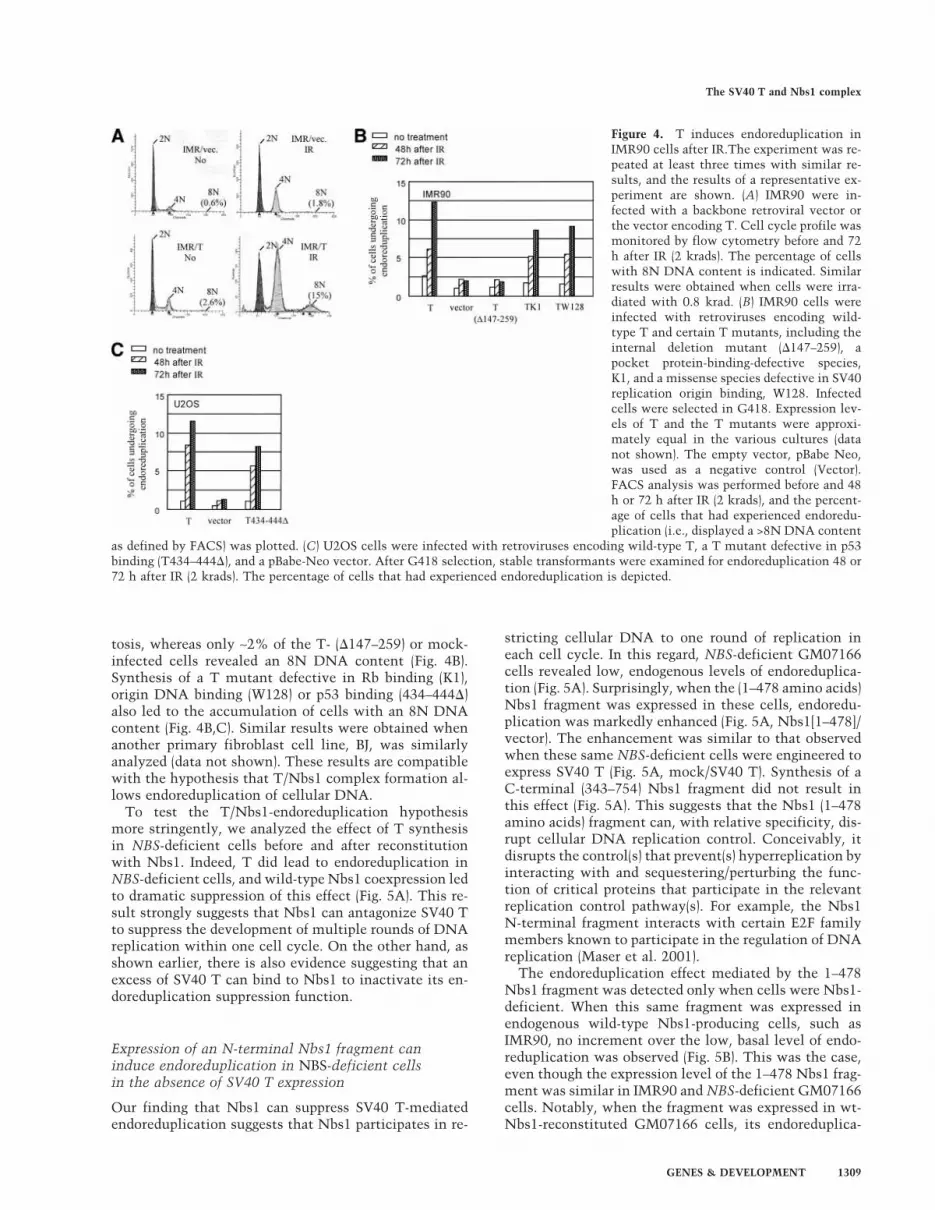
tosis, whereas only ∼2% of the T- (�147–259) or mock-infected cells revealed an 8N DNA content (Fig. 4B).Synthesis of a T mutant defective in Rb binding (K1),origin DNA binding (W128) or p53 binding (434–444�)also led to the accumulation of cells with an 8N DNAcontent (Fig. 4B,C). Similar results were obtained whenanother primary fibroblast cell line, BJ, was similarlyanalyzed (data not shown). These results are compatiblewith the hypothesis that T/Nbs1 complex formation al-lows endoreduplication of cellular DNA.
To test the T/Nbs1-endoreduplication hypothesismore stringently, we analyzed the effect of T synthesisin NBS-deficient cells before and after reconstitutionwith Nbs1. Indeed, T did lead to endoreduplication inNBS-deficient cells, and wild-type Nbs1 coexpression ledto dramatic suppression of this effect (Fig. 5A). This re-sult strongly suggests that Nbs1 can antagonize SV40 Tto suppress the development of multiple rounds of DNAreplication within one cell cycle. On the other hand, asshown earlier, there is also evidence suggesting that anexcess of SV40 T can bind to Nbs1 to inactivate its en-doreduplication suppression function.
Expression of an N-terminal Nbs1 fragment caninduce endoreduplication in NBS-deficient cellsin the absence of SV40 T expression
Our finding that Nbs1 can suppress SV40 T-mediatedendoreduplication suggests that Nbs1 participates in re-
stricting cellular DNA to one round of replication ineach cell cycle. In this regard, NBS-deficient GM07166cells revealed low, endogenous levels of endoreduplica-tion (Fig. 5A). Surprisingly, when the (1–478 amino acids)Nbs1 fragment was expressed in these cells, endoredu-plication was markedly enhanced (Fig. 5A, Nbs1[1–478]/vector). The enhancement was similar to that observedwhen these same NBS-deficient cells were engineered toexpress SV40 T (Fig. 5A, mock/SV40 T). Synthesis of aC-terminal (343–754) Nbs1 fragment did not result inthis effect (Fig. 5A). This suggests that the Nbs1 (1–478amino acids) fragment can, with relative specificity, dis-rupt cellular DNA replication control. Conceivably, itdisrupts the control(s) that prevent(s) hyperreplication byinteracting with and sequestering/perturbing the func-tion of critical proteins that participate in the relevantreplication control pathway(s). For example, the Nbs1N-terminal fragment interacts with certain E2F familymembers known to participate in the regulation of DNAreplication (Maser et al. 2001).
The endoreduplication effect mediated by the 1–478Nbs1 fragment was detected only when cells were Nbs1-deficient. When this same fragment was expressed inendogenous wild-type Nbs1-producing cells, such asIMR90, no increment over the low, basal level of endo-reduplication was observed (Fig. 5B). This was the case,even though the expression level of the 1–478 Nbs1 frag-ment was similar in IMR90 and NBS-deficient GM07166cells. Notably, when the fragment was expressed in wt-Nbs1-reconstituted GM07166 cells, its endoreduplica-
Figure 4. T induces endoreduplication inIMR90 cells after IR.The experiment was re-peated at least three times with similar re-sults, and the results of a representative ex-periment are shown. (A) IMR90 were in-fected with a backbone retroviral vector orthe vector encoding T. Cell cycle profile wasmonitored by flow cytometry before and 72h after IR (2 krads). The percentage of cellswith 8N DNA content is indicated. Similarresults were obtained when cells were irra-diated with 0.8 krad. (B) IMR90 cells wereinfected with retroviruses encoding wild-type T and certain T mutants, including theinternal deletion mutant (�147–259), apocket protein-binding-defective species,K1, and a missense species defective in SV40replication origin binding, W128. Infectedcells were selected in G418. Expression lev-els of T and the T mutants were approxi-mately equal in the various cultures (datanot shown). The empty vector, pBabe Neo,was used as a negative control (Vector).FACS analysis was performed before and 48h or 72 h after IR (2 krads), and the percent-age of cells that had experienced endoredu-plication (i.e., displayed a >8N DNA content
as defined by FACS) was plotted. (C) U2OS cells were infected with retroviruses encoding wild-type T, a T mutant defective in p53binding (T434–444�), and a pBabe-Neo vector. After G418 selection, stable transformants were examined for endoreduplication 48 or72 h after IR (2 krads). The percentage of cells that had experienced endoreduplication is depicted.
The SV40 T and Nbs1 complex
GENES & DEVELOPMENT 1309

tion-inducing effect was largely suppressed (Fig. 5C).This suggests that the Nbs1 fragment can disrupt cellu-lar DNA replication control only when endogenous,wild-type Nbs1 function is deficient. This implies thatNBS deficiency is a prerequisite for the Nbs1 N-terminalfragment to induce endoreduplication and suggests thatintact Nbs1 may establish a safeguard mechanism thatprevents DNA hyperreplication, even when certain rep-lication control pathways are disturbed.
NBS deficiency leads to reinitiation of DNAreplication at regions close to putative replicationinitiation sites
To more directly test the Nbs1 replication control hy-pothesis, we performed Fluorescence In Situ Hybridiza-tion (FISH) analysis on interphase cells using a DIG-la-beled probe for sequences at a specific site near the �-glo-bin locus where DNA replication is initiated (Kitsberg etal. 1993; Aladjem et al. 1995; Avni et al. 2003). FITC-conjugated secondary antibody against DIG was used todetect replication initiation foci at this site (Fig. 6A;Table 1A). Diploid cells in which the specific segment tobe probed is unreplicated should reveal two FISH dots,three dots if one of the two relevant replication originshas fired, or four dots if both have fired. If there are more
than four dots overlying two chromosomes, at least onesegment of the probed region has rereplicated in the rel-evant cell cycle.
In a culture of unperturbed IMR90, most cells con-tained two, three, or four dots, and only ∼2% of cellscontained more than four dots (Table 1A). In contrast,>20% of the aforementioned NBS-deficient cell line con-tained more than four dots—even in the unperturbedstate (Table 1A). Examples of NBS-deficient cells withtwo, three, four, or more than four replication dots areillustrated in Figure 6A. Expression of wild-type Nbs1 inNBS-deficient cells reduced the percentage of cells withmore than four dots by more than fourfold (Table 1A).Probes that recognize DNA regions containing knownsites of DNA synthesis initiation near the HSP70 andLamin B2 genes (Taira et al. 1994; Abdurashidova et al.2000) yielded similar results (data not shown). Almost allNBS-deficient cells (GM07166) were characterized bynormal chromosomal numbers.
We also performed FISH analysis on metaphase chro-mosomes. In >97% of IMR90 cells (wtNbs1), we detectedtwo pairs of dots at the �-globin replication initiationregion per cell, one on each chromatid, implying that, inG2-arrested cells, the adjacent DNA has replicated once(Table 1B). In contrast, ∼26% of NBS-deficient cells(GM07166) contained two or more �-globin replicationdots on one chromatid, and 10% contained two or more
Figure 5. Ectopic Nbs1 expression suppresses T-medi-ated endoreduplication. Each experiment was repeatedat least three times with similar results, and the resultsof a representative experiment are shown in each case.(A) The NBS-deficient cell line, GM07166, immortal-ized by expression of hTERT, was infected with retro-viruses encoding wild-type Nbs1 (Nbs1), myc-taggedwild-type Nbs1 (myc-Nbs1), 1–478 and 343–754 Nbs1fragments, and the empty pBabe-puro IRES-GFP vector(mock). After drug selection, the infected cells were su-perinfected with a wild-type T-encoding retrovirus(SV40 T) or empty vector (Vector). Standardized West-ern blotting revealed similar levels of T expression inall lines infected with T-encoding virus, and there wasequal expression of all transfected Nbs1 species in thisexperiment (data not shown). The percentage of cellsthat underwent endoreduplication is shown. (B) Ali-quots of IMR90 were infected with retroviruses encod-ing wild-type Nbs1 (Nbs1), myc-tagged wild-type Nbs1(myc-Nbs1), the 1–478 Nbs1 fragment, and the pBabepuro IRES-GFP vector (vector). All cultures were as-sayed for endoreduplication as noted above. IMR90 in-fected by a T-encoding virus underwent endoreduplica-tion, and they represented a positive control here. Ex-pression of the 1–478 Nbs1 N-terminal fragment didnot induce endoreduplication in IMR90 cells. (C) Themyc-Nbs1 (1–478) fragment was introduced into NBS-deficient cells GM07166 and its derivative reconsti-tuted with wild-type Nbs1 by retroviral infection.When the 1–478 Nbs1 fragment was expressed in theGM07166 cells, endoreduplication was detected. Thisphenotype was suppressed when the same fragmentwas expressed in GM07166 cells reconstituted withwild-type Nbs1.
Wu et al.
1310 GENES & DEVELOPMENT

dots on more than one chromatid (Fig. 6B; Table 1B).Occasionally, we observed that certain GM07166 cellscarry an additional Chromosome 11 (three copies ofChromosome 11), where the �-globin gene is located(<1%; Fig. 6B). These findings strongly suggest that NBSdeficiency permits more than one round of DNA repli-cation at certain loci close to replication origins. Theadditional round of DNA replication appears regionaland does not cover the entire chromosome, in keepingwith the prior observation that only low/background lev-els of gross chromosomal endoreduplication and hyper-ploidy were detected in NBS-deficient cells (see Fig. 5A).
Nbs1 suppresses SV40 T-mediated viralDNA replication
Our study suggests that SV40 T binds to Nbs1 and,thereby, perturbs the normal control of cellular DNAreplication initiation. One outcome appears to be a stateof cellular endoreduplication. This being the case, itseemed reasonable to ask whether this process is rel-evant to the phenomenon of autonomous replication ofthe viral genome, an event that depends on repetitivefiring of the viral origin in a single S phase.
In an effort to address this question, we introduced anintegrated SV40 replication origin into IMR90 cells byinfecting with a retroviral vector. The vector encodes themurine ecotropic retroviral receptor (ER) and contains anSV40 replication origin. Subsequently, these cells wereinfected with another retrovirus encoding wild-type T orwith an empty vector (pBaba-Neo-oric−) that carries aneo-resistance gene but lacks an SV40 replication origin.After several passages, genomic DNA from these cul-tures was purified and digested with SalI and HindIII, astep that results in the generation of an intact andunique SV40 origin-containing fragment detectable bySouthern blotting (Fig. 7A). The integrated SV40 origin-containing fragment became amplified when T was ex-pressed (Fig. 7B). In contrast, cells infected with emptyvector or T(�147–259), which encodes a protein defectivein replication origin binding, did not amplify this frag-ment (Fig. 7B).
We also asked whether wild-type Nbs1 affects this pro-cess. Forced expression of an ectopic, wild-type Nbs1allele (amino acids 1–754) in the NBS-deficient cells sup-pressed T-mediated viral DNA replication in these cells(Fig. 7C, cf. lanes 2 and 4). In contrast, the Nbs1(1–478)fragment led to enhanced T-mediated viral DNA repli-
Figure 6. NBS deficiency leads to rerep-lication of DNA sequences near replica-tion initiation sites. (A) RepresentativeFISH analysis performed on interphaseNBS-deficient cells (GM07166). Fixedcells were hybridized with a BAC probecorresponding to a segment covering the�-globin locus (clone RP11-645I8), andthe hybridization images appear as greenspots. This cloned genomic segment con-tains a putative replication origin (Kits-berg et al. 1993; Aladjem et al. 1995).Chromosomal DNA was stained withDAPI (blue). Examples of GM07166 cellsthat contain two, three, four, or more dotsat the �-globin locus are shown. (B) FISHof metaphase nuclei of GM07166 cells.Cells were treated with colchicine to se-lect for metaphase cells, and metaphasespreads were prepared. The fixed cellswere hybridized with the above-noted�-globin probe (green), and chromosomeswere stained with PI (red). FISH-positivesegments of chromatids are identifiedwith arrows in magnified copies of therelevant segments of each spread. In thevarious images are depicted GM07166nuclei containing two pairs of �-globindots (normal; one on each sister chroma-tid [sc]); multiple dots on one sc; multipledots on more than one sc; and three pairs
of dots each on a chromosome (indicating the presence of three No. 11 chromosomes). In some cases, the highly packed metaphasechromosomes likely “opened up” in certain locations during preparation, possibly allowing DNA to loop out, which, in turn, led toFISH signals extending out from the main body of the chromosome. An example is shown in the “multiple dots on one sc” and ismarked with a red arrow. Here, the amplification of the �-globin DNA sequence of interest in the indicated chromatid is reflected bythe presence of multiple FISH dots in a linear array extending out from the relevant chromatid.
The SV40 T and Nbs1 complex
GENES & DEVELOPMENT 1311

cation (Fig. 7C, cf. lanes 6 and 2), implying that the pro-cesses that elicit Nbs1(1–478)-dependent cellular endo-reduplication and autonomous firing of the SV40 repli-cation origin are, at least in part, related.
Thus, we propose that T targets Nbs1, suppressing itsability to down-regulate the autonomous firing of bothcellular and viral replication origins. The data also implythat Nbs1 is involved in suppressing the development ofmore than one round of replication in a given S phase andsuggest that Nbs1 regulates cellular and viral replicationby a related mechanism(s).
Discussion
SV40 induces cellular endoreduplication when it infectshost cells, and SV40 T is required for this process (Tegt-meyer 1972; Chou et al. 1974; Botchan et al. 1979). Themechanisms by which SV40 T drives more than oneround of cellular DNA replication in a given S phasehave been largely unclear. In this report, we have iden-tified an association of the Nbs1/Mre11/Rad50 complexwith SV40 T. The data suggest that this association un-derlies, at least in part, the ability of SV40 T to inactivatethe processes that guarantee one round of DNA replica-tion of cellular replicons in a given cell cycle. One out-come of this T-mediated perturbation is endoreduplica-tion of chromosomal DNA. For example, the T deletionmutant (�147–259) was both impaired in Nbs1 bindingand unable to induce host chromosomal endoreduplica-tion. This is consistent with the previous observationthat the N-terminal 1–259 region of T induced a tetra-ploid DNA content in CV-1 cells—a line that is permis-sive for viral DNA replication (Perry and Lehman 1998).Expression of SV40 T also induced much higher levels ofcellular endoreduplication in NBS-deficient cells than in
wild-type Nbs1-reconstituted isogenic cell lines. Thesefindings are compatible with a hypothesis in which SV40T targets Nbs1, thereby inactivating its rereplicationsuppression function and inducing multiple rounds ofcellular DNA replication in a given cell cycle.
Given the finding that excess Nbs1 antagonized T-me-diated cellular endoreduplication, one might predict thatNBS deficiency would result in signs of rereplication.Results of FISH analysis support this hypothesis. We ob-served that DNA at positions close to certain replicationorigins was hyperreplicated in >22% of NBS-deficientfibroblast cells (GM07166), although the data do not ex-plicitly demonstrate that rereplication initiated from therelevant origins. Where analyzed, ectopic expression ofwild-type Nbs1 efficiently suppressed this regional hy-perreplication phenomenon.
Although a large fraction of NBS-deficient cells spon-taneously rereplicated their DNA in regions close to rep-lication initiation sites, FACS analysis did not reveal sig-nificant numbers of cells with DNA contents of >8N.However, when the N-terminal fragment (amino acids1–478) of Nbs1 was expressed in NBS-deficient cells, pro-found endoreduplication was observed independent ofSV40 T function. This phenomenon was observed onlyin NBS-deficient cells, and not in NBS-proficient cellssuch as IMR90. Taken together, these findings, in part,suggest that Nbs1 may contribute to the activation of acheckpoint that prevents additional refiring of replica-tion origins that have already fired.
However, florid DNA rereplication requires additionalmolecular disruptions beyond simple loss of NBS func-tion. It appears likely that the rereplication suppres-sion function of Nbs1 is inhibited following SV40 T/Nbs1 complex formation. Yet, because T was requiredto maximize endoreduplication in NBS-deficient cells,
Table 1. �-Globin locus FISH analysis of NBS-deficient cells (GM07166)
A) FISH of unsynchronized cellsa
Cell linesTotal
number 2 dots 3 or 4 dots >4 dots
IMR90 807 423 (52.4%) 365 (45.2%) 19 (2.4%)NBS−/− 219 67 (30.6%) 102 (46.6%) 50 (22.8%)NBS−/− (Nbslwt) 432 232 (53.7%) 177 (41.0%) 23 (5.3%)
B) FISH of metaphase nucleib
Cell linesTotal
number 1 on each sc 2 or more on one sc2 or more on
more than one sc
IMR90 129 125 (96.9%) 4 (3.1%) 0 (0.0%)NBS−/− 110 70 (63.6%) 29 (26.4%) 11 (10.0%)
A BAC clone containing a large DNA segment (clone RP11-645I8) of the �-globin locus, which includes a DNA replication initiationsite (Kitsberg et al. 1993; Aladjem et al. 1995), was used as a FISH probe.aInterphase nuclei from primary IMR90 cells, NBS-deficient GM07166 cells (NBS), and wild-type (wt) Nbs1-reconstituted GM07166cells [NBS−/− (Nbslwt)] were analyzed by FISH using this probe. The number of cells containing two, three, four, or more than four dotsat the �-globin locus is indicated.bMetaphase nuclei of IMR90 and GM07166 cells were analyzed by FISH, using the above-noted probe. The number of IMR90 andGM07166 cells with two pairs of �-globin dots, one on each sister chromatid (sc one on each sc), with multiple dots on one sc (twoor more on one sc), and with multiple dots on more than one sc (two or more on more than one sc), are shown. The percentage of cellsin each category is shown in parentheses.
Wu et al.
1312 GENES & DEVELOPMENT

florid, T-mediated endoreduplication likely requires atleast one as-yet-undefined function of this viral protein.Whatever this step(s) may be, like T, the 1–478 fragmentof Nbs1 also induced marked endoreduplication. Nota-bly, it was only detected in NBS-deficient cells. Conceiv-ably, unopposed by wild-type Nbs1, this mutant poly-peptide interacts in an abnormal manner with certaincell protein(s) (which might include one or more poly-peptides with which intact Nbs1 normally interacts), anoutcome of which is a gross override of rereplication con-trol. It will eventually be interesting to determinewhether T and this particular Nbs1 mutant operate byperturbing the same pathway(s).
Marked amplification of the viral genome is essentialfor SV40 propagation, but the factors that optimize thisprocess have, in the past, been incompletely defined. Inthis regard, we observed that a chromosomally inte-grated SV40 origin-containing segment was more exten-sively amplified in NBS-deficient than in wild-typeNbs1-reconstituted GM07166. This suggests that, by tar-geting Nbs1, SV40 T creates an environment that con-tributes to maximal amplification of its own genome.
Results suggesting that Nbs1 regulates DNA replica-tion control—both cellular and viral—are consistentwith the observation that Nbs1 interacts with E2F1, a
protein known to associate with certain replication ini-tiation sites, and locates near these regions in S-phasecells (Maser et al. 1997). Because T can also perturb E2F1function by displacing it from the Rb protein, one won-ders whether it can also displace it and its partner Nbs1from certain replication origins. This is, however, notlikely the case, because the association of Nbs1 with the�-globin and B2 lamin replication initiation sites was notaffected by T, as revealed by the results of chromatinimmunoprecipitation (ChIP) assays (X. Wu and D. Liv-ingston, unpubl.).
Nbs1 also operates in S-phase checkpoint control. Inresponse to IR delivered during S phase, the protein be-comes phosphorylated by ATM at several sites, andthese phosphorylation events are important for Nbs1function in the ensuing intrareplication checkpoint (Limet al. 2000). Although it remains unclear how Nbs1 con-tributes to the slowdown of DNA replication followingDNA damage, it is conceivable that, when S-phase DNAdamage is sensed or after replicons have fired during anormal cell cycle, Nbs1 communicates with the DNAreplication machinery to ensure that no further initia-tion events occur.
However, it should also be noted that the signals thatactivate Nbs1 checkpoint function and its hyperreplica-
Figure 7. Nbs1 suppresses replication initiationfrom the SV40 replication origin. (A) The SV40 rep-lication origin present in the pBabe vector used inthis experiment is flanked by SalI and HindIII re-striction sites. The indicated probe was used forSouthern blot analysis. Genomic DNA surroundedthe viral replication origin may be amplified whenthe viral origin fires multiple times in a given cellcycle. (B) IMR90 cells were infected by a retrovirus(pBabe-hygro-ER) encoding a murine ecotropic retro-viral receptor (ER). The vector also contained anSV40 replication origin. Cells were subsequently in-fected with retroviruses encoding wild-type T, theT(�147–259) mutant, or empty vector pBabe-Neo-oric− (Vec.). Standardized quantities of purified ge-nomic DNA from the indicated cultures were di-gested with SalI and HindIII. Southern blot analysiswas performed using the SalI to HindIII probe iden-tified in A. An EtBr-stained agarose gel depicts thequantities of SalI + HindIII-digested genomic DNAanalyzed. The expression of T and the deletion mu-tant T(�147–259) was assessed by Western blotanalysis. (C) NBS-deficient cells (GM07166) immor-talized by hTERT were infected with pBabe-hygro-ER containing an SV40 origin. They were then re-constituted by infection with myc-tagged wild-typeNbs1 retrovirus, myc-Nbs1 fragment (1–478) virus,or empty vector virus. T or empty vector (Vec.) viruswas subsequently used to superinfect these cultures.Southern blot analysis of standardized amounts ofSalI- and HindIII-digested genomic DNA from eachof these cultures was then performed in search of
SV40 origin-containing DNA, using the above-noted SalI–HindIII fragment as probe (see A). An EtBr-stained gel revealed the quantityof DNA from each culture that was loaded onto the relevant gel. The expression level of T in each culture is shown by Westernblotting.
The SV40 T and Nbs1 complex
GENES & DEVELOPMENT 1313

tion suppression function might be different. For ex-ample, T–Nbs1 complex formation did not interfere withDNA damage-induced Nbs1 phosphorylation (Gatei etal. 2000; Lim et al. 2000; Wu et al. 2000; Zhao et al.2000), and one or more of these phosphorylation eventsparticipates in the activation of the intrareplicationcheckpoint (Lim et al. 2000). Consistently, this check-point operated normally in the presence of T. In contrast,the interaction of T and Nbs1 did interfere with the func-tion of Nbs1 in preventing hyperreplication. How signalsare generated and transduced to Nbs1 when the duplica-tion of a given replicon has been initiated and/or is com-plete remains unclear.
Nbs1 forms a tight complex with Mre11 and Rad50(Carney et al. 1998). Both of these proteins are essentialfor DSB repair (Johzuka and Ogawa 1995; Moore andHaber 1996). In mammalian cells, damage-induced focicontaining Mre11 are detected during S phase, and focusformation depends on Nbs1 function (Maser et al. 1997;Carney et al. 1998; Dong et al. 1999). Depletion of Mre11from Xenopus extracts leads to dramatic accumulationof DSBs during DNA replication (Costanzo et al. 2001).The Nbs1 S-phase checkpoint function suppresses DNAreplication initiation once damage is sensed, and Nbs1activates Mre11 and Rad50 through a direct association,leading to repair of the relevant DNA damage. The stud-ies described in this report reveal a new function forNbs1 in preventing DNA hyperreplication during thecell cycle. This finding reinforces the notion that Nbs1 isa multifunctional protein that contributes to properDNA replication and the maintenance of genome stabil-ity.
Materials and methods
Cell culture
GM07166 was obtained from the Coriell Human Mutant CellRepository. IMR90, 293T, 293, and U2OS cells were fromATCC. Murine ecotropic retroviral receptor (ER; Albritton et al.1989) was first introduced into IMR90 and GM07166 cells byretroviral infection. These viruses were generated using thePhoenix amphotropic packaging cell line from ATCC. Subse-quently wild-type Nbs1, Nbs1 mutants, wild-type T, and T mu-tants were introduced into these ER+ cells with suitable recom-binant murine ecotropic retroviruses generated in Phoenix eco-tropic packaging cells (ATCC). GM07166 cells wereimmortalized by infection with a retrovirus (pBabe/hygro) en-coding hTERT (Meyerson et al. 1997). U2OS cells that stablyexpress SV40 T [U2OS(T)] were generated by transfecting U2OScells with CMV T neo (Campbell et al. 1997) and selecting forG418-resistant cells.
Antibodies
Monoclonal antibodies against SV40 T (pAB419 and pAB416)and against Nbs1 (EE15 and D29) have been described previ-ously (Harlow et al. 1981; Wu et al. 2000). Polyclonal Ab (D27)against Mre11 was raised against the C-terminal fragment ofhuman Mre11 (Mre11360–709). Monoclonal antibody againstRad50 was purchased from Novus Biologicals.
Plasmids
Full-length Nbs1 and Nbs1 fragments were subcloned into amammalian expression vector, pCDNA3�, that carries the se-quence encoding the myc epitope (Chen et al. 1998). Full-lengthT and its derivative mutants were subcloned into eitherpCDNA3� (Scully et al. 1997) or pSG5 (Stratagene). Alleles en-coding full-length Nbs1 with or without a myc-tag and myc-tagged Nbs1 fragments (1–478, 343–754) were inserted into aretroviral vector, pBabe-puro, that was modified by adding anIRES-EGFP sequence from pIRES-EGFP (Clontech). The retro-viral vector, pBabe/neo/oric−, was generated by replacing theSV40 promoter of pBabe/neo with the comparable DNA regionof pBaba/puro/oric−, a gift from Kathy Rundell (NorthwesternUniversity, Chicago, IL). The retroviral vector, pBabe-Blastici-dine, encoding ER, was constructed by replacing the Hygromarker of pBabe-hygro-ER with a Blasticidine marker, a giftfrom Peiqing Sun (The Scripps Research Institute, LaJolla, CA).Detailed information on plasmid construction will be providedupon request.
Immunoassays
Cells were lysed in NETN (150 mM NaCl, 1 mM EDTA, 20 mMTris-Cl at pH 8.0, 0.5% NP-40). Primary antibodies were incu-bated with cell lysates at 4°C for 3 h, followed by adding proteinA- or G-Sepharose beads for another hour’s incubation. Beadswere washed four times with NETN before SDS-gel samplebuffer was added. Primary antibodies were routinely rockedwith blots overnight at 4°C. HRP-conjugated secondary anti-bodies were used at 1:3000 dilution (Amersham).
For immunostaining, cells were fixed in 70% methanol and30% acetone for 15 min at −20°C. After 1 h of drying at roomtemperature and three subsequent PBS washes, cells werestained with antibody recognizing Nbs1 (rabbit polyclonal; On-cogene) and/or T (monoclonal pAB419) for 4 h at room tempera-ture. Subsequently, cells were stained with FITC-conjugatedanti-mouse and Rhodamine-conjugated anti-rabbit antibodies(Jackson Immuno-Research) for 1 h at room temperature.
Analysis of DNA content by flow cytometry
At each time point, cells were trypsinized, washed once withPBS, and fixed with prechilled 70% ethanol. After incubating forseveral hours or overnight at 4°C, cells were washed with PBSand resuspended in a buffer containing 38 mM Na Citrate, 70µM propidium iodide (PI), and 20 µg/mL RNase A, incubated for30 min at 37°C in the dark, and subjected to flow cytometryanalysis.
Fluorescence In Situ Hybridization (FISH) analysis
FISH was performed as described (Heng et al. 1992; Heng andTsui 1993; Boggs and Chinault 1997). Human BAC clones thatwere used as probes for DNA sequences at the B2-Lamin,HSP70, and �-globin loci were obtained through the Children’sHospital Oakland Research Institute (CHORI). The BAC clonenumbers are: B2-Lamin, RP11-211I3; HSP70, RP11-425A7; and�-globin, RP11-645I8.
Acknowledgments
We thank James DeCaprio and Ole Gjoerup for sharing SV40 TDNA-encoding plasmids and for many helpful discussions. Wealso thank William Hahn and Robert Weinberg for providingpBabe-hygro-hTERT, Peiqing Sun for pBabe-hygro-ER, and
Wu et al.
1314 GENES & DEVELOPMENT

Kathy Rundell for pBabe-puro-oric−. William Hahn graciouslyhelped us in the immortalization of GM07166 with hTERT.
The publication costs of this article were defrayed in part bypayment of page charges. This article must therefore be herebymarked “advertisement” in accordance with 18 USC section1734 solely to indicate this fact.
References
Abdurashidova, G., Deganuto, M., Klima, R., Riva, S., Biamonti,G., Giacca, M., and Falaschi, A. 2000. Start sites of bidirec-tional DNA synthesis at the human lamin B2 origin. Science287: 2023–2026.
Aladjem, M.I., Groudine, M., Brody, L.L., Dieken, E.S., Fournier,R.E., Wahl, G.M., and Epner, E.M. 1995. Participation of thehuman �-globin locus control region in initiation of DNAreplication. Science 270: 815–819.
Albritton, L.M., Tseng, L., Scadden, D., and Cunningham, J.M.1989. A putative murine ecotropic retrovirus receptor geneencodes a multiple membrane-spanning protein and conferssusceptibility to virus infection. Cell 57: 659–666.
Ali, S.H. and DeCaprio, J.A. 2001. Cellular transformation bySV40 large T antigen: Interaction with host proteins. Semin.Cancer Biol. 11: 15–23.
Avni, D., Yang, H., Martelli, F., Hofmann, F., ElShamy, W.M.,Ganesan, S., Scully, R., and Livingston, D.M. 2003. Activelocalization of the retinoblastoma protein in chromatin andits response to S phase DNA damage. Mol. Cell 12: 735–746.
Boggs, B.A. and Chinault, A.C. 1997. Analysis of DNA replica-tion by fluorescence in situ hybridization. Methods 13: 259–270.
Botchan, M., Topp, W., and Sambrook, J. 1979. Studies on sim-ian virus 40 excision from cellular chromosomes. ColdSpring Harb. Symp. Quant. Biol. 43: 709–719.
Campbell, K.S., Mullane, K.P., Aksoy, I.A., Stubdal, H., Zalvide,J., Pipas, J.M., Silver, P.A., Roberts, T.M., Schaffhausen, B.S.,and DeCaprio, J.A. 1997. DnaJ/hsp40 chaperone domain ofSV40 large T antigen promotes efficient viral DNA replica-tion. Genes & Dev. 11: 1098–1110.
Carney, J.P., Maser, R.S., Olivares, H., Davis, E.M., Le Beau, M.,Yates III, J.R., Hays, L., Morgan, W.F., and Petrini, J.H. 1998.The hMre11/hRad50 protein complex and Nijmegen break-age syndrome: Linkage of double-strand break repair to thecellular DNA damage response. Cell 93: 477–486.
Challberg, M.D. and Kelly, T.J. 1989. Animal virus DNA repli-cation. Annu. Rev. Biochem. 58: 671–717.
Chen, J., Silver, D.P., Walpita, D., Cantor, S.B., Gazdar, A.F.,Tomlinson, G., Couch, F.J., Weber, B.L., Ashley, T., Living-ston, D.M., et al. 1998. Stable interaction between the prod-ucts of the BRCA1 and BRCA2 tumor suppressor genes inmitotic and meiotic cells. Mol. Cell 2: 317–328.
Chou, J.Y., Avila, J., and Martin, R.G. 1974. Viral DNA synthe-sis in cells infected by temperature-sensitive mutants ofsimian virus 40. J. Virol. 14: 116–124.
Costanzo, V., Robertson, K., Bibikova, M., Kim, E., Grieco, D.,Gottesman, M., Carroll, D., and Gautier, J. 2001. Mre11 pro-tein complex prevents double-strand break accumulationduring chromosomal DNA replication. Mol. Cell 8: 137–147.
Diffley, J.F. 1996. Once and only once upon a time: Specifyingand regulating origins of DNA replication in eukaryoticcells. Genes & Dev. 10: 2819–2830.
———. 2001. DNA replication: Building the perfect switch.Curr. Biol. 11: R367–R370.
Dong, Z., Zhong, Q., and Chen, P.L. 1999. The Nijmegen break-age syndrome protein is essential for Mre11 phosphorylation
upon DNA damage. J. Biol. Chem. 274: 19513–19516.Featherstone, C. and Jackson, S.P. 1998. DNA repair: The
Nijmegen breakage syndrome protein. Curr. Biol. 8: R622–R625.
Friedrich, T.D., Laffin, J., and Lehman, J.M. 1992. Simian virus40 large T-antigen function is required for induction of tet-raploid DNA content during lytic infection. J. Virol. 66:4576–4579.
———. 1994. Induction of tetraploid DNA content by simianvirus 40 is dependent on T-antigen function in the G2 phaseof the cell cycle. J. Virol. 68: 4028–4030.
Gatei, M., Young, D., Cerosaletti, K.M., Desai-Mehta, A.,Spring, K., Kozlov, S., Lavin, M.F., Gatti, R.A., Concannon,P., and Khanna, K. 2000. ATM-dependent phosphorylationof nibrin in response to radiation exposure. Nat. Genet. 25:115–119.
Harlow, E., Crawford, L.V., Pim, D.C., and Williamson, N.M.1981. Monoclonal antibodies specific for simian virus 40 tu-mor antigens. J. Virol. 39: 861–869.
Hatanaka, M. and Dulbecco, R. 1966. Induction of DNA syn-thesis by SV40. Proc. Natl. Acad. Sci. 56: 736–740.
Heng, H.H. and Tsui, L.C. 1993. Modes of DAPI banding andsimultaneous in situ hybridization. Chromosoma 102: 325–332.
Heng, H.H., Squire, J., and Tsui, L.C. 1992. High-resolutionmapping of mammalian genes by in situ hybridization to freechromatin. Proc. Natl. Acad. Sci. 89: 9509–9513.
Hurwitz, J., Dean, F.B., Kwong, A.D., and Lee, S.H. 1990. The invitro replication of DNA containing the SV40 origin. J. Biol.Chem. 265: 18043–18046.
Johzuka, K. and Ogawa, H. 1995. Interaction of Mre11 andRad50: Two proteins required for DNA repair and meiosis-specific double-strand break formation in Saccharomycescerevisiae. Genetics 139: 1521–1532.
Kalderon, D. and Smith, A.E. 1984. In vitro mutagenesis of aputative DNA binding domain of SV40 large-T. Virology139: 109–137.
Kierstead, T.D. and Tevethia, M.J. 1993. Association of p53binding and immortalization of primary C57BL/6 mouse em-bryo fibroblasts by using simian virus 40 T-antigen mutantsbearing internal overlapping deletion mutations. J. Virol. 67:1817–1829.
Kitsberg, D., Selig, S., Keshet, I., and Cedar, H. 1993. Replicationstructure of the human �-globin gene domain. Nature 366:588–590.
Lai, J.S. and Herr, W. 1992. Ethidium bromide provides a simpletool for identifying genuine DNA-independent protein asso-ciations. Proc. Natl. Acad. Sci. 89: 6958–6962.
Lim, D.S., Kim, S.T., Xu, B., Maser, R.S., Lin, J., Petrini, J.H., andKastan, M.B. 2000. ATM phosphorylates p95/nbs1 in an S-phase checkpoint pathway. Nature 404: 613–617.
Lin, J.Y. and DeCaprio, J.A. 2003. SV40 large T antigen pro-motes dephosphorylation of p130. J. Biol. Chem. 278: 46482–46487.
Luo, G., Yao, M.S., Bender, C.F., Mills, M., Bladl, A.R., Bradley,A., and Petrini, J.H. 1999. Disruption of mRad50 causes em-bryonic stem cell lethality, abnormal embryonic develop-ment, and sensitivity to ionizing radiation. Proc. Natl. Acad.Sci. 96: 7376–7381.
Maser, R.S., Monsen, K.J., Nelms, B.E., and Petrini, J.H. 1997.hMre11 and hRad50 nuclear foci are induced during the nor-mal cellular response to DNA double-strand breaks. Mol.Cell. Biol. 17: 6087–6096.
Maser, R.S., Mirzoeva, O.K., Wells, J., Olivares, H., Williams,B.R., Zinkel, R.A., Farnham, P.J., and Petrini, J.H. 2001.Mre11 complex and DNA replication: Linkage to E2F and
The SV40 T and Nbs1 complex
GENES & DEVELOPMENT 1315

sites of DNA synthesis. Mol. Cell. Biol. 21: 6006–6016.McVey, D., Brizuela, L., Mohr, I., Marshak, D.R., Gluzman, Y.,
and Beach, D. 1989. Phosphorylation of large tumour antigenby cdc2 stimulates SV40 DNA replication. Nature 341: 503–507.
Meyerson, M., Counter, C.M., Eaton, E.N., Ellisen, L.W.,Steiner, P., Caddle, S.D., Ziaugra, L., Beijersbergen, R.L.,Davidoff, M.J., Liu, Q., et al. 1997. hEST2, the putative hu-man telomerase catalytic subunit gene, is up-regulated intumor cells and during immortalization. Cell 90: 785–795.
Mirzoeva, O.K. and Petrini, J.H. 2001. DNA damage-dependentnuclear dynamics of the Mre11 complex. Mol. Cell. Biol. 21:281–288.
Moore, J.K. and Haber, J.E. 1996. Cell cycle and genetic require-ments of two pathways of nonhomologous end-joining repairof double-strand breaks in Saccharomyces cerevisiae. Mol.Cell. Biol. 16: 2164–2173.
Nelms, B.E., Maser, R.S., MacKay, J.F., Lagally, M.G., and Pet-rini, J.H. 1998. In situ visualization of DNA double-strandbreak repair in human fibroblasts. Science 280: 590–592.
Paulovich, A.G. and Hartwell, L.H. 1995. A checkpoint regu-lates the rate of progression through S phase in S. cerevisiaein response to DNA damage. Cell 82: 841–847.
Peden, K.W., Spence, S.L., Tack, L.C., Cartwright, C.A., Sriniva-san, A., and Pipas, J.M. 1990. A DNA replication-positivemutant of simian virus 40 that is defective for transforma-tion and the production of infectious virions. J. Virol. 64:2912–2921.
Perry, M.B. and Lehman, J.M. 1998. Activities of SV40 T antigennecessary for the induction of tetraploid DNA content inpermissive CV-1 cells. Cytometry 31: 251–259.
Prives, C. 1990. The replication functions of SV40 T antigen areregulated by phosphorylation. Cell 61: 735–738.
Santocanale, C. and Diffley, J.F. 1998. A Mec1- and Rad53-de-pendent checkpoint controls late-firing origins of DNA rep-lication. Nature 395: 615–618.
Scully, R., Chen, J., Plug, A., Xiao, Y., Weaver, D., Feunteun, J.,Ashley, T., and Livingston, D.M. 1997. Association ofBRCA1 with Rad51 in mitotic and meiotic cells. Cell 88:265–275.
Seigneur, M., Ehrlich, S.D., and Michel, B. 2000. RuvABC-de-pendent double-strand breaks in dnaBts mutants requirerecA. Mol. Microbiol. 38: 565–574.
Shiloh, Y. 1997. Ataxia-telangiectasia and the Nijmegen break-age syndrome: Related disorders but genes apart. Annu. Rev.Genet. 31: 635–662.
Shirahige, K., Hori, Y., Shiraishi, K., Yamashita, M., Takahashi,K., Obuse, C., Tsurimoto, T., and Yoshikawa, H. 1998. Regu-lation of DNA-replication origins during cell-cycle progres-sion. Nature 395: 618–621.
Simmons, D.T., Wun-Kim, K., and Young, W. 1990. Identifica-tion of simian virus 40 T-antigen residues important for spe-cific and nonspecific binding to DNA and for helicase activ-ity. J. Virol. 64: 4858–4865.
Stillman, B. 1989. Initiation of eukaryotic DNA replication invitro. Annu. Rev. Cell Biol. 5: 197–245.
Taira, T., Iguchi-Ariga, S.M., and Ariga, H. 1994. A novel DNAreplication origin identified in the human heat shock protein70 gene promoter. Mol. Cell. Biol. 14: 6386–6397.
Tegtmeyer, P. 1972. Simian virus 40 deoxyribonucleic acid syn-thesis: The viral replicon. J. Virol. 10: 591–598.
Varon, R., Vissinga, C., Platzer, M., Cerosaletti, K.M., Chrza-nowska, K.H., Saar, K., Beckmann, G., Seemanova, E., Coo-per, P.R., Nowak, N.J., et al. 1998. Nibrin, a novel DNAdouble-strand break repair protein, is mutated in Nijmegenbreakage syndrome. Cell 93: 467–476.
Wu, X., Ranganathan, V., Weisman, D.S., Heine, W.F., Ciccone,D.N., O’Neill, T.B., Crick, K.E., Pierce, K.A., Lane, W.S.,Rathbun, G., et al. 2000. ATM phosphorylation of Nijmegenbreakage syndrome protein is required in a DNA damageresponse [see comments]. Nature 405: 477–482.
Wun-Kim, K., Upson, R., Young, W., Melendy, T., Stillman, B.,and Simmons, D.T. 1993. The DNA-binding domain of sim-ian virus 40 tumor antigen has multiple functions. J. Virol.67: 7608–7611.
Xiao, Y. and Weaver, D.T. 1997. Conditional gene targeted de-letion by Cre recombinase demonstrates the requirement forthe double-strand break repair Mre11 protein in murine em-bryonic stem cells. Nucleic Acids Res. 25: 2985–2991.
Yamaguchi-Iwai, Y., Sonoda, E., Sasaki, M.S., Morrison, C.,Haraguchi, T., Hiraoka, Y., Yamashita, Y.M., Yagi, T.,Takata, M., Price, C., et al. 1999. Mre11 is essential for themaintenance of chromosomal DNA in vertebrate cells.EMBO J. 18: 6619–6629.
Zhao, S., Weng, Y.C., Yuan, S.S., Lin, Y.T., Hsu, H.C., Lin, S.C.,Gerbino, E., Song, M.H., Zdzienicka, M.Z., Gatti, R.A., et al.2000. Functional link between ataxia-telangiectasia andNijmegen breakage syndrome gene products [see com-ments]. Nature 405: 473–477.
Zhu, J., Petersen, S., Tessarollo, L., and Nussenzweig, A. 2001.Targeted disruption of the Nijmegen breakage syndromegene NBS1 leads to early embryonic lethality in mice. Curr.Biol. 11: 105–109.
Zou, H. and Rothstein, R. 1997. Holliday junctions accumulatein replication mutants via a RecA homolog-independentmechanism. Cell 90: 87–96.
Wu et al.
1316 GENES & DEVELOPMENT


