Embed Size (px)
Citation preview

The
Journ
al o
f Exp
erim
enta
l M
edic
ine
ARTICLE
1087
JEM Vol. 202, No. 8, October 17, 2005 1087–1098 www.jem.org/cgi/doi/10.1084/jem.20042530
Chemokine receptor CCR5 promotes leukocyte trafficking to the brain and survival in West Nile virus infection
William G. Glass,
1
Jean K. Lim,
1
Rushina Cholera,
1
Alexander G. Pletnev,
2
Ji-Liang Gao,
1
and Philip M. Murphy
1
1
Laboratory of Molecular Immunology and
2
Laboratory of Infectious Diseases, National Institute of Allergy and Infectious Diseases, National Institutes of Health, Bethesda, MD 20892
The molecular immunopathogenesis of West Nile virus (WNV) infection is poorly understood. Here, we characterize a mouse model for WNV using a subcutaneous route of infection and delineate leukocyte subsets and immunoregulatory factors present in the brains of infected mice. Central nervous system (CNS) expression of the chemokine receptor CCR5 and its ligand CCL5 was prominently up-regulated by WNV, and this was associated with CNS infiltration of CD4
�
and CD8
�
T cells, NK1.1
�
cells and macrophages expressing the receptor. The significance of CCR5 in pathogenesis was established by mortality studies in which infection of CCR5
�
/
�
mice was rapidly and uniformly fatal. In the brain, WNV-infected CCR5
�
/
�
mice had increased viral burden but markedly reduced NK1.1
�
cells, macrophages, and CD4
�
and CD8
�
T cells compared with WNV-infected CCR5
�
/
�
mice. Adoptive transfer of splenocytes from WNV-infected CCR5
�
/
�
mice into infected CCR5
�
/
�
mice increased leukocyte accumulation in the CNS compared with transfer of splenocytes from infected CCR5
�
/
�
mice into infected CCR5
�
/
�
mice, and increased survival to 60%, the same as in infected CCR5
�
/
�
control mice. We conclude that CCR5 is a critical antiviral and survival determinant in WNV infection of mice that acts by regulating trafficking of leukocytes to the infected brain.
West Nile virus (WNV) is an enveloped, neu-rotropic, single stranded (
�
) sense RNAflavivirus, first isolated from a woman inUganda in 1937 (1). Since then it has reemergedmultiple times: first in Europe, and most recentlyduring the summer of 1999 in the northeasternUnited States (2, 3). The virus, which hasspread rapidly throughout North America,may cause severe illness especially in the elderly(manifested by meningitis and encephalitis)that may lead to paralysis, coma, and death.During the 1999–2003 outbreaks of WNV inthe USA, 13,491 cases were reported, including533 deaths (
�
4%) (3, 4).A zoonosis, WNV is maintained in a natural
cycle between avian hosts and mosquito vectors,although humans, horses, and several othermammals can also serve as hosts (5–8). Thelevel of exposure required for infection anddisease is undefined; however, epidemiologicstudies indicate that
�
80% of WNV infection
remains subclinical whereas
�
20% progressesto a febrile illness. Of these,
�
30% of cases in theUS progress to meningitis and/or encephalitis.Survivors of severe neurological disease typicallyrequire prolonged rehabilitation. There is novaccine or specific antiviral treatment for WNVinfection in humans.
Although immunocompromised hosts appearto have increased susceptibility to WNV, specificinformation on the human innate and adaptivecellular immune response to the virus is limited(2, 9–11). Several mouse models of infection thatdiffer with regard to mouse and viral strain, routeof infection, and clinical outcome have beendeveloped to provide insight into immunopatho-genesis (12–15). We have used a model in whichthe highly neurotropic and neurovirulent WNV-NY99 strain is injected s.c., resulting in viralreplication and leukocyte infiltration in thebrain 4–7 d after infection (13, 16–19). Avariant of this model has been used previouslyto demonstrate that both B and T cells are re-quired for viral clearance, because 100% of bothRAG1
�
/
�
mice and
�
MT
�
/
�
mice rapidlysuccumb to viral infection, whereas only
�
35%
W.G. Glass and J.K. Lim contributed equally to this work.W.G. Glass’s present address is Centocor Global R&D, Infectious Diseases, Radnor, PA 19087.
CORRESPONDENCEPhilip M. Murphy: [email protected]
Abbreviations used: CNS, central nervous system; ffu, focus-forming unit; WNV, West Nile virus.
on June 28, 2015jem
.rupress.orgD
ownloaded from
Published October 17, 2005

CCR5 AND WEST NILE VIRUS PATHOGENESIS | Glass et al.
1088
of mice with intact immune systems die (19, 20). Theprimary role of humoral immunity is thought to limitviral dissemination to the central nervous system (CNS).CD8
�
T cells play a prominent role in viral clearancefrom the CNS, as infected CD8
�
/
�
mice have increasedmortality (
�
85%) and higher CNS viral burden and viralpersistence (13).
Encephalitis caused by other neurotropic/neurovirulentviruses is often characterized by CNS entry of macrophages,CD4
�
and CD8
�
T cells, NKT cells and dendritic cells.However, little information is available regarding the molec-ular factors responsible for their recruitment. Here, we pro-vide a profile of the leukocyte subsets and proinflammatoryand immunoregulatory molecules present in the CNS of
Figure 1. West Nile virus induces production of specific immuno-regulatory factors in the brains of C57BL/6 mice. (A) RNA analysis by RPA. Leftmost lane in each of the four panels is unprotected probe, and the factors analyzed are named to the left; note that the protected fragments run slightly lower on gel. The names of factors scoring positive in the assay are given to the right of each band. GAPDH is provided as an RNA loading control. Time after infection (days), is indicated at the top of each panel. Mock-infected mice were analyzed on day 7 of the experiment. Each lane on each panel corresponds to RNA from a single mouse brain. The results shown are from two independent experiments with six mice analyzed
for each time point, except for mock-infected mice (n � 3). Lanes corresponding in location on each of the four gels represent RNA from the same mouse. (B) RNA analysis by RT-PCR. �-Actin is provided as a loading control. Mock-infected mice were analyzed at day 7. Each lane represents a single mouse. (C) Protein analysis. The factor analyzed is identified at the top left of each graph. Time and virus variables for each factor are specified at the bottom of the panel. Data were pooled from three experiments with three to five in each group, and are presented asmean � SEM pg/g protein in supernatants from total brain homogenates as a function of time after infection.
on June 28, 2015jem
.rupress.orgD
ownloaded from
Published October 17, 2005
JEM VOL. 202, October 17, 2005
1089
ARTICLE
WNV-infected mice. We have used this profile to identifythe chemokine receptor CCR5 as a candidate protective fac-tor, and have tested this hypothesis genetically using CCR5knockout mice.
RESULTS
C57BL/6 mice were infected s.c. with 10
0
–10
6
focus-formingunits (ffu) of strain WNV-NY99, which imitates the naturalroute of WNV infection in man. All mice infected with 1 or 10ffu survived. Mortality was first observed in animals injectedwith 100 ffu and was 30% at that dose (unpublished data). Mor-tality was relatively insensitive to further increases in WNVdose reaching only 40% at 10
6
ffu, the highest dose tested. Thisis consistent with previously published data for WNV-NY99infection of this strain of mice using a footpad injection (19).Based on these results, we infected mice with 10
4
ffu WNV s.c.for all subsequent studies. Using this dose, all deaths occurredbetween 7 and 12 d after infection.
WNV infection induces immunoregulatory mediators in the CNS
To identify molecules that might be important in WNV en-cephalitis, we screened a total of 45 different immunoregula-tory factors for differential expression in the brains of mock-and WNV-infected mice on days 7 and 12 after infection.Of these, 37 (9 cytokines, 8 cytokine receptors, 11 chemo-kines, and 9 chemokine receptors) were screened at theRNA level by either RPA or RT-PCR of mouse brain (Fig.1, A and B) and positive samples were quantified usingGAPDH or
�
-actin signals for normalization. Althoughthere was some variability in expression among the micetested, we accepted as candidates those factors for which ex-pression was increased in at least one mouse. 13 of these fac-tors were expressed in a WNV-dependent manner. 8 out ofthe 13 (IFN
�
, IL-12 receptor
�
1 and the chemokinesCXCL10, MIP-2, CCL2, CCL3, CCL4, and CCL5) werenot detected in the brains of mock-infected mice, but wereinduced specifically by WNV; the other 5 were all chemo-kine receptors (CCR1, CCR2, CCR5, CXCR3, CX
3
CR1)and were constitutively expressed in mock-infected mousebrains but were further induced by WNV. 12 out of these13 factors typically are associated with Th1 immune re-sponses. The major Th2 cytokines IL-4, IL-5, and IL-13were not detected in the brains from either mock- or virus-infected animals. Thus, WNV appears to generate a Th1cell–polarized immune response in this model. We identified10 other factors constitutively expressed at the RNA levelthat were not further induced by WNV (IL-10 and its recep-tor; IL-15; IL-11 receptor; gp130;
and
�
chains of theIFN-
�
receptor; the chemokines CXCL4 and CX
3
CL1; andthe chemokine receptor CXCR4).
Nine of the factors up-regulated at the RNA level werealso tested at the protein level and were positive (Fig. 1 C).Of these, seven were factors induced by WNV (IFN-
�
andthe chemokines CCL2–5, MIP-2, and CXCL10). CCL5was particularly strongly and durably induced. We screened
an additional seven factors in the brain at the protein leveland found that all were highly induced by WNV. Three ofthe seven—IFN-
, TNF-
and CXCL9—were not consti-tutively detected. IFN-
and TNF-
expression was tran-
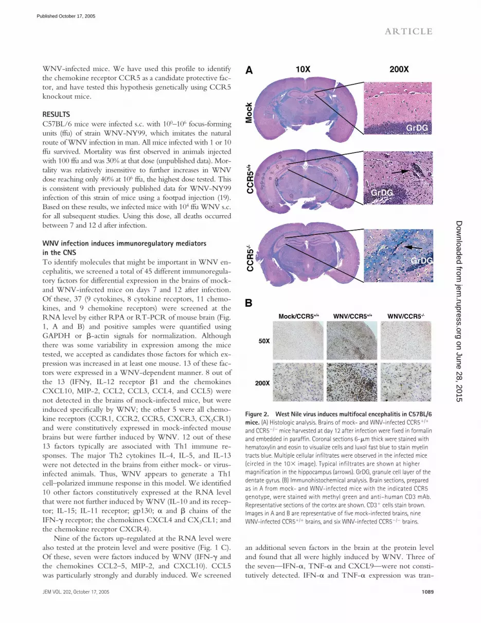
Figure 2. West Nile virus induces multifocal encephalitis in C57BL/6 mice. (A) Histologic analysis. Brains of mock- and WNV-infected CCR5�/� and CCR5�/� mice harvested at day 12 after infection were fixed in formalin and embedded in paraffin. Coronal sections 6-�m thick were stained with hematoxylin and eosin to visualize cells and luxol fast blue to stain myelin tracts blue. Multiple cellular infiltrates were observed in the infected mice (circled in the 10 image). Typical infiltrates are shown at higher magnification in the hippocampus (arrows). GrDG, granule cell layer of the dentate gyrus. (B) Immunohistochemical analysis. Brain sections, prepared as in A from mock- and WNV-infected mice with the indicated CCR5 genotype, were stained with methyl green and anti–human CD3 mAb. Representative sections of the cortex are shown. CD3� cells stain brown. Images in A and B are representative of five mock-infected brains, nine WNV-infected CCR5�/� brains, and six WNV-infected CCR5�/� brains.
on June 28, 2015jem
.rupress.orgD
ownloaded from
Published October 17, 2005

CCR5 AND WEST NILE VIRUS PATHOGENESIS | Glass et al.
1090
sient, with large amounts of cytokine found on day 7 afterinfection, but not on day 12. Constitutive expression of theother four factors (IL-1
�
, the adhesion molecule ICAM-1,and the metalloproteinase MMP-9 and its inhibitor TIMP-1)was markedly increased by virus and remained elevatedthroughout the 12 d period of observation. These moleculesmay serve to arrest activated leukocytes in cerebral bloodvessels and break down the blood brain barrier to facilitateleukocyte penetration into infected brain tissue. With regardto candidate leukocyte chemotactic systems that might guidethis process, our data point clearly to the non-ELR CXCchemokines CXCL9 and CXCL10 and their receptorCXCR3; and the CC chemokines CCL2, CCL3, CCL4,and CCL5 and their receptors CCR1, CCR2, and CCR5.The RNA data suggest that the CX
3
C chemokine CX
3
CL1and its receptor CX
3
CR1 may also play a role. For the re-mainder of this paper, we focus our analysis on the role ofCCR5 in WNV pathogenesis.
CCR5
�
leukocytes accumulate in WNV-infected mouse brain
We examined histopathological sections of mock- andWNV-infected mice and visualized intense multifocal cellu-
lar infiltrates, primarily localized to hippocampus and exter-nal capsule, evident in WNV-infected but not mock-infected mouse brain stained either by hematoxylin andeosin or anti-CD3 (Fig. 2, A and B). Previously publishedwork (21, 22), confirmed by us (unpublished data), hasshown that WNV infects neurons.
Consistent with the histological analysis and immunohis-tochemistry results, we observed a notable increase in leuko-cytes isolated from WNV-infected wild-type mouse brains atboth 7 and 12 d after infection (Figs. 3 and 4). The totalamount of CD4
�
T cells rose from 1.2
10
3
/brain (0.2% oftotal leukocytes) in mock-infected brains to 2
10
4
/brain(1.8% of total leukocytes) and 9.6
10
4
/brain (5.0% of totalleukocytes) on days 7 and 12 after infection, respectively.The amount of CD8
�
T cells rose from 1.8
10
3
/brain(0.3% of total leukocytes) in mock-infected brains to 2.3
10
4
/brain (2.0% of total leukocytes) and 1.0
10
5
/brain(5.3% of total leukocytes) on days 7 and 12 after infection,respectively. The number of NK1.1
�
cells increased from1.4
10
3
/brain (0.3% of total leukocytes) in mock-infectedbrains to 2.2
10
4
/brain (1.9% of total leukocytes) and 4.1
10
4
/brain (2.1% of total leukocytes) on days 7 and 12 after
Figure 3. WNV induces accumulation of leukocytes in mouse brain. Representative FACS analysis dot plots of mock- and WNV-infected CCR5�/� and CCR5�/� mice. Percentages are calculated based on total events; histograms to the right of the corresponding dot plot show CCR5
staining within single positive CD4� or CD8� T cells, NK cells, and the three F4/80�/CD45� subsets. The blue line in the histogram represents isotype control staining, and the red shaded line represents CCR5 staining. Bar shows which portion of the histogram was considered CCR5 positive.
on June 28, 2015jem
.rupress.orgD
ownloaded from
Published October 17, 2005

JEM VOL. 202, October 17, 2005
1091
ARTICLE
infection, respectively. The amount of resident microglia(F4/80
�
/CD45
lo) remained relatively constant throughoutthe course of disease. However, activated resident microglia
(F4/80�/CD45med) rose from a baseline of 0.24 105/brain(4.4% of total leukocytes) in mock-infected brains to 1.2 105 (11.0% of total leukocytes) and 2.2 105 (11.1% of totalleukocytes) on days 7 and 12 after infection, respectively.The most impressive increase was seen in infiltrating mac-rophages (F4/80�/CD45hi). Mock-infected mouse brainshad only 7.3 103/brain (1.3% of total leukocytes) of thesecells, whereas the amount in infected brains increased to2.7 105/brain (23.0% of total leukocytes) and 5.5 105/brain (28.3% of total leukocytes) on days 7 and 12 after in-fection, respectively (Figs. 3 and 4).
CCR5 has been reported to be expressed mainly on sub-sets of monocytes, macrophages, NK cells, and T lympho-cytes. Consistent with this, on day 7 after infection, 12% ofCD8� T cells, and 25% each of CD4� T cells, NK1.1� NKcells, and F4/80� CD45hi activated infiltrating macrophages,which were induced to accumulate in the brain by WNV,expressed CCR5 (Figs. 3 and 4 A). On day 12 after infec-tion, expression of CCR5 on NK cells and activated mac-rophages in WNV-infected CNS had decreased to 16% and15%, respectively, whereas expression on CD4� and CD8�
T cells was not statistically significantly different from ex-pression on day 7. In mock-infected mouse brain, of the fewleukocytes present, 4% of resident microglial cells (F4/80�/CD45lo) expressed CCR5 (Fig. 3 and 4 A). Together, thesedata support the hypothesis that CCR5 may play a role inthe migration of a portion of CD4� and CD8� T cells, NKcells and activated tissue-infiltrating macrophages to brainsinfected with WNV.
Loss of CCR5 results in impaired leukocyte trafficking to the CNSWe tested this hypothesis by comparing the outcomes ofWNV infection between CCR5�/� and CCR5�/� mice.On day 12 after infection, CCR5�/� mice had markedly re-duced cellular infiltrates (�1 lesion per 200 hpf in coronalsections of the hippocampus and external capsule comparedwith 5–8 lesions per 200 hpf for WNV-infected wild typemice (Fig. 2 A). This was confirmed by direct immunohis-tochemical analysis of the T cell antigen CD3, which re-vealed markedly decreased staining of infected CCR5�/�
mouse brains compared with the brains of infected CCR5�/�
mice (Fig. 2 B). FACS analysis allowed for direct quantitationof this decrease (Figs. 3 and 4). Specifically, on day 7 inCCR5�/� mice, we found a �50% decrease in the numberof CD4� T cells, NK1.1� cells, F4/80�/CD45med activatedmicroglia and F4/80�/CD45hi infiltrating macrophages com-pared with values for WNV-infected CCR5�/� controls.The number of CD8� T cells was not significantly decreasedat this time point. By day 12 after infection, the reduction inthe cell infiltrate to the WNV-infected CCR5�/� mousebrain was even more evident: 60% reduction in CD4� Tcells, 46% reduction in CD8� T cells, 84% reduction inNK1.1� cells, 63% reduction in activated microglia, and a76% reduction in infiltrating macrophages compared withinfected CCR5�/� control mice (Figs. 3 and 4).
Figure 4. CCR5 regulates accumulation of T cells, NK cells, and macrophages in the WNV-infected mouse brain. (A) WNV induces accumulation of CCR5� leukocytes in the mouse brain. CCR5 surface expression was examined on specific leukocyte subsets isolated from mouse brain and the percent positive cells was determined. (B and C) Lack of CCR5 blunts leukocyte accumulation in the mouse brain in response to WNV infection. Data were pooled from three experiments, with three to five mice analyzed at each time point/experiment and are presented as the mean � SEM. *, P � 0.04 for the indicated comparison by Student’s t test.
on June 28, 2015jem
.rupress.orgD
ownloaded from
Published October 17, 2005
CCR5 AND WEST NILE VIRUS PATHOGENESIS | Glass et al.1092
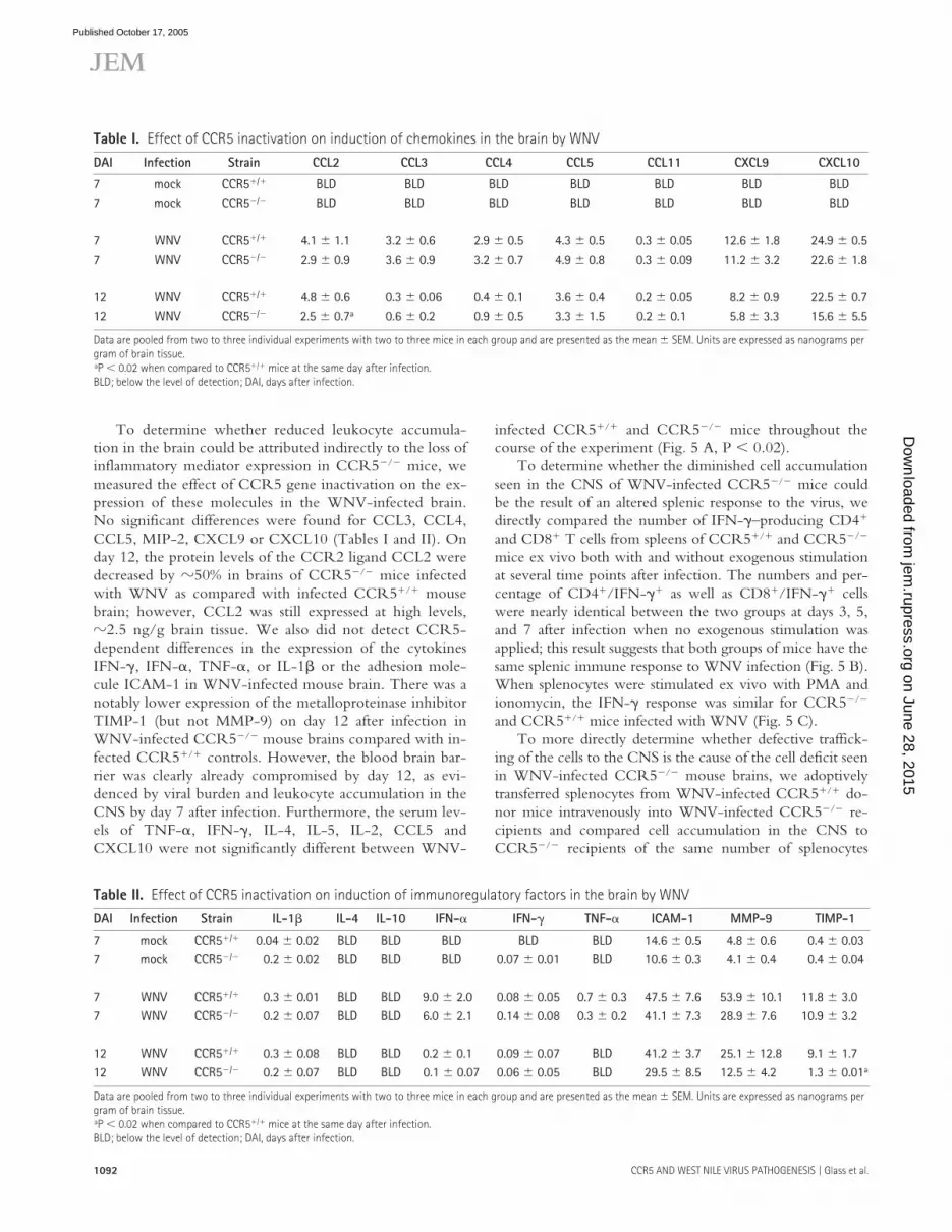
To determine whether reduced leukocyte accumula-tion in the brain could be attributed indirectly to the loss ofinflammatory mediator expression in CCR5�/� mice, wemeasured the effect of CCR5 gene inactivation on the ex-pression of these molecules in the WNV-infected brain.No significant differences were found for CCL3, CCL4,CCL5, MIP-2, CXCL9 or CXCL10 (Tables I and II). Onday 12, the protein levels of the CCR2 ligand CCL2 weredecreased by �50% in brains of CCR5�/� mice infectedwith WNV as compared with infected CCR5�/� mousebrain; however, CCL2 was still expressed at high levels,�2.5 ng/g brain tissue. We also did not detect CCR5-dependent differences in the expression of the cytokinesIFN-�, IFN-, TNF-, or IL-1� or the adhesion mole-cule ICAM-1 in WNV-infected mouse brain. There was anotably lower expression of the metalloproteinase inhibitorTIMP-1 (but not MMP-9) on day 12 after infection inWNV-infected CCR5�/� mouse brains compared with in-fected CCR5�/� controls. However, the blood brain bar-rier was clearly already compromised by day 12, as evi-denced by viral burden and leukocyte accumulation in theCNS by day 7 after infection. Furthermore, the serum lev-els of TNF-, IFN-�, IL-4, IL-5, IL-2, CCL5 andCXCL10 were not significantly different between WNV-
infected CCR5�/� and CCR5�/� mice throughout thecourse of the experiment (Fig. 5 A, P � 0.02).
To determine whether the diminished cell accumulationseen in the CNS of WNV-infected CCR5�/� mice couldbe the result of an altered splenic response to the virus, wedirectly compared the number of IFN-�–producing CD4�
and CD8� T cells from spleens of CCR5�/� and CCR5�/�
mice ex vivo both with and without exogenous stimulationat several time points after infection. The numbers and per-centage of CD4�/IFN-�� as well as CD8�/IFN-�� cellswere nearly identical between the two groups at days 3, 5,and 7 after infection when no exogenous stimulation wasapplied; this result suggests that both groups of mice have thesame splenic immune response to WNV infection (Fig. 5 B).When splenocytes were stimulated ex vivo with PMA andionomycin, the IFN-� response was similar for CCR5�/�
and CCR5�/� mice infected with WNV (Fig. 5 C).To more directly determine whether defective traffick-
ing of the cells to the CNS is the cause of the cell deficit seenin WNV-infected CCR5�/� mouse brains, we adoptivelytransferred splenocytes from WNV-infected CCR5�/� do-nor mice intravenously into WNV-infected CCR5�/� re-cipients and compared cell accumulation in the CNS toCCR5�/� recipients of the same number of splenocytes
Table I. Effect of CCR5 inactivation on induction of chemokines in the brain by WNV
DAI Infection Strain CCL2 CCL3 CCL4 CCL5 CCL11 CXCL9 CXCL10
7 mock CCR5�/� BLD BLD BLD BLD BLD BLD BLD7 mock CCR5�/� BLD BLD BLD BLD BLD BLD BLD
7 WNV CCR5�/� 4.1 � 1.1 3.2 � 0.6 2.9 � 0.5 4.3 � 0.5 0.3 � 0.05 12.6 � 1.8 24.9 � 0.57 WNV CCR5�/� 2.9 � 0.9 3.6 � 0.9 3.2 � 0.7 4.9 � 0.8 0.3 � 0.09 11.2 � 3.2 22.6 � 1.8
12 WNV CCR5�/� 4.8 � 0.6 0.3 � 0.06 0.4 � 0.1 3.6 � 0.4 0.2 � 0.05 8.2 � 0.9 22.5 � 0.712 WNV CCR5�/� 2.5 � 0.7a 0.6 � 0.2 0.9 � 0.5 3.3 � 1.5 0.2 � 0.1 5.8 � 3.3 15.6 � 5.5
Data are pooled from two to three individual experiments with two to three mice in each group and are presented as the mean � SEM. Units are expressed as nanograms per gram of brain tissue.aP � 0.02 when compared to CCR5�/� mice at the same day after infection.BLD; below the level of detection; DAI, days after infection.
Table II. Effect of CCR5 inactivation on induction of immunoregulatory factors in the brain by WNV
DAI Infection Strain IL-1� IL-4 IL-10 IFN- IFN-� TNF- ICAM-1 MMP-9 TIMP-1
7 mock CCR5�/� 0.04 � 0.02 BLD BLD BLD BLD BLD 14.6 � 0.5 4.8 � 0.6 0.4 � 0.037 mock CCR5�/� 0.2 � 0.02 BLD BLD BLD 0.07 � 0.01 BLD 10.6 � 0.3 4.1 � 0.4 0.4 � 0.04
7 WNV CCR5�/� 0.3 � 0.01 BLD BLD 9.0 � 2.0 0.08 � 0.05 0.7 � 0.3 47.5 � 7.6 53.9 � 10.1 11.8 � 3.07 WNV CCR5�/� 0.2 � 0.07 BLD BLD 6.0 � 2.1 0.14 � 0.08 0.3 � 0.2 41.1 � 7.3 28.9 � 7.6 10.9 � 3.2
12 WNV CCR5�/� 0.3 � 0.08 BLD BLD 0.2 � 0.1 0.09 � 0.07 BLD 41.2 � 3.7 25.1 � 12.8 9.1 � 1.712 WNV CCR5�/� 0.2 � 0.07 BLD BLD 0.1 � 0.07 0.06 � 0.05 BLD 29.5 � 8.5 12.5 � 4.2 1.3 � 0.01a
Data are pooled from two to three individual experiments with two to three mice in each group and are presented as the mean � SEM. Units are expressed as nanograms per gram of brain tissue.aP � 0.02 when compared to CCR5�/� mice at the same day after infection.BLD; below the level of detection; DAI, days after infection.
on June 28, 2015jem
.rupress.orgD
ownloaded from
Published October 17, 2005

JEM VOL. 202, October 17, 2005 1093
ARTICLE
from WNV-infected CCR5�/� donors. WNV-infectedCCR5�/� mice that received CCR5�/� splenocytes had asignificantly greater amount of CD4� and CD8� T cells,NK1.1� cells, and infiltrating macrophages (increased 2.3-,5.3-, 3.1-, and 7.9-fold, respectively) compared with WNV-infected CCR5�/� mice that received CCR5�/� spleno-cytes (Fig. 5 D, P � 0.02). In the CCR5�/� mice that re-ceived CCR5�/� splenocytes, we were able to determine byFACS that 4.7% of the CD4� T cells, 9.6% of CD8� T cells,and 0.9% of NK1.1� cells in the brain expressed CCR5,demonstrating that the transferred cells were able to migrateto the brain.
CCR5 is critical to viral control and survivalWe next tested the importance of CCR5 on viral clearanceand survival. Viral load in the spleens of CCR5�/� andCCR5�/� mice was not statistically different in onset, magni-tude, duration, or clearance as both groups had a maximal vi-ral load on day 3 after infection and surviving mice hadcleared virus from the spleen by day 9 after infection (Fig. 6A), further indicating that the immune response to WNV inCCR5�/� mice is not altered. WNV-infected CCR5�/�
mice had nearly identical CNS viral loads as CCR5�/� con-trols on day 7 after infection (Fig. 6 B). However, whereassurviving WNV-infected CCR5�/� mice began to clear vi-rus by day 12, the few surviving CCR5�/� mice available foranalysis showed no decrease in CNS viral load on day 12, butrather a 32-fold increase relative to controls. This was evidenteven when CCR5�/� mice were selected to match the poorclinical status of CCR5�/� mice at this time point (Fig. 6 B).Consistent with this result, infection with WNV was uni-formly fatal in CCR5�/� mice (n � 38), whereas in CCR5�/�
mice mortality was 35% (Fig. 6 C, n � 50). All infectedCCR5�/� mice were dead by day 13. In contrast, WNV-infection in mice lacking either CCR1 or CX3CR1, whichare two chemokine receptors related to CCR5 and shownby RNA analysis (Fig. 1) to be up-regulated by WNV infec-tion, resulted in mortality that was only slightly increased rel-ative to that observed in wild-type control mice (Fig. 6 C).Finally, adoptive transfer of splenocytes from WNV-infected
Figure 5. CCR5 deficiency affects immune responses to WNV selectively in the brain. (A) Serum cytokine analysis. The factor analyzed is identified at the bottom of the graph. Data were pooled from two experiments
with five to seven mice in each group, and are presented as mean � SEM pg/ml serum as a function of time after infection. (B and C) Intracellular cytokine staining for WNV-induced IFN-�. Splenocytes were isolated from mice at the indicated days after WNV infection and assayed without exogenous stimulation (B) or after 4 h of stimulation with ionomycin and PMA (C). Data presented are from one experiment with five mice at each time point. Values at the top of each bar are the percentage of total cells represented by the indicated cell type. (D) Adoptive transfer of CCR5�/� and CCR5�/� splenocytes from WNV-infected mice into WNV-infected CCR5�/� mice. Infected CCR5�/� recipient mice, designated (WT → KO) and (KO → KO) received splenocytes from infected CCR5�/� or infected CCR5�/� donor mice, respectively, 4 d after s.c. infection. FACS analysis was performed 4 d after transfer (8 d after infection). Data are presented as the mean � SEM and are from one experiment with five mice in each group. (B and C) Percents shown are the percentage of total cells. *, P � 0.02 for the indicated comparison (of total cell number) by Student’s t test.
on June 28, 2015jem
.rupress.orgD
ownloaded from
Published October 17, 2005

CCR5 AND WEST NILE VIRUS PATHOGENESIS | Glass et al.1094
CCR5�/� donor mice to WNV-infected CCR5�/� recipi-ents (WT → KO) reduced mortality to the level observed forwild-type C57BL/6 mice. In contrast, transfer of the samenumber of splenocytes from WNV-infected CCR5�/� miceto WNV-infected CCR5�/� recipients (KO → KO) hadonly a minor protective effect (Fig. 6 D).
DISCUSSIONThis study provides a detailed analysis of the molecular andcellular immune response in the brain during WNV infec-tion and demonstrates that expression of the chemokine re-ceptor CCR5 is crucial for viral clearance and survival in amouse model of disease. WNV induced production of allthree CCR5 ligands in the brain, with particularly high anddurable induction of CCL5, which is consistent with localaccumulation of CCR5-expressing NK cells, macrophages,and CD4� and CD8� T lymphocytes that we observed inthe model. Several lines of evidence lead us to believe thattrafficking of leukocytes expressing CCR5 to the WNV-infected mouse brain is critical for survival. First, genetic dis-ruption of CCR5 markedly reduced accumulation of thesecell types in brains of WNV-infected mice, without signifi-cantly affecting CNS expression of other immunoregulatoryfactors, including the CCR5 ligands CCL3, CCL4, andCCL5. Second, there was no defect in clearance of the virusfrom the spleen in CCR5�/� mice. Third, WNV-inducedproduction of IFN-� by CD4� and CD8� T cells in thespleen was identical for CCR5�/� and CCR5�/� mice.Fourth, when splenocytes from WNV-infected CCR5�/�
or CCR5�/� mice were adoptively transferred into WNV-infected CCR5�/� mice, we found much greater leuko-cyte accumulation in the brain after CCR5�/� than afterCCR5�/� cell transfer and detected CCR5� leukocytes in
Figure 6. West Nile virus infection is uniformly fatal in CCR5 knockout mice. (A) Time course of viral clearance from spleen. Virus was quantified in the spleens of the indicated strains of surviving mice at the indicated times by focus formation assay. Mock-infected mice were analyzed on day 7 of the experiment. (B) Time course of viral clearance from brain. Virus was quantified in the brain of the indicated strains of mice at the indicated times by focus formation assay. Data were pooled from three to four experiments with three to five mice at each time point indicated and are presented as the mean � SEM. *, P � 0.02 for the indicated comparison by Student’s t test. N/A, not applicable, because all infected CCR5�/� die by day 13. (C) Kaplan-Meier analysis of survival of WNV-infected mice. Results shown are pooled data from three independent experiments for CCR5�/� mice (n � 50 total mice) and CCR5�/� mice (n � 34), and two independent experiments for CCR1�/� mice (n � 30) and CX3CR1�/� mice (n � 32). Similar results were obtained for both wild-type C57BL6/J and C57BL/6NTac control mice; however, only the C57BL6/J results are shown (CCR5�/�). (D) Kaplan-Meier analysis of survival of adoptive transfer mice. WT → KO, transfer of splenocytes from WNV-infected CCR5�/� mice into WNV-infected CCR5�/� mice; KO → KO, transfer of splenocytes from WNV-infected CCR5�/� mice to WNV-infected CCR5�/� mice. The following control groups were included that did not undergo adoptive transfer: Mock, mock-infected CCR5�/�mice; CCR5�/�, WNV-infected CCR5�/� mice; and CCR5�/�, WNV-infected CCR5�/� mice. Data represent a single experiment with 10 mice in each group.
on June 28, 2015jem
.rupress.orgD
ownloaded from
Published October 17, 2005

JEM VOL. 202, October 17, 2005 1095
ARTICLE
the brains of CCR5�/� recipients. This experiment, whichinvolves direct injection of WNV-activated CCR5�/� orCCR5�/� splenocytes into the blood, bypasses the splenicegress step, and suggests strongly, when coupled with unal-tered splenic T cell or viral clearance responses in CCR5�/�
mice, that CCR5 functions by promoting trafficking of leu-kocytes from the blood to the brain in response to WNVinfection, for the purpose of containing and clearing the vi-rus. This is consistent with previously published work on WNVwhich demonstrated that RAG�/� mice have dramaticallyincreased CNS viral burden and succumb to infection within12 d, the same time frame that we found for CCR5�/� mice(19). Furthermore, mice lacking CD8� T cells also have in-creased viral burden in the CNS and mortality when in-fected with WNV (13, 18, 23–25). Shirato et al. have re-cently reported an increased expression of mRNA forCCR5 ligands when mice were infected i.p. with a lethalversus nonlethal strain of WNV (14). Our data confirm andextend these results in a distinct model by providing mRNAand protein analysis for these and numerous other immuno-regulatory molecules and by directly testing the importanceand role of CCR5.
The critical role of CCR5 in WNV pathogenesis appearsto be unique compared with other neurotropic viruses thathave been tested to date. CNS infection with lymphocyticchoriomeningitis virus results in equivalent viral burden andmortality in CCR5�/� and CCR5�/� mice suggesting thatthis receptor does not play a role in pathogenesis in this model(26). Likewise, infection with the neurovirulent retrovirusFR98 results in identical mortality rates and CNS viral burdenin CCR5�/� and CCR5�/� mice (27). Infection with a neu-rovirulent strain of mouse hepatitis virus (MHV) results in sim-ilar viral titers in the CNS of CCR5�/� and CCR5�/� mice,but knockouts have reduced demyelination, the major mani-festation of disease in this model. The mechanism involvesCCR5-dependent recruitment of macrophages and CD4� andCD8� T cells to the CNS (28). Thus, CCR5 promotes demy-elinating disease in MHV infection, not antiviral host defenseas it does in WNV infection. CCR5 has also been demon-strated to regulate leukocyte trafficking to the brain, but notthe lung, after Cryptococcus neoformans infection (29).
The importance of CCR5 also varies among noninfec-tious, immunologically mediated, CNS disease. CCR5 defi-ciency has no effect on experimental autoimmune encepha-litis (EAE), despite the fact that CCR5 ligands are highlyexpressed in the CNS of mice with EAE (30). In addition,wild-type and CCR5-deficient mice have similar outcomesin experimental autoimmune neuritis, including similar lev-els of leukocytes recruited to the cauda equina (31). Theseexamples show the potential for chemokine receptors to playspecific and important roles or more redundant roles de-pending on the disease context, and the spatial, temporal,and quantitative details of cognate ligand expression.
Our data raise new questions regarding whether CCR5may play a more general role in the immune response toother flaviviruses. In this regard, in a recent study of 10 cy-
tokines in patients infected with Japanese Encephalitis virus(the prototype flavivirus), increased serum CCL5 alone wasassociated with a fatal outcome (32). The mechanism wasnot established, but could conceivably involve desensitiza-tion of leukocyte CCR5 in the bloodstream, thereby pre-venting trafficking to the infected CNS.
We found that CCR1, CCR2, CXCR3, and CX3CR1and their ligands were also up-regulated in mouse brains byWNV. These systems could work cooperatively with CCR5and could explain why leukocytes accumulate in small num-bers in the CNS of WNV-infected CCR5�/� mice and whymany of the leukocytes in the WNV-infected brain areCCR5 negative. Induction of CXCR3 was particularlystrong, and is consistent with induction of a Th1-polarizedcytokine response in the brain (18, 23, 24). However, ourresults using knockout mice do not support an important,nonredundant role for CCR1 or CX3CR1 in this model.Our gene and protein expression data also suggest new andtestable hypotheses regarding the role in control of WNVinfection of TNF-, the IL-12/IFN-� axis, IFN-, the leu-kocyte adhesion molecule ICAM-1, the matrix metallopro-teinase MMP-9, and the metalloproteinase inhibitor TIMP-1(33). WNV has been reported to be very sensitive to bothexogenous type I and II interferons in vitro (34), but theyare only able to prevent infection in vivo in mice whengiven before challenge (35). IFN- may be even less effec-tive in humans because infection with WNV has been re-ported in patients actively being treated with IFN- and ri-bavirin for hepatitis C (36). Matrix metalloproteinases maybe important in disrupting the blood brain barrier to allowhematogenous spread of WNV to the CNS, as has been pre-viously suggested (19). Although the role of TNF- in thebrain is unclear, recent work has clarified its importance inthe periphery where it is produced in a TLR3-dependentmanner and mediates permeabilization of the blood brainbarrier to facilitate WNV entry (12). The specific roles inpathogenesis of these and other factors induced in the brainby WNV are currently under investigation.
An important question raised by our findings iswhether CCR5 is also protective in WNV infection ofhumans. In limited studies, T cells and macrophages havebeen reported to accumulate in WNV-infected humanbrains as they do in our mouse model (25, 37). Moreover,like humans, the mouse is easily infected with WNV, andthe major clinical manifestations of disease are similar inboth species: encephalitis and mortality in a subset of in-fected individuals. If CCR5 is protective in humans, indi-viduals homozygous for the CCR5�32 mutation, wholack functional CCR5 and include 1% of North AmericanCaucasians (38), may be at greater risk for fatal encephalitisfrom WNV infection. A related issue is the safety ofCCR5 blocking agents under development for patientswith HIV/AIDS who become infected with WNV.
In conclusion, our data identify CCR5 as a critical protec-tive factor in fatal encephalitis caused by WNV in a mousemodel. The effect is highly specific relative to other neurotro-
on June 28, 2015jem
.rupress.orgD
ownloaded from
Published October 17, 2005

CCR5 AND WEST NILE VIRUS PATHOGENESIS | Glass et al.1096
pic viruses and at least two other relevant chemokine recep-tors. Loss of CCR5 results in decreased ability to recruit/maintain leukocytes into the infected CNS where they mayfunction to clear the virus. The data also raise important newquestions about the potential roles of other immunoregulatorysystems that are induced by WNV in this model and in man.
MATERIALS AND METHODSMouse strains. Mouse studies were performed in an animal biosafety levelthree facility under a protocol approved by the National Institute of Allergyand Infectious Diseases/National Institutes of Health (NIH) Animal Care andUse Committee. CCR5�/� mice (B6;129P2-Ccr5tm1Kuz), its approximate ge-netic match mouse strain B6129PF2 and C57BL6/J mice were all purchasedfrom the Jackson Laboratory. Infection of B6129PF2 and C57BL6/J micewith 104 ffu WNV induced similar changes and time courses with regard toall parameters that were compared (mortality, CNS viral load, CNS accumu-lation of leukocyte subsets [total and CCR5� T cells, NK cells, and macro-phages], and CNS chemokine protein [CCL3, CCL4, CCL5, and CXCL10];unpublished data). In all experiments shown, data for CCR5�/� mice weregenerated using the C57BL6/J mouse. CX3CR1�/� mice (F10 backcross onthe C57BL/6NTac background), CCR1�/� mice (F10 backcross on theC57BL/6NTac background), and control C57BL/6NTac mice were ob-tained from Taconic. All experiments were initiated using female mice 8–12wk old. Mice were selected randomly for phenotypic analysis at day 7 afterinfection with WNV. On day 12 after WNV infection, all survivingCCR5�/� mice were severely ill (limp tail, hunched back, ruffled fur, mini-mal activity), and therefore infected C57BL6/J mice with similar clinical signswere selected for phenotypic analysis at this time point.
Viral infections. WNV strain NY99-35262 was provided by R. Lanciotti(Centers for Disease Control and Prevention, Fort Collins, CO). Originallyisolated from a Chilean flamingo at the Bronx Zoo in 1999 (3), this isolatehad been passaged once in Vero cell culture that contained 10% FBS. It wasthen reamplified by two passages in Vero cells (WHO seed passage 143) thatdid not contain FBS. A virus suspension was prepared that had a titer of 2 107 ffu/ml, as determined using an immunostaining focus-forming assay andWNV-specific mouse antibodies (39). Mice were injected s.c. in the back ofthe neck with either 104 ffu WNV-NY99 suspended in 50 �l HBSS (Invi-trogen) or with HBSS alone (mock infected). Mice were monitored visuallyand weighed daily. Brains of killed mice were aseptically removed, weighedand placed directly in either 2 ml HBSS (for ELISA and viral titer assay) or5 ml FACS buffer (HBSS � 1% BSA � 0.1% NaN3; Sigma-Aldrich).
ELISA. Brains were homogenized in sterile HBSS and frozen at �80 C.Samples were thawed and centrifuged at 1,500 g for 25 min. The aqueousfraction of the homogenate was serially diluted, and then analyzed for thepresence of specific cytokine and chemokine proteins using murine Quan-tikine Immunoassay kits according to the manufacturer’s recommendations(R&D systems). Samples were used only once.
RT-PCR. Total RNA was extracted from homogenized brain tissue usingTRIzol (Invitrogen). 2 ng of total RNA was converted to cDNA using aSuperscript III RT kit (Invitrogen), with random primers. PCR was per-formed using 2 �l cDNA in a reaction mix with Taq polymerase (Invitro-gen). PCR reactions were optimized to determine the number of cycleswhere the visual signal was in the linear range. These experiments deter-mined that at �35 cycles, the signal plateaued for all targets tested. There-fore, all PCR reactions were run for 30 cycles using the following primerpairs: �-actin forward (5�-AGCCATGTACGTAGCCATCC-3�), �-actinreverse (5�-TCTCAGCTGTGGTGGTGAAG-3�); CXCR3 forward(5�-TGCTAGATGCCTCGGACTTT-3�), CXCR3 reverse (5�-CGCT-GACTCAGTAGCACAGC-3�); CXCL4 forward (5�-AGTCCTGAGCT-GCTGCTTCT-3�), CXCL4 reverse (5�-GGCAAATTTTCCTCCCATTC-3�); CXCR4 forward (5�-TCAGTGGCTGACCTCCTCTT-3�), CXCR4
reverse (5�-TTTCAGCCAGCAGTTTCCTT-3�); CX3CL1 forward(5�-AGCCCTGTGACATTTTCTGG-3�), CX3CL1 reverse (5�-CCTC-ACTCTCAGGAGCCAAC-3�); and CX3CR1 forward (5�-GGAGAC-TGGAGCCAACAGAG-3�), CX3CR1 reverse (5�-TCTTGTCTGGCT-GTGTCCTG-3�). PCR products were separated and visualized onethidium bromide-stained 2% agarose/TBE gels.
Ribonuclease protection assay. Cytokine, chemokine, cytokine recep-tor, and chemokine receptor mRNA transcripts were analyzed from totalbrain RNA using mCK-1, mCK-5, mCR-1, and mCR-5 multi-templateprobe sets, respectively (BD Biosciences). RPA analysis was performed us-ing 10 �g of total RNA using a previously described protocol (28). Forquantitation of signal intensity, autoradiographs were scanned and individ-ual chemokine, cytokine, or chemokine/cytokine receptor transcript bandswere normalized as the ratio of band intensity to the GAPDH control.Analysis was performed using NIH Image J1.32i software.
Cell isolation and FACS analysis. Mice were killed by cervical disloca-tion. Organs were aseptically removed but not perfused before analysis. Sin-gle cell suspensions of leukocytes were obtained from brain using a previ-ously described method (40). In brief, brains were collected in FACS buffer(HBSS � 1% BSA � 0.1% NaN3) and homogenized using the plunger por-tion of a 6-cc syringe on ice in a petri dish. The suspension was brought to7 ml with FACS buffer and 3 ml of 90% percoll in PBS was added. After 5min at 22 C, the suspension was underlaid with 1 ml of 70% Percoll (inRPMI), and was then centrifuged at 2,470 rpm for 20 min at 22 C at roomtemperature. The leukocytes at the interphase were isolated and washedthree times in FACS buffer. Cells were fixed in 100 �l BD Cytofix (BDBiosciences) at 4 C for 20 min, counted using a hemacytometer, and thenfiltered though a 70-�m mesh screen. Cells were washed three times withFACS buffer and incubated with Fc block (Becton Dickinson) in 100 �lCD16/32 Ab (BD Biosciences) diluted 1:200 in FACS buffer. Cells werethen incubated with specific antibodies for 45 min at 4 C, washed threetimes and suspended in 400 �l FACS buffer. Antibodies used for flow cy-tometry were FITC-conjugated anti–mouse CD4 (GK1.5), CD19 (1D3),CD45 (LCA, Ly-5; BD Biosciences) and F4/80 (Serotec); PE-conjugatedanti–mouse CCR5, NK1.1 (PK136), and CD45 (LCA, Ly-5); APC-conju-gated anti-mouse CD8 (Ly-2) and NK1.1 (PK136; BD Biosciences); andPerCP-conjugated anti-mouse CD4 (L3T4) and CD45 (LCA, Ly-5). In allcases, an isotype-matched FITC-, PE-, APC-, or PerCP-conjugated anti-body was used as the control. Cells were analyzed on a FACSCalibur (Bec-ton Dickinson) where leukocytes were further refined by gating on cellswith the appropriate forward and side scatter profile. 100,000 events werecaptured for analysis for each brain and each staining set. Data were ana-lyzed using FlowJo software and are presented as the percentage of positivecells within the gated population.
Intracellular cytokine staining. Mouse splenocytes (3 106 cells/well)were incubated without exogenous stimulation for 4 h or with 50 ng/mlPMA (Sigma-Aldrich) plus 500 ng/ml ionomycin (Sigma-Aldrich) for 4 hat 37 C. Cells were then washed and blocked with Fc block for 20 min at4 C. Cells were washed twice with FACS buffer, and surface markers werestained with CD19-FITC, CD8-PE, and CD4-PerCP for 30 min at 4 C.Cells were washed twice with FACS buffer and resuspended in 100 �lCytofix/Cytoperm (Becton Dickinson) for 20 min at 4 C to permeabilizethe plasma membrane and facilitate antibody staining of IFN-�. We washedthe cells twice with Perm/Wash solution and stained them with an APC-conjugated mAb to mouse IFN-� (Becton Dickinson) for 30 min at 4 C.Finally, cells were washed twice and resuspended in FACS buffer and ana-lyzed on a FACSCalibur (Becton Dickinson). In all samples 100,000 eventswere captured. Data were analyzed using FloJo software.
Adoptive transfer. CCR5�/� and CCR5�/� donor mice were infectedwith 104 ffu WNV s.c. 3 d later, CCR5�/� recipient mice were infectedwith 104 ffu WNV s.c. After recipients had been infected for 4 d, a duration
on June 28, 2015jem
.rupress.orgD
ownloaded from
Published October 17, 2005

JEM VOL. 202, October 17, 2005 1097
ARTICLE
sufficient for virus to enter the brain, 107 splenocytes from donors (infectedfor 7 d at this time point) were transferred IV to recipients. Splenocyteswere isolated using lympholyte M (Cedarlane Labs). CCR5�/� recipients ofdonor CCR5�/� splenocytes are referred to as KO → KO. CCR5�/�
recipients of donor CCR5�/� splenocytes are referred to as WT → KO.
Viral titers. Vero cells were maintained in OptiPro SFM (Invitrogen)until passaged for the viral titer assay at which time they were grown inOptiPro SFM � 2% FBS � 50 �g/ml gentamicin. The virus was quanti-tated by growth on confluent Vero cell monolayers in 12 well plates. 200�l of brain homogenate supernatant was diluted in culture media andplaced on the cells in duplicate. Plates were incubated for 1 h at 37 C toallow virus attachment, and then cells were overlaid with 2 ml of sterileOpti-MEM (Invitrogen) � methyl cellulose 8 g/liter (EM Science) � 2%FBS � 50 �g/ml gentamicin. Plates were incubated for 2 d at 37 C, andthen washed three times with PBS (Biosource International) and incu-bated for 1 h at 37 C with 500 �l of diluted anti-WNV anti-sera/well(HMAF; American Type Culture Collection VR-82). After washing,samples were incubated at 37 C with 500 �l of diluted anti–mouse/anti–rabbit (1:10)-labeled polymer (DAKO Cytomation) for 1 h. After wash-ing, 1 ml DAB mix (4.5 mg DAB [Sigma-Aldrich] /10 ml PBS � 4.5 �l30% H202 [Fisher Scientific]/10 ml) was added at 22 C for 10 min to 1 hto visualize WNV foci. The reaction was stopped by the addition of wa-ter. The average of duplicate assays was used to determine viral titers,which are expressed as ffu per gram of brain tissue.
Histology. Brains were aseptically removed from mice after cervical dis-location and placed directly into 10% normal buffered formalin (FisherChemicals) for 24 h. Samples were paraffin-embedded and cut into 6-�msections (Histoserve). RNase protection (gloves, RNase-free water andsterilized instruments) was used at all stages and slides were baked at 60 Cfor 1 h after sectioning. Sections were stained with hematoxylin and eosin(for visualization of cells) as well as luxol fast blue (for visualization of my-elin). Separate sections were stained with an anti–human CD3 antibody(DAKO Cytomation) or with hyperimmune mouse ascites fluid collectedafter inoculation with WNV (HMAF). In brief, slides were deparaffinizedwith Citrosolve (National Diagnostics) and graded ethanol followed by awater wash. Slides were heated to 90 C and washed with water twice,blocked with hydrogen peroxide for 10 min, and washed in water. Slideswere blocked with BSA for 20 min, and then incubated with primary an-tibody overnight at a 1:100 dilution. Next, we washed the slides, incu-bated them with biotinylated secondary antibody for 30 min at 1:500 di-lution, and then rewashed. Slides were incubated with streptavidin for 30min and washed and developed with DAB (DAKO Cytomation). Finally,slides were counterstained with hematoxylin and dehydrated with gradedethanol and Citrosolve.
Statistical analysis. Statistically significant differences between groups ofmice were determined by Student’s t test using Microsoft Excel MAC v.Xsoftware, and p-values �0.05 were considered significant.
We thank the animal care staff at the NIH for technical assistance. We also thank J. Ward and L. Brinster for expert analysis of neuropathology.
This research was supported by the Intramural Research Program of the National Institute of Allergy and Infectious Diseases, NIH.
The authors have no conflicting financial interests.
Submitted: 13 December 2004Accepted: 30 August 2005
REFERENCES1. Smithburn, K.C., T.P. Hughes, A.W. Burke, and J.H. Paul. 1940.
Neurotropic virus isolated from the blood of a native of Uganda. Am.J. Trop. Med. 20:471–472.
2. Hubalek, Z., and J. Halouzka. 1999. West Nile fever–a reemerging
mosquito-borne viral disease in Europe. Emerg. Infect. Dis. 5:643–650.3. Lanciotti, R.S., J.T. Roehrig, V. Deubel, J. Smith, M. Parker, K.
Steele, B. Crise, K.E. Volpe, M.B. Crabtree, J.H. Scherret, et al. 1999.Origin of the West Nile virus responsible for an outbreak of encephali-tis in the northeastern United States. Science. 286:2333–2337.
4. Centers for Disease Control and Prevention. 2004. West Nile virus ac-tivity–United States, October 6-12, 2004. MMWR Morb. Mortal.Wkly. Rep. 53:950–951.
5. van der Poel, W.H. 1999. West Nile-like virus is the cause of enceph-alitis in humans and horses and the death of hundreds of birds in NewYork. [In Dutch.] Tijdschr. Diergeneeskd. 124:704–705
6. Steele, K.E., M.J. Linn, R.J. Schoepp, N. Komar, T.W. Geisbert,R.M. Manduca, P.P. Calle, B.L. Raphael, T.L. Clippinger, T. Larsen,et al. 2000. Pathology of fatal West Nile virus infections in native andexotic birds during the 1999 outbreak in New York City, New York.Vet. Pathol. 37:208–224.
7. Ostlund, E.N., J.E. Andresen, and M. Andresen. 2000. West Nile en-cephalitis. Vet. Clin. North Am. Equine Pract. 16:427–441.
8. Briese, T., W.G. Glass, and W.I. Lipkin. 2000. Detection of West Nilevirus sequences in cerebrospinal fluid. Lancet. 355:1614–1615.
9. Asnis, D.S., R. Conetta, G. Waldman, and A.A. Teixeira. 2001. TheWest Nile virus encephalitis outbreak in the United States (1999-2000): from Flushing, New York, to beyond its borders. Ann. NYAcad. Sci. 951:161–171.
10. Asnis, D.S., R. Conetta, A.A. Teixeira, G. Waldman, and B.A. Samp-son. 2000. The West Nile Virus outbreak of 1999 in New York: theFlushing Hospital experience. Clin. Infect. Dis. 30:413–418.
11. Tsai, T.F., F. Popovici, C. Cernescu, G.L. Campbell, and N.I.Nedelcu. 1998. West Nile encephalitis epidemic in southeastern Ro-mania. Lancet. 352:767–771.
12. Wang, T., T. Town, L. Alexopoulou, J.F. Anderson, E. Fikrig, andR.A. Flavell. 2004. Toll-like receptor 3 mediates West Nile virus entryinto the brain causing lethal encephalitis. Nat. Med. 10:1366–1373;10.1038/nm1140.
13. Shrestha, B., and M.S. Diamond. 2004. Role of CD8� T cells in con-trol of West Nile virus infection. J. Virol. 78:8312–8321.
14. Shirato, K., T. Kimura, T. Mizutani, H. Kariwa, and I. Takashima.2004. Different chemokine expression in lethal and non-lethal murineWest Nile virus infection. J. Med. Virol. 74:507–513.
15. Mashimo, T., M. Lucas, D. Simon-Chazottes, M.P. Frenkiel, X. Mon-tagutelli, P.E. Ceccaldi, V. Deubel, J.L. Guenet, and P. Despres. 2002.A nonsense mutation in the gene encoding 2�-5�-oligoadenylate syn-thetase/L1 isoform is associated with West Nile virus susceptibility inlaboratory mice. Proc. Natl. Acad. Sci. USA. 99:11311–11316.
16. Licon Luna, R.M., E. Lee, A. Mullbacher, R.V. Blanden, R. Langman,and M. Lobigs. 2002. Lack of both Fas ligand and perforin protects fromflavivirus-mediated encephalitis in mice. J. Virol. 76:3202–3211.
17. Diamond, M.S., E.M. Sitati, L.D. Friend, S. Higgs, B. Shrestha, andM. Engle. 2003. A critical role for induced IgM in the protectionagainst West Nile virus infection. J. Exp. Med. 198:1853–1862.
18. Diamond, M.S., B. Shrestha, E. Mehlhop, E. Sitati, and M. Engle.2003. Innate and adaptive immune responses determine protectionagainst disseminated infection by West Nile encephalitis virus. Viral Im-munol. 16:259–278.
19. Diamond, M.S., B. Shrestha, A. Marri, D. Mahan, and M. Engle.2003. B cells and antibody play critical roles in the immediate defenseof disseminated infection by West Nile encephalitis virus. J. Virol. 77:2578–2586.
20. Ceccaldi, P.E., M. Lucas, and P. Despres. 2004. New insights on theneuropathology of West Nile virus. FEMS Microbiol. Lett. 233:1–6.
21. Shrestha, B., D. Gottlieb, and M.S. Diamond. 2003. Infection and injuryof neurons by West Nile encephalitis virus. J. Virol. 77:13203–13213.
22. Lucas, M., T. Mashimo, M.P. Frenkiel, D. Simon-Chazottes, X. Mon-tagutelli, P.E. Ceccaldi, J.L. Guenet, and P. Despres. 2003. Infection ofmouse neurones by West Nile virus is modulated by the interferon-inducible 2�-5� oligoadenylate synthetase 1b protein. Immunol. CellBiol. 81:230–236.
23. Wang, Y., M. Lobigs, E. Lee, and A. Mullbacher. 2003. CD8� T cells
on June 28, 2015jem
.rupress.orgD
ownloaded from
Published October 17, 2005

CCR5 AND WEST NILE VIRUS PATHOGENESIS | Glass et al.1098
mediate recovery and immunopathology in West Nile virus encephali-tis. J. Virol. 77:13323–13334.
24. Ben-Nathan, D., I. Huitinga, S. Lustig, N. van Rooijen, and D. Ko-biler. 1996. West Nile virus neuroinvasion and encephalitis induced bymacrophage depletion in mice. Arch. Virol. 141:459–469.
25. Omalu, B.I., A.A. Shakir, G. Wang, W.I. Lipkin, and C.A. Wiley.2003. Fatal fulminant pan-meningo-polioencephalitis due to West Nilevirus. Brain Pathol. 13:465–472.
26. Nansen, A., J.P. Christensen, S.O. Andreasen, C. Bartholdy, J.E.Christensen, and A.R. Thomsen. 2002. The role of CC chemokine re-ceptor 5 in antiviral immunity. Blood. 99:1237–1245.
27. Peterson, K.E., J.S. Errett, T. Wei, D.E. Dimcheff, R. Ransohoff,W.A. Kuziel, L. Evans, and B. Chesebro. 2004. MCP-1 and CCR2contribute to non-lymphocyte-mediated brain disease induced by Fr98polytropic retrovirus infection in mice: role for astrocytes in retroviralneuropathogenesis. J. Virol. 78:6449–6458.
28. Glass, W.G., M.T. Liu, W.A. Kuziel, and T.E. Lane. 2001. Reducedmacrophage infiltration and demyelination in mice lacking thechemokine receptor CCR5 following infection with a neurotropiccoronavirus. Virology. 288:8–17.
29. Huffnagle, G.B., L.K. McNeil, R.A. McDonald, J.W. Murphy, G.B.Toews, N. Maeda, and W.A. Kuziel. 1999. Cutting edge: role of C-Cchemokine receptor 5 in organ-specific and innate immunity to Cryp-tococcus neoformans. J. Immunol. 163:4642–4646.
30. Tran, E.H., W.A. Kuziel, and T. Owens. 2000. Induction of experi-mental autoimmune encephalomyelitis in C57BL/6 mice deficient ineither the chemokine macrophage inflammatory protein-1alpha or itsCCR5 receptor. Eur. J. Immunol. 30:1410–1415.
31. Duan, R.S., Z. Chen, L. Bao, H.C. Quezada, I. Nennesmo, B. Win-blad, and J. Zhu. 2004. CCR5 deficiency does not prevent P0 peptide180-199 immunized mice from experimental autoimmune neuritis.Neurobiol. Dis. 16:630–637.
32. Winter, P.M., N.M. Dung, H.T. Loan, R. Kneen, B. Wills, T. Thu le,
D. House, N.J. White, J.J. Farrar, C.A. Hart, and T. Solomon. 2004.Proinflammatory cytokines and chemokines in humans with Japaneseencephalitis. J. Infect. Dis. 190:1618-1626.
33. Rosenzweig, S.D., and S.M. Holland. 2004. Congenital defects in theinterferon-gamma/interleukin-12 pathway. Curr. Opin. Pediatr. 16:3–8.
34. Anderson, J.F., and J.J. Rahal. 2002. Efficacy of interferon alpha-2b andribavirin against West Nile virus in vitro. Emerg. Infect. Dis. 8:107–108.
35. Morrey, J.D., C.W. Day, J.G. Julander, L.M. Blatt, D.F. Smee, andR.W. Sidwell. 2004. Effect of interferon-alpha and interferon-inducerson West Nile virus in mouse and hamster animal models. Antivir.Chem. Chemother. 15:101–109.
36. Hrnicek, M.J., and M.E. Mailliard. 2004. Acute west nile virus in twopatients receiving interferon and ribavirin for chronic hepatitis C. Am.J. Gastroenterol. 99:957.
37. Yang, J.S., J.J. Kim, D. Hwang, A.Y. Choo, K. Dang, H. Maguire, S.Kudchodkar, M.P. Ramanathan, and D.B. Weiner. 2001. Induction ofpotent Th1-type immune responses from a novel DNA vaccine forWest Nile virus New York isolate (WNV-NY1999). J. Infect. Dis. 184:809–816.
38. Lu, Y., V.R. Nerurkar, W.M. Dashwood, C.L. Woodward, S. Ablan,C.M. Shikuma, A. Grandinetti, H. Chang, H.T. Nguyen, Z. Wu, etal. 1999. Genotype and allele frequency of a 32-base pair deletion mu-tation in the CCR5 gene in various ethnic groups: absence of mutationamong Asians and Pacific Islanders. Int. J. Infect. Dis. 3:186–191.
39. Pletnev, A.G., R. Putnak, J. Speicher, E.J. Wagar, and D.W. Vaughn.2002. West Nile virus/dengue type 4 virus chimeras that are reducedin neurovirulence and peripheral virulence without loss of immunoge-nicity or protective efficacy. Proc. Natl. Acad. Sci. USA. 99:3036–3041.
40. Lane, T.E., M.T. Liu, B.P. Chen, V.C. Asensio, R.M. Samawi, A.D.Paoletti, I.L. Campbell, S.L. Kunkel, H.S. Fox, and M.J. Buchmeier.2000. A central role for CD4(�) T cells and RANTES in virus-induced central nervous system inflammation and demyelination. J. Vi-rol. 74:1415–1424.
on June 28, 2015jem
.rupress.orgD
ownloaded from
Published October 17, 2005


