Embed Size (px)
Citation preview

Tissue Engineering& Regenerative MedicineInternational Society
The Official Journal of
TissueEngineeringPart C: Methods
ISSN 19373384
VOLUME 17 NUMBER 7JULY 2011
45839_TEC_cover.indd 1 6/22/11 5:03 PM

Chitosan Scaffolds Containing Hyaluronic Acidfor Cartilage Tissue Engineering
Clara R. Correia, M.Sc.,1,2 Liliana S. Moreira-Teixeira, M.Sc.,3 Lorenzo Moroni, Ph.D.,3 Rui L. Reis, Ph.D.,1,2
Clemens A. van Blitterswijk, Ph.D.,3 Marcel Karperien, Ph.D.,3 and Joao F. Mano, Ph.D.1,2
Scaffolds derived from natural polysaccharides are very promising in tissue engineering applications and re-generative medicine, as they resemble glycosaminoglycans in the extracellular matrix (ECM). In this study, wehave prepared freeze-dried composite scaffolds of chitosan (CHT) and hyaluronic acid (HA) in different weightratios containing either no HA (control) or 1%, 5%, or 10% of HA. We hypothesized that HA could enhancestructural and biological properties of CHT scaffolds. To test this hypothesis, physicochemical and biologicalproperties of CHT/HA scaffolds were evaluated. Scanning electron microscopy micrographs, mechanicalproperties, swelling tests, enzymatic degradation, and Fourier transform infrared (FTIR) chemical maps wereperformed. To test the ability of the CHT/HA scaffolds to support chondrocyte adhesion and proliferation, live–dead and MTT assays were performed. Results showed that CHT/HA composite scaffolds are noncytotoxic andpromote cell adhesion. ECM formation was further evaluated with safranin-O and alcian blue staining methods, andglycosaminoglycan and DNA quantifications were performed. The incorporation of HA enhanced cartilage ECMproduction. CHT/5HA had a better pore network configuration and exhibited enhanced ECM cartilage formation.On the basis of our results, we believe that CHT/HA composite matrixes have potential use in cartilage repair.
Introduction
Cartilage damage frequently occurs because of sportsor progressive ageing. Once damaged, cartilage cannot
be spontaneously repaired because of its avascularity andlow cellular mitotic activity.1 A range of clinical optionsemerged to repair focal lesions and damage to the articularsurface. These approaches may reduce pain and increasemobility, but only to a limited extent and over a short-termperiod.2,3 Current strategies are not able to fully restore thenative structure of cartilage, which raises concerns about thelong-term performance of repaired cartilage.4 These partialsuccesses lead to significant research efforts to develop tissueengineering therapies for cartilage repair.
The main challenge for cartilage tissue engineering is thechondrocytes massive transition into fibroblastic cells duringthe in vitro culture process. Those dedifferentiated cells formfibrocartilage instead of hyaline cartilage.5 Additionally,chondrocytes availability is limited because of the size ofbiopsies that can be collected from the patient’s own tissue,without causing the risk of morbidity of the site of explant.
Moreover, the development of biodegradable polymers toperform the role of a temporary matrix is an important factorin the success of cell transplantation.6,7
Extracellular matrices (ECMs) provide a microenviron-ment for cells to maintain homeostasis and to retain the re-quired differentiated state for specific tissues.8 Among themany different ECM molecules, hyaluronic acid (HA) is themain glycosaminoglycan (GAG) in the mesenchyme duringthe early stage of chondrogenesis.9,10 In addition, HA isknown to influence chondrocytes by triggering a sophisti-cated signaling pathway leading to enhancement of cellularfunctions.11,12 However, the concentration of HA must beconfined to a relatively low amount, as concentrations mayreduce cell adhesion because of its negative charge.13 Thisdisadvantage can be overcome by combining HA with pos-itively charged polycations such as chitosan (CHT).14 CHT, apartially deacetylated derivative from chitin, has the abilityto interact with negatively charged molecules.15 Because ofits biocompatibility and biodegradability, CHT has beenwidely applied in tissue engineering strategies.16–19 Ad-ditionally, CHT is structurally similar to various GAGs
13B’s Research Group—Biomaterials, Biodegradables, and Biomimetics, Headquarters of the European Institute of Excellence on TissueEngineering and Regenerative Medicine, University of Minho, Guimaraes, Portugal.
2PT Associated Laboratory, IBB—Institute for Biotechnology and Bioengineering, Guimaraes, Portugal.3Department of Tissue Regeneration, MIRA—Institute for Biomedical Technology and Technical Medicine, Twente University, Enschede,
The Netherlands.
TISSUE ENGINEERING: Part CVolume 17, Number 7, 2011ª Mary Ann Liebert, Inc.DOI: 10.1089/ten.tec.2010.0467
717

found in articular cartilage.20 Combining the advantages ofboth polysaccharides, CHT/HA scaffolds can be suitablecandidates for cartilage tissue regeneration. The blend wouldbe stabilized by an ionic interaction between the positivelycharged CHT and the negatively charged HA.
CHT has been already conjugated with HA to obtain abiomimetic matrix for chondrocytes.16,21–24 Yamane et al.22
developed CHT-based HA polymer fibers by the wetspin-ning method. In the hybrid fibers, chondrocyte adhesion,proliferation, and also the synthesis of aggrecan and type IIcollagen were significantly higher when compared withCHT fibers. Hsu et al.24 studied CHT–alginate–hyaluronanscaffolds, with or without covalent attachment, with RGD-containing protein. The cell-seeded scaffolds showed neo-cartilage formation in vitro in the presence or absence ofRGD. Tan et al.21 demonstrated that CHT-HA hydrogels al-lowed cell survivor, and cells retained the chondrogenicmorphology.
The reported studies are focused on the superior biologicaleffects that HA could provide, and its incorporation has notbeen studied in terms of physicochemical effects. Ad-ditionally, to our knowledge, the effect of varying the ratiobetween CHT and HA, and consequently, its influence interms of both physicochemical and biological properties, hasnot been yet reported. On the other hand, because of HAhigh water uptake and the lack of mechanical strength pro-vided by natural polymers, the existing strategies combiningCHT and HA for cartilage tissue engineering are very limitedand usually consist in hydrogels.21,25 However, these hy-drogels do not have the required mechanical strength tomaintain the initial shape of the implanted scaffold. Conse-quently, they cannot be transplanted into large cartilaginouslesions in advanced degenerative diseases, such as osteoar-thritis and rheumatoid arthritis. Moreover, the existingCHT/HA blends are usually used to produce scaffolds bytechniques more complex than the freeze-drying method, inwhich further research is needed to determine the adequateshape, pore size, and mechanical properties of a three-dimensional fabrication for cartilage TE.
The aim of this work was to prepare a new class of hybridscaffolds composed of CHT as a framework containing lowconcentrations of HA to mimic cartilage ECM composition.The application of HA as a component of the cartilage scaf-fold biomaterial could be a reasonable approach for en-hancing chondrogenesis. We hypothesized that HA couldprovide superior effects on the formation and structural andbiological properties of CHT scaffolds, by providing a suit-able microenvironment where chondrocytes may produce acartilage-specific matrix for cartilage regeneration. To testthis hypothesis, HA was added to CHT solution in a finalpolymer concentration of 2% (w/v) to prepare three-dimensional scaffolds by freeze-drying method.19 Using thismethod, porous scaffolds are easily prepared with control-lable pore size, uniform pore distribution, and functionalfeatures. Four types of scaffolds were obtained containingeither no HA (control, CHT) or 1% (CHT/1HA), 5% (CHT/5HA), and 10% of HA (CHT/10HA). The physicochemi-cal properties and biological behavior of the developedscaffolds were evaluated to study the HA influence in theCHT scaffolds in different parameters, including pore sizeand geometry, mechanical properties, and cartilage ECMproduction.
Materials and Methods
Materials
Medium-molecular-weight CHT (Mw: 190,000–310,000,75%–85% deacetylation degree, viscosity: 200–800 cps) andHA (#81, Lot Dev 00453, Mw: 1.8 million) were purchasedfrom Sigma-Aldrich. Before being used, CHT was purified byrecrystallization. CHT was dissolved in 1% (w/v) acetic acidsolution and then filtered under vacuum through porousmembranes (Whatman ashes filter paper, 20–25mm) into aBuckner flask. By adding a solution of sodium hydroxide, thepH of the solution was adjusted to 8, resulting in flocculationdue to deprotonation and insolubility of the polymer at neu-tral pH. The polymer solution was then neutralized until thepH was equal to that of distilled water. Samples were frozenat -80�C and lyophilized. Lysozyme from chicken egg white(lyophilized powder, *100,000 U/mg, stored at 4�C) andhyaluronidase from bovine tests (Type VIII, 300 U/mg, storedat - 20�C) were purchased from Sigma-Aldrich.
Methods
Preparation of CHT and HA scaffolds. CHT and HAwere first separately dissolved (1%, w/v) overnight at roomtemperature in diluted acetic acid (1%, v/v) and subsequentlymixed in different ratios (w/v) in a final polymer concentrationof 2% (w/v). To obtain a cylindrical shape, the polymeric so-lutions were placed into plastic tubes and frozen for 1 day at- 80�C. The plastic tubes were then cut into small pieces toobtain a scaffold dimension of 5 mm · Ø 6 mm. After freeze-drying, the scaffolds were neutralized with a solution of NaOH(0.1 M) and then freeze-dried again. Four types of scaffoldswere obtained, containing 0% (control), 1%, 5%, or 10% of HA(CHT; CHT/1HA; CHT/5HA; and CHT/10HA, respectively).
Physicochemical characterization. Morphology and poresize: The morphology of CHT/HA scaffolds was character-ized by scanning electron microscopy (SEM). The scaffoldswere gold-coated using a sputter coater (Cressington) for 60 sat a current of 40 mA. Cross-sectional morphologies wereviewed using a Philips XL 30 ESEM-FEG operated at 10 kVaccelerating voltage. Pore size was measured using a sub-menu, consisting on a ruler tool, incorporated in the softwareof the SEM equipment. Once the overall cross-sectional im-ages of the scaffolds were obtained, several pores ( > 20) wereselected. In each selected pore, the ruler tool measured thediameter of the pore by measuring the distance between twopoints. In each cross-sectional image of the scaffolds, theaverage pore size was calculated.
Fourier transform infrared spectroscopy imaging measure-ments: To confirm the presence of chitosan or hyaluronic acidin the different scaffold’s formulations, Fourier transforminfrared (FTIR) measurements were performed using a Per-kin-Elmer Spectrum Spotlight 200 FTIR Microscope Systemin reflectance mode. As a monochromotic light beam reachesthe sample, subsequently, the light reflected is measured fordifferent wavelengths. The reflected light at a determinedwavelength is characteristic for each functional group, thus itcan be identified, confirming its presence in the sampleanalyzed. The four scaffold formulations and a sample ofpure HA were analyzed without further modifications inthe range of 720–1800 cm - 1. The selected region for CHT
718 CORREIA ET AL.

identification was 1650 cm - 1, which corresponds to C=Ostretching of amide I.26 The selected region for HA was1045 cm - 1, which corresponds to C-O-C bond stretching vi-bration of symmetric ester band.27,28 To obtain the chemicalmaps, a submenu incorporated in the software of the FTIRequipment was used. Two ranges and three colors were se-lected. For CHT, the range 1630–1670 cm - 1 integrates theamide I characteristic peak and it is represented in green. ForHA, the range 1025–1065 cm - 1 integrates the ester character-istic peak and it is represented in red. The color yellow wasselected to represent the presence of the two characteristicpeaks simultaneously. Spectra were collected in continuousscan mode for sample areas of 85 · 85mm2, with a spectralresolution of 16 cm - 1 by averaging 15 scans for each spectrum.
Mechanical properties: Mechanical compression tests of thescaffolds were performed using an INSTRON 5543 (InstronInt. Ltd.) up to 60% of strain, at room temperature. Thetesting machine was equipped with a 1 kN load cell and theloading rate was 2 mm/min. The compressive modulus wascalculated in the initial linear section of the stress–straincurve, when the strain was lower than 10%. The mechanicalproperties of the scaffolds were tested in both dry and wetstates. In the wet state assay, scaffolds were immersed inphosphate-buffered saline (PBS) at pH 7.4 for 24 h in ashaking water bath at 37�C to simulate in vivo conditions andfor complete hydration. Compressive mechanical tests wereperformed in n = 6 per scaffold formulation in both assays.
Swelling properties: The water sorption capacity of thescaffolds was determined by swelling of freeze-dried scaf-folds (with known weights) in PBS (Gibco) at pH 7.4 for 3days at 37�C. The swollen scaffolds were removed at pre-determined time intervals (15 min, 30 min, 1 h, 2 h, 4 h, 6 h,8 h, 24 h, 2 days, and 3 days) and immediately weighed withan analytical balance (Scaltec) after the removal of excess ofwater by keeping the surfaces on a filter paper (WhatmanPergamyn Paper, 100 · 100 mm). The swelling ratio (SR) wascalculated using the following equation (1):
SR¼ (Ww�Wd)=Wd (1)
where Ww and Wd are the weights of the scaffolds at theswelling state and the dry state, respectively.
Enzymatic degradation: CHT/HA scaffolds were placed atpH 7.4 in PBS (control) or at pH 7.11 in an enzymatic solutioncontaining 2 mg/mL of lysozyme29 and 0.33 mg/mL of hy-aluronidase30 in a shaking water bath at 37�C. The mediumwas replaced every third day. At predetermined time inter-vals (7 and 14 days), scaffolds were taken from the solutionsand washed with distilled water three times to remove salts.The weight was measured after the scaffolds were immersedin 100% ethanol for 2 h and dried for 1 day at room tem-perature. The percentage weight loss (%WL) of scaffolds wascalculated according to the following equation (2):
%WL¼ [(Wi�Wf)=Wi] · 100% (2)
where Wi is the initial dry weight of scaffold and Wf is theweight of the dry scaffold after incubation in the PBS orenzymatic solution.
Bovine articular chondrocyte culture. Bovine articularchondrocytes were isolated from freshly collected cartilage
from a calf knee through enzymatic digestion, under sterileconditions. Briefly, cartilage was minced in small pieces,which were incubated overnight in Dulbecco’s modifiedEagle’s medium (DMEM) with 0.2% collagenase II (Wor-thington). Afterward, cells were washed with PBS (Gibco)and resuspended in chondrocyte proliferation medium con-taining DMEM high glucose (Invitrogen), fetal bovine serum(10%; Sigma-Aldrich), nonessential amino acids (0.1 mM;Sigma-Aldrich), penicillin/streptomycin (100 U/100 mg/mL;Invitrogen), proline (0.4 mM; Sigma-Aldrich), and Asap(0.2 mM; Invitrogen). Ultimately, cells were seeded in plastictissue culture flasks and incubated in a humidified atmo-sphere with 5% CO2 at 37�C. Adherent chondrocytes wereexpanded and the medium was changed every third day,until the cells achieved 80% confluence.
Prior to cell seeding, the scaffolds were sterilized with 70%(v/v) ethanol for 2 h, rinsed three times in distilled water,and then immersed in PBS for 2 days. The seeding wasperformed by injection of a cell suspension, on which the cellconcentration was adjusted to 0.5 · 106 cells in 20 mL of me-dium (per scaffold). After incubation for 4 h at 37�C in a 5%CO2 atmosphere incubator, chondrocyte proliferation me-dium or chondrocyte differentiation medium [DMEM, 2 mMglutamax (Gibco), 0.2 mM Asap (Invitrogen), 100mg/mLpenicillin/streptomycin, 0.4 mM proline (Sigma-Aldrich),100mg/mL sodium pyruvate (Sigma-Aldrich), and 50 mg/mL ITS + premix (BD biosciences]. Immediately before use,10 ng/mL TGF-b3 (R&D Systems) and 0.1 mM dexametha-sone (Sigma-Aldrich) were added to the seeded scaffolds,according to the type of assay performed.
Biological assays. To analyze cell viability, proliferation,and adhesion of CHT/HA scaffolds, articular bovine chon-drocytes were seeded by injection at passage 3 in prolifera-tion medium. Live/dead and MTT assays were performed at1, 3, 7, 14, and 21 days, according to manufacturer’s speci-fications, and scaffolds were further observed by SEM. Toevaluate the chondrogenic phenotype maintenance, articularbovine chondrocytes were seeded by injection at passage 1 indifferentiation medium. Safranin-O and alcian blue stainingmethods, quantitative GAG and DNA assays, and SEM ob-servation were performed. Chondrocyte medium was chan-ged every third day to maintain an adequate supply of cellnutrients.
Live/dead assay: Scaffolds were deposited in a 48-well plate.To perform this assay, chondrocyte proliferation mediumwas aspirated from the wells and the seeded scaffolds wereincubated with ethidium homodimer-1 (6mM) and calcein-AM (4 mM) (Invitrogen) for 30 min in the dark at 37�C in a5% CO2 atmosphere incubator. Scaffolds were immediatelyexamined in an inverted fluorescent microscope (NikonEclipse E600) using an FITC/Texas Red Filter. The imageswere captured using a color camera (Nikon FDX-35) and theQCapture software. Calcein-AM is enzymatically converted,producing fluorescent living cells, as live cells have intra-cellular esterase activity. Ethidium homodimer-1 is only ableto enter dead cells, and after binding to nuclei acids, a redfluorescent signal is produced.
MTT assay: Scaffolds were deposited in a 48-well plate andincubated with 1 mL of chondrocytes proliferation mediumand 20mL MTT solution (5 mg/mL; Gibco) per well for 2 h at37�C in a 5% CO2 atmosphere incubator. MTT is a pale
CHITOSAN SCAFFOLDS CONTAINING HYALURONIC ACID 719
yellow substrate, which is reduced by living cells to for-mazan, which stains dark purple. This process requires ac-tive mitochondria and is thus an accurate measure of themetabolic activity of cells in a culture. At the referred timeintervals, MTT solution was added to each well. During thisperiod, viable cells could reduce the MTT formazan. Imageswere captured using a color camera (Nikon SMZ-10A) andthe MATRIX Vison SRGB 32Bit software.
SEM observation: Chondrocytes adhesion on scaffolds wasanalyzed by SEM (n = 2). CHT/HA scaffolds were immersedin 10% (v/v) formalin overnight at 4�C. Specimens were thendehydrated using sequential ethanol series (70%, 80%, 90%,96%, and 100% (v/v), 1 h in each, and critical point driedusing a Balzers CPD 030 machine. The scaffolds were gold-sputtered as previously described for further observation bySEM operated at 10 kV accelerating voltage.
Histology: Safranin-O and alcian blue staining methodswere used to analyze cartilage tissue formation. Scaffoldswere washed with PBS, fixed overnight in 10% formalin,and then dehydrated using sequential ethanol series [70%,80%, 90%, 96%, and 100% (v/v), 1 h in each]. Once dehy-drated, they were incubated in butanol overnight and thenin a solution containing butanol and paraffin (50:50) for 8 h.Ultimately, the scaffolds were embedded in paraffin and5-mm-thick sections were cut using a microtome. After de-paraffinization with xylene and rehydration using a gradedethanol series [from 100% to 70% (v/v)], the samples werestained. For safranin-O staining, sections were counterstained with hematoxylin (Sigma-Aldrich) and fast green(Merk) to visualize cells and cell nuclei, respectively, andsafranin-O (Sigma-Aldrich) for visualization of GAGs inred. For alcian blue staining, sections were stained withalcian blue solution (1% in acetic acid) for 30 min to visu-alize extracellular GAGs in blue. After the washing steps,the samples were counterstained with nuclear fast red for5 min. Slides were assembled with resinous medium andmounted slides were examined under a light micro-scope (Nikon Eclipse E600). Representative images were
captured using a digital camera (Nikon FDX-35) andQCapture software. Each assay was performed at either 1,14, 21, and 35 days of culture in duplicate (n = 2 per scaffoldformulation).
Quantitative GAG and DNA assays: CHT/HA scaffolds werewashed with PBS and frozen at - 80�C for quantitative anal-ysis of GAG expression and cell number. Subsequently, theywere digested with 1 mg/mL proteinase K (Sigma-Aldrich) inTris/EDTA buffer (pH 7.6) containing 18.5mg/mL iodoace-tamide and 1mg/mL pepstatin A (Sigma-Aldrich) for 20 h at56�C. GAG content was spectrophotometrically determinedwith DMMB (Sigma-Aldrich) staining in PBE buffer [PBScontaining 14.2 g/L Na2HPO4 and 3.72 g/L Na2EDTA (pH6.5)] with a monochromatic microplate reader (TECAN Safire2) at an absorbance of 520 nm. The standard curve for theGAG analysis was generated using chondroitin sulfate A(Sigma-Aldrich). Quantification of total DNA was determinedwith the CyQuant DNA kit, according to the manufacturer’sdescription (Molecular Probes), using a fluorescent platereader (emission: 520 nm; excitation: 480 nm; Perkin-Elmer).The standard curve for DNA analysis was generated with lDNA provided with the CyQuant DNA kit. The assays wereperformed at 1, 14, 21, and 35 days of culture.
Statistical analysis. Each experiment was carried out intriplicate unless otherwise specified. All results are presentedas mean – standard deviation (SD). The experimental datawere analyzed using single-factor analysis of variance tech-nique to assess the statistical significance of results. Statisticalsignificance was set at a p-value of £ 0.05 (*) or £ 0.01 (**).
Results and Discussion
Physicochemical characterization of CHTand HA scaffolds
Scaffolds preparation and microstructure observa-tion. Concentrations of HA-based scaffolds are usuallylimited to 2% (w/v), or less, because higher concentrations
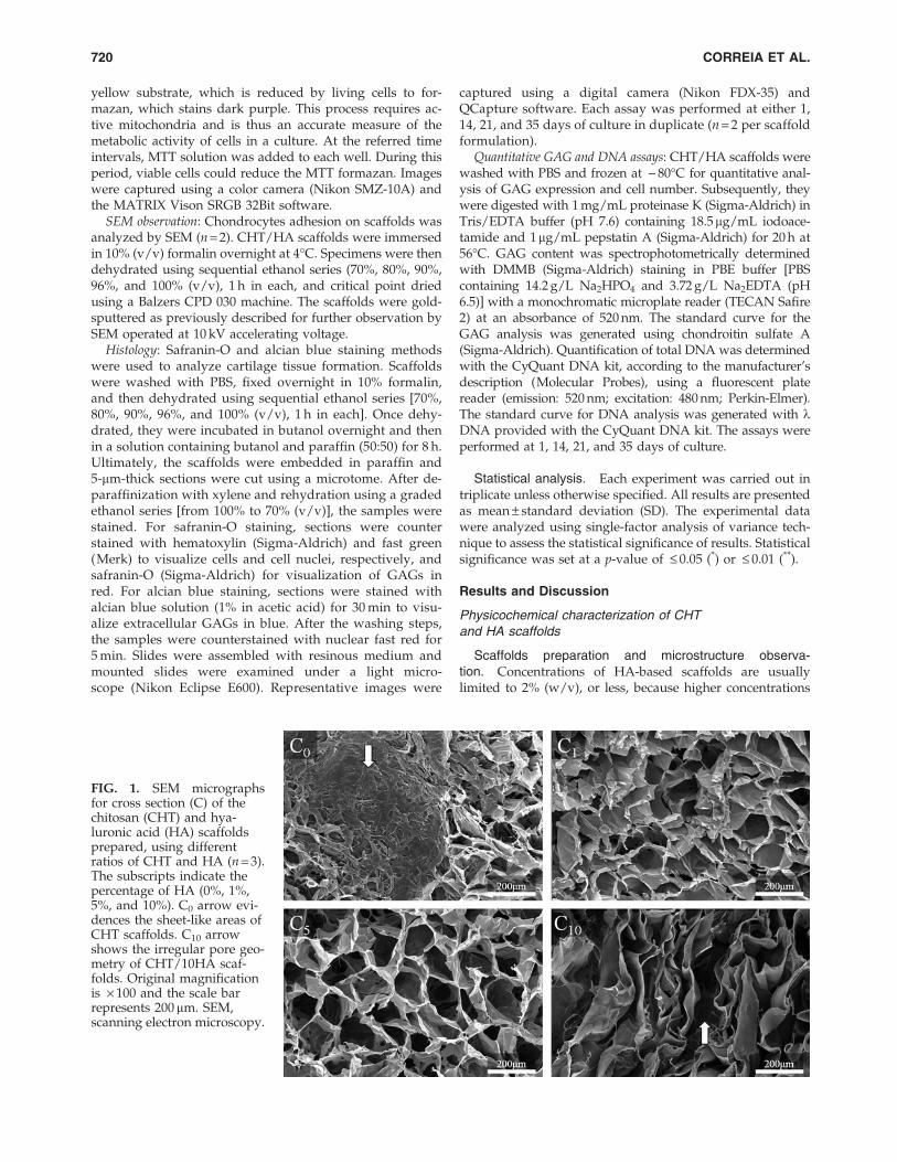
FIG. 1. SEM micrographsfor cross section (C) of thechitosan (CHT) and hya-luronic acid (HA) scaffoldsprepared, using differentratios of CHT and HA (n = 3).The subscripts indicate thepercentage of HA (0%, 1%,5%, and 10%). C0 arrow evi-dences the sheet-like areas ofCHT scaffolds. C10 arrowshows the irregular pore geo-metry of CHT/10HA scaf-folds. Original magnificationis · 100 and the scale barrepresents 200 mm. SEM,scanning electron microscopy.
720 CORREIA ET AL.

are too viscous for adequate mixing.31 The morphologies ofthe scaffolds were investigated based on different ratios be-tween CHT and HA. To characterize the microstructuremorphology of CHT and HA scaffolds, cross-sectional SEMimages were obtained (Fig. 1). CHT scaffolds showed aclosed network pore configuration with sheet-like areas (ar-row in Fig. 1C0). In contrast, CHT/1HA and CHT/5HAscaffolds showed a more open network structure. These twoformulations had high porosity and uniformly distributedpore structures. In CHT/10HA scaffolds, pore geometrywas very irregular with closed or oversized collapsed pores(arrow in Fig. 1C10). This influence on porosity and porestructure when HA was incorporated in scaffolds of poly-cationic polymers was also found by Ren et al.13 The dif-ferences between the pore architecture are at least partiallyrelated to the homogeneity of the polymeric solution, thatis, how well are the two polymers mixed and dispersed inthe solution. As CHT and HA have an opposite charge, anionic bond between the carboxyl groups of HA and theamino groups of CHT is expected.15 As the concentration ofHA increased in the CHT/HA formulation, the solutionbecame more opaque. The incorporation of HA up to aconcentration of 5% in a CHT solution yielded a well-dispersed colloidal suspension. CHT/5HA suspension wasstable, indicating the formation of submicrometer com-plexes. In contrast, higher concentrations of HA jeopardizedefficient mixture of both polymers, resulting in white pre-cipitates. These precipitates tended to aggregate and, con-sequently, after freeze-drying, resulted in a nonuniformpore geometry (Fig. 1C10). Mao et al.32 also studied CHTand HA blends and correlated the formation of white pre-cipitates with the high density of carboxyl groups in HAand amino groups in CHT.
Pore size increased with concentrations of HA in CHTscaffolds (Table 1). Fan et al.31 reported the same influence ofHA on scaffold pore size. CHT/10HA also exhibited thehighest standard deviation in pore sizes because of thenonuniform pore geometry. The open pore network and arange in pore size between *77 and 97 mm, as observed inCHT/1HA and CHT/5HA scaffolds, rendered these scaf-folds appropriate for cellular infiltration. Particularly, wehypothesize that articular bovine chondrocytes can be in-corporated into the aforementioned scaffolds.
Fourier transform infrared spectroscopy imaging mea-surements. FTIR measurements (Fig. 2) confirmed thepresence of CHT and HA in all CHT/HA scaffolds. Char-acteristic peaks of both polymers (Fig. 2a) were found,namely a distinct band at 1650 cm - 1 (amide I) and at1580 cm - 1 (amide II).26 The ester group (1045 cm - 1), a HA
characteristic peak, is the main difference between the twospectra. The referred characteristic peaks were also found inall CHT/HA scaffolds spectra (Fig. 2b). Particularly, CHT/10HA had a notable higher intensity in the ester peak, as it isthe formulation with higher amount of HA.
The homogeneity of the CHT/HA blends was investi-gated by FTIR mapping (Fig. 3). As expected, chemical mapsof CHT scaffolds (Fig. 3a) and pure HA (Fig. 3b) onlyshowed the selected color for the pure materials (green andred, respectively). As showed in Figure 3d, CHT/5HAscaffolds had an improved polymer mixture, in which thetwo polymers were well dispersed in the length scale ana-
Table 1. Pore Size (mm) and Compressive Modulus (kPa) of the Chitosan/Hyaluronic Acid Scaffolds
Prepared with Different Amounts of Hyaluronic Acid in Dry and Wet States
Samplecode
Chitosan(% wt)
Hyaluronicacid (% wt)
Pore size(lm)
Compressive modulusin dry state (kPa)
Compressive modulusin wet state (kPa)
CHT 100 0 55.80 – 10.04 291.03 – 82.73 2.84 – 0.41CHT/1HA 99 1 77.40 – 15.01 144.33 – 28.32 2.43 – 0.07CHT/5HA 95 5 97.43 – 6.56 144.00 – 25.68 2.30 – 0.06CHT/10HA 90 10 148.73 – 99.24 40.73 – 18.31 1.21 – 0.47
Values are reported as mean – SD.
FIG. 2. Fourier transform infrared (FTIR) spectra of (a)CHT scaffolds (solid line) and pure HA (dashed line) and of(b) CHT/1HA (solid line), CHT/5HA (dashed line), andCHT/10HA (dotted line) scaffolds. The rectangles present atFigure 2a indicate the chosen regions for the integrationprocedure used to obtain the images of Figure 3, each onecentered at the selected characteristic peak of chitosan(1670 cm - 1) or HA (1045 cm - 1).
CHITOSAN SCAFFOLDS CONTAINING HYALURONIC ACID 721

lyzed (tens of microns). On the other hand, in CHT/10HAscaffolds the quality of the blend was compromised. Therewere more green and red areas, which correspond to CHTand HA alone, respectively, and less yellow areas, whichcorrespond to the homogeneously blended biomaterials.
Mechanical properties. The mechanical properties of thescaffolds in tissue engineering applications are of great im-portance because of the necessity of structural stability towithstand stresses incurred during in vitro culture and in vivoimplantation.31
Compressive mechanical tests were performed usingacellular scaffolds in dry and wet states. The compressivemodulus of the scaffolds is shown in Table 1. All samplesexhibited a sponge-like behavior. For both assays, the addi-tion of HA in CHT networks reduced the mechanicalstrength of the scaffold, while it increased its flexibility. Thisbehavior may essentially be due to the increase in pore sizeby increasing the HA concentration. It was interesting to notethat CHT/1HA and CHT/5HA scaffolds had similar com-pressive modulus in dry and wet states, probably because oftheir similar pore network.
In addition to an increase in pore size, O’Brien et al.33 re-ported a correlation between decreasing mechanical strengthand nonuniform pore shape in HA-based scaffolds. This mayexplain the reason why CHT/10HA had a much lowercompressive modulus.
In the dry state, the compressive modulus of CHT/HAscaffolds were lower but in the same order of magnitudewhen compared with the modulus of articular cartilage re-ported by Korhonen et al.34 The compressive modulus in wetstate was much lower compared with the dry state becauseof HA’s ability of high water uptake. As CHT and HA aretwo natural polymers, this drawback in terms of mechanical
strength was expected. However, the principal aim of in-corporating HA into CHT scaffolds was to mimic the nativecartilage ECM. The compressive modulus may further im-prove by the deposition of an ECM by chondrocytes inCHT/HA scaffolds.
Swelling properties. Diffusion and exchange of nutrients(e.g., oxygen) and waste throughout the entire scaffold arerelated to the swelling properties of the scaffolds. Both CHTand HA have an abundant number of hydrophilic groups,such as hydroxyl, amino, and carboxyl groups, which canpromote water uptake in the structure.25 The swelling abilitywas evaluated by soaking the scaffolds in PBS at 37�C for 3days (Fig. 4). The ratio between CHT and HA significantlyaffected the scaffolds’ swelling properties. In the first 2 h, allsamples rapidly increased their weight. This rapid weightincrease continued until 8 h. Then, the swelling ratio of all
FIG. 3. Chemical maps obtained by FTIR microscopy of the cross sections of the scaffolds and pure HA: (a) CHT, (b) HA, (c)CHT/1HA, (d) CHT/5HA, and (e) CHT/10HA. Green indicates the presence of CHT and red indicates the presence of HA.The inset images individualize the presence of the pure polymers in the blends. The inset images next to (a) and (b) representthe images control for HA and CHT, respectively. Color images available online at www.liebertonline.com/tec
FIG. 4. Swelling ratio of CHT and HA scaffolds in phos-phate-buffered saline at 37�C for 3 days. Values are reportedas mean – standard deviation (SD) (n = 3).
722 CORREIA ET AL.

samples seemed to stabilize, and a slight increase could beobserved with time. CHT scaffolds had the lowest valuesduring the entire experiment, whereas CHT/10HA had thehighest. CHT/1HA and CHT/5HA had similar swelling.The swelling results indicated that HA incorporation cangreatly improve hydrophilicity and wetting of CHT scaf-folds. Hence, scaffolds with higher content of HA showedhigher values of swelling. This aspect may imply that theabsorption and diffusion of solutes through the interior poresin CHT scaffolds are greatly improved by incorporation oflow concentrations of HA.
In vitro enzymatic degradation. Scaffolds for tissue en-gineering are usually required to either degrade or be re-absorbed by the body after successful tissue regeneration.CHT/HA scaffolds were incubated in a solution with lyso-zyme and hyaluronidase or in a solution with PBS (control),both at 37�C for 7 and 14 days, to evaluate the weight de-crease in the biopolymers due to enzymatic degradation. Theweight loss percentage of scaffolds as a function of incuba-tion time is illustrated in Figure 5.
To distinguish between enzymatic degradation and sim-ple dissolution, we compared the weight loss of samples thathad been placed in PBS (Fig. 5a) with those that had beenplaced in PBS supplemented with lysozyme and hyaluroni-dase at 7 and 14 days (Fig. 5b). According to Figure 5b, thepresence of HA rendered CHT scaffolds more susceptible toenzymatic degradation, which is most likely due to higherporosity and better accessibility of cleavage sites by the en-zymes. The hydrophilicity of HA also contributed to a higherdegradation of CHT/HA scaffolds as it will enhance theinteraction of the biomaterial with the enzymatic solution.After a high initial weight loss at day 7, the degradation rate
of the samples generally appeared to slow down at day 14,which is most likely due to the gradual disappearance ofhexasaccharide sequences susceptible for enzymatic degra-dation. The control experiment in PBS (Fig. 5a) showed thatpractically none of the scaffolds degrade in PBS after 14 days.The degradation of CHT has been already tested in severalin vitro studies.35–38 Most of these studies were, however,performed at low pH for optimal lysozyme activity. In ourstudy, the enzymatic degradation experiments were per-formed at higher pH, which resembles better degradation ofthe scaffolds in physiological conditions. Hyaluronidase andlysozyme concentrations used in this study (0.33 and 2 mg/mL, respectively) are not representatives of their concentra-tion in vivo. The largest single reservoir of hyaluronidase isthe synovial fluid of diarthrodial joints (0.5–4 mg/mL).39
Lysozyme commonly exists in various human body fluidsand tissues, with concentrations ranging from 4 to 13 mg/Lin serum40 and from 450 to 1230 mg/L in tears.41
In vitro articular bovine chondrocyte culture
Cell viability, proliferation, and adhesion studies on CHT/HA scaffolds. In a live–dead assay, all materials exhibitedvery good biocompatibility with hardly any detectable celldeath (Fig. 6). Further, cells were metabolically active (Fig. 7)and well spread throughout the scaffolds in all formulations.The presence of increasing concentrations of HA did notinfluence cell loading of CHT scaffolds. This behavior is es-sentially due to the low capacity for cell attachment of CHT.
The cell morphology and proliferation on CHT/HA scaf-folds was further studied by SEM (Fig. 8). In all scaffolds,chondrocytes were primarily localized in the superficial areaat days 1 and 3 and displayed a spherical morphology (cell
FIG. 5. Weight loss percentage of CHT/HA scaffolds as a function of incubation time (7 and 14 days) at 37�C. Scaffoldswere embedded in (a) phosphate-buffered saline or (b) enzymatic solution containing both lysozyme and hyaluronidase.Values are reported as mean – SD (n = 3).
CHITOSAN SCAFFOLDS CONTAINING HYALURONIC ACID 723

size diameter: *10 mm). At day 7, a limited increase in cellnumber was observed but cells did not penetrate into thescaffolds. At days 14 and 21, chondrocytes seeded in CHT/5HA scaffolds significantly proliferated into the inner areasof the scaffolds. In all scaffolds, even after 21 days of culture,the cells continued to display a normal spherical morphol-ogy, as observed in native cartilage.
Maintenance of chondrogenic phenotype. The roundshape of chondrocytes is an indicator of phenotype reten-tion and is essential for matrix formation.42 The newlyformed matrix was stained with safranin-O (Fig. 9) andalcian blue (Fig. 10), which showed GAG secretion. As HAis a nonsulfated GAG, it stains blue in alcian blue staining.Low amounts of HA in the scaffold can be easily distin-guished from newly deposited ECM by the chondrocytes bycomparing alcian blue staining at 1 (Fig. 10A1–A35) withstaining at later time intervals. At day 1, chondrocytes didnot attach to the walls of CHT scaffolds, whereas in CHT/HA scaffolds, the cells attached to walls, forming small
aggregates. At day 14, in CHT/5HA scaffolds (Fig. 9C14
and Fig. 10C14), it was interesting to note that most chon-drocytes agglomerated to form very large aggregates ad-herent to the scaffolds. The aggregates were located on thesuperficial area. Hardly any penetration of cells in thescaffolds was observed. These events were also reported byZhao et al.43 CHT/1HA and CHT/5HA showed enhancedcell–material interactions and ECM production, with en-hanced GAG production (Figs. 9 and 10). At day 35, CHT/5HA scaffolds showed better cell dispersion and higherECM production and GAG deposition when compared withthe other formulations. Chondrocytes seeded in CHT scaf-folds, even at day 35, remained in small aggregates (Fig.9A35 and Fig. 10A35).
Enhanced cartilage tissue formation can be also qualita-tively detected by SEM analysis (Fig. 11). At day 1, chon-drocytes were in aggregates in all specimens, but at day 14they were better spread and attached. At days 14 and 21,improved chondrocyte dispersion could be noticed in CHT/1HA and CHT/5HA scaffolds. In all specimens, lacunae
FIG. 6. Live–dead assay results of chondrocytes at passage 3 in proliferation medium seeded on CHT/HA scaffolds at days1, 7, 14, and 21. Live cells were stained green by calcein and dead cells were stained red by ethidium (n = 3). Letters A–Dcorrespond to each of the four scaffold’s formulations, namely CHT, CHT/1HA, CHT/5HA or CHT/10HA, respectively. Thesubscripts indicate the time period. Scale bar is 200 mm. Color images available online at www.liebertonline.com/tec
724 CORREIA ET AL.

FIG. 7. MTT assay results of chondrocytes at passage 3 in proliferation medium seeded on CHT/HA scaffolds at days 1, 7,14, and 21 (n = 3). Metabolic active cells were stained dark purple. Letters A–D correspond to each of the four scaffold’sformulations, namely CHT, CHT/1HA, CHT/5HA or CHT/10HA, respectively. The subscripts indicate the time period.Original magnification is · 7.5. Scale bar is 3 cm. Color images available online at www.liebertonline.com/tec
FIG. 8. SEM micrographs (10 kV) of CHT/HA scaffolds seeded with articular bovine chondrocytes at passage 3 in pro-liferation medium (n = 2). Letters A–D correspond to each of the four scaffold’s formulations, namely CHT, CHT/1HA, CHT/5HA or CHT/10HA, respectively. The subscripts indicate the time period. Original magnification is · 500. Scale bar is 50mm.Arrows point to chondrocytes and/or cartilage extracellular matrix deposition and stars indicate scaffold material.
725

formation was seen in the matrix surrounding the chon-drocytes. The morphology of these lacunas shows similari-ties to natural cartilage.
Chondrogenic activity evaluation. It is known that whenchondrocytes maintain their natural spherical shape theyproduce more GAGs and higher collagen type II versuscollagen type I ratio.44–46 Cell proliferation and ECM for-mation were quantified by DNA and GAG assays, respec-tively. In GAG quantification assay (Fig. 12a), theincorporation of HA seemed to initiate an earlier GAG de-position, particularly at day 14 for CHT/1HA and CHT/5HA scaffolds. Results showed that the incorporation of HAseemed to favor GAG deposition, because at day 35 all thescaffolds containing HA had more GAG deposition whencompared with CHT scaffolds. For DNA quantification assay(Fig. 12b), CHT/5HA had the highest amount of DNA at day14. However, at day 35, CHT/1HA scaffolds significantlysupported a higher amount of cells when compared withother formulations.
A higher value of GAG/DNA ratio indicates a higherdegree of differentiation of the cells.47 For GAG/DNA ratio(Fig. 12c), at day 35, scaffolds with higher amount of HA(CHT/5HA and CHT/10HA) induced a higher ratio. Al-though CHT/1HA had the highest amount of cells in DNAquantification, CHT/5HA had the highest GAG versus DNAratio. This may imply that CHT/5HA scaffolds are the for-mulation that most favors cartilage ECM production, ren-dering them attractive for cartilage TE.
Physicochemical effects of CHT/HA scaffoldson cartilage regeneration
The key challenge for biomedical researchers is how todesign and control material properties to achieve a specificbiological response.48 It has been well established that thephysicochemical properties of materials can regulate bio-logical responses, such as differentiation of stem cells,49
proliferation of fibroblasts,50 gene delivery,51 and celldeath.52
FIG. 9. Histological cross-sections show glycosaminoglycan (GAG) deposition (stained red) in CHT/HA scaffolds bysafranin-O staining at 1, 14, 21, and 35 days of chondrocyte culture in differentiation medium (n = 2). Letters A–D correspondto each of the four scaffold’s formulations, namely CHT, CHT/1HA, CHT/5HA or CHT/10HA, respectively. Subscriptsindicate time intervals. Scale bar is 100mm. Color images available online at www.liebertonline.com/tec
726 CORREIA ET AL.

FIG. 10. Histological cross-sections show GAG deposition (stained blue) in CHT/HA scaffolds by alcian blue staining at 1, 14,21, and 35 days of chondrocyte culture in differentiation medium (n = 2). Letters A–D correspond to each of the four scaffold’sformulations, namely CHT, CHT/1HA, CHT/5HA or CHT/10HA, respectively. Subscripts indicate time intervals. Scale bar is100mm. Color images available online at www.liebertonline.com/tec
FIG. 11. SEM micrographs (10 kV) of CHT/HA scaffolds seeded with articular bovine chondrocytes at passage 1 in dif-ferentiation medium (n = 2). Letters A–D correspond to each of the four scaffold’s formulations, namely CHT, CHT/1HA,CHT/5HA or CHT/10HA, respectively. The subscripts indicate the time period. Original magnification is · 500. Scale bar is50 mm. Arrows point to chondrocytes and/or cartilage extracellular matrix deposition and stars indicate scaffold material.
727

CHT/1HA and CHT/5HA scaffolds exhibited a uniformpore network with high porosity (Fig. 1) and a homogeneouspolymer dispersion (Fig. 3), rendering them suitable forbovine chondrocyte culture. The pore configuration allowedcellular infiltration into the inner pores of the scaffolds(Fig. 8). On the other hand, CHT and CHT/10HA scaffoldshad a very irregular pore network configuration with closedpores. Consequently, in these scaffolds, the adhesion andproliferation of chondrocytes were jeopardized when com-pared with the other formulations. The scaffolds’ high wateruptake ability (Fig. 4) allowed the chondrocyte medium toreach the interior pores of the scaffolds. Consequently, dif-fusion and exchange of nutrients and waste throughout thescaffolds were ensured, allowing cells to proliferate and bemetabolically active (Figs. 6 and 7).
Besides appropriate pore network and water uptakeability, CHT/1HA and, especially, CHT/5HA had an im-proved GAG dispersion and deposition (Figs. 9 and 10).Additionally, the two referred formulations had similarcompressive modulus. Hence, physical and chemical factorsseem to simultaneously influence the biological outcome.
Cartilage regeneration seemed to be favored simultaneouslyby chemical aspects, that is, intermediate amounts of HA (1%and 5%), and also by physical aspects, that is, the referredscaffold formulations also have similar compressive modu-lus and a uniform pore network configuration. Therefore, inthis case scenario, it is not possible to distinguish case by casethe detailed mechanisms of how physical properties affectthe biological performance of the CHT/HA scaffolds. De-spite this unclear contribution of each parameter, the resultsobtained in our study are not compromised.
Conclusions
The incorporation of HA up to a concentration of 5% inCHT scaffolds improved the physicochemical properties andbiological properties of CHT scaffolds. It enhanced pore net-work configuration and increased pore size, swelling ratio,and degradation rate when compared with pure CHT scaf-folds. Higher concentrations of HA resulted in heterogeneousblends with irregular and collapsed pore networks, particu-larly CHT/5HA scaffolds favored chondrocyte adhesion, cell
FIG. 12. GAG and DNA assays on CHT/HA scaffolds at 1, 14, 21, and 35 days of culture: (a) GAG assay, (b) DNA assay, (c)GAGs per DNA ratio. Values are reported as mean – standard deviation (n = 3). Significant differences for *p £ 0.05 and**p £ 0.01 were found.
728 CORREIA ET AL.

proliferation, and cartilage matrix production when comparedwith pure CHT scaffolds and other CHT/HA blends.
The biological outcome of CHT/HA scaffolds seems to beinfluenced by physicochemical factors, such as polymer dis-persion, pore network configuration, pore size, water uptakeability, and mechanical strength. However, other studies arerequired to elucidate case by case the dependence of biologicalresponse on physical properties and to categorize the relativeweight of different physical and chemical factors.
Our results suggest that the freeze-dried scaffolds of CHTand HA could have potential use in the regeneration ofcartilaginous lesions caused by various joint diseases, in-cluding osteoarthritis and rheumatoid arthritis.
Disclosure Statement
No competing financial interests exist.
References
1. Wu, S.C., Chang, J.K., Wang, C.K., Wang, C.J., and Ho, M.L.Enhancement of chondrogenesis of human adipose derivedstem cells in a hyaluronan-enriched microenvironment.Biomaterials 31, 631, 2010.
2. Bobic, V., and Noble, J. Articular cartilage, to repair or not torepair. J Bone Joint Surg Br 82, 165, 2000.
3. Temenoff, J.S., and Mikos, A.G. Review: tissue engineering forregeneration of articular cartilage. Biomaterials 21, 431, 2000.
4. Gobbi, A., Nunag, P., and Malinowski, K. Treatment of fullthickness chondral lesions of the knee with microfracture ina group of athletes. Knee Surg Sports Traumatol Arthrosc13, 213, 2005.
5. Lee, J., Lee, J.Y., Lee, E., and Son, Y. Newborn calf serumretards loss of the chondrocytic phenotype during in vitrocell expansion. J Tissue Eng Regen Med 6, 229, 2009.
6. Lin, Y.C., Tan, F.J., Marra, K.G., Jan, S.S., and Liu, D.C.Synthesis and characterization of collagen/hyaluronan/chitosan composite sponges for potential biomedical appli-cations. Acta Biomater 5, 2591, 2009.
7. Mano, J.F., and Reis, R.L. Osteochondral defects: presentsituation and tissue engineering approaches. J Tissue EngRegen Med 1, 261, 2007.
8. Chen, G., Ushida, T., and Tateishi, T. Scaffold design fortissue engineering. Macromol Biosci 2, 67, 2002.
9. Goldring, M.B., Tsuchimochi, K., and Ijiri, K. The control ofchondrogenesis. J Cell Biochem 97, 33, 2006.
10. Yoo, H.S., Lee, E.A., Yoon, J.J., and Park, T.G. Hyaluronicacid modified biodegradable scaffolds for cartilage tissueengineering. Biomaterials 26, 1925, 2005.
11. Kim, I.Y., Seo, S.J., Moon, H.S., Yoo, M.K., Park, I.Y., Kim,B.C., and Cho, C.S. Chitosan and its derivatives for tissueengineering applications. Biotechnol Adv 26, 1, 2008.
12. Ghosh, P., and Guidolin, D. Intra-articular hyaluronan ther-apy in osteoarthritis. Semin Arthritis Rheum 32, 10, 2002.
13. Ren, Y.J., Zhou, Z.Y., Liu, B.F., Xu, Q.Y., and Cui, F.Z. Pre-paration and characterization of fibroin/hyaluronic acidcomposite scaffold. Int J Biol Macromol 44, 372, 2009.
14. Tian, W.M., Hou, S.P., Ma, J., Zhang, C.L., Xu, Q.Y., and Lee,I.S. Hyaluronic acid–poly-D-lysine-based three-dimensionalhydrogel for traumatic brain injury. Tissue Eng 11, 513, 2005.
15. Oyarzun-Ampuero, F.A., Brea, J., Loza, M.I., Torres, D., andAlonso, M.J. Chitosan-hyaluronic acid nanoparticles loadedwith heparin for the treatment of asthma. Int J Pharm 381,
122, 2009.
16. Tan, H., Wu, J., Lao, L., and Gao, C. Gelatin/chitosan/hyaluronan scaffold integrated with PLGA microspheres forcartilage tissue engineering. Acta Biomater 5, 328, 2009.
17. Martino, A.D., Sittinger, M., and Risbud, M.V. Chitosan: aversatile biopolymer for orthopaedic tissue-engineering.Biomaterials 26, 5983, 2005.
18. Kim, S.E., Park, J.H., Cho, Y.W., Chung, H., Jeong, S.Y., Lee,E.B., and Kwon, I.C. Porous chitosan scaffold containingmicrospheres loaded with transforming growth factor-b1:implications for cartilage tissue engineering. J Control Re-lease 91, 365, 2003.
19. Oliveira, J.M., Rodrigues, M.T., Silva, S.S., Malafaya, P.B.,Gomes, M.E., Viegas, C.A., Dias, I.R., Azevedo, J.T., Mano,J.F., and Reis, R.L. Novel hydroxyapatite/chitosan bilayeredscaffold for osteochondral tissue-engineering applications:scaffold design and its performance when seeded with goatbone marrow stromal cells. Biomaterials 27, 6123, 2006.
20. Nge, T.T., Nogi, M., Yano, H., and Sugiyama, J. Micro-structure and mechanical properties of bacterial cellulose/chitosan porous scaffold. Cellulose 17, 349, 2010.
21. Tan, H., Chu, C.R., Payne, K.A., and Marra, K.G. Injectablein situ forming biodegradable chitosan-hyaluronic acidbased hydrogels for cartilage tissue engineering. Biomater-ials 30, 2499, 2009.
22. Yamane, S., Iwasaki, N., Majima, T., Funakoshi, T., Masuko,T., Harada, K., Minami, A., Monde, K., and Nishimura, S.I.Feasibility of chitosan-based hyaluronic acid hybrid bioma-terial for a novel scaffold in cartilage tissue engineering.Biomaterials 26, 611, 2005.
23. Yamane, S., Iwasaki, N., Kasahara, Y., Harada, K., Majima,T., Monde, K., Nishimura, S.I., and Minami, A. Effect of poresize on in vitro cartilage formation using chitosan-basedhyaluronic acid hybrid polymer fibers. J Biomed Mater ResA 81A, 587, 2006.
24. Hsu, S.H., Whu, S.W., Hsieh, S.C., Tsai, C.L., Chen, D.C.,and Tan, T.S. Evaluation of chitosan-alginate-hyaluronatecomplexes modified by an RGD-containing protein as tissueengineering scaffolds for cartilage regeneration. Artif Organs28, 693, 2004.
25. Chen, J.P., and Cheng, T.H. Preparation and evaluation ofthermo-reversible copolymer hydrogels containing chitosanand hyaluronic acid as injectable cell carriers. Polymer 50,
107, 2009.26. Chung, T.W., Yang, J., Akaike, T., Cho, K.Y., Nah, J.W., Kim,
S., II, and Cho, C.S. Preparation of alginate/galactosylatedchitosan scaffold for hepatocyte attachment. Biomaterials 23,
2827, 2002.27. Ji, Y., Ghosh, K., Shu, X.Z., Li, B., Sokolov, J.C., Prestwic,
G.D., Clark, R.A.F., and Rafailovich, M.H. Electrospun three-dimensional hyaluronic acid nanofibrous scaffolds. Bioma-terials 27, 3782, 2006.
28. Antunes, J.C., Oliveira, J.M., Reis, R.L., Soria, J.M., Gomez-Ribelles, J.L., and Mano, J.F. Novel poly (L-lactic acid)/hyaluronic acid macroporous hybrid scaffolds: character-ization and assessment of cytotoxicity. J Biomed Mater ResA 94, 856, 2010.
29. Greenwald, R.A., Josephson, A.S., Diamond, H.S., andTsang, A. Human cartilage lysozyme. J Clin Invest 51, 2264,1972.
30. Jin, R., Moreira-Teixeira, L.S., Krouwels, A., Dijkstra, P.J.,van Blitterswijk, C.A., Karperien, M., and Feijen, J. Synthesisand characterization of hyaluronic acid-peg hydrogels viaMichael addition: an injectable biomaterial for cartilage re-pair. Acta Biomater 6, 1968, 2010.
CHITOSAN SCAFFOLDS CONTAINING HYALURONIC ACID 729

31. Fan, J., Shang, Y., Yuan, Y., and Yang, J. Preparation andcharacterization of chitosan/galactosylated hyaluronic acidscaffolds for primary hepatocytes culture. J Mater Sci MaterMed 21, 319, 2010.
32. Mao, J.S., Liu, H.F., Yin, Y.J., and Yao, K.D. The properties ofchitosan–gelatin membranes and scaffolds modified withhyaluronic acid by different methods. Biomaterials 24, 1621,2003.
33. O’Brien, F.J., Harley, B.A., Yannas, I.V., and Gibson, L. In-fluence of freezing rate on pore structure in freeze-driedcollagen–GAG scaffolds. Biomaterials 25, 1077, 2004.
34. Korhonen, R.K., Laasanen, M.S., Toyras, J., Rieppo, J., Hir-vonen, J., Helminen, H.J., and Jurvelin, J.S. Comparison ofthe equilibrium response of articular cartilage in unconfinedcompression, confined compression and indentation. J Bio-mech 35, 903, 2002.
35. Pangburn, S.H., Trescony, P.V., and Heller, J. Lysozymedegradation of partially deacetylated chitin, its films andhydrogels. Biomaterials 3, 105, 1982.
36. Tomihata, K., and Ikada, Y. In vitro and in vivo degradationof films of chitin and its deacetylated derivatives. Bioma-terials 18, 567, 1997.
37. Lee, K.Y., Ha, W.S., and Park, W.H. Blood compatibility andbiodegradability of partially N-acylated chitosan deriva-tives. Biomaterials 16, 1211, 1995.
38. Hirano, S., Tsuchida, H., and Nagao, N. N-acetylation inchitosan and the rate of its enzymatic hydrolysis. Bioma-terials 10, 574, 1989.
39. Fraser, J.R.E., Laurent, T.C., and Laurent, U.B.G. Hyalur-onan: its nature, distribution, functions and turnover. J In-tern Med 242, 27, 1997.
40. Nordtveit, R.J., Varum, K.M., and Smidsrød, O. Degradationof partially N-acetylated chitosans with hen egg white andhuman lysozyme. Carbohydr Polym 29, 163, 1996.
41. Telmel, A., Kazokoglu, H., and Taga, Y. Tear lysozyme levelsin contact lens wearers. Ann Ophthalmol 23, 191, 1991.
42. Von der Mark, K., Gauss, V., von der Mark, H., and Muller,P. Relationship between cell shape and type of collagensynthesized as chondrocytes lose their cartilage phenotypein culture. Nature 267, 531, 1977.
43. Zhao, K., Deng, Y., Chen, J.C., and Chen, G.Q. Poly-hydroxyalkanoate (PHA) scaffolds with good mechanicalproperties and biocompatibility. Biomaterials 24, 1041, 2003.
44. Woodfield, T.B.F., Miot, S., Martin, I., van Blitterswijk, C.A.,and Riesle, J. The regulation of expanded human nasalchondrocyte re-differentiation capacity by substrate com-position and gas plasma surface modification. Biomaterials27, 1043, 2006.
45. Miot, S., Woodfield, T.B.F., Daniels, A.U., Suetterlin, R.,Peterschmitt, I., Heberer, M., van Blitterswijk, C.A., Riesle, J.,and Martin, I. Effects of scaffold composition and architec-ture on human nasal chondrocyte redifferentiation and car-tilaginous matrix deposition. Biomaterials 26, 2479, 2005.
46. Barry, J.J., Gidda, H.S., Scotchford, C.A., and Howdle, S.M.Porous methacrylate scaffolds: supercritical fluid fabricationand in vitro chondrocyte responses. Biomaterials 25, 3559,2004.
47. Moroni, L., Schotel, R., Hamann, D., de Wijn, J.R., and vanBlitterswijk, C.A. 3D fiber-deposited electrospun integratedscaffolds enhance cartilage tissue formation. Adv FunctMater 18, 53, 2008.
48. Mitragotri, S., and Lahann, J. Physical approaches to bio-material design. Nat Mater 8, 15, 2009.
49. Graziano, A., d’Aquino, R., Cusella-De Angelis, M.G.,Francesco, F., Giordano, A., Laino, G., Piattelli, A., Traini, T.,Rosa, A., and Papaccio, G. Scaffold’s surface geometry sig-nificantly affects human stem cell bone tissue engineering.J Cell Physiol 214, 166, 2008.
50. Milner, K.R., and Siedlecki, C.A. Submicron poly(L-lacticacid) pillars affect fibroblast adhesion and proliferation.J Biomed Mater Res A 82, 80, 2007.
51. Shen, H., Tan, J., and Saltzman, W.M. Surface-mediatedgene transfer from nanocomposites of controlled texture.Nat Mater 3, 569, 2004.
52. Chen, C.S., Mrksich, M., Huang, S., Whitesides, G.M., andIngber, D.E. Geometric control of cell life and death. Science276, 1425, 1997.
Address correspondence to:Joao F. Mano, Ph.D.
3B’s Research Group—Biomaterials,Biodegradables, and Biomimetics
Headquarters of the European Institute of Excellenceon Tissue Engineering and Regenerative Medicine
University of MinhoAvePark
Zona Industrial da GandraS. Claudio do Barco
Guimaraes 4806-909Portugal
E-mail: [email protected]
Received: August 9, 2010Accepted: March 7, 2011
Online Publication Date: April 22, 2011
730 CORREIA ET AL.


