Embed Size (px)
Citation preview

This article was originally published in a journal published byElsevier, and the attached copy is provided by Elsevier for the
author’s benefit and for the benefit of the author’s institution, fornon-commercial research and educational use including without
limitation use in instruction at your institution, sending it to specificcolleagues that you know, and providing a copy to your institution’s
administrator.
All other uses, reproduction and distribution, including withoutlimitation commercial reprints, selling or licensing copies or access,
or posting on open internet sites, your personal or institution’swebsite or repository, are prohibited. For exceptions, permission
may be sought for such use through Elsevier’s permissions site at:
http://www.elsevier.com/locate/permissionusematerial
Autho
r's
pers
onal
co
py
Cholesterol biosensor based onN-(2-aminoethyl)-3-aminopropyl-trimethoxysilane
self-assembled monolayer
Sunil K. Arya a,b, Arun K. Prusty a, S.P. Singh a, Pratima R. Solanki a,Manoj K. Pandey a, Monika Datta b, Bansi D. Malhotra a,*
a Biomolecular Electronics and Conducting Polymer Research Group, National Physical Laboratory, K. S. Krishnan Marg, New Delhi 110012, Indiab Department of Chemistry, University of Delhi, Delhi 110007, India
Received 23 October 2006Available online 26 January 2007
Abstract
Cholesterol oxidase (ChOx) has been covalently immobilized onto two-dimensional self-assembled monolayer (SAM) of N-(2-amino-ethyl)-3-aminopropyl-trimethoxysilane (AEAPTS) deposited on the indium–tin oxide (ITO) coated glass plates using N-ethyl-N 0-(3-dim-ethylaminopropyl) carbodiimide and N-hydroxysuccinimide (EDC/NHS) chemistry. These ChOx/AEAPTS/ITO bioelectrodes arecharacterized using contact angle (CA) measurements, UV–visible spectroscopy, atomic force microscopy (AFM), electrochemicalimpedance technique, and Fourier transform infrared (FT–IR) technique. The covalently immobilized ChOx-modified AEAPTS bioelec-trodes are used for the estimation of cholesterol in solution using UV–visible technique. These cholesterol sensing bioelectrodes showlinearity as 50 to 500 mg/dl for cholesterol solution, detection limit as 25 mg/dl, sensitivity as 4.499 · 10�5 Abs (mg/dl)�1, Km valueas 58.137 mg/dl (1.5 mM), apparent enzyme activity as 1.81 · 10�3 U cm�2, shelf life of approximately 10 weeks, and electrode reusabil-ity as 10 times.� 2007 Elsevier Inc. All rights reserved.
Keywords: Cholesterol; Cholesterol oxidase (ChOx); N-(2-aminoethyl)-3-aminopropyl-trimethoxysilane (AEAPTS); Self-assembled monolayer (SAM);Electrochemical impedance
Rapid growth in the development of new biomaterialsand the improvements in sensing techniques have givenboost to the evolution of new biosensors [1–5]. The immo-bilization and stability of enzymes, antibodies and DNAetc. onto solid support are crucial factors for the fabrica-tion of a biosensor. Various immobilization methods, suchas physical adsorption [6], entrapment [7], cross-linking [8],and covalent attachment [9]—have been used for the tech-nological development of a biosensor. Among these meth-ods, the covalent binding of an enzyme, involving theformation of covalent bonds between an enzyme and a sup-porting matrix, has the added advantage of stronger
attachment resulting in reduced leaching of an enzyme.For improved response and binding, various immobiliza-tion matrices, such as Langmuir–Blodgett films [10], con-ducting polymers [8], nanomaterials [11], sol gel films[12], and self-assembled monolayers (SAMs)1 [13] havebeen used. SAMs are considered to be more suitable thanothers because they are easy to form and have increased
0003-2697/$ - see front matter � 2007 Elsevier Inc. All rights reserved.
doi:10.1016/j.ab.2007.01.029
* Corresponding author. Fax: +91 11 25726938.E-mail address: [email protected] (B.D. Malhotra).
1 Abbreviations used: SAM, self-assembled monolayer; GOx, glucoseoxidase; AEAPTS, N-(2-aminoethyl)-3-aminopropyl-trimethoxysilane;ITO, indium-tin-oxide; ChOx, cholesterol oxidase; EDC/NHS, N-ethyl-N 0-(3-dimethylaminopropyl) carbodiimide and N-hydroxysuccinimide;HRP, horseradish peroxidase; CA, contact angle; AFM, atomic forcemicroscopy; FT–IR, Fourier transform infrared; EIS, electrochemicalimpedance spectroscopy; PBS, phosphate-buffered saline; ATR, attenu-ated total reflection.
www.elsevier.com/locate/yabio
Analytical Biochemistry 363 (2007) 210–218
ANALYTICAL
BIOCHEMISTRY
Autho
r's
pers
onal
co
py
stability, reusability, ordered arrangement and high repro-ducibility etc. [14,15]. In addition, the properties of SAMscan be easily manipulated via changing functional groupsthat become compatible for the immobilization of biomol-ecules [16–22], and they can be used to orient the desiredbiomolecules without denaturation, facilitating electrontransfer between biomolecules and electrode surface.
Recently, monolayers of functionalized silanes haveattracted much attention for anchoring organic materialssuch as proteins, enzymes and DNA [23–26]. These silanescan act as cross-linkers because they contain Si-O bondsthat react with surface hydroxyl groups of metal oxide,glass, silicon, quartz etc. and the pendant-functionalizedhydrocarbon chain that binds with organic material andbiomolecules. Hence, SAM formation by silanization pro-vides a uniform layer on a desired surface and the mediumto tailor the surface properties. Babu and coworkers havereported that the silanization of matrix for binding helpsin the stabilization of immobilized glucose oxidase (GOx)against thermal inactivation [23]. Chrisey and coworkershave reported the use of trimethoxysilylpropyldiethylenetri-amine and N-(2-aminoethyl)-3-aminopropyl-trimethoxysi-lane (AEAPTS) for covalent binding of DNA on siliconsurface [24]. They suggested that the use of aminosilanemonolayers to anchor DNA oligomers via the use ofcross-linking molecules may be useful for certain applica-tions where the thin, covalently attached DNA films or pat-terned DNA surfaces are required. Francia and coworkershave reported label-free optical biosensor [25]. They modi-fied the porous silicon surface by trimethoxy-3-bromoace-tamidopropylsilane for the binding of single-strandedDNA and showed that enhancement of light emissionoccurs when modified surface reacts with the complemen-tary strand. Nakagawa and coworkers have used the ami-nosilane-coated silicon wafer for the extraction of DNAfrom the blood [26].
Fabrication of silane-based SAMs onto indium–tinoxide (ITO) surface has been used because it offers desiredphysical and chemical properties such as stability underphysiological conditions, transparency, and conductivity[27]. Hatton and coworkers have employed SAM of silanederivatized ITO electrodes for fabricating an organic elec-troluminescent device [28]. They have reported that the sil-anization of ITO improves the durability of theelectroluminescent device. Moore and coworkers have used3-aminopropyl-trimethoxysilane silane onto ITO films forDNA binding [29]. Yang and Li have used the SAM ofepoxysilane for the immobilization of anti-Escherichia coli
antibody [30]. Ruan and coworkers have developed imped-ance biosensor chips based on the immobilization of affin-ity-purified antibodies onto ITO electrode surface fordetection of E. coli O157:H7 [31]. They have used theSAM of epoxysilane to serve as a template for chemicalanchoring of antibodies onto ITO plate.
The estimation of cholesterol on a periodical basis isessential for living beings because the abnormal levels ofcholesterol are related to heart diseases. In addition, cho-
lesterol and its fatty acid esters are essential componentsof nerve cells and are precursors of many biomaterials suchas bile acid and steroid hormones. Attempts have beenmade recently for the development of cholesterol biosensorusing conducting polymers [32,33], sol gels [12], carbonelectrodes [34,35], and monolayers of different materials[36–38]. Biosensors based on these matrices, however, suf-fer from low range of linearity, harsh conditions for fabri-cation or short shelf life.
There are reports on the use of silane monolayers forDNA binding and immunosensor fabrication. We arereporting the results of our studies relating to cholesterolbiosensor based on covalent immobilization of cholesteroloxidase (ChOx) onto SAM of AEAPTS using N-ethyl-N 0-(3-dimethylaminopropyl) carbodiimide and N-hydroxy-succinimide (EDC/NHS) chemistry to create covalentbonding between the NH2 group of AEAPTS and theCOOH group of ChOx by spectrophotometric technique.
Materials and methods
Chemicals and reagents
ChOx (EC 1.1.36 from Pseudomonas fluorescens) withspecific activity of 24 U/mg and horseradish peroxidase(HRP, EC 1.11.1.7) with specific activity of 200 U/mg solidwere purchased from Sigma–Aldrich (USA). AEAPTS wasprocured from Merck (India). All other chemicals were ofanalytical grade and used without further purification.
Enzyme solution preparation
The solutions of ChOx (24 U/ml) and HRP (200 U/ml)were prepared freshly in phosphate buffer (50 mM, pH 7.0)prior to being used. Stock solution of cholesterol was pre-pared in 10% Triton X-100 and stored at 4 �C. This stocksolution was further diluted to make different concentra-tions of cholesterol solution. O-Dianisidine (1%) solutionwas prepared freshly in deionized water.
Preparation of SAM of AEAPTS and immobilization of
ChOx
The ITO plates were precleaned with acetone, ethanol,and a copious amount of deionized water. To obtain a uni-formly distributed OH group on ITO surface, the ITOplates were immersed in a solution of H2O2/NH4OH/H2O (1:1:5, v/v) for 30 min at 80 �C for hydrolysis, afterwhich they were rinsed thoroughly with deionized waterand dried. The hydrolyzed ITO plates were immersed in1% (v/v) solution of AEAPTS in toluene overnight at roomtemperature for silanization. After the coupling reaction,the electrodes were rinsed with toluene and water toremove the physically absorbed silanes from the surface.The modified electrodes were then dried under a streamof nitrogen. The ChOx was covalently attached toAEAPTS SAM using EDC/NHS (Scheme 1). For immobi-
Cholesterol biosensor based on AEAPTS SAM / S.K. Arya et al. / Anal. Biochem. 363 (2007) 210–218 211
Autho
r's
pers
onal
co
py
lization, the amount of enzyme used was optimized andmaximum binding was observed when 5 ll of ChOx solu-tion was diluted to 30 ll in distilled water containing0.4 M EDC and 0.1 M NHS. The enzyme solution waspoured onto a 1-cm2 area of ITO and was kept for approx-imately 4 h in a humid chamber at room temperature forChOx binding. ChOx immobilization occurs via a couplingreaction between the amino group of silane and the EDC/NHS activated carboxylic group of ChOx. The electrode(ChOx/AEAPTS/ITO) formed was washed thoroughlywith phosphate buffer (50 mM, pH 7.0) containing 0.9%NaCl and 0.05% Tween 20 to remove any unbound enzymeand was stored at 4 �C when not in use.
Characterization
The electrodes fabricated were characterized usingcontact angle (CA) measurements, UV–visible spectros-copy, atomic force microscopy (AFM), electrochemicalimpedance technique, and Fourier transform infrared(FT–IR) technique. CA measurements were carried outby the Sessile drop method [39] using a drop shape ana-lyzer (DSA 100, DSA/V 1.9, Kruss, Germany). Theenzyme activity measurements were carried out using aUV–visible spectrophotometer (model 160A, Shimadzu).AFM images were taken in air using a Digital Instru-ments NanoScope IIIa with a silicon cantilever operatedin tapping mode to examine the modification of sur-faces. The measurements were made in air at room tem-perature. Electrochemical impedance spectroscopy (EIS)measurements were carried out in the frequency rangeof 0.01 to 105 Hz, amplitude 5 mV, on an AutolabPotentiostat/Galvanostat (Eco Chemie, The Netherlands)using a three-electrode system in phosphate-buffer saline
(PBS) (50 mM, pH 7.0, 0.9% NaCl) containing 5 mMFeðCNÞ63�=4� FT–IR spectra were recorded with a Perk-inElmer (Spectrum BX) and an attenuated total reflec-tion (ATR) accessory. For each sample, 256 scanswith the wave number resolution of 2 cm�1 wererecorded.
Enzyme activity measurements
One unit of enzyme activity (U cm�2) is defined asthe activity that results in the production of 1 lmolof enzymatic product per minute. To calculate theamount of actively working enzyme units, the apparentenzyme activity (U cm�2) was calculated using themethod based on the difference of absorbance observedbefore and after the incubation of enzyme-bound elec-trode [40]:
aenzappðU cm�2Þ ¼ AV =ets ð1Þ
where A is the difference in absorbance before and afterincubation, V is the total volume (3.14 cm3), e is the mil-limolar extinction coefficient (7.5 for o-dianisidine at500 nm), t is the reaction time (min), and s is the surfacearea (cm2) of the electrode. For the measurement, a solu-tion of 20 ll HRP, 20 ll dye, and 100 ll of 200 mg/dlcholesterol was diluted by adding 3 ml PBS (pH 7.0)and was kept in a thermostat at 25 �C. The ChOx/AEAPTS/ITO plate was immersed and incubated forapproximately 7 min. Then, the plate was removed andthe absorbance of the solution was measured at 500 nmusing a double beam spectrophotometer to determinethe product produced as a result of enzymatic reaction.The apparent enzyme activity (U cm�2) was evaluatedusing Eq. (1).
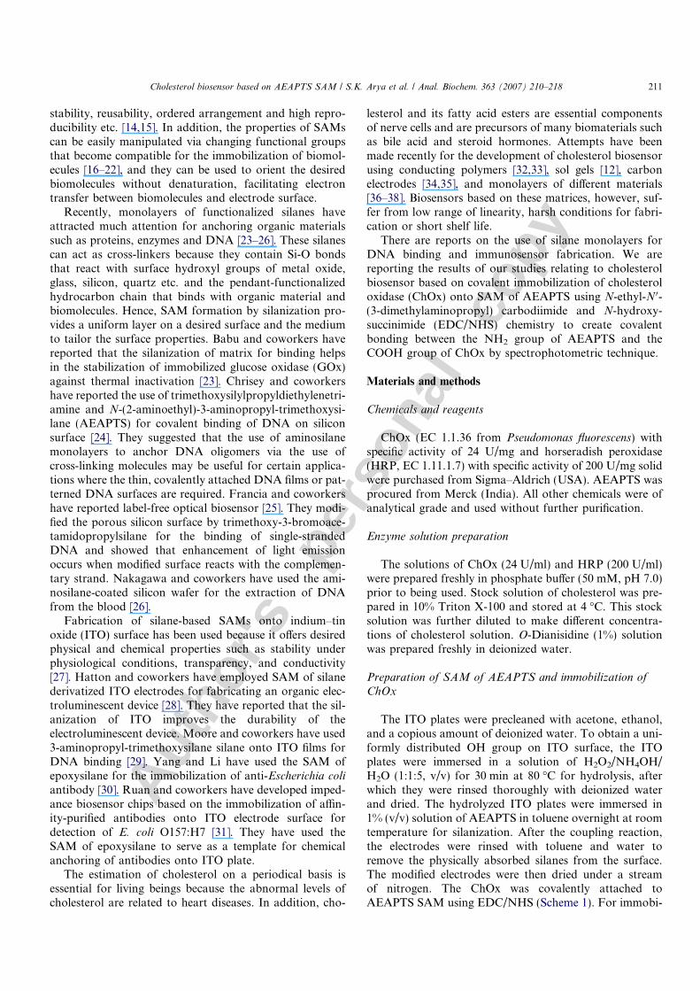
OH
OH
OH
OH
OH
OH
O
HO-Si-O-Si-O-Si-O-Si-OH
OOO
R R R R
N N N NH2 H2 H2 H2
Where RNH2 = -CH2-CH2-CH2-NH-CH2-CH2-NH2
H2O2/NH4OH/H2O (1:1:5),
80 oC, 30 Min
HydrolysisITO
H2NR-Si(OCH3)3/Toluene (1%)
Silanization
O
HO-Si-O-Si-O-Si-O-Si-OH
OOO
R R R R
N N N NH2 H2 H2 H2
O
HO-Si-O-Si-O-Si-O-Si-OH
OOO
R R R REDC/NHS/ HOOC-Enzyme
NH
CO
NH
CO
Enz
Enz
NH2
NH2
Immobilization
Scheme 1. The schematic of covalent immobilization of ChOx onto AEAPTS/ITO surface using EDC/NHS chemistry.
212 Cholesterol biosensor based on AEAPTS SAM / S.K. Arya et al. / Anal. Biochem. 363 (2007) 210–218
Autho
r's
pers
onal
co
py
Results and discussion
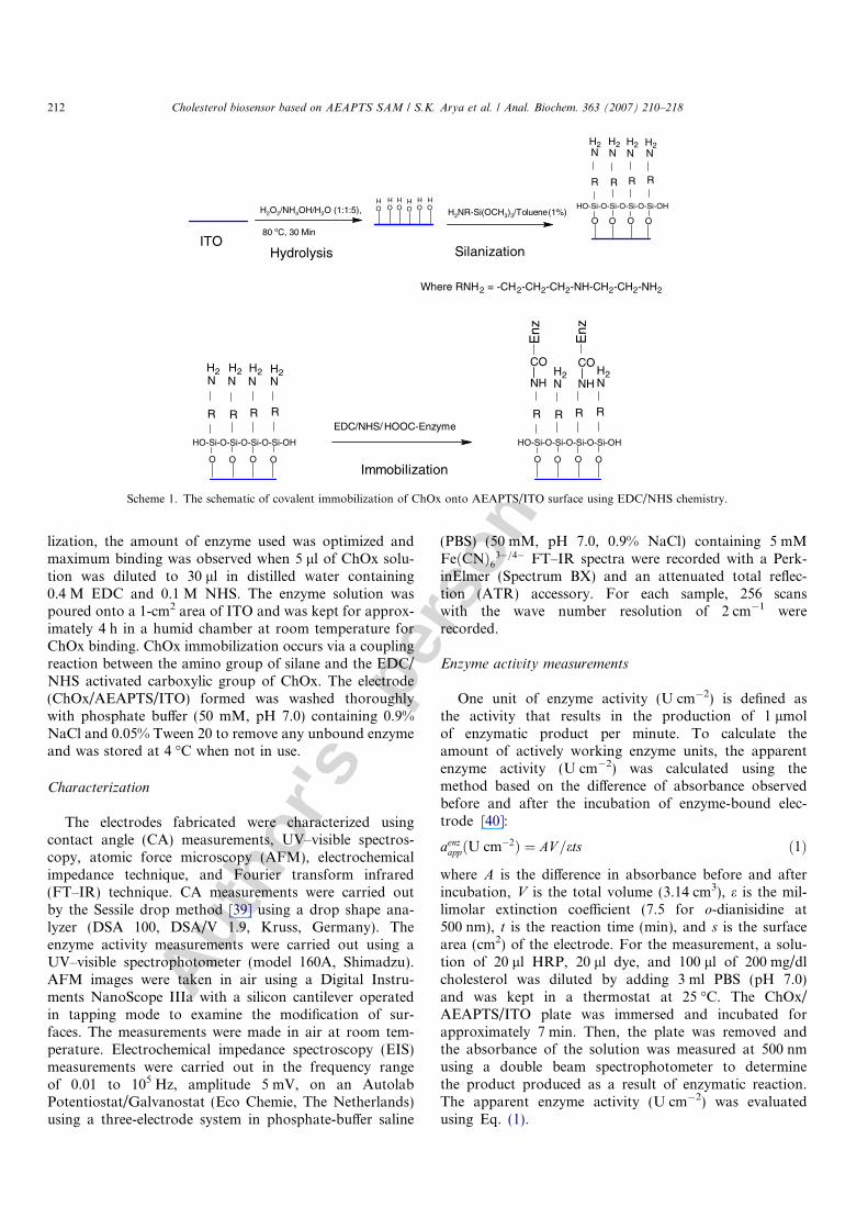
Contact angle (CA) studies
CA measurements were carried out to investigate thesilane SAM formation and ChOx immobilization usingthe Sessile drop method. The change in the value of theCA reveals the hydrophobic/hydrophilic character of thesurface, which in turn can be related to silane SAM forma-tion as well as the immobilization of enzyme.
Figs. 1A, B, and C show the variation of CA of hydro-lyzed ITO, AEAPTS/ITO, and ChOx/AEAPTS/ITO,respectively. The decrease in the value of CA from 68�for ITO to 49� for silane/ITO indicates the formation ofSAM of AEAPTS. The value of CA decreases to 30� afterenzyme (ChOx) immobilization. This change in the valueof CA on ChOx binding indicates the immobilization ofChOx on AEAPTS/ITO surface.
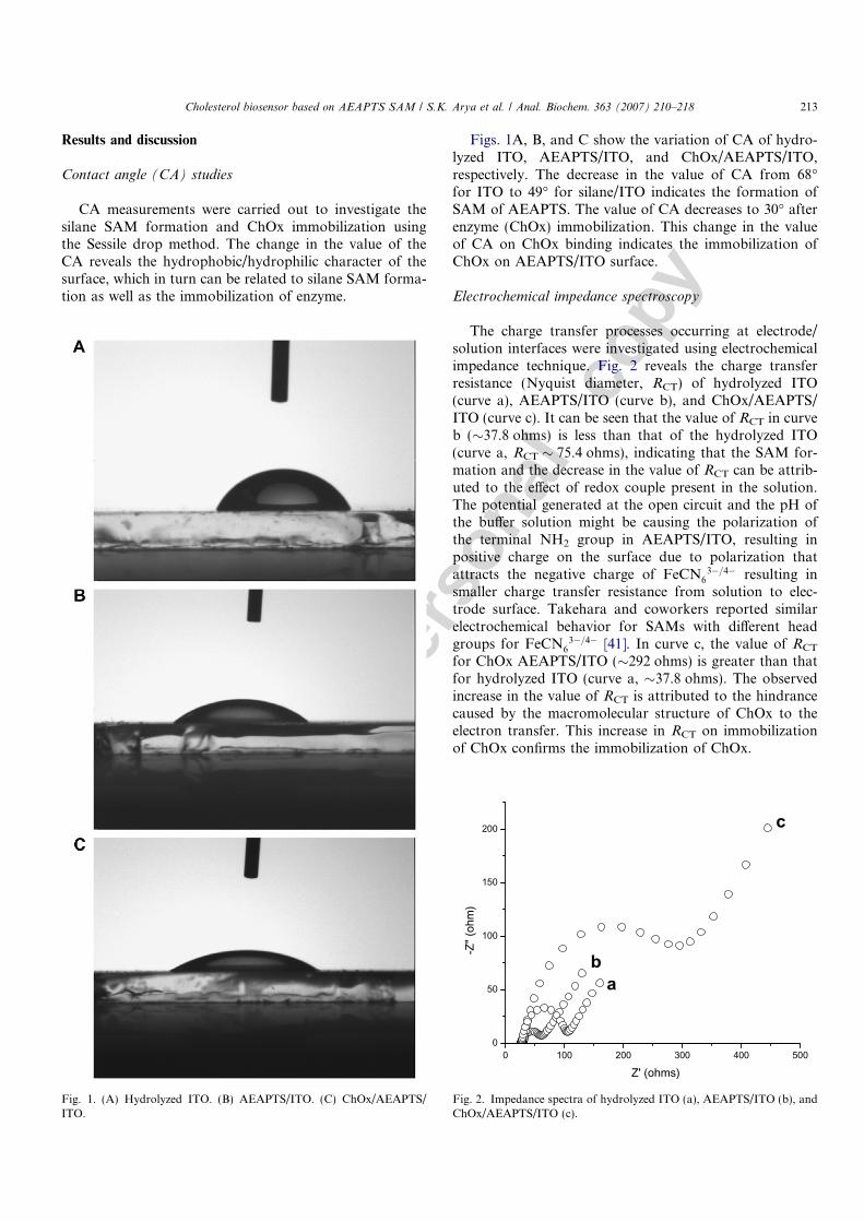
Electrochemical impedance spectroscopy
The charge transfer processes occurring at electrode/solution interfaces were investigated using electrochemicalimpedance technique. Fig. 2 reveals the charge transferresistance (Nyquist diameter, RCT) of hydrolyzed ITO(curve a), AEAPTS/ITO (curve b), and ChOx/AEAPTS/ITO (curve c). It can be seen that the value of RCT in curveb (�37.8 ohms) is less than that of the hydrolyzed ITO(curve a, RCT � 75.4 ohms), indicating that the SAM for-mation and the decrease in the value of RCT can be attrib-uted to the effect of redox couple present in the solution.The potential generated at the open circuit and the pH ofthe buffer solution might be causing the polarization ofthe terminal NH2 group in AEAPTS/ITO, resulting inpositive charge on the surface due to polarization thatattracts the negative charge of FeCN6
3�=4� resulting insmaller charge transfer resistance from solution to elec-trode surface. Takehara and coworkers reported similarelectrochemical behavior for SAMs with different headgroups for FeCN6
3�=4� [41]. In curve c, the value of RCT
for ChOx AEAPTS/ITO (�292 ohms) is greater than thatfor hydrolyzed ITO (curve a, �37.8 ohms). The observedincrease in the value of RCT is attributed to the hindrancecaused by the macromolecular structure of ChOx to theelectron transfer. This increase in RCT on immobilizationof ChOx confirms the immobilization of ChOx.
Fig. 1. (A) Hydrolyzed ITO. (B) AEAPTS/ITO. (C) ChOx/AEAPTS/ITO.
0 100 200 300 400 5000
50
100
150
200 c
ba
-Z" (
ohm
)
Z' (ohms)
Fig. 2. Impedance spectra of hydrolyzed ITO (a), AEAPTS/ITO (b), andChOx/AEAPTS/ITO (c).
Cholesterol biosensor based on AEAPTS SAM / S.K. Arya et al. / Anal. Biochem. 363 (2007) 210–218 213
Autho
r's
pers
onal
co
py
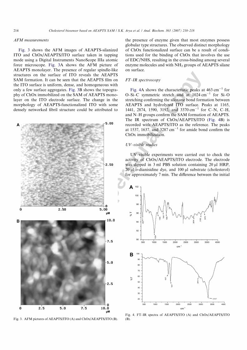
AFM measurements
Fig. 3 shows the AFM images of AEAPTS-silanizedITO and ChOx/AEAPTS/ITO surface taken in tappingmode using a Digital Instruments NanoScope IIIa atomicforce microscope. Fig. 3A shows the AFM picture ofAEAPTS monolayer. The presence of regular spindle-likestructures on the surface of ITO reveals the AEAPTSSAM formation. It can be seen that the AEAPTS film onthe ITO surface is uniform, dense, and homogeneous withonly a few surface aggregates. Fig. 3B shows the topogra-phy of ChOx immobilized on the SAM of AEAPTS mono-layer on the ITO electrode surface. The change in themorphology of AEAPTS-functionalized ITO with somedensely networked fibril structure could be attributed to
the presence of enzyme given that most enzymes possessglobular type structures. The observed distinct morphologyof ChOx functionalized surface can be a result of condi-tions used for the binding of ChOx that involves the useof EDC/NHS, resulting in the cross-binding among severalenzyme molecules and with NH2 groups of AEAPTS silaneon surface.
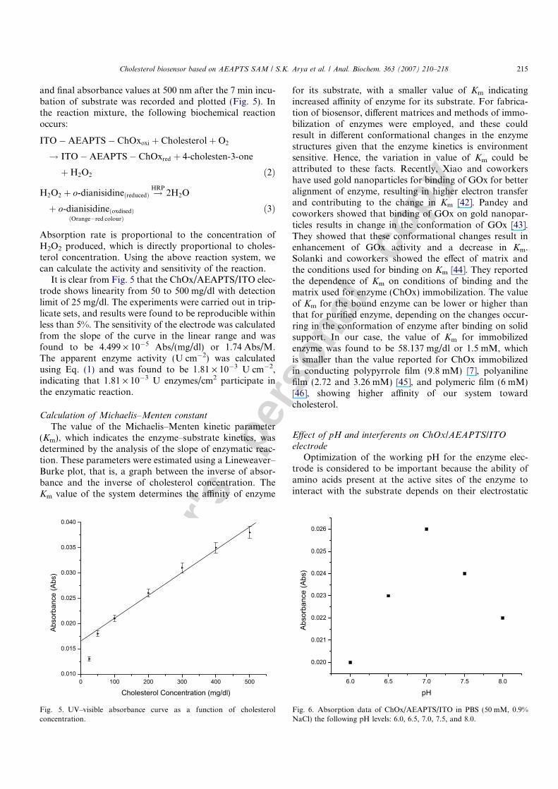
FT–IR spectroscopy
Fig. 4A shows the characteristic peaks at 463 cm�1 forO–Si–C symmetric stretch and at 1024 cm�1 for Si–Ostretching confirming the siloxane bond formation betweenAEAPTS and hydrolyzed ITO surface. Peaks at 1165,1441, 2874, 1590, 3192, and 3370 cm�1 for C–N, C–H,and N–H groups confirm the SAM formation of AEAPTS.The IR spectrum of ChOx/AEAPTS/ITO (Fig. 4B) isrecorded with AEAPTS/ITO as the reference. The peaksat 1537, 1637, and 3287 cm�1 for amide bond confirm theChOx immobilization.
UV–visible studies
UV–visible experiments were carried out to check theactivity of ChOx/AEAPTS/ITO electrode. The electrodewas dipped in 3 ml PBS solution containing 20 ll HRP,20 ll o-dianisidine dye, and 100 ll substrate (cholesterol)for approximately 7 min. The difference between the initial
Fig. 3. AFM pictures of AEAPTS/ITO (A) and ChOx/AEAPTS/ITO (B).Fig. 4. FT–IR spectra of AEAPTS/ITO (A) and ChOx/AEAPTS/ITO(B).
214 Cholesterol biosensor based on AEAPTS SAM / S.K. Arya et al. / Anal. Biochem. 363 (2007) 210–218
Autho
r's
pers
onal
co
py
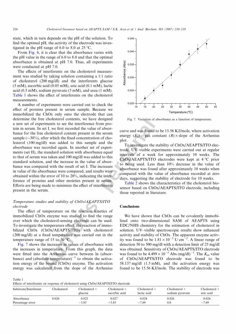
and final absorbance values at 500 nm after the 7 min incu-bation of substrate was recorded and plotted (Fig. 5). Inthe reaction mixture, the following biochemical reactionoccurs:
ITO�AEAPTS � ChOxoxi þ CholesterolþO2
! ITO�AEAPTS� ChOxred þ 4-cholesten-3-one
þH2O2 ð2Þ
H2O2 þ o-dianisidineðreducedÞ !HRP
2H2O
þ o-dianisidineðoxdisedÞðOrange�red colourÞ
ð3Þ
Absorption rate is proportional to the concentration ofH2O2 produced, which is directly proportional to choles-terol concentration. Using the above reaction system, wecan calculate the activity and sensitivity of the reaction.
It is clear from Fig. 5 that the ChOx/AEAPTS/ITO elec-trode shows linearity from 50 to 500 mg/dl with detectionlimit of 25 mg/dl. The experiments were carried out in trip-licate sets, and results were found to be reproducible withinless than 5%. The sensitivity of the electrode was calculatedfrom the slope of the curve in the linear range and wasfound to be 4.499 · 10�5 Abs/(mg/dl) or 1.74 Abs/M.The apparent enzyme activity (U cm�2) was calculatedusing Eq. (1) and was found to be 1.81 · 10�3 U cm�2,indicating that 1.81 · 10�3 U enzymes/cm2 participate inthe enzymatic reaction.
Calculation of Michaelis–Menten constant
The value of the Michaelis–Menten kinetic parameter(Km), which indicates the enzyme–substrate kinetics, wasdetermined by the analysis of the slope of enzymatic reac-tion. These parameters were estimated using a Lineweaver–Burke plot, that is, a graph between the inverse of absor-bance and the inverse of cholesterol concentration. TheKm value of the system determines the affinity of enzyme
for its substrate, with a smaller value of Km indicatingincreased affinity of enzyme for its substrate. For fabrica-tion of biosensor, different matrices and methods of immo-bilization of enzymes were employed, and these couldresult in different conformational changes in the enzymestructures given that the enzyme kinetics is environmentsensitive. Hence, the variation in value of Km could beattributed to these facts. Recently, Xiao and coworkershave used gold nanoparticles for binding of GOx for betteralignment of enzyme, resulting in higher electron transferand contributing to the change in Km [42]. Pandey andcoworkers showed that binding of GOx on gold nanopar-ticles results in change in the conformation of GOx [43].They showed that these conformational changes result inenhancement of GOx activity and a decrease in Km.Solanki and coworkers showed the effect of matrix andthe conditions used for binding on Km [44]. They reportedthe dependence of Km on conditions of binding and thematrix used for enzyme (ChOx) immobilization. The valueof Km for the bound enzyme can be lower or higher thanthat for purified enzyme, depending on the changes occur-ring in the conformation of enzyme after binding on solidsupport. In our case, the value of Km for immobilizedenzyme was found to be 58.137 mg/dl or 1.5 mM, whichis smaller than the value reported for ChOx immobilizedin conducting polypyrrole film (9.8 mM) [7], polyanilinefilm (2.72 and 3.26 mM) [45], and polymeric film (6 mM)[46], showing higher affinity of our system towardcholesterol.
Effect of pH and interferents on ChOx/AEAPTS/ITO
electrodeOptimization of the working pH for the enzyme elec-
trode is considered to be important because the ability ofamino acids present at the active sites of the enzyme tointeract with the substrate depends on their electrostatic
0 100 200 300 400 5000.010
0.015
0.020
0.025
0.030
0.035
0.040
Abso
rban
ce (A
bs)
Cholesterol Concentration (mg/dl)
Fig. 5. UV–visible absorbance curve as a function of cholesterolconcentration.
6.0 6.5 7.0 7.5 8.0
0.020
0.021
0.022
0.023
0.024
0.025
0.026
Abso
rban
ce (A
bs)
pH
Fig. 6. Absorption data of ChOx/AEAPTS/ITO in PBS (50 mM, 0.9%NaCl) the following pH levels: 6.0, 6.5, 7.0, 7.5, and 8.0.
Cholesterol biosensor based on AEAPTS SAM / S.K. Arya et al. / Anal. Biochem. 363 (2007) 210–218 215
Autho
r's
pers
onal
co
py
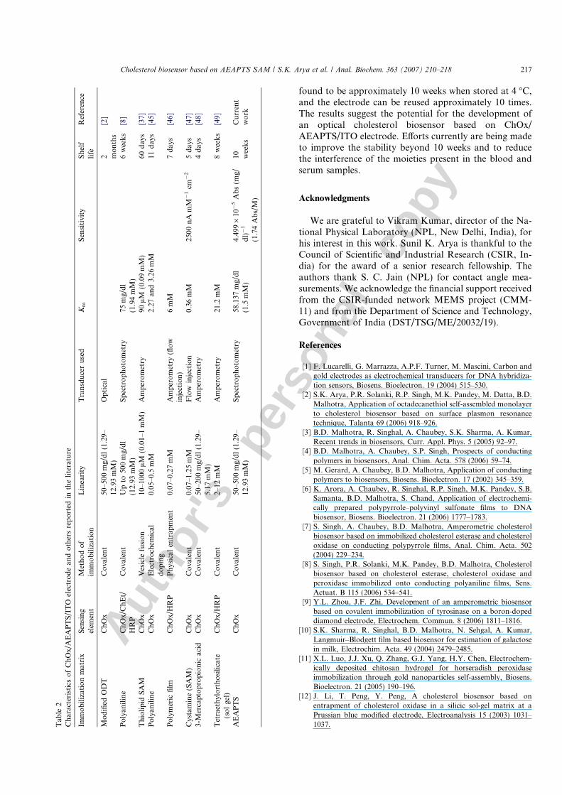
state, which in turn depends on the pH of the solution. Tofind the optimal pH, the activity of the electrode was inves-tigated in the pH range of 6.0 to 8.0 at 25 �C.
From Fig. 6, it is clear that the absorbance varies withthe pH value in the range of 6.0 to 8.0 and that the optimalabsorbance is obtained at pH 7.0. Thus, all experimentswere conducted at pH 7.0.
The effects of interferents on the cholesterol measure-ment was studied by taking solution containing a 1:1 ratioof cholesterol (200 mg/dl) and the interferents glucose(5 mM), ascorbic acid (0.05 mM), uric acid (0.1 mM), lacticacid (0.5 mM), sodium pyruvate (5 mM), and urea (1 mM).Table 1 shows the effect of interferents on the cholesterolmeasurements.
A number of experiments were carried out to check theeffect of proteins present in serum sample. Because weimmobilized the ChOx only onto the electrode that candetermine the free cholesterol contents, we have designeda new set of experiments to see the interference from pro-tein in serum. In set I, we first recorded the value of absor-bance for the free cholesterol content present in the serumsample (�30%), after which the fixed concentration of cho-lesterol (100 mg/dl) was added to this sample and theabsorbance was recorded again. In another set of experi-ments (set II), the standard solution with absorbance equalto that of serum was taken and 100 mg/dl was added to thisstandard solution, and the increase in the value of absor-bance was compared with the result of set I. The increasesin value of the absorbance were compared, and results wereobtained within the error of 10 to 20%, indicating the inter-ference of proteins and other moieties present in serum.Efforts are being made to minimize the effect of interferentspresent in the serum.
Temperature studies and stability of ChOx/AEAPTS/ITO
electrode
The effect of temperature on the reaction kinetics ofimmobilized ChOx enzyme was studied to find the rangeover which the cholesterol-sensing electrode can be used.To investigate the temperature effect, the reaction of immo-bilized ChOx (ChOx/AEAPTS/ITO) with cholesterol(200 mg/dl) at a fixed temperature was carried out in thetemperature range of 15 to 50 �C.
Fig. 7 shows the increases in values of absorbance withthe increases in temperature. From this graph, the datawere fitted into the Arrhenius curve between ln (absor-bance) and (absolute temperature)�1 to obtain the activa-tion energy of the bound (ChOx) enzyme. The activationenergy was calculated from the slope of the Arrhenius
curve and was found to be 15.56 KJ/mole, where activationenergy (Ea) = gas constant (R) · slope of the Arrheniusplot.
To investigate the stability of ChOx/AEAPTS/ITO elec-trode, UV–visible experiments were carried out at regularintervals of a week for approximately 10 weeks. TheChOx/AEAPTS/ITO electrodes were kept at 4 �C priorto being used. Less than 10% decrease in the value ofabsorbance was found after approximately 10 weeks whencompared with the value of absorbance recorded at zerodays, suggesting the stability of electrode for 10 weeks.
Table 2 shows the characteristics of the cholesterol bio-sensor based on ChOx/AEAPTS/ITO electrode, includingthose reported in literature.
Conclusions
We have shown that ChOx can be covalently immobi-lized onto two-dimensional SAM of AEAPTS usingEDC/NHS chemistry for the estimation of cholesterol insolution. UV–visible spectroscopic results show enhancedactivity and stability of ChOx. The apparent enzyme activ-ity was found to be 1.81 · 10�3 U cm�2. A linear range ofdetection 50 to 500 mg/dl with a detection limit of 25 mg/dlwas obtained. Sensitivity of ChOx/AEAPTS/ITO electrodewas found to be 4.499 · 10�5 Abs (mg/dl)�1. The Km valueof ChOx/AEAPTS/ITO electrode was found to be58.137 mg/dl (1.5 mM), and the activation energy wasfound to be 15.56 KJ/mole. The stability of electrode was
Table 1Effects of interferents on response of cholesterol using ChOx/AEAPTS/ITO electrode
Substrate/Interferent Cholesterol Cholesterol +glucose
Cholesterol +ascorbic acid
Cholesterol +lactic acid
Cholesterol +sodium pyruvate
Cholesterol +uric acid
Absorbance 0.026 0.025 0.027 0.024 0.026 0.024Percentage error �3.85 +3.85 �7.69 0.0 �7.69
10 15 20 25 30 35 40 45 50 550.020
0.025
0.030
0.035
0.040
0.045
Abso
rban
ce (A
bs)
Temperature (ºC)
Fig. 7. Variation of absorbance as a function of temperature.
216 Cholesterol biosensor based on AEAPTS SAM / S.K. Arya et al. / Anal. Biochem. 363 (2007) 210–218
Autho
r's
pers
onal
co
py
found to be approximately 10 weeks when stored at 4 �C,and the electrode can be reused approximately 10 times.The results suggest the potential for the development ofan optical cholesterol biosensor based on ChOx/AEAPTS/ITO electrode. Efforts currently are being madeto improve the stability beyond 10 weeks and to reducethe interference of the moieties present in the blood andserum samples.
Acknowledgments
We are grateful to Vikram Kumar, director of the Na-tional Physical Laboratory (NPL, New Delhi, India), forhis interest in this work. Sunil K. Arya is thankful to theCouncil of Scientific and Industrial Research (CSIR, In-dia) for the award of a senior research fellowship. Theauthors thank S. C. Jain (NPL) for contact angle mea-surements. We acknowledge the financial support receivedfrom the CSIR-funded network MEMS project (CMM-11) and from the Department of Science and Technology,Government of India (DST/TSG/ME/20032/19).
References
[1] F. Lucarelli, G. Marrazza, A.P.F. Turner, M. Mascini, Carbon andgold electrodes as electrochemical transducers for DNA hybridiza-tion sensors, Biosens. Bioelectron. 19 (2004) 515–530.
[2] S.K. Arya, P.R. Solanki, R.P. Singh, M.K. Pandey, M. Datta, B.D.Malhotra, Application of octadecanethiol self-assembled monolayerto cholesterol biosensor based on surface plasmon resonancetechnique, Talanta 69 (2006) 918–926.
[3] B.D. Malhotra, R. Singhal, A. Chaubey, S.K. Sharma, A. Kumar,Recent trends in biosensors, Curr. Appl. Phys. 5 (2005) 92–97.
[4] B.D. Malhotra, A. Chaubey, S.P. Singh, Prospects of conductingpolymers in biosensors, Anal. Chim. Acta. 578 (2006) 59–74.
[5] M. Gerard, A. Chaubey, B.D. Malhotra, Application of conductingpolymers to biosensors, Biosens. Bioelectron. 17 (2002) 345–359.
[6] K. Arora, A. Chaubey, R. Singhal, R.P. Singh, M.K. Pandey, S.B.Samanta, B.D. Malhotra, S. Chand, Application of electrochemi-cally prepared polypyrrole–polyvinyl sulfonate films to DNAbiosensor, Biosens. Bioelectron. 21 (2006) 1777–1783.
[7] S. Singh, A. Chaubey, B.D. Malhotra, Amperometric cholesterolbiosensor based on immobilized cholesterol esterase and cholesteroloxidase on conducting polypyrrole films, Anal. Chim. Acta. 502(2004) 229–234.
[8] S. Singh, P.R. Solanki, M.K. Pandey, B.D. Malhotra, Cholesterolbiosensor based on cholesterol esterase, cholesterol oxidase andperoxidase immobilized onto conducting polyaniline films, Sens.Actuat. B 115 (2006) 534–541.
[9] Y.L. Zhou, J.F. Zhi, Development of an amperometric biosensorbased on covalent immobilization of tyrosinase on a boron-dopeddiamond electrode, Electrochem. Commun. 8 (2006) 1811–1816.
[10] S.K. Sharma, R. Singhal, B.D. Malhotra, N. Sehgal, A. Kumar,Langmuir–Blodgett film based biosensor for estimation of galactosein milk, Electrochim. Acta. 49 (2004) 2479–2485.
[11] X.L. Luo, J.J. Xu, Q. Zhang, G.J. Yang, H.Y. Chen, Electrochem-ically deposited chitosan hydrogel for horseradish peroxidaseimmobilization through gold nanoparticles self-assembly, Biosens.Bioelectron. 21 (2005) 190–196.
[12] J. Li, T. Peng, Y. Peng, A cholesterol biosensor based onentrapment of cholesterol oxidase in a silicic sol-gel matrix at aPrussian blue modified electrode, Electroanalysis 15 (2003) 1031–1037.T
able
2C
har
acte
rist
ics
of
Ch
Ox/
AE
AP
TS
/IT
Oel
ectr
od
ean
do
ther
sre
po
rted
inth
eli
tera
ture
Imm
ob
iliz
atio
nm
atri
xS
ensi
ng
elem
ent
Met
ho
do
fim
mo
bil
izat
ion
Lin
eari
tyT
ran
sdu
cer
use
dK
mS
ensi
tivi
tyS
hel
fli
feR
efer
ence
Mo
difi
edO
DT
Ch
Ox
Co
vale
nt
50–5
00m
g/d
l(1
.29–
12.9
3m
M)
Op
tica
l2 m
on
ths
[2]
Po
lyan
ilin
eC
hO
x/C
hE
t/H
RP
Co
vale
nt
Up
to50
0m
g/d
l(1
2.93
mM
)S
pec
tro
ph
oto
met
ry75
mg/
dl
(1.9
4m
M)
6w
eek
s[8
]
Th
ioli
pid
SA
MC
hO
xV
esic
lefu
sio
n10
–100
0lM
(0.0
1–1
mM
)A
mp
ero
met
ry90
lM(0
.09
mM
)60
day
s[3
7]P
oly
anil
ine
Ch
Ox
Ele
ctro
chem
ical
do
pin
g0.
05–0
.5m
M2.
27an
d3.
26m
M11
day
s[4
5]
Po
lym
eric
film
Ch
Ox/
HR
PP
hys
ical
entr
apm
ent
0.07
–0.2
7m
MA
mp
ero
met
ry(fl
ow
inje
ctio
n)
6m
M7
day
s[4
6]
Cys
tam
ine
(SA
M)
Ch
Ox
Co
vale
nt
0.07
–1.2
5m
MF
low
inje
ctio
n0.
36m
M25
00n
Am
M�
1cm�
25
day
s[4
7]3-
Mer
cap
top
rop
ion
icac
idC
hO
xC
ova
len
t50
–200
mg/
dl
(1.2
9–5.
17m
M)
Am
per
om
etry
4d
ays
[48]
Tet
raet
hyl
ort
ho
sili
cate
(so
lge
l)C
hO
x/H
RP
Co
vale
nt
2–12
mM
Am
per
om
etry
21.2
mM
8w
eek
s[4
9]
AE
AP
TS
Ch
Ox
Co
vale
nt
50–5
00m
g/d
l(1
.29–
12.9
3m
M)
Sp
ectr
op
ho
tom
etry
58.1
37m
g/d
l(1
.5m
M)
4.49
9·
10�
5A
bs
(mg/
dl)�
1
(1.7
4A
bs/
M)
10 wee
ks
Cu
rren
tw
ork
Cholesterol biosensor based on AEAPTS SAM / S.K. Arya et al. / Anal. Biochem. 363 (2007) 210–218 217
Autho
r's
pers
onal
co
py
[13] S.K. Arya, P.R. Solanki, S.P. Singh, K. Kaneto, M.K. Pandey, M.Datta, B.D. Malhotra, Poly-(3-hexylthiophene) self-assembledmonolayer-based cholesterol biosensor using surface plasmonresonance technique, Biosens. Bioelectron. (2006), doi:10.1016/j.bios.2006.10.011.
[14] K.V. Gobi, F. Mizutani, Layer-by-layer construction of activemultilayer enzyme electrode applicable for direct amperometricdetermination of cholesterol, Sens. Actuat. B 80 (2001) 272–277.
[15] H. Zhao, H. Ju, Multilayer membranes for glucose biosensing vialayer-by-layer assembly of multiwall carbon nanotubes and glucoseoxidase, Anal. Biochem. 350 (2006) 138–144.
[16] S. Dong, J. Li, Self-assembled monolayers of thiols on gold electrodesfor bioelectrochemistry and biosensors, Bioelectrochem. Bioenerg. 42(1997) 7–13.
[17] A. Subramanian, J. Irudayaraj, T. Ryan, Mono and dithiol surfaceson surface plasmon resonance biosensors for detection of Staphylo-
coccus aureus, Sens. Actuat. B 114 (2006) 192–198.[18] E. Mauriz, A. Calle, L.M. Lechuga, J. Quintana, A. Montoya, J.J.
Mancl’us, Real-time detection of chlorpyrifos at part per trillionlevels in ground, surface, and drinking water samples by a portablesurface plasmon resonance immunosensor, Anal. Chim. Acta. 561(2006) 40–47.
[19] E. Mauriz, A. Calle, A. Montoya, L.M. Lechuga, Determination ofenvironmental organic pollutants with a portable optical immuno-sensor, Talanta 69 (2006) 359–364.
[20] J.W. Chung, S.D. Kim, R. Bernhardt, J.C. Pyun, Application of SPRbiosensor for medical diagnostics of human hepatitis B virus (hHBV),Sens. Actuat. B 111/112 (2005) 416–422.
[21] J.W. Lee, S.J. Sim, S.M. Cho, J. Lee, Characterization of a self-assembled monolayer of thiol on a gold surface and the fabricationof a biosensor chip based on surface plasmon resonance fordetecting anti-GAD antibody, Biosens. Bioelectron. 20 (2005) 1422–1427.
[22] X. Su, F.T. Chew, S.F.Y. Li, Self-assembled monolayer-basedpiezoelectric crystal immunosensor for the quantification of totalhuman immunoglobulin E, Anal. Biochem. 273 (1999) 66–72.
[23] V.R.S. Babu, M.A. Kumar, N.G. Karnath, M.S. Thakur, Stabiliza-tion of immobilized glucose oxidase against thermal inactivation bysilanization for biosensor applications, Biosens. Bioelectron. 19(2004) 1337–1341.
[24] L.A. Chrisey, G.U. Lee, C.E. O’Ferrall, Covalent attachment ofsynthetic DNA to self-assembled monolayer films, Nucleic Acids Res.24 (1996) 3031–3039.
[25] G.D. Francia, V.L. Ferrara, S. Manzo, S. Chiavarini, Towards label-free optical porous silicon DNA sensor, Biosens. Bioelectron. 21(2005) 661–665.
[26] T. Nakagawa, T. Tanaka, D. Niwa, T. Osaka, H. Takeyama, T.Matsunaga, Fabrication of amino silane-coated microchip for DNAextraction from whole blood, J. Biotechnol. 116 (2005) 105–111.
[27] H. Hillebrandt, M. Tanaka, Electrochemical characterization of self-assembled alkylsiloxane monolayers on indium–tin oxide semicon-ductor electrodes, J. Phys. Chem. B 105 (2001) 4270–4276.
[28] R.A. Hatton, S.R. Day, M.A. Chesters, M.R. Willis, Organicelectroluminescent devices: Enhanced carrier injection using anorganosilane self-assembled monolayer (SAM) derivatized ITO elec-trode, Thin Solid Films 394 (2001) 292–297.
[29] E. Moore, D. O’Connell, P. Galvin, Surface characterization ofindium–tin oxide thin electrode films for use as a conducting substratein DNA sensor development, Thin Solid Films 515 (2006) 2612–2617.
[30] L. Yang, Y. Li, AFM and impedance spectroscopy characterizationof the immobilization of antibodies on indium–tin oxide electrodethrough self-assembled monolayer of epoxysilane and their capture ofEscherichia coli O157:H7, Biosens. Bioelectron. 20 (2005) 1407–1416.
[31] C. Ruan, L. Yang, Y. Li, Immunobiosensor chips for detectionof Escherichia coli O157:H7 using electrochemical impedancespectroscopy, Anal. Chem. 74 (2002) 4814–4820.
[32] S. Singh, P.R. Solanki, M.K. Pandey, B.D. Malhotra, Covalentimmobilization of cholesterol esterase and cholesterol oxidase onpolyaniline films for application to cholesterol biosensor, Anal. Chim.Acta. 568 (2006) 126–132.
[33] J.C. Vidal, E.G. Ruiz, J. Espuelas, T. Aramendia, J.R. Castillo,Comparison of biosensors based on entrapment of cholesterol oxidaseand cholesterol esterase in electropolymerized films of polypyrroleand diaminonaphthalene derivatives for amperometric determinationof cholesterol, Anal. Bioanal. Chem. 377 (2003) 273–280.
[34] X. Tan, M. Li, P. Cai, L. Luo, X. Zou, An amperometric cholesterolbiosensor based on multiwalled carbon nanotubes and organicallymodified sol-gel/chitosan hybrid composite film, Anal. Biochem. 337(2005) 111–120.
[35] G. Li, J.M. Liao, G.Q. Hu, N.Z. Ma, P.J. Wu, Study of carbonnanotube modified biosensor for monitoring total cholesterol inblood, Biosens. Bioelectron. 20 (2005) 2140–2144.
[36] M.K. Ram, P. Bertoncello, H. Ding, S. Paddeu, C. Nicolini,Cholesterol biosensors prepared by layer-by-layer technique, Biosens.Bioelectron. 16 (2001) 849–856.
[37] M.P. Bokoch, A. Devadoss, M.S. Palencsar, J.D. Burgess, Steady-state oxidation of cholesterol catalyzed by cholesterol oxidase in lipidbilayer membranes on platinum electrodes, Anal. Chim. Acta. 519(2004) 47–55.
[38] L.C.S. Chou, C.C. Liu, Development of a molecular imprinting thickfilm electrochemical sensor for cholesterol detection, Sens. Actuat. B110 (2005) 204–208.
[39] M.Y. Hong, Y.J. Kim, J.W. Lee, K. Kim, J.H. Lee, J.S. Yoo, S.H.Bae, B.S. Choi, H.S. Kim, Synthesis and characterization oftri(ethylene oxide)-attached poly(amidoamine) dendrimer layers ongold, J. Coll. Interf. Sci. 274 (2004) 41–48.
[40] A. Kumar, R. Malhotra, B.D. Malhotra, S.K. Grover, Co-immobi-lization of cholesterol oxidase and horseradish peroxidase in a sol-gelfilm, Anal. Chim. Acta. 414 (2000) 43–50.
[41] K. Takehara, H. Takemura, Y. Ide, Electrochemical studies of theterminally substituted alkanethiol monolayers formed on a goldelectrode: Effects of the terminal group on the redox responses ofFeðCNÞ63, RuðNHÞ63þ, and ferrocenedimethanol, Electrochim. Acta.39 (1994) 817–822.
[42] Y. Xiao, F. Patolsky, E. Katz, J.F. Hainfeld, I. Willner, ‘‘Plugginginto enzymes’’: Nanowiring of redox enzymes by a gold nanoparticle,Science 299 (2003) 1877–1881.
[43] P. Pandey, S.P. Singh, S.K. Arya, V. Gupta, M. Datta, S. Singh, B.D.Malhotra, Application of thiolated gold nanoparticles for enhance-ment of glucose oxidase activity, Langmuir (2007), doi:10.1021/la062901c.
[44] P.R. Solanki, S.K. Arya, S.P. Singh, M.K. Pandey, B.D. Malhotra,Application of electrochemically prepared poly-N-methylpyrrole-p-toluenesulfonate films to cholesterol biosensor, Sens. Actuat. B(2006), doi:10.1016/j.snb.2006.10.046.
[45] H.Y. Wang, S.L. Mu, Bioelectrochemical characteristics of choles-terol oxidase immobilized in a polyaniline film, Sens. Actuat. B 56(1999) 22–30.
[46] C. Bongiovanni, T. Ferri, A. Poscia, M. Varalli, R. Santucci, A.Desideri, An electrochemical multienzymatic biosensor for determi-nation of cholesterol, Bioelectrochemistry 54 (2001) 17–22.
[47] J.C. Vidal, J. Espuelas, J.R. Castillo, Amperometric cholesterolbiosensor based on in situ reconstituted cholesterol oxidase on animmobilized monolayer of flavin adenine dinucleotide cofactor, Anal.Biochem. 333 (2004) 88–98.
[48] J. Shen, C.C. Liu, Development of a screen-printed cholesterolbiosensor: Comparing the performance of gold and platinum as theworking electrode material and fabrication using a self-assemblyapproach, Sens. Actuat. B 120 (2007) 417–425.
[49] A. Kumar, R.R. Pandey, B. Brantley, Tetraethylorthosilicate filmmodified with protein to fabricate cholesterol biosensor, Talanta 69(2006) 700–705.
218 Cholesterol biosensor based on AEAPTS SAM / S.K. Arya et al. / Anal. Biochem. 363 (2007) 210–218


