Embed Size (px)
Citation preview

Durham Research Online
Deposited in DRO: 08 October 2009
Peer-review status: Peer-reviewed
Publication status of attached file: Accepted for publication
Citation for published item: Dijkerman, H. C. and McIntosh, R. D. and Schindler, I. and Nijboer, T. C. W. and Milner, A. D. (2009) 'Choosing between alternative wrist postures : action planning needs perception.', Neuropsychologia., 47 (6). pp. 1476-1482.
Further information on publisher’s website: http://dx.doi.org/10.1016/j.neuropsychologia.2008.12.002
Use policy The full-text may be used and/or reproduced, and given to third parties in any format or medium, without prior permission or charge, for personal research or study, educational, or not-for-profit purposes provided that :
a full bibliographic reference is made to the original source a link is made to the metadata record in DRO the full-text is not changed in any way
The full-text must not be sold in any format or medium without the formal permission of the copyright holders. Please consult the full DRO policy for further details.
Durham University Library, Stockton Road, Durham DH1 3LY, United Kingdom Tel : +44 (0)191 334 2975 | Fax : +44 (0)191 334 2971
http://dro.dur.ac.uk

SHORTENED TITLE: Action planning needs perception
Choosing between alternative wrist postures: action planning needs perception
H.C. Dijkerman1*
, R.D. McIntosh2, I. Schindler
3, T.C.W. Nijboer
1, A.D. Milner
4
1Department of Experimental Psychology, Helmholtz Institute, Utrecht University,
2Human Cognitive Neuroscience, Psychology, University of Edinburgh,
3Department
of Psychology, University of Hull, 4Cognitive Neuroscience Research Unit, Wolfson
Research Institute, University of Durham, Queen's Campus
*Corresponding author:
Department of Experimental Psychology,
Helmholtz Institute,
Utrecht University,
Heidelberglaan 2,
3584 CS Utrecht, the Netherlands
email: [email protected]
Tel: +31-30-2533395
Fax: +31-30-2534511

2
Abstract
When normal subjects grasp with their right hand a rectangular object placed at
different orientations in the horizontal plane, they change from a „thumb left‟
(clockwise) to a „thumb right‟ (anti-clockwise) grasp when the orientation exceeds
about 110°, with respect to the mid-sagittal plane. This suggests planning of the final
grip orientation at or before the start of the prehension movement. The current study
assessed performance of two visual agnosic patients (SB and DF) on a grasping task
requiring the planning of final grip posture. Five healthy subjects were also tested.
Subjects were required to grasp a triangular-section block, which was presented at one
of seven different orientations (80-140°). The healthy subjects showed a consistent
relation between object orientation and hand orientation just before contact. In
addition, they consistently used a clockwise grasp when object orientation was less
than 100°, and an anti-clockwise grasp when it was more than 110°, with a sharply-
defined switch-point being identifiable for each subject. For both visual agnosic
patients, hand orientation was also reliably related to object orientation. However, the
selection of grasp posture was markedly abnormal: they did not consistently switch
between clockwise and anti-clockwise grasps within the normal orientation range, and
the switch, when it did occur, was not at all sharply defined. These results suggest that
the planning of hand orientation during a grasp depends on a perceptually-based
judgement of the awkwardness of alternative movements. This would presumably
involve ventral stream processing, which is disrupted in the visual agnosic patients.
Keywords: Visual agnosia; ventral stream; orientation; grasping; end-state comfort

3
Introduction
In the last fifteen years or so, several studies have investigated the relative
contribution of the two visual cortical processing streams in different visually-based
tasks. Early studies investigating task-dependent processing suggested that the dorsal
stream is critically necessary for the immediate online control of goal-directed action,
whilst the ventral stream is crucial for the recognition of objects. Evidence for this
dissociation came originally from monkey neurophysiology (Sakata, Taira, Murata, &
Mine, 1995) and human neuropsychological single case studies (Goodale, Milner,
Jakobson, & Carey, 1991; Milner & Goodale, 1995; Milner et al., 1991), but has more
recently been supported through other methodologies such as functional
neuroimaging (Culham et al., 2003; James, Culham, Humphrey, Milner, & Goodale,
2003) and TMS (Desmurget et al., 1999; Rice, Tunik, & Grafton, 2006). Particularly
important has been the study of visual form agnosic patient DF, whose damage
includes ventral stream area LO bilaterally (James et al., 2003). This patient could not
identify the width of a rectangular shape, nor was she able to report the orientation of
a slot (Goodale et al., 1991; Milner et al., 1991). Nevertheless, she was able to use the
same visual information for grasping the rectangular shape or posting an object
through the slot.
Other studies have confirmed her ability to use visual input about the orientation of an
object for online guidance of hand orientation during a grasping movement (Carey,
Harvey, & Milner, 1996; Dijkerman, Milner, & Carey, 1996). However, these studies
also showed impairments in grasping behaviour under particular task conditions.
Carey et al. (1996) reported that DF did not consistently grasp the appropriate part of
everyday utensils, despite being able to adjust hand orientation to object orientation.
This suggests that she was unable to use stored knowledge about the function of an
4
object to guide the selection of a semantically appropriate grasp, although she could
still use orientation information to execute the selected grasp efficiently. Overall,
these findings suggest that ventral stream processing may be crucial for certain
aspects of hand orientation during reaching and grasping, for example when
recognition of the object is required.
It is well known that many aspects of a visuomotor act need to be pre-planned based
on the available visual input. For example, Rosenbaum, Heugten, & Caldwell (1996)
reported the “end-state comfort” effect when grasping an object in order to make a
second movement with it. They observed that the handle was grasped with such a
hand orientation that a comfortable hand configuration was achieved at the end of the
second movement, even if the intermediate hand configuration at the end of the first
movement was not always comfortable. The end-state comfort effect can only be
achieved through planning at the start of the movement what the end posture will be.
Another example comes from a study by Stelmach, Castiello, & Jeannerod (1994).
When normal subjects grasp an elongated object with a triangular cross-section placed
at different orientations in the horizontal plane with their right hand, they change from
a „thumb left‟ (clockwise) to a „thumb right‟ (anti-clockwise) grip when the
orientation exceeds about 110°, with respect to the mid-sagittal plane. This suggests
that, the final grip orientation (and thereby the direction of hand rotation during the
movement) is chosen at or before the start of the prehension movement. This type of
planning is influenced by contextual visual illusions such as the rod and frame illusion
(Craje, van der Kamp, & Steenbergen, 2008) and is considered to depend on visual
processing within the ventral stream (Goodale & Milner, 2004; Milner & Goodale,
1995, see also Liu, Chua, & Enns, 2008), predicting that the ability should be severely
disrupted by bilateral ventral stream lesions. The current study tested this prediction
5
in two patients with visual agnosia, DF and SB, using a version of Stelmach et al‟s
task (1994), which requires the planning of final grip posture. Some of the data
collected with SB have been reported previously in study on visuomotor abilities of
this patient (Dijkerman, Le, Demonet, & Milner, 2004). Movement execution of SB
has been analyzed more carefully in this study and compared to that of DF and
healthy controls, allowing more comprehensive results/conclusions.
Methodology
Participants
DF: This patient experienced carbon monoxide poisoning in 1988, resulting in a
severe visual form agnosia (Milner et al., 1991). Recent high-resolution structural
MRI has confirmed a dense bilateral lesion in lateral prestriate cortex, which
functional MRI has shown to coincide with the lateral occipital area (LO), an area in
the ventral stream that is implicated in object perception (James et al., 2003).
Functional MRI also shows that the anterior intraparietal area (AIP) in DF‟s dorsal
stream remains functional during object grasping. DF performed the present
experiment twice, once at the age of 45 and a second time at the age of 48.
SB: This patient, a right-handed man, suffered from an attack of viral meningo-
encephalitis at the age of 3 years. At the time of testing, at the age of 31 years, SB
retained a severe object, letter and face recognition deficit. Although he can
describe the contours of a visually-presented object, he cannot identify the object
in most cases. SB‟s perceptual capacities and pattern of brain damage have already
been described in detail in an earlier paper (Le et al., 2002). For more extensive
descriptions of SB‟s visuomotor abilities see Dijkerman, Le, Demonet, & Milner

6
(2004). MRI structural scans revealed large lesions of occipito-parietal and occipito-
temporal regions in the right hemisphere, and at the occipito-temporal junction in the
left hemisphere (Le et al., 2002). The right-hemisphere lesion includes complete or
partial damage to the human counterparts of the monkey‟s V2, V3, V4 and MT, and
also to area LO. In addition there is limited damage to the right inferior parietal lobule
in the region of the supramarginal gyrus. The spared regions in the right occipital pole
include the calcarine fissure (primary visual cortex, V1) at least in its rostral and
superior aspects. In the left hemisphere, the lesions involve mainly the ventrolateral
visual cortex, including a complete destruction of the fusiform gyrus and area LO. In
summary, the lesions seem to have all but destroyed the visual ventral stream
bilaterally, while sparing the occipital pole and the left dorsal stream.
Five healthy female control subjects (mean age 30.8 years, range 24-41 years) with
normal (or corrected to normal) vision were also tested.
Fig. 1: The two different ways of orienting the finger opposition space in the present grasping task. Normal subjects grasp objects that are placed at orientations less than 90° (with 0° being aligned parallel to the midsagittal axis) with their thumb on the left and their index finger on the right (clockwise, top picture). They switch to an ‘anti-clockwise’ grasp when the object is placed at 110° or higher (bottom picture).

7
Procedure and experimental set up
Following a task devised by Stelmach et al. (1994), we asked participants to grasp,
without lifting, a triangular-section prism block (6 cm long by 2.5 cm wide), made out
of dark grey plastic. Because its section was an equilateral triangle, the object offered
only one effective grip pattern, with the thumb and fingertips in opposition at the two
ends: it could not be picked up sideways, without it slipping out of one‟s grip (see Fig
1). The object was placed on a white table top, with its centre 30 cm away from the
starting position along the subject‟s midsagittal axis. The starting position was 5 cm
away from the table edge. The target object was presented at one of seven different
orientations (80-140° in steps of 10°, with 0° being with its main axis parallel to the
mid-sagittal axis). Each target orientation was presented ten times in a pseudo-
randomized order.
INSERT FIG 1 ABOUT HERE
The Minibird (Ascension Technology Corporation) magnetic recording system was
used for recording the reaching and grasping movements. The positions of markers
attached to the nails of the thumb and forefinger of the right hand were tracked for 3 s
at a sampling rate of 103Hz. Start and end times of the grasping movement were
determined by using a velocity based criterion (5 cm/s for the thumb marker).
Data analyses
Grasp orientation was determined throughout the movement. This was achieved by
calculating the angle of a straight line drawn through the markers on the index finger
and thumb, with respect to the sagittal plane, for each frame. Several variables were
extracted from the grasp orientation data. First, the reaching movements were
normalized with respect to time, with each movement being divided into 100 samples.
The grasp orientation measured at 2 normalized samples before the end of the

8
movement was examined as a function of the object orientation. Second, the grasp
posture was classed as clockwise if this orientation was signed positively (with 0
degrees being the index finger-thumb axis being aligned parallel to the midsagittal
axis), and as anti-clockwise if it was signed negatively (see Figure 1). For each
participant, the percentage of anti-clockwise grasps was calculated for each object
orientation. In order to describe the dependence of grasp posture on object orientation
in a manner analogous to a psychophysical analysis, the best-fitting sigmoid curve
was calculated for each participant‟s data. Provided that a reliable fit was obtained,
two (pseudo-psychophysical) parameters were derived. The „switch-point‟ was
calculated as the object orientation at which the frequency of anti-clockwise grasps
was equal to 50% (analogous to the point of subjective equality in a standard
psychophysical analysis). The „switch-sharpness‟ was calculated as the range of
object orientations between anti-clockwise frequencies of 25 and 75% (analogous to
one just noticeable difference in a standard psychophysical analysis).
We also assessed whether hand orientation was reliably related to object orientation 2
samples before the end of the movement. For this we calculated hand orientation
irrespective of whether a clockwise or anti-clockwise grip was used. We further
divided the grasps into „natural‟ or „awkward‟ depending on whether control subjects
performed a grasp for that particular object orientation using a clockwise or anti-
clockwise grip. This meant that all clockwise grasps were classed as „natural‟ when
performed for object orientations between 80-110 degrees, while anti-clockwise
grasps were considered to be natural when object orientation was between 100-140
degrees. All other grasps were considered to be „awkward‟. The „awkward grasps
were excluded from the analyses as biomechanical constraints when performing these

9
uncomfortable grasps might have influenced hand orientation. A linear regression
analyses was performed for each participant only using data from the „natural‟ grasps
with object orientation as independent variable and hand orientation as dependent
variable.
To assess whether the grasp posture had been pre-planned, we examined the change
of hand orientation over the course of each reach. Hand orientation was plotted as a
function of normalized time. Hand orientation at 0, 10, 30, 50 and 70% of movement
duration was calculated in relation to object orientation and to final grip posture
(clockwise, anti-clockwise). The data were analysed for each subject using t-tests to
determine the stage in the movement at which hand orientation began to differ
according to whether the final grasp posture on that trial was to be „clockwise‟ or
„anti-clockwise‟. The t-tests were carried out on hand orientation measured at 0, 10,
30, 50 and 70 percent into the movement, with final hand position (clockwise-anti-
clockwise) as the independent variable.
Finally, standard kinematic measures such as movement time (MT), maximum grip
aperture (MGA), time to maximum grip aperture (TMGA), and time to maximum grip
aperture as percentage of total movement time (%TMGA) were calculated.
Results
Relation between hand posture and target orientation
Figure 2 depicts the percentage of clockwise grasps for each orientation per subject. It
is clear from this figure that the control subjects consistently switch from a
„clockwise‟ (positive values) to an „anti-clockwise‟ (negative values) grip at 100 –

10
110 degrees. The „switch-point‟ for the control subjects varied between 98.9 and
111.6 degrees. The „switch-sharpness‟ (range of object orientations between anti-
clockwise frequencies of 75 and 25%) varied between 0.49 and 3.89 degrees, showing
that the control subjects changed quite sharply between clockwise and anti-clockwise
grips.
Fig 2: The best-fitting sigmoid curves for grasp posture across object orientations in a manner analogous to a psychophysical analysis for each participant. Two (pseudo-psychophysical) parameters were derived. The ‘switch-point’ was calculated as the object orientation at which the frequency of anti-clockwise grasps was equal to 50% (analogous to the point of subjective equality in a standard psychophysical analysis). The ‘switch-sharpness’ was calculated as the range of object orientations between anti-clockwise frequencies of 25 and 75% (analogous to one just noticeable difference in a standard psychophysical analysis).
As can be seen in Figure 2, SB and DF showed a different pattern. In the first testing
session DF did not change hand orientations at a particular object orientation. Instead
she used both grips over a range of object orientations (90-120 degrees) and only
showed a consistent grip for the extreme object orientations (80 degrees: clockwise;
130-140 degrees anti-clockwise). Indeed, although her „switch-point‟ was within the
normal range for this session (110.10 degrees), the „switch sharpness‟ was not (8.50
degrees). In the second session, the „switch-point‟ as well as the „switch sharpness‟
are outside the normal range (129.9 degrees and 6.2 degrees respectively) suggesting

11
that DF only changes to an anti-clockwise grasp for the object orientations 130 and
140 degrees. As we already described previously (Dijkerman et al., 2004), SB also
did not change between grips at a certain object orientation. Instead, he always
grasped the object in an anti-clockwise manner, except for three trials at 80 and 90
degrees. It was not possible to calculate the „switch-point‟ or „switch sharpness‟ for
his data, since no sigmoid function could be fitted reliably to his data.
Fig 3: Hand orientation 2 normalized frames before the end of the movement as a function of object orientation, irrespective of whether the grasp was clockwise or anti-clockwise. Visual agnosic participants DF (two sessions) and SB are depicted in the first three graphs on the top row. The healthy control subjects are shown in the remaining graph on the top row and in the bottom four graphs. The hand orientations are coded according to whether they are natural (e.g. ‘clockwise’ grip for object orientations 90-110 or ‘anti-clockwise’ grip for object orientations 100-140, filled circles) or awkward (all other trials, open circles). A linear regression was performed for natural trials only. Hand orientation just before the end of the movement was highly related to object orientation for all participants including DF and SB.
Figure 3 depicts hand orientations at two frames before the end of the movement
irrespective of whether the grip was clockwise or anti-clockwise. A linear regression
analysis for each individual performed on the „natural‟ grasps showed that, for all

12
participants, including the agnosic patients, hand orientation just prior to the end of
the movement was reliably related to object orientation (minimum r2 = 0.70). This
suggests that visual input about the object orientation was used to adjust the hand
orientation during the grasping movement. Thus, although the agnosic patients adjust
their hand orientation during the grasping movement to the object orientation, they do
not consistently pre-select a particular hand-grip posture, in contrast to all control
subjects.
Next we determined the point during the grasping movement at which it became clear
that the object was going to be grasped with a clockwise or anti-clockwise grip.
Control subjects presumably pre-plan this decision. If the agnosic patients, however,
are impaired in planning the grasping movement, the decision to adopt a certain grip
may be deferred until after the movement has started. We used independent samples t-
tests to assess at what part of the movement a reliable difference between hand
orientations leading to a 'clockwise' and an 'anti-clockwise' grasp could be observed.
This was done for DF only, as SB performed only three grasps with a 'thumb to the
left' grip. The results show that for four out of five control subjects a difference
between „clockwise‟ and 'anti-clockwise' grip can already be detected after 10% of the
movement (see Table 1 and Figure 4). For the remaining control subject, the
difference becomes significant at 30% into the movement. For DF, the difference
between the two types of grip is also significant at 10% of the total movement in both
sessions. DF therefore performs similarly to the control subjects in that she selects her
grip prior to, or early during, the movement, in a normal fashion. However, a
significant difference was also found in both sessions for DF‟s initial hand orientation
at the first frame of the movement (see Table 1). This was not the case for any of the
control subjects and suggests that DF‟s final grip may partly be determined by her
13
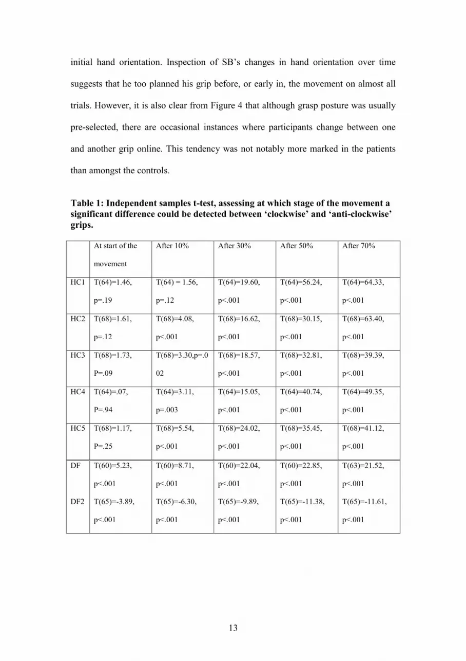
initial hand orientation. Inspection of SB‟s changes in hand orientation over time
suggests that he too planned his grip before, or early in, the movement on almost all
trials. However, it is also clear from Figure 4 that although grasp posture was usually
pre-selected, there are occasional instances where participants change between one
and another grip online. This tendency was not notably more marked in the patients
than amongst the controls.
Table 1: Independent samples t-test, assessing at which stage of the movement a
significant difference could be detected between ‘clockwise’ and ‘anti-clockwise’
grips.
At start of the
movement
After 10% After 30% After 50% After 70%
HC1 T(64)=1.46,
p=.19
T(64) = 1.56,
p=.12
T(64)=19.60,
p<.001
T(64)=56.24,
p<.001
T(64)=64.33,
p<.001
HC2 T(68)=1.61,
p=.12
T(68)=4.08,
p<.001
T(68)=16.62,
p<.001
T(68)=30.15,
p<.001
T(68)=63.40,
p<.001
HC3 T(68)=1.73,
P=.09
T(68)=3.30,p=.0
02
T(68)=18.57,
p<.001
T(68)=32.81,
p<.001
T(68)=39.39,
p<.001
HC4 T(64)=.07,
P=.94
T(64)=3.11,
p=.003
T(64)=15.05,
p<.001
T(64)=40.74,
p<.001
T(64)=49.35,
p<.001
HC5 T(68)=1.17,
P=.25
T(68)=5.54,
p<.001
T(68)=24.02,
p<.001
T(68)=35.45,
p<.001
T(68)=41.12,
p<.001
DF
DF2
T(60)=5.23,
p<.001
T(65)=-3.89,
p<.001
T(60)=8.71,
p<.001
T(65)=-6.30,
p<.001
T(60)=22.04,
p<.001
T(65)=-9.89,
p<.001
T(60)=22.85,
p<.001
T(65)=-11.38,
p<.001
T(63)=21.52,
p<.001
T(65)=-11.61,
p<.001

14
Fig 4: Hand orientation during the course of the movement for each individual trial. Note that for all participants, including the two agnosic patients, the clockwise and anti-clockwise grips are clearly distinguishable very early in the movement.
Standard kinematic measures
The means and standard errors of the MT, MGA, TMGA and %TMGA are shown in
Table 2. DF and SB perform within the normal range on most measures. SB was
slightly outside the normal range in terms of MGA (3mm) and DF on TMGA.
However %TMGA and MT were similar to those of the control subjects.

15
Table 2: Mean and standard error (between brackets) of movement time (MT),
maximum grip aperture (MGA) and time to maximum grip aperture (TMGA)
and time to maximum grip aperture as a percentage of total movement time
(%TMGA).
MT MGA TMGA %TMGA
HC1 829ms (13.04) 104.98 mm (0.47) 545ms (10.74) 66.21% (1.30)
HC2 1142ms (21.95) 105.42mm(0.80) 781ms (30.15) 68.13% (2.47)
HC3 768ms (15.25) 103.10mm (0.76) 572ms(10.24) 76.05% (1.70)
HC4 1013ms (24.05) 111.67mm (0.69) 587ms (17.87) 59.25% (1.88)
HC5 952ms (12.40) 104.43mm (0.77) 780ms (14.37) 82.79% (1.74)
SB 778ms (23.65) 114.45mm (0.97) 568ms (12.91) 75.67% (1.98)
DF
DF2
1163ms (18.89)
1087ms (24.23)
106.39mm (0.88)
101.96mm (1.22)
898ms (26.16)
886ms (19.38)
78.08% (2.24)
82.60% (1.49)
Discussion
In this study, the prediction that planning of the final, comfortable hand orientation
would be impaired after bilateral damage to the ventral stream was tested in two
patients with visual agnosia. Both DF and SB fail to show the normal sharp switch of
wrist posture between clockwise and anti-clockwise as the orientation of a centrally
placed elongated object changes with respect to the subject (Stelmach et al., 1994; see
Dijkerman et al., 2004 for an earlier report of this finding for SB). In healthy subjects
this switch in posture occurs at approximately 100-110 degrees clockwise from the
sagittal axis. Of course all subjects can generally still grasp the object even when they
have chosen the „wrong‟ wrist posture to adopt, but doing so is uncomfortable, and
often results in poorer orientation scaling (see Figure 3, DF session 2) and fumbling
movements to secure the grasp. Indeed, informal observations during the testing of
both DF and SB revealed awkward grasps during several trials. Healthy subjects

16
therefore usually pre-emptively avoid this problem by planning the more appropriate
posture of the hand before initiating the reach.
The absence of a normal „switch‟ point in DF and SB‟s data is presumably due to the
need for a perceptually-based anticipatory judgement of the awkwardness of the
alternative movements, and then a postural decision based on this analysis. Both SB
and DF therefore would be expected to have difficulty because they are impaired in
the necessary perceptual analysis that must inform such a decision (Le et al., 2002;
Milner et al., 1991). Both DF and SB have suffered severe damage to the ventral
stream systems that underlie the perception of shape and pattern, causing visual form
agnosia (James et al., 2003; Le et al., 2002; Milner et al., 1991). We may infer that
both patients would therefore have an imperfect or impoverished perceptual
representation of the solid shape that was presented to them on each trial, including its
orientation on the table. It may be assumed that their well-documented perceptual
deficits prevent adequate action selection.
Although both patients are impaired when selecting their grip, they manifest their
deficits differently. While SB almost always grasps with an anti-clockwise grip, DF
shows a more variable response, which nevertheless does not show the sharp switch
point as observed in the control subjects. Several factors may contribute to this
difference between the two patients. First, although both SB and DF suffered bilateral
damage to the ventral stream, the lesion was more extensive in SB, which may have
resulted in his qualitatively different pattern of grasp selection (e.g. very few switches
between clockwise and anti-clockwise grasps). A second explanation may be that the
two patients, though both faced with an inability to select the correct grip on a
perceptual estimate of the object orientation, resorted to different strategies to deal

17
with this; for example adopting a „default‟ selection based either on the hand
orientation at the start of the movement (DF), or their own preference for one type of
grip over the other (SB).
Yet however inappropriately selected their actions are, both patients still proceed to
program their grasps (as evidenced by standard kinematic measures) and particularly
their finger-thumb grip orientation to the object orientation with considerable skill, in
agreement with previous studies (Carey et al., 1996; Dijkerman et al., 2004; Goodale
et al., 1991; Milner et al., 1991). Indeed as Figure 4 shows, DF and SB executed
grasps comfortably in most of the trials, with pre-programming of the movement from
the outset (cf Milner et al., 1991). The data thus reveal a dissociation between two
different requirements for the successful performance of a visuomotor action. Each
patient has a preserved ability to execute a visually-guided grasp, despite performing
abnormally on planning the appropriate overall posture for the action in the first
place. To put it another way, our patients cannot successfully use visual orientation
information to make an initial action selection, but are able to use visual information
to calibrate their actions once the action has been selected, despite partial damage to
dorsal stream visual areas in both patients.
Carey et al. (1996) showed that DF was impaired when required to select to
appropriate parts of everyday utensils for grasping, again despite normal grip
orientation. Grip selection in this task presumably requires recognition of the object
and retrieval of semantic information about the function of the tool. In the current
study, object recognition and semantic information were not required to select the
correct grip. This suggests that ventral stream involvement in grasp selection does not

18
depend necessarily on object recognition or semantic processing but may also be
required when predicting the final posture of a grasp. In general, the ventral stream
may to be critically involved in visuomotor behaviour when whenever anticipatory
mental simulation of an action is required for selection of the form of the action.
Previous studies have shown that patient DF is impaired in grasping, pointing, or
making saccades based on memorized visual input (Goodale, Jakobson, & Keillor,
1994; Milner, Dijkerman, & Carey, 1999). In contrast, after posterior parietal lesions,
optic ataxic patients improve their visuomotor performance after a delay between
target presentation and grasping or pointing response (Milner et al., 2001; Milner,
Paulignan, Dijkerman, Michel, & Jeannerod, 1999). The current study suggests the
ventral stream is also involved when planning a grasp depends on predictions about
its consequences. Thus, based on the assumption that the performance of DF and SB
show the dorsal stream operating in isolation, the current findings reveal another
limitation to visuomotor processing in this stream. Whereas it is capable to adjust
hand orientation to the object orientation during the grasp, the dorsal stream is not
able to predict the awkwardness of the final posture and adjust the grasp accordingly.
Similarly, the ventral stream and frontal areas appear to be required for the control of
new as opposed to overlearned visuomotor skills, again illustrating the limitations of
dorsal–stream processing (Gonzalez, Ganel, Whitwell, Morrissey, & Goodale, 2008;
Grol, de Lange, Verstraten, Passingham, & Toni, 2006).
A critical distinction has been made elsewhere between the planning and
programming of an action (Goodale & Milner, 2004; Milner & Goodale, 2008).
Programming of an action involves pre-specification of movement parameters based
on visual information about the object‟s size, shape, orientation and egocentric

19
position. These movement parameters bear a direct relation to the visual
characteristics, e.g. maximum grip aperture depends on size of object, and hand
orientation on object orientation. As such there is a relatively direct translation of
visual parameters in motor parameters. In contrast, planning of an action does not
involve direct visual to motor transformations, but rather relates to the initial selection
of higher order aspects of the movement such as the type of grip with which the
object is grasped, or whether to grasp it with one or with two hands (van Doorn et al.,
2007). This can be influenced by previous motor experience, but also by stored
knowledge about the object to be grasped (Carey et al., 1996). This distinction
between planning and programming has been blurred by some writers, who have
conflated both aspects of action preparation as “planning” (Glover, 2004). It is clear
that the unfolding of DF and SB‟s actions, as shown in Figure 4, must be based on
intact action programming, despite faulty action planning.

20
References
Carey, D. P., Harvey, M., & Milner, A. D. (1996). Visuomotor sensitivity for shape
and orientation in a patient with visual form agnosia. Neuropsychologia, 34,
329-337.
Craje, C., van der Kamp, J., & Steenbergen, B. (2008). The effect of the "rod-and-
frame" illusion on grip planning in a sequential object manipulation task.
Experimental Brain Research, 185, 53-62.
Culham, J. C., Danckert, S. L., DeSouza, J. F., Gati, J. S., Menon, R. S., & Goodale,
M. A. (2003). Visually guided grasping produces fMRI activation in dorsal
but not ventral stream brain areas. Experimental Brain Research, 153, 180-
189.
Desmurget, M., Epstein, C. M., Turner, R. S., Prablanc, C., Alexander, G. E., &
Grafton, S. T. (1999). Role of the posterior parietal cortex in updating
reaching and grasping movements to a visual target. Nature Neuroscience, 2,
563-567.
Dijkerman, H. C., Le, S., Demonet, J. F., & Milner, A. D. (2004). Visuomotor
performance in a patient with visual agnosia due to an early lesion. Cognitive
Brain Research, 20, 12-25.
Dijkerman, H. C., Milner, A. D., & Carey, D. P. (1996). The perception and
prehension of objects oriented in the depth plane. I. Effects of visual form
agnosia. Experimental Brain Research, 112, 442-451.
Glover, S. (2004). Separate visual representations in the planning and control of
action. Behavioral and Brain Sciences, 27, 3-24; discussion 24-78.
Gonzalez, C. L., Ganel, T., Whitwell, R. L., Morrissey, B., & Goodale, M. A. (2008).
Practice makes perfect, but only with the right hand: sensitivity to perceptual

21
illusions with awkward grasps decreases with practice in the right but not the
left hand. Neuropsychologia, 46(2), 624-631.
Goodale, M. A., Jakobson, L. S., & Keillor, J. M. (1994). Differences in the visual
control of pantomimed and natural grasping movements. Neuropsychologia,
32, 1159-1178.
Goodale, M. A., & Milner, A. D. (2004). Plans for action. Behavioral and Brain
Sciences, 27, 37-40.
Goodale, M. A., Milner, A. D., Jakobson, L. S., & Carey, D. P. (1991). A
neurological dissociation between perceiving objects and grasping them.
Nature, 349, 154-156.
Grol, M. J., de Lange, F. P., Verstraten, F. A., Passingham, R. E., & Toni, I. (2006).
Cerebral changes during performance of overlearned arbitrary visuomotor
associations. J Neurosci, 26, 117-125.
James, T. W., Culham, J., Humphrey, G. K., Milner, A. D., & Goodale, M. A. (2003).
Ventral occipital lesions impair object recognition but not object-directed
grasping: an fMRI study. Brain, 126, 2463-2475.
Le, S., Cardebat, D., Boulanouar, K., Henaff, M. A., Michel, F., Milner, D., et al.
(2002). Seeing, since childhood, without ventral stream: a behavioural study.
Brain, 125, 58-74.
Liu, G., Chua, R., & Enns, J. T. (2008). Attention for perception and action: task
interference for action planning, but not for online control. Experimental
Brain Research, 185, 709-717.
Milner, A. D., Dijkerman, H. C., & Carey, D. P. (1999). Visuospatial processing in a
pure case of visual-form agnosia. In N. Burgess, K. Jeffery & J. O'Keefe

22
(Eds.), Spatial functions of the hippocampal formation and the parietal lobe
(pp. 441-466). Oxford: Oxford University Press.
Milner, A. D., Dijkerman, H. C., Pisella, L., McIntosh, R. D., Tilikete, C., Vighetto,
A., et al. (2001). Grasping the past: delay can improve visuomotor
performance. Current Biology, 11, 1896-1901.
Milner, A. D., & Goodale, M. A. (1995). The visual brain in action. Oxford: Oxford
University Press.
Milner, A. D., & Goodale, M. A. (2008). Two visual systems re-viewed.
Neuropsychologia, 46, 774-785.
Milner, A. D., Paulignan, Y., Dijkerman, H. C., Michel, F., & Jeannerod, M. (1999).
A paradoxical improvement of optic ataxia with delay: new evidence for two
separate neural systems for visual localization. Proceedings of the Royal
Society of London B, 266, 2225-2229.
Milner, A. D., Perrett, D. I., Johnston, R. S., Benson, P. J., Jordan, T. R., Heeley, D.
W., et al. (1991). Perception and action in 'visual form agnosia'. Brain, 114,
405-428.
Rice, N. J., Tunik, E., & Grafton, S. T. (2006). The anterior intraparietal sulcus
mediates grasp execution, independent of requirement to update: new insights
from transcranial magnetic stimulation. Journal of Neuroscience, 26, 8176-
8182.
Rosenbaum, D. A., Heugten, C. M., & Caldwell, G. E. (1996). From cognition to
biomechanics and back: The end-state comfort effect and the middle-is-faster
effect. Acta Psychologia, 94, 59-85.

23
Sakata, H., Taira, M., Murata, A., & Mine, S. (1995). Neural mechanisms of visual
guidance of hand action in the parietal cortex of the monkey. Cerebral Cortex,
5, 429-438.
Stelmach, G. E., Castiello, U., & Jeannerod, M. (1994). Orienting the finger
opposition space during prehension movements. Journal of Motor Behavior,
26, 176-186.
van Doorn, H., van der Kamp, J., & Savelsbergh, G. J. P. (2007). Grasping the
Muller-Lyer illusion: The contributions of vision for perception in action.
Neuropsychologia, 45, 1939–1947.


