Embed Size (px)
Citation preview

Hassan El-Garem, Ayman Ammer, Hany Shehab, Olfat Shaker, Mohammed Anwer, Wafaa El-Akel, Heba Omar
Hassan El-Garem, Ayman Ammer, Hany Shehab, Moham-med Anwar, Wafaa El-Akel, Heba Omar, Endemic Medicine and Hepatology Department, Faculty of Medicine, Cairo Univer-sity, Cairo 12613, EgyptOlfat Shaker, Medical Biochemistry and Molecular Biology Department, Faculty of Medicine, Cairo University, Cairo 12613, EgyptAuthor contributions: El-Garem H contributed to the study de-sign, revision; Ammer A contributed to the study design; Shehab H contributed to the study design and drafting the manuscript; Shaker O contributed to the study design, biochemical analysis of samples; Anwer M contributed to the drafting manuscript and revision; El-Akel W contributed to the study design and statistical analysis; Omar H contributed to the study design, samples and data collection, manuscript drafting.Correspondence to: Heba Omar, Msc, PhD, Endemic Medi-cine and Hepatology Department, Faculty of medicine, 16 Azam Street, Helwan, Cairo 12613, Egypt. [email protected]: +20-2-01007032146 Received: April 25, 2014 Revised: October 10, 2014 Accepted: October 23, 2014Published online: November 27, 2014
AbstractAIM: To explore the potential usefulness of serum miR-122 and miR-221 as non-invasive diagnostic mark-ers of hepatitis C virus (HCV)-related hepatocellular carcinoma (HCC).
METHODS: This prospective study was conducted on 90 adult patients of both sex with HCV-related chronic liver disease and chronic hepatitis C related HCC. In addition to the 10 healthy control individuals, patients were stratified into; interferon-naïve chronic hepatitis C (CH) (n = 30), post-hepatitis C compensated cirrhosis (LC) (n = 30) and treatment-naïve HCC (n = 30). All patients and controls underwent full clinical assessment
and laboratory investigations in addition to the evalua-tion of the level of serum miRNA expression by RT-PCR.
RESULTS: There was a significant fold change in se-rum miRNA expression in the different patient groups when compared to normal controls; miR-122 showed significant fold increasing in both CH and HCC and significant fold decrease in LC. On the other hand, miR-221 showed significant fold elevation in both CH and LC groups and significant fold decrease in HCC group (P = 0.01). Comparing fold changes in miRNAs in HCC group vs non HCC group (CH and Cirrhosis), there was non-significant fold elevation in miR-122 (P = 0.21) and significant fold decreasing in miR-221 in HCC vs non-HCC (P = 0.03). ROC curve analysis for miR-221 yielded 87% sensitivity and 40% specificity for the dif-ferentiation of HCC patients from non-HCC at a cutoff 1.82.
CONCLUSION: Serum miR-221 has a strong potential to serve as one of the novel non-invasive biomarkers of HCC.
© 2014 Baishideng Publishing Group Inc. All rights reserved.
Key words: MiRNA; Hepatocellular carcinoma; Serum
Core tip: In the current study a signature of circulat-ing miRNAs (miR-122 and miR-221) was evaluated. miR-221 was differentially expressed between patients with hepatocellular carcinoma and those without (chronic hepatitis and liver cirrhosis) with lower serum level of miR-221 in former group of patients in compari-son to later one. miR-221 yielded 87% sensitivity and 40% specificity in differentiating between both groups at a cutoff 1.82 folds. The present study emphasizes that circulating miR-221 deserves further attention as a potential non-invasive biomarkers for hepatocellular
CASE CONTROL STUDY
Submit a Manuscript: http://www.wjgnet.com/esps/Help Desk: http://www.wjgnet.com/esps/helpdesk.aspxDOI: 10.4254/wjh.v6.i11.818
November 27, 2014|Volume 6|Issue 11|WJH|www.wjgnet.com
World J Hepatol 2014 November 27; 6(11): 818-824ISSN 1948-5182 (online)
© 2014 Baishideng Publishing Group Inc. All rights reserved.
Circulating microRNA, miR-122 and miR-221 signature in Egyptian patients with chronic hepatitis C related hepatocellular carcinoma
818
carcinoma.
El-Garem H, Ammer A, Shehab H, Shaker O, Anwer M, El-Akel W, Omar H. Circulating microRNA, miR-122 and miR-221 signature in Egyptian patients with chronic hepatitis C related hepatocellular carcinoma. World J Hepatol 2014; 6(11): 818-824 Available from: URL: http://www.wjgnet.com/1948-5182/full/v6/i11/818.htm DOI: http://dx.doi.org/10.4254/wjh.v6.i11.818
INTRODUCTIONHepatocellular carcinoma (HCC) is the most common primary liver cancer, and the fifth most common cancer worldwide[1]. The last decade has witnessed a significant rise in the incidence of HCC[2-4] with a specially high incidence reported in Egypt[5]. A direct role of hepatitis C virus (HCV) in hepatocarcinogenesis has been sug-gested[6]. However, it seems that cirrhosis is the common route through which several risk factors act and induce carcinogenesis[7].
Currently available blood tumor markers are far from optimal. Alfa-fetoprotein (AFP), Lens culinaris aggluti-nin-reactive AFP (AFP-L3) and des-carboxyprothrombin (DCP) perform poorly in the surveillance mode and early detection of HCC[8]. The Practice Guidelines of the American Association for the Study of Liver Diseases (AASLD) (July 2010) rejected AFP whether for the sur-veillance or the diagnosis of HCC. This highlights the need for other methods that would be minimally-invasive, simple and reliable for the early detection of HCC.
MicroRNA (miRNA) is a non-coding RNA gene product that negatively controls gene expression by alter-ing the stability or translational efficiency of its target mRNAs[2]. MiRNAs regulate several biological processes, such as cell differentiation, apoptosis and proliferation. miRNAs have been reported to be aberrantly present in cancers whether through up- or down-regulation in neo-plastic cells compared with their normal counterparts[3,4]. What makes miRNAs even more interesting is that sev-eral recent studies have demonstrated that miRNAs are detectable and stable in plasma and serum[4-6].
The goal of the present study was to evaluate circu-lating serum miRNAs (miR-122 and miR-221) expres-sion levels in Egyptian patients with HCC as well as in patients with HCV-related chronic liver disease to explore their potential as novel non-invasive markers for diagno-sis of HCV-related HCC.
MATERIALS AND METHODSDuring the period between March and June 2012 serum samples were collected from consecutive HCV-infected patients presenting to our outpatient department: 30 with chronic HCV alone (CH), 30 with HCV-related cirrho-sis (LC) and 30 with HCV-related HCC. Serum samples were also collected from 10 age and gender-matched
healthy volunteers (defined as those with normal trans-aminases, normal hepatic ultrasound and negative for HBsAg, HBc-Ab and HCV RNA-PCR). All patients were recruited after a written informed consent and the study protocol was approved by the ethics review committee of Cairo University hospital. Exclusion criteria included: patients with chronic HBV infection or any other identi-fiable cause for chronic hepatitis other than HCV, previ-ous treatment for HCC or antiviral therapy for HCV and any associated malignancies other than HCC.
RNA extraction For the real-time PCR RNAs were extracted from serum using TRIzol according to the manufacturer’s instruction. The RNA purity was assessed by the RNA concentra-tion and quantified by NanoDrop ND-1000 (Nanodrop, United States). Single-stranded cDNAs were generated using the RT kit (Qiagen, Valencia, CA, United States) according to the manufacturer’s directions (miSCript miRNA PCR system, miRneasy mini kit for miRna ex-traction, miScript RT Ⅱ for miRna reverse transcription, miSCript Primer Assay and miSCript SYBR Green PCR Kit for PCR amplification.
RNA quantificationPCR quantification experiments were performed with PCR (Applied Biosystems; Foster City, CA) using the SYBR Green PCR Master Mix according to the manu-facturer’s protocol. The primers for microRNA-122, -221 and housekeeping gene were supplied by Qiagene, Germany (catalog numbers 3416, 3857 and 33712). The housekeeping miRNA SNORD68 was used as the endogenous control. Fluorescence measurements were made in every cycle and the cycling conditions used were: 95 ℃ for 30 s, and 40 cycles of 95 ℃ for 5 s and 60 ℃ for 34 s.
Expression of miRNAs was reported as ΔCt value. The ΔCt was calculated by subtracting the Ct values of miRNA SNORD68 from the Ct values of the target miRNAs. As there is an inverse correlation between ΔCt and miRNA expression level, lower ΔCt values were asso-ciated with increased miRNA. The resulting normalized ΔCt values were used in calculating relative expression values by using 2- Δ(Ct), these values are directly related to the miRNA expression levels. The 2-ΔΔ (Ct) method was used to determine relative-quantitative levels of indi-vidual miRNAs.
Statistical analysisPatients were categorized into 4 groups; normal, CH, Cir-rhosis and HCC. Further comparisons were performed between HCC group and Non-HCC (CH and cirrhosis). Quantitative variables were expressed by mean ± SD or expressed by median and inter quartile range (IQR) for non-parametric data. They were compared by t-student or ANOVA test when appropriate. Qualitative variables were compared by χ 2 or Fischer’s exact test when appro-priate. AFP levels were transformed into their log values
November 27, 2014|Volume 6|Issue 11|WJH|www.wjgnet.com
El-Garem H et al . Circulating miRNA in hepatocellular carcinoma
819
to undergo parametric statistical tests. Receiver operator characteristic (ROC) curves were constructed to assess the value of miRNA in diagnosing HCC and to assess area un-der the curve (AUROC). AUROC less than 0.60 with P val-ue > 0.05 is considered unreliable for ROC curve. Spear-men and Pearson correlations were done for correlating quantitative variables. In all tests, P value was considered significant if less than 0.05.
RESULTSThis study was conducted on 100 participants stratified into: Group1: thirty patients with HCV-related HCC who were diagnosed according to EASL guidelines 2012; Group2: thirty patients with hepatitis C related liver cirrhosis; Group 3: thirty non-cirrhotic patients with chronic hepatitis C viral infection (CH), while Group 4 included ten age and gender-matched healthy volunteers
(defined as those with normal hepatic ultrasound and transaminases and negative for hepatitis B and C by PCR) considered as internal reference.
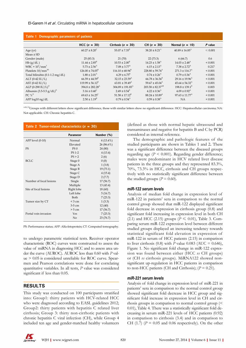
The demographic and pathologic features of the studied participants are shown in Tables 1 and 2. There was a significant difference between the diseased groups regarding age (P < 0.001). Regarding gender difference; males were predominant in HCV related liver disease patients in the three groups and they represented 83.3%, 70%, 73.3% in HCC, cirrhosis and CH groups respec-tively with no statistically significant difference between the studied groups (P = 0.60).
miR-122 serum levelsAnalysis of median fold change in expression level of miR-122 in patients’ sera in comparison to the normal control group showed that miR-122 displayed significant fold decrease in expression in cirrhosis group (0.8) and significant fold increasing in expression level in both CH (2.1) and HCC (2.15) groups (P ≤ 0.01), Table 3. Com-paring serum miR-122 expression level between different studied groups displayed an increasing tendency towards statistical significant fold elevation in expression of miR-122 in serum of HCC patients (2.15) in comparison to liver cirrhosis (0.8) with P value 0.083 (AUC = 0.646), Figure 1. No significant fold change in miR-122 expres-sion was found between either (HCC vs CH groups) or (CH vs cirrhosis groups). MiRNA122 showed non-significant up-regulation in HCC patients in comparison to non-HCC patients (CH and Cirrhosis); (P = 0.21).
miR-221 serum levelsAnalysis of fold change in expression level of miR-221 in patients’ sera in comparison to the normal control group showed significant fold decrease in HCC group and sig-nificant fold increase in expression level in CH and cir-rhosis groups in comparison to normal control group (< 0.01), Table 4. There was a statistically significant fold de-creasing in serum miR-221 levels of HCC patients (0.92) in comparison to cirrhosis (3.4) and in comparison to CH (1.7) (P = 0.05 and 0.06 respectively). On the other
820 November 27, 2014|Volume 6|Issue 11|WJH|www.wjgnet.com
HCC (n = 30) Cirrhosis (n = 30) CH (n = 30) Normal (n = 10) P value
Age (yr) Mean ± SD
60.27 ± 8.20C 55.07 ± 7.35B 38.20 ± 8.21A 40.89 ± 16.85A≤ 0.001
Gender (male) 25 (83.3) 21 (70) 22 (73.3) 6 (66.7) 0.6 Hb (g/dL ) 11.44 ± 2.85B 10.53 ± 2.00B 14.23 ± 1.58A 14.03 ± 2.48A < 0.001 WBC × 103/mm3 5.73 ± 2.56A 7.08 ± 3.77A 6.15 ± 2.13A 7.38 ± 2.72A 0.217 Platelets 10/mm3D 126.00 ± 74.05B 116.10 ± 68.94B 228.80 ± 59.74A 271.3 ± 116.7A < 0.001 Total bilirubin (0.1-1.2 mg/dL) 1.88 ± 2.01A 4.29 ± 6.75B 0.74 ± 0.26A 0.79 ± 0.36A < 0.001 ALT (0-42 IU/L) 66.59 ± 44.59B 32.15 ± 23.59A 66.78 ± 36.56B 29.16 ± 19.96A < 0.001 AST (0-42 IU/L) 119.99 ± 56.12B 63.81 ± 39.49A 59.67 ± 45.04A 45.64 ± 54.32A < 0.001 ALP (0-290 IU/L)D 394.8 ± 282.28A 304.89 ± 191.85A 203.58 ± 82.37AB 198.8 ± 139.1B 0.003 Albumin (3.5-5.5 g/dL)D 3.16 ± 0.40C 2.49 ± 0.54B 4.22 ± 0.36A 4.09 ± 0.92A < 0.001 PC %D 69.63 ± 16.25C 51.58 ± 17.12B 88.24 ± 10.89A 97.63 ± 11.77A < 0.001 AFP log10 ng/dL 2.50 ± 1.19B 0.79 ± 0.54A 0.59 ± 0.38A NA < 0.001
Table 1 Demographic parameters of patients
A,B,C,DGroups with different letters show significant difference, those with similar letters show no significant difference. HCC: Hepatocellular carcinoma; NA: Not applicable. CH: Chronic hepatitis C.
Parameter Number (%)
AFP level (0-10) Normal 4 (13.4%)Elevated 26 (86.6%)
PS PS 0 24 (80)PS 1-2 4 (13.4)PS > 2 2 (6)
BCLC Stage 0 0 (0)Stage A 1 (3.8)Stage B 19 (73.1)Stage C 4 (15.4)Stage D 2 (7.7)
Number of focal lesions Single 17 (56.7)Multiple 13 (43.4)
Site of focal lesions Right lobe 18 (60)Left lobe 5 (16.7)
Both 7 (23.3) Tumor size by CT < 3 cm 1 (3.3)
3-5 cm 12 (40)> 5 cm 17 (56.7)
Portal vein invasion Yes 7 (23.3)No 23 (76.7)
Table 2 Tumor-related characteristics (n = 30)
PS: Performance status; AFP: Alfa-fetoprotein; CT: Computed tomography.
El-Garem H et al . Circulating miRNA in hepatocellular carcinoma

821 November 27, 2014|Volume 6|Issue 11|WJH|www.wjgnet.com
to use tumour markers, mainly AFP, for the screening of HCC. However, performance of tumor markers has not been optimal with the sensitivity and specificity of AFP and PIVKA-II in the range of 39%-64% and 76%-91%, and 41%-77% and 72%-98%, respectively[9,10], the quest for an optimal tumor marker hence continues. miRNAs have been implicated in roles affecting cellular prolifera-tion and oncogenesis[11]. Cellular miRNAs have been linked with HCC[12]. Their availability in the circulation makes them a tempting target for early tumor detec-tion[12]. The aim of the present study was to explore the potential usefulness of serum miR-122 and miR-221 as novel noninvasive markers for diagnosis of HCV related hepatocellular carcinoma in Egyptian patients.
Most of our HCC patients were within Child-Pugh A and B classifications (56.7%, 36.7% respectively), 73.1% were stage B on BCLC scoring system[13]. This could be explained by the fact that most of them were recruited
hand, there was no statistical significant fold change in serum miR-221 expression level between (CH vs cirrhosis groups) (P = 0.214).
miRNA-221 displayed significant fold decrease in HCC group (0.92) compared to non-HCC patients (CH and cirrhosis) (1.81) (P value 0.03). At a cut-off of 1.82 folds, miR-221 yielded 87% sensitivity and 40% specificity in differentiating between both groups. Figure 2, Table 5.
Correlation of miRNA’s with features of HCC There was no statistically significant correlation between serum expression level of studied miRNAs and serum AFP level in the different studied groups of patients. No significant correlation was found between the two miR-NAs and tumor size, Child-pugh grade in HCC group of patients.
Correlation of miRNA’s with severity of hepatic dysfunctionSerum miRNA-122 expression level showed statistically significant correlation with serum necro-inflammatory markers of the liver [aspartate transaminase (AST) and alanine transaminase (ALT) levels) in CH group (P value 0.034 and 0.030 respectively), Table 1.
DISCUSSIONOver the last 2 decades it has become common practice
Fold of change comparing to normal
Group HCCn = 30
Cirrhosisn = 30
CHn= 30
P value
miR-122 (IQR) 2.15 (7.3) 0.8 (3.7) 2.1 (9) < 0.010.0831
0.5722
0.4173
Group HCCn = 30
Non HCC(CH + cirrhosis)
n = 60
P value
miR-122 (IQR) 2.15 (7.3) 1.75 (6.8) 0.21 (NS)
Table 3 MiR-122 serum levels in the different groups
1HCC vs cirrhosis; 2HCC vs CH; 3Cirrhosis vs CH. HCC: Hepatocellular carcinoma; CH: Chronic hepatitis C; IQR: Inter quartile range; NS: Non significant; PVP: Positive predictive value; NVP: Negative predictive value.
Fold of change in comparison to normal
HCCn = 30
Cirrhosisn = 30
CHn = 30
P value
miR-221 (IQR)
0.92 (0.88) 3.4 (19.2) 1.7 (2.6) > 0.010.051
0.062
0.2143
HCC Non HCC0.92 (0.88) 1.81 (7.75) 0.03 (S)
Table 4 MiR-221 serum levels
1HCC vs LC; 2HCC vs CH; 3LC vs CH. HCC: Hepatocellular carcinoma; CH: Chronic hepatitis C; IQR: Inter quartile range; LC: Liver cirrhosis.
0.0 0.2 0.4 0.6 0.8 1.01-Specificity
Sens
itivi
ty
1.0
0.8
0.6
0.4
0.2
0.0
0.0 0.2 0.4 0.6 0.8 1.01-Specificity
Sens
itivi
ty
1.0
0.8
0.6
0.4
0.2
0.0
ROC curve
Figure 1 Receiver operator characteristic curve for miR-122 as a discrimi-nant of hepatocellular carcinoma vs cirrhosis patients.
Figure 2 Receiver operator characteristic curve for miR-221 as a discrimi-nant between hepatocellular carcinoma vs non-hepatocellular carcinoma.
ROC curve
El-Garem H et al . Circulating miRNA in hepatocellular carcinoma

822 November 27, 2014|Volume 6|Issue 11|WJH|www.wjgnet.com
while being assessed for the possibility of interventional treatment. Another possible explanation for rather good liver condition seen in HCC series could be attributed to that with implementing surveillance programs, allowing detecting tumors at an early stage in well compensated patients. Alfa fetoprotein level was normal (< 10 ng/dL) in 13.4% of recruited HCC patients. Similar find-ing was observed by Tateishi et al[14] who suggested that not all tumors secrete AFP, and serum levels are normal in up to 40% of small HCCs. It was also showed that α-Fetoprotein alone is not recommended for the diag-nosis of HCC and studies showed that its cut off value should be set at 200 ng/mL.
Analysis of fold changes in expression level of miR-122 displayed significant fold increase in expression level in chronic hepatitis C group (2.1) and significant fold decrease in expression in cirrhotics (0.8) in compari-son to normal controls. miR-122 is present abundantly in hepatocytes with much lower levels in the circulation in healthy subjects. With hepatocyte injury miR-122 is released in the circulation more readily and serum levels rise. With the eventual loss of hepatocytes and develop-ment of fibrosis with proliferation of myelofibroblasts and accumulation of extracellular matrix the circulating miR-122 levels drop again[15].
In the current study there was significant fold rise in serum expression level of miR-122 in HCC group in comparison to normal control group (P value < 0.01). Matching our results, Trebicka et al[15] who studied hepatic miR-122 expression in 43 HCV related HCC in compari-son to 3 healthy liver samples using qRT-PCR; miR-122 was strongly up-regulated in malignant liver nodules in comparison to healthy liver. They suggested that miR-122 might down regulate target mRNA of unknown tumor suppressor genes and thus lead to further tumor growth[15].
In a study on hepatitis B patients Xu et al[16] suggested that cancer-induced hepatocyte damage would release the abundant intracellular miR-122 into the circulation, the stability of miRNA would be reflected by easily de-tectable high blood levels[17]. In contrast to our results, significant down regulation of miR-122 in HCC com-pared to normal liver tissue was reported by Meng et al[18], Wang et al[19] and Huang et al[20] who compared miR-122 expression profile of 3 different pairs of tumor and nor-mal human liver-derived RNA and 20 HCC liver tissues (mixed etiologies) to normal tissues respectively using mi-croarray[18,19,20]. Similarly a significant down regulation in miR-122 in 19 HBV related HCC liver tissue in compari-son to paired healthy liver by next-generation sequencing
was reported by Connolly et al[21]. Ladeiro et al[22] have established significant down ex-
pression of miR-122 in 28 HCC liver tissues (mixed eti-ologies) in comparison to 4 healthy liver tissues by qRT-PCR.
In our series, no statistically significant correlation could be verified between serum miR122 expression level and patient characters (age), liver synthetic functions tests (Albumin, bilirubin and PC), or serum AFP level in HCC vs non HCC group (CH and cirrhosis). However, in the chronic hepatitis groups serum miR-122 was correlated with higher AST and ALT levels, further solidifying the theory regarding the initial rise in miR-122 levels due to hepato-cyte inflammation and destruction followed by a drop in the levels with the developing fibrosis. Köberle et al[23] also reported significant correlation between serum miR-122 expression level and necro-inflammatory markers (AST, ALT), and Albumin but no significant correlation was found with bilirubin in HCC patients[23].
Perhaps the most significant finding in our study was related to miR-221. Analysis of fold change in expres-sion level of miR-221 in patients’ sera of HCV associ-ated liver disease (CH and cirrhosis) in comparison to normal control group showed significant fold increase in expression level in CH and cirrhosis groups in compari-son to normal control group (< 0.01). Also a significant fold decrease in serum miR-221 in HCC group (0.92) in comparison to normal control was noticed. We assumed that with the progression of liver disease from chronic hepatitis to cirrhosis the increased activity of hepatic stel-late cells was associated with increase miR-221 expres-sion level, such high level stimulated tumorigenesis and increase level of miR-221 in tissue, but as miR-221 is anti apoptotic so serum miR-221 didn’t show similar increase. In contrast to our results many studies established up regulation of miR-221 in HCC in relation to normal con-trol, e.g.,[18,19,20,24]. However, most these studies assessed tissue miR-221 rather than serum levels .The different results could also be explained by technical variations including sampling methods and freezing and RNA iso-lation procedures. The etiology of liver disease is also variable in different studies including viral and alcoholic. The stage of the disease is also a source of variation es-pecially that it is still not evident how miRNA expression changes with fibrosis progression. Different studies have also used different control samples for normalization, e.g., non-HCC tissue from the same patient, healthy liver tissue from another subject or patients with the same pa-thology but not HCC, this is especialy relevant to studies assessing tissue miRNA levels[25].
Similar to what was previously reported by Rong et al[26], we found no statistically significant correlation could be verified between serum miR-221 expression level and patient characters (age), laboratory values (AST, ALT), liver synthetic functions tests (Albumin, bilirubin and PC), or serum a-fetoprotein level in HCC vs non HCC group (CH and cirrhosis) and no statistically significant correlation could be found between the clinic-pathological parameters of hepatic focal lesion, e.g., (number of focal lesions, Child
AUC P value Best cutoff Sensitivity Specificity PPV NPV
0.655 0.03 < 1.82 0.87 0.40 0.47 0.83
Table 5 Diagnostic performance of miR-221 for discriminating patients with hepatocellular carcinoma from those without
HCC: Hepatocellular carcinoma; PVP: Positive predictive value; NVP: Negative predictive value.
El-Garem H et al . Circulating miRNA in hepatocellular carcinoma

823 November 27, 2014|Volume 6|Issue 11|WJH|www.wjgnet.com
score, biggest diameter of focal lesion BCLC, and portal vein invasion) and miR-221 expression level (P ≥ 0.05)[26].
Circulating miR-221 level is significantly up-regulated in the serum of HCV infected patients. It has some value in the differentiation between HCV patients with hepatocellular carcinoma and those without with 87% sensitivity and 40% specificity. It may be able to serve as a promising non-invasive diagnostic marker for HCC. Better results could be obtained if combined with other markers and testing a panel of miRNA’s collectively could ultimately serve as a reliable diagnostic test for HCC.
COMMENTSBackgroundHepatocellular carcinoma (HCC), the most common type of liver cancer, is amongst the top three leading causes of cancer-related deaths worldwide with a median survival of only six to eight months. This poor outcome is related to the late detection, with more than two thirds of patients diagnosed at advanced stages of disease. Thus, surveillance of populations at-risk may detect tumors at an early stage when curative interventions can be implemented. The perfor-mance of available circulating biomarkers in the screening and diagnostic set-tings of HCC is sub-optimal.Research frontiersMiRNAs constitute a large class of genes that encode short RNAs (19-24 nucleotides long), which play key roles in development and differentiation, by the post-transcriptional regulation of protein coding genes. At present, miRNAs have a widely recognized role in human carcinogenesis, including hepatocar-cinogenesis, and many experimental evidences indicate that they may act as oncogenes or tumor suppressor genes regulating the expression of crucial pro-tein coding genes. MiRNAs have been proposed as possible novel biomarkers for cancer diagnosis.Innovations and breakthroughsIn the current study a signature of circulating miRNAs (miR-122 and miR-221) was evaluated. MiR-221 was differentially expressed between patients with HCC and those without (chronic hepatitis and cirrhosis) with lower serum level of miR-221 in former group of patients in comparison to later one. MiR-221 yielded 87% sensitivity and 40% specificity in differentiating between both groups at a cutoff 1.82 folds.ApplicationsThe present study emphasis that circulating miR-221 deserves much attention as potential non invasive biomarkers for HCC in the diagnostic setting.TerminologyHCC: Hepatocellular carcinoma; Non HCC: Chronic hepatis C group of patients and patients with liver cirrhosis.Peer reviewThe manuscript entitled “Circulating microRNA, miR-122 and miR221 Signature in Egyptian Patients with Chronic Hepatitis C Related Hepatocellular Carci-noma”. The manuscript is interesting.
REFERENCES1 Parkin DM, Bray FI, Devesa SS. Cancer burden in the year
2000. The global picture. Eur J Cancer 2001; 37 Suppl 8: S4-S66 [PMID: 11602373 DOI: 10.1016/S0959-8049(01)00267-2]
2 El-Serag HB. Hepatocellular carcinoma: an epidemiologic view. J Clin Gastroenterol 2002; 35: S72-S78 [PMID: 12394209 DOI: 10.1097/00004836-200211002-000028]
3 Velázquez RF, Rodríguez M, Navascués CA, Linares A, Pérez R, Sotorríos NG, Martínez I, Rodrigo L. Prospective analysis of risk factors for hepatocellular carcinoma in pa-tients with liver cirrhosis. Hepatology 2003; 37: 520-527 [PMID: 12601348 DOI: 10.1053/jhep.2003.50093]
4 Bruix J, Sherman M. Management of hepatocellular carci-noma. Hepatology 2005; 42: 1208-1236 [PMID: 16250051 DOI: 10.1002/hep.20933]
5 El-Attar IA. Cancer databases in the Arab world. Ethn Dis 2005; 15: S1-3-S1-4 [PMID: 15787030]
6 El-Nady GM, Ling R, Harrison TJ. Gene expression in HCV-associated hepatocellular carcinoma--upregulation of a gene encoding a protein related to the ubiquitin-conjugating enzyme. Liver Int 2003; 23: 329-337 [PMID: 14708893 DOI: 10.1034/j.1478-3231.2003.00862.x]
7 Fattovich G, Giustina G, Sanchez-Tapias J, Quero C, Mas A, Olivotto PG, Solinas A, Almasio P, Hadziyannis S, Degos F, de Moura MC, Krogsgaard K, Pantalena M, Realdi G, Cor-rocher R, Schalm SW. Delayed clearance of serum HBsAg in compensated cirrhosis B: relation to interferon alpha therapy and disease prognosis. European Concerted Action on Viral Hepatitis (EUROHEP) Am J Gastroenterol 1998; 93: 896-900 [PMID: 9647014 DOI: 10.1111/j.1572-0241.1998.00272.x]
8 Marrero JA, Feng Z, Wang Y, Nguyen MH, Befeler AS, Rob-erts LR, Reddy KR, Harnois D, Llovet JM, Normolle D, Dal-hgren J, Chia D, Lok AS, Wagner PD, Srivastava S, Schwartz M. Alpha-fetoprotein, des-gamma carboxyprothrombin, and lectin-bound alpha-fetoprotein in early hepatocellular carci-noma. Gastroenterology 2009; 137: 110-118 [PMID: 19362088 DOI: 10.1053/j.gastro.2009.04.005]
9 Okuda H, Nakanishi T, Takatsu K, Saito A, Hayashi N, Ta-kasaki K, Takenami K, Yamamoto M, Nakano M. Serum lev-els of des-gamma-carboxy prothrombin measured using the revised enzyme immunoassay kit with increased sensitivity in relation to clinicopathologic features of solitary hepatocel-lular carcinoma. Cancer 2000; 88: 544-549 [PMID: 10649245 DOI: 10.1002/(SICI)1097-0142(20000201)88]
10 Marrero JA, Lok AS. Newer markers for hepatocellular carcinoma. Gastroenterology 2004; 127: S113-S119 [PMID: 15508074 DOI: 10.1053/j.gastro.2004.09.024]
11 Bushati N, Cohen SM. microRNA functions. Annu Rev Cell Dev Biol 2007; 23: 175-205 [PMID: 17506695 DOI: 10.1146/an-nurev.cellbio.23.090506.123406]
12 Llovet JM, Burroughs A, Bruix J. Hepatocellular carcinoma. Lancet 2003; 362: 1907-1917 [PMID: 14667750 DOI: 10.1016/S0140-6736(03)14964-1]
13 Chen DS, Sung JL, Sheu JC, Lai MY, How SW, Hsu HC, Lee CS, Wei TC. Serum alpha-fetoprotein in the early stage of human hepatocellular carcinoma. Gastroenterology 1984; 86: 1404-1409 [PMID: 6201411]
14 Tateishi R, Yoshida H, Matsuyama Y, Mine N, Kondo Y, Omata M. Diagnostic accuracy of tumor markers for hepato-cellular carcinoma: a systematic review. Hepatol Int 2008; 2: 17-30 [PMID: 19669276 DOI: 10.1007/s12072-007-9038-x]
15 Trebicka J, Anadol E, Elfimova N, Strack I, Roggendorf M, Viazov S, Wedemeyer I, Drebber U, Rockstroh J, Sauerbruch T, Dienes HP, Odenthal M. Hepatic and serum levels of miR-122 after chronic HCV-induced fibrosis. J Hepatol 2013; 58: 234-239 [PMID: 23085648 DOI: 10.1016/j.jhep.2012.10.015]
16 Xu J, Wu C, Che X, Wang L, Yu D, Zhang T, Huang L, Li H, Tan W, Wang C, Lin D. Circulating microRNAs, miR-21, miR-122, and miR-223, in patients with hepatocellular car-cinoma or chronic hepatitis. Mol Carcinog 2011; 50: 136-142 [PMID: 21229610 DOI: 10.1002/mc.20712]
17 Varnholt H, Drebber U, Schulze F, Wedemeyer I, Schirm-acher P, Dienes HP, Odenthal M. MicroRNA gene expres-sion profile of hepatitis C virus-associated hepatocellular carcinoma. Hepatology 2008; 47: 1223-1232 [PMID: 18307259 DOI: 10.1002/hep.22158]
18 Meng F, Henson R, Wehbe-Janek H, Ghoshal K, Jacob ST, Pa-tel T. MicroRNA-21 regulates expression of the PTEN tumor suppressor gene in human hepatocellular cancer. Gastroen-terology 2007; 133: 647-658 [PMID: 17681183 DOI: 10.1053/j.gastro.2007.05.022]
19 Wang Y, Lee AT, Ma JZ, Wang J, Ren J, Yang Y, Tantoso E, Li KB, Ooi LL, Tan P, Lee CG. Profiling microRNA expression in hepatocellular carcinoma reveals microRNA-224 up-regula-tion and apoptosis inhibitor-5 as a microRNA-224-specific
COMMENTS
El-Garem H et al . Circulating miRNA in hepatocellular carcinoma

824 November 27, 2014|Volume 6|Issue 11|WJH|www.wjgnet.com
target. J Biol Chem 2008; 283: 13205-13215 [PMID: 18319255 DOI: 10.1074/jbc.M707629200]
20 Huang XH, Wang Q, Chen JS, Fu XH, Chen XL, Chen LZ, Li W, Bi J, Zhang LJ, Fu Q, Zeng WT, Cao LQ, Tan HX, Su Q. Bead-based microarray analysis of microRNA expression in hepatocellular carcinoma: miR-338 is downregulated. Hepatol Res 2009; 39: 786-794 [PMID: 19473441 DOI: 10.1111/j.1872-034X.2009.00502.x]
21 Connolly E, Melegari M, Landgraf P, Tchaikovskaya T, Ten-nant BC, Slagle BL, Rogler LE, Zavolan M, Tuschl T, Rogler CE. Elevated expression of the miR-17-92 polycistron and miR-21 in hepadnavirus-associated hepatocellular carcinoma contributes to the malignant phenotype. Am J Pathol 2008; 173: 856-864 [PMID: 18688024 DOI: 10.2353/ajpath.2008.080096]
22 Ladeiro Y, Couchy G, Balabaud C, Bioulac-Sage P, Pelletier L, Rebouissou S, Zucman-Rossi J. MicroRNA profiling in hepa-tocellular tumors is associated with clinical features and on-cogene/tumor suppressor gene mutations. Hepatology 2008;
47: 1955-1963 [PMID: 18433021 DOI: 10.1002/hep.22256]23 Köberle V, Kronenberger B, Pleli T, Trojan J, Imelmann E,
Peveling-Oberhag J, Welker MW, Elhendawy M, Zeuzem S, Pi-iper A, Waidmann O. Serum microRNA-1 and microRNA-122 are prognostic markers in patients with hepatocellular carci-noma. Eur J Cancer 2013; 49: 3442-3449 [PMID: 23810247 DOI: 10.1016/j.ejca.2013.06.002]
24 Zekri A, Youssef AS, Ahmed O. Serum MicroRNAs as dif-ferential markers and a molecular therapeutic target for HCV associated liver disease. AASLD 2013; poster
25 Borel F, Konstantinova P, Jansen PL. Diagnostic and thera-peutic potential of miRNA signatures in patients with he-patocellular carcinoma. J Hepatol 2012; 56: 1371-1383 [PMID: 22314424 DOI: 10.1016/j.jhep.2011.11.026]
26 Rong M, Chen G, Dang Y. Increased miR-221 expression in hepatocellular carcinoma tissues and its role in enhancing cell growth and inhibiting apoptosis in vitro. BMC Cancer 2013; 13: 21 [PMID: 23320393 DOI: 10.1186/1471-2407-13-21]
P- Reviewer: Hwang KC, Jiang CP, Zou ZM S- Editor: Ji FF L- Editor: A E- Editor: Wu HL
El-Garem H et al . Circulating miRNA in hepatocellular carcinoma

© 2014 Baishideng Publishing Group Inc. All rights reserved.
Published by Baishideng Publishing Group Inc8226 Regency Drive, Pleasanton, CA 94588, USA
Telephone: +1-925-223-8242Fax: +1-925-223-8243
E-mail: [email protected] Desk: http://www.wjgnet.com/esps/helpdesk.aspx
http://www.wjgnet.com