Embed Size (px)
Citation preview
Guadagni et al. BMC Cancer (2022) 22:660 https://doi.org/10.1186/s12885-022-09770-3
RESEARCH
Circulating tumour cell gene expression and chemosensitivity analyses: predictive accuracy for response to multidisciplinary treatment of patients with unresectable refractory recurrent rectal cancer or unresectable refractory colorectal cancer liver metastasesStefano Guadagni1*, Francesco Masedu1, Giammaria Fiorentini2, Donatella Sarti2, Caterina Fiorentini3, Veronica Guadagni4, Panagiotis Apostolou5, Ioannis Papasotiriou6, Panagiotis Parsonidis5, Marco Valenti1, Enrico Ricevuto1, Gemma Bruera1, Antonietta R. Farina1, Andrew R. Mackay1† and Marco Clementi1†
Abstract
Background: Patients with unresectable recurrent rectal cancer (RRC) or colorectal cancer (CRC) with liver metasta-ses, refractory to at least two lines of traditional systemic therapy, may receive third line intraarterial chemotherapy (IC) and targeted therapy (TT) using drugs selected by chemosensitivity and tumor gene expression analyses of liquid biopsy-derived circulating tumor cells (CTCs).
Methods: In this retrospective study, 36 patients with refractory unresectable RRC or refractory unresectable CRC liver metastases were submitted for IC and TT with agents selected by precision oncotherapy chemosensitivity assays performed on liquid biopsy-derived CTCs, transiently cultured in vitro, and by tumor gene expression in the same CTC population, as a ratio to tumor gene expression in peripheral mononuclear blood cells (PMBCs) from the same individual. The endpoint was to evaluate the predictive accuracy of a specific liquid biopsy precision oncotherapy CTC purification and in vitro culture methodology for a positive RECIST 1.1 response to the therapy selected.
Results: Our analyses resulted in evaluations of 94.12% (95% CI 0.71–0.99) for sensitivity, 5.26% (95% CI 0.01–0.26) for specificity, a predictive value of 47.06% (95% CI 0.29–0.65) for a positive response, a predictive value of 50% (95% CI 0.01–0.98) for a negative response, with an overall calculated predictive accuracy of 47.22% (95% CI 0.30–0.64).
© The Author(s) 2022. Open Access This article is licensed under a Creative Commons Attribution 4.0 International License, which permits use, sharing, adaptation, distribution and reproduction in any medium or format, as long as you give appropriate credit to the original author(s) and the source, provide a link to the Creative Commons licence, and indicate if changes were made. The images or other third party material in this article are included in the article’s Creative Commons licence, unless indicated otherwise in a credit line to the material. If material is not included in the article’s Creative Commons licence and your intended use is not permitted by statutory regulation or exceeds the permitted use, you will need to obtain permission directly from the copyright holder. To view a copy of this licence, visit http:// creat iveco mmons. org/ licen ses/ by/4. 0/. The Creative Commons Public Domain Dedication waiver (http:// creat iveco mmons. org/ publi cdoma in/ zero/1. 0/) applies to the data made available in this article, unless otherwise stated in a credit line to the data.
Open Access
†Andrew R. Mackay and Marco Clementi shared last authors.
*Correspondence: [email protected]
1 Department of Applied Clinical and Biotechnological Sciences, University of L’Aquila, 67100 L’Aquila, ItalyFull list of author information is available at the end of the article

Page 2 of 15Guadagni et al. BMC Cancer (2022) 22:660
BackgroundClinical and biological prognostic markers identify patients with differing risks of a specific outcome regard-less of treatment, such as progression or death [1], and can select individuals at high risk of relapse, as potential candidates for alternative treatments. In contrast, pre-dictive markers are associated with response (benefit) or lack of response to a particular therapy relative to other available therapies and identify patients more likely to benefit from a particular treatment [2]. Within this con-text, tissue and liquid biopsy precision oncotherapy holds great promise in improving therapeutic outcomes, as both represent important methods for detecting prog-nostic and predictive markers, with additional potential to identify therapeutic targets. This is particularly true for less invasive, serially repeatable liquid biopsies, which are potentially more relevant to disseminated disease.
Tissue biopsies, in accordance with American Soci-ety of Clinical Oncology (ASCO) and European Society for Medical Oncology (ESMO) recommendations [3], can be used to detect oncogenic mutations, oncogene overexpression and to evaluate chemosensitivity [4]. Liquid biopsies (blood, ascites, urine, pleural effusion, or cerebrospinal fluid) can be used to do the same but also permit purification and analysis of tumour-derived components including circulating tumor cells (CTCs), exosomes, circulating tumor DNAs (ctDNA), microR-NAs (miRNAs), long non-coding RNAs (lncRNAs) and proteins [5, 6], with enhanced potential for identifying prognostic markers, predictive markers and therapeutic targets.
When compared to tumor-derived non-cellular com-ponents, intact CTCs not only provide a valuable source of quality tumour cell-derived nucleic acids and proteins for immunocytological, fluorescence in situ hybridiza-tion (FISH), DNA sequence, PCR, RT-PCR and multi-plex RNA analysis but also the opportunity for transient in vitro culture, enabling chemosensitivity and tumor gene expression analysis to be performed on the same CTC population. Up to 2010, CTC detection and purifi-cation methodologies were based upon either biophysi-cal (i.e. Biocoll/Ficoll®, Oncoquick® or Screen-Cell® Cyto IS - Sarcelles, France) or antigenic (i.e. CellSearch®
system - Menarini Silicon Biosystems) properties. Fol-lowing 2010, progress in microfluidic-based technolo-gies, devices and principles have resulted in the use of novel antibody-based marker-dependent platforms, physical characteristic-based platforms, and secretome and transcriptome-based platforms, the latter of which, however, involves CTC lysis, eliminating the possibility of CTC culture for drug screening.
The FDA has approved liquid biopsy [7] and has vali-dated the Cell-SearchTM system (Veridex LCC, Raritan, NJ, USA) as a diagnostic tool for CTC identification and enumeration in a variety of tumours, including colorec-tal cancer (CRC) and currently holds a dominant posi-tion amongst competitors [8]. This system is employed to detect CD45 (Cluster of differentiation 45) negative, EpCAM (Epithelial adhesion molecule) and cytokerat-ins CK8/18/19 positive CTCs, using anti human CK8/18/19/20 antibodies conjugated with ferrous beads for magnetic purification [8]. Cytokeratins and particu-larly CK20, are well-established markers of epithelial cell and metastatic CRC CTCs [9] and are employed also for semi-quantitative qRT-PCR analyses [10] and the major-ity of studies using this system report CTC enumera-tion as a prognostic marker for progression free survival (PFS) and overall survival (OS) [11]. Drawbacks with this approach, however, include potential CTC underestima-tion due to epithelial to mesenchymal transition (EMT) and loss of epithelial marker expression, technical bot-tlenecks and cost, which reduce routine use [8]. Alter-native isolation and analytical methods and techniques have also been reported that overcome some of these problems (i.e. leukapheresis and Hydro-Seq technology) [8] and flow cytometry has also been reported to detect CTCs with high sensitivity and specificity [12].
To date, few papers have investigated the therapeutic predictive potential of CTC isolation and enumeration in CRC patients [13–16] or the promise of combin-ing molecular profiling with chemosensitivity analysis, considered for some time to be the best way to improve disease characterisation and selection of individualised therapy [17, 18]. Within this context, patients present-ing with unresectable recurrent rectal cancer (RRC) or unresectable CRC liver metastases, refractory to at
Conclusions: This is the first reported estimation of predictive accuracy derived from combining chemosensitiv-ity and tumor gene expression analyses on liquid biopsy-derived CTCs, transiently cultured in vitro which, despite limitations, represents a baseline and benchmark which we envisage will be improve upon by methodological and technological advances and future clinical trials.
Keywords: Predictive accuracy, Precision oncotherapy, Liquid biopsies, Circulating tumor cells, Chemosensitivity tests, Tumor gene expressions analyses, Recurrent rectal cancer, Colorectal cancer liver metastases, Intraarterial chemotherapy, Targeted therapy

Page 3 of 15Guadagni et al. BMC Cancer (2022) 22:660
least two lines of standard systemic chemotherapy, tar-geted and radiation therapy, represent good potential candidates for determining the predictive accuracy of combined in vitro (ex-vivo) CTC chemosensitivity and gene expression analysis.
CRC is currently the third most common cancer type, with upwards of ≈ 1.8 million new cases and ≈ 9% of all cancer-related deaths reported annually worldwide [19]. Treatment strategies depend upon disease stage, patient condition, molecular mechanisms, economic parameters, health care systems and unexpected fac-tors, such as the current Sars-Covid-2 pandemic. In highly developed countries, 5-year survival rates for localized CRC of ≈ 90% plummet to ≈ 14% for met-astatic or relapsed disease. For stage IV metastatic or recurrent CRC, devoid of known cancer driving muta-tions or targetable biomarkers (≈ 85% of patients), current systemic therapeutic regimes include: FOL-FOX (leucovorin plus 5-fluorouracil plus oxaliplatin), CAPEOX (capecitabine plus oxaliplatin), FOLFIRI (leucovorin plus 5-fluorouracil plus irinotecan), FOL-FOXIRI (leucovorin plus 5-fluorouracil plus oxaliplatin plus irinotecan), typically combined with bevacizumab or cetuximab or aflibercept and recently new oral drugs such as regorafenib and trifluridine/tipiracil [18, 20–28]. Patients with dominant liver metastatic dis-ease or limited extrahepatic disease, may receive liver-directed intraarterial therapies such as hepatic arterial chemotherapy infusion, chemoembolization [20] and radioembolization to improve local tumor response and to reduce systemic side effects [18]. For all of these patients, liquid biopsy-derived CTCs deserve thorough investigation as a potential source of important pre-dictive information with respect chemosensitivity and chemoresistance, enhancing the possibility of identify-ing novel alternative therapeutic strategies and reduc-ing toxicity.
For stage IV metastatic or recurrent CRCs refractory to standard systemic therapies, that exhibit detectable overexpression or mutation of known driver oncogenes (≈ 4–15%), precision oncotherapy represents a viable therapeutic option, in conditions of: i) deficient mis-match repair protein expression (dMMR) and/or DNA microsatellite instability (MSI-H/high), treatable with pembrolizumab and nivolumab or a combination of nivolumab and ipilimumab check-point inhibitors in first and subsequent lines of treatment; ii) specific BRAFV600E mutation treatable with a combination of encorafenib and cetuximab in second or third lines; iii) EGFR overex-pression treatable with cetuximab and panitumumab; iv) HER-2 3+ overexpression or HER2 FISH/ISH amplifica-tion treatable with trastuzumab, lapatinib, tucatinib and deruxtecan/trastuzumab; v) neurotrophic tropomyosin
receptor kinases (NTRKs) overexpression or expression of novel NTRK chimeric fusions treatable with larotrec-tinib or entrectinib [21–28].
Within this context, we report a retrospective study of the accuracy of in vitro ex vivo CTC chemosensitiv-ity and tumour gene expression analyses in predicting response to treatment in CRC patients presenting with unresectable refractory RRC or unresectable refractory CRC liver metastases. In this setting, chemotherapeutic agents and monoclonal antibodies used for the multidis-ciplinary treatment of patients with refractory CRC, were chosen from chemosensitivity assays performed on tran-siently in vitro cultured liquid biopsy-derived CTCs and from the ratio of tumor gene expression exhibited by the same CTC populations compared to purified peripheral blood mononuclear cells from the same patient.
MethodsPatientsThis study involved patients with unresectable and pre-dictable disease course and was approved by ASL n.1 Ethics committee, Abruzzo, Italy (Chairperson: G. Pic-cioli; protocol number 10/CE/2018; approved: 19 July 2018 (n.1419)]. Drug selection, clinical treatments and evaluations were performed at the University of L’Aquila, L’Aquila, Italy. CTC isolation, culture, gene expression and chemosensitivity analyses were performed at the Research Genetic Cancer Centre, Florina, Greece. All patients provided written consent and received complete information about their disease and the implications of the proposed conventional treatment, in accordance with the Helsinki Declaration and the University of L’Aquila committee on human experimentation.
From a cohort of 168 CRC patients, enrolled from 2007 to 2019, comprising 62 patients with unresectable recurrent rectal cancer in progression following two lines of systemic chemotherapy and radiotherapy and 106 patients with unresectable CRC liver metastases in pro-gression after two lines of systemic chemotherapy, 36 patients were retrospectively selected based on the fol-lowing criteria: submission for precision oncotherapy by combined intraarterial chemotherapy and systemic venous targeted therapy, using drugs selected by com-parative chemosensitivity and gene expression analyses performed on in vitro cultured CTCs and PBMCs from the same patient. To ensure the stability and consistency of sample collection and detection, only patients submit-ted for combined locoregional intraarterial chemother-apy and systemic venous targeted-therapy, whose CTCs had been purified using the same methodology, were included in this retrospective study, whereas patients submitted for systemic venous chemotherapy and/or sys-temic targeted therapy, and those in which CTC isolation

Page 4 of 15Guadagni et al. BMC Cancer (2022) 22:660
and culture had been performed with more recent tech-niques and methodologies, not available at the beginning of the study, were excluded.
Decisions concerning unresectability and precision oncotherapy were made by experienced surgeons, oncol-ogists and radiologists during multidisciplinary meet-ings. Inclusion criteria were: i) histologically confirmed colorectal cancer diagnosis and complete primary tumor resection; ii) failure of two lines of systemic chemother-apy; iii) Eastern Cooperative Oncology Group (ECOG) performance status of < 4; iv) adequate liver or renal dys-function (total bilirubin serum levels < 3 mg/dL, serum albumin level > 20 g/L, serum creatinine level < 2 mg/dL). In all cases, systemic chemotherapy ceased 4 weeks prior to the 1st cycle of tailored intraarterial chemo-therapy in association with targeted therapy. Patients with inadequate medical records were excluded from this study. Demographic and clinical characteristics of the 36 patients are presented in Table 1.
Liquid biopsy, CTC gene expression and chemosensitivity assaysSample collection, storage and transportationLiquid biopsies (≥ 20 mL venous blood) were collected from each patient in sterile 50 mL Falcon tubes, con-taining 7 ml of 0.02 M ethylenediaminetetraacetic acid (EDTA) anticoagulant (E0511.0250, Duchefa Biochemie B.V., Haarlem, The Netherlands), transported in impact-resistant containers, under refrigeration at 2–8 °C, and analysed within 80 hours [29].
CTC isolationFor CTC isolation, blood samples were layered over 4 ml polysucrose solution (Biocoll 1077, Biochrom, Berlin, Germany) and centrifuged for 20 min at 2500×g. Periph-eral blood mononuclear cells (PBMCs) and CTCs (the buffy coat) were collected and washed with phosphate-buffered saline (PBS, P3813, Sigma-Aldrich, Germany). Cell pellets were resuspended for 10 min in erythrocyte lysis buffer [154 mM NH4Cl (31,107, Sigma-Aldrich, Germany), 10 mM KHCO3 (4854, Merck, Germany) and 0.1 mM EDTA]. Cells were then collected by centrifuga-tion, washed in PBS, and incubated with mouse mono-clonal anti-human CD45 antibody-conjugated magnetic beads (39-CD45–250, Gentaur, Belgium) for 30 min at 4 °C. Anti CD45 bead-bound cells were collected in a magnetic field and saved for use as non-cancer control PBMCs in qRT-PCR gene expression analyses. Remain-ing cells were incubated with mouse monoclonal Anti-Pan cytokeratin-conjugated microbeads (PCK/CK4, CK5, CK6, CK8, CK10, CK13 and CK18) (MA1081-M, Gen-taur, Belgium) for 30 min at 4 °C and PCK bead-bound cells (CTCs) collected in a magnetic field and washed in
PBS. Isolated viable CTCs (IV-CTCs) were counted, and samples containing ≥5 viable CTCs per ml of blood were cultured for 6 days in order to obtain sufficient cell num-bers for gene expression and chemosensitivity assays.
CTC culturePurified pan-cytokeratin positive/CD45-negative bead-bound cells were cultured in RPMI-1640 (R0883, Sigma-Aldrich, Germany) containing 10% Fetal Bovine Serum-FBS (F4135, Sigma-Aldrich, Germany) and 2 mmol/l glutamine (G5792; Sigma), in 12-well cell culture plates (Corning, Merck, Germany), without
Table 1 Patient demographic and clinical characteristics
IQR Interquartile range, SBTT Survival from recurrence/metastases date and first treatment of tailored intraarterial chemotherapy in association with targeted therapy, ECOG Eastern Cooperative Oncology Group performance status before first treatment of tailored intraarterial chemotherapy in association with targeted therapy, PFSTT Progression free survival after tailored therapy, OSTT Overall survival after tailored therapy, KRAS Kirsten rat sarcoma virus gene, NRAS Neuroblastoma RAS gene, BRAF v-Raf murine sarcoma viral oncogene homolog B
Median; [IQR] Number (%)
Age (years) 60.5; [55.5–67.5]
- < 75 35 (97.2)
- ≥ 75 1 (2.8)
Gender - M 22 (61.1)
- F 14 (38.9)
Site of recurrence/metastases - Pelvis 27 (75)
- Liver 9 (25)
Presence of other metastases - Yes 18 (50)
- No 18 (50)
Number of previous systemic chemotherapy lines - 2 35 (97.2)
- > 2 1 (2.8)
KRAS, NRAS, BRAF genotype status - KRAS exon 2 codon 12 3 (8.3)
- NRAS mutations 0 (0)
- BRAF mutations 0 (0)
- Unknown 13 (36.1)
SBTT (months) 16.0; [12–20.5]
ECOG - 0 0 (0)
- 1 9 (25)
- 2 13 (36.1)
- 3 14 (38.9)
- 4 0 (0)
PFSTT (months) 7.5; [5–12.5]
OSTT (months) 34.0; [23.5–41.5]

Page 5 of 15Guadagni et al. BMC Cancer (2022) 22:660
antibiotics, at 37 °C, 5% CO2, and culture medium was changed every 2 days, as previously described [30].
CTC validation post‑cultureCTC validation was confirmed by positive CK18, CK19, and negative CD45, CD31, N-Cadherin qRT-PCR (see section 2.2.5 for methodology) and by positive Pan-Cytokeratin-APC, and EpCAM-FITC, and negative CD45-PE Immunofluorescence (IF), in 6-day CTC cul-tures (Fig. 1). Briefly for IF, purified CTCs and PBMCs from the same patient were initially stained with anti-human CD45-PE (phycoerythin) conjugated mouse monoclonal antibody (304,008, Biolegend, CA, USA) and anti-human EpCAM-FITC (fluorescein isothioy-anate) conjugated mouse monoclonal antibody (324,204, Biolegend, CA, USA), at recommended dilutions, and subsequently, cells were stained with anti-human Pan-Cytokeratin-APC (allophycocyanin, ab201807, Cam-bridge, UK) conjugated mouse monoclonal antibody (SAB4700666, Sigma-Aldrich, MO, USA), using Leuco-perm staining protocol (BUF09C, Bio-Rad, CA, USA). Nuclei were counterstained with DAPI (4′,6-diamidino-2-phenylindole, Abbott Molecular, Illinois, USA) and cells were visualised under a Nikon Eclipse 50i micro-scope, armed with Cytovision software (Leica Biosys-tems, United States).
CTC gene expressionFor gene expression analysis, RNAs were purified from CTC cultures and from corresponding PBMCs using RNeasy Mini Kits, as directed by the manufacturer (74,105, Qiagen, Hilden, Germany). RNAs (1 μg) were reverse transcribed using a PrimeScript RT Reagent Kit, as directed by the manufacturer (RR037A, Takara, Bei-jing, China) and subjected to KAPA SYBR Fast Master Mix (2×) Universal (KK4618, KAPA Biosystems, MA, USA) real-time qPCR, in a final volume of 20 μl. Real-time qRT-PCR reactions were performed in a final vol-ume of 20 μl and characterised by 2 min denaturation at 95 °C, followed by 40 cycles consisting of 10 sec denatura-tion at 95 °C and 30 sec annealing at 59 °C. Melting-curve analysis was performed from 70 °C to 90 °C, with 0.5 °C increments of 5 s, at each step. Reactions were employed to evaluate the expression of multidrug resistance gene-ABCB1 (MDR1), thymidylate synthase (TYMS), dihydro-folate reductase (DHFR), DNA excision repair protein (ERCC1), glutathione S-transferase (GST), epidermal growth factor (EGF), vascular epidermal growth fac-tor (VEGF), 18S ribosomal RNA (18S rRNA), β actin (ACTB), glyceraldehyde 3-phosphate dehydrogenase (GAPDH), the specific primers for which have been pre-viously reported [31]. All reactions were performed in triplicate, compared to template-free negative controls,
and analysed by Livak relative quantification [32]. Gene expression was quantified using the following equations:
and classified as low (< 50%) or high (> 50%) over-expression, as previously described [31].
CTC chemosensitivityFor chemosensitivity assays, 6 day CTC cultures in 12-well plates were treated with the following drug concentrations: 1 μM alkeran (Μ2011, Sigma-Aldrich, Germany), 1 μM doxorubicin (D1515, Sigma-Aldrich, Germany), 1 μM cisplatin (P4394, Sigma-Aldrich, Ger-many), 10 μM 5-fluorouracil (F6627, Sigma-Aldrich, Germany), 1.12 μM oxaliplatin (O9512, Sigma-Aldrich, Germany), 1 μM carboplatin (41575–94-4, Sigma-Aldrich, Germany), 5 μM irinotecan (I1406, Sigma-Aldrich, Germany), 1 μM raltitrexed (112887–68-0, Sigma-Aldrich, Germany) and 2 μM mitomycin C (M4287, Sigma-Aldrich, Germany), in complete culture medium. Cell viability was assessed by Annexin V-PE (559,763; BD Bioscience, USA) flow cytometry (BD Instruments Inc., San José, CA, USA), and the percentage of living, dead and dying cells evaluated using BD Cell-Quest Software (BD Instruments Inc., USA (Fig. 2). Con-trols included untreated cells incubated in the presence and absence of chemotherapeutic drugs and cell-free counterparts. The percentage of non-viable CTCs was calculated under non-drug and drug-treated conditions, and chemosensitivity was classified as either: i) non-sen-sitive < 35% death; ii) partially sensitive 35–80% death, or iii) highly sensitive > 80% death.
Precision oncotherapy protocol criteriaDecisions concerning precision oncotherapy were made by experienced oncologists, surgeons and radiologists during multidisciplinary meetings based on gene expres-sion and chemosensitivity analyses, previous systemic chemotherapeutic protocols, and in vitro drug cytotoxic-ity under hypoxic conditions [33]. For intraarterial chem-otherapy, drug regimens were selected according to the
�Ct(threshold Cycle) = Cttarget − Ctβ-actin
��Ct = �Ct(treated CTCs)-�Ct(non-cancer cells)
Relative expression level = 2-��Ct
%Gene expression = 100× 2-��Ct-1

Page 6 of 15Guadagni et al. BMC Cancer (2022) 22:660
following criteria: i) mono-chemotherapy for CTCs with high sensitivity (> 80% of dead and dying cells) for one or more drug, with highest chemosensitivity indicating the
drug to be used, ii) poly-chemotherapy for CTCs exhibit-ing partial sensitivity, and iii) with respect to drug activ-ity under conditions of hypoxia. For targeted therapy, the
Fig. 1 Representative: A) qRT-PCR CTC validation and B) accompanying histogram, demonstrating β-actin, CK18 and CK19 but not CD45, CD31 or N-Cadherin mRNA expression in a 6-day CTC culture, plus C) IF validation demonstrating positive IF immunoreactivity for EpCAM, Pan-CK but not CD45 in a 6 day CTC culture (left panels), and positive IF immunoreactivity for CD45 but not EpCAM or Pan-CK in PBMCs from the same patient (right panels) (bar = 50 μm)

Page 7 of 15Guadagni et al. BMC Cancer (2022) 22:660
Fig. 2 Representative flow cytometric analyses, with corresponding tables, showing the percentage changes in live, dead, late apoptotic and apoptotic Annexin-V positive CTCs, in 72 hour-chemosensitivity assays of cultured CTCs from the same individual (Case 1), demonstrating chemosensitivity to Mitomycin-C (MMC, 2 μM) (A) but not Flurouracil (5-FU, 10 μM) (B), compared to respective untreated CTC controls

Page 8 of 15Guadagni et al. BMC Cancer (2022) 22:660
presence/absence of KRAS and NRAS mutations in exon 2 (codons 12 and 13), exon 3 (codons 59 and 61) and/or exon 4 (codons 117 and 146) was verified, and subse-quent drug selection was based upon existing therapeutic options already recommended by evolving international guidelines for clinical practice, and high CTC: PBMC gene over-expression ratios, with the highest percentage used to select each drug. Specifically, for EGFR overex-pression cetuximab was selected and for high VEGFR overexpression bevacizumab was selected. If CTC: PBMC expression ratios were > 50%, target-therapy selec-tion was based upon the highest ratio of tumour gene overexpression.
Intraarterial chemotherapy techniquesIntraarterial chemotherapeutic procedures for unre-sectable refractory RRC and for unresectable refrac-tory CRC liver metastases, together with eligibility criteria, have been described previously [17, 18]. For pelvic recurrences, hypoxic pelvic perfusion (HPP) requires specialized surgical skill and can also be per-formed percutaneously [34], whereas regional intraar-terial chemotherapy for CRC liver metastases requires an interventional radiologist [18, 35]. Both procedures included hemofiltration to reduce toxicity [17, 18].
Response and adverse events criteriaResponses, assessed by computed tomography (CT), magnetic resonance imaging (MRI) and positron emis-sion tomography (PET), were evaluated 3 months follow-ing the first cycle of intraarterial chemotherapy combined with targeted therapy and classified using Response Evaluation Criteria in Solid Tumors (version 1.1), as either complete responses (CR), partial responses (PR), corresponding to a minimum 30% reduction in tumour volume, stable disease (SD) or progressive disease (PD) [36]. Patient responses prior to 2009 were re-classified retrospectively. Adverse reactions were evaluated using National Cancer Institute-Common Terminology Crite-ria for Adverse Events software (version 4.03) and classi-fied from 0 to 4.
Statistical analysesDue to the low sample size and plausible deviations from distributional symmetry, patient demographic and clini-cal characteristics are summarized as percentages and median values. Cut-off values for chemosensitivity tests and tumor gene expression analyses were calculated from combined optima of positive predictive values. Specifi-cally, this pair of cut-off values was derived from the pair of thresholds that maximized predictive accuracy, con-sidering all possible confusion matrices. Binomial exact confidence intervals of 95% are provided for sensitivity,
specificity, positive predictive, negative predictive and accuracy values.
ResultsCTC chemosensitivity and gene expression analyses, used for selecting precision oncotherapy protocols, and patient therapeutic responsesWith respect to CTC purification, the CTC detection rate in this cohort of advanced stage metastatic CRC patients was 100%, the mean (± s.e.) number of purified CTCs per millilitre of blood was 10.3 ± 0.35 (range 6.2 to 16.3) (Table 2). For 25 mls blood samples this translated into total numbers of CTCs per patient ranging from ≈ 150 to 400, which was expanded by 6-day in vitro culture to between ≈ 35,000 to 100,000 CTCs per patient for the gene expression and chemosensitivity assays used to select locoregional chemotherapy and systemic targeted therapy protocols.
RECIST 1.1 responses to combined intraarterial chem-otherapy and targeted therapy, selected by CTC che-mosensitivity and gene expression assays, and the KRAS mutational status of tumors are presented in Tables 2 and 3.
Response predictive accuracy of precision oncotherapy protocols, selected by chemosensitivity and gene expression analyses performed on in vitro cultured liquid biopsy‑derived CTCs and PBMCsWith respect to the 36 patients assessed in this study, intraarterial chemotherapy combined with targeted ther-apy elicited 17 partial responses (PR), 18 stable disease (SD) responses and 1 progressive disease (PD) response. RECIST 1.1 responses subdivided into positive (CR + PR) and negative (SD + PD) responses, in relation to CTCs chemosensitivity and tumor gene expression tests for targeted therapy, are presented in Table 4. Tests were defined as positive when ≥70% of drug-treated CTCs were killed and CTC: PBMC tumor gene expression ratios were ≥ 50%. Tests were defined as negative when < 70% of drug-treated CTCs were killed and CTC: PBMC tumor gene expression ratios were < 50%.
Considering both types of therapy (intraarterial and targeted) and associated respective cut-offs for assay positivity, we report 16 true positive responses (TP), 1 false positive response (FP), 18 true negative responses (TN) and 1 false negative response (FN), which translate into a sensitivity of 94.12% (16/17) (95% CI 0.71–0.99), specificity of 5.26% (1/19) (95% CI 0.01–0.26), a positive predictive value of 47.06% (16/34) (95% CI 0.29–0.65), a negative predictive value of 50% (1/2) (95% CI 0.01–0.98) and an over-all predictive accuracy of 47.22% (17/36) (95% CI 0.30–0.64).

Page 9 of 15Guadagni et al. BMC Cancer (2022) 22:660
Table 2 Protocols of intraarterial chemotherapy based on CTC chemosensitivity, and RECIST 1.1 responses
Pt IV‑CTCs Intraarterial chemotherapy protocols Response in association with targeted therapy
Drug % CTC chemosensitivity Dose administered
1R 12.2/ml MMC 82 25 mg/m2 PR2R 16.2/ml 5-FU 70 100 mg/m2 PR
OX 72 80 mg/m2
3R 8.4/ml MMC 80 25 mg/m2 PROX 75 80 mg/m2
RAL 70 3 mg/m2
4R 12.2/ml MMC 70 25 mg/m2 PRCIS 70 70 mg/m2
RAL 70 3 mg/m2
5R 7.5/ml MMC 80 25 mg/m2 PRRAL 70 3 mg/m2
6R 6.9/ml MMC 80 25 mg/m2 PRRAL 75 3 mg/m2
7R 15.1/ml ALK 70 30 mg/m2 PRCAR 75 100 mg/m2
8R 16.3/ml ALK 75 30 mg/m2 PRCAR 75 100 mg/m2
9R 8.9/ml OX 70 80 mg/m2 PR10R 10/ml OX 70 80 mg/m2 PR
MMC 75 25 mg/m2
11R 9.5/ml OX 70 80 mg/m2 PRMMC 75 25 mg/m2
12R 8.9/ml OX 75 80 mg/m2 PRMMC 75 25 mg/m2
13R 7.5/ml MMC 80 25 mg/m2 PR14R 8.9/ml MMC 82 25 mg/m2 PR15R 6.9/ml MMC 75 25 mg/m2 PR16R 12.2/ml MMC 82 30 mg/m2 PR17R 16.2/ml 5-FU 60 600 mg/m2 PR
OX 72 150 mg/m2
18R 7.5/ml OX 75 80 mg/m2 SDIRI 80 100 mg/m2
19R 9/ml MMC 85 25 mg/m2 SD20R 8.4/ml OX 85 80 mg/m2 SD21R 9.7/ml MMC 83 25 mg/m2 SD22R 6.9/ml MMC 80 25 mg/m2 SD23R 9.8/ml MMC 75 25 mg/m2 SD24R 16.2/ml DOX 72 35 mg/m2 SD
OX 65 80 mg/m2
25R 9.8/ml MMC 70 25 mg/m2 SDRAL 70 3 mg/m2
26R 8.9/ml MMC 80 25 mg/m2 SD27R 6.9/ml MMC 80 25 mg/m2 SD28 L 9.4/ml MMC 85 25 mg/m2 SD29 L 16.2/ml MMC 80 25 mg/m2 SD30 L 7.5/ml OX 75 150 mg/m2 SD
IRI 80 200 mg/m2
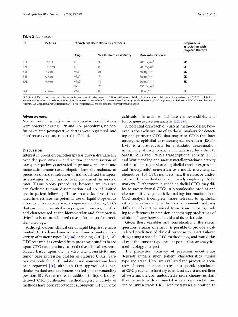
Page 10 of 15Guadagni et al. BMC Cancer (2022) 22:660
Adverse eventsNo technical, hemodynamic or vascular complications were observed during HPP and HAI procedures, no per-fusion-related postoperative deaths were registered, and all adverse events are reported in Table 5.
DiscussionInterest in precision oncotherapy has grown dramatically over the past 20 years and routine characterisation of oncogenic pathways activated in primary, recurrent and metastatic tumour tissue biopsies form the mainstay of precision oncology selection of individualised therapeu-tic strategies, which has led to improvements in survival rates. Tissue biopsy procedures, however, are invasive, can facilitate tumour dissemination and are of limited use in patient follow-up. These drawbacks have stimu-lated interest into the potential use of liquid biopsies, as a source of tumour-derived components including CTCs that can be enumerated as a prognostic marker, purified and characterised at the biomolecular and chemosensi-tivity levels to provide predictive information for preci-sion oncology.
Although current clinical use of liquid biopsies remains limited, CTCs have been isolated from patients with a variety of tumour types [37, 38], including CRC [17, 18]. CTC research has evolved from prognostic studies based upon CTC enumeration, to predictive clinical response studies based upon the in vitro chemosensitivity and tumor gene expression profiles of cultured CTCs. Vari-ous methods for CTC isolation and enumeration have been reported [10], although FDA approval of a par-ticular method and equipment has led to a commanding position [8]. Furthermore, in addition to liquid biopsy-derived CTC purification methodologies, a variety of methods have been reported for subsequent CTC in vitro
cultivation in order to facilitate chemosensitivity and tumor gene expression analysis [12, 39].
A potential drawback of current methodologies, how-ever, is the exclusive use of epithelial markers for detect-ing and purifying CTCs that may miss CTCs that have undergone epithelial to mesenchymal transition (EMT). EMT is a pre-requisite for metastatic dissemination in majority of carcinomas, is characterised by a shift to SNAIL, ZEB and TWIST transcriptional activity, TGFβ and Wnt signaling and matrix metalloproteinase activity and results in repression of epithelial marker expression and “metaplastic” conversion to a motile mesenchymal phenotype [10]. CTCs numbers may, therefore, be under-estimated by methods that exclusively employ epithelial markers. Furthermore, purified epithelial CTCs may dif-fer to mesenchymal CTCs in biomolecular profiles and chemosensitivity, potentially making information from CTC analysis incomplete, more relevant to epithelial rather than mesenchymal tumour components and may differ to information gained from tissue biopsies, lead-ing to differences in precision oncotherapy predictions of clinical efficacy between liquid and tissue biopsies.
Given these variables and considerations, the salient question remains whether it is possible to provide a cal-culated prediction of clinical response to select tailored drugs using a specific CTC methodology, and would this alter if the tumour type, patient population or analytical methodology changes?
The predictive accuracy of precision oncotherapy depends initially upon patient characteristics, tumor type and stage. Here, we evaluated the predictive accu-racy of precision oncotherapy on a specific population of CRC patients, refractory to at least two standard lines of systemic therapy, undoubtedly more chemo-resistant than patients with unresectable recurrent rectal can-cer or unresectable CRC liver metastases submitted to
Table 2 (continued)
Pt IV‑CTCs Intraarterial chemotherapy protocols Response in association with targeted therapy
Drug % CTC chemosensitivity Dose administered
31 L 10/ml IRI 90 200 mg/m2 SD32 L 16.2/ml IRI 80 200 mg/m2 SD33 L 7.5/ml MMC 81 30 mg/m2 SD34 L 9.8/ml MMC 70 30 mg/m2 SD35 L 9.9/ml MMC 75 30 mg/m2 SD
OX 70 150 mg/m2
36 L 6.9/ml MMC 80 30 mg/m2 PD
Pt Patient, R Patient with unresectable refractory recurrent rectal cancer, L Patient with unresectable refractory colo-rectal cancer liver metastases, IV-CTCs Isolated viable circulating tumor cells in patient blood prior to culture, 5-FU 5 fluorouracil, MMC Mitomycin, IRI Irinotecan, OX Oxaliplatin, RAL Raltitrexed, DOX Doxorubicin, ALK Alkeran, CIS Cisplatin, CAR Carboplatin, PR Partial response, SD stable disease, PD Progressive disease

Page 11 of 15Guadagni et al. BMC Cancer (2022) 22:660
first line treatment. The predictive accuracy of precision oncotherapy also depends upon the methodology used for CTC isolation, enrichment, in vitro culture, chemo-sensitivity, tumor gene expression, etc. In this study, due to the long accrual period, the predictive accuracy of a
particular methodology for CTC purification, culture and analysis, was evaluated. This methodology has now been improved by recent technical advances, which if available during the period of study, may have enhanced predictive accuracy.
Table 3 Targeted therapy protocols selected according to CTC: PBMC percentage gene overexpression ratios, and associated RECIST 1.1 responses
CTC Circulating tumor cells, PMBCs Peripheral mononuclear blood cells, Pt Patient, R Patient with unresectable refractory recurrent rectal cancer, L Patient with unresectable refractory colo-rectal cancer liver metastases, EGFR Epidermal growth factor receptor, VEGFR Vascular endothelial growth factor receptor, MDR1 Multidrug resistance gene (ABCB1 gene), TYMS Thymidylate synthase gene, DHFR Dihydrofolate reductase, ERCC1 DNA excision repair protein, GST Glutathione S-transferases, BEV Bevacizumab (dosage administered = 5 mg/kg), CET Cetuximab (dosage administered = 250 mg/m2), PR Partial response, SD Stable disease, PD Progressive disease
*KRAS (Kirsten rat sarcoma virus gene) mutated in codon 12, exon 2
Pt EGFR % VEGFR % MDR1% TYMS % DHFR % ERCC1% GST % Protocol of targeted therapy
Response in association with intraarterial chemotherapy
1R 45 70 80 0 0 0 18 BEV PR2R 45 70 80 0 0 0 10 BEV PR3R 50 65 65 0 0 0 10 BEV PR4R 50 20 55 0 0 0 10 CET PR5R 65 50 70 0 0 0 5 CET PR6R 50 80 70 0 0 0 10 BEV PR7R 50 20 80 10 0 25 20 CET PR8R 50 20 60 0 0 0 10 CET PR9R 50 30 65 10 0 0 10 CET PR10R 70 60 70 0 0 0 0 CET PR11R 60 55 65 5 0 0 10 CET PR12R 60 55 65 5 0 0 15 CET PR13R 40 70 80 5 10 0 10 BEV PR14R 50 70 70 0 0 0 10 BEV PR15R 50 20 65 0 0 0 15 CET PR16R 65 70 80 0 0 0 18 BEV PR17R 55 70 80 0 0 0 10 BEV PR18R 60 85 70 0 0 0 10 BEV SD19R 60 55 60 0 0 0 10 CET SD20R 70 45 58 0 0 0 12 CET SD21R 10 65 65 0 0 0 20 BEV SD22R 50 80 70 0 0 0 10 BEV SD23R 60 70 70 0 0 0 10 BEV SD24R 80 35 60 0 25 0 0 CET SD25R 60 70 70 0 0 0 0 BEV SD26R 40 80 70 5 0 15 15 BEV SD27R 40 60 60 0 0 0 10 BEV SD28 L 50 70 70 10 0 0 10 BEV SD29 L 40 50 60 0 10 0 10 BEV SD30 L 60 85 70 0 0 0 10 BEV SD31*L 65 80 83 0 0 0 10 BEV SD32 L 50 80 60 0 0 0 8 BEV SD33 L 60 90 70 0 0 0 10 BEV SD34 L 60 30 70 0 0 0 10 CET SD35*L 45 55 70 0 0 0 10 BEV SD36*L 50 80 70 0 0 0 10 BEV PD
Page 12 of 15Guadagni et al. BMC Cancer (2022) 22:660
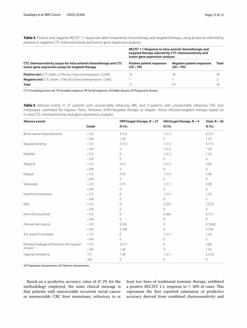
Based on a predictive accuracy value of 47.2% for the methodology employed, the main clinical message is that patients with unresectable recurrent rectal cancer or unresectable CRC liver metastases, refractory to at
least two lines of traditional systemic therapy, exhibited a positive RECIST 1.1. response in ≈ 50% of cases. This represents the first reported estimation of predictive accuracy derived from combined chemosensitivity and
Table 4 Positive and negative RECIST 1.1 responses after intraarterial chemotherapy and targeted therapy, using protocols selected by positive or negative CTC chemosensitivity and tumor gene expression analyses
CTC Circulating tumor cell, CR Complete response, PR Partial response, SD Stable disease, PD Progressive disease
RECIST 1.1 Response to intra‑arterial chemotherapy and targeted therapy selected by CTC chemosensitivity and tumor gene expression analyses
CTC chemosensitivity assays for intra‑arterial chemotherapy and CTC tumor gene expression assays for targeted therapy
Positive patient responses (CR + PR)
Negative patient responses (SD + PD)
Total
Positive test [CTC death ≥70% plus Gene overexpression ≥50%] 16 18 34
Negative test [CTC death < 70% plus Gene overexpression < 50%] 1 1 2
Total 17 19 36
Table 5 Adverse events in 27 patients with unresectable refractory RRC and 9 patients with unresectable refractory CRC liver metastases submitted for Hypoxic Pelvic Perfusion (HPP)/targeted therapy or Hepatic Artery Infusion/targeted therapy based on in vitro CTC chemosensitivity and gene expressions analyses
AST Aspartate transaminase, ALT Alanine transaminase
Adverse events HPP/target‑therapy, N = 27 HAI/target therapy, N = 9 Total, N = 36Grade N (%) N (%) N (%)
Bone marrow hypocellurarity −1/2 3 (11) 1 (11) 4 (11)
−3/4 1 (4) 0 1 (3)
Nausea/vomiting −1/2 3 (11) 1 (11) 4 (11)
−3/4 0 1 (11) 1 (3)
Diarrhea −1/2 0 1 (11) 1 (3)
−3/4 0 0 0
Alopecia −1/2 2 (7) 1 (11) 3 (8)
−3/4 0 0 0
Fatigue −1/2 2 (7) 1 (11) 3 (8)
−3/4 0 0 0
Neuropaty −1/2 2 (7) 1 (11) 3 (8)
−3/4 0 0 0
Hand Foot Syndrome −1/2 0 1 (11) 1 (3)
−3/4 0 0 0
Rash −1/2 0 2 (22) 2 (5.5)
−3/4 0 0 0
Non-infectious fever −1/2 0 4 (44) 4 (11)
−3/4 0 0 0
Perineal skin toxicity −1/2 6 (22) 0 6 (16.6)
−3/4 5 (18) 0 5 (14)
AST and ALT increased −1/2 0 1 (11) 1 (3)
−3/4 0 0 0
Persistent leakage of fluid from the inguinal incision
−1/2 3 (11) 0 3 (8)
−3/4 1 (4) 0 1 (3)
Inguinal hematoma -1/2 1 (4) 1 (11) 2 (5.5)
-3/4 0 0 0

Page 13 of 15Guadagni et al. BMC Cancer (2022) 22:660
tumor gene expression analysis of in vitro cultured CTCs and extends a recent meta-analysis reporting a predictive value of chemosensitivity assays alone for individualized CRC chemotherapy [40]. Furthermore, the combined positivity cut-off of ≥70% cell death for chemosensitiv-ity and ≥ 50% for CTC: PBMC tumor gene expression ratios, were selected considering the positive predictive value, which essentially focus on an optimized RECIST 1.1 response, maximizing predictive accuracy. Choosing pairs of lower cut-off values, potential oncotherapy preci-sion protocols should consider chemotherapeutic agents for which CTCs exhibit greater resistance and/or mono-clonal antibodies for less expressed CTC antigens.
The reported predictive accuracy value of 47.2%, is indeed impressive considering that the CTCs from 77.8% of patients exhibited over-expression of the multi-drug resistance gene MDR1 (≥ 65%), CTCs from 19.5% of patients exhibited significant over-expression of ERCC1 and GST (> 15%), involved in platinum resistance [41] and CTCs from 22.8% of patients exhibited ≥5% over-expression of TYMS or DHFR, involved in 5-fluorouracil resistance [42, 43].
Another important message from this study is that transient in vitro culture of liquid biopsy-derived CTCs can provide sufficient cell numbers for screening antican-cer compounds including agents not normally prescribed for any particular tumor type, of relevance for “drug repurposing” [39]. Indeed, 72% of CTCs in the refractory CRC patient group exhibited sensitivity to Mitomycin C, 5.5% exhibited sensitivity to Alkeran and 2.7% exhibited sensitivity to Doxorubicin, agents that are not currently recognized as particularly active against CRC and have been reported to be ≈10 times more cytotoxic under hypoxic conditions [33]. In the present study, CTC che-mosensitivity assays were not performed under hypoxic conditions, suggesting that the cytotoxic potential of these agents could be further enhanced via intraarte-rial administration to improve access to hypoxic tumour regions or using therapeutic protocols that promote tis-sue hypoxia, such as hypoxic pelvic perfusion (HPP), in order to take therapeutic advantage of chronic or tran-sient tumour tissue hypoxia [44].
Limitations of this study, include: i) the small patient sample size, which in any case provided a confidence interval for predictive accuracy of 95% (CI 0.30–0.64); ii) the inclusion of data from CTC populations obtained from patients with recurrent rectal cancer and CRC liver metastases, mitigated somewhat by the need to compare CTCs disseminating from recurrent and overt metastatic sites; iii) transient in vitro CTC culture, used to obtain sufficient numbers for assay, which may have altered gene expression, chemosensitivity and reduced predictive accuracy, deemed necessary for reasons of
methodological homogeneity for this study, initiated 2007 and terminated in 2019, which also explains why novel miniaturized system-based methods were not used; iv) the use of methodology for the evaluation of apoptosis that may have underestimated total death by not measur-ing paraptosis, ferroptosis and/or necroptosis; and v) lack of novel recent methodological improvements (micro-fluidic based systems, anti-CK20 antibodies and EMT markers), also for reasons of methodological homogene-ity for the duration of this 2007–2019 retrospective study.
Despite these limitations and emphasizing combined treatment with intraarterial chemotherapy and systemic target therapy for advanced CRC patients, refractory to at least two lines of systemic therapy, this study provides interesting biostatistical information for multidiscipli-nary oncological teams for quantifying the predictive accuracy of the particular CTC-isolation/assay method-ology employed. We do not, however, exclude the pos-sibility that predictive accuracy could be improved by recent technological and biomolecular innovations (see above) nor does this study evaluate the relative impor-tance of intraarterial chemotherapy in determining tumor response.
ConclusionsThe biomolecular methodology utilised in this retro-spective study of patients with unresectable refractory RRC and unresectable refractory CRC liver metastases, provides a predictive accuracy of ≈ 50% for response to liquid biopsy precision oncology-selected intraarterial chemotherapy and targeted therapy. We envisage that this value will be improved by novel technologies and therapeutic agents, and by upgrading methodology to ensure purification of all potentially metastatic epithe-lial, quasi-mesenchymal and mesenchymal CTC sub-populations, to include physiologically relevant controls in addition to PBMCs, and by eventual methodological standardization. Despite the challenges in applying these procedures to wider populations in non-research set-tings, the encouraging result of this retrospective study, employing CTC purification procedures relevant to 2007–2019, sets the stage for potential improvements in predictive accuracy in subsequent clinical trials employ-ing current and future technological and methodological improvements.
AbbreviationsFDA: Food and Drug Administration; BRAF: v-Raf murine sarcoma viral onco-gene homolog B; CK-20: Cytokeratin-20; EGFR: Epithelial Growth Factor Recep-tor; EMT: Epithelial-Mesenchymal-Transition; CD45: Cluster of differentiation 45; EpCAM: Epithelial adhesion molecule; pan-CK: Pan-Cytokeratin; qRT-PCR: Reverse Transcriptase quantitative Polymerase Chain Reaction; SNAIL: Gene family of zinc-finger transcription factors; ZEB: Gene family of zinc finger tran-scription factors; TWIST: Twist gene family; TGFβ: Transforming growth factor

Page 14 of 15Guadagni et al. BMC Cancer (2022) 22:660
β; Wnt: Wingless and Int − 1 signaling pathway; CT: Computed tomography; MRI: Magnetic resonance imaging; PET: Positron emission tomography; CD31: Platelet endothelial cell adhesion molecule; DAPI: 4′,6-diamidino-2-phe-nylindole; EpCAM-FITC: Antibody against Epithelial cell adhesion molecule conjugated to fluorescein isothioyanate; CD45-PE: Antibody against Cluster of differentiation 45 conjugated to phycoerythin; Pan-Cytokeratin-APC: Antibody against a panel of cytokeratins conjugated to allophycocyanin; ACTB: β actin; 18S rRNA: 18S ribosomal RNA; 5-FU: 5 fluorouracil; GAPDH: Glyceraldehyde 3-phosphate dehydrogenase; MMC: Mitomycin; IRI: Irinotecan; OX: Oxali-platin; RAL: Raltitrexed; DOX: Doxorubicin; ALK: Alkeran; CIS: Cisplatin; CAR : Carboplatin.
AcknowledgmentsNone.
Conflict of interestThe authors report no proprietary or commercial interest in any product mentioned or concept discussed in this article. None of the authors have any COI/ Financial Disclosure.
Authors’ contributionsS.G., F.M., A.R.M, P.A., I.P. wrote the main manuscript text. S.G., P.P., A.R.F., prepared figures and Tables. D.S., G.B., C.F., V.G. made data curation. P.A., P.P., made biomolecular analyses. G.S., M.C., G.F., E.R., G.B. made clinical treatments. M.V. supervised statistical analysis. All authors reviewed the manuscript. The author(s) read and approved the final manuscript.
FundingThis research received no external funding.
Availability of data and materialsThe datasets generated and/or analysed during the current study are not pub-licly available due to privacy reasons but are available from the corresponding author on reasonable request.
Declarations
Ethics approval and consent to participateThis study was conducted according to the guidelines of the Declaration of Helsinki, and was approved by the Ethics Committee of the “Azienda Sanitaria Locale” (ASL) n.1 of the “Regione Abruzzo”, Italy (Chairperson: G. Piccioli; proto-col number 10/CE/2018; approved: 19 July 2018 (n.1419)].Members of ASL n.1 Ethics committee
Gianlorenzo PICCIOLI Chairperson; Magistrate
Claudio FERRI Clinician
Marco Valenti Biostatistic
Domenico PARISE Substitute of Hospital Health Director
Maurizio PAOLONI Clinician
Marco CARMIGNANI Pharmacologist
Elvira D’ALESSANDRO Geneticist
Goffredo DEL ROSSO Clinician
Mario DI PIETRO Pediatrician
Antonio BARILE Radiologist
Roberto BERRETTONI Medical Devices
Ester LIBERATORE Hospital Pharmacy
Patrizia MASCIOVECCHIO Coroner
Fabrizio ANDREASSI Clinical Engineering
Carmine ORLANDI Nutritionist
Luciano LIPPA Clinician
Giovanni MUTTILLI Nurse
Eleonora CORONA Patient Organization
Carlo DI STANISLAO Clinical Secretary
Marivera DE ROSA Political Secretary
Informed consent was obtained from all subjects in the study.
Consent for publicationNot applicable.
Competing interestsAll authors declare that they have no competing interests.
Author details1 Department of Applied Clinical and Biotechnological Sciences, University of L’Aquila, 67100 L’Aquila, Italy. 2 Department of Oncology and Hematology, Azienda Ospedaliera “Ospedali Riuniti Marche Nord”, Pesaro, Italy. 3 Department of Prevention and Sports Medicine, University Hospital Klinikum rechts der Isar, Technical University of Munich, Munich, Germany. 4 Department of Physiol-ogy and Pharmacology, Cumming School of Medicine, University of Calgary, Calgary, AB, Canada. 5 Research Genetic Cancer Centre S.A, Florina, Greece. 6 Research Genetic Cancer Centre International GmbH, Zug, Switzerland.
Received: 7 February 2022 Accepted: 8 June 2022
References 1. Sargent DJ, Conley BA, Allegra C, Collette L. Clinical trial designs for
predictive marker validation in cancer treatment trials. J Clin Oncol. 2005;23:2020–7.
2. McShane LM. Statistical challenges in the development and evaluation of marker-based clinical tests. BMC Med. 2012;10:52.
3. Sepulveda AR, Hamilton SR, Allegra CJ, Grody W, Cushman-Vokoun AM, Funkhouseret WK, et al. Molecular biomarkers for the evaluation of colorectal cancer. Guideline from the American Society for Clinical Pathology, College of American Pathologists, Association for Molecular Pathology, and American Society of Clinical Oncology. Arch Pathol Lab Med. 2017;141:625–57.
4. Yoon YS, Kim JC. Recent applications of chemosensitivity tests for colo-rectal cancer treatment. World J Gastroenterol. 2014;20:16398–408.
5. Fernández-Lázaro D, García Hernández JL, García AC, Córdova Martínez A, Mielgo-Ayuso J, Cruz-Hernández JJ. Liquid biopsy as novel tool in preci-sion medicine: origins, properties, identification and clinical perspective of cancer’s biomarkers. Diagnostics. 2020;10:215.
6. Wong C-H, Chen Y-C. Clinical significance of exosomes as potential biomarkers in cancer. World J Clin Cases. 2019;7:171–90.
7. Karachaliou N, Mayo-de-Las-Casas C, Molina-Vila MA, Rosell R. Real-time liquid biopsies become a reality in cancer treatment. Ann Transl Med. 2015;3:36.
8. Ding Y, Li W, Wang K, Xu C, Hao M, Ding L. Perspectives of the applica-tion of liquid biopsy in colorectal Cancer. Biomed Res Int. 2020:6843180. https:// doi. org/ 10. 1155/ 2020/ 68431 80.
9. Welinder C, Jansson B, Lindell G, Wenner J. Cytokeratin 20 improves the detection of circulating tumor cells in patients with colorectal cancer. Cancer Lett. 2015;358:43–6. https:// doi. org/ 10. 1016/j. canlet. 2014. 12. 024.
10. Hendricks A, Brandt B, Geisen R, Dall K, Röder C, Schafmayer C, et al. Isola-tion and enumeration of CTC in colorectal Cancer patients: introduction of a novel cell imaging approach and comparison to cellular and molecu-lar detection techniques. Cancers. 2020;12:2643. https:// doi. org/ 10. 3390/ cance rs120 92643.
11. Vacante M, Ciuni R, Basile F, Biondi A. The liquid biopsy in the Manage-ment of Colorectal Cancer: an overview. Biomedicines. 2020;8:308. https:// doi. org/ 10. 3390/ biome dicin es809 0308.
12. Papasotiriou I, Chatziioannou M, Pessiou K, Retsas I, Dafouli G, Kyriazopou-lou A, et al. Detection of circulating tumor cells in patients with breast, prostate, pancreatic, Colon and Melanoma Cancer: a blinded compara-tive study using healthy donors. J C T. 2015;6:543–53. https:// doi. org/ 10. 4236/ jct. 2015. 67059.

Page 15 of 15Guadagni et al. BMC Cancer (2022) 22:660
13. Seeberg LT, Waage A, Brunborg C, Hugenschmidt H, Renolen A, Stav I, et al. Circulating tumor cells in patients with colorectal liver metastasis predict impaired survival. Ann Surg. 2015;261:164–71.
14. Arrazubi V, Mata E, Antelo ML, Tarifa A, Herrera J, Zazpe C, et al. Circulating tumor cells in patients undergoing resection of colorectal cancer liver metastases. Clinical utility for long-term outcome: a prospective trial. Ann Surg Oncol. 2019;26:2805–11.
15. Wang L, Zhou S, Zhang W, Wang J, Wang M, Hu X, et al. Circulating tumor cells as an independent prognostic factor in advanced colorectal cancer: a retrospective study in 121 patients. Int J Color Dis. 2019;34:589–97.
16. Musella V, Pietrantonio F, Di Buduo E, Iacovelli R, Martinetti A, Sottotetti E, et al. Circulating tumor cells as a longitudinal biomarker in patients with advanced chemorefractory, RAS-BRAF wild-type colorectal cancer receiv-ing cetuximab or panitumumab. Int J Cancer. 2015;137:1467–74.
17. Guadagni S, Fiorentini G, De Simone M, Masedu F, Zoras O, Mackay AR, et al. Precision oncotherapy based on liquid biopsies in multidisciplinary treatment of unresectable recurrent rectal cancer: a retrospective cohort study. J Cancer Res Clin Oncol. 2020;146:205–19. https:// doi. org/ 10. 1007/ s00432- 019- 03046-3.
18. Guadagni S, Clementi M, Mackay AR, Ricevuto E, Fiorentini G, Sarti D, et al. Real-life multidisciplinary treatment for unresectable colorectal cancer liver metastases including hepatic artery infusion with chemo-filtration and liquid biopsy precision oncotherapy. Observational cohort study. J Cancer Res Clin Oncol. 2020;146:1273–90.
19. Bray F, Ferlay J, Soerjomataram I, Siegel RL, Torre LA, Jemal A. Global cancer statistics 2018: GLOBOCAN estimates of incidence and mor-tality worldwide for 36 cancers in 185 countries. CA Cancer J Clin. 2018;68:394–424.
20. Aliberti C, Carandina R, Sarti D, Mulazzani L, Pizzirani E, Guadagni S, et al. Chemoembolization adopting polyethylene glycol drug-eluting embolics loaded with doxorubicin for the treatment of hepatocellular carcinoma. Am J Roentgenol. 2017;209:430–4.
21. Bruera G, Cannita K, Di Giacomo D, Lamy A, Frébourg T, Sabourin JC, et al. Worse prognosis of KRAS c.35 G > a mutant metastatic colorectal cancer (MCRC) patients treated with intensive triplet chemotherapy plus bevaci-zumab (FIr-B/FOx). BMC Med. 2013;11:59.
22. Bruera G, Cannita K, Tessitore A, Russo A, Alesse E, Ficorella C, et al. The prevalent KRAS exon 2 c.35 G > a mutation in metastatic colorectal cancer patients: a biomarker of worse prognosis and potential benefit of bevacizumab-containing intensive regimens? Crit Rev Oncol Hematol. 2015;93:190–202.
23. Bruera G, Pepe F, Malapelle U, Pisapia P, Dal Mas A, Di Giacomo D, et al. KRAS, NRAS and BRAF mutations detected by next generation sequenc-ing, and differential clinical outcome in metastatic colorectal cancer (MCRC) patients treated with first line FIr-B/FOx adding bevacizumab (BEV) to triplet chemotherapy. Oncotarget. 2018;9:26279–90.
24. Morelli MF, Santomaggio A, Ricevuto E, Cannita K, De Galitiis F, Tudini M, et al. Triplet schedule of weekly 5-fluorouracil and alternating irinotecan or oxaliplatin in advanced colorectal Cancer: a dose-finding and phase II study. Oncol Rep. 2010;23:1635–40.
25. Bruera G, Santomaggio A, Cannita K, Lanfiuti Baldi P, Tudini M, De Galitiis F, et al. “Poker” association of weekly alternating 5-fluorouracil, Irinotecan, bevacizumab and Oxaliplatin (FIr-B/FOx) in first line treatment of meta-static colorectal cancer: a phase II study. BMC Cancer. 2010;10:567.
26. Bruera G, Ricevuto E. Intensive chemotherapy of metastatic colorectal cancer: weighing between safety and clinical efficacy. Evaluation of Masi G, Loupakis F, Salvatore L, et al. Bevaizumab with FOLFOXIRI (irinotecan, oxaliplatin, fluorouracil, and folinate) as first-line treatment for metastatic colorectal cancer: a phase 2 trial. Lancet Oncol 2010; 11:845-52. Expert Opin Biol Ther. 2011;11:821–4.
27. Bruera G, Cannita K, Giuliante F, Lanfiuti Baldi P, Vicentini R, Marchetti P, et al. Effectiveness of liver metastasectomies in metastatic colorectal Can-cer (MCRC) patients treated with triplet chemotherapy plus bevacizumab (FIr-B/FOx). Clin Colorectal Cancer. 2012;11:119–26.
28. Sartore-Bianchi A, Pietrantonio F, Lonardi S, Mussolin B, Rua F, Fenoc-chio E, et al. Phase II study of anti-EGFR rechallenge therapy with panitumumab driven by circulating tumor DNA molecular selection in metastatic colorectal cancer: The CHRONOS trial (abstract). J Clin Oncol. 2021;39(suppl 15) Abstract available online at https:// meeti nglib rary. asco. org/ record/ 195971/ abstr act (Accessed on 08 June 2021).
29. Apostolou P, Ntanovasilis DA, Papasotiriou I. Evaluation of a simple method for storage of blood samples that enables isolation of circulating tumor cells 96 h after sample collection. J Biol Res Thessalon. 2017;24:11.
30. Toloudi M, Ioannou E, Chatziioannou M, Apostolou P, Kiritsis C, Manta S, et al. Comparison of the growth curves of Cancer cells and Cancer stem cells. Curr Stem Cell Res Ther. 2014;9:112–6.
31. Apostolou P, Iliopoulos AC, Parsonidis P, Papasotiriou I. Gene expression profiling as a potential predictor between normal and cancer samples in gastrointestinal carcinoma. Oncotarget. 2019;10:3328–38.
32. Livak KJ, Schmittgen TD. Analysis of relative gene expression data using real- time quantitative PCR and the 22DDCT method. Methods. 2001;25:402–8.
33. Teicher BA, Lazo JS, Sartorelli AC. Classification of antineoplastic agents by their selective toxicities toward oxygenated and hypoxic tumor cells. Cancer Res. 1981;41:73–81.
34. Guadagni S, Fiorentini G, Clementi M, Palumbo P, Mambrini A, Masedu F. Mitomycin C hypoxic pelvic perfusion for unresectable recurrent rectal cancer: pharmacokinetic comparison of surgical and percutane-ous techniques. Updat Surg. 2017;69:403–10. https:// doi. org/ 10. 1007/ s13304- 017- 0480-6.
35. Mambrini A, Bassi C, Pacetti P, Torri T, Iacono C, Ballardini M, et al. Prognos-tic factors in patients with advanced pancreatic adenocarcinoma treated with intra-arterial chemotherapy. Pancreas. 2008;36:56–60.
36. Eisenhauer EA, Therasse P, Bogaerts J, Schwartz LH, Sargent D, Ford R, et al. New response evaluation criteria in solid tumors: revised RECIST guideline (version 1.1). Eur J Cancer. 2009;45:228–47.
37. Guadagni S, Clementi M, Masedu F, Fiorentini G, Sarti D, Deraco M, et al. A pilot study of the predictive potential of chemosensitivity and gene expression assays using circulating tumour cells from patients with recur-rent ovarian cancer. Int J Mol Sci. 2020;21:4813. https:// doi. org/ 10. 3390/ ijms2 11348 13.
38. Guadagni S, Fiorentini G, Papasotiriou I, Apostolou P, Masedu F, Sarti D, et al. Circulating tumour cell liquid biopsy in selecting therapy for recur-rent cutaneous melanoma with locoregional pelvic metastases: a pilot study. BMC Res Notes. 2020;13:176.
39. Popova AA, Levkin PA. Precision Medicine in Oncology: In Vitro Drug Sensitivity and Resistance Test (DSRT) for Selection of Personalized Anticancer Therapy. Adv Therap. 2020;3:1900100. https:// doi. org/ 10. 1002/ adtp. 20190 0100.
40. Blom K, Nygren P, Larsson R, Andersson CR. Predictive value of ex vivo Chemosensitivity assays for individualized Cancer chemotherapy: a Meta-analysis. SLAS Technol. 2017;22:306–14.
41. Yu Y, Kanwar SS, Patel BB, Nautiyal J, Sarkar FH, Majumdar APN. Elimina-tion of Colon Cancer stem-like cells by the combination of curcumin and FOLFOX. Transl Oncol. 2009;2:321–8.
42. Jensen SA, Vainer B, Witton CJ, Jørgensen JT, Sørensen JB. Prognostic significance of numeric aberrations of genes for thymidylate synthase, thymidine phosphorylase and Dihydrofolate reductase in colorectal Cancer. Acta Oncol. 2008;47:1054–61.
43. Di Paolo A, Chu E. The role of thymidylate synthase as a molecular bio-marker. Clin Cancer Res. 2004;10:411–2.
44. Farina AR, Cappabianca L, Sebastiano M, Zelli V, Guadagni S, Mac-kay AR. Hypoxia-induced alternative splicing: the 11th Hallmark of Cancer. J Exp Clin Cancer Res. 2020;39:110. https:// doi. org/ 10. 1186/ s13046- 020- 01616-9.
Publisher’s NoteSpringer Nature remains neutral with regard to jurisdictional claims in pub-lished maps and institutional affiliations.


