Embed Size (px)
Citation preview

RESEARCH PAPERbph_1777 368..377
Maximizing tumourexposure to anti-neuropilin-1 antibodyrequires saturation ofnon-tumour tissueantigenic sinks in miceDaniela Bumbaca1, Hong Xiang1, C Andrew Boswell1, Ruediger E Port1,Shannon L Stainton2, Eduardo E Mundo1, Sheila Ulufatu2, Anil Bagri3,Frank-Peter Theil1, Paul J Fielder1, Leslie A Khawli1 and Ben-Quan Shen1
1Department of Pharmacokinetic and Pharmacodynamic Sciences, Genentech Research and Early
Development, South San Francisco, CA, USA, 2Department of Investigational Safety Assessment,
Genentech Research and Early Development, South San Francisco, CA, USA, and 3Department of
Molecular Biology, Genentech Research and Early Development, South San Francisco, CA, USA
CorrespondenceBen-Quan Shen, Department ofPharmacokinetic andPharmacodynamic Sciences,Genentech, Inc., 1 DNA Way,MS# 463A, South San Francisco,CA 94080, USA. E-mail:shen.ben@gene.com----------------------------------------------------------------
Keywordsneuropilin-1; antibody; doseescalation; biodistribution----------------------------------------------------------------
Received10 August 2011Revised6 October 2011Accepted21 October 2011
BACKGROUND AND PURPOSENeuropilin-1 (NRP1) is a VEGF receptor that is widely expressed in normal tissues and is involved in tumour angiogenesis.MNRP1685A is a rodent and primate cross-binding human monoclonal antibody against NRP1 that exhibits inhibition oftumour growth in NPR1-expressing preclinical models. However, widespread NRP1 expression in normal tissues may affectMNRP1685A tumour uptake. The objective of this study was to assess MNRP1685A biodistribution in tumour-bearing mice tounderstand the relationships between dose, non-tumour tissue uptake and tumour uptake.
EXPERIMENTAL APPROACHNon-tumour-bearing mice were given unlabelled MNRP1685A at 10 mg·kg-1. Tumour-bearing mice were given 111In-labelledMNRP1685A along with increasing amounts of unlabelled antibody. Blood and tissues were collected from all animals todetermine drug concentration (unlabelled) or radioactivity level (radiolabelled). Some animals were imaged using singlephoton emission computed tomography – X-ray computed tomography.
KEY RESULTSMNRP1685A displayed faster serum clearance than pertuzumab, indicating that target binding affected MNRP1685Aclearance. I.v. administration of 111In-labelled MNRP1685A to tumour-bearing mice yielded minimal radioactivity in the plasmaand tumour, but high levels in the lungs and liver. Co-administration of unlabelled MNRP1685A with the radiolabelledantibody was able to competitively block lungs and liver radioactivity uptake in a dose-dependent manner while augmentingplasma and tumour radioactivity levels.
CONCLUSIONS AND IMPLICATIONSThese results indicate that saturation of non-tumour tissue uptake is required in order to achieve tumour uptake andacceptable exposure to antibody. Utilization of a rodent and primate cross-binding antibody allows for translation of theseresults to clinical settings.
AbbreviationsAUC, area under the antibody serum concentration versus time curve; CL, clearance; CPM, counts per minute; CUBdomain, complement C1r/C1s, Uegf, and Bmf1 domain; DOTA, 1,4,7,10-tetraazacyclododecane-N,N′,N′′,N′′′-tetraaceticacid; ID, injected dose; MAM domain, Meprin, A5-protein, and protein-tyrosine phosphatase-Mu domain; MTD,maximum tolerated dose; NRP1, neuropilin-1; PK, pharmacokinetics; SPECT-CT, single photon emission computedtomography – X-ray computed tomography
BJP British Journal ofPharmacology
DOI:10.1111/j.1476-5381.2011.01777.xwww.brjpharmacol.org
368 British Journal of Pharmacology (2012) 166 368–377 © 2011 Genentech Inc.British Journal of Pharmacology © 2011 The British Pharmacological Society

IntroductionNeuropilin-1 (NRP1) is a 130–140 kDa transmembraneprotein with a prominent role in regulating angiogenesis(Bagri et al., 2009) and contains three structural motifs: (i) anextracellular structure containing two CUB homologydomains (a1, a2), two coagulation factor V/VIII homologydomains (b1, b2) and a MAM domain (c); (ii) a transmem-brane domain; and (iii) a short cytoplasmic domain (Sokeret al., 1998). NRP1 acts as a co-receptor for semaphorins andmembers of the VEGF family. NRP1 binds to VEGF165 toenhance its binding to VEGF receptor 2, increasing endothe-lial cell chemotaxis (Soker et al., 1998; Bielenberg et al., 2006).Mice with targeted NRP1 deletion have severe defects in vas-cular remodelling (Bagri et al., 2009). NRP1 is expressed byvascular endothelial and epithelial cells of numerous normaladult human tissues, including heart, placenta, breast,endometrium, kidneys, lungs, liver, pancreas and skeletalmuscle (Soker et al., 1998; Bielenberg et al., 2006). Over-expression of NRP1 occurs on some tumour cells, includingthose of prostate, mammary, bladder, kidney, colon, pancreas,skin, ovary and lung carcinomas (Bielenberg et al., 2006).
A rodent and primate cross-reactive human IgG1 withsimilar binding affinity across species was generated againstthe b1b2 domain, VEGF165 binding domain (Mamluk et al.,2002), of NRP1 that slowed tumour growth in xenograftmodels (Liang et al., 2007). This anti-NRP1 antibody,MNRP1685A, blocked tumour growth in HM7-tumours, amodel with very little tumour epithelial cell NRP1 expression,but with endothelial cell expression, suggesting thatMNRP1685A mediates its effect through blocking the angio-genesis necessary to support tumour growth (Liang et al.,2007). In addition, in select xenograft models, MNRP1685Atherapy coupled with concomitant anti-VEGF treatmentenhanced the effect of slowing tumour growth comparedwith that of anti-VEGF treatment alone (Pan et al., 2007),pointing to MNRP1685A’s promise as a potential anti-tumourbiotherapeutic agent.
Numerous monoclonal antibodies are being developedagainst various tumour targets with the goal of suppressingtumour growth (Sliwkowski et al., 1999; Herbst et al., 2001;Buchsbaum et al., 2003; Ferrara et al., 2004; Adams andWeiner, 2005). A thorough understanding of the pharmaco-kinetics (PK) and biodistribution of such molecules wouldinform their clinical development. Fast clearance or non-linear plasma PK profiles of antibody therapeutics can oftenbe attributed to target-mediated clearance (Lammerts vanBueren et al., 2006) and, in such cases, biodistribution datacan identify the tissue in which the target is located (Baxteret al., 1995; Lobo et al., 2004; Lammerts van Bueren et al.,2006). Tissue distribution studies can also reveal non-antigen-dependent, off-target binding, which may also influenceplasma PK (Jain, 1999; Bumbaca et al., 2011). Targets havingwidespread non-tumour tissue expression may affect tumouruptake of a target therapeutic and vice versa. Additionally,attributes of the antibody such as size, molecular weight andtarget affinity (Adams et al., 2001; Minchinton and Tannock,2006) as well as tumour physiology, including degree of vas-cularity, antigen density and necrosis (Jain, 1999; Minchin-ton and Tannock, 2006; Thurber et al., 2008; Tabrizi et al.,2010) can influence antibody distribution to and within the
tumour, so it is understandable that all tumour-associatedtarget antigens may not be accessible to the antibody. Theimplications of these limitations on therapeutic efficacywarrant a detailed understanding of the relationship betweenantibody PK, biodistribution and tumour uptake.
Although NRP1 has widespread distribution in normaladult tissues, little is known about the level of expression onthe surface of cells, and therefore, it was unclear how theNRP1 expression level in normal tissues would affectMNRP1685A biodistribution to tumour tissue, if at all. Thus,this work sought to evaluate the biodistribution ofMNRP1685A by exploring the relationship between dose,non-tumour tissue uptake and tumour uptake to support itsclinical development. First, MNRP1685A serum concentra-tions were determined in non-tumour-bearing mice and com-pared with those of pertuzumab, a humanized anti-HER2antibody that does not cross-react with murine HER2 antigenand therefore does not have target-mediated clearance inmice (Adams et al., 2006). Furthermore, pertuzumab andMNRP1685A use the same consensus frameworks of IgG1 andhave the same molecular weight (150 kDa), making pertu-zumab a suitable control. This comparison should ascertainwhat effect, if any, normal tissue NRP1 expression had onantibody PK. Subsequently, a biodistribution study was con-ducted in tumour-bearing mice, with dose escalation of unla-belled antibody and a fixed dose of radiolabelled antibody.This competitive binding approach should determine thedegree of saturation in each tissue at each dose, delineatingthe relationship between dose, non-tumour tissue uptake andtumour uptake. In addition, the results were complementedwith non-invasive live animal imaging and ex vivo assaysevaluating blood cell and tumour binding.
Methods
DOTA conjugation and 111In incorporationAliquots containing 5 mg of MNRP1685A (Genentech,South San Francisco, CA, USA) or an IgG1 control antibody(Genentech) were exchanged from their respective formula-tion buffers (proprietary) into aqueous 50 mM sodiumborate, pH 8.5 using illustra NAP5™ columns (GE Health-care Life Sciences, Piscataway, NJ, USA). Exactly 5 molarequivalents of the N-hydroxysuccinimidyl ester of 1,4,7,10-tetraazacyclododecane-N,N′,N′′,N′′′-tetraacetic acid (DOTA-NHS) in 0.68 mL of dimethyl formamide was added to600 mL borate-buffered protein solutions. Reaction mixtureswere gently agitated for 1 h at 37°C. The reaction was ter-minated by promptly applying the mixtures to NAP5™columns pre-equilibrated in aqueous 0.3 M ammoniumacetate buffer, pH 7.0.
111In was incorporated into DOTA through the addition ofa 3 mL (30.3 MBq) aliquot of 111InCl (MDS Nordion, Ottawa,ON, Canada) to a 17 mL aliquot of the ammonium acetate-buffered DOTA-MNRP1685A (59.4 mg) or DOTA-control anti-body conjugate (70.2 mg). Reaction mixtures were gentlyagitated for 1 h at 37°C. A 5 mL aliquot of 50 mM aqueousEDTA challenge solution was added, followed by an addi-tional 75 mL aliquot of aqueous 0.3 M ammonium acetatebuffer, pH 7.0. The mixture was applied to a NAP5 TM column,
BJPNon-tumour sinks impact MNRP1685A tumour uptake
British Journal of Pharmacology (2012) 166 368–377 369

from which the radiolabelled protein eluted in a 500-mL frac-tion of PBS. The specific activity of [111In]-DOTA-MNRP1685Aand [111In]-DOTA-control antibody were 0.177 and0.284 MBq·mg-1 respectively. The radiolabelled antibodieswere shown to maintain ~80% antigen binding to theirunmodified counterparts by ELISA and were shown to beintact by size-exclusion HPLC.
RadioiodinationMNRP1685A and control antibody were radioiodinated with125I using the indirect iodogen addition method as previouslydescribed (Chizzonite et al., 1991). The radiolabelled proteinswere purified using NAP5TM columns pre-equilibrated in PBS.The specific activities of [125I]-MNRP1685A and [125I]-controlantibody were 0.132 and 0.123 MBq·mg-1 respectively. Theradiolabelled antibodies were shown to maintain ~90%antigen binding to their unmodified counterparts by ELISA
and were shown to be intact by size-exclusion HPLC.
PK studyAll in vivo protocols, housing, and anaesthesia were approvedby the Institutional Animal Care and Use Committees ofGenentech Laboratory Animal Resources, in compliance withthe Association for Assessment and Accreditation of Labora-tory Animal Care regulations. Female athymic nude mice(nu/nu) (n = 3) within the age range of 6–8 weeks (CharlesRiver Laboratories, Hollister, CA, USA) were given 10 mg·kg-1
MNRP1685A i.v., as a bolus. Serum samples were collectedat 5 min; 1, 4 and 8 h; and 1, 1.5, 2, 3, 4, 7, 10, 14, 21,and 28 days post dose and analysed by ELISA for antibodyconcentration.
The concentration–time data were used to estimate PKparameters with WinNonlin Enterprise, Version 5.1.1 (Phar-sight Corporation, Mountain View, CA, USA). A naïve pooledapproach was used to provide one estimate for the treatmentgroup. Non-compartmental analysis (NCA) [WinNonlinModel 201; i.v. Bolus input using uniform weighting andlinear trapezoidal (linear interpolation) rule] was used to esti-mate area under the antibody serum concentration versustime curve (AUC). Clearance (CL) was calculated using thefollowing equation: CL = Dose/AUC. The PK results from thisstudy with MNRP1685A were compared with those obtainedfrom a separate study conducted with pertuzumab, as previ-ously described (Adams et al., 2006).
Ex vivo blood cell binding[125I]-MNRP1685A or [125I]-control antibody with or without100 mg·mL-1 excess unlabelled antibody was introduced intohuman whole blood (Bioreclamation LLC, Hicksville, NY,USA). Six 0.5 mL aliquots of blood were incubated for 1 h at37°C. Three of the six aliquots were centrifuged at 12,800¥ gfor 5 min at 4°C to separate the plasma from the cell pellets,after which the cell pellets were washed with 0.5 mL cold PBS.To re-calcify the blood, calcium chloride was added to theremaining three aliquots of whole blood at a final concentra-tion of 25 mM, immediately inverted and then further incu-bated at ambient temperature for 30 min until the blood hadcompletely clotted. The samples were centrifuged as above toseparate serum from the clots, which were washed once with0.5 mL cold PBS. All samples were counted for radioactivity
on a gamma counter (Wallac 1480 Wizard 3″, EC&G Wallac.Turku, Finland). The data are presented as the percent of totalradioactivity. Significance was assessed using a one-wayANOVA followed by the Tukey post test (GraphPad Prismv5.04, La Jolla, CA, USA).
In vivo biodistribution in tumourbearing miceFemale mice of the same strain and weight range as the PKstudy were inoculated s.c. with 5 x 10⁶ HM7 cells (humancolon carcinoma, ATCC, Manassas, VA, USA) suspended inHBSS in their right flanks 7 days before receiving the radio-labelled material. When the tumours were between 100 and200 mm3, the mice were co-dosed with 7.4 MBq·kg-1 [111In]-DOTA-MNRP1685A (0.05 mg·kg-1) along with unlabelledantibody at 0, 0.1, 0.3, 0.5, 1, 2.5, 5, 7.5, 10, 15, 20, 25, 40 or80 mg·kg-1 via i.v. bolus (multiple studies). At 15 min and 6,24, 48 and 120 h post dose, blood (processed for plasma),tumour, lungs, liver, kidneys and muscle (gastrocnemius)were collected (n = 4). The radioactivity level in each samplewas calculated and expressed as percentage of injected doseper gram or millilitre of sample (%ID·g-1 or %ID·mL-1). Sig-nificance was assessed using a one-way ANOVA followed by theTukey’s post test (GraphPad Prism). Plasma samples were alsoanalysed by size-exclusion HPLC.
Size-exclusion HPLC separation was carried out on a Phe-nomenex™ BioSep-SEC-S 3000, 300 ¥ 7.8 mm, 5 mm column(Torrance, CA). The mobile phase was PBS, and the flow ratewas 0.5 mL·min-1 (isocratic) for 30 min. The ChemStationanalogue-to-digital converter was set to 25 000 units per mV,peak width 2 s, slit 4 nM (Agilent Technologies, Santa Clara,CA, USA). [111In] was detected with a raytest Ramona 90(Raytest USA, Inc., Wilmington, NC, USA) in line with astandard Agilent 1100 HPLC module system. The data werecollected using ChemStation for LC 3D Systems (revisionB.01.03[204]).
Single photon emission computed tomography– X-ray computed tomography (SPECT-CT)Non-tumour-bearing mice of the same strain and weightrange as the biodistribution study received an i.v. bolus con-sisting of [111In]-DOTA-MNRP1685A (3.69 MBq) or [111In]-DOTA-control antibody (4.03 MBq) in 200 mL of saline (n =1). Initiation of non-invasive SPECT-CT (Gamma-Medica,Northridge, CA, USA) imaging was executed 24 h from thetime of tracer injection. Image acquisition times for CT andSPECT were an average of 2 and 45 min, respectively, withapproximately 20 000 counts per projection (20 s per projec-tion) for the latter. The SPECT and CT data was transferred to256 ¥ 256 matrix and fused using AMIRA® graphics software(TGS, San Diego, CA, USA), allowing the achievement ofsimultaneous scintigraphic and anatomic information in alltomographic scans in the three different spatial axes.
Ex vivo tumour bindingFemale mice of the same strain and weight range as thebiodistribution study were inoculated with HM7 cells asdescribed above. When the tumours were 100–200 mm3, themice received an i.v. bolus of unlabelled MNRP1685A at 0, 1, 2,5, 10, 20 and 40 mg·kg-1. Tumours were collected at 120 h post
BJP D Bumbaca et al.
370 British Journal of Pharmacology (2012) 166 368–377

dose (n = 3). Each tumour was placed inside a culture dish anddiced in 2 mL of RPMI-1640/10% FBS/10 mg·mL-1 non-specificIgG (media). The diced tumours were carefully transferred to4 mL cryovials. The culture dish was rinsed with 1 mL mediato remove any residual cells, and the rinse was combined withthe rest of the tumour pieces. Samples were gently agitated for30 min at room temperature to block non-specific binding.Tumour sections were washed once by centrifugation at 1000¥g for 3 min at 4°C and re-suspended in fresh media, containing[125I]-MNRP1685A at 1 ¥ 106 counts per minute (cpm)·mL-1.Following gentle agitation for 4 h at 4°C to allow for specificbinding of the radiolabelled antibody, the tumour sectionswere washed twice by centrifugation, saving both the mediaand wash. All fractions were counted for total radioactivity in
a gamma counter. The radioactivity level in each sample wascalculated and expressed as a percentage of total radioactivityper gram (%Total·g-1).
Results
PK in non-tumour-bearing miceFollowing 10 mg·kg-1 i.v. administration to athymic nudemice, MNRP1685A cleared much faster than pertuzumab(Figure 1), an IgG1 antibody with no target-mediated clear-ance in mice. The clearance for MNRP1685A at 10 mg·kg-1
was about 70 mL·day-1·kg-1, whereas that for pertuzumab,over the dose range 3–90 mg·kg-1 was 5.6–9.2 mL·day-1·kg-1
(Adams et al., 2006).
Ex vivo blood cell bindingThe plasma fraction of whole blood contained most of theradioactivity (~99%), for both MNRP1685A and the isotype-matched control antibody whether in the absence or pres-ence of excess unlabelled antibody (Table 1). In the wholeblood samples, processed for serum and blood clot, a majorityof the radioactivity (80–90%) was detected in the serum frac-tion for both antibodies, regardless of unlabelled antibodylevels, but was generally lower that was detected in theplasma. The difference in radioactivity between the plasmaand serum fractions is an artefact of the differing ability toseparate the plasma from the cell pellets and the serum fromthe blood clots. There was no statistical difference betweenMNRP1685A and control antibody plasma (P = 0.465 and0.308 for the tracer and tracer + unlabelled antibody, respec-tively) and serum radioactivity levels (P = 0.189 and 0.262 forthe tracer and tracer + unlabelled antibody, respectively).These data demonstrate minimal or no binding to blood cells.
Biodistribution in tumour-bearing miceThe tumour-bearing mice received tracer amounts of [111In]-DOTA-MNRP1685A along with 0, 5, 10, 20, 40 and 80 mg·kg-1
Figure 1Serum PK of MNRP1685A and pertuzumab. Serum concentrationversus time profile of MNRP1685A following i.v. administration at10 mg·kg-1 in athymic nude mice. Pertuzumab was administered at30 mg·kg-1 in the same mouse strain and the serum concentrationversus time profile presented in the figure was normalized to a10 mg·kg-1 dose based on its linear PK profile (Adams et al., 2006).
Table 1Percent recovery of [125I]-MNRP1685A or [125I]-control IgG1 � unlabelled antibody following incubation in human whole blood (average � SD)
Matrix Antibody [125I]-antibody
[125I]-antibody +100 mg·mL-1 unlabelledantibody
Plasma MNRP1685A 99.1 � 0.200 98.9 � 0.200
Control IgG1 99.9 � 0.0360 99.9 � 0.220
Cell pellet MNRP1685A 0.900 � 0.200 1.10 � 0.200
Control IgG1 0.0853 � 0.0360 0.0949 � 0.0220
Serum MNRP1685A 86.5 � 6.30 83.5 � 7.60
Control IgG1 92.6 � 2.03 90.1 � 4.30
Blood clot MNRP1685A 13.5 � 6.30 16.5 � 7.60
Control IgG1 7.43 � 2.03 9.92 � 4.30
There were no statistically significant differences between the MNRP1685A radioactivity levels and those of the control antibody or betweenthe tracer radioactivity levels and those of the tracer + unlabelled antibody (P > 0.05).
BJPNon-tumour sinks impact MNRP1685A tumour uptake
British Journal of Pharmacology (2012) 166 368–377 371
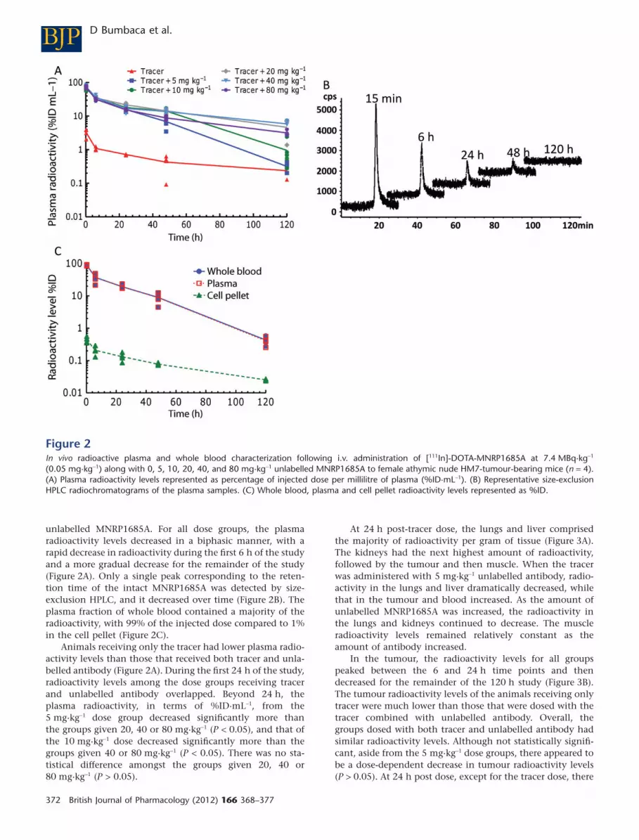
unlabelled MNRP1685A. For all dose groups, the plasmaradioactivity levels decreased in a biphasic manner, with arapid decrease in radioactivity during the first 6 h of the studyand a more gradual decrease for the remainder of the study(Figure 2A). Only a single peak corresponding to the reten-tion time of the intact MNRP1685A was detected by size-exclusion HPLC, and it decreased over time (Figure 2B). Theplasma fraction of whole blood contained a majority of theradioactivity, with 99% of the injected dose compared to 1%in the cell pellet (Figure 2C).
Animals receiving only the tracer had lower plasma radio-activity levels than those that received both tracer and unla-belled antibody (Figure 2A). During the first 24 h of the study,radioactivity levels among the dose groups receiving tracerand unlabelled antibody overlapped. Beyond 24 h, theplasma radioactivity, in terms of %ID·mL-1, from the5 mg·kg-1 dose group decreased significantly more thanthe groups given 20, 40 or 80 mg·kg-1 (P < 0.05), and that ofthe 10 mg·kg-1 dose decreased significantly more than thegroups given 40 or 80 mg·kg-1 (P < 0.05). There was no sta-tistical difference amongst the groups given 20, 40 or80 mg·kg-1 (P > 0.05).
At 24 h post-tracer dose, the lungs and liver comprisedthe majority of radioactivity per gram of tissue (Figure 3A).The kidneys had the next highest amount of radioactivity,followed by the tumour and then muscle. When the tracerwas administered with 5 mg·kg-1 unlabelled antibody, radio-activity in the lungs and liver dramatically decreased, whilethat in the tumour and blood increased. As the amount ofunlabelled MNRP1685A was increased, the radioactivity inthe lungs and kidneys continued to decrease. The muscleradioactivity levels remained relatively constant as theamount of antibody increased.
In the tumour, the radioactivity levels for all groupspeaked between the 6 and 24 h time points and thendecreased for the remainder of the 120 h study (Figure 3B).The tumour radioactivity levels of the animals receiving onlytracer were much lower than those that were dosed with thetracer combined with unlabelled antibody. Overall, thegroups dosed with both tracer and unlabelled antibody hadsimilar radioactivity levels. Although not statistically signifi-cant, aside from the 5 mg·kg-1 dose groups, there appeared tobe a dose-dependent decrease in tumour radioactivity levels(P > 0.05). At 24 h post dose, except for the tracer dose, there
Figure 2In vivo radioactive plasma and whole blood characterization following i.v. administration of [111In]-DOTA-MNRP1685A at 7.4 MBq·kg-1
(0.05 mg·kg-1) along with 0, 5, 10, 20, 40, and 80 mg·kg-1 unlabelled MNRP1685A to female athymic nude HM7-tumour-bearing mice (n = 4).(A) Plasma radioactivity levels represented as percentage of injected dose per millilitre of plasma (%ID·mL-1). (B) Representative size-exclusionHPLC radiochromatograms of the plasma samples. (C) Whole blood, plasma and cell pellet radioactivity levels represented as %ID.
BJP D Bumbaca et al.
372 British Journal of Pharmacology (2012) 166 368–377

was no difference in tumour : plasma ratios between the dosegroups (P > 0.05; Figure 3C). The dose groups higher than5 mg·kg-1 had similar tumour : plasma ratios at 48 h postdose (P > 0.05) and at the 120 h time point, there was nodifference among the 20, 40 and 80 mg·kg-1 dose groups(P > 0.05). In a separate study, when the radiolabelled anti-body was co-dosed with 0, 0.1, 0.3, 0.5, 1, 2.5, 5, 7.5, 10, 15and 25 mg·kg-1 unlabelled antibody, at 24 h post dose, adose-dependent increase in radioactivity was observed inthe tumours up to about 2.5–5 mg·kg-1, after which theradioactivity appeared to reach a plateau (Figure 3D). Thetumour : plasma ratios also increased with dose before reach-ing a plateau starting with the 2.5 mg·kg-1 dose.
SPECT-CT imagingQualitatively, the radioactivity in the [111In]-DOTA-MNRP1685A dosed mouse was more localized in the lungs
and liver, while that of the [111In]-DOTA-control antibody-dosed mouse was more diffuse throughout the animal, con-sistent with presence in the blood pool (Figure 4).
Ex vivo tumour bindingIn general, at the 120 h time point, the radioactivity associ-ated with the tumours decreased as the amount of unlabelledantibody dose increased, with tumour binding reaching aplateau at the 10 mg·kg-1 dose (Figure 5). This indicates thatsaturation of antibody uptake in the tumour was reachedwith a minimal dose of 10 mg kg-1.
Discussion and conclusions
This work assessed the biodistribution of MNRP1685A tounderstand the relationship between dose, non-tumour
70A
C
B
D
5030
25
TracerTracer + 5 mg kg–1
Tracer + 10 mg kg–1
Tracer + 20 mg kg–1
Tracer + 40 mg kg–1
Tracer + 80 mg kg–1
20
15
Tis
sue r
adio
activity (
%ID
g–1)
Tum
our
radio
activity (
%ID
g–1)
Radio
activity level (%
IDg
–1)
Tum
our
pla
sm
a r
atio
10
5
0Wholeblood
Plasma Cellpellet
Tumor Lungs Liver Kidneys Muscle 0 20 40 60
Time (h)
Tumour
Plasma
80 100 120
0
2
24 h 48 h 120 h
4
6
15
10
5
0
25
20
15
10
5
025 3020151050
TracerTracer + 5 mg kg–1
MNRP 1685A dose (mg kg–1)
Tracer + 10 mg kg–1
Tracer + 20 mg kg–1
Tracer + 40 mg kg–1
Tracer + 80 mg kg–1
Tracer
Tracer + 5 mg kg–1
Tracer + 10 mg kg–1
Tracer + 20 mg kg–1
Tracer + 40 mg kg–1
Tracer + 80 mg kg–1
Figure 3In vivo tissue radioactivity levels and tumour : plasma ratios following i.v. administration of [111In]-DOTA-MNRP1685A at 7.4 MBq·kg-1
(0.05 mg·kg-1) along with 0, 5, 10, 20, 40 and 80 mg·kg-1 unlabelled MNRP1685A to female athymic nude HM7 tumour-bearing mice (n = 4).(A) Tissue radioactivity levels at 24 h post dose, represented as percentage of injected dose per gram of tissue (%ID·g-1). (B) Tumour radioactivitylevels over time represented as %ID·g-1. (C) Average tumour : plasma radioactivity levels over time. (D) Plasma and tumour radioactivity levelsfollowing an i.v. administration of [111In]-DOTA-MNRP1685A at 7.4 MBq·kg-1 (0.05 mg·kg-1) along with 0, 0.1, 0.3, 0.5, 1, 2.5, 5, 7.5, 10, 15 and25 mg·kg-1 unlabelled MNRP1685A at 24 h post dose (n = 1), represented as %ID·g-1.
BJPNon-tumour sinks impact MNRP1685A tumour uptake
British Journal of Pharmacology (2012) 166 368–377 373

tissue uptake and tumour uptake. First, plasma concentra-tions of MNRP1685A were determined in non-tumour-bearing mice to ascertain the effect, if any, on antibody PK ofNRP1 expression in normal tissue. Then, MNRP1685A biodis-tribution was evaluated in tumour-bearing mice utilizing afixed radiolabelled antibody dose along with increasingamounts of unlabelled antibody. This competitive bindingapproach should determine the degree of saturation in eachtissue at each dose.
MNRP1685A displayed fast serum clearance in mice com-pared with that of pertuzumab (Figure 1), an antibodywithout target-mediated clearance in mice, suggesting thatwidespread NRP1 expression in non-tumour tissues was likelyto affect antibody clearance. Both an ex vivo blood cell
binding assay (Table 1) and in vivo whole blood fractionation(Figure 2C) indicated that there was little or no binding ofMNRP1685A to either human or mouse blood cells respec-tively. These data suggest that blood cell binding is notresponsible for the apparent fast clearance of the antibody.Furthermore, no evidence of antibody degradation, aggrega-tion or complex formation in plasma was detected by size-exclusion HPLC that could have affected the antibodyclearance rate (Figure 2B). However, target-mediated distribu-tion to the lungs, liver and kidneys was likely to contribute,in part, to the rapid plasma clearance of MNRP1685A(Figure 3A). At the tracer dose, 5- to 10-fold less control IgG1was detected in the liver and lungs, respectively, supportingthe specificity of MNRP1685A distribution to these tissues(Figure S1). SPECT-CT imaging also confirmed the specificlocalization of MNRP1685A in the lungs and liver, in contrastto the more diffuse distribution of the control antibody(Figure 4). Increasing the antibody dose from tracer to5 mg·kg-1 prompted a marked decrease in radioactivity in thelungs and liver (Figure 3A). A further decrease was noted inthe lungs and kidneys as the MNRP1685A dose increased. Astarget-mediated biodistribution did not account for the fastplasma clearance of the antibody at higher doses ofMNRP1685A, further investigation is warranted to determineother contributing factors. As NRP1 is a vascular targetexpressed in several adult non-tumour tissues (Bielenberget al., 2006), the lungs and liver appear to be acting as anti-genic sinks for MNRP1685A. Given that the lungs, with 235 �
111 mL blood per gram of tissue, and the liver, with 42.0 �
7.71 mL·g-1 (Boswell et al., 2010) are much more perfused rela-tive to the tumour (~20 mL·g-1; in-house data), MNRP1685Awould have more opportunity to bind to its target in thesewell perfused tissues, thereby decreasing observed levels inthe tumour. Alternatively, the NRP1 expression level in thesetissues may be higher than that of the tumour. Whatever theexplanation, a sufficiently high dose of MNRP1685A was
Figure 4SPECT-CT images of [111In]-DOTA-MNRP1685A (3.69 MBq; 25 mg)and [111In]-DOTA-control antibody dosed mice (4.03 MBq; 27 mg).Twenty-four-hour post-dose reconstructed three-dimensionalvolume rendered SPECT-CT fusion images of [111In]-DOTA-MNRP1685A (A) and [111In]-DOTA-control antibody (D) respectively(n = 1). False-coloured SPECT images in arbitrary uptake units arefused onto the X-ray CT images. The three-dimensional volumerendered SPECT image without the CT image for [111In]-DOTA-MNRP1685A and [111In]-DOTA-control antibody are shown in panelsB and E respectively. The two-dimensional coronal view of SPECT-CTfusion image for [111In]-DOTA-MNRP1685A and [111In]-DOTA-controlantibody are shown in panels C and F respectively.
Figure 5Radioactivity associated with the tumours following ex vivo incuba-tion with [125I]-MNRP1685A. HM7-tumour-bearing mice were giveni.v. 0, 1, 2, 5, 10, 20 or 40 mg·kg-1 unlabelled MNRP1685A 120-hbefore tumour harvest (n = 3). The radioactivity is represented aspercentage of total per gram of tissue (%Total·g-1).
BJP D Bumbaca et al.
374 British Journal of Pharmacology (2012) 166 368–377

required to satisfy the lungs and liver uptake, making theantibody available for tumour uptake. This phenomenon of anormal tissue antigenic sink has previously been observed innon-Hodgkin’s lymphoma patients in whom anti-CD20 anti-bodies cleared quickly from blood at low protein doses due tospecific splenic uptake (Kaminski et al., 1993; Press et al.,1993). Higher doses elevated circulating antibody levels,leading to enhanced tumour antibody concentrations. Thebenefit of utilizing a rodent and primate cross-reactive anti-body in our study allowed us to demonstrate this sink effectpreclinically and use this information to guide the clinicaldevelopment of MNRP1685A.
Although normal tissues were identified as antigenic sinksfor MNRP1685A, this antibody was well tolerated in preclini-cal toxicology studies conducted in rat and cynomolgusmonkeys, with no gross or microscopic pathology observedafter up to nine weekly doses at 100 mg·kg-1 (de Zafra et al.,2010). MNRP1685A was also well tolerated in a phase IAsingle-agent dose escalation study up to a 30 mg·kg-1 doseevery 3 weeks (Weekes et al., 2010). The most frequentlyreported drug-related adverse events were grade 1 and 2 acuteinfusion site reactions, which were controlled by premedica-tion with dexamethasone.
The xenograft model used in this analysis, HM7cells, is amodel with very low NRP1 expression on tumour epithelialcells, but with vascular endothelial cell expression (Lianget al., 2007), and was chosen precisely for that characteristicas the desired mechanism of action of MNRP1685A is anti-angiogenesis. Initial studies with the antibody indicated thatat unlabelled antibody doses lower than 5 mg·kg-1, therewas a dose-dependent increase in tumour radioactivity(Figure 3D), and this increase was not simply due toincreased blood radioactivity levels but was due to specificbinding of the antibody in the tumour, as observed by thehigh tumour : plasma radioactivity ratios at these lowerdoses. The tumour : plasma ratio, a specificity ratio relatingspecific binding in the tumour to unbound antibody inplasma (Fujimori et al., 1989; 1990; Thurber et al., 2008), wascalculated with the purpose of comparing the relativetumour exposures at each dose. Conceptually, a plateau intumour : plasma ratio versus dose indicates that no addi-tional specific binding at higher doses occurred and there-fore no additional benefit of those higher doses (Fujimoriet al., 1989). Based on the results of the initial study, doses of5–80 mg·kg-1 were chosen to determine how long this expo-sure could be maintained as no statistically significant dif-ferences in tumour radioactivity levels were observed overtime (Figure 3B), probably due to contributions from bothreceptor-bound and unbound radiolabelled antibody in thetumour interstitium. At 24 h post dose, doses �5 mg·kg-1
had similar tumour : plasma ratios; however, doses �10 and�20 mg·kg-1 had similar tumour : plasma ratios at 48 and120 h post dose, respectively (Figure 3C), illustrating thatdifferent doses maximize specific binding for differentlengths of time. Avoiding a change in MNRP1685Atumour : plasma ratio for a given duration requires a dosethat not only satisfies uptake in non-tumour antigenic tissuesinks but also overcomes antibody clearance in the tumourduring that time period. The 20 mg·kg-1 dose appeared to bethe lowest dose that maximized tumour exposure for the5 day study.
In order to confirm the effect of dose on tumour exposureobserved in vivo, an ex vivo tumour binding assay wasemployed to assess the binding of radiolabelled antibody tounoccupied receptors in the tumour ex vivo, thereby remov-ing the limitations present in the in vivo system. Such limi-tations in the tumour are the degree of vascularity,permeability of the vessel walls, heterogeneity of blood flowrate, antigen density, interstitial fluid pressure and necrosis(Jain, 1999; Minchinton and Tannock, 2006; Thurber et al.,2008; Tabrizi et al., 2010). Additionally, antibody targetbinding affinity, molecular charge, molecular weight and PKcould further limit penetration of the tumour by the anti-body (Fujimori et al., 1989; 1990; Adams et al., 2001;Minchinton and Tannock, 2006; Dennis et al., 2007). Therewas a dose-dependent decrease in radioactivity in the assay(Figure 5) with maximum exposure reached with the10 mg·kg-1 dose, indicating that there were fewer unoccupiedreceptors at the higher doses. This is consistent with the trendobserved in vivo. The fact that maximum binding wasobserved at the 10 mg·kg-1 dose rather than the 20 mg·kg-1
dose is most likely an artefact of the tumour processing,potentially making more receptors available ex vivo than theantibody can reach in vivo.
The characterization of the relationship between tumouruptake and MNRP1685A dose provided additional informa-tion for clinical development. It is well established in the fieldof oncology therapeutics that finding a balance between effi-cacy and toxicity is crucial for selecting the optimal phase IIclinical dose and regimen (Wolf et al., 2004; Jain et al., 2010;Rubin and Anderson, 2010). Traditionally, the phase Imaximum tolerated dose (MTD) determines the phase II dose(Jain et al., 2010; Rubin and Anderson, 2010), but this maynot necessarily be the best dose. It is possible, especially fortargeted therapeutics, that a dose lower than the MTD resultsin the maximum biological effect, and so dosing at the MTDserves to increase toxicity while not improving efficacy (Wolfet al., 2004; Jain et al., 2010; Rubin and Anderson, 2010).Additionally, it has been suggested that phase II doses shouldbe based on the minimum effective dose (Haines, 2008) oroptimum biological dose (Wolf et al., 2004). Theoretically,the dose that maintained saturating levels in the tumour forthe desired dosing interval should be the highest dose thatwould need to be administered, assuming equivalent efficacyat all doses. However, there are challenges in using preclinicaldata to help predict clinical success, such as potential differ-ences among animals and humans in terms of antibody dis-position, tumour growth and/or physiology (Morgan, 2001;Kerbel, 2003; Bleeker et al., 2008). Nevertheless, some positivecorrelations have been found between preclinical efficacy inxenografts and clinical efficacy in human tumours (Bovenet al., 1992; Johnson et al., 2001; Kerbel, 2003; Peterson andHoughton, 2004). As such, understanding antibody absorp-tion, distribution and clearance in a preclinical model aids inpredicting the optimal dose and regimen for patients(Morgan, 2001; Bleeker et al., 2008). For instance, Luo andcolleagues showed that mouse plasma concentrations ofcetuximab were fairly comparable with the concentrations inpatients at the dose they identified to be the highest neededto achieve maximum efficacy (Luo et al., 2005). The data fromour study was translated into a clinically relevant dose bydetermining the trough plasma concentration at 5 days and
BJPNon-tumour sinks impact MNRP1685A tumour uptake
British Journal of Pharmacology (2012) 166 368–377 375

identifying the dose and regimen from the phase I clinicaltrial that maintains the plasma concentrations (based oninter-species scaling) at or above this level (11 mg·kg-1 everyother week or 18 mg·kg-1 every 3 weeks). These results werecorroborated with other preclinical and clinical data (datanot shown).
In conclusion, this work has demonstrated that wide-spread expression of NRP1 in non-tumour tissue did limitdistribution of MNRP1685A to tumour tissue, in a dose-dependent manner, because of the influence of non-tumourtissue antigenic sinks. The use of a rodent and primate cross-binding antibody allowed for the translation of these findingsfrom the preclinical to clinical setting, indicating that ahigher than expected clinical dose would need to be admin-istered to achieve acceptable tumour exposure.
Acknowledgements
The authors would like to thank Michelle Schweiger, Eliza-beth Torres, Mike Reiche, Amy Oldendorp, Misia Howell,Kirsten Messick and Noore Kadri for excellent animal studiessupport; Yanmei Lu for ELISA support; Amy Gilbert for helpwith the PK studies; and Yan Xin and Enzo Palma for helpfuldiscussion.
Conflicts of interest
All authors are employees of Genentech, Inc., a member ofthe Roche Group, or Roche, and hold financial interest inRoche.
ReferencesAdams CW, Allison DE, Flagella K, Presta L, Clarke J, Dybdal Net al. (2006). Humanization of a recombinant monoclonal antibodyto produce a therapeutic HER dimerization inhibitor, pertuzumab.Cancer Immunol Immunother 55: 717–727.
Adams GP, Weiner LM (2005). Monoclonal antibody therapy ofcancer. Nat Biotechnol 23: 1147–1157.
Adams GP, Schier R, McCall AM, Simmons HH, Horak EM,Alpaugh RK et al. (2001). High affinity restricts the localization andtumor penetration of single-chain fv antibody molecules. CancerRes 61: 4750–4755.
Bagri A, Tessier-Lavigne M, Watts RJ (2009). Neuropilins in tumorbiology. Clin Cancer Res 15: 1860–1864.
Baxter LT, Zhu H, Mackensen DG, Butler WF, Jain RK (1995).Biodistribution of monoclonal antibodies: scale-up from mouse tohuman using a physiologically based pharmacokinetic model.Cancer Res 55: 4611–4622.
Bielenberg DR, Pettaway CA, Takashima S, Klagsbrun M (2006).Neuropilins in neoplasms: expression, regulation, and function. ExpCell Res 312: 584–593.
Bleeker WK, Munk ME, Mackus WJ, van den Brakel JH, Pluyter M,Glennie MJ et al. (2008). Estimation of dose requirements forsustained in vivo activity of a therapeutic human anti-CD20antibody. Br J Haematol 140: 303–312.
Boswell CA, Ferl GZ, Mundo EE, Schweiger MG, Marik J, Reich MPet al. (2010). Development and evaluation of a novel method forpreclinical measurement of tissue vascular volume. Mol Pharm 7:1848–1857.
Boven E, Winograd B, Berger DP, Dumont P, Braakhuis B,Fodstad O et al. (1992). Phase II preclinical drug screening inhuman xenografts: a first European multicenter collaborative study.Cancer Res 52: 5940–5947.
Buchsbaum DJ, Zhou T, Grizzle WE, Oliver PG, Hammond CJ,Zhang S et al. (2003). Antitumor efficacy of TRA-8 anti-DR5monoclonal antibody alone or in combination with chemotherapyand/or radiation therapy in a human breast cancer model. ClinCancer Res 9 (10 Pt 1): 3731–3741.
Bumbaca D, Wong A, Drake E, Reyes AE 2nd, Lin BC, Stephan JPet al. (2011). Highly specific off-target binding identified andeliminated during the humanization of an antibody against FGFreceptor 4. MAbs 3: 376–386.
Chizzonite R, Truitt T, Podlaski FJ, Wolitzky AG, Quinn PM,Nunes P et al. (1991). IL-12: monoclonal antibodies specific for the40-kDa subunit block receptor binding and biologic activity onactivated human lymphoblasts. J Immunol 147: 1548–1556.
Dennis MS, Jin H, Dugger D, Yang R, McFarland L, Ogasawara Aet al. (2007). Imaging tumors with an albumin-binding Fab, a noveltumor-targeting agent. Cancer Res 67: 254–261.
Ferrara N, Hillan KJ, Gerber HP, Novotny W (2004). Discovery anddevelopment of bevacizumab, an anti-VEGF antibody for treatingcancer. Nat Rev Drug Discov 3: 391–400.
Fujimori K, Covell DG, Fletcher JE, Weinstein JN (1989). Modelinganalysis of the global and microscopic distribution ofimmunoglobulin G, F(ab’)2, and Fab in tumors. Cancer Res 49:5656–5663.
Fujimori K, Covell DG, Fletcher JE, Weinstein JN (1990). Amodeling analysis of monoclonal antibody percolation throughtumors: a binding-site barrier. J Nucl Med 31: 1191–1198.
Haines IE (2008). Dose selection in phase I studies: why we shouldalways go for the most effective. J Clin Oncol 26: 3650–3652,author reply 3652–3653.
Herbst RS, Kim ES, Harari PM (2001). IMC-C225, an anti-epidermalgrowth factor receptor monoclonal antibody, for treatment of headand neck cancer. Expert Opin Biol Ther 1: 719–732.
Jain RK (1999). Transport of molecules, particles, and cells in solidtumors. Annu Rev Biomed Eng 1: 241–263.
Jain RK, Lee JJ, Hong D, Markman M, Gong J, Naing A et al. (2010).Phase I oncology studies: evidence that in the era of targetedtherapies patients on lower doses do not fare worse. Clin CancerRes 16: 1289–1297.
Johnson JI, Decker S, Zaharevitz D, Rubinstein LV, Venditti JM,Schepartz S et al. (2001). Relationships between drug activity in NCIpreclinical in vitro and in vivo models and early clinical trials. Br JCancer 84: 1424–1431.
Kaminski MS, Zasadny KR, Francis IR, Milik AW, Ross CW,Moon SD et al. (1993). Radioimmunotherapy of B-cell lymphomawith [131I]anti-B1 (anti-CD20) antibody. N Engl J Med 329:459–465.
Kerbel RS (2003). Human tumor xenografts as predictive preclinicalmodels for anticancer drug activity in humans: better thancommonly perceived-but they can be improved. Cancer Biol Ther 2(4 Suppl. 1): S134–S139.
BJP D Bumbaca et al.
376 British Journal of Pharmacology (2012) 166 368–377

Lammerts van Bueren JJ, Bleeker WK, Bogh HO, Houtkamp M,Schuurman J, van de Winkel JG et al. (2006). Effect of targetdynamics on pharmacokinetics of a novel therapeutic antibodyagainst the epidermal growth factor receptor: implications for themechanisms of action. Cancer Res 66: 7630–7638.
Liang WC, Dennis MS, Stawicki S, Chanthery Y, Pan Q, Chen Yet al. (2007). Function blocking antibodies to neuropilin-1generated from a designed human synthetic antibody phagelibrary. J Mol Biol 366: 815–829.
Lobo ED, Hansen RJ, Balthasar JP (2004). Antibodypharmacokinetics and pharmacodynamics. J Pharm Sci 93:2645–2668.
Luo FR, Yang Z, Dong H, Camuso A, McGlinchey K, Fager K et al.(2005). Correlation of pharmacokinetics with the antitumor activityof Cetuximab in nude mice bearing the GEO human coloncarcinoma xenograft. Cancer Chemother Pharmacol 56: 455–464.
Mamluk R, Gechtman Z, Kutcher ME, Gasiunas N, Gallagher J,Klagsbrun M (2002). Neuropilin-1 binds vascular endothelialgrowth factor 165, placenta growth factor-2, and heparin via itsb1b2 domain. J Biol Chem 277: 24818–24825.
Minchinton AI, Tannock IF (2006). Drug penetration in solidtumours. Nat Rev Cancer 6: 583–592.
Morgan P (2001). The use of preclinical pharmacokinetic andpharmacodynamic data to predict clinical doses: current and futureperspectives. Int Congr Ser 1220: 1–12.
Pan Q, Chanthery Y, Liang WC, Stawicki S, Mak J, Rathore N et al.(2007). Blocking neuropilin-1 function has an additive effect withanti-VEGF to inhibit tumor growth. Cancer Cell 11: 53–67.
Peterson JK, Houghton PJ (2004). Integrating pharmacology and invivo cancer models in preclinical and clinical drug development.Eur J Cancer 40: 837–844.
Press OW, Eary JF, Appelbaum FR, Martin PJ, Badger CC, Nelp WBet al. (1993). Radiolabeled-antibody therapy of B-cell lymphomawith autologous bone marrow support. N Engl J Med 329:1219–1224.
Rubin EH, Anderson KM (2010). Finding the right dose for cancertherapeutics–can we do better? Clin Cancer Res 16: 1085–1087.
Sliwkowski MX, Lofgren JA, Lewis GD, Hotaling TE, Fendly BM,Fox JA (1999). Nonclinical studies addressing the mechanism ofaction of trastuzumab (Herceptin). Semin Oncol 26 (4 Suppl. 12):60–70.
Soker S, Takashima S, Miao HQ, Neufeld G, Klagsbrun M (1998).Neuropilin-1 is expressed by endothelial and tumor cells as anisoform-specific receptor for vascular endothelial growth factor. Cell92: 735–745.
Tabrizi M, Bornstein GG, Suria H (2010). Biodistributionmechanisms of therapeutic monoclonal antibodies in health anddisease. AAPS J 12: 33–43.
Thurber GM, Schmidt MM, Wittrup KD (2008). Factors determiningantibody distribution in tumors. Trends Pharmacol Sci 29: 57–61.
Weekes CD, Hedge P, Xin Y, Yu R, Xiang H, Beeram M et al. (2010).A first-in-human phase I study to evaluate the fully humanmonoclonal antibody MNRP1685A (anti-NRP1) administeredintravenously every three weeks in patients with advanced solidtumors (Abstract). 2010 ASCO Annual Meeting, Chicago, IL. J ClinOncol 28: 15s, 2010 (Suppl.; abstr 3014).
Wolf M, Swaisland H, Averbuch S (2004). Development of thenovel biologically targeted anticancer agent gefitinib: determiningthe optimum dose for clinical efficacy. Clin Cancer Res 10:4607–4613.
de Zafra CLZ, Allamneni K, Kennedy D, Cain G, Xiang H,Auyeung-Kim DL et al. (2010). Nonclinical toxicology evaluation ofa monoclonal antibody against the VEGF co-receptor neuropilin-1(Abstract). 49th Annual Meeting of the Society of Toxicology; Salt LakeCity, UT. Toxicologist 2010.
Supporting information
Additional Supporting Information may be found in theonline version of this article:
Figure S1 Tissue distribution at 24 h post dose following i.v.administration of [111In]-DOTA-MNRP1685A or [111In]-DOTA-control IgG1 at 7.4 MBq·kg-1 (0.05 mg·kg-1) (n = 4). The radio-activity is represented as percentage of injected dose per gramof tissue (%ID·g-1).
Please note: Wiley-Blackwell are not responsible for thecontent or functionality of any supporting materials suppliedby the authors. Any queries (other than missing material)should be directed to the corresponding author for the article.
BJPNon-tumour sinks impact MNRP1685A tumour uptake
British Journal of Pharmacology (2012) 166 368–377 377


