Embed Size (px)
Citation preview
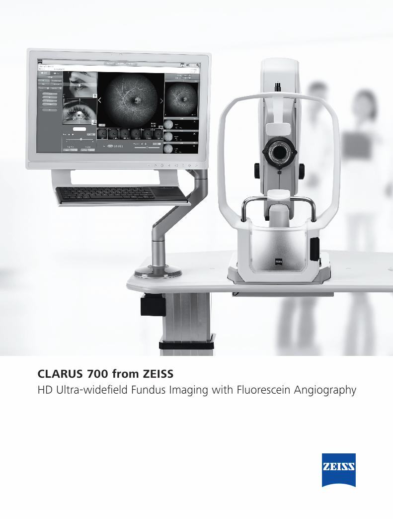
CLARUS 700 from ZEISSHD Ultra-widefield Fundus Imaging with Fluorescein Angiography
2
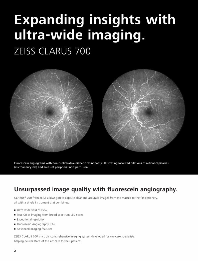
Fluorescein angiograms with non-proliferative diabetic retinopathy, illustrating localized dilations of retinal capillaries(microaneurysms) and areas of peripheral non-perfusion.
Expanding insights with ultra-wide imaging.ZEISS CLARUS 700
Unsurpassed image quality with fluorescein angiography.CLARUS® 700 from ZEISS allows you to capture clear and accurate images from the macula to the far periphery,
all with a single instrument that combines:
• Ultra-wide field of view
• True Color imaging from broad spectrum LED scans
• Exceptional resolution
• Fluorescein Angiography (FA)
• Advanced imaging features
ZEISS CLARUS 700 is a truly comprehensive imaging system developed for eye care specialists,
helping deliver state-of-the-art care to their patients.
3
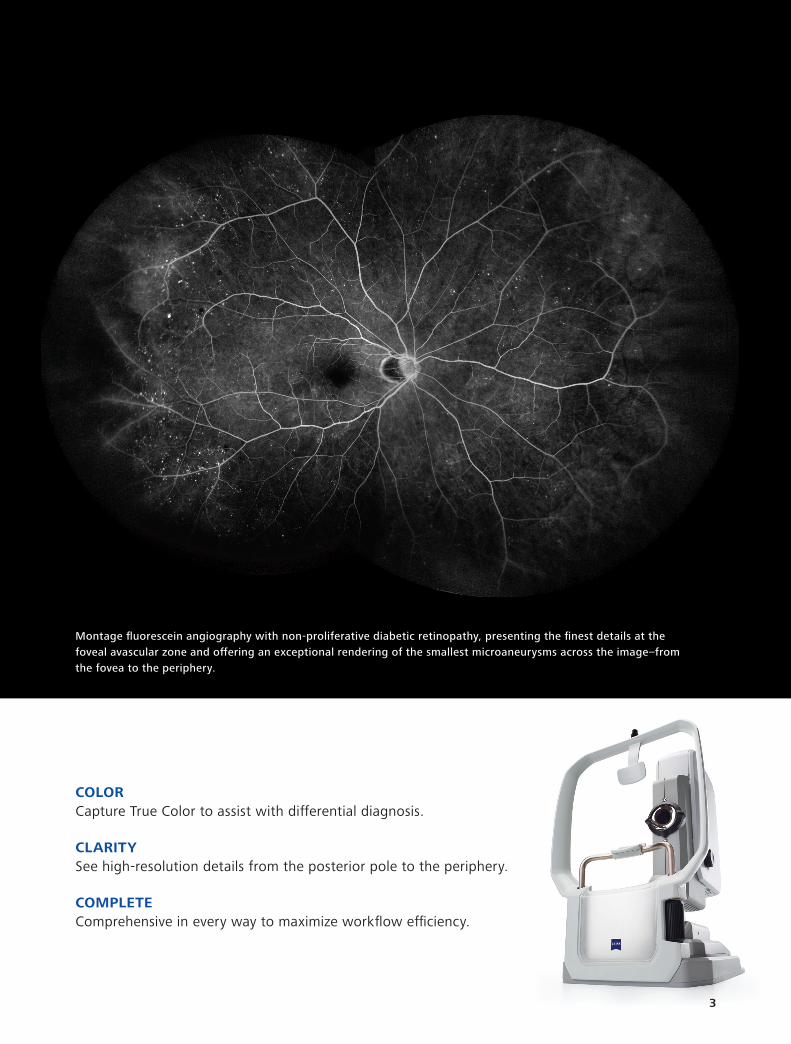
Montage fluorescein angiography with non-proliferative diabetic retinopathy, presenting the finest details at thefoveal avascular zone and offering an exceptional rendering of the smallest microaneurysms across the image–fromthe fovea to the periphery.
3
COLOR Capture True Color to assist with differential diagnosis.
CLARITY See high-resolution details from the posterior pole to the periphery.
COMPLETE Comprehensive in every way to maximize workflow efficiency.
A Comprehensive Imaging System
Now you can manage all fundus
imaging modalities without
compromising on clarity—viewing
high resolution in ultra-widefield.
• Image from the superior and inferior
retina with less peripheral distortion
• Capture clear detail of vessel
structure from early to late phase of
fluorescein angiography
• AutoBright control automatically
optimizes the angiogram series
preserving change in signal
Combining ultra-widefield imaging
with True Color, excellent clarity and
a full suite of imaging modalities,
ZEISS CLARUS 700 empowers you
with features and capabilities that
maximize workflow efficiency.
• Quickly and easily compare images
over time and between image
capture modes
• Provide a comfortable patient
experience that ensures image
integrity, with ergonomic chin and
head rests to swivel motion and live
IR preview
True Color Imaging
Powered by Broad Line Technology,
the ZEISS CLARUS 700 captures
images that closely resemble the
coloration of the fundus as seen
during clinical examination.
Unlike CSLO (confocal scanning laser), Broad Line Technology enables the combination of ultra-wide fields of view and a full range of retinal imaging modes to generate images with high dynamic range, contrast, resolution and natural colors through sequential illumination of broad-spectrum red, green and blue light emitting diodes.1
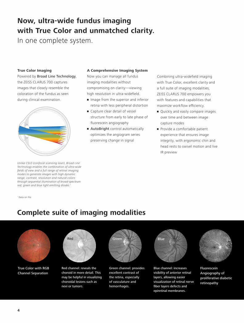
True Color with RGB Channel Separation
Complete suite of imaging modalities
Fluorescein Angiography of proliferative diabeticretinopathy
Red channel: reveals the choroid in more detail. This may be helpful in visualizing choroidal lesions such as nevi or tumors.
Green channel: provides excellent contrast of the retina, especially of vasculature and hemorrhages.
Blue channel: increases visibility of anterior retinal layers, allowing easier visualization of retinal nerve fiber layers defects and epiretinal membranes.
4
Red Green Blue
1 Data on file.
Now, ultra-wide fundus imaging with True Color and unmatched clarity. In one complete system.
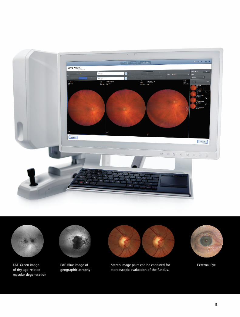
FAF-Green image of dry age-related macular degeneration
FAF-Blue image of geographic atrophy
External EyeStereo image pairs can be captured for stereoscopic evaluation of the fundus.
5
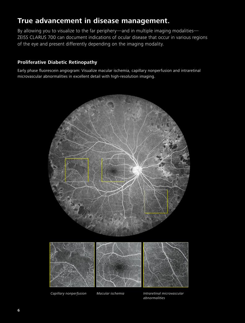
Proliferative Diabetic Retinopathy
True advancement in disease management.By allowing you to visualize to the far periphery—and in multiple imaging modalities— ZEISS CLARUS 700 can document indications of ocular disease that occur in various regions of the eye and present differently depending on the imaging modality.
6
Early phase fluorescein angiogram: Visualize macular ischemia, capillary nonperfusion and intraretinal microvascular abnormalities in excellent detail with high-resolution imaging.
Capillary nonperfusion Macular ischemia Intraretinal microvascular abnormalities
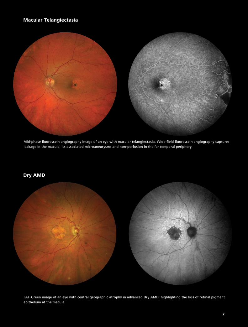
Mid-phase fluorescein angiography image of an eye with macular telangiectasia. Wide-field fluorescein angiography captures leakage in the macula, its associated microaneurysms and non-perfusion in the far temporal periphery.
FAF-Green image of an eye with central geographic atrophy in advanced Dry AMD, highlighting the loss of retinal pigment epithelium at the macula.
Macular Telangiectasia
Dry AMD
7
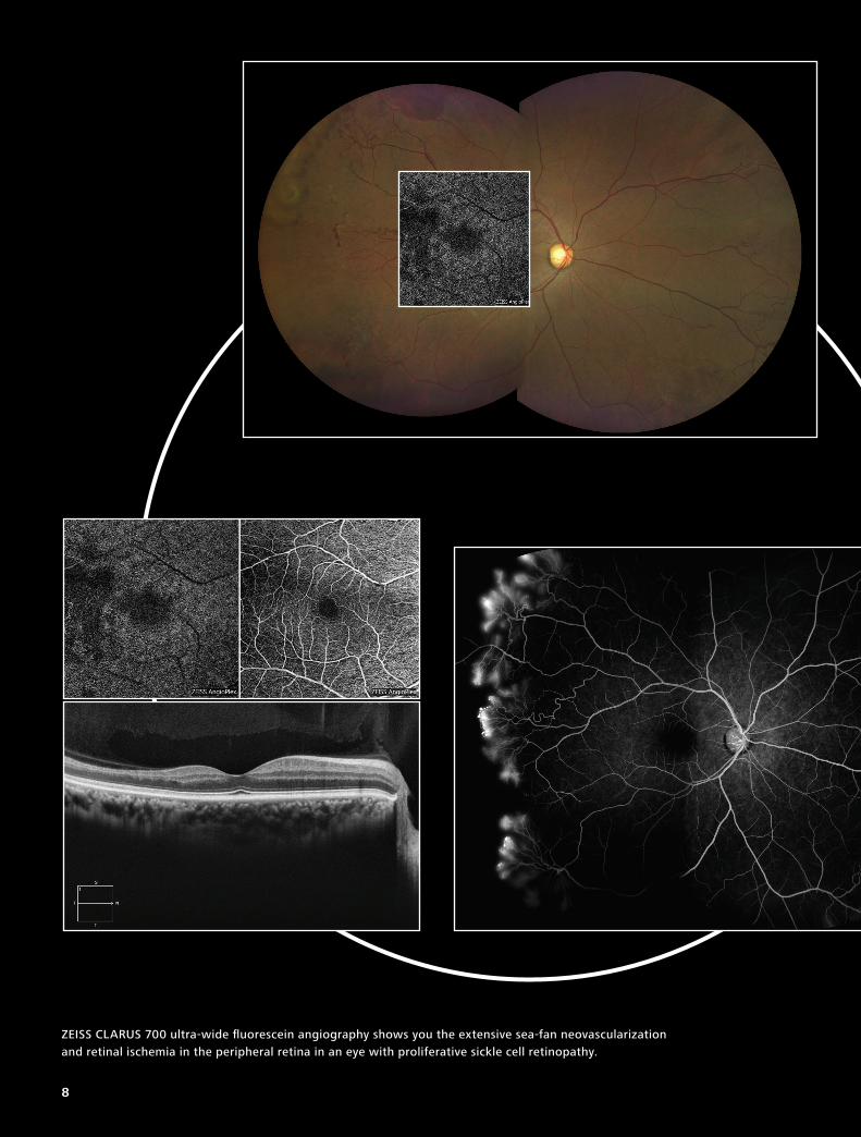
ZEISS CLARUS 700 ultra-wide fluorescein angiography shows you the extensive sea-fan neovascularization and retinal ischemia in the peripheral retina in an eye with proliferative sickle cell retinopathy.
8
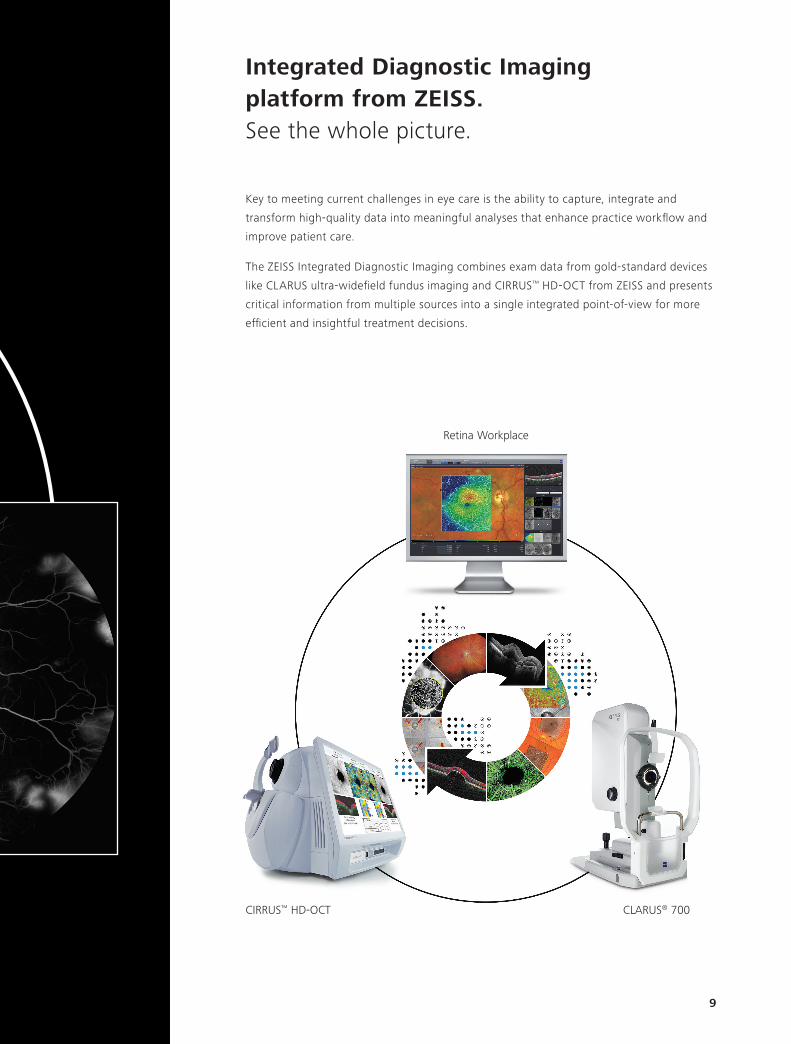
Integrated Diagnostic Imaging platform from ZEISS.See the whole picture.
Key to meeting current challenges in eye care is the ability to capture, integrate and
transform high-quality data into meaningful analyses that enhance practice workflow and
improve patient care.
The ZEISS Integrated Diagnostic Imaging combines exam data from gold-standard devices
like CLARUS ultra-widefield fundus imaging and CIRRUS™ HD-OCT from ZEISS and presents
critical information from multiple sources into a single integrated point-of-view for more
efficient and insightful treatment decisions.
Retina Workplace
CLARUS® 700CIRRUS™ HD-OCT
9
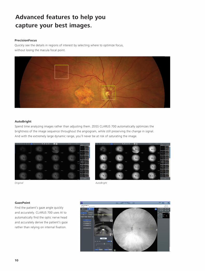
PrecisionFocus
Quickly see the details in regions of interest by selecting where to optimize focus,
without losing the macula focal point.
AutoBright
Spend time analyzing images rather than adjusting them. ZEISS CLARUS 700 automatically optimizes the
brightness of the image sequence throughout the angiogram, while still preserving the change in signal.
And with the extremely large dynamic range, you’ll never be at risk of saturating the image.
Advanced features to help you capture your best images.
10
Original AutoBright
GazePoint
Find the patient’s gaze angle quickly
and accurately. CLARUS 700 uses AI to
automatically find the optic nerve head
and accurately derive the patient’s gaze
rather than relying on internal fixation.
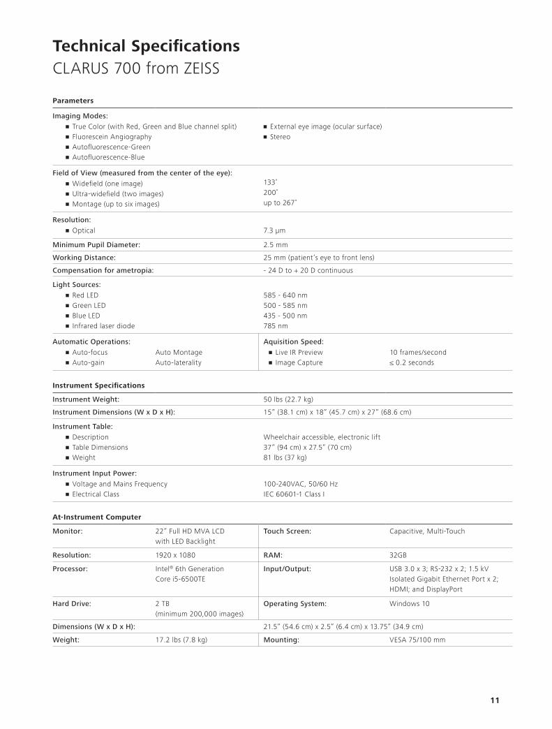
Technical SpecificationsCLARUS 700 from ZEISS
11
Parameters
Imaging Modes:• True Color (with Red, Green and Blue channel split)• Fluorescein Angiography• Autofluorescence-Green• Autofluorescence-Blue
• External eye image (ocular surface)• Stereo
Field of View (measured from the center of the eye):• Widefield (one image)• Ultra-widefield (two images)• Montage (up to six images)
133˚200˚up to 267˚
Resolution:• Optical 7.3 µm
Minimum Pupil Diameter: 2.5 mm
Working Distance: 25 mm (patient’s eye to front lens)
Compensation for ametropia: - 24 D to + 20 D continuous
Light Sources:• Red LED• Green LED• Blue LED• Infrared laser diode
585 - 640 nm500 - 585 nm435 - 500 nm785 nm
Automatic Operations: Aquisition Speed:• Auto-focus• Auto-gain
Auto MontageAuto-laterality
• Live IR Preview• Image Capture
10 frames/second≤ 0.2 seconds
Instrument Specifications
Instrument Weight: 50 lbs (22.7 kg)
Instrument Dimensions (W x D x H): 15” (38.1 cm) x 18” (45.7 cm) x 27” (68.6 cm)
Instrument Table:• Description• Table Dimensions• Weight
Wheelchair accessible, electronic lift 37” (94 cm) x 27.5” (70 cm) 81 lbs (37 kg)
Instrument Input Power:• Voltage and Mains Frequency• Electrical Class
100-240VAC, 50/60 HzIEC 60601-1 Class I
At-Instrument Computer
Monitor: 22” Full HD MVA LCD with LED Backlight
Touch Screen: Capacitive, Multi-Touch
Resolution: 1920 x 1080 RAM: 32GB
Processor: Intel® 6th Generation Core i5-6500TE
Input/Output: USB 3.0 x 3; RS-232 x 2; 1.5 kV Isolated Gigabit Ethernet Port x 2; HDMI; and DisplayPort
Hard Drive: 2 TB (minimum 200,000 images)
Operating System: Windows 10
Dimensions (W x D x H): 21.5” (54.6 cm) x 2.5” (6.4 cm) x 13.75” (34.9 cm)
Weight: 17.2 lbs (7.8 kg) Mounting: VESA 75/100 mm
Download the ZEISS Image Library App directly from the App Store. Explore a wide selection of modalities such as ultra-widefield and OCTA.
CAM
.114
45 C
Z VI
I/201
9 Un
ited
Stat
es e
ditio
n: O
nly
for s
ale
in s
elec
ted
coun
tries
. Th
e co
nten
ts o
f the
bro
chur
e m
ay d
iffer
from
the
curre
nt s
tatu
s of
app
rova
l of t
he p
rodu
ct o
r ser
vice
offe
ring
in y
our c
ount
ry. P
leas
e co
ntac
t our
regi
onal
repr
esen
tativ
es fo
r m
ore
info
rmat
ion.
Sub
ject
to c
hang
es in
des
ign
and
scop
e of
del
iver
y an
d du
e to
ong
oing
tech
nica
l dev
elop
men
t. CL
ARUS
700
and
CIR
RUS-
HDD-
OCT
are
eith
er tr
adem
arks
or
regi
ster
ed tr
adem
arks
of C
arl Z
eiss
Med
itec
AG o
r oth
er c
ompa
nies
of t
he Z
EISS
Gro
up in
Ger
man
y an
d / o
r oth
er c
ount
ries.
©
Car
l Zei
ss M
edite
c, In
c., 2
019.
All
right
s re
serv
ed.
Carl Zeiss Meditec AGGoeschwitzer Strasse. 51-5207745 JenaGERMANYwww.zeiss.com/clarus700www.zeiss.com/med/contacts
Carl Zeiss Meditec, Inc.5160 Hacienda DriveDublin, CA 94568USAwww.zeiss.com/us/clarus700 www.zeiss.com/us/med
0297
CLARUS 700


