Embed Size (px)
Citation preview

Page number not for citation purposes 1
Performance of LED fluorescence microscopy for the detection of tuberculosis in
Rwanda using Zeiss Primo Star
Alaine Umubyeyi Nyaruhirira1, Martine Toussaint2, Bennett Nemser3, Greet Vandebriel4, Michel Gasana2, Yanis Ben Amor3,&
1Management Sciences for Health, Center for Pharmaceutical Management, Pretoria, South Africa, 2TB Division, Rwanda Biomedical Center,
Institute of HIV/AIDS, Disease Prevention & Control, Kigali, Rwanda, 3The Earth Institute, Columbia University, New York, New York, USA, 4CAP
Columbia University, Rwanda
&Corresponding author: Yanis Ben Amor, The Earth Institute Columbia University 475 Riverside Drive New York, New York 10025 USA
Key words: Tuberculosis, fluorescence microscopy, iLED microscopy, Rwanda
Received: 17/11/2014 - Accepted: 04/07/2015 - Published: 14/07/2015
Abstract
Introduction: Ziehl-Neelsen (ZN) bright-field microscopy is time-consuming, with poor sensitivity, even under optimal conditions. Introduction of
Primo Star iLED fluorescent microscopy (FM) may improve TB case finding at referral hospitals in Rwanda. The study aimed to determine the
acceptability and effectiveness of iLED in a low resource setting. Methods: Between June 2009 and May 2010, the Rwandan TB Program and
National Reference Laboratory carried out demonstration studies with iLED at a referral hospital in the capital, Kigali, and a rural district hospital in
Nyamata, taking conventional FM as Gold Standard. Results: Agreement between the iLED and rechecking at the Reference Laboratory were
deemed “almost perfect” (kappa = 0.81-1.00) across three of four site-phase combinations. The exception was Nyamata District Hospital during
the validation phase, which was deemed “substantial” agreement (kappa = 0.61-0.80). However, the 100% concordance at both demonstration
sites during the continuation phase shows technicians' rapid command of the new iLED microscope in a relatively short time. The lower overall
positivity rate obtained in the rural clinic is not related to the performance of the microscope (or technicians), but is attributable to a significant
increase in total number of patients and samples screened through active case finding. Conclusion: Laboratory technicians demonstrated high
acceptance of iLED. Additionally, fluorescent microscopy reduces the time necessary for examination by more than half. The high level of
agreement between iLED and FM during implementation in both sites provides initial evidence for iLED to replace current methods.
Pan African Medical Journal. 2015; 21:198 doi:10.11604/pamj.2015.21.198.5776
This article is available online at: http://www.panafrican-med-journal.com/content/article/21/198/full/
© Alaine Umubyeyi Nyaruhirira et al. The Pan African Medical Journal - ISSN 1937-8688. This is an Open Access article distributed under the terms of the Creative
Commons Attribution License (http://creativecommons.org/licenses/by/2.0), which permits unrestricted use, distribution, and reproduction in any medium, provided the original work is properly cited.
Pan African Medical Journal – ISSN: 1937- 8688 (www.panafrican-med-journal.com) Published in partnership with the African Field Epidemiology Network (AFENET). (www.afenet.net)
Research
Open Access

Page number not for citation purposes 2
Introduction
Sputum smear microscopy for acid-fast bacilli (AFB) using Ziehl-
Neelsen (ZN) staining remains the most cost-efficient tool available
to diagnose tuberculosis (TB) in low-resource countries. This
method is rapid, inexpensive, and highly specific for detecting AFB
in high-burden settings. However, the main limitation is its low and
variable sensitivity, exacerbated in high HIV prevalence settings [1].
High TB-HIV co-infection rates and low TB case detection impede
disease control in many TB endemic settings, notably sub-Saharan
Africa [2]. Furthermore, where workloads are high, the amount of
time spent examining smears compromises sensitivity [3]. A recent
systematic review demonstrated that fluorescence microscopy (FM)
is, on average, 10% more sensitive than conventional bright-field
microscopy in detecting AFB in clinical specimens, with comparable
specificity, and takes significantly less time [4]. However,
widespread implementation of FM in disease-endemic settings
remains limited due to several factors, including the short life and
high cost of mercury vapor lamps; difficulty in maintaining
machines; the need for a darkroom; and strict requirements for
electrical power supply. Light-emitting diodes (LEDs) for FM have
been identified as an alternative to conventional FM for screening of
AFB [5, 6]. LED lamps do not have the disadvantages of mercury
vapor lamps, with life expectancy averaging around 50,000-100,000
hours (10-20 years) of use [5]. They can also run on batteries
[5, 7, 8]. Several commercial LED systems are now available, either
as stand-alone microscopes or as add-on adapters to conventional
microscopes [9]. Data published so far on LED microscopy for TB
show that results in terms of sensitivity and specificity are
comparable or better with LED than mercury vapor lamps [6, 7, 10-
13]. Study objectives: This demonstration project evaluated the
effectiveness of employing the Primo Star iLED fluorescence
microscope (subsequently referred to as iLED) for case finding of TB
under routine conditions in one referral and one rural setting in
Rwanda. Microscopists without prior experience in FM were solicited
in order to determine operational and clinical performance, as well
as acceptability of the technology to laboratory staff. Study
design: This project was conducted at two sites: Nyamata district
hospital (DH) in Bugesera district and the Centre Hospitalier
Universitaire de Kigali (CHUK) in Kigali. Nyamata DH is a 100-bed
hospital with a catchment area of about 300,000 people. CHUK is a
509-bed national referral hospital (RH) serving the capital city,
Kigali, with a catchment area of approximately 1 million people. The
implementation of iLED was carried out in five phases: (1) ZN
baseline; (2) iLED training and appraisal (five days); (3) validation
(one month); (4) implementation (three months); and (5)
continuation (six months).
Methods
Ethics statement: The evaluation was approved by the Ethical
Review Committee of the Ministry of Health (Kigali, Rwanda) under
Protocol Number 58/RNEC/2009 and by the Institutional Review
Board of Columbia University IRB-AAAC6248 (New York, NY, USA).
Written consent was not obtained because microscopy for AFB
smears is the standard of care in Rwanda as part of regular clinic
monitoring and evaluation of Tuberculosis. During validation, all
sputum results obtained though iLED were rechecked systematically
by the National Reference Laboratory before results were provided
to patients for management. The implementation phase was only
allowed once iLED was validated as a replacement for light
microscopy with similar or better sensitivity, therefore not placing
patients at risk of misdiagnosis. Written consent to participate in the
study was not sought from the microscopists or their supervisors
because the introduction of the iLED only minimally increased the
workload and only for a short duration (1 month) during the whole
study. Participants were informed about the purpose and impact of
the study, and microscopists readily participated enthusiastically.
The need for collecting documented informed consent was waived
by the IRB. Phase 1: ZN Baseline: The aim of this one-month
phase was to establish a baseline, under study conditions, of false
positivity and negativity rates for ZN. TB treatment decisions were
based on ZN results. All incoming sputum smears were stained for
ZN examination under routine conditions. Slides were read using the
available conventional bright-field microscope (1000x). After
reading, all slides were kept in slide boxes, which were labeled to
specify the study phase, study site, box number, and slide ID. Once
every two weeks, the National Reference Laboratory (NRL) study
supervisor collected all boxes. NRL study technicians rechecked all
slides using conventional bright-field microscopes. Discrepant slides,
if any, were sent to the Supra National Reference Laboratory (SNRL)
in Germany for rereading. Phase 2: Training and Appraisal: A
standardized five-day training course for microscopists and
supervisors participating in the study was conducted. All eight
participants had skills in ZN microscopy but not conventional FM.
Participants after learning the purpose and impact of the study,
participated readily. Following the training, all technicians involved

Page number not for citation purposes 3
in the project filled out a questionnaire about several features of the
iLED, including installation and first use, training, and optics and
handling. Phase 3: validation: The validation phase lasted one
month. Each sputum sample at the study sites was stained using
Auramine O and examined by the iLED at 400x magnification.
Patient management was based solely on the rechecking results
carried out by the NRL. Staining solutions were prepared by NRL
using Merck staining reagents (Catalogue 41000, Auramine O, item
number 1013010050, lot number ZC 253201532) and provided to
the study sites once per month, taking into consideration the limited
shelf life of Auramine O. All readings (including rechecking) were
done within 48 hours of staining. Results were quantified according
to the scale presented in Table 1. NRL rechecked all slides using
conventional FM. Rechecked results were provided to study sites the
next day for timely patient management. The semi-quantitative
scale for rechecking by NRL was different than the one used by
study sites (Table 2). Discordant slides, if any, were sent to the
SNRL for final discussion. The study sites were allowed to proceed
to phase four only if the following performance targets were met:
(1) 95% accordance between validation results of microscopy center
and supervisory site; (2) quality of Auramine O stains acceptable in
100% of slides examined; and (3) fewer than two false results in a
proficiency testing panel of 10 pre-defined slides.
Phase 4: implementation phase: The procedures were the same
as during the validation phase. The duration of this phase was three
months, and patient management was now based on iLED results.
Supervision and rechecking by the NRL study supervisor were
carried out using Lot Quality Assurance Sampling (LQAS). The
sample size was calculated by NTP/NRL based on the positivity and
number of negative smears, but the frequency of rechecking was
decreased from daily to once every two weeks. Rereading by NRL
was done using conventional FM at 400x magnification. Discordant
slides, if any, were sent to the SNRL in Germany for umpire reading.
Rechecked results were provided to study sites. Phase 5:
continuation and expansion: The continuation phase lasted six
months, and patient management was based on iLED results.
Supervision and rechecking by NRL supervisors was carried out
according to national Rwandan External Quality Assurance
guidelines. Fifteen slides were collected quarterly and rechecked by
NRL using conventional FM at 400 x magnification. Discordant
slides, if any, were sent to the SNRL in Germany for umpire reading.
Rechecked results were provided to study sites. After the six-month
continuation phase, and following the availability of the compiled
results of the previous phase, the demonstration project coordinator
allowed all sites to use the iLED method routinely under program
conditions. Data entry and analysis: All data and results were
recorded in phase-specific forms and sent to NRL and NTP. An
electronic database was completed on-site. Positivity agreement
between methods at the study laboratories (DH or CHUK) and the
National Reference Laboratory was assessed using Cohen's Kappa,
which corrects for agreement by chance. Strength of agreement
was evaluated using guidelines from Land is and Koch [14]: <0 =
poor; 0-0.20 = slight; 0.21-0.40 = fair; 0.41-0.60 = moderate;
0.61-0.80 = substantial; 0.81-1.00 = almost perfect.
Results
Baseline: At the DH in Nyamata, all incoming sputum samples
(100) from 37 patients, using the Spot-Morning-Spot criteria, were
examined using ZN staining during the baseline phase. The
positivity rate of the slides was 11% (11/100 -Table 3) with only
one low false positive (LFP) as determined following rechecking. The
LFP result had no negative public health consequence on the
accurate diagnosis of the patient. There were no poorly stained
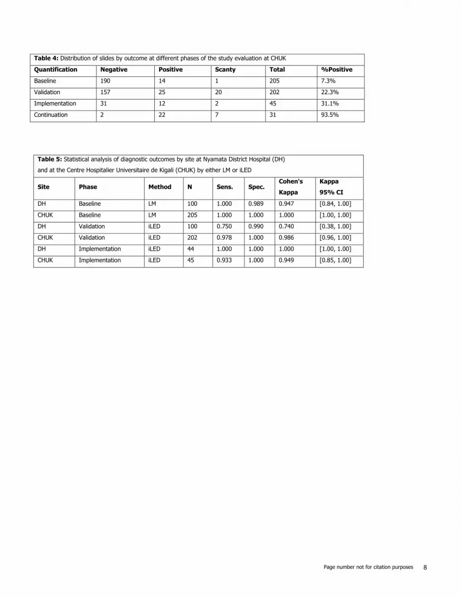
slides reported by the NRL. At CHUK in Kigali, all incoming sputum
samples (205) from 94 patients (Spot-Morning-Spot) were analyzed.
The positivity rate for the samples was 7.3% (15/205 - Table 4)
and two quantification errors (QEs) were reported following
rechecking at NRL. The two QEs, corresponding to two samples
from two different patients, had no negative public health
consequence. There were no poorly stained slides reported by the
NRL for CHUK during the rechecking of the baseline. For the
comparison between results obtained by the DH in Nyamata and the
NRL, Kappa was 0.947 [95% CI: 0.84, 1.00], which indicates an
almost perfect agreement between the two laboratories (see results
in Table 5). For the comparison between CHUK and NRL, Kappa
was 1.000 due to full agreement (Table 5).
Validation phase: In Nyamata, all incoming samples (100) from
39 patients were screened. The positivity rate decreased to 4%
(4/100 - see Table 3) and there were 1 low false positive and 1 low
false negative (LFN) errors reported. Additionally, 15 samples were
reported as having poor stains. The LFN and LFP errors,
corresponding to two samples from the same patient, would not
have a negative public health consequence and were probably due
to administrative errors. However, as per protocol, diagnosis was
made solely on the basis of the rechecking by the NRL. Kappa was

Page number not for citation purposes 4
0.740 [95% CI: 0.38, 1.00], which indicates substantial agreement
between the laboratories (Table 5). In CHUK, all incoming samples
(202) from 87 patients were screened. The positivity rate
significantly increased from baseline to 22.3% (45/202 - see Table
4) and there was one LFN reported. Since only one sample was
collected for that particular patient, the LFN error would have had a
negative public health consequence on the accurate diagnosis of the
patient. The patient was adequately treated following rechecking by
NRL. Nine samples were reported as having poor stains by the NRL.
Kappa was 0.986 [95% CI: 0.96, 1.00'> 1.00], which indicates an
almost perfect agreement between laboratories (Table 5). No
errors were detected at the proficiency panel test. Both sites were
allowed to proceed to the implementation phase.
Implementation phase: In Nyamata, following LQAS, 44 samples
(corresponding to 44 patients) were rechecked by the NRL study
supervisor. The positivity rate for samples increased from validation
to 9.1% (4/44) but remained lower than in the baseline phase
(Table 3). No reading/diagnosis errors were reported by the NRL.
However, there were six poorly stained samples. Kappa was 1.000
due to full agreement (Table 5). In CHUK, following LQAS, 45
samples from 45 patients were rechecked. The positivity rate for
samples increased further from baseline and validation to 31.1%
(14/45), as shown in Table 4. Only one LFN and three poor stains
were reported by the NRL. It is not possible to say whether the LFN
error had a negative public health consequence, since the other two
samples from this particular patient were not rechecked and could
have been positive. Kappa was 0.949 [95% CI: 0.85; 1.00], which
indicates an almost perfect agreement between laboratories (Table
5).
Continuation phase: In Nyamata, 16 samples were read from
January through March by iLED at the site and conventional FM at
NRL, and 100% concordance was observed. From April through
June, an additional 11 samples were rechecked and all results
concurred. At CHUK, there were 15 samples screened from January
through March and 100% concordance was observed. Sixteen
additional samples were screened from April through June and
100% concordance was also observed. Technicians' appraisal:
the appraisal took place after training of the
technicians. Installation and first use: All technicians felt that
installation of the iLED was easy and that the manual was
comprehensive and easy to read and understand. Training: The
technicians participating in the iLED project felt that for technicians
already trained in ZN microscopy (such as themselves), an iLED
training of 1-5 days was suitable. However, for technicians not
familiar with ZN microscopy, an iLED training of 3-20 days was
suitable. Technicians also felt that NTPs can readily use the current
manual developed by the manufacturer for implementation of LED
microscopy without major changes.
Optics and handling: All technicians were satisfied with the
contrast, color, intensity, and signal-to-noise ratio of the iLED. All
technicians were very satisfied with the resolution and depth of
focus of the iLED. All technicians also felt that the field of view of
the iLED is more homogenously illuminated compared to the
standard view. All of them were very satisfied with the overall
handling features of the microscope. All technicians also felt that it
was convenient or very convenient to switch between bright field
and fluorescence. Only one technician surveyed felt that the toggle
field was not robust. All technicians felt that no darkroom was
needed when using iLED, a really convenient feature of iLED
compared to regular FM. All technicians also agreed that the
dazzling protection for the eyepieces was useful. None of the
technicians surveyed reported any technical problems with the
microscope overall.
Discussion
Compared to classical FM with mercury vapor lamp, LED FM is more
user-friendly and benefits from a high acceptability by technicians.
Microscopes do not require warm-up and cool-down time, a
considerable advantage when power supply is erratic, and the LED
light source is considered safer than the mercury vapor lamp. LED
systems developed for AFB smears consist either of modules that
can be fitted to a conventional microscope, or a complete
microscope with built-in LED as the light source, such as the iLED.
While Partech (Münster, Germany) and Cytoscience (Fontaines,
Switzerland) have both developed complete LED FM microscopes,
these microscopes are less appropriate for TB than iLED since they
are monocular. Using a camera and monitor might be an
appropriate solution, but not in less-developed countries. iLED, a
binocular FM microscope with built-in LED lamp for epi-fluorescence
has produced very good results in reference laboratories [13]. So
far, very few reports on these systems exist. These rare reports,
however, show excellent performance compared to ZN microscopy
[15, 16]. Our study is the first direct evaluation of iLED in Rwanda.
LED add-on kits have been designed for different common types of

Page number not for citation purposes 5
bright field microscopes. The complete installation is not difficult,
but it requires slightly more time and care, which could be a
disadvantage from the end-user perspective. As difficulty in
acceptance by inexperienced microscopists seems to be the main
obstacle to the use of FM outside referral laboratories, this may
prove to be a major advantage of transmitted LED light FM, as
reported earlier from Tanzania [8]. It also remains to be seen
whether complete binocular LED microscopes using epi-
fluorescence, rather than transmitted light, will meet the same
acceptance level with the progressive decentralization of FM to
peripheral hospitals and health centers. Our study shows that the
acceptability amongst the staff using iLED was extremely high and
proficiency in adequate usage was rapid. Compared to traditional
bright-field methods, LED fluorescence methods using Auramine O
staining allows up to four times faster screening. The detection rate
is also estimated to be at least 10% higher. While we did not
directly compare the positivity rates between ZN and iLED, we
monitored the positivity rate over time during our study.
In Nyamata, the positivity rate surprisingly decreased during the
validation phase (Table 5). This result compares with previously
established data from the National TB Programme, which has shown
that in the last quarter of 2009 and first quarter of 2010, during
which our study took place, there was an overall decrease in
positivity rate that is not related to the performance of the
microscope (or the technicians) but is rather attributable to a
significant increase in total number of patients and samples
screened. Indeed, the positivity rate dropped to 7.7% during our
study, compared to 9.2% the year before. This increase in the total
number of patients screened may be the result of the impact of
Community Health Workers (CHWs) in Bugesera District (where
Nyamata DH is located), who have been involved in active case
finding, therefore casting a wider net for overall screening of TB
suspects and decreasing the positivity rate at the Nyamata Center.
One of the expected issues associated with the change from ZN to
Auramine staining for the purpose of our study was a difficulty in
preparing and then subsequently reading the slides adequately
using the Auramine protocol. Indeed, a few of the smears in the
various phases were reported as having poor stains. We believe
these could be explained by the fact that some laboratory
technicians were not completely proficient in staining the slides
appropriately, as can also sometimes be the case for ZN. However,
this was not an issue throughout the study as the number of slides
poorly stained gradually decreased at both sites. Agreement
between the iLED and rechecking at the Reference Laboratory were
deemed “almost perfect” (kappa = 0.81-1.00) across three of four
site-phase combinations. The exception was Nyamata District
Hospital during the validation phase, which was deemed
“substantial” agreement (kappa = 0.61-0.80). However, the 100%
concordance at both demonstration sites during the continuation
phase shows technicians' rapid command of the new iLED
microscope in a relatively short time. Technicians can therefore be
easily trained to switch from ZN microscopy to LED FM with a high
success rate. This should be of interest to national TB control
programs that are interested in improving their overall case
detection rate but cannot yet invest in the newer, molecular-based
technologies currently being rolled out.
Conclusion
In our study, the use of iLED FM module in both a referral hospital
and rural clinic setting was associated with a high concordance rate
as compared to a Reference Laboratory using conventional FM. The
high level of agreement between iLED and FM during our study in
multiple sites, combined to the fact that fluorescent microscopy
reduces the time necessary for examination by more than half,
provides initial evidence for the iLED to replace current standard
methods. The iLED microscope also excelled in terms of user-
friendliness and acceptance by users.
Competing interests
The authors declare no competing interests.
Authors’ contributions
AUN, MG, and YBA conceived the study and participated in its
design and coordination. MT and GV participated in the design and
coordination of the study. BN performed all statistical analysis. All
authors helped to draft the manuscript and read and approved the
final manuscript.

Page number not for citation purposes 6
Acknowledgments
We would like to thank CN Paramasivan, Fabrice Ingabire,
Sebazungu Emmanuel, Alida Ngwije, Eliane Kamanzi, Jean Baptiste
Gatabazi, and Ruben Sahabofor their help with our study. Finally,
we would like to thank Timmons Barbara and Catherine Mundy for
the revision of the manuscript.
Tables
Table 1: Semi-quantitative scale used for reading with Iled
Table 2: Semi-quantitative scale for rechecking with conventional
FM
Table 3: Distribution of slides by outcome at different phases of the
study evaluation at Nyamata Hospital
Table 4: Distribution of slides by outcome at different phases of the
study evaluation at CHUK
Table 5: Statistical analysis of diagnostic outcomes by site at
Nyamata District Hospital (DH) and at the Centre Hospitalier
Universitaire de Kigali (CHUK) by either LM or iLED
References
1. Elliott AM, Halwiindi B, Hayes RJ, Luo N, Tembo G, Machiels L,
Bem C, Steenbergen G, Pobee JO, Nunn PP, Hayes RJ,
McAdam KP. The impact of human immunodeficiency virus on
presentation and diagnosis of tuberculosis in a cohort study in
Zambia. J Trop Med Hyg . 1993; 96(1): 1-
11. PubMed | Google Scholar
2. World Health Organization. Global tuberculosis control:
surveillance, planning, financing. Geneva. 2014; WHO. Google
Scholar
3. Cambanis A, Ramsay A, Wirkom V, Tata E, Cuevas LE.
Investing time in microscopy: an opportunity to optimise
smear-based case detection of tuberculosis. Int J Tuberc Lung
Dis. 2007; 11(1):40-45. PubMed |Google Scholar
4. Steingart KR, Henry M, Ng V, Hopewell PC, Ramsay A,
Cunningham J, Urbanczik R, Perkins M, Aziz MA, Pai M.
Fluorescence versus conventional sputum smear microscopy
for tuberculosis: a systematic review. Lancet Infect Dis. 2006;
6(9):570-581. PubMed | Google Scholar
5. Anthony RM, Kolk AH, Kuijper S, Klatser PR. Light emitting
diodes for auramine O fluorescence microscopic screening of
Mycobacterium tuberculosis. Int J Tuberc Lung Dis. 2006;
10(9):1060-1062. PubMed |Google Scholar
6. Marais BJ, Brittle W, Painczyk K, Hesseling AC, Beyers N,
Wasserman E, van Soolingen D, Warren RM. Use of light-
emitting diode fluorescence microscopy to detect acid-fast
bacilli in sputum. Clin Infect Dis. 2008; 47(2):203-
207. PubMed | Google Scholar
7. Affolabi D, Torrea G, Odoun M, Senou N, Ali Ligali M,
Anagonou S, Van Deun A. Comparison of two LED fluorescence
microscopy build-on modules for acid-fast smear microscopy.
Int J Tuberc Lung Dis. 2010; 14(2): 160-
164. PubMed | Google Scholar
8. Van Deun A, Chonde TM, Gumusboga M, Rienthong S.
Performance and acceptability of the FluoLED Easy module for
tuberculosis fluorescence microscopy. Int J Tuberc Lung Dis.
2008; 12(9): 1009-1014. PubMed |Google Scholar
9. Minion J, Sohn H, Pai M. Light-emitting diode technologies for
TB diagnosis: what is on the market?. Expert Rev Med Devices.
2009; 6(4):341-345. PubMed | Google Scholar
10. Albert H, Manabe Y, Lukyamuzi G, Ademun P, Mukkada S,
Nyesiga B, Joloba M, Paramasivan CN, Perkins MD.
Performance of three LED-based fluorescence microscopy
systems for detection of tuberculosis in Uganda. PLOS ONE.
2010; 5(12):e15206. PubMed | Google Scholar
11. Bonnet M, Gagnidze L, Githui W, Guerin PJ, Bonte L, Varaine F,
Ramsay A. Performance of LED-based fluorescence microscopy
to diagnose tuberculosis in a peripheral health centre in
Nairobi. PLOS ONE. 2011; 6(2):e17214. PubMed | Google
Scholar

Page number not for citation purposes 7
12. Hung NV, Sy DN, Anthony RM, Cobelens FG, van Soolingen D.
Fluorescence microscopy for tuberculosis diagnosis. Lancet
Infect Dis. 2007; 7(4):238-239; author reply 239-
240. PubMed | Google Scholar
13. Nabeta P, Ha DT, Michaels JS, Hofmann H, Krapp F. LED-based
fluorescence microscope for TB detection: evaluation in
reference laboratories. Int J Tuberc Lung Dis. 2009; Suppl:
S123. PubMed |Google Scholar
14. Kundel HL, Polansky M. Measurement of observer agreement.
Radiology. 2003; 228(2):303-308. PubMed | Google Scholar
15. Minion J, Pai M, Ramsay A, Menzies D, Greenaway C.
Comparison of LED and conventional fluorescence microscopy
for detection of acid fast bacilli in a low-incidence setting. PLOS
ONE. 2011; 6(7):e22495. PubMed |Google Scholar
16. Turnbull ER, Kaunda K, Harris JB, Kapata N, Muvwimi MW,
Kruuner A, Henostroza G, Reid SE. An evaluation of the
performance and acceptability of three LED fluorescent
microscopes in Zambia: lessons learnt for scale-up. PLOS ONE.
2011; 6(11):e27125. PubMed | Google Scholar
Table 1: Semi-quantitative scale used for reading with iLED
IUATLD Scale (1000field =HPF)
Result
iLED (400x magnification: 1 length =40 fields = 200HPF
Negative Zero AFB /1 length
Scanty 1–19 AFB/1 length
1+ 20–199 AFB/1 length
2+ 5–50 AFB/1 field on average
3+ >50 AFB/1 field on average
Table 2: Semi-quantitative scale for rechecking with conventional FM
IUATLD Scale (1000field =HPF)
Result
Conventional FM (200-250x magnification: 1 length
=30 fields = 300HPF
Negative Zero AFB /1 length
Scanty 1–9 AFB/1 length
1+ 30–299 AFB/1 length
2+ 10–100 AFB/1 field on average
3+ >100 AFB/1 field on average
Table 3: Distribution of slides by outcome at different phases of the study evaluation at Nyamata Hospital
Quantification Negative Positive Scanty Total %Positive
Baseline 89 8 3 100 11.0%
Validation 96 1 3 100 4.0%
Implementation 40 3 1 44 9.1%
Continuation 0 23 4 27 100.0%
Page number not for citation purposes 8
Table 4: Distribution of slides by outcome at different phases of the study evaluation at CHUK
Quantification Negative Positive Scanty Total %Positive
Baseline 190 14 1 205 7.3%
Validation 157 25 20 202 22.3%
Implementation 31 12 2 45 31.1%
Continuation 2 22 7 31 93.5%
Table 5: Statistical analysis of diagnostic outcomes by site at Nyamata District Hospital (DH)
and at the Centre Hospitalier Universitaire de Kigali (CHUK) by either LM or iLED
Site Phase Method N Sens. Spec. Cohen's
Kappa
Kappa
95% CI
DH Baseline LM 100 1.000 0.989 0.947 [0.84, 1.00]
CHUK Baseline LM 205 1.000 1.000 1.000 [1.00, 1.00]
DH Validation iLED 100 0.750 0.990 0.740 [0.38, 1.00]
CHUK Validation iLED 202 0.978 1.000 0.986 [0.96, 1.00]
DH Implementation iLED 44 1.000 1.000 1.000 [1.00, 1.00]
CHUK Implementation iLED 45 0.933 1.000 0.949 [0.85, 1.00]


