Embed Size (px)
Citation preview
REVIEW PAPER
Class III HD-ZIPs govern vascular cell fate: an HD view on patterning and differentiation
Prashanth Ramachandran1, Annelie Carlsbecker1,* and J. Peter Etchells2,*1 Physiological Botany, Department of Organismal Biology and Linnean Centre for Plant Biology in Uppsala, Uppsala University, Ulls väg 24E, SE-756 51 Uppsala, Sweden2 Department of Biosciences, Durham University, South Road, Durham DH1 3LE, UK
* Correspondence: [email protected] or [email protected]
Received 4 August 2016; Editorial decision 15 September 2016; Accepted 22 September 2016
Editor: Simon Turner, University of Manchester
Abstract
Plant vasculature is required for the transport of water and solutes throughout the plant body. It is constituted of xylem, specialized for transport of water, and phloem, that transports photosynthates. These two differentiated tis-sues are specified early in development and arise from divisions in the procambium, which is the vascular meristem during primary growth. During secondary growth, the xylem and phloem are further expanded via differentiation of cells derived from divisions in the cambium. Almost all of the developmental fate decisions in this process, including vascular specification, patterning, and differentiation, are regulated by transcription factors belonging to the class III homeodomain-leucine zipper (HD-ZIP III) family. This review draws together the literature describing the roles that these genes play in vascular development, looking at how HD-ZIP IIIs are regulated, and how they in turn influence other regulators of vascular development. Themes covered vary, from interactions between HD-ZIP IIIs and auxin, cytokinin, and brassinosteroids, to the requirement for exquisite spatial and temporal regulation of HD-ZIP III expres-sion through miRNA-mediated post-transcriptional regulation, and interactions with other transcription factors. The literature described places the HD-ZIP III family at the centre of a complex network required for initiating and main-taining plant vascular tissues.
Key words: Auxin, (pro)cambium, cytokinin, HD-ZIP III, miR165/166, root, shoot, transcription factors, vascular development, xylem.
Introduction
Homeodomain transcription factors have been synonymous with regulation of development since their identification in patterning of the fly more than 30 years ago. In plants, mem-bers of the class III homeodomain-leucine zipper (HD-ZIP III) transcription factor family are an excellent example of the incredibly broad range of developmental processes that HD transcription factors regulate. HD-ZIP IIIs act from cradle to grave, with roles in patterning of the embryo, meristem main-tenance, leaf development, inflorescence architecture, ovule
development, growth response to environmental signals, and senescence. Characterization of mutations in REVOLUTA, one of five HD-ZIP III genes present in the model plant Arabidopsis thaliana, represents the first description of the consequences of loss of HD-ZIP III function (Talbert et al., 1995). While this study is notable for its description of the pleiotropic defects present in HD-ZIP III mutants, clues begin to emerge as to their importance in controlling vas-cular development. In particular, Talbert et al. (1995) noted
© The Author 2016. Published by Oxford University Press on behalf of the Society for Experimental Biology. All rights reserved. For permissions, please email: [email protected]
Journal of Experimental Botany, Vol. 68, No. 1 pp. 55–69, 2017doi:10.1093/jxb/erw370 Advance Access publication 7 October 2016
Dow
nloaded from https://academ
ic.oup.com/jxb/article/68/1/55/2627443 by guest on 15 January 2022
that there were changes to the numbers of xylem and phloem cells in rev mutants compared with wild-type plants, accom-panied by changes to fibre differentiation. In subsequent years, our understanding of the role of REV, which is also known as INTERFASCICULAR FIBRELESS1 (IFL1) or AMPHIVASAL VASCULAR BUNDLES1, and the other members of the HD-ZIP III family in Arabidopsis, Arabidopsis thaliana HOMEOBOX8 (ATHB8), PHABULOSA (PHB)/ATHB14, PHAVOLUTA (PHV)/ATHB9, and CORONA (CNA)/INCURVATA4/ATHB15, has been considerably elab-orated in multiple aspects of vascular development. In this review, we will describe in detail these roles in vascular pat-terning and xylem differentiation in both the shoot and root.
Radial patterning of vascular tissues in the shoot
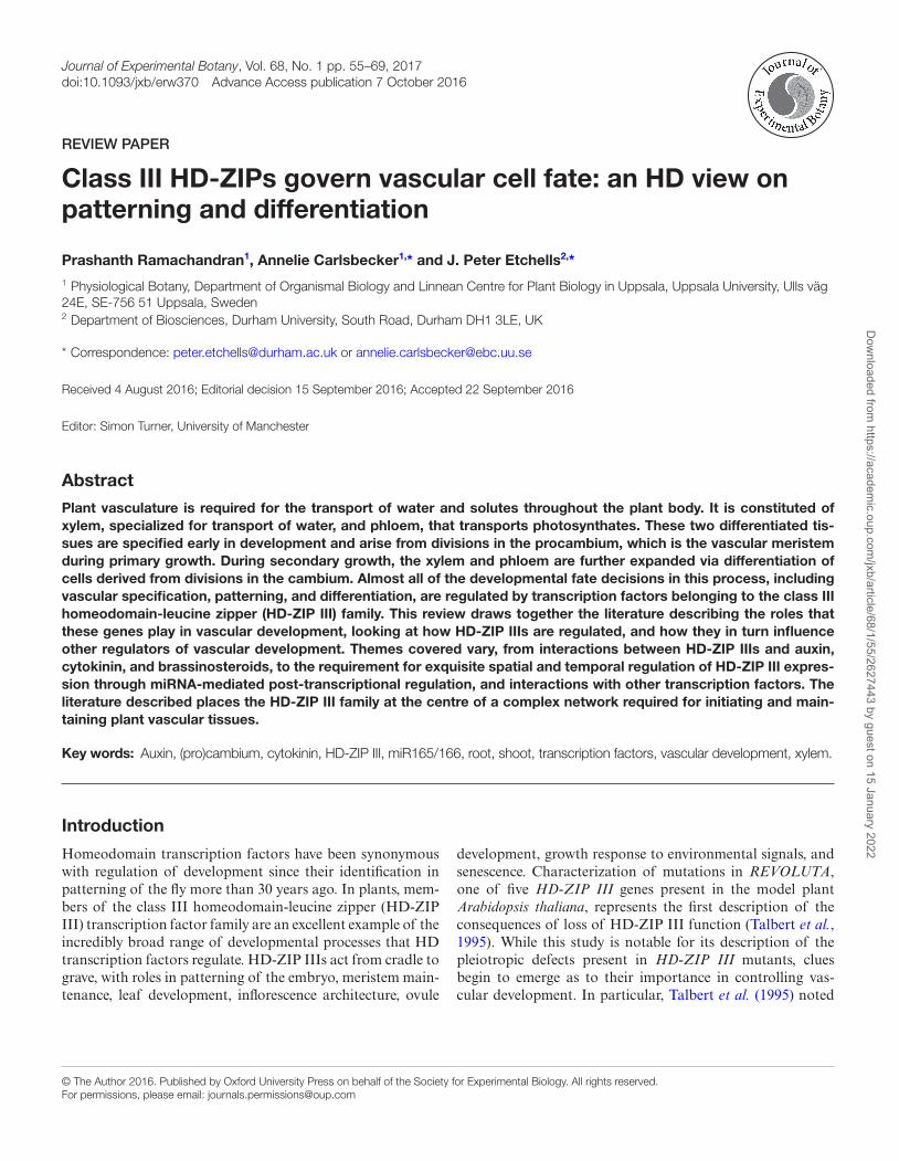
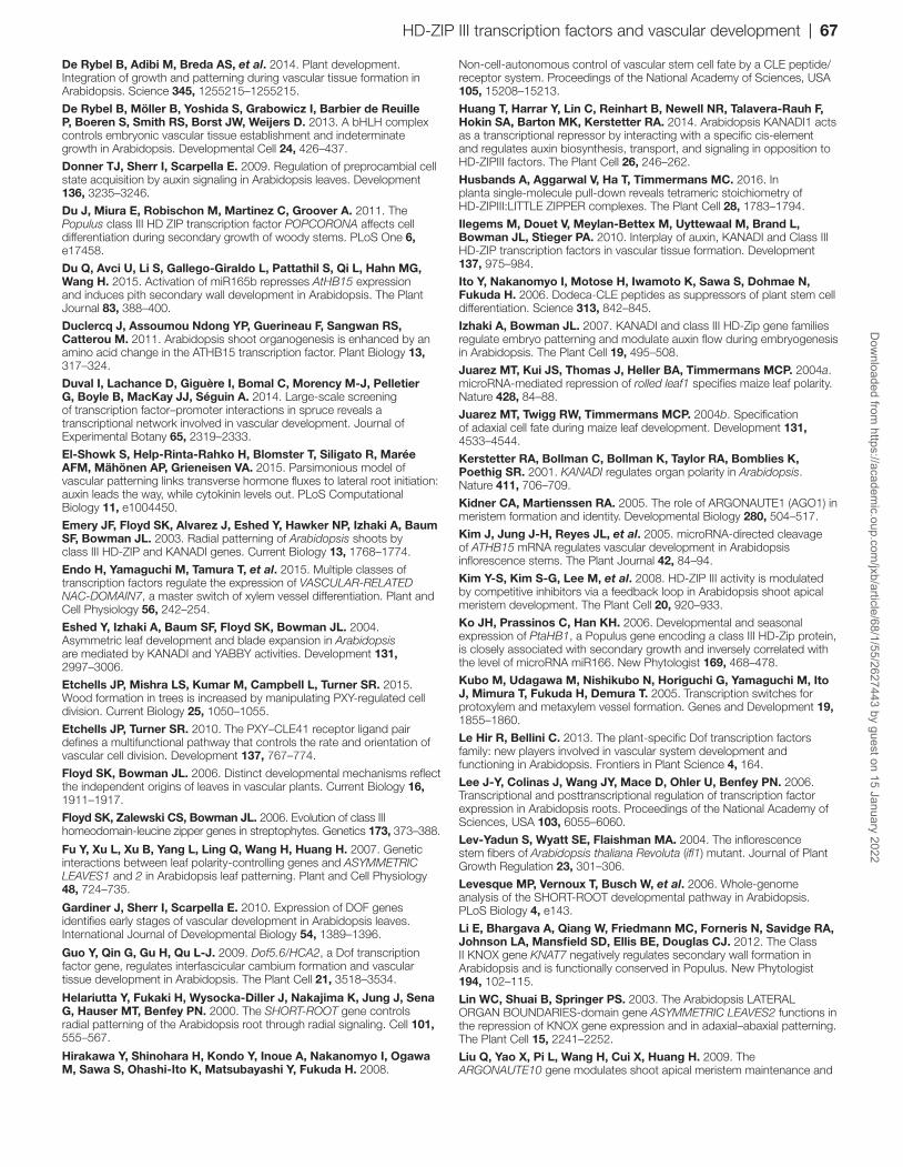
Vascular tissue specification and differentiation occur in the wider developmental context of organs such as the leaf, stem, or root. Several HD-ZIP III mutants were initially identified in screens aimed at identifying regulators of leaf develop-ment, and these mutants also demonstrated vascular defects (McConnell and Barton, 1998; McConnell et al., 2001). Leaves are initiated at the flanks of the shoot apical meris-tem. They develop an upper (adaxial or dorsal) side special-ized for light capture, and a lower (abaxial or ventral) side specialized for gas exchange. The vascular strands are typi-cally positioned where the adaxial and abaxial domains meet. Xylem is present in the adaxial position and phloem is posi-tioned abaxially. The question of how these specific patterns arise in the leaf was addressed in early experiments, where the initiating leaf primordium was surgically separated from the apical meristem from which it arose. The aim of these experiments was to determine if all information required for normal leaf formation is present within the initiating primor-dium or if leaf patterning requires communication with the meristem (Sussex, 1954). These experiments are pertinent to understanding vascular development within the shoot as they also represent some of the first observations of changes in vascular patterns. In a series of elegant papers in the 1950s, Sussex demonstrated that radially symmetric leaves were the consequence of surgically separating initiating primordia from the apical meristem in potato, arguing that a mobile signal emanating from the apical meristem must be involved in leaf patterning. Within these radially symmetric leaves, the vascular tissues were also clearly perturbed (Sussex, 1955). Subsequently, similar experiments using willowherb (Epilobium) also revealed radialized leaves that lacked xylem–phloem asymmetry (Snow and Snow, 1959)(Fig. 1A–C).
The first study to place the observations of asymmetry loss in a genetic context made use of the snapdragon (Antirrhinum) phantastica (phan) mutants, which had radialized leaves simi-lar to those observed in the surgical experiments (Fig. 1D, E). phan was described as a ‘dorsalizing factor’, namely a gene that specifies the upper (and therefore xylem) side of the leaf (Waites and Hudson, 1995). phan encodes a myb transcription factor (Waites et al., 1998), and its Arabidopsis
Fig. 1. Vascular tissue formation within radialized leaves. Separation of incipient leaf primordium (I1) from apical meristem by cut ‘x’ (A) leads to loss of adaxial–abaxial leaf asymmetry (B) and amphicribal vascular tissue (C) with phloem surrounding xylem in Epilobium. Cut ‘y’ (A) represents the separation between meristem and initiating leaf performed by Sussex (1955) with similar results. P1, P2, and P3 denote leaf primordia formed by the meristem prior to the cut. phan mutant from Antirrhinum (E) with radialized vascular tissue similar to that described in (C), compared with that of a wild-type Antirrhinum leaf which demonstrates adaxial–abaxial asymmetry (D). Phenotype of the phb-1d mutant with radially symmetric trumpet-shaped leaves (G) with amphivasal vascular tissue (I) compared with that of wild-type plants (F, H), in which xylem is restricted to the adaxial domain and phloem to the abaxial domain. (H, I) Toluidine blue-stained cross-sections of leaf petioles. Scale bars are 50 μm (D), 5 mm (F, G) and 20 μm (H, I). x, p, pa, and ve are xylem, phloem, parenchyma, and ventral epidermis, respectively. (A–C) Reproduced from Snow and Snow (1959), with permission from Wiley. (D, E) Reproduced from Waites and Hudson (1995), with permission from the Company of Biologists. (F–I) Reproduced from McConnell and Barton (1995), with permission from the Company of Biologusts.
56 | Ramachandran et al.D
ownloaded from
https://academic.oup.com
/jxb/article/68/1/55/2627443 by guest on 15 January 2022
orthologue ASYMMETRIC LEAVES 1 (AS1) (Byrne et al., 2000) was subsequently shown to act as a positive regulator of the expression of PHB, PHV, and REV (Fu et al., 2007) (Fig. 2). Such observations were consistent with phenotypes of dominant gain-of-function phb-1d (Fig. 1F–I) and phv-1d alleles, which had earlier been described as having amphivasal vascular bundles with xylem surrounding phloem (i.e. xylem present in both adaxial and abaxial positions) and there-fore gain of adaxial identity (McConnell and Barton, 1998; McConnell et al., 2001). This phenotype is opposite to that observed in loss-of-function phan, which has amphicribal bundles where phloem surrounds xylem (thus demonstrat-ing a loss of adaxial identity) (Waites and Hudson, 1995). Cloning of the gain-of-function phb and phv alleles enabled comparisons of sequences with previously described genes. Similarities were found with ATHB8, an early marker of vascularization (Baima et al., 1995), and with REV. While dominant phb-1d and phv-1d alleles demonstrated the most dramatic loss of asymmetry due to the presence of xylem in positions where phloem might be expected to form (Fig. 1I), loss-of-function alleles demonstrated only subtle, if any, aberrations as single mutants. However, multiple HD-ZIP III knockouts resulted in phenotypes opposite to those observed in the dominant alleles (phloem present in positions where xylem forms in the wild type) (Emery et al., 2003; Prigge et al., 2005). The influence of the five HD-ZIP III genes on asymmetry determination is not equal. The phylogenetically
relatively closely related PHB, PHV, and REV clearly play predominant roles, but their paralogous couple ATHB8 and CNA may also contribute to the radial patterning process as ATHB8 overexpression leads to an increase in the formation of xylem tissue (Baima et al., 2001), and the dominant icu4 alleles of CNA display some characteristics of plants with changes to adaxial–abaxial asymmetry (Ochando et al., 2006, 2008). All five HD-ZIP III genes therefore, to a greater or lesser extent, promote adaxial (and therefore xylem) identity within the leaf. Both gain-of-function and loss-of-function HD-ZIP III mutants also demonstrate radial patterning defects in the stem, with dominant alleles characterized by vascular bundles having xylem surrounding phloem (as seen in Fig. 3C), and recessive alleles by phloem surrounding xylem (Emery et al., 2003).
miRNA-mediated restriction of HD-ZIP III activity domains
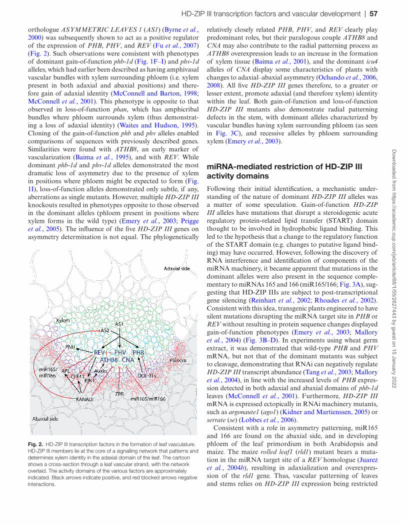
Following their initial identification, a mechanistic under-standing of the nature of dominant HD-ZIP III alleles was a matter of some speculation. Gain-of-function HD-ZIP III alleles have mutations that disrupt a steroidogenic acute regulatory protein-related lipid transfer (START) domain thought to be involved in hydrophobic ligand binding. This led to the hypothesis that a change to the regulatory function of the START domain (e.g. changes to putative ligand bind-ing) may have occurred. However, following the discovery of RNA interference and identification of components of the miRNA machinery, it became apparent that mutations in the dominant alleles were also present in the sequence comple-mentary to miRNAs 165 and 166 (miR165/166; Fig. 3A), sug-gesting that HD-ZIP IIIs are subject to post-transcriptional gene silencing (Reinhart et al., 2002; Rhoades et al., 2002). Consistent with this idea, transgenic plants engineered to have silent mutations disrupting the miRNA target site in PHB or REV without resulting in protein sequence changes displayed gain-of-function phenotypes (Emery et al., 2003; Mallory et al., 2004) (Fig. 3B–D). In experiments using wheat germ extract, it was demonstrated that wild-type PHB and PHV mRNA, but not that of the dominant mutants was subject to cleavage, demonstrating that RNAi can negatively regulate HD-ZIP III transcript abundance (Tang et al., 2003; Mallory et al., 2004), in line with the increased levels of PHB expres-sion detected in both adaxial and abaxial domains of phb-1d leaves (McConnell et al., 2001). Furthermore, HD-ZIP III mRNA is expressed ectopically in RNAi machinery mutants, such as argonaute1 (ago1) (Kidner and Martienssen, 2005) or serrate (se) (Lobbes et al., 2006).
Consistent with a role in asymmetry patterning, miR165 and 166 are found on the abaxial side, and in developing phloem of the leaf primordium in both Arabidopsis and maize. The maize rolled leaf1 (rld1) mutant bears a muta-tion in the miRNA target site of a REV homologue (Juarez et al., 2004b), resulting in adaxialization and overexpres-sion of the rld1 gene. Thus, vascular patterning of leaves and stems relies on HD-ZIP III expression being restricted
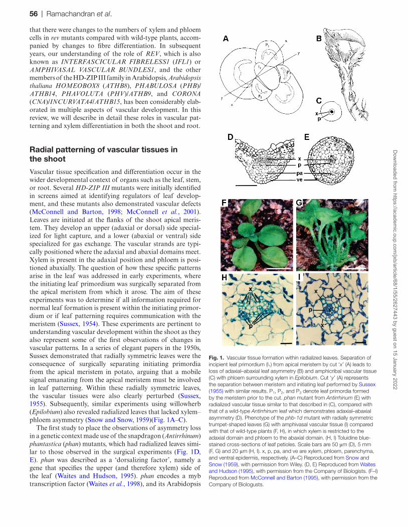
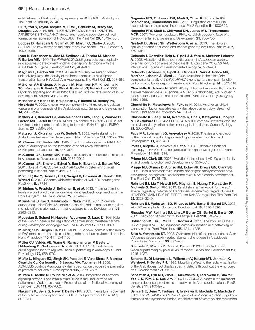
Fig. 2. HD-ZIP III transcription factors in the formation of leaf vasculature. HD-ZIP III members lie at the core of a signalling network that patterns and determines xylem identity in the adaxial domain of the leaf. The cartoon shows a cross-section through a leaf vascular strand, with the network overlaid. The activity domains of the various factors are approximately indicated. Black arrows indicate positive, and red blocked arrows negative interactions.
HD-ZIP III transcription factors and vascular development | 57D
ownloaded from
https://academic.oup.com
/jxb/article/68/1/55/2627443 by guest on 15 January 2022
through miRNA-mediated removal of HD-ZIP III mRNA from abaxial domains in both eudicots and monocots. Interestingly, in situ hybridization of miR166 localization in the maize leaf primordium revealed a dynamic and graded distribution on the abaxial/phloem side of the leaf, leading Juarez et al. (2004a) to note that it behaved as a movable signal.
Focusing of miR166 to the abaxial side of the maize leaf is thought to be the result of the action of trans-acting short-interfering RNAs (ta-siRNAs; for review, see Chitwood et al., 2007). Briefly, in contrast to conventional miRNA-directed cleavage which results in the degradation of the target mRNA (e.g. miR165/166 action on HD-ZIP III transcripts described above), cleavage of a non-coding TAS RNA enables it to become a target for RNA-dependent RNA polymerases. The resulting dsRNA is subject to further processing from which 21 bp ta-siRNAs are generated. ta-siRNAs guide cleavage of mRNA targets in a similar manner to miRNAs. ta-siR-NAs are derived from miRNA action on non-coding TAS transcripts. In Arabidopsis, ta-siRNAs, derived from TAS3 that has been subjected to cleavage by miR390, negatively regulate ETTIN (ETT), also known as AUXIN RESPONSE FACTOR3 (ARF3), and ARF4, two genes that act
redundantly in abaxial leaf identity (Chitwood et al., 2007). In maize, LEAFBLADELESS1 (LBL1) encodes a zinc finger protein required for the generation of ta-siRNAs, and in lbl1 mutants, the localization of miR166 is no longer restricted to the abaxial domain of the initiating leaf primordium, but is expressed throughout. lbl mutants demonstrate a clear loss of adaxial–abaxial asymmetry (Nogueira et al., 2007), con-sistent with downstream changes to levels of HD-ZIP III transcript (Nogueira et al., 2009). One possibility is that these small RNAs could act non-cell autonomously and thus are candidates for the ‘Sussex signal’, proposed in the early surgi-cal experiments described above that are involved in crosstalk between the shoot apical meristem and initiating leaf primor-dium (Chitwood et al., 2007).
Disruption of the interactions between miRNA and mRNA target has provided particular insight into the roles that HD-ZIP IIIs play in vascular tissue forma-tion. HD-ZIP IIIs are required for vascular tissue in the leaves, as overexpression of one of the two genes encod-ing miR165, MIR165A, results in leaves that entirely lack vascular tissue (Zhou et al., 2007). An activation tagging line, jabba-1d (jba-1d) that resulted in increases in expres-sion of MIR166G, one of the seven miR166-encoding genes,
Fig. 3. Dominant HD-ZIP III alleles discussed in this review. (A) HD-ZIP III domain structure, with miRNA complementary site marked. Protein (upper) and mRNA nucleotide (lower) sequences from the different HD-ZIP III alleles are shown below. (B–D) Toluidine blue-stained cross-sections of vascular bundles from the inflorescence stems of the wild type (B) which has xylem to the centre of the stem and phloem towards the outside, compared with that of rev-10d (C) where xylem surrounds the phloem. In plants expressing a version of REV harbouring silent point mutations in the miRNA target site (D; rev-δmiRNA), some vascular bundles (lower right in D) demonstrate similar phenotypes to rev-10d (C), with xylem surrounding phloem. ph is phloem, xy is xylem; arrowheads point to xylem cells. (B–D) Reproduced from Emery et al. (2003) with permission from Elsevier.
58 | Ramachandran et al.D
ownloaded from
https://academic.oup.com
/jxb/article/68/1/55/2627443 by guest on 15 January 2022
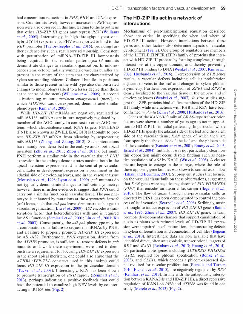
had concomitant reductions in PHB, PHV, and CNA expres-sion. Counterintuitively, however, increases in REV expres-sion were also observed in this line, leading to the hypothesis that other HD-ZIP III genes may repress REV (Williams et al., 2005). Interestingly, in high-throughput yeast one-hybrid (Y1H) experiments, PHV was reported to bind to the REV promoter (Taylor-Teeples et al., 2015), providing fur-ther evidence for such a regulatory relationship. Consistent with perturbation of miRNA–HD-ZIP III homeostasis being required for the vascular pattern, jba-1d mutants demonstrate changes to vascular organization. In inflores-cence stems, ectopic radially symmetric vascular bundles are present in the centre of the stem that are characterized by xylem surrounding phloem. Collateral bundles in positions similar to those present in the wild type also demonstrated changes to morphology (albeit to a lesser degree than those at the centre of the stem) (Williams et al., 2005). A second activation tag mutant, meristem enlargement1 (men1), in which MIR166A was overexpressed, demonstrated similar phenotypes (Kim et al., 2005).
While HD-ZIP III mRNAs are negatively regulated by miR165/166, miRNAs are in turn negatively regulated by a member of the AGO family. In contrast to other AGO pro-teins, which cleave/silence small RNA targets, PINHEAD (PNH; also known as ZWILLE/AGO10) is thought to pro-tect HD-ZIP III mRNA from silencing by sequestering miR165/166 (Zhang and Zhang, 2012). Such interactions have mainly been described in the embryo and shoot apical meristem (Zhu et al., 2011; Zhou et al., 2015), but might PNH perform a similar role in the vascular tissue? PNH expression in the embryo demonstrates maxima both in the apical meristem expression and in the central provascular cells. Later in development, expression is prominent in the adaxial side of developing leaves, and in the vascular tissue (Moussian et al., 1998; Lynn et al., 1999). pnh mutants do not typically demonstrate changes to leaf vein asymmetry; however, there is further evidence to suggest that PNH could carry out a similar function in vascular tissue. The pnh phe-notype is enhanced by mutations at the asymmetric leaves2 (as2) locus, such that as2 pnh leaves demonstrate changes to vascular organization (Liu et al., 2009). AS2 encodes a tran-scription factor that heterodimerizes with and is required for AS1 function (Semiarti et al., 2001; Lin et al., 2003; Xu et al., 2003). Consequently, the as2 pnh phenotype may be a combination of a failure to sequester miRNAs by PNH, and a failure to properly promote HD-ZIP III expression by AS1-AS2. Furthermore, PNH expression, driven from the ATHB8 promoter, is sufficient to restore defects in pnh mutants, and, while these experiments were used to dem-onstrate a requirement for focusing HD-ZIP III expression in the shoot apical meristem, one could also argue that the ATHB8::YFP-ZLL construct used in this analysis could focus HD-ZIP III expression in the provascular domain (Tucker et al., 2008). Interestingly, REV has been shown to promote transcription of PNH rapidly (Reinhart et al., 2013), perhaps indicating a positive feedback that could have the potential to canalize high REV levels by counter-acting miR165/166s (Fig. 2).
The HD-ZIP IIIs act in a network of interactions
Mechanisms of post-transcriptional regulation described above are critical in specifying the when and where of HD-ZIP III action. However, interactions between these genes and other factors also determine aspects of vascular development (Fig. 2). One group of regulators are members of the LITTLE ZIPPER (ZPR) family of proteins that inter-act with HD-ZIP III proteins by forming complexes, through interactions at the zipper domain, and thereby preventing HD-ZIP III binding to DNA (Wenkel et al., 2007; Kim et al., 2008; Husbands et al., 2016). Overexpression of ZPR genes results in vascular defects including cellular proliferation adjacent to veins in the leaf and changes to xylem–phloem asymmetry. Furthermore, expression of ZPR1 and ZPR3 is clearly localized to the vascular tissue in the embryo and in developing leaves (Wenkel et al., 2007). In vitro studies sug-gest that ZPR proteins bind all five members of the HD-ZIP III family, while interactions with PHB and REV have been confirmed in planta (Kim et al., 2008; Husbands et al., 2016).
Genes of the KANADI family of GRAS-type transcription factors were shown a number of years ago to act in opposi-tion to HD-ZIP IIIs in radial patterning. In particular, where HD-ZIP IIIs specify the adaxial side of the leaf and the xylem side of the vascular tissue, KAN genes, of which there are four, specify the abaxial side of the leaf and the phloem side of the vasculature (Kerstetter et al., 2001; Emery et al., 2003; Eshed et al., 2004). Initially, it was not particularly clear how this opposition might occur, despite findings such as nega-tive regulation of AS2 by KAN1 (Wu et al., 2008). A clearer picture began to emerge in the embryo, where the role of these opposing gene families was shown to control auxin flow (Izhaki and Bowman, 2007). Subsequent studies that focused on the vascular tissue built on these observations, suggesting that KAN genes were negative regulators of PIN-FORMED1 (PIN1) that encodes an auxin efflux carrier (Ilegems et al., 2010). The flow of auxin through pre-provascular cells, as directed by PIN1, has been demonstrated to control the pro-cess of leaf venation (Scarpella et al., 2006). Strikingly, auxin is thought to induce expression of HD-ZIP III genes (Baima et al., 1995; Zhou et al., 2007). HD ZIP III genes, in turn, promote developmental changes that support canalization of auxin as plants with reduced levels of HD-ZIP III expres-sion were impaired in cell maturation, demonstrating defects in xylem differentiation and connection of cell files (Ilegems et al., 2010). Interestingly, data are now available that have identified direct, often antagonistic, transcriptional targets of REV and KAN1 (Reinhart et al., 2013; Huang et al., 2014). Of particular note, genes including ALTERED PHLOEM (APL), required for phloem specification (Bonke et al., 2003), and CLE41, which encodes a phloem-expressed sig-nal required for vascular proliferation (Etchells and Turner, 2010; Etchells et al., 2015), are negatively regulated by REV (Reinhart et al., 2013). In line with the antagonistic interac-tion between KANADIs and HD-ZIP IIIs, a direct repressive regulation of KAN1 on PHB and ATHB8 was found in one study (Merelo et al., 2013) (Fig. 2).
HD-ZIP III transcription factors and vascular development | 59D
ownloaded from
https://academic.oup.com
/jxb/article/68/1/55/2627443 by guest on 15 January 2022
Cell to cell movement of miR165/166 patterns the root vasculature
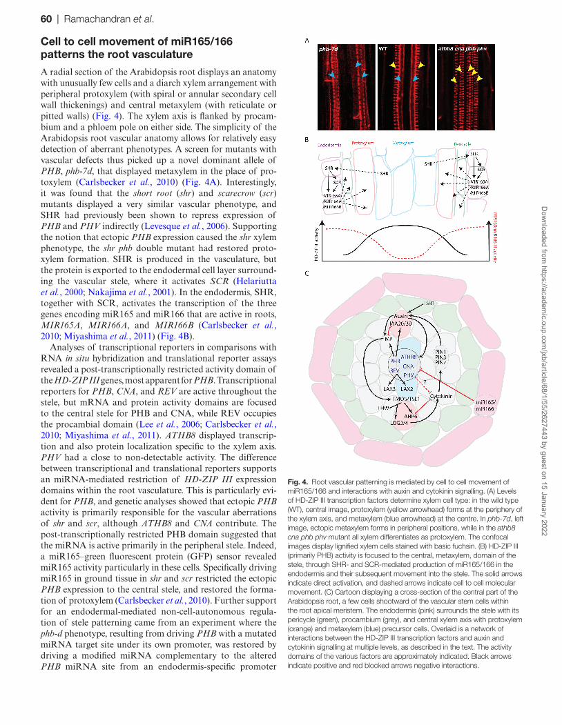
A radial section of the Arabidopsis root displays an anatomy with unusually few cells and a diarch xylem arrangement with peripheral protoxylem (with spiral or annular secondary cell wall thickenings) and central metaxylem (with reticulate or pitted walls) (Fig. 4). The xylem axis is flanked by procam-bium and a phloem pole on either side. The simplicity of the Arabidopsis root vascular anatomy allows for relatively easy detection of aberrant phenotypes. A screen for mutants with vascular defects thus picked up a novel dominant allele of PHB, phb-7d, that displayed metaxylem in the place of pro-toxylem (Carlsbecker et al., 2010) (Fig. 4A). Interestingly, it was found that the short root (shr) and scarecrow (scr) mutants displayed a very similar vascular phenotype, and SHR had previously been shown to repress expression of PHB and PHV indirectly (Levesque et al., 2006). Supporting the notion that ectopic PHB expression caused the shr xylem phenotype, the shr phb double mutant had restored proto-xylem formation. SHR is produced in the vasculature, but the protein is exported to the endodermal cell layer surround-ing the vascular stele, where it activates SCR (Helariutta et al., 2000; Nakajima et al., 2001). In the endodermis, SHR, together with SCR, activates the transcription of the three genes encoding miR165 and miR166 that are active in roots, MIR165A, MIR166A, and MIR166B (Carlsbecker et al., 2010; Miyashima et al., 2011) (Fig. 4B).
Analyses of transcriptional reporters in comparisons with RNA in situ hybridization and translational reporter assays revealed a post-transcriptionally restricted activity domain of the HD-ZIP III genes, most apparent for PHB. Transcriptional reporters for PHB, CNA, and REV are active throughout the stele, but mRNA and protein activity domains are focused to the central stele for PHB and CNA, while REV occupies the procambial domain (Lee et al., 2006; Carlsbecker et al., 2010; Miyashima et al., 2011). ATHB8 displayed transcrip-tion and also protein localization specific to the xylem axis. PHV had a close to non-detectable activity. The difference between transcriptional and translational reporters supports an miRNA-mediated restriction of HD-ZIP III expression domains within the root vasculature. This is particularly evi-dent for PHB, and genetic analyses showed that ectopic PHB activity is primarily responsible for the vascular aberrations of shr and scr, although ATHB8 and CNA contribute. The post-transcriptionally restricted PHB domain suggested that the miRNA is active primarily in the peripheral stele. Indeed, a miR165–green fluorescent protein (GFP) sensor revealed miR165 activity particularly in these cells. Specifically driving miR165 in ground tissue in shr and scr restricted the ectopic PHB expression to the central stele, and restored the forma-tion of protoxylem (Carlsbecker et al., 2010). Further support for an endodermal-mediated non-cell-autonomous regula-tion of stele patterning came from an experiment where the phb-d phenotype, resulting from driving PHB with a mutated miRNA target site under its own promoter, was restored by driving a modified miRNA complementary to the altered PHB miRNA site from an endodermis-specific promoter
Fig. 4. Root vascular patterning is mediated by cell to cell movement of miR165/166 and interactions with auxin and cytokinin signalling. (A) Levels of HD-ZIP III transcription factors determine xylem cell type: in the wild type (WT), central image, protoxylem (yellow arrowhead) forms at the periphery of the xylem axis, and metaxylem (blue arrowhead) at the centre. In phb-7d, left image, ectopic metaxylem forms in peripheral positions, while in the athb8 cna phb phv mutant all xylem differentiates as protoxylem. The confocal images display lignified xylem cells stained with basic fuchsin. (B) HD-ZIP III (primarily PHB) activity is focused to the central, metaxylem, domain of the stele, through SHR- and SCR-mediated production of miR165/166 in the endodermis and their subsequent movement into the stele. The solid arrows indicate direct activation, and dashed arrows indicate cell to cell molecular movement. (C) Cartoon displaying a cross-section of the central part of the Arabidopsis root, a few cells shootward of the vascular stem cells within the root apical meristem. The endodermis (pink) surrounds the stele with its pericycle (green), procambium (grey), and central xylem axis with protoxylem (orange) and metaxylem (blue) precursor cells. Overlaid is a network of interactions between the HD-ZIP III transcription factors and auxin and cytokinin signalling at multiple levels, as described in the text. The activity domains of the various factors are approximately indicated. Black arrows indicate positive and red blocked arrows negative interactions.
60 | Ramachandran et al.D
ownloaded from
https://academic.oup.com
/jxb/article/68/1/55/2627443 by guest on 15 January 2022
(Miyashima et al., 2011). Hence, miR165/166 derived from the endodermis move several cells away to restrict the mRNA activity domain of the HD-ZIP III transcription factors (pri-marily PHB) within the stele, and thereby control vascular patterning (Fig. 4B).
The critical role of cell to cell trafficking in root vascular patterning was further confirmed by blocking plasmodesmata connections. Gain-of-function alleles of callose synthase 3 (cals3-d) overproduce callose at plasmodesmata, hindering macromolecular cell to cell passage. This results in a root vascular phenotype similar to that of a phb-d or shr mutant. In these lines, PHB is ectopically active throughout the stele, and SHR movement into the endodermis fails (Vatén et al., 2011). Driving a dominant and inducible version of cals3 by tissue-specific promoters further allowed Vatén et al. (2011) to analyse the consequence of blocking plasmodesmata con-nections between the ground tissue and the stele on miR165 accumulation. In this experiment, miR165 and callose syn-thase were simultaneously induced in the ground tissue of a shr mutant. In situ hybridization revealed that miR165 accu-mulated in the ground tissue, compared with controls. Thus, these findings demonstrated plasmodesmata-mediated cell to cell mobility of the miRNA.
Ectopic expression of miR165 throughout the stele results in protoxylem forming in metaxylem positions in the xylem axis. In line with this, plants harbouring mutations in four of the five HD-ZIP III genes also display protoxylem through-out the xylem axis, while lower order mutants may display formation of a central metaxylem strand flanked by several protoxylem files (Carlsbecker et al., 2010) (Fig. 4A). The quintuple HD-ZIP III mutant does not form xylem at all. These phenotypes, together with that of phb-d mutants where metaxylem replaces protoxylem, indicate that HD-ZIP III transcription factors determine xylem cell identity in a dose-dependent fashion, with high dosage resulting in metaxylem and lower dosage in protoxylem (Carlsbecker et al., 2010). Notably, phb-d affects not only xylem cell type formation, but also pericycle cell identity (Miyashima et al., 2011). Thus, miR165/166 may form a morphogenetic gradient emanating from the endodermal cell layer, determining stele cell identity.
HD-ZIP III activity intersects with auxin and cytokinin signalling for proper xylem patterning
The HD-ZIP III-miRNA gradients in the root is overlaid by balanced auxin and cytokinin signalling domains shown to establish xylem and procambium cell identity, respectively (Bishopp et al., 2011) (Fig. 4C). Multiple points of intersec-tion between these two hormones and the HD-ZIP III tran-scription factors occur during root vascular patterning. Auxin biosynthesis is primarily tryptophan dependent, and conse-quently requires the enzyme TRYPTOPHAN SYNTHASE. Two alleles (trp2-12 and trp2-13) of the gene encoding the β-subunit (TSB1/TRP2) of this enzyme were identified from a screen for mutants with altered root vascular develop-ment. The trp2 mutants along with other auxin biosynthesis
mutants that are defective in downstream biosynthesis steps, such as the weak ethylene insensitive 8 tryptophan aminotrans-ferase related 2 (wei8 tar2) double mutant or a quintuple yucca mutant, displayed defective metaxylem development and protoxylem formation in the metaxylem position, suggesting that auxin biosynthesis is required for metaxylem formation (Ursache et al., 2014). A similar phenotype was observed in axr3-3, which harbours a gain-of-function mutation in IAA17 that inhibits auxin signalling. The vascular defects in trp2 were rescued by treatment with l-tryptophan while treatment with l-kynurenine (Kyn), which blocks TAA1/TAR-mediated auxin biosynthesis, phenocopied the auxin biosynthesis mutants with the formation of protoxylem in the metaxylem position. In line with the similarity of this phe-notype to higher order HD-ZIP III mutants, the expression of PHB, PHV, CNA, and ATHB8 was greatly reduced in the trp2 mutants and upon Kyn treatment of the wild type. Kyn resistance was brought about by driving PHB expression by an auxin-non-responsive promoter. Taken together with the partial rescue of the phb-7d xylem phenotype by Kyn treat-ment, this revealed an auxin biosynthesis-mediated, HD-ZIP III-dependent, vascular development pathway required pri-marily for metaxylem formation (Ursache et al., 2014).
The interconnection between HD-ZIP III and auxin was previously shown by the auxin-inducible characteristic of ATHB8 (Baima et al., 1995). Studies on vascular pattern-ing in the leaf showed that the accumulation of the DR5 auxin reporter preceded procambium formation, and was closely followed by activation of the auxin response factor ARF5/MONOPTEROS (MP) and ATHB8 (Mattsson et al., 2003). Donner et al. (2009) subsequently demonstrated that ATHB8 transcription is directly regulated by MP. However, neither in the leaf nor in the root meristem is there a precise correlation between domains of high auxin signalling and transcription domains of the five HD-ZIP III genes. Hence, other as yet unidentified factors probably contribute to their activation and/or restriction. Efforts to identify gene regula-tory networks around the HD-ZIP III genes may be probed for such candidates (Brady et al., 2011; Taylor-Teeples et al., 2015) (see also later).
In the post-embryonic root meristem, auxin response reporters suggest an auxin sink at the position of the imma-ture xylem axis. The accumulation of auxin is brought about by polar auxin transport, via PIN1 and procambially local-ized PIN3 and PIN7 mediating lateral auxin transport. Inhibition of polar auxin transport by exogenous supply of N-1-naphthylphthalamic acid (NPA) led to loss of protox-ylem strand formation in a dose-dependent manner (Bishopp et al., 2011) (Fig. 4C). In the protoxylem domain, the auxin maximum activates an inhibitor of cytokinin signalling, ARABIDOPSIS HISTIDINE PHOSPHOTRANSFER PROTEIN 6 (AHP6). In ahp6 mutants, the protoxylem strand integrity is affected similarly to the wild-type root subjected to exogenous cytokinin treatment, such that protoxylem becomes replaced by procambial cells. On the other hand, cytokinin depletion or a block in cytokinin signalling led to differentiation of all vascular cells as protoxylem (Mähönen et al., 2006). Therefore, inhibition of cytokinin signalling
HD-ZIP III transcription factors and vascular development | 61D
ownloaded from
https://academic.oup.com
/jxb/article/68/1/55/2627443 by guest on 15 January 2022
in the xylem axis is necessary for vessel formation, and the presence of cytokinin signalling in the procambial cells is required for maintaining them in an undifferentiated state. Interestingly, the phb-7d mutant lacks expression of AHP6 while, in contrast, athb8 cna phb phv quadruple mutants demonstrate expansion of the AHP6 expression domain to the entire xylem axis (Carlsbecker et al., 2010) (Fig. 4C). To predict the minimal molecular signalling circuits required for proper radial patterning in the Arabidopsis root, Muraro et al. (2014) generated a mathematical model with which they were able to reconstitute a realistic radial pattern, but only by integrating SHR–miR165/166–PHB with the above-described auxin and cytokinin signalling loop. In their model, they predicted PHB to act as repressor of AHP6 expression in the metaxylem domain. In support of this prediction, the expression of AHP6 rapidly increases upon induction of miR165 (Müller et al., 2016), although it is unknown if this interaction is direct, or occurs via the effect that HD-ZIP III transcription factors have on auxin signalling.
Several observations suggest that levels of HD-ZIP III transcription factors affect auxin signalling: auxin signalling reporters revealed considerable increases in activity in the athb8 cna phb phv mutant compared with the wild type, while phb-7d mutants displayed severely impaired auxin signal-ling in the xylem axis that could not be revived by exogenous auxin treatments (Müller et al., 2016). Similarly, up-regula-tion of miR165 resulted in a wider auxin reporter expression domain, and a number of core auxin signalling genes were increased, along with a down-regulation of primarily PHB, PHV, and CNA (Ilegems et al., 2010; Müller et al., 2016). However, despite being auxin inducible, MP, IAA20, and IAA30 were down-regulated upon miR165 induction, and PHB was found to bind the promoters of MP and IAA20 in vivo, suggesting that PHB is required at their promoters for proper activation (Müller et al., 2016). In contrast to most AUX/IAA proteins, IAA20 and IAA30 lack the canonical domain II, recognized by the auxin/TIR receptor complex, and are therefore not degraded even in the presence of high auxin levels. Their interactions with ARFs, however, are not altered, and they may therefore act as ARF scavengers and dampen auxin signalling (Sato and Yamamoto, 2008). The double mutant iaa20 iaa30 displays formation of extra pro-toxylem strands, suggesting that a balanced auxin response is required for proper root vascular patterning (Müller et al., 2016). Similarities in the phenotypes of a weak mp mutant and lines overexpressing IAA30 indicate that IAA30 (and IAA20) probably represses the activity of MP. Activation by PHB (and other HD-ZIP IIIs) of components both promot-ing and suppressing auxin signalling may balance the vascular auxin response, and genetic data suggest that this promotes a stable xylem axis patterning.
Thus, several studies show a tight link between HD-ZIP III transcription factors and auxin signalling on many differ-ent levels (recently reviewed by Turchi et al., 2015). A direct binding of REV to the promoters of the auxin influx car-riers AUX1, LAX2, and LAX3 was identified (Baima et al., 2014; Huang et al., 2014), and the expression of these genes was significantly altered upon the induction of miR165 in the
root and shoot (Baima et al., 2014; Müller et al., 2016). The triple aux1 lax1 lax2 mutant has aberrant protoxylem forma-tion (El-Showk et al., 2015) and, along with previously men-tioned results obtained by blocking polar auxin transport, this supports the notion that the activity of both auxin influx and efflux carriers is required to attain sufficient auxin accu-mulation for proper protoxylem and metaxylem formation. As a consequence of auxin accumulation in the xylem axis, a number of downstream genes that play a role in xylem cell specification and differentiation are switched on (see below).
A role for HD-ZIP III genes in restricting procambial cell proliferation?
In the embryo, the first vascular cells are initiated in the cen-tral globular staged embryo (Scheres et al., 1995). Analyses of expression revealed the presence of REV, PHB, PHV, and CNA expression in apical parts of the embryo from the early globular stage, while ATHB8 appears a little later, at the early heart stage, in the provascular cells where it is later joined by the other family members (Baima et al., 1995; Prigge et al., 2005; Smith and Long, 2010). Thus, although expression of the HD-ZIP III genes is initiated early, their activity domains are not perfectly overlapping that of the first vascular cells, suggesting that their activity in the procambium follows the initiation of the first vascular cells. A pathway mediated by TARGET OF MONOPTEROS 5 (TMO5) along with its interaction partner LONESOME HIGHWAY (LHW) con-trols periclinal cell divisions in the embryo essential for the radial vascular axis and also for the maintenance of vascular cell number in the post-embryonic root meristem (De Rybel et al., 2013; Ohashi-Ito et al., 2013) (Fig. 4C). Alterations in cell number have been attributed to shifts in the auxin–cyto-kinin balance as long-term treatment with NPA increases the vascular cell number and subsequently the number of xylem poles (Bishopp et al., 2011), while impaired cytokinin signalling results in reduced procambial cell proliferation (Mähönen et al., 2000). TMO5 and its homologue TMO5-LIKE1 (T5L1) are expressed specifically in the xylem axis. As dimers with LHW they directly control the expression of the rate-limiting cytokinin biosynthesis genes LONELY GUY3 (LOG3) and LOG4 (De Rybel et al., 2014; Ohashi-Ito et al., 2014), which would serve to increase cytokinin levels in the xylem axis. However, cytokinin reporters reveal that signalling primarily occurs in the procambium. Potentially, activation of AHP6 by T5L1/LHW may restrict the effect of cytokinin from the xylem domain (Ohashi-Ito et al., 2014). However, AHP6 is not active in the central metaxylem/PHB activity domain of the xylem axis. It is possible that PHB contributes by other means to the reduced cytokinin respon-siveness of these cells; a recent publication may provide a possible mechanism, as it was found that PHB can prevent the activity of B-type response regulators (B-ARRs) poten-tially by preventing B-ARR DNA binding, especially under high cytokinin level conditions (Sebastian et al., 2015). The role for PHB and the other HD-ZIP III transcription fac-tors as potential regulators of procambial cell proliferation
62 | Ramachandran et al.D
ownloaded from
https://academic.oup.com
/jxb/article/68/1/55/2627443 by guest on 15 January 2022
needs to be substantiated by more research; however, several observations suggest a role for the HD-ZIP IIIs in regulat-ing procambial cell divisions. The athb8 cna phb phv mutant has a significant increase in the number of root procambial cells compared with the wild type, resulting in a triarch or tetrarch vascular arrangement. Driving miR165 in the stele also causes a similar increase in the number of vascular cells (Carlsbecker et al., 2010; Ilegems et al., 2010). Conversely, the phb-d alleles contain fewer stele cells (Carlsbecker et al., 2010). While there are as yet only clues as to how HD-ZIP IIIs might ultimately regulate this process, one possibility is that expres-sion of HD-ZIP IIIs in the procambium may be regulated by DOF transcription factors. Seven different DOF genes were found to interact with the promoters of PHB and PHV, and in certain cases a single DOF could act as an activator of one HD-ZIP III gene while repressing another (Brady et al., 2011) (Fig. 2). DOF transcription factors are expressed early in procambium formation in the leaf (Gardiner et al., 2010), and some members of the gene family act to control vascu-lar cell division (Guo et al., 2009; for a review, see Le Hir and Bellini, 2013). Complex networks of interactions such as this are present around HD-ZIP III transcription factors as shown in transcriptional regulatory network analysis for both the stele and xylem (Brady et al., 2011; Taylor-Teeples et al., 2015). The connections in such networks point to interesting regulatory relationships. In the case of DOF regulation of PHB and PHV, further work is required to understand the significance of this interaction.
HD-ZIP III regulated differentiation of xylem cells
While the analysis HD-ZIP III function described above looks at changes to vascular patterning and organization, HD-ZIP IIIs also function post-patterning, in particular in differentiation of the xylem. Early work on the role of REV in xylem differentiation followed the independent isolation of REV loss-of-function alleles by Zhong and Ye (ifl1 alleles of
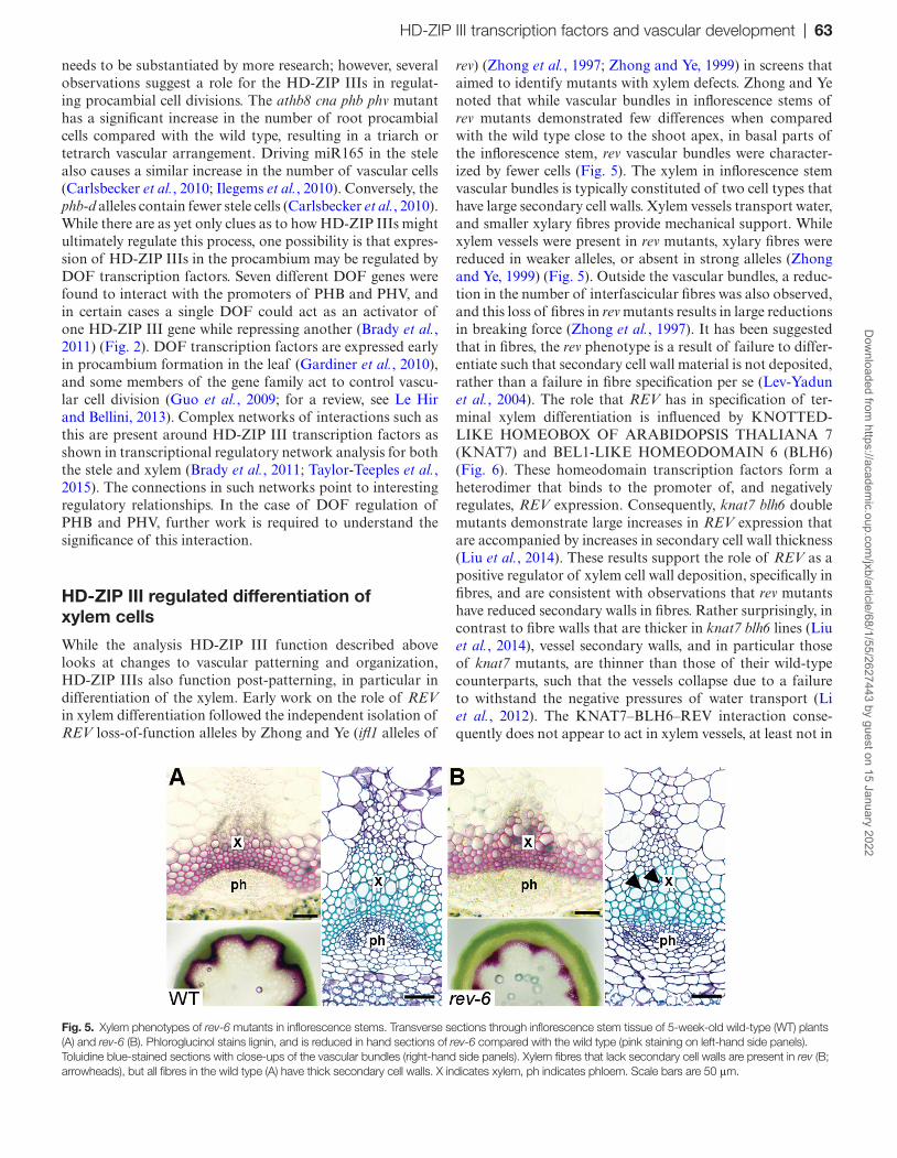
rev) (Zhong et al., 1997; Zhong and Ye, 1999) in screens that aimed to identify mutants with xylem defects. Zhong and Ye noted that while vascular bundles in inflorescence stems of rev mutants demonstrated few differences when compared with the wild type close to the shoot apex, in basal parts of the inflorescence stem, rev vascular bundles were character-ized by fewer cells (Fig. 5). The xylem in inflorescence stem vascular bundles is typically constituted of two cell types that have large secondary cell walls. Xylem vessels transport water, and smaller xylary fibres provide mechanical support. While xylem vessels were present in rev mutants, xylary fibres were reduced in weaker alleles, or absent in strong alleles (Zhong and Ye, 1999) (Fig. 5). Outside the vascular bundles, a reduc-tion in the number of interfascicular fibres was also observed, and this loss of fibres in rev mutants results in large reductions in breaking force (Zhong et al., 1997). It has been suggested that in fibres, the rev phenotype is a result of failure to differ-entiate such that secondary cell wall material is not deposited, rather than a failure in fibre specification per se (Lev-Yadun et al., 2004). The role that REV has in specification of ter-minal xylem differentiation is influenced by KNOTTED-LIKE HOMEOBOX OF ARABIDOPSIS THALIANA 7 (KNAT7) and BEL1-LIKE HOMEODOMAIN 6 (BLH6) (Fig. 6). These homeodomain transcription factors form a heterodimer that binds to the promoter of, and negatively regulates, REV expression. Consequently, knat7 blh6 double mutants demonstrate large increases in REV expression that are accompanied by increases in secondary cell wall thickness (Liu et al., 2014). These results support the role of REV as a positive regulator of xylem cell wall deposition, specifically in fibres, and are consistent with observations that rev mutants have reduced secondary walls in fibres. Rather surprisingly, in contrast to fibre walls that are thicker in knat7 blh6 lines (Liu et al., 2014), vessel secondary walls, and in particular those of knat7 mutants, are thinner than those of their wild-type counterparts, such that the vessels collapse due to a failure to withstand the negative pressures of water transport (Li et al., 2012). The KNAT7–BLH6–REV interaction conse-quently does not appear to act in xylem vessels, at least not in
Fig. 5. Xylem phenotypes of rev-6 mutants in inflorescence stems. Transverse sections through inflorescence stem tissue of 5-week-old wild-type (WT) plants (A) and rev-6 (B). Phloroglucinol stains lignin, and is reduced in hand sections of rev-6 compared with the wild type (pink staining on left-hand side panels). Toluidine blue-stained sections with close-ups of the vascular bundles (right-hand side panels). Xylem fibres that lack secondary cell walls are present in rev (B; arrowheads), but all fibres in the wild type (A) have thick secondary cell walls. X indicates xylem, ph indicates phloem. Scale bars are 50 μm.
HD-ZIP III transcription factors and vascular development | 63D
ownloaded from
https://academic.oup.com
/jxb/article/68/1/55/2627443 by guest on 15 January 2022
the same way that it regulates wall deposition in fibres. One explanation of this phenotype is that KNAT7/BLH6 acts independently from REV in vessel element differentiation.
A number of observations have supported a role for other members of the HD-ZIP III family in xylem develop-ment and differentiation. Analysis of HD-ZIP III expres-sion in Zinnia elegans leaves found that REV homologues, ZeHB11 and ZeHB12, demonstrated xylem expression, as did ATHB8 and CNA orthologues (ZeHB-10 and ZeHB-13, respectively), albeit in an expression domain consistent with these genes having a role in early xylem specification, rather than in deposition of cell wall polymers (Ohashi-Ito and Fukuda, 2003). Such a hypothesis is supported by the obser-vation that constitutive overexpression of MIR165B, which results in reductions in CNA expression, and probably that of other HD-ZIP IIIs leads to ectopic deposition of second-ary cell wall material in the pith of Arabidopsis stems (Du et al., 2015). Subsequent work, which tested genetic redun-dancy between rev and the other HD-ZIP III transcription factors, showed that phb and phv were strong enhancers of the rev phenotype in the xylem; in extreme cases, rev phb/+ and rev phv mutants displayed vascular bundles with remarkably few lignified cells (Prigge et al., 2005). In contrast, lignifica-tion of xylem tissue and interfascicular fibres was restored in athb8 cna rev triple mutants (i.e. athb8 cna suppressed the rev phenotype). The idea that ATHB8 and CNA have distinct functions from those of PHB, PHV, and REV is supported by experiments in a rev mutant background where expression of HD-ZIP III family members was driven from the REV
promoter. While REV::REV, REV::PHB, and REV::PHV constructs rescued the rev mutant phenotype, REV::ATHB8 and REV::CNA did not (Prigge et al., 2005). Taken together, these observations suggest that early xylem specification may be controlled by ATHB8 and CNA, while differentiation to mature xylem is repressed by these genes. In contrast, REV, PHV, and PHB are positive regulators of the final stages of xylem differentiation.
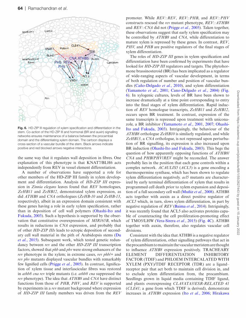
The roles of HD-ZIP III genes in xylem specification and differentiation have been confirmed by experiments that have looked for HD-ZIP III regulators and targets. The phytohor-mone brassinosteroid (BR) has been implicated as a regulator of wide-ranging aspects of vascular development, in terms of both regulation of number and position of vascular bun-dles (Caño-Delgado et al., 2010), and xylem differentiation (Yamamoto et al., 2001; Cano-Delgado et al., 2004) (Fig. 6). In xylogenic cultures, levels of BR have been shown to increase dramatically at a time point corresponding to entry into the final stages of xylem differentiation. Rapid induc-tion of REV homologue transcripts, ZeHB11 and ZeHB12, occurs upon BR treatment. In contrast, expression of the same transcripts is repressed upon treatment with unicona-zole, a BR inhibitor (Yamamoto et al., 2001, 2007; Ohashi-Ito and Fukuda, 2003). Intriguingly, the behaviour of the ATHB8 orthologue ZeHB10 is similarly regulated, and while ZeHB13, a CNA orthologue, is not repressed upon perturba-tion of BR signalling, its expression is also increased upon BR induction (Ohashi-Ito and Fukuda, 2003). This begs the question of how apparently opposing functions of ATHB8/CNA and PHB/PHV/REV might be reconciled. The answer probably lies in the position that each gene controls within a complex network. ACAULIS5 (ACL5) is a gene encoding a thermospermine synthase, which has been shown to regulate xylem differentiation negatively. acl5 mutants are character-ized by early terminal differentiation of xylem that results in programmed cell death prior to xylem expansion and deposi-tion of a full secondary cell wall (Muñiz et al., 2008). ATHB8 acts together with auxin as a direct positive regulator of ACL5 which, in turn, slows xylem differentiation, in part by negative regulation of REV (Baima et al., 2014). Intriguingly, it was recently found that ACL5 also activates proteins capa-ble of counteracting the cell proliferation-promoting effect of TMO5/LHW (Vera-Sirera et al., 2015) (Fig. 4C). ATHB8 together with auxin, therefore, also regulates vascular cell divisions.
Consistent with the idea that ATHB8 is a negative regulator of xylem differentiation, other signalling pathways that act in the procambium to maintain the vascular meristem are thought to influence ATHB8 expression positively. TRACHEARY ELEMENT DIFFERENTIATION INHIBITORY FACTOR (TDIF) and PHLOEM INTERCALATED WITH XYLEM (PXY)/TDIF RECEPTOR (TDR) are a ligand–receptor pair that act both to maintain cell division in, and to exclude xylem differentiation from, the procambium. Seedlings grown in liquid media containing TDIF ligand, and plants overexpressing CLAVATA3/ESR-RELATED 41 (CLE41; a gene from which TDIF is derived), demonstrate increases in ATHB8 expression (Ito et al., 2006; Hirakawa
Fig. 6. HD-ZIP III regulation of xylem specification and differentiation in the stem. Co-action of the HD-ZIP III and hormonal (BR and auxin) signalling networks ensures maintenance of a balance between the procambial domain and the differentiating xylem domain. The cartoon displays a cross-section of a vascular bundle of the stem. Black arrows indicate positive and red blocked arrows negative interactions.
64 | Ramachandran et al.D
ownloaded from
https://academic.oup.com
/jxb/article/68/1/55/2627443 by guest on 15 January 2022
et al., 2008; Etchells and Turner, 2010) (Fig. 6), which sup-ports the idea that ATHB8 acts to slow xylem differentiation.
Genetic analysis has supported a function for REV, PHB, and PHV in promoting xylem differentiation as described above. These observations were supported by recent experi-ments suggesting that REV (and PHV) both bind to the promoter of the xylem master regulator VASCULAR-RELATED NAC DOMAIN7 (VND7) (Fig. 6). In assays where constructs containing the VND7 promoter control-ling expression of a luciferase reporter were co-bombarded into Arabidopsis leaves with a 35S::REV construct, a 3-fold increase in promoter activity was observed compared with controls (Endo et al., 2015). Expression of VND7 has previ-ously been shown to result in adoption of xylem fate (Kubo et al., 2005). This leads to a model whereby adjacent to the procambium, where ATHB8/CNA show expression maxima, the xylem differentiation process is slowed by positive regu-lation of ACL5. However, expression of ATHB8/CNA and consequently ACL5 expression is lowered further from the procambium. Therefore, REV (and possibly PHB and PHV) would be released from this negative regulation by ACL5, enabling promotion of expression of VND7.
The HD-ZIP III transcription factors lie at the centre of a network that is required to fine-tune dynamic changes in gene expression throughout vascular development. High-throughput Y1H screens have recently been used to place PHB, PHV, and REV in a network that regulates secondary cell wall deposition. Interactions within this network include both VND7 and PHV binding to the promoter of REV, hint-ing at complex regulatory mechanisms. In particular, VND7 was reported to regulate REV expression negatively. REV, in turn, binds to the promoter and negatively regulates the expression of PHENYLALANINE AMMONIA LYASE4 (PAL4) (Taylor-Teeples et al., 2015), a gene involved in lignin biosynthesis (Sewalt et al., 1997). However, as REV has previ-ously been reported to regulate expression of VND7 positively (Endo et al., 2015), these results suggest that an understanding at cell type-specific resolution is required to understand how these interactions control commitment to xylem differentia-tion, fibre formation, and deposition of the secondary cell wall.
HD-ZIP III regulation of wood formation in trees
In tree species such as poplar, vascular tissue expansion is present in a continuous ring in the stem and is the main driver of secondary growth. It is clear that members of the HD-ZIP III family have an important role in regulating this process as REV, CNA, and ATHB8 orthologues, popREVOLUTA (PRE), POPCORONA (PCN), and PtrHB7, are expressed in poplar vascular tissue, and perturbations to the expression of these genes lead to defects in organization and wood deposi-tion (Du et al., 2011; Robischon et al., 2011; Zhu et al., 2013). While transgenic trees overexpressing a miRNA-resistant form of PCN had relatively subtle defects including early onset of secondary growth (Du et al., 2011), phenotypes of miRNA-resistant PRE overexpressers demonstrated much
more dramatic phenotypes, including areas of xylem present on both sides of the cambium. This is in contrast to wild-type poplar (and other woody species), where xylem is strictly restricted to the inner side of the cambium (Robischon et al., 2011). In another experiment, overexpression in poplar of the native REV homologue also resulted in reduction in the fibre to vessel ratio and associated changes in many genes relating to cell wall synthesis (Côté et al., 2010). Interestingly, genome-wide association studies identify links between the multiple splice variants in the 3' end of the REV locus and wood cellulose content in poplar (Porth et al., 2014).
One of the striking features of perennial woody plants is the annual rings that form in the wood due to differences in seasonal growth. In hybrid aspen, miR166 has been shown to be seasonally regulated, with a large peak in expression in the winter months. Elevated winter miR166 coincides with reduc-tions in expression of PtaHB1, a REV orthologue (Ko et al., 2006), suggesting that seasonal control of REV-directed wood development is at least in part via miR166 regulation.
It may be interesting to observe the roles that HD-ZIP IIIs and miRNAs might have in patterning of plants with unu-sual cambial organizations, for example those with included phloem such as Avicennia and Bougainvillea (Studholme and Philipson, 1966; Zamski, 1979), or plants that develop phloem wedges, such as members of the Bignonieae (Pace et al., 2009; Spicer and Groover, 2010).
Aside from miRNA-mediated regulation of HD-ZIP IIIs, other regulatory interactions are likely to be conserved across plants with differing growth habits. One such regula-tory interaction is that between PttHB8 (an ATHB8 ortho-logue) and poplar ACL5 (POPACAULIS5). POPACAULIS5 represses PttHB8 expression, while in contrast PttHB8 pro-motes expression of POPACAULIS5 expression, suggesting that thermospermine levels and PttHB8 expression are bal-anced by feedback control (Milhinhos et al., 2013).
Conifers and other gymnosperms also display extensive sec-ondary development, and also here HD-ZIP III transcripts are associated with secondary xylem (Côté et al., 2010; Duval et al., 2014). However, in conifers, the xylem tissues contain only tracheids, while vessels and fibres are missing. Potentially reflecting this, conifers have relatively few NAC-domain-containing VND homologues, while this gene family has expanded considerably in angiosperms (Nystedt et al., 2013). A recent study employing Agrobacterium-mediated transforma-tion of embryonic spruce cells to test for promoter–transcrip-tion factor interactions in planta in a semi-high-throughput manner found evidence for the regulation of multiple genes regulating secondary cell wall formation by a NAC-domain transcription factor (Duval et al., 2014), including interaction with a homologue to the angiosperm HD-ZIPIII genes from Picea glauca. However, the NAC-domain transcription factor most closely related to the VNDs, which also displayed expres-sion during secondary growth, did not show interaction with the HD-ZIP promoters tested. Thus, despite the ~300 million years of separate evolution, molecular circuits connecting HD-ZIP IIIs and NACs may be at least partially conserved. It will be interesting to learn whether the HD-ZIP IIIs are also important for conifer tracheid formation.
HD-ZIP III transcription factors and vascular development | 65D
ownloaded from
https://academic.oup.com
/jxb/article/68/1/55/2627443 by guest on 15 January 2022
Perspectives and outlook
While clearly a considerable amount is now known about the roles that HD-ZIP IIIs play in multiple aspects of vascular development, there are still a number of unanswered ques-tions, in particular pertaining to the apparently very com-plex loops of regulation these factors act in. Omics-based methods such as transcriptome analyses, Chip-seq, together with high-throughput interaction screening using Y1H, have revealed a complex transcriptional network around these fac-tors. Furthermore, despite the apparent redundancy these five factors display in certain genetic analyses, they sometimes act antagonistically, and the molecular basis for this will probably continue to be revealed by large-scale approaches. However, it is conceivable, or even likely, that different cellular, tissue, and organ contexts provide opportunities for different positions in molecular networks of the five family members. Therefore, improvements in techniques for cellular and tissue resolu-tion of large-scale omics assays, in methods for determining molecular interactions, and in modelling of both networks and development, are promising.
To complicate the image further, the HD-ZIP IIIs are, as mentioned, also regulated post-transcriptionally by miRNA, providing additional levels of complexity. In addition, HD-ZIP III protein activity is most probably closely regu-lated as well; the presence of the highly conserved START domain strongly suggests interactions with an as yet uniden-tified ligand. Furthermore, the C-terminus is occupied by a conserved domain, the MEHKLA domain, displaying simi-larity to Per Arnt Sim (PAS)-domains known to sense light, redox, or other stimuli (Mukherjee and Burglin, 2006). Thus far its function is not clear: the MEHKLA domain has been shown to be a site for protein–protein interactions (Chandler et al., 2007); alternative folding of this domain regulates REV activity (Magnani and Barton, 2011) and a point mutation in the MEHKLA domain of the hoc allele of CNA confers high regeneration competence, even in the absence of hormones (Duclercq et al., 2011). Intriguingly, whereas the MEHKLA domain might be redox sensitive, DNA binding of HD-ZIP IIIs can also be redox regulated (Comelli and Gonzalez, 2007; Xie et al., 2014). Considering that HD-ZIP III transcription factors appear active in the plant vasculature after its devel-opment programme is complete, it is tempting to speculate that these factors not only regulate the development of the vascular tissues, but also contribute to the function of the vasculature as an information highway, perhaps by transmit-ting information from one part of the plant to another.
The HD-ZIP III–miR165/166 regulon is highly conserved, and found not only in vascular plants but in all land plants, including mosses and liverworts (Floyd and Bowman, 2006; Floyd et al., 2006; Prigge and Clark, 2006). Strikingly, a HD-ZIP III gene from the moss (i.e. pre-vascular) species Physcomitrella patens regulates moss leaf development, including the conducting tissues, and partially suppresses the Arabidopsis rev phenotype (Prigge and Clark, 2006; Yip et al., 2016). In early vascular plants, lycophytes, and ferns, HD-ZIP III genes are associated with leaf development and procambium (Floyd and Bowman, 2006; Vasco et al., 2016).
It is conceivable that the HD-ZIP III–miR165/166 regulon evolved from an ancestral function in leaf patterning and growth also to govern vascular differentiation of cells with secondary cell walls. Analyses of the molecular networks in which the moss and liverwort HD-ZIP III homologues act will likely contribute not only to our understanding of vas-cular plant evolution, but perhaps also to the function of the famous five in the complex processes of patterning and differentiation of vascular tissues in Arabidopsis, and other vascular plants.
AcknowledgementsThis work was supported by the Swedish Research Council for Environment, Agricultural Sciences and Spatial Planning (FORMAS; 2013-953) to AC, and an EU-FP7 Marie Skłodowska-Curie fellowship to JPE.
ReferencesBaima S, Forte V, Possenti M, Peñalosa A, Leoni G, Salvi S, Felici B, Ruberti I, Morelli G. 2014. Negative feedback regulation of auxin signaling by ATHB8/ACL5–BUD2 transcription module. Molecular Plant 7, 1006–1025.
Baima S, Nobili F, Sessa G, Lucchetti S, Ruberti I, Morelli G. 1995. The expression of the Athb-8 homeobox gene is restricted to provascular cells in Arabidopsis thaliana. Development 121, 4171–4182.
Baima S, Possenti M, Matteucci A, Wisman E, Altamura MM, Ruberti I, Morelli G. 2001. The Arabidopsis ATHB-8 HD-Zip protein acts as a differentiation-promoting transcription factor of the vascular meristems. Plant Physiology 126, 643–655.
Bishopp A, Help H, El-Showk S, Weijers D, Scheres B, Friml J, Benková E, Mähönen AP, Helariutta Y. 2011. A mutually inhibitory interaction between auxin and cytokinin specifies vascular pattern in roots. Current Biology 21, 917–926.
Bonke M, Thitamadee S, Mahonen AP, Hauser MT, Helariutta Y. 2003. APL regulates vascular tissue identity in Arabidopsis. Nature 426, 181–186.
Brady SM, Zhang L, Megraw M, et al. 2011. A stele-enriched gene regulatory network in the Arabidopsis root. Molecular Systems Biology 7, 459.
Byrne ME, Barley R, Curtis M, Arroyo JM, Dunham M, Hudson A, Martienssen RA. 2000. Asymmetric leaves1 mediates leaf patterning and stem cell function in Arabidopsis. Nature 408, 967–971.
Caño-Delgado A, Lee J-Y, Demura T. 2010. Regulatory mechanisms for specification and patterning of plant vascular tissues. Annual Review of Cell and Developmental Biology 26, 605–637.
Cano-Delgado A, Yin YH, Yu C, Vafeados D, Mora-Garcia S, Cheng JC, Nam KH, Li JM, Chory J. 2004. BRL1 and BRL3 are novel brassinosteroid receptors that function in vascular differentiation in Arabidopsis. Development 131, 5341–5351.
Carlsbecker A, Lee J-Y, Roberts CJ, et al. 2010. Cell signalling by microRNA165/6 directs gene dose-dependent root cell fate. Nature 465, 316–321.
Chandler JW, Cole M, Flier A, Grewe B, Werr W. 2007. The AP2 transcription factors DORNROSCHEN and DORNROSCHEN-LIKE redundantly control Arabidopsis embryo patterning via interaction with PHAVOLUTA. Development 134, 1653–1662.
Chitwood DH, Guo M, Nogueira FTS, Timmermans MCP. 2007. Establishing leaf polarity: the role of small RNAs and positional signals in the shoot apex. Development 134, 813–823.
Comelli RN, Gonzalez DH. 2007. Conserved homeodomain cysteines confer redox sensitivity and influence the DNA binding properties of plant class III HD-Zip proteins. Archives of Biochemistry and Biophysics 467, 41–47.
Côté CL, Boileau F, Roy V, Ouellet M, Levasseur C, Morency M-J, Cooke JE, Séguin A, MacKay JJ. 2010. Gene family structure, expression and functional analysis of HD-Zip III genes in angiosperm and gymnosperm forest trees. BMC Plant Biology 10, 1–17.
66 | Ramachandran et al.D
ownloaded from
https://academic.oup.com
/jxb/article/68/1/55/2627443 by guest on 15 January 2022
De Rybel B, Adibi M, Breda AS, et al. 2014. Plant development. Integration of growth and patterning during vascular tissue formation in Arabidopsis. Science 345, 1255215–1255215.
De Rybel B, Möller B, Yoshida S, Grabowicz I, Barbier de Reuille P, Boeren S, Smith RS, Borst JW, Weijers D. 2013. A bHLH complex controls embryonic vascular tissue establishment and indeterminate growth in Arabidopsis. Developmental Cell 24, 426–437.
Donner TJ, Sherr I, Scarpella E. 2009. Regulation of preprocambial cell state acquisition by auxin signaling in Arabidopsis leaves. Development 136, 3235–3246.
Du J, Miura E, Robischon M, Martinez C, Groover A. 2011. The Populus class III HD ZIP transcription factor POPCORONA affects cell differentiation during secondary growth of woody stems. PLoS One 6, e17458.
Du Q, Avci U, Li S, Gallego-Giraldo L, Pattathil S, Qi L, Hahn MG, Wang H. 2015. Activation of miR165b represses AtHB15 expression and induces pith secondary wall development in Arabidopsis. The Plant Journal 83, 388–400.
Duclercq J, Assoumou Ndong YP, Guerineau F, Sangwan RS, Catterou M. 2011. Arabidopsis shoot organogenesis is enhanced by an amino acid change in the ATHB15 transcription factor. Plant Biology 13, 317–324.
Duval I, Lachance D, Giguère I, Bomal C, Morency M-J, Pelletier G, Boyle B, MacKay JJ, Séguin A. 2014. Large-scale screening of transcription factor–promoter interactions in spruce reveals a transcriptional network involved in vascular development. Journal of Experimental Botany 65, 2319–2333.
El-Showk S, Help-Rinta-Rahko H, Blomster T, Siligato R, Marée AFM, Mähönen AP, Grieneisen VA. 2015. Parsimonious model of vascular patterning links transverse hormone fluxes to lateral root initiation: auxin leads the way, while cytokinin levels out. PLoS Computational Biology 11, e1004450.
Emery JF, Floyd SK, Alvarez J, Eshed Y, Hawker NP, Izhaki A, Baum SF, Bowman JL. 2003. Radial patterning of Arabidopsis shoots by class III HD-ZIP and KANADI genes. Current Biology 13, 1768–1774.
Endo H, Yamaguchi M, Tamura T, et al. 2015. Multiple classes of transcription factors regulate the expression of VASCULAR-RELATED NAC-DOMAIN7, a master switch of xylem vessel differentiation. Plant and Cell Physiology 56, 242–254.
Eshed Y, Izhaki A, Baum SF, Floyd SK, Bowman JL. 2004. Asymmetric leaf development and blade expansion in Arabidopsis are mediated by KANADI and YABBY activities. Development 131, 2997–3006.
Etchells JP, Mishra LS, Kumar M, Campbell L, Turner SR. 2015. Wood formation in trees is increased by manipulating PXY-regulated cell division. Current Biology 25, 1050–1055.
Etchells JP, Turner SR. 2010. The PXY–CLE41 receptor ligand pair defines a multifunctional pathway that controls the rate and orientation of vascular cell division. Development 137, 767–774.
Floyd SK, Bowman JL. 2006. Distinct developmental mechanisms reflect the independent origins of leaves in vascular plants. Current Biology 16, 1911–1917.
Floyd SK, Zalewski CS, Bowman JL. 2006. Evolution of class III homeodomain-leucine zipper genes in streptophytes. Genetics 173, 373–388.
Fu Y, Xu L, Xu B, Yang L, Ling Q, Wang H, Huang H. 2007. Genetic interactions between leaf polarity-controlling genes and ASYMMETRIC LEAVES1 and 2 in Arabidopsis leaf patterning. Plant and Cell Physiology 48, 724–735.
Gardiner J, Sherr I, Scarpella E. 2010. Expression of DOF genes identifies early stages of vascular development in Arabidopsis leaves. International Journal of Developmental Biology 54, 1389–1396.
Guo Y, Qin G, Gu H, Qu L-J. 2009. Dof5.6/HCA2, a Dof transcription factor gene, regulates interfascicular cambium formation and vascular tissue development in Arabidopsis. The Plant Cell 21, 3518–3534.
Helariutta Y, Fukaki H, Wysocka-Diller J, Nakajima K, Jung J, Sena G, Hauser MT, Benfey PN. 2000. The SHORT-ROOT gene controls radial patterning of the Arabidopsis root through radial signaling. Cell 101, 555–567.
Hirakawa Y, Shinohara H, Kondo Y, Inoue A, Nakanomyo I, Ogawa M, Sawa S, Ohashi-Ito K, Matsubayashi Y, Fukuda H. 2008.
Non-cell-autonomous control of vascular stem cell fate by a CLE peptide/receptor system. Proceedings of the National Academy of Sciences, USA 105, 15208–15213.
Huang T, Harrar Y, Lin C, Reinhart B, Newell NR, Talavera-Rauh F, Hokin SA, Barton MK, Kerstetter RA. 2014. Arabidopsis KANADI1 acts as a transcriptional repressor by interacting with a specific cis-element and regulates auxin biosynthesis, transport, and signaling in opposition to HD-ZIPIII factors. The Plant Cell 26, 246–262.
Husbands A, Aggarwal V, Ha T, Timmermans MC. 2016. In planta single-molecule pull-down reveals tetrameric stoichiometry of HD-ZIPIII:LITTLE ZIPPER complexes. The Plant Cell 28, 1783–1794.
Ilegems M, Douet V, Meylan-Bettex M, Uyttewaal M, Brand L, Bowman JL, Stieger PA. 2010. Interplay of auxin, KANADI and Class III HD-ZIP transcription factors in vascular tissue formation. Development 137, 975–984.
Ito Y, Nakanomyo I, Motose H, Iwamoto K, Sawa S, Dohmae N, Fukuda H. 2006. Dodeca-CLE peptides as suppressors of plant stem cell differentiation. Science 313, 842–845.
Izhaki A, Bowman JL. 2007. KANADI and class III HD-Zip gene families regulate embryo patterning and modulate auxin flow during embryogenesis in Arabidopsis. The Plant Cell 19, 495–508.
Juarez MT, Kui JS, Thomas J, Heller BA, Timmermans MCP. 2004a. microRNA-mediated repression of rolled leaf1 specifies maize leaf polarity. Nature 428, 84–88.
Juarez MT, Twigg RW, Timmermans MCP. 2004b. Specification of adaxial cell fate during maize leaf development. Development 131, 4533–4544.
Kerstetter RA, Bollman C, Bollman K, Taylor RA, Bomblies K, Poethig SR. 2001. KANADI regulates organ polarity in Arabidopsis. Nature 411, 706–709.
Kidner CA, Martienssen RA. 2005. The role of ARGONAUTE1 (AGO1) in meristem formation and identity. Developmental Biology 280, 504–517.
Kim J, Jung J-H, Reyes JL, et al. 2005. microRNA-directed cleavage of ATHB15 mRNA regulates vascular development in Arabidopsis inflorescence stems. The Plant Journal 42, 84–94.
Kim Y-S, Kim S-G, Lee M, et al. 2008. HD-ZIP III activity is modulated by competitive inhibitors via a feedback loop in Arabidopsis shoot apical meristem development. The Plant Cell 20, 920–933.
Ko JH, Prassinos C, Han KH. 2006. Developmental and seasonal expression of PtaHB1, a Populus gene encoding a class III HD-Zip protein, is closely associated with secondary growth and inversely correlated with the level of microRNA miR166. New Phytologist 169, 468–478.
Kubo M, Udagawa M, Nishikubo N, Horiguchi G, Yamaguchi M, Ito J, Mimura T, Fukuda H, Demura T. 2005. Transcription switches for protoxylem and metaxylem vessel formation. Genes and Development 19, 1855–1860.
Le Hir R, Bellini C. 2013. The plant-specific Dof transcription factors family: new players involved in vascular system development and functioning in Arabidopsis. Frontiers in Plant Science 4, 164.
Lee J-Y, Colinas J, Wang JY, Mace D, Ohler U, Benfey PN. 2006. Transcriptional and posttranscriptional regulation of transcription factor expression in Arabidopsis roots. Proceedings of the National Academy of Sciences, USA 103, 6055–6060.
Lev-Yadun S, Wyatt SE, Flaishman MA. 2004. The inflorescence stem fibers of Arabidopsis thaliana Revoluta (ifl1) mutant. Journal of Plant Growth Regulation 23, 301–306.
Levesque MP, Vernoux T, Busch W, et al. 2006. Whole-genome analysis of the SHORT-ROOT developmental pathway in Arabidopsis. PLoS Biology 4, e143.
Li E, Bhargava A, Qiang W, Friedmann MC, Forneris N, Savidge RA, Johnson LA, Mansfield SD, Ellis BE, Douglas CJ. 2012. The Class II KNOX gene KNAT7 negatively regulates secondary wall formation in Arabidopsis and is functionally conserved in Populus. New Phytologist 194, 102–115.
Lin WC, Shuai B, Springer PS. 2003. The Arabidopsis LATERAL ORGAN BOUNDARIES-domain gene ASYMMETRIC LEAVES2 functions in the repression of KNOX gene expression and in adaxial–abaxial patterning. The Plant Cell 15, 2241–2252.
Liu Q, Yao X, Pi L, Wang H, Cui X, Huang H. 2009. The ARGONAUTE10 gene modulates shoot apical meristem maintenance and
HD-ZIP III transcription factors and vascular development | 67D
ownloaded from
https://academic.oup.com
/jxb/article/68/1/55/2627443 by guest on 15 January 2022
establishment of leaf polarity by repressing miR165/166 in Arabidopsis. The Plant Journal 58, 27–40.
Liu Y, You S, Taylor-Teeples M, Li WL, Schuetz M, Brady SM, Douglas CJ. 2014. BEL1-LIKE HOMEODOMAIN6 and KNOTTED ARABIDOPSIS THALIANA7 interact and regulate secondary cell wall formation via repression of REVOLUTA. The Plant Cell 26, 4843–4861.
Lobbes D, Rallapalli G, Schmidt DD, Martin C, Clarke J. 2006. SERRATE: a new player on the plant microRNA scene. EMBO Reports 7, 1052–1058.
Lynn K, Fernandez A, Aida M, Sedbrook J, Tasaka M, Masson P, Barton MK. 1999. The PINHEAD/ZWILLE gene acts pleiotropically in Arabidopsis development and has overlapping functions with the ARGONAUTE1 gene. Development 126, 469–481.
Magnani E, Barton MK. 2011. A Per-ARNT-Sim-like sensor domain uniquely regulates the activity of the homeodomain leucine zipper transcription factor REVOLUTA in Arabidopsis. The Plant Cell 23, 567–582.
Mähönen AP, Bishopp A, Higuchi M, Nieminen KM, Kinoshita K, Törmäkangas K, Ikeda Y, Oka A, Kakimoto T, Helariutta Y. 2006. Cytokinin signaling and its inhibitor AHP6 regulate cell fate during vascular development. Science 311, 94–98.
Mähönen AP, Bonke M, Kauppinen L, Riikonen M, Benfey PN, Helariutta Y. 2000. A novel two-component hybrid molecule regulates vascular morphogenesis of the Arabidopsis root. Genes and Development 14, 2938–2943.
Mallory AC, Reinhart BJ, Jones-Rhoades MW, Tang G, Zamore PD, Barton MK, Bartel DP. 2004. MicroRNA control of PHABULOSA in leaf development: importance of pairing to the microRNA 5′ region. EMBO Journal 23, 3356–3364.
Mattsson J, Ckurshumova W, Berleth T. 2003. Auxin signaling in Arabidopsis leaf vascular development. Plant Physiology 131, 1327–1339.
McConnell JR, Barton MK. 1995. Effect of mutations in the PINHEAD gene of Arabidopsis on the formation of shoot apical meristems. Developmental Genetics 16, 358–366.
McConnell JR, Barton MK. 1998. Leaf polarity and meristem formation in Arabidopsis. Development 125, 2935–2942.
McConnell JR, Emery J, Eshed Y, Bao N, Bowman J, Barton MK. 2001. Role of PHABULOSA and PHAVOLUTA in determining radial patterning in shoots. Nature 411, 709–713.
Merelo P, Xie Y, Brand L, Ott F, Weigel D, Bowman JL, Heisler MG, Wenkel S. 2013. Genome-wide identification of KANADI1 target genes. PLoS One 8, e77341.
Milhinhos A, Prestele J, Bollhöner B, et al. 2013. Thermospermine levels are controlled by an auxin-dependent feedback loop mechanism in Populus xylem. The Plant Journal 75, 685–698.
Miyashima S, Koi S, Hashimoto T, Nakajima K. 2011. Non-cell-autonomous microRNA165 acts in a dose-dependent manner to regulate multiple differentiation status in the Arabidopsis root. Development 138, 2303–2313.
Moussian B, Schoof H, Haecker A, Jurgens G, Laux T. 1998. Role of the ZWILLE gene in the regulation of central shoot meristem cell fate during Arabidopsis embryogenesis. EMBO Journal 17, 1799–1809.
Mukherjee K, Burglin TR. 2006. MEKHLA, a novel domain with similarity to PAS domains, is fused to plant homeodomain-leucine zipper III proteins. Plant Physiology 140, 41142–41150.
Müller CJ, Valdés AE, Wang G, Ramachandran P, Beste L, Uddenberg D, Carlsbecker A. 2016. PHABULOSA mediates an auxin signaling loop to regulate vascular patterning in Arabidopsis. Plant Physiology 170, 956–970.
Muñiz L, Minguet EG, Singh SK, Pesquet E, Vera-Sirera F, Moreau-Courtois CL, Carbonell J, Blázquez MA, Tuominen H. 2008. ACAULIS5 controls Arabidopsis xylem specification through the prevention of premature cell death. Development 135, 2573–2582.
Muraro D, Mellor N, Pound MP, et al. 2014. Integration of hormonal signaling networks and mobile microRNAs is required for vascular patterning in Arabidopsis roots. Proceedings of the National Academy of Sciences, USA 111, 857–862.
Nakajima K, Sena G, Nawy T, Benfey PN. 2001. Intercellular movement of the putative transcription factor SHR in root patterning. Nature 413, 307–311.
Nogueira FTS, Chitwood DH, Madi S, Ohtsu K, Schnable PS, Scanlon MJ, Timmermans MCP. 2009. Regulation of small RNA accumulation in the maize shoot apex. PLoS Genetics 5, e1000320.
Nogueira FTS, Madi S, Chitwood DH, Juarez MT, Timmermans MCP. 2007. Two small regulatory RNAs establish opposing fates of a developmental axis. Genes and Development 21, 750–755.
Nystedt B, Street NR, Wetterbom A, et al. 2013. The Norway spruce genome sequence and conifer genome evolution. Nature 497, 579–584.
Ochando I, González-Reig S, Ripoll J-J, Vera A, Martínez-Laborda A. 2008. Alteration of the shoot radial pattern in Arabidopsis thaliana by a gain-of-function allele of the class III HD-Zip gene INCURVATA4. International Journal of Developmental Biology 52, 953–961.
Ochando I, Jover-Gil S, Ripoll JJ, Candela H, Vera A, Ponce MR, Martínez-Laborda A, Micol JL. 2006. Mutations in the microRNA complementarity site of the INCURVATA4 gene perturb meristem function and adaxialize lateral organs in Arabidopsis. Plant Physiology 141, 607–619.
Ohashi-Ito K, Fukuda H. 2003. HD-Zip III homeobox genes that include a novel member, ZeHB-13 (Zinnia)/ATHB-15 (Arabidopsis), are involved in procambium and xylem cell differentiation. Plant and Cell Physiology 44, 1350–1358.
Ohashi-Ito K, Matsukawa M, Fukuda H. 2013. An atypical bHLH transcription factor regulates early xylem development downstream of auxin. Plant and Cell Physiology 54, 398–405.
Ohashi-Ito K, Saegusa M, Iwamoto K, Oda Y, Katayama H, Kojima M, Sakakibara H, Fukuda H. 2014. A bHLH complex activates vascular cell division via cytokinin action in root apical meristem. Current Biology 24, 2053–2058.
Pace MR, Lohmann LG, Angyalossy V. 2009. The rise and evolution of the cambial variant in Bignonieae Bignoniaceae. Evolution and Development 11, 465–479.
Porth I, Klápště J, McKown AD, et al. 2014. Extensive functional pleiotropy of REVOLUTA substantiated through forward genetics. Plant Physiology 164, 548–554.
Prigge MJ, Clark SE. 2006. Evolution of the class III HD-Zip gene family in land plants. Evolution and Development 8, 350–361.
Prigge MJ, Otsuga D, Alonso JM, Ecker JR, Drews GN, Clark SE. 2005. Class III homeodomain-leucine zipper gene family members have overlapping, antagonistic, and distinct roles in Arabidopsis development. The Plant Cell 17, 61–76.
Reinhart BJ, Liu T, Newell NR, Magnani E, Huang T, Kerstetter R, Michaels S, Barton MK. 2013. Establishing a framework for the ad/abaxial regulatory network of Arabidopsis: ascertaining targets of class III HOMEODOMAIN LEUCINE ZIPPER and KANADI regulation. The Plant Cell 25, 3228–3249.
Reinhart BJ, Weinstein EG, Rhoades MW, Bartel B, Bartel DP. 2002. MicroRNAs in plants. Genes and Development 16, 1616–1626.
Rhoades MW, Reinhart BJ, Lim LP, Burge CB, Bartel B, Bartel DP. 2002. Prediction of plant microRNA targets. Cell 110, 513–520.
Robischon M, Du J, Miura E, Groover A. 2011. The Populus Class III HD ZIP, popREVOLUTA, influences cambium initiation and patterning of woody stems. Plant Physiology 155, 1214–1225.
Sato A, Yamamoto KT. 2008. Overexpression of the non-canonical Aux/IAA genes causes auxin-related aberrant phenotypes in Arabidopsis. Physiologia Plantarum 133, 397–405.
Scarpella E, Marcos D, Friml J, Berleth T. 2006. Control of leaf vascular patterning by polar auxin transport. Genes and Development 20, 1015–1027.
Scheres B, Di Laurenzio L, Willemsen V, Hauser MT, Janmaat K, Weisbeek P, Benfey PN. 1995. Mutations affecting the radial organisation of the Arabidopsis root display specific defects throughout the embryonic axis. Development 121, 53–62.
Sebastian J, Ryu KH, Zhou J, Tarkowská D, Tarkowski P, Cho Y-H, Yoo S-D, Kim E-S, Lee J-Y. 2015. PHABULOSA controls the quiescent center-independent root meristem activities in Arabidopsis thaliana. PLoS Genetics 11, e1004973.
Semiarti E, Ueno Y, Tsukaya H, Iwakawa H, Machida C, Machida Y. 2001. The ASYMMETRIC LEAVES2 gene of Arabidopsis thaliana regulates formation of a symmetric lamina, establishment of venation and repression
68 | Ramachandran et al.D
ownloaded from
https://academic.oup.com
/jxb/article/68/1/55/2627443 by guest on 15 January 2022
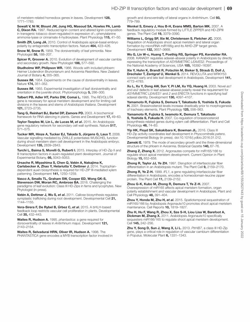
of meristem-related homeobox genes in leaves. Development 128, 1771–1783.
Sewalt V, Ni W, Blount JW, Jung HG, Masoud SA, Howles PA, Lamb C, Dixon RA. 1997. Reduced lignin content and altered lignin composition in transgenic tobacco down-regulated in expression of l-phenylalanine ammonia-lyase or cinnamate 4-hydroxylase. Plant Physiology 115, 41–50.
Smith ZR, Long JA. 2010. Control of Arabidopsis apical–basal embryo polarity by antagonistic transcription factors. Nature 464, 423–426.
Snow M, Snow R. 1959. The dorsiventrality of leaf primordia. New Phytologist 58, 188–207.
Spicer R, Groover A. 2010. Evolution of development of vascular cambia and secondary growth. New Phytologist 186, 577–592.
Studholme WP, Philipson WR. 1966. Woods with included phloem: Heimer Llodendron Brunonianum and Avicennia Resinifera. New Zealand Journal of Botany 4, 355–365.
Sussex IM. 1954. Experiments on the cause of dorsiventrality in leaves. Nature 174, 351–352.
Sussex IM. 1955. Experimental investigation of leaf dorsiventrality and orientation in the juvenile shoot. Phytomorphology 5, 286–300.
Talbert PB, Adler HT, Parks DW, Comai L. 1995. The REVOLUTA gene is necessary for apical meristem development and for limiting cell divisions in the leaves and stems of Arabidopsis thaliana. Development 121, 2723–2735.
Tang G, Reinhart BJ, Bartel DP, Zamore PD. 2003. A biochemical framework for RNA silencing in plants. Genes and Development 17, 49–63.
Taylor-Teeples M, Lin L, de Lucas M, et al. 2015. An Arabidopsis gene regulatory network for secondary cell wall synthesis. Nature 517, 571–575.
Tucker MR, Hinze A, Tucker EJ, Takada S, Jürgens G, Laux T. 2008. Vascular signalling mediated by ZWILLE potentiates WUSCHEL function during shoot meristem stem cell development in the Arabidopsis embryo. Development 135, 2839–2843.
Turchi L, Baima S, Morelli G, Ruberti I. 2015. Interplay of HD-Zip II and III transcription factors in auxin-regulated plant development. Journal of Experimental Botany, 66, 5043–5053.
Ursache R, Miyashima S, Chen Q, Vatén A, Nakajima K, Carlsbecker A, Zhao Y, Helariutta Y, Dettmer J. 2014. Tryptophan-dependent auxin biosynthesis is required for HD-ZIP III-mediated xylem patterning. Development 141, 1250–1259.
Vasco A, Smalls TL, Graham SW, Cooper ED, Wong GK-S, Stevenson DW, Moran RC, Ambrose BA. 2016. Challenging the paradigms of leaf evolution: Class III HD-Zips in ferns and lycophytes. New Phytologist (in press).
Vatén A, Dettmer J, Wu S, et al. 2011. Callose biosynthesis regulates symplastic trafficking during root development. Developmental Cell 21, 1144–1155.
Vera-Sirera F, De Rybel B, Úrbez C, et al. 2015. A bHLH-based feedback loop restricts vascular cell proliferation in plants. Developmental Cell 35, 432–443.
Waites R, Hudson A. 1995. phantastica: a gene required for dorsoventrality of leaves in Antirrhinum majus. Development 121, 2143–2154.
Waites R, Selvadurai HRN, Oliver IR, Hudson A. 1998. The PHANTASTICA gene encodes a MYB transcription factor involved in
growth and dorsoventrality of lateral organs in Antirrhinum. Cell 93, 779–789.
Wenkel S, Emery J, Hou B-H, Evans MMS, Barton MK. 2007. A feedback regulatory module formed by LITTLE ZIPPER and HD-ZIPIII genes. The Plant Cell 19, 3379–3390.
Williams L, Grigg SP, Xie M, Christensen S, Fletcher JC. 2005. Regulation of Arabidopsis shoot apical meristem and lateral organ formation by microRNA miR166g and its AtHD-ZIP target genes. Development 132, 3657–3668.
Wu G, Lin W-c, Huang T, Poethig RS, Springer PS, Kerstetter RA. 2008. KANADI1 regulates adaxial–abaxial polarity in Arabidopsis by directly repressing the transcription of ASYMMETRIC LEAVES2. Proceedings of the National Academy of Sciences, USA 105, 16392–16397.
Xie Y, Huhn K, Brandt R, Potschin M, Bieker S, Straub D, Doll J, Drechsler T, Zentgraf U, Wenkel S. 2014. REVOLUTA and WRKY53 connect early and late leaf development in Arabidopsis. Development 141, 4772–4783.
Xu L, Xu Y, Dong AW, Sun Y, Pi LM, Xu YQ, Huang H. 2003. Novel as1 and as2 defects in leaf adaxial–abaxial polarity reveal the requirement for ASYMMETRIC LEAVES1 and 2 and ERECTA functions in specifying leaf adaxial identity. Development 130, 4097–4107.
Yamamoto R, Fujioka S, Demura T, Takatsuto S, Yoshida S, Fukuda H. 2001. Brassinosteroid levels increase drastically prior to morphogenesis of tracheary elements. Plant Physiology 125, 556–563.
Yamamoto R, Fujioka S, Iwamoto K, Demura T, Takatsuto S, Yoshida S, Fukuda H. 2007. Co-regulation of brassinosteroid biosynthesis-related genes during xylem cell differentiation. Plant and Cell Physiology 48, 74–83.
Yip HK, Floyd SK, Sakakibara K, Bowman JL. 2016. Class III HD-Zip activity coordinates leaf development in Physcomitrella patens. Developmental Biology (in press). doi:10.1016/j.ydbio.2016.01.012.
Zamski E. 1979. The mode of secondary growth and the three-dimensional structure of the phloem in Avicennia. Botanical Gazette 140, 67–76.
Zhang Z, Zhang X. 2012. Argonautes compete for miR165/166 to regulate shoot apical meristem development. Current Opinion in Plant Biology 15, 652–658.
Zhong R, Taylor JJ, Ye ZH. 1997. Disruption of interfascicular fiber differentiation in an Arabidopsis mutant. The Plant Cell 9, 2159–2170.
Zhong R, Ye Z-H. 1999. IFL1, a gene regulating interfascicular fiber differentiation in Arabidopsis, encodes a homeodomain-leucine zipper protein. The Plant Cell 11, 2139–2152.
Zhou G-K, Kubo M, Zhong R, Demura T, Ye Z-H. 2007. Overexpression of miR165 affects apical meristem formation, organ polarity establishment and vascular development in Arabidopsis. Plant and Cell Physiology 48, 391–404.
Zhou Y, Honda M, Zhu H, et al. 2015. Spatiotemporal sequestration of miR165/166 by Arabidopsis Argonaute10 promotes shoot apical meristem maintenance. Cell Reports 10, 1819–1827.
Zhu H, Hu F, Wang R, Zhou X, Sze S-H, Liou Lisa W, Barefoot A, Dickman M, Zhang X. 2011. Arabidopsis Argonaute10 specifically sequesters miR166/165 to regulate shoot apical meristem development. Cell 145, 242–256.
Zhu Y, Song D, Sun J, Wang X, Li L. 2013. PtrHB7, a class III HD-Zip gene, plays a critical role in regulation of vascular cambium differentiation in Populus. Molecular Plant 6, 1331–1343.
HD-ZIP III transcription factors and vascular development | 69D
ownloaded from
https://academic.oup.com
/jxb/article/68/1/55/2627443 by guest on 15 January 2022


