Embed Size (px)
Citation preview

1
Clinical Haematology
T. Rashad Saleh M. Alkhwlany
MSc in medical microbiology

2
Clinical Haematology T/ Rashad Saleh Alkhwlany
COMPOSITION AND FUNCTIONS OF BLOOD
Hematology: is the study of the blood-forming tissues and circulating blood
components.
o Physical properties of blood:
Your total blood volume is: 5-6 liters (8% of body weight or 80 ml/kg body
weight).
Specific gravity: 1050-1060
Viscosity: 4-5 times that of water
PH: 7.4 + - 0.05; alkaline
Composition of Blood
If anticoagulant is add to blood sample and allowed to stand in a narrow tube, it
separates out into cells and plasma.
o Cells of blood
The cellular elements of blood represent 45% of the total blood volume, called
Packed Cell Volume (PCV) or Haematocrit. It includes:
1. Erythrocytes or Red Blood Cells (RBC‟s):
Normal count 5 million/ mm3 (5 x 10
6 per mm
3).
2. Leucocytes or White Blood Cells (WBC‟s):
Normal count 4,000 -11,000/ mm3 (4-11 x 10
3 per mm
3).
3. Platelets or Thrombocytes:
Normal count: 150000-400000 PLTs/ mm3 (150 - 400 x 10
3 per mm3).
o Plasma
Plasma is a clear, straw colored fluid portion of the blood and represents 55% of
the total blood volume. It composed of serum and fibrinogen. The serum contains:
1. 91% water.
2. 9% solids. The solids comprises of:
a. 1% inorganic molecules and
b. 8% organic molecules.
The major inorganic molecules are:
o Na+, Ca
2+, Cl
-, HCO3
- (mainly extracellular).

3
Clinical Haematology T/ Rashad Saleh Alkhwlany
o K+, Mg
2+, Cu
2+, PO4 (mainly intracellular).
o Fe2+
, Fe3+
.
Of 8% total organic molecules:
o 7% are plasma proteins.
o 1% is other substances like NON protein Nitrogenous (NPN)
substances, sugar, fats, enzymes and hormones.
Plasma Proteins
Normal value: 6.4-8.3 gm%
Components;
1. 55% Albumin: 3-5 gm% (Average: 4.8 gm %)
2. 38% Globulin: 2-3 gm% (Average: 2.3 gm %)
i. 13% α - Globulin: 0.79-0.84 gm%
ii. 14% β - Globulin : 0.78-0.81 gm%
iii. 11% γ - Globulin : 0.66-0.70 gm%
A/G ratio; Albumin: Globulin: : 1.7
3. 7% Fibrinogen: 0.3 gm%
4. Prothrombin: 40 mg%
Non-Protein Nitrogenous (NPN) Substances
Normal: 28-40 mg%
These are derivatives of food and in parts are the waste products of tissue
catabolism. These include:
1. Urea : 20-40 mg%
2. Uric acid : 2-4 mg%
3. Creatine : 1-2 mg%
4. Creatinine : 0.6-1.2 mg%
5. Xanthine : Traces
6. Hypoxanthine : Traces
Other Substances
These include:
1. Neutral „fats (triglycerides) : 30-150 mg%
2. Phospholipids e.g. Lecithin, sphingomyelin, cephalin etc. : 150-300 mg%

4
Clinical Haematology T/ Rashad Saleh Alkhwlany
3. Glucose (fasting) : 70-90 mg%
4. Cholesterol : 150-240 mg%
Functions of Blood
1. Respiratory: Transport of oxygen from lungs to the tissues and of carbon-
dioxide (CO2) from the tissues to the lungs.
2. Nutritive: Transport absorbed food materials, glucose, amino acids, fatty acids,
vitamins, electrolytes and trace metals from the alimentary canal to the tissues
for utilization and storage.
3. Excretory: Transports the metabolic wastes e.g. urea, uric acid, creatinine etc.
to the kidney, skin and intestine for their removal.
4. „Homeostatic‟ for water, pH and electrolyte concentration: Blood forms internal
environment of the cell. Buffering power of haemoglobin helps to maintain
constancy of blood pH.
5. Regulation of body temperature: Blood preserves the very narrow range in body
temperature. How?
Blood whose major constituent is water has:
i. High specific heat - This buffer sudden change (rise or fall) in body
temperature.
ii. High conductivity - This helps to take out heat from an organ for
uniform distribution throughout the body.
iii. High latent heat of evaporation.
6. Chemical for communication and protection:
i. Concentration of hormones and various substances in the blood is
regulated through feedback mechanisms.
ii. Within blood circulates the entire complex of humoral antibodies
important in defence against infection, initiation of inflammation and
regulation of Haemostasis (clotting mechanism).
7. Plasma proteins functions:
i. Regulation of the osmotic pressure which influences the exchange of
fluid between blood and tissues.

5
Clinical Haematology T/ Rashad Saleh Alkhwlany
ii. Act as a reservoir of proteins.
iii. Combine with many substances e.g. iron, thyroxine and steroid
hormones to form transportable complexes from which the active
components are released at the appropriate sites.
Serum
If the blood is allowed to clot in a test tube, then the clot retracts and gives out
serum. Therefore, serum is plasma minus fibrinogen and clotting factors (II, V and
VIII), because these factors get consumed during clotting (remaining do not).
Serum has higher serotonin (5 hydroxy-tryptamine - 5HT) content because of the
breakdown of platelets during clotting.

6
Clinical Haematology T/ Rashad Saleh Alkhwlany
Haematopoiesis
(From Ancient Greek: haima: blood; poiesis to make) (Or hematopoiesis in the United
States; sometimes also haemopoiesis or hemopoiesis).
Is the formation of blood cellular components. All cellular blood components are
derived from haematopoietic stem cells. In a healthy adult person, approximately 1011
–
1012
new blood cells are produced daily in order to maintain blood levels in the
peripheral circulation.
o Site of haematopoiesis:
1. Fetus: 0-2 months: yolk sac.
2-7 months: liver and spleen.
5-9 months: bone marrow.
2. Infants: Bone marrow (practically all bones).
3. Adults: Vertebrae, ribs, sternum, skull, sacrum and pelvis. Proximal ends of
femur and humeri.
In the first few weeks of gestation the yolk sac is the main site of haernopoiesis.
However, definitive haernopoiesis derives from a population of stem cells first
observed on the dorsal aorta termed the AGM (aorta-gonads-mesonephros) region.
These common precursors of endothelial and haemopoi¬etic cells (haemangioblasts)
are believed to seed the liver, spleen and bone marrow.
o Extramedullary
In some cases, such as some disease, the liver, thymus, and spleen may resume
their haematopoietic function, if necessary. This is called extramedullary
haematopoiesis. It may cause these organs to increase in size substantially.
o Stages of haematopoiesis:
Stem cells:
All cellular blood components are derived from haematopoietic stem cells. The
stem cells found within the bone marrow are the origin of all blood cells. Blood
cells are formed by a process of differentiation from stem cell. The stem cell has
the capability of self-renewal (so that marrow cellularity remains constant in a
7
Clinical Haematology T/ Rashad Saleh Alkhwlany
normal healthy steady state). One stem cell is capable of producing about 106
mature blood cells after 20 cell divisions. The precursor cells are, however,
capable of responding to haemopoietic growth factors with increased production
of one or other cell line when the need arises.
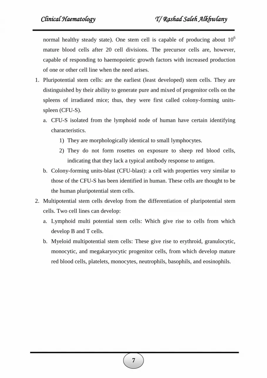
1. Pluripotential stem cells: are the earliest (least developed) stem cells. They are
distinguished by their ability to generate pure and mixed of progenitor cells on the
spleens of irradiated mice; thus, they were first called colony-forming units-
spleen (CFU-S).
a. CFU-S isolated from the lymphoid node of human have certain identifying
characteristics.
1) They are morphologically identical to small lymphocytes.
2) They do not form rosettes on exposure to sheep red blood cells,
indicating that they lack a typical antibody response to antigen.
b. Colony-forming units-blast (CFU-blast): a cell with properties very similar to
those of the CFU-S has been identified in human. These cells are thought to be
the human pluripotential stem cells.
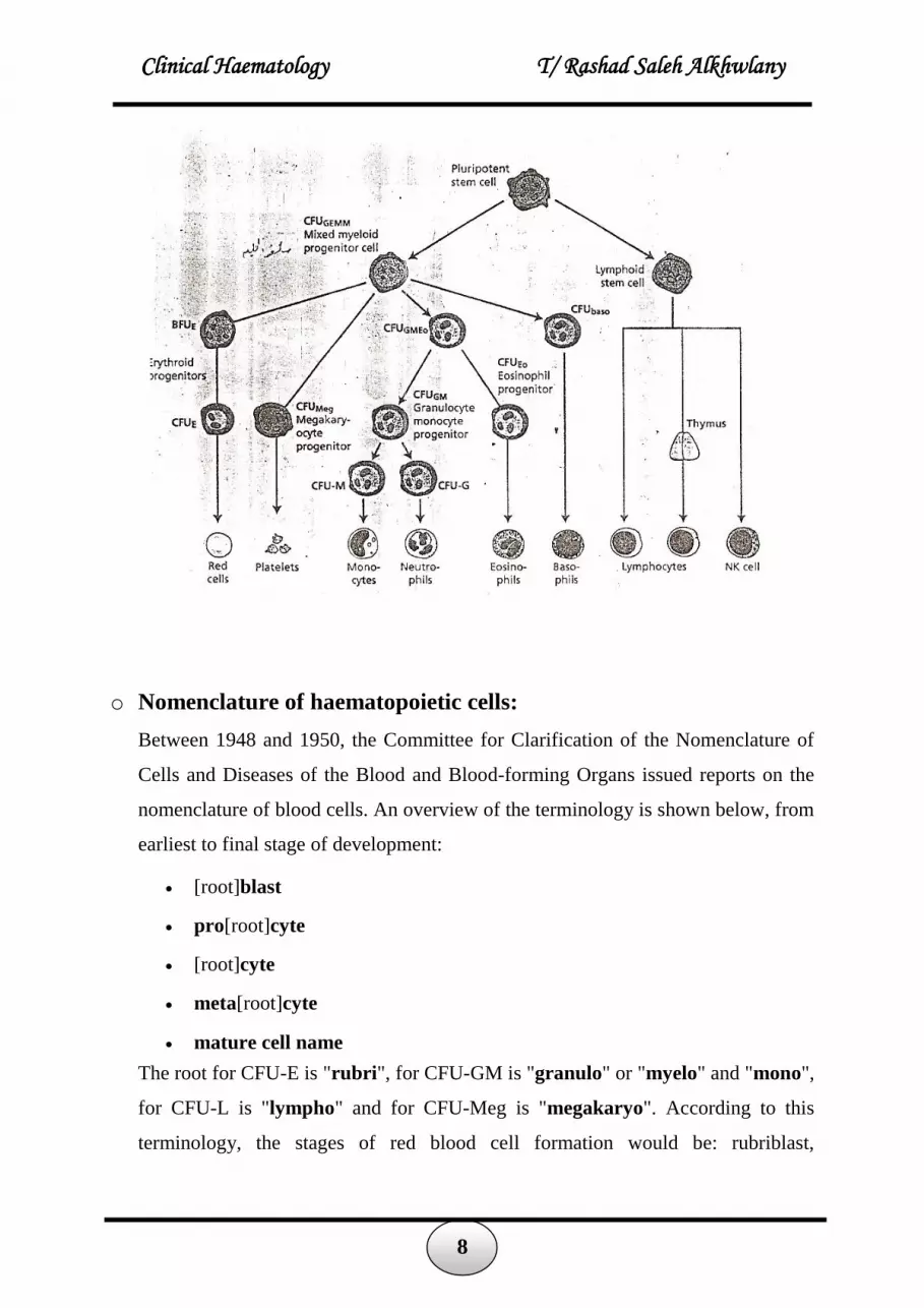
2. Multipotential stem cells develop from the differentiation of pluripotential stem
cells. Two cell lines can develop:
a. Lymphoid multi potential stem cells: Which give rise to cells from which
develop B and T cells.
b. Myeloid multipotential stem cells: These give rise to erythroid, granulocytic,
monocytic, and megakaryocytic progenitor cells, from which develop mature
red blood cells, platelets, monocytes, neutrophils, basophils, and eosinophils.
8
Clinical Haematology T/ Rashad Saleh Alkhwlany
o Nomenclature of haematopoietic cells:
Between 1948 and 1950, the Committee for Clarification of the Nomenclature of
Cells and Diseases of the Blood and Blood-forming Organs issued reports on the
nomenclature of blood cells. An overview of the terminology is shown below, from
earliest to final stage of development:
[root]blast
pro[root]cyte
[root]cyte
meta[root]cyte
mature cell name
The root for CFU-E is "rubri", for CFU-GM is "granulo" or "myelo" and "mono",
for CFU-L is "lympho" and for CFU-Meg is "megakaryo". According to this
terminology, the stages of red blood cell formation would be: rubriblast,

9
Clinical Haematology T/ Rashad Saleh Alkhwlany
prorubricyte, rubricyte, metarubricyte, and erythrocyte. However, the following
nomenclature seems to be, at present, the most prevalent:
Committee "lympho" "rubri" "granulo" or "myelo" "mono" "megakaryo"
Lineage Lymphoid Myeloid Myeloid Myeloid Myeloid
CFU CFU-L CFU-
GEMM→CFU-E CFU-GEMM→CFU-GM→CFU-G
CFU-
GEMM→CFU-
GM→CFU-M
CFU-
GEMM→CFU-
Meg
Process lymphocytopoiesis erythropoiesis granulocytopoiesis monocytopoiesis thrombocytopoiesis
[root]blast Lymphoblast Proerythroblast Myeloblast Monoblast Megakaryoblast
pro[root]cyte Prolymphocyte Polychromatophilic
erythrocyte Promyelocyte Promonocyte Promegakaryocyte
[root]cyte - Normoblast Eosino/neutro/basophilic myelocyte Megakaryocyte
meta[root]cyte Large lymphocyte Reticulocyte
Eosinophilic/neutrophilic/basophilic
metamyelocyte,
Eosinophilic/neutrophilic/basophilic
band cell
Early monocyte -
mature cell
name Small lymphocyte Erythrocyte
granulocytes
(Eosino/neutro/basophil) Monocyte
thrombocytes
(Platelets)
Osteoclasts also arise from haemopoietic cells of the monocyte/neutrophil lineage,
specifically CFU-GM.
o Colony-forming units
There are various kinds of colony-forming units:
Colony-forming unit lymphocyte (CFU-L)
Colony-forming unit erythrocyte (CFU-E)

10
Clinical Haematology T/ Rashad Saleh Alkhwlany
Colony-forming unit granulo-monocyte (CFU-GM)
Colony-forming unit megakaryocyte (CFU-Me)
Colony-forming unit Basophil (CFU-B)
Colony-forming unit Eosinophil (CFU-Eo)
o The regulation of haemopoiesis
Haemopoiesis starts with stem cell division in which one cell replaces the stem cell
(self-renewal) and the other is committed to differentiation. These early committed
progenitors express low levels of transcription factors that may commit them to
discrete cell lineages. Which cell lineage is selected for differentiation may depend
both on chance and on the external signals received by progenitor cells.
Several transcription factors have been isolated that regulate differentiation along
the major cell lineages. For instance, PU.l commits cells to the myeloid lineage
whereas GATA-l has an essential role in erythropoietic and megakaryocytic
differentiation.
o Haematopoietic growth factors
Red and white blood cell production is regulated in healthy humans, and the
production of granulocytes is rapidly increased during infection. The proliferation
and self-renewal of these cells depend on stem cell growth factor. Growth factors
are glycoprotein that regulates the proliferation, also stimulate differentiation,
maturation, prevent apoptosis and affect the function of mature cells that enter the
blood from the marrow. The more factors are:
Human hematopoietic growth factors
Growth Factor Source Major Function
GM-CSF T-Lymphocyte, endothelial cells, Fibroblasts Stimulates production of neutrophils, eosinophils, monocytes, red cells and platelets.
G-CSF Monocytes, Fibroblasts Stimulates production of neutrophils. M-CSF Macrophages, endothelia cells Stimulates production of Monocytes ERYTHROPOIETIN Peritubular cells, Liver, Macrophages Stimulates production of red cells.
IL-1 Macrophages, activated lymphes, endothelial cells.
Cofactor for IL-3 and IL-6. Activated T cells
IL-2 Activated T cells T cell growth factor. Stimulates IL-1 synthesis. Activated B cells and NK cells

11
Clinical Haematology T/ Rashad Saleh Alkhwlany
IL-3 T cells Stimulates production of all non-lymphoid cells.
IL-4 Activated T cells Growth factor for activated B cells, resting T cells and mast cells.
IL-5 T cells Induces differentiation of activated B cells and eosinophils.
IL-6 T cells Stimulates CFU-GEMM. Stimulates Ig synthesis
IL-7 T cells, Fibroblasts, Endothelial cells Growth factor for pre B cells

12
Clinical Haematology T/ Rashad Saleh Alkhwlany
Erythropoiesis
The term erythropoiesis (erythro = RBC, and poiesis = to make) used to describe
the process of RBC formation or production. In humans, erythropoiesis occurs
almost exclusively in the red bone marrow.
There are approximately 1012
new erythrocytes (red cells) each day by the complex
and finely regulated process of erythropoiesis.
Erythropoiesis passes from the stem cell through the progenitor cells colony-
forming unit granulocyte, erythroid, monocyte and megakaryocyte (CFUGEMM),
burst-forming unit erythroid (BFUE) and erythroid CFU (CFUE) to the first
recognizable erythrocyte precursor in the bone marrow, the pronormoblast.
Erythrocyte differentiation
1. Pluripotent hematopoietic stem cell
2. Multipotent stem cell
3. Myeloid stem cell
4. Pronormoblast
5. Basophilic normoblast/early normoblast
6. Polychromatophilic normoblast/intermediate normoblast
7. Orthochromatic normoblast/late normoblast
8. Reticulocyte
9. Mature RBCs In blood circulation
Table
In bone marrow
(5 days)
Spends 1-2 days in
the marrow and also
circulates in the
peripheral blood for
1-2 days before
maturing, mainly in
the spleen.

13
Clinical Haematology T/ Rashad Saleh Alkhwlany
College of American Pathologists (CAP) vs. American Society for Clinical Pathology (ASCP)
Terminology for Red Cells
Pathologists (CACAP ASCP
Pronormoblast Rubriblast
Basophilic normoblast Prorubricyte
Polychromatophilic normoblast Rubricyte
Orthochromic normoblast Metarubricyte
Reticulocyte Reticulocyte
Erythrocyte Erythrocyte
Pronormoblast
o Size: 18 to 20 μm, the largest immature, the “mother cell”.
o N:C ratio: 4:1.
o Nuclear chromatin: Round nucleus with condensation chromatin, evenly
distributed, fine texture with deep violet color, nucleoli may be present but are
difficult to visualize.
o Cytoplasm: Dark marine blue.
Basophilic Normoblast
o Size: 16 μm.
o N:C ratio: 4:1.
o Nuclear chromatin: Round nucleus with crystalline chromatin, red-purple
color to chromatin.
o Cytoplasm: Blue.
Polychromatophilic Normoblast
o Size: 13 μm.
o N:C ratio: 2:1.
o Nuclear chromatin: Chromatin is condensed.
o Cytoplasm: A color mixture, blue layered with tinges of orange red, the
hemoglobin begins to be synthesized.

14
Clinical Haematology T/ Rashad Saleh Alkhwlany
Orthochromic Normoblast-Nucleated (nRBC)
o Size: 8 μm.
o N:C ratio: 1:1.
o Nuclear chromatin: Dense, velvet-appearing homogeneous chromatin.
o Cytoplasm: Increased volume, with orange-red color with slight blue.
Reticulocyte
o Size: 8 μm.
o Appearance: Remnant of RNA visualized as reticulum, filamentous structure
in chains or as a single dotted structure in new methylene blue stain.
o Nucleus: The cell has extruded its nucleus, but is still capable of producing
hemoglobin.
Mature Red Cell
o Size: 6 to 8 μm.
o Appearance: Disk-shaped cell filled with hemoglobin, having an area of
central pallor of 1 to 3 μm.
Major changes and features of erythropoiesis
• Decrease the total size of the cell.
• Increase amount of cytoplasm.
• Decrease basophilic staining.
• Increase haemoglobin synthesis.
• Condensation of chromatin.
• The N:C ratio decreases as the cell matures.
Note:
Previous cell are studied from where:
o Cytoplasm (amount, color, texture).
o Nucleus (Size, color, position, texture).
o Nucleoli (presence and number).
o Ratio of cytoplasm to nucleus.

15
Clinical Haematology T/ Rashad Saleh Alkhwlany
Control of erythropoiesis:
By erythropoietin (formed in kidneys) is released in response to lowered tissue
oxygen. Erythropoietin is glycoprotein and stimulates erythropoiesis.
It act on the BFU-E (burst-forming units), CFU-E (colony-forming units) and on
pronormoblast.

16
Clinical Haematology T/ Rashad Saleh Alkhwlany
Hemoglobin
Hemoglobin (also spelled haemoglobin and abbreviated Hb or Hgb) is the iron-
containing oxygen-transport metalloprotein in the red blood cells. The hemoglobin
was discovered by Hünefeld in 1840.
إ انىظفة األساسة نكشات انذو انحشاء ه قم األوكسج ي انشئت إنى األسجة وحم ثا أكسذ انكشبى
.ي األسجة إنى انشئت. وهز انؼهة تتى بىاسطة انهىجهىب.
كى ي , أيا انهى فتانجهىبي انهىجهىب تكى ي أسبغ رسات ي انهى وأسبغ رسات ي جزيءكم
Fانحذذ انثائ ++
وأيا انجهىب فهى بشوت تكى ي سالسم ي ػذذات انببتذ Porphyrineوانبىسفش
Polypeptide وانت تتكى ي تجغ ي األحاض األيةamino acid.
Each molecule of haemoglobin contains four polypeptide (globin) chains and four
molecules of haem.
Globin in adult consist of two alpha (α) chain containing 141 amino acid and two beta (β)
chains containing 146 amino acids.
.Immature RBCsانجهىب تكى ف خاع انؼظى وبانزات ف سبىسىيات انخالا انحشاء انغش اضجة
. Mitochondriaس ف انتىكىذساأيا انهى فتكى بشكم أسا
و Vitamin Cو Folic acidو Vitamin B12هاك انؼذذ ي انؼاصش انت تساػذ ف تكى انهىجهىب يثم
Fe وانحذذ Vitamine 6انحاس و ++
.

17
Clinical Haematology T/ Rashad Saleh Alkhwlany
Types in humans
o In the embryo:
Gower 1 (δ2ε2)
Gower 2 (α2ε2)
Hemoglobin Portland (δ2γ2)
o In the fetus:
Hemoglobin F (α2γ2)
o In adults:
Hemoglobin A (α2β2) - a normal amount 96-98%
Hemoglobin A2 (α2δ2) - δ chain synthesis begins late in the third trimester
and in adults, it has a normal range of 1.5-3.2%
Hemoglobin F (α2γ2) - In adults Hemoglobin F is restricted to a limited
population of red cells called F-cells, it has a normal range of 0.5-0.8%

18
Clinical Haematology T/ Rashad Saleh Alkhwlany
However, the level of Hb F can be elevated in persons with sickle-cell
disease and thalassemia.
Hb A هى انشائغ ف جسى اإلسا بؼذ انشهش انثانث, أيا ف األطفال كىHb F نك هز %80-60بسبة
نث وإرا نى تتشاجغ فأها تؤدي نإلصابة باأليشاض.انسبة تتشاجغ بؼذ انشهش انثا
Genetics
Subunit Name Gene Chromosomal Locus
Hb α1 HBA1 Chromosome 16 p13.3
Hb α2 HBA2 Chromosome 16 p13.3
Hb β HBB Chromosome 11 p15.5
Hb derivatives:
o Oxyhemoglobin
o Deoxyhemoglobin
o Methaemoglobin
o Carboxyhaemoglobin
o Sulfhaemoglobin
Abnormal Hb:
o Thalassemia (β4)
o HbS (α2βS
2)
o HbC (α2βC
2)
o HbSC

19
Clinical Haematology T/ Rashad Saleh Alkhwlany
Red Blood Cells (RBCs)
Red blood cells are also known as RBCs, red blood corpuscles (an archaic term),
erythrocytes (from Greek erythros for "red" and cyte translated as "cell". The term
Red Blood Cells is the proper name.
The first person to describe red blood cells or erythrocytes was a Dutch biologist
Jan Swammerdam, in 1658. Anton van Leeuwenhoek provided further descriptions
of the RBCs in 1674.
The color of erythrocytes is due to the heme group of hemoglobin. The blood
plasma alone is straw-colored, but the red blood cells change color depending on
the state of the hemoglobin:
when combined with oxygen (oxyhemoglobin) is scarlet.
when oxygen has been released (deoxyhemoglobin) is darker.
RBCs function:
RBCS transport the respiratory gases O2 and Co2
Life Span:
120 days, then destroy at RE (reticuloendothelial system) system. The important
breakdown products are heam and globin that recirculated in the body. The heme
are broken down into Fe and biliverdin. The biliverdin is reduced to bilirubin, which
is released into the plasma and recirculated to the liver bound to albumin. The iron is
released into the plasma to be recirculated by a carrier protein called transferrin.
Almost all erythrocytes are removed in this manner from the circulation before they
are old enough to hemolyze. Hemolyzed hemoglobin is bound to a protein in plasma
called haptoglobin which is not excreted by the kidney.
RBCs structure:
Red cell is structure from cell membrane and cytoplasm filled completely with Hb.
The RBCs lack the others organelles (nucleus, ribosome, Golgi apparatus and
mitochondria).
Prior to discharge from B.M (bone marrow) into P.B. (peripheral blood)
RBCs shed their nuclei. This gives the advantages:
1. Reduced weight.

20
Clinical Haematology T/ Rashad Saleh Alkhwlany
2. Transformation into biconcave shape with increased flexibility compared
with rigid nucleated cell.
RBCs is biconcave shape (from side) and spherical (disc) with central pallor from
top (⅓ diameter of cell) with 7.5-8 µm in diameter, must be able to pass repeatedly
through the microcirculation whose minimum diameter is 3.5-5 µm, in order to:
1. Maintain Hb in reduced 9ferrous Fe++
) form.
2. Maintain osmotic equilibrium inside the cell.
RBCs membrane:
The red cell membrane comprises a lipid bilayer, integral membrane proteins with
surface carbohydrate.
10% carbohydrate On the surface
Ags of blood group.
50% protein
Which include α and β spectrin
Ankyrin
Protein band 4.1 and 4.2
Actin
Band 3
These protein formed the RBCs skeleton.
These protein are important in maintain biconcave shape and transmembrane.
Any change or defect in these protein may cases some alteration in RBCs
shape eg: hereditary spherocytosis and elliptocytosis.
40% lipid:

21
Clinical Haematology T/ Rashad Saleh Alkhwlany
Which include cholesterol and phospholipid.
Alterations in lipid composition may be associated with others shape
abnormalitiis eg:
Increase in cholesterol and phospholipid causes target cells formation.
Large increase in cholesterol cause acanthocyte formation.
RBCs metabolism:
RBCs able to produce energy that required it as:
ATP:
From anaerobic glycolysis (Embden Meyerhof pathway).
To maintain RBCs shape, volume, flexibility.
NADH:
Also from anaerobic glycolysis
Reducing power, to maintain Hb in reduced form (reduced met Hb
normal Hb)
NADPH:
5% from glycolysis enter HMP (Hexose monophosphate pathway) .
As NADH also to remove the toxicity from RBCs (antioxidant).
2,3 DPG (2,3 diphosphoglycerate):
From Luebering-Rapoport (part from glycolysis)
Regulation of Hb-oxygen affinity.

22
Clinical Haematology T/ Rashad Saleh Alkhwlany
Embden Meyerhof pathway
The red blood cells diseases include:
Anemia are diseases characterized by low oxygen transport capacity of the
blood, because of low red cell count or some abnormality of the red blood cells
or the hemoglobin.
Polycythemia are diseases characterized by increase of red blood cells.

23
Clinical Haematology T/ Rashad Saleh Alkhwlany
LEUKOCYTES (WHITE BLOOD CELLS)
Several types of leukocytes, or white blood cells (WBCs), are found in the blood. The
normal WBC count is ~4,000 to 11,000/µL. Leukocytes are usually divided into
granulocytes, which have specific granules, and agranulocytes, which lack specific
granules.
Granulocytes are divided into neutrophils (with faintly staining granules), eosinophils
(with large reddish or eosinophilic granules), and basophils (with large dark blue or
basophilic granules). Agranulocytes are divided into lymphocytes and monocytes.
Although they are called white blood cells, leukocytes predominantly function in
tissues. They are only in the blood transiently, while they travel to their site of action.
Functions of neutrophils
About half the neutrophils circulating in the blood (included in WBC count).
The other half, marginate along the walls of blood vessels and in capillaries, (not
included in WBC count).
Neutrophils have a short life-span. After circulating in the blood for 6–10 hours
they pass into the tissues where they become phagocytes, they are important in
defending the body from infection.
They mobilize and migrate to sites of infection or inflammation, they attracted
by chemical substances released by bacteria, complement components, damaged
tissue, and other leukocytes (process called chemotaxis).
Neutrophils have receptors for IgG antibody and complement (C3b) and are
therefore able to recognize, phagocytose, and kill bacteria coated with IgG or
C3b. Bacteria are ingested and destroyed by chemicals released from neutrophil
granules.
Following phagocytosis, neutrophils die at the site of infection, forming pus
cells. Neutrophils not involved in the inflammatory process are removed by the
spleen after 1–2 days.
Functions of eosinophils
Eosinophils circulate in blood for about 8 hours after which they enter the
tissues. They are found mainly in the skin, gastrointestinal tract and lungs where

24
Clinical Haematology T/ Rashad Saleh Alkhwlany
they are involved in hypersensitivity reactions, e.g. asthma, hay fever, eczema.
Eosinophils contain substances that are able to inactivate histamine and factors
released during anaphylaxis.
Eosinophils are important in parasitic helminth immune responses in which IgG
and IgE antibodies are produced. Lymphokines released from T lymphocytes
stimulate the production of IgE which binds to mast cells at the site of parasitic
infection.
Parasitic antigens cause chemotactic substances to be released from mast cells
which attract eosinophils to the site. Eosinophils bind to the antibody-coated
parasite and release cytotoxic substances which damage the surface of the
parasite, leading to its destruction.
Antibody dependent cell-mediated cytotoxicity by eosinophils is effective
against parasites which are located in the tissues, or have a tissue invading stage,
e.g. schistosomes, filarial worms, hookworms, Trichinella, Strongyloides,
Ascaris, Fasciola, Fasciolopsis, Paragonimus, and Toxocara.
Functions of basophils
Basophils circulate only in the blood. They are not found in tissues (mast cells
are tissue equivalents to basophils). Basophils bind IgE on their surface and are
involved in anaphylactic, hypersensitivity, and inflammatory reactions.
Basophils interact with eosinophils and macrophages in allergic reactions. When
specific antigen reacts with IgE bound to basophils, degranulation of the cells
occurs with the release of inflammatory substances including heparin, histamine
and platelet activating factor.
Functions of monocytes and macrophages
Monocytes pass from the bone marrow into the blood circulation. Within 2–3
days they reach the tissues where they develop into macrophages, becoming
fixed tissue macrophages in the spleen, liver (Küpffer cells), lymph nodes,
connective tissues, and central nervous system, and free macrophages in lung
alveoli, peritoneum and inflammatory granulomas.
25
Clinical Haematology T/ Rashad Saleh Alkhwlany
Macrophages form the mononuclear phagocytic system, i.e. reticuloendothelial
(RE) system. Unlike neutrophils, mononuclear phagocytes do not die following
phagocytosis. Macrophages live in the tissues for several months or longer.
As phagocytic cells, monocytes ingest microorganisms and cellular debris,
including malaria pigment. The RE system is involved in the destruction of
bacteria, viruses, fungi, protozoal parasites, and malignant cells.
Macrophages process and present antigens to T lymphocytes, and regulate many
T and B cell activities.
When activated, macrophages synthesize and secrete cytokines (e.g.
interleukins, tumour necrosis factor, GM-CSF) which are involved in the
activation of lymphocytes, the inflammatory process (particularly chronic
inflammation), cell-mediated immune responses and haematopoiesis.
An increase in circulating monocytes (monocytosis) is found in protozoal
parasitic infections, and also in tuberculosis and other chronic bacterial
infections and rickettsial infections.
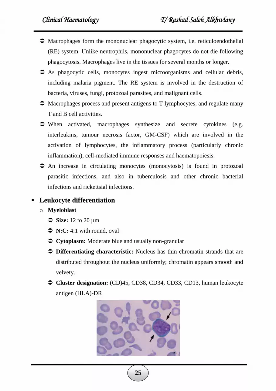
Leukocyte differentiation
o Myeloblast
Size: 12 to 20 µm
N:C: 4:1 with round, oval
Cytoplasm: Moderate blue and usually non-granular
Differentiating characteristic: Nucleus has thin chromatin strands that are
distributed throughout the nucleus uniformly; chromatin appears smooth and
velvety.
Cluster designation: (CD)45, CD38, CD34, CD33, CD13, human leukocyte
antigen (HLA)-DR

26
Clinical Haematology T/ Rashad Saleh Alkhwlany
o Promyelocyte
Size: 15 to 21 µm
N:C: 3:1, oval, round
Cytoplasm: Moderate blue color but difficult to observe because fine to
large blue-red azurophilic granules are scattered; granules are
NONSPECIFIC
Differentiating characteristic: Cell is larger than the blast with large
prominent nucleoli, nuclear chromatin is slightly coarse.
Cluster designation: CD45, CD33, CD13, CD15
o Myelocyte
Size: 10 to 18 µm
N:C: 2:1
Cytoplasm: Specific granules present, neutrophilic granules are dusty, fine,
and red-blue; eosinophilic granules are large red-orange, and singular;
basophil granules are large, deep blue purple
Differentiating characteristic: Small pink-purple granules for the
neutrophilic myelocyte, nucleus stains deeper color, granular pattern to the
chromatin.
Cluster designation: CD45, CD33, CD13, CD15, CD11b/11c

27
Clinical Haematology T/ Rashad Saleh Alkhwlany
o Metamyelocyte
Size: 10 to 15 µm
N:C: 1:1
Cytoplasm: Pale blue to pinkish tan with moderate specific granules
Differentiating characteristics: Nuclear indentation and condensed
chromatin with no nuclei.
Cluster designation: CD markers are the same as for the myelocyte
o Band
Size: 9 to 15 µm
Chromatin: Band shaped like a cigar band, C or S shaped, unable to see
filament, coarselyclumped almost like leopard spot coarseness
Cytoplasm: Brown-pink, with many fine secondary granules
Differentiating characteristics: No filament, may resemble a
metamyelocyte but indentation is more severe and chromatin is more
clumped.
Cluster designation: CD45, CD13, CD15, CD11b/11c


