Embed Size (px)
Citation preview

BioMed CentralJournal of Translational Medicine
ss
Open AcceResearchCMV pp65 and IE-1 T cell epitopes recognized by healthy subjectsStefanie L Slezak, Maria Bettinotti, Silvia Selleri, Sharon Adams, Francesco M Marincola and David F Stroncek*Address: Department of Transfusion Medicine, Warren G. Magnuson Clinical Center National Institutes of Health, Bethesda, Maryland, USA
Email: Stefanie L Slezak - [email protected]; Maria Bettinotti - [email protected]; Silvia Selleri - [email protected]; Sharon Adams - [email protected]; Francesco M Marincola - [email protected]; David F Stroncek* - [email protected]
* Corresponding author
AbstractBackground: Adoptive immune and vaccine therapies have been used to prevent cytomegalovirus(CMV) disease in recipients of hematopoietic progenitor cell transplants, but the nature of T cellresponses to CMV have not been completely characterized.
Methods: Peptide pools and individual peptides derived from the immune-dominant CMV proteinspp65 and IE-1 and antigen-specific, cytokine flow cytometry were used to characterize theprevalence and frequency of CD4+ and CD8+ memory T cells in 20 healthy CMV-seropositivesubjects.
Results: CD8+ T cell responses to pp65 were detected in 35% of subjects and to IE-1 in 40% ofsubjects. CD4+ T cell responses to pp65 were detected in 50% of subjects, but none were detectedto IE-1. Several new IE-1 HLA class I epitopes were identified, including 4 restricted to HLA-Cantigens. One region of IE-1 spanning amino acids 300 to 327 was rich in class I epitopes. The HLAclass I restrictions of IE-1 peptides were more promiscuous than those of pp65 peptides.
Conclusion: Since naturally occurring CD4+ and CD8+ T cell responses to pp65 were detectablein many subjects, but only CD8+ T cell responses to IE-1 were detected, pp65 may be better thanIE-1 for use in vaccine and adoptive immune therapies.
BackgroundCytomegalovirus (CMV) is a persistent virus in normalhosts in which primary infection is typically controlledthrough a combination of adaptive and innate immuneresponses. Viral latency follows primary infection andcontinues for life, usually without major symptoms.Although 60% to 80% of adults that test seropositive forCMV antibodies show no symptoms of infection, inimmunocompromised hosts, such as patients undergoinghematopoietic stem cell transplantation (HSCT) or recip-ients of organ transplants, CMV can cause severe dis-
ease[1]. In the past 10 years, the incidence of CMV diseasein allogeneic HSCT recipients during the first 100 daysafter transplantation fell from 40% to 5% as a result ofimproved diagnosis and prophylactic or preemptive treat-ment with antiviral drugs. However, the effect of theimproved therapy has delayed disease onset to 6 to 12months after transplantation, and the risk of severe CMVdisease remains in 10% of allogeneic HSCT recipients[2].
In order to control rather than delay the incidence of CMVdisease after HSCT, the host's anti-CMV immunity must
Published: 28 March 2007
Journal of Translational Medicine 2007, 5:17 doi:10.1186/1479-5876-5-17
Received: 9 January 2007Accepted: 28 March 2007
This article is available from: http://www.translational-medicine.com/content/5/1/17
© 2007 Slezak et al; licensee BioMed Central Ltd. This is an Open Access article distributed under the terms of the Creative Commons Attribution License (http://creativecommons.org/licenses/by/2.0), which permits unrestricted use, distribution, and reproduction in any medium, provided the original work is properly cited.
Page 1 of 16(page number not for citation purposes)

Journal of Translational Medicine 2007, 5:17 http://www.translational-medicine.com/content/5/1/17
be restored. Because CMV immunotherapy is growing inimportance, the study of the immune response to CMV isclinically relevant. T cell immunity is believed to be themost important component in immune response to CMVdisease. This is supported by the fact that Natural Killercells recover early after HSCT, before the peak incidence ofCMV disease, and by the failure of infusion of immu-noglobulin (Ig) preparations containing antiviral anti-bodies to control the disease [3,4]. In contrast, clinicaltrials involving adoptive transfer of CMV-specific T cellswere successful in restoring immunity against the virus[5,6]. An alternative to adoptive immunotherapy is vacci-nation of HSCT donors against CMV with the aim of pro-viding specific immunity to the recipient for protectionagainst primary infection or reactivation[7].
Two CMV proteins, phosphoprotein 65 (pp65) andimmediate early protein-1 (IE-1), have been found to bemajor targets of the cellular immune response [8-12].Immunodominant epitopes in these proteins have beendefined for some human leukocyte antigen (HLA) alleles,but further definition for specific major-histocompatibil-ity complex (MHC) classes and HLA allelic restrictionscould be useful for adoptive immune therapy, vaccinetherapy, and in screening for reactivation after immuno-suppression[13,14].
The purpose of this study was to characterize the naturalrepertoire of immunodominant pp65 and IE-1 epitopesin 20 CMV-seropositive, healthy subjects. Donor CD4+and CD8+ T cell responses to CMV pp65 and IE-1 15-merpeptide libraries were defined and followed downstreamto the individual 15-mer and nanomer peptides, respec-tively. This process identified epitopes without biasesintroduced by focusing on specific immunodominantregions for individual HLA types.
MethodsStudy designIn this study, 20 CMV-seropositive and 5 CMV-seronega-tive healthy donors were evaluated. To determine theCD8+ and CD4+ T cell responses to CMV in the 25donors, peripheral blood mononuclear cells (PBMCs)were stimulated in vitro with a cocktail of 15-mer peptidesoverlapping by 11 residues and covering the pp65 and IE-1 proteins. Antigen-specific cytokine flow cytometry wasused to detect specific T cell responses[15,16]. To defineimmunodominant epitopes, PBMCs were also stimulatedwith subpools of pp65 and IE-1, each subpool containing10 overlapping 15-mer peptides. PBMCs reactive to a sub-pool were then tested further against the individual pep-tides contained within the subpool to determine thereactive 15-mer(s). In order to further characterizeepitopes presented to CD8+ T cells by HLA class I anti-gens, PBMCs from donors with reactive CD8+ cells were
tested again with nanomer peptides spanning the reactive15-mer peptide and overlapping at 8 amino acids (Figures1 and 2). These studies were approved by a National Insti-tutes of Health Institutional Review Board on the use ofHuman Subjects in Research.
Collection of PBMCsPBMC concentrates from 20 CMV-seropositive and 5CMV-seronegative healthy donors were collected byapheresis, (Fenwal CS-3000 Plus, Baxter Healthcare Corp,Deerfield, IL) and peripheral blood mononuclear cellswere isolated by ficoll-hypaque density gradient separa-tion and frozen in aliquots of 1 × 10⁸ cells. Donors weretyped for HLA class I and class II alleles using sequence-specific primers and, in some cases, direct sequencing(HLA Laboratory, DTM, CC, NIH, Bethesda, MD). Thepresence of IgG and IgM CMV antibodies in each subjectwas analyzed by passive agglutination (BD, Microbiologi-cal Systems, Cockeysville, MD).
Peptide libraries and poolsLibraries for CMV proteins pp65 and IE-1 were made upof peptides 15 amino acids long that overlapped by 11residues and covering the complete pp65 (CMV Towne)[17] and IE-1 (CMV AD169) proteins[18]. The pp65library was made up of 138 peptides and the IE-1 libraryof 120 peptides, and both were commercially synthesized(Princeton Biomolecules, Langhorne, PA). Peptides werediluted in dimethyl sulfoxide and pooled into cocktailscontaining the complete pp65 and IE-1 protein sequencesand into subpools containing 10 consecutive peptideseach. Pp65 was divided into 13 subpools of 10 peptidesand 1 subpool of 8 peptides, and IE-1 was divided into 12subpools of 10 peptides (Figures 1 and 2).
PBMC stimulation and flow cytometric assessmentT cell activation was assessed by measuring intracellularinterferon gamma (IFN-γ) production by flow cytometryafter PBMC stimulation. PBMCs were plated at 1–5 × 10⁶
per mL in 24-well plates with 1 mL of complete medium(CM) supplemented with 10% heat-inactivated humanAB serum, 10 μM HEPES buffer, 100 U/mL penicillin-streptomycin, 0.03% L-glutamine, and 10 mg/mL cipro-floxacin. Following overnight resting at 37°C, PBMCswere stimulated with 1 μg/mL of pp65 and IE-1 libraries,sub-pools, or individual peptides and 0.5 mg/mL of sta-phylococcal enterotoxin B (SEB) (Sigma, St Louis, MO) asa positive control.
After 1 h of incubation at 37°C, 2 μg/ml of Brefeldin A(BFA) (Sigma) was added, and the cells were incubated foran additional 5 h before harvesting, fixed in FACS LysingSolution (BD Biosciences, San Jose, CA) at room temper-ature and rested overnight at 4°C. The following dayPBMCs were washed, permeabilized in Perm2 Solution
Page 2 of 16(page number not for citation purposes)

Journal of Translational Medicine 2007, 5:17 http://www.translational-medicine.com/content/5/1/17
and stained with monoclonal antibodies against CD3-PerCP, CD8-PE, CD4-APC and IFNγ-FITC (all from BDBiosciences) and incubated at 4°C for 30 minutes. MouseIg isotype controls were also used (BD Biosciences). The 2-laser FACSCalibur flow cytometer and CellQuest Pro soft-ware (BD Biosciences) were used for analysis by acquiring200,000 events, and determining the viable lymphocytepopulation by light scatter. Gates were designed for CD3+expression and for characterization of CD8+ and CD4+subpopulations. The proportion of reactive CD8+ andCD4+ cells was expressed as a percent of the total numberof CD8+ and CD4+ cells analyzed, respectively.
Reactive populations met 2 criteria: (1) well-defined cellpopulation reactive with both IFN-γ and CD8+ or CD4+,
and (2) the percentage of IFN-γ producing cells wasgreater than 3 standard deviations of the percentage ofunstimulated IFN-γ producing cells. Scattered reactivecells were not sufficient to be considered a positive popu-lation.
Determination of HLA restrictionEpitope HLA restrictions were determined by testing theability of peptide-pulsed Epstein-Barr Virus (EBV) trans-formed lymphoblastoid B cell lines (EBV-LCL) to stimu-late T cells that were sensitized in vitro with the samepeptide. In vitro sensitization involved an 8 to10 day cellculture in the presence of the determined CMV nanomersor 15-mers. PBMCs were plated at a concentration of 1 ×10⁷ PBMC per mL in a 24 well-plate with 2 mL CM, and
Identification of CMV pp65 epitopes using 15-mer and nanomer peptidesFigure 1Identification of CMV pp65 epitopes using 15-mer and nanomer peptides. PBMCs were stimulated with a library of 138 15-mer peptides overlapping by 11 residues and covering the entire pp65 protein. IFN-γ flow cytometry was used to detect T cell responses. To identify specific epitopes, PBMCs were stimulated with subpools containing 10 overlapping 15-mer peptides. PBMCs from donors reactive with CD4+ T cells were tested further with individual 15-mers. The results of analysis of CD4+ T cell responses in donor 14 to pp65 peptides are shown.
1. MESRGRRCPEMASVL
2. GRRCPEMASVLGPIS
3. PEMASVLGPISGHVL
4. SVLGPISGHVLKAVF
5. PISGHVLKAVFSRGD
6. HVLKAVFSRGDTPVL
7. AVFSRGDTPVLPHET
------
--------
---------
133. GVWQPAAQPKRRRHR
134. PAAQPKRRRHRQDAL
135. PKRRRHRQDALPGPC
136. RHRQDALPGPCIAST
137. DALPGPCIASTPKKH
138. LPGPCIASTPKKHRG
Peptide maxi pool:
138 peptides
pp65 protein
MESRGRRCPEMASVLGPISGHVLKAVFSRGDTPVLPHETRLLQTGIHVRVSQPSLILVSQYTPDSTPCHRGDNQLQVQHTYFTGSEVENVSVNVHNPTG
RSICPSQEPMSIYVYALPLKMLNIPSINVHHYPSAAERKHRHLPVADAVIHASGKQMWQARLTVSGLAWTRQQNQWKEPDVYYTSAFVFPTKDVALRHV
VCAHELVCSMENTRATKMQVIGDQYVKVYLESFCEDVPSGKLFMHVTLGSDVEEDLTMTRNPQPFMRPHERNGFTVLCPKNMIIKPGKISHIMLDVAFT
SHEHFGLLCPKSIPGLSISGNLLMNGQQIFLEVQAIRETVELRQYDPVAALFFFDIDLLLQRGPQYSEHPTFTSQYRIQGKLEYRHTWDRHDEGAAQGD
DDVWTSGSDSDEELVTTERKTPRVTGGGAMAGASTSAGRKRKSASSATACTAGVMTRGRLKAESTVAPEEDTDEDSDNEIHNPAVFTWPPWQAGILARN
LVPMVATVQGQNLKYQEFFWDANDIYRIFAELEGVWQPAAQPKRRRHRQDALPGPCIASTPKKHRG
14 mini pools:
10 peptides each
pp65 pool 11. MESRGRRCPEMASVL
2. GRRCPEMASVLGPIS
3. PEMASVLGPISGHVL
4. SVLGPISGHVLKAVF
5. PISGHVLKAVFSRGD
6. HVLKAVFSRGDTPVL
7. AVFSRGDTPVLPHET
8. RGDTPVLPHETRLLQ
9. PVLPHETRLLQTGIH
10.HETRLLQTGIHVRVS
pp65 pool 2
pp65 pool –
pp65 pool 14
131. YRIFAELEGVWQPAA
132. AELEGVWQPAAQPKR
133. GVWQPAAQPKRRRHR
134. PAAQPKRRRHRQDAL
135. PKRRRHRQDALPGPC
136. RHRQDALPGPCIAST
137. DALPGPCIASTPKKH
138. LPGPCIASTPKKHRG
Single peptides:
1 through 138
1. MESRGRRCPEMASVL
2. GRRCPEMASVLGPIS
-------------
138. LPGPCIASTPKKHRG
IFN-γ
CD
4
100
101
102
103
104
IFNG-FITC
Data.008
pp65 library
1.34%
100
101
102
103
104
IFNG-FITC
Data.019
Sub-pool 10
0.96%
100
101
102
103
104
IFNG-FITC
Data.024
15-mer pp65365-379
0.56%
Page 3 of 16(page number not for citation purposes)
Journal of Translational Medicine 2007, 5:17 http://www.translational-medicine.com/content/5/1/17
directly stimulated with 1 μg/mL of pre-determined pep-tide. Recombinant human interleukin-2 (50 U/mL, Chi-ron, Emeryville, CA) was added to the cells at 24 hoursand every other day.
Partially HLA matched EBV-LCL panels were developedfor each donor, to match HLA class I or class II antigens.EBV-LCLs in CM were pulsed with 1 μg/mL of peptide andincubated at 37°C for 1 h, mixing vigorously at 30 mintervals. Washed EBV-LCLs were resuspended in CM at 1× 10⁶ cells per mL, and 1 mL was added to the sensitizedcells which were plated according to the methodsdescribed above. The plates were centrifuged for 2 min-utes at 500 rpm and incubated for 1 h at 37°C before add-ing 2 μg/mL of BFA and incubating for an additional 5 h.Samples were treated and analyzed as described above.
ResultsPeripheral blood was obtained from 25 healthy subjects;20 CMV-seropositive and 5 CMV-seronegative, similar inage, gender, and race (Table 1). The HLA types of the sub-jects were representative of the general population (Table2). PBMCs from all 25 subjects were tested against thecomplete pp65 peptide library and subpools (Figure 1)and the complete IE-1 peptide library and subpools (Fig-ure 2).
T cell responses to the complete pp65 and IE-1 peptide librariesNeither the complete pp65 or IE-1 libraries stimulatedCD8+ or CD4+ T cells from the 5 CMV-seronegative sub-jects (Table 3). The proportion of CD8+ cells producingintracellular IFN-γ in response to the pp65 library rangedfrom 0.02% to 0.09%, and in response to the IE-1 library
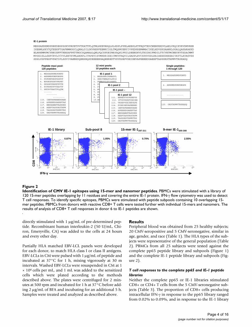
Identification of CMV IE-1 epitopes using 15-mer and nanomer peptidesFigure 2Identification of CMV IE-1 epitopes using 15-mer and nanomer peptides. PBMCs were stimulated with a library of 120 15-mer peptides overlapping by 11 residues and covering the entire IE-1 protein. IFN-γ flow cytometry was used to detect T cell responses. To identify specific epitopes, PBMCs were stimulated with peptide subpools containing 10 overlapping 15-mer peptides. PBMCs from donors with reactive CD8+ T cells were tested further with individual 15-mers and nanomers. The results of analysis of CD8+ T cell responses in donor 6 to IE-1 peptides are shown.
IE-1 protein
MESSAKRKMDPDNPDEGPSSKVPRPETPVTKATTFLQTMLRKEVNSQLSLGDPLFPELAEESLKTFEQVTEDCNENPEKDVLAELVKQIKVRVDMVRHRIKEHMLKKYTQTEEKFTGAFNMMGGCLQNALDILDKVHEPFEEMKCIGLTMQSMYENYIVPEDKREMWMACIKELHDVSKGAANKLGGALQAKARAKKDELRRKMMYMCYRNIEFFTKNSAFPKTTNGCSQAMAALQNLPQCSPDEIMAYAQKIFKILDEERDKVLTHIDHIFMDILTTCVETMCNEYKVTSDACMMTMYGGISLLSEFCRVLCCYVLEETSVMLAKRPLITKPEVISVMKRRIEEICMKVFAQYILGADPLRVCSPSVDDLRAIAEESDEEEAIVAYTLATAGVSSSDSLVSPPESPVPATIPLSSVIVAENSDQEESEQSDEEEEEGAQEEREDTVSVKSEPVSEIEEVAPEEEEDGAEEPTASGGKSTHPMVTRSKADQ
1. MESSAKRKMDPDNPD 2. AKRKMDPDNPDEGPS3. PDPDNPDEGPSSKVP4. NPDEGPSSKVPRPET5. GPSSKVPRPETPVTK6. KVPRPETPVTKATTF7. PETPVTKATTFLQTM
--------------
---------115. IEEVAPEEEEDGAEE116. APEEEEDGAEEPTAS117. EEDGAEEPTASGGKS118. AEEPTASGGKSTHPM119. TASGGKSTHPMVTRS120. GKSTHPMVTRSKADQ
Peptide maxi pool:120 peptides
IE-1 pool 11. MESRGRRCPEMASVL2. GRRCPEMASVLGPIS3. PEMASVLGPISGHVL4. SVLGPISGHVLKAVF5. PISGHVLKAVFSRGD6. HVLKAVFSRGDTPVL7. AVFSRGDTPVLPHET8. RGDTPVLPHETRLLQ9. PVLPHETRLLQTGIH 10.HETRLLQTGIHVRVS
IE-1 pool 2
IE-1 pool –
IE-1 pool 12111. EEREDTVSVKSEPVS112. DTVSVKSEPVSEIEE113. VKSEPVSEIEEVAPE 114. PVSEIEEVAPEEEED 115. IEEVAPEEEEDGAEE 116. APEEEEDGAEEPTAS 117. EEDGAEEPTASGGKS 118. AEEPTASGGKSTHPM 119. TASGGKSTHPMVTRS120. GKSTHPMVTRSKADQ
12 mini pools: 10 peptides each
Single peptides:1 through 120
1. MESSAKRKMDPDNPD
2. AKRKMDPDNPDEGPS
-------------
120. GKSTHPMVTRSKADQ
IFN-γ
CD
8
10 0 10 1 10 2 10 3 10 4
IFNG-FITC
9-mer IE-1300-308
2.55%
100 101 102 103 104
IFNG-FITC
IE-1 library
0.87%
100 101 102 103 104
IFNG-FITC
Sub-pool 8
1.05%
100 101 102 103 104
IFNG-FITC
15-mer IE-1297-311
0.70%
Page 4 of 16(page number not for citation purposes)

Journal of Translational Medicine 2007, 5:17 http://www.translational-medicine.com/content/5/1/17
ranged from 0.00% to 0.05%. The proportion of CD4+cells producing IFN-γ in response to the pp65 libraryranged from 0.00% to 0.04% and in response to the IE-1library ranged from 0.01% to 0.04%. PBMCs were alsotested against the subpools, none of which induced signif-icant IFN-γ.
Testing PBMCs from each of the 20 CMV-seropositive sub-jects revealed that the pp65 and IE-1 libraries stimulatedCD8+ T cells in 7 (35%) and 8 (40%) subjects, respec-tively. IFN-γ producing cells ranged from 0.12% to 0.70%for pp65 and 0.32% to 2.06% for IE-1 (Table 4). CD4+cells were stimulated by the pp65 library in 10 (50%) sub-jects with the percent of IFN-γ producing cells rangingfrom 0.11% to 3.33%, but were not stimulated in anydonors by the IE-1 library (Table 4).
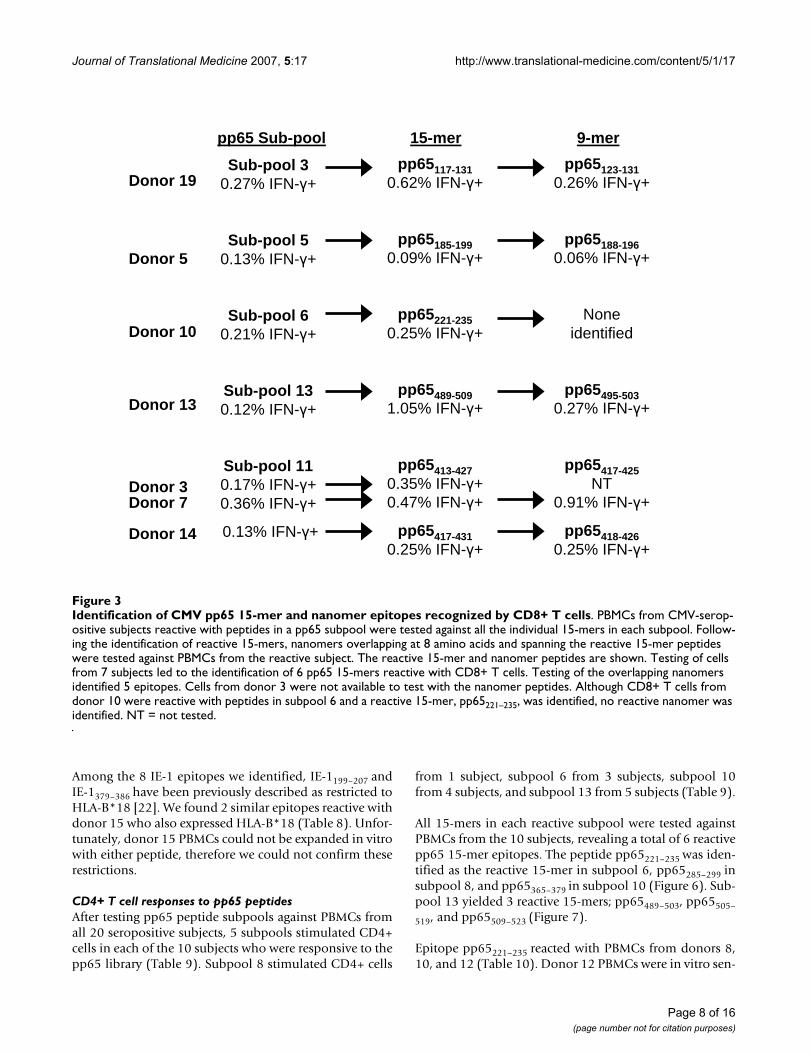
CD8+ T cell responses to pp65 peptidesTesting of PBMCs from all 20 CMV-seropositive donorsagainst the pp65 subpools revealed that all 7 subjects withCD8+ cells that were reactive to the pp65 library reactedto at least 1 pp65 subpool. We identified 5 reactive pp65subpools, with subpool 11 stimulating CD8+ cells from 3subjects (Table 5).
All of the 15-mer peptides in each of the reactive sub-pools, 3, 5, 6, 11, and 13 were tested against PBMCs fromthe corresponding subjects, and we identified 6 reactivepp65 15-mer peptides (Figure 3). Four of the 6 reactivepeptides each elicited a CD8+ T cell response in 1 subject,donors 5, 10, 13, and 19. The other 2 reactive peptideswere overlapping 15-mers that elicited responses indonors 3, 7 and 14.
Since HLA class I antigens usually present peptides of 9 or10 amino acids long to CD8+ T cells, nanomers were syn-thesized that spanned each reactive 15-mer and that over-lapped by 8 amino acids. We identified 5 epitopes by
testing each nanomer against PBMCs from donor(s) reac-tive with the corresponding 15-mer (Figure 3). Three ofthe 15-mers each yielded 1 epitope, pp65123–131, pp65188–
196, and pp65495–503, in donors 19, 5, and 13, respectively(Figure 3). The nanomers spanning 2 overlapping 15-mers, pp65413–427 and pp65417–431, yielded 2 epitopes,pp65417–425 and pp65418–426 (Figure 3). However, nano-mers covering pp65221–235, did not stimulate any cells.
Both pp65417–425 and pp65418–426 stimulated CD8+ cellsfrom donors 3, 7, and 14, all of whom expressed HLA-B*07 (Table 6). The restriction to HLA-B*07 was con-firmed by testing donor 14 PBMCs sensitized in vitro for10 days with pp65418–426 against EBV-LCLs pulsed withpp65418–426. CD8+ IFN-γ production was detected whenthe in vitro sensitized PBMCs were tested against peptide-loaded EBV-LCLs expressing HLA-B*07, but not thoseexpressing the other donor 14 HLA class I antigens. Simi-lar epitopes, pp65415–429 and pp65417–426 have been previ-ously found to be presented by HLA-B*07[8,19].
The peptide pp65123–131 has been described as restricted toHLA-B*35, and we found this peptide to stimulate CD8+cells from donor 19, who also expressed HLA-B*35 (Table6)[20]. This restriction was confirmed by testing donor 19PBMCs that were ex vivo sensitized with pp65123–131against EBV-LCLs loaded with pp65123–131. CD8+ T cellsproduced IFN-γ when tested against pp65123–131-loadedEBV-LCLs expressing HLA-B*35 but not those expressingother donor 19 HLA class I antigens.
The epitope pp65495–503 stimulated CD8+ cells fromdonor 13, who expressed HLA-A*02 (Table 6). Since thisepitope has been described by several groups as beingrestricted to HLA-A*02, we did not test this peptide fur-ther[8,13,21].
Table 1: Age, Gender, and Race of Subjects Studied
CMV-Seropositive n = 20 CMV-Seronegative n = 5 All n = 25
Age, years
Median 45.1 42.8 44.6Range 17–64 35–49 17–64Gender
Male, % 80 80 80Race
White, % 55 60 56Black, % 40 20 36Hispanic, % 5 0 4Unknown, % 0 20 4
Page 5 of 16(page number not for citation purposes)

Journal of Translational Medicine 2007, 5:17 http://www.translational-medicine.com/content/5/1/17
The epitope pp65188–196 stimulated CD8+ cells fromdonor 5, who expressed HLA-A*05; -B*11, 68, and -C*05,06 (Table 6). Unfortunately, no PBMCs were available totest. However, pp65186–196 has been described as beingrestricted to HLA-B*35 and HLA-A*6801/02[20], so it ispossible that in this donor, pp65188–196 is restricted toHLA-A*68[8,21].
CD8+ T cell responses to IE-1 peptidesThe 12 IE-1 subpools were tested against all 20 subjectsand 4 subpools induced IFN-γ in 8 subjects (Table 7). Atleast 1 IE-1 subpool induced IFN-γ production in CD8+ Tcells from all 8 subjects with cells reactive with the IE-1library. One subpool stimulated CD8+ cells in 5 subjects,another stimulated CD8+ cells in 3 subjects, and 2 othersubpools stimulated CD8+ cells from 1 subject (Table 7).
Testing the 15-mers in IE-1 subpool 5 against PBMCsfrom donor 15 identified IE-1197–211 as the reactive 15-merand IE-1198–206 as the reactive nanomer (Figure 4). Testingthe 15-mers in subpool 10 against cells from donor 15revealed that IE-1373–387 as the reactive 15-mer and IE-1378–386 as the reactive nanomer (Figure 4), and testing ofsubpool 3 peptides against PBMCs from donors 5, 10, and11 identified IE-181–95and IE-187–95 as the reactive 15-merand nanomer (Figure 4).
Testing subpool 8 peptides revealed that one 15-mer and1 nanomer were reactive with cells from donor 6, IE-1297–
311 (Figure 5a) and IE-1300–308 (Figure 5b), respectively.Testing of subpool 8 peptides against cells from donors 2,5, and 7 identified 3 reactive 15-mers: IE-1305–319, IE-1309–
323, and IE-1313–327(Figure 5a), and 4 reactive nanomers:
Table 2: HLA Genotype of the 20 CMV-Seropositive Subjects Studied
HLA Type
Donor Race Class I Class ll
A* B* C* DRB1* DRB- DQB1*
1 Black 01, 0301 15, 81 05, 18 0901, 11 3*00, 4*01 02, 062 Caucasian 24, 32 07, 40 02, 07 0401, 1302 3*0301, 4*0103 03, 06093 Caucasian 01, 31 07, 27 0202, 0702 0801, 1501 5*01, 5*01 04, 064 Black 30, 3601 53, 53 04, 04 03, 1001 3*01, 3*01 04, 055 Hispanic 11, 68 44, 45 05, 06 0404, 1101 3*02, 4*0103 03, 036 Black 26, 30 35, 57 04, 18 11, 16 3*02, 5*02 0301, 057 Caucasian 01, 0201 07, 35 04, 07 0401, 0701 4*01, 4*01 0303, 038 Black 0202, 30 4901, 53 04, 07 13031, 1503 3*02, 3*02 02, 069 Caucasian 23, 32 35, 41 04, 17 11, 11 3*02, 3*02 03, 0610 Black 0301, 68 53, 58 04, 06 0701, 1302 3*0301, 4*0401 0202, 060411 Black 26, 74 15, 5802 0602, 1601 0102, 1101 3*0202, 3*0202 0301, 050112 Black 0202, 68 5301, 5301 04, 04 03021, 1503 3*01, 5*01 04, 0613 Caucasian 0201, 0201 15, 18 03, 07 13, 1501 3*02, 5*01 06, 0614 Caucasian 010101, 2601 070201, 3801 070201, 120301 0402, 1101 3*02, 4*0103 0301, 030215 Caucasian 0201, 30 18, 44 0501, 0501 04, 13 3*02, 4*01 03, 0616 Caucasian 0201, 11 07, 40 03, 07 0301, 1501 3*01, 5*01 02, 0617 Caucasian 0201, 0201 15, 18 03, 07 13, 1501 3*02, 5*01 06, 0618 Black 2301, 24 07, 18 0202, 0702 07, 15 4*01, 5*01 02, 0619 Caucasian 03, 29 35, 44 04, 1601 01, 0701 4*01, 4*01 0202, 050120 Caucasian 03, 11 07, 5601 01, 07 0101, 0701 4*01, 4*01 02, 0501
Table 3: Proportion of Peripheral Blood CD8+ and CD4+ T Cells from 5 Healthy CMV-Seronegative Subjects Producing Intracellular IFN-γ Following Stimulation With a Library of 138 CMV pp65 or 120 CMV IE-1 15-mer Peptides
Proportion of T Cells Producing Intracellular IFN-γ(%) in Each Donor
Library Cell Type 1 2 3 4 5
pp65 CD8+ 0.02 0.03 0.03 0.08 0.09IE-1 CD8+ 0.04 0.02 0.01 0.00 0.05
pp65 CD4+ 0.00 0.03 0.02 0.02 0.03IE-1 CD4+ 0.01 0.01 0.02 0.01 0.04
Page 6 of 16(page number not for citation purposes)

Journal of Translational Medicine 2007, 5:17 http://www.translational-medicine.com/content/5/1/17
IE-1305–313, IE-1308–316, IE-1312–320, and IE-1319–327 (Figure5c).
Among the 8 IE-1 epitopes identified, IE-187–95, IE-1300–
308, IE-1305–313, IE-1312–320, and IE-1319–327 have not beenpreviously described. The peptide IE-187–95 stimulatedCD8+ cells from 3 donors, all of whom expressed HLA-C*06 (Table 8) and this restriction was confirmed by test-ing donor 11 in vitro sensitized PBMCs against IE-187–95-loaded EBV-LCLs.
The peptide IE-1305–313 stimulated cells from donor 7,who expressed HLA-A*01, 0201; -B*07, 35; and -C*04,07. Testing donor 7 PBMCs that were in vitro sensitizedwith IE-1305–313 against IE-1305–313-loaded EBV-LCLs con-firmed that this epitope was restricted to HLA-C*07(Table 8).
The peptide IE-1308–316 stimulated cells from donors 2 and7, both expressing HLA-B*07 and -C*07. Testing donor 7
PBMCs that were in vitro sensitized with IE-1308–316against IE-1308–316-loaded EBV-LCLs confirmed the restric-tion to HLA-C*07 (Table 8). However, an overlappingpeptide, IE-1309–317, has been reported to be restricted toHLA-B*0702[9].
The peptide IE-1312–320 stimulated cells from donor 7 andtesting of PBMCs that were in vitro sensitized with IE-1312–320 against peptide-loaded EBV-LCLs revealed restric-tion to HLA-C*07 (Table 8).
The peptide IE-1319–327 stimulated cells from donor 5,who expressed A*11,68; -B*44,45; and -C*05,06. TestingPBMCs that were in vitro sensitized with IE-1319–327against peptide-loaded EBV-LCLs revealed that IE-1319–327was restricted to HLA-A*68.
The peptide IE-1300–308 stimulated CD8+ cells from donor6 however, no PBMCs were available for testing.
Table 5: Proportion of Peripheral Blood CD8+ T Cells From 20 Healthy CMV-Seropositive Subjects Producing Intracellular IFN-γ Following Stimulation With a Library of 138 CMV pp65 15-mer Peptides and Each of 14 subpools of 10 Peptides*
CD8+ T Cells Producing Intracellular IFN-γ in Each Donor (%)
1 2 3 4 5 6 7 8 9 10 11 12 13 14 15 16 17 18 19 20
Library 0.02 0.06 0.37 0.10 0.27 0.13 0.70 0.13 0.13 0.27 0.11 0.18 0.12 0.27 0.07 0.08 0.06 0.05 0.18 0.03Subpool
1 0.01 0.04 0.07 0.03 0.01 0.02 0.02 0.02 0.01 0.01 0.12 0.09 0.14 0.03 0.01 0.08 0.01 0.00 0.02 0.032 0.01 0.05 0.02 0.03 0.17 0.05 0.11 0.05 0.06 0.03 0.03 0.08 0.01 0.03 0.06 0.08 0.03 0.03 0.03 0.023 0.02 0.02 0.07 0.13 0.04 0.06 0.15 0.01 0.03 0.02 0.03 0.16 0.03 0.04 0.03 0.03 0.05 0.01 0.27 0.024 0.02 0.04 0.03 0.01 0.10 0.08 0.18 0.08 0.06 0.09 0.01 0.08 0.02 0.05 0.07 0.02 0.00 0.05 0.05 0.025 0.04 0.01 0.02 0.03 0.13 0.04 0.08 0.07 0.11 0.00 0.04 0.06 0.08 0.02 0.03 0.08 0.01 0.01 0.03 0.026 0.00 0.01 0.00 0.14 0.08 0.03 0.04 0.05 0.08 0.21 0.05 0.06 0.03 0.03 0.11 0.08 0.03 0.04 0.01 0.027 0.03 0.06 0.03 0.03 0.04 0.01 0.11 0.07 0.07 0.08 0.03 0.07 0.08 0.15 0.03 0.03 0.02 0.10 0.03 0.028 0.02 0.00 0.03 0.01 0.03 0.10 0.01 0.10 0.03 0.01 0.03 0.08 0.03 0.02 0.02 0.10 0.02 0.04 0.03 0.059 0.05 0.03 0.00 0.06 0.02 0.03 0.03 0.02 0.03 0.02 0.05 0.12 0.02 0.04 0.02 0.05 0.03 0.04 0.03 0.1310 0.06 0.02 0.00 0.05 0.02 0.10 0.14 0.06 0.03 0.04 0.06 0.05 0.02 0.08 0.03 0.06 0.03 0.03 0.04 0.0211 0.02 0.02 0.17 0.01 0.02 0.06 0.36 0.04 0.06 0.04 0.05 0.12 0.01 0.13 0.01 0.05 0.01 0.01 0.00 0.0212 0.12 0.08 0.03 0.00 0.07 0.02 0.06 0.05 0.09 0.01 0.03 0.04 0.01 0.03 0.00 0.08 0.02 0.03 0.02 0.0513 0.05 0.05 0.02 0.00 0.01 0.02 0.06 0.06 0.07 0.04 0.08 0.07 0.12 0.03 0.04 0.03 0.09 0.03 0.01 0.0214 0.03 0.01 0.02 0.01 0.10 0.04 0.06 0.07 0.08 0.08 0.06 0.07 0.02 0.01 0.03 0.02 0.02 0.05 0.04 0.00
* Subpool 14 contained 8 peptidesPositive CD8+ T cell assays are indicated in bold and italics.
Table 4: Proportion of Peripheral Blood CD8+ and CD4+ T Cells From 20 Healthy CMV-Seropositive Subjects Producing Intracellular IFN-γ Following Stimulation With a Library of 138 CMV pp65 or 120 CMV IE-1 15-mer Peptides
Proportion of T Cells Producing Intracellular IFN-γ(%) in Each Donor
Library Cell 1 2 3 4 5 6 7 8 9 10 11 12 13 14 15 16 17 18 19 20
pp65 CD8+ 0.02 0.06 0.37 0.10 0.27 0.13 0.70 0.13 0.13 0.27 0.11 0.18 0.12 0.27 0.07 0.08 0.06 0.05 0.18 0.03IE-1 CD8+ 0.05 0.46 2.06 0.04 1.65 0.87 1.37 0.13 0.15 0.33 0.32 0.06 0.02 0.12 1.41 0.05 0.05 0.02 0.03 0.08
pp65 CD4+ 0.16 0.06 0.00 0.06 0.11 0.76 0.11 0.65 0.08 0.21 3.33 0.18 0.20 1.34 0.23 0.02 0.15 0.03 0.04 0.01IE-1 CD4+ 0.02 0.04 0.01 0.03 0.02 0.14 0.03 0.05 0.01 0.02 0.08 0.06 0.04 0.13 0.09 0.02 0.05 0.02 0.00 0.10
*Positive CD8+ T cell assays are indicated in bold, italics.
Page 7 of 16(page number not for citation purposes)
Journal of Translational Medicine 2007, 5:17 http://www.translational-medicine.com/content/5/1/17
Among the 8 IE-1 epitopes we identified, IE-1199–207 andIE-1379–386 have been previously described as restricted toHLA-B*18 [22]. We found 2 similar epitopes reactive withdonor 15 who also expressed HLA-B*18 (Table 8). Unfor-tunately, donor 15 PBMCs could not be expanded in vitrowith either peptide, therefore we could not confirm theserestrictions.
CD4+ T cell responses to pp65 peptidesAfter testing pp65 peptide subpools against PBMCs fromall 20 seropositive subjects, 5 subpools stimulated CD4+cells in each of the 10 subjects who were responsive to thepp65 library (Table 9). Subpool 8 stimulated CD4+ cells
from 1 subject, subpool 6 from 3 subjects, subpool 10from 4 subjects, and subpool 13 from 5 subjects (Table 9).
All 15-mers in each reactive subpool were tested againstPBMCs from the 10 subjects, revealing a total of 6 reactivepp65 15-mer epitopes. The peptide pp65221–235 was iden-tified as the reactive 15-mer in subpool 6, pp65285–299 insubpool 8, and pp65365–379 in subpool 10 (Figure 6). Sub-pool 13 yielded 3 reactive 15-mers; pp65489–503, pp65505–
519, and pp65509–523 (Figure 7).
Epitope pp65221–235 reacted with PBMCs from donors 8,10, and 12 (Table 10). Donor 12 PBMCs were in vitro sen-
Identification of CMV pp65 15-mer and nanomer epitopes recognized by CD8+ T cellsFigure 3Identification of CMV pp65 15-mer and nanomer epitopes recognized by CD8+ T cells. PBMCs from CMV-serop-ositive subjects reactive with peptides in a pp65 subpool were tested against all the individual 15-mers in each subpool. Follow-ing the identification of reactive 15-mers, nanomers overlapping at 8 amino acids and spanning the reactive 15-mer peptides were tested against PBMCs from the reactive subject. The reactive 15-mer and nanomer peptides are shown. Testing of cells from 7 subjects led to the identification of 6 pp65 15-mers reactive with CD8+ T cells. Testing of the overlapping nanomers identified 5 epitopes. Cells from donor 3 were not available to test with the nanomer peptides. Although CD8+ T cells from donor 10 were reactive with peptides in subpool 6 and a reactive 15-mer, pp65221–235, was identified, no reactive nanomer was identified. NT = not tested.
pp65 Sub-pool 15-mer 9-mer
Sub-pool 30.27% IFN-γ+
Sub-pool 50.13% IFN-γ+
Sub-pool 60.21% IFN-γ+
Sub-pool 130.12% IFN-γ+
Sub-pool 110.17% IFN-γ+ 0.36% IFN-γ+
0.13% IFN-γ+
pp65117-1310.62% IFN-γ+
pp65185-1990.09% IFN-γ+
pp65221-235 0.25% IFN-γ+
pp65489-5091.05% IFN-γ+
pp65413-4270.35% IFN-γ+ 0.47% IFN-γ+
pp65417-4310.25% IFN-γ+
pp65123-1310.26% IFN-γ+
pp65188-1960.06% IFN-γ+
None identified
pp65495-503 0.27% IFN-γ+
pp65417-425NT
0.91% IFN-γ+
pp65418-426 0.25% IFN-γ+
Donor 19
Donor 5
Donor 10
Donor 13
Donor 14
Donor 7Donor 3
Page 8 of 16(page number not for citation purposes)

Journal of Translational Medicine 2007, 5:17 http://www.translational-medicine.com/content/5/1/17
sitized with pp65221–235, and reacted with pp65221–235,-loaded EBV-LCLs expressing DRB1*15 and DRB5*01. It islikely that pp65221–235 is presented by DRB1*15 since 2 ofthe 3 donors expressed DRB1*15 but only donor 12expressed DRB5*01. In addition, pp65225–239 has previ-ously been reported to be restricted to DRB1*15[23].
Pp65365–379 stimulated cells from 4 donors all expressingHLA-DRB1*11 (Table 10). PBMCs from donor 11 were invitro sensitized with pp65365–379, and produced IFN-γwhen tested against pp65365–379 -loaded EBV-LCLsexpressing HLA-DRB1*11, but not when tested againstthose expressing HLA-DRB1*0102, DRB3*0202,DQB1*0301, and DQB1*0501. The testing of this pep-tide against a second subject, donor 6, revealed similarresults. Two similar epitopes, pp65361–376 and pp65361–
375, have been described as restricted to HLA-DR11[11,24].
Epitope pp65489–503 reacted with 3 donors, all of whomexpressed HLA-DRB3*02 (Table 10). Donor 6 PBMCsthat were in vitro sensitized with pp65489–503, producedIFN-γ when tested against EBV-LCLs expressing HLA-
DRB3*02, but not when tested against the other donor 6class II antigens. Although we found that pp65489–503 wasrestricted to DRB3*02, others have described this peptideas restricted to DRB1*11 and DRB1*03 [24].
Epitope pp65505–519 was reactive with PBMCs fromdonors 6 and 17 and testing of donor 6 pp65505–519 exvivo sensitized PBMCs against pp65505–519-loaded EBV-LCLs revealed presentation by DQB1*0502. Donor 17 didnot express DQB1*0502, but cells were not available foradditional testing. The epitope pp65505–523 has beenreported to be restricted to DRB1*01[23] and DR52[11].
Epitope pp65509–523 reacted with PBMCs from donors 12and 14, and testing donor 12 in vitro sensitized PBMCsagainst loaded EBV-LCLs revealed presentation byDRB3*0101. Donor 14 did not express DRB3*0101 how-ever, an adequate number of cells were not available for invitro sensitization and testing. The epitope pp65509–524was previously reported to be restricted to DRB1*03[24].
Pp65285–299 reacted with cells from donor 7, whoexpressed both HLA-DRB1*0401 and HLA-DRB1*0701
Table 7: Proportion of Peripheral Blood CD8+ T Cells From 20 Healthy CMV-Seropositive Subjects Producing Intracellular IFN-γ Following Stimulation With a Library of 120 CMV IE-1 15-mer Peptides and Each of 12 Subpools of 10 Peptides
CD8+ T Cells Producing Intracellular IFN-γ in Each Donor (%)
1 2 3 4 5 6 7 8 9 10 11 12 13 14 15 16 17 18 19 20
Library 0.05 0.46 2.06 0.04 1.65 0.87 1.37 0.13 0.15 0.33 0.32 0.06 0.02 0.12 1.41 0.05 0.05 0.02 0.03 0.08Subpool
1 0.04 0.03 0.08 0.05 0.01 0.03 0.01 0.11 0.04 0.05 0.04 0.05 0.04 0.05 0.03 0.05 0.02 0.02 0.01 0.002 0.05 0.07 0.06 0.01 0.07 0.04 0.06 0.05 0.10 0.03 0.02 0.04 0.02 0.02 0.01 0.05 0.01 0.01 0.01 0.013 0.06 0.05 0.04 0.04 0.95 0.03 0.02 0.08 0.02 0.26 0.29 0.10 0.01 0.05 0.02 0.08 0.01 0.01 0.01 0.024 0.06 0.07 0.16 0.05 0.01 0.05 0.00 0.00 0.07 0.04 0.04 0.06 0.03 0.03 0.02 0.08 0.04 0.03 0.00 0.025 0.03 0.04 0.05 0.00 0.05 0.04 0.05 0.07 0.17 0.03 0.07 0.03 1.28 0.04 1.60 0.08 0.03 0.06 0.01 0.026 0.02 0.02 0.17 0.03 0.05 0.10 0.01 0.06 0.06 0.06 0.06 0.06 0.03 0.12 0.05 0.11 0.02 0.01 0.04 0.007 0.04 0.01 0.02 0.03 0.01 0.03 0.03 0.09 0.01 0.04 0.05 0.07 0.06 0.03 0.07 0.05 0.01 0.02 0.02 0.008 0.06 0.43 1.65 0.00 0.33 1.05 1.39 0.07 0.02 0.01 0.05 0.04 0.03 0.12 0.06 0.00 0.03 0.01 0.06 0.009 0.04 0.02 0.03 0.08 0.02 0.05 0.02 0.14 0.03 0.04 0.09 0.08 0.03 0.04 0.11 0.02 0.02 0.04 0.04 0.0010 0.04 0.07 0.08 0.03 0.03 0.05 0.00 0.07 0.08 0.05 0.05 0.07 0.04 0.05 0.25 0.05 0.01 0.04 0.04 0.0011 0.01 0.01 0.09 0.03 0.05 0.06 0.05 0.09 0.04 0.02 0.08 0.07 0.02 0.01 0.05 0.07 0.03 0.01 0.01 0.0212 0.02 0.05 0.11 0.06 0.07 0.03 0.02 0.06 0.03 0.03 0.05 0.08 0.06 0.04 0.04 0.10 0.02 0.04 0.03 0.02
Positive assays are indicated in bold and italics.
Table 6: Individual pp65 Epitopes that Induced Intracellular IFN-γ Production by CD8+ T Cells, the HLA Type of Donors With Reactive CD8+ T Cells, and HLA Antigen Restrictions of Each Epitope
Epitope(s) Donor HLA-A* HLA-B* HLA-C* HLA Antigen Restriction Published Antigen Restriction
pp65123–131 19 03, 29 35, 44 04, 1601 B*35 pp65123–131 B*3501 [20]pp65188–196 05 11, 68 44, 45 05, 06 No cells available pp65186–196 A*6801/2 [21]pp65417–425 and pp65418–426 03 01, 31 07, 27 0202, 0702 No cells available
07 01, 0201 07, 35 04, 07 No cells available pp65417–426 B*0702 [8,19]14 010101, 2601 070201, 3801 070201, 120301 HLA-B*07
pp65 495–503 13 0201, 0201 15, 18 03, 07 Not tested pp65495–503 A*0201 [13,30]
The HLA antigen restrictions identified are shown in bold.
Page 9 of 16(page number not for citation purposes)

Journal of Translational Medicine 2007, 5:17 http://www.translational-medicine.com/content/5/1/17
(Table 10). Although additional PBMCs were not availa-ble for testing, a similar epitope, pp65281–295, was previ-ously found to be restricted to both HLA-DR*4 and HLA-DR*7[11].
CD4+ T cell responses to IE-1Testing with both the IE-1 library and the 12 subpools didnot reveal active CD4+ populations from any subjects(Table 11).
DiscussionThe aim of this investigation was to map CMV pp65 andIE-1 epitopes in 20 CMV-seropositive healthy subjects inorder to identify peptides that may be important in vacci-nation, adoptive immunotherapy, and the monitoring oftransplant recipients. Among the 20 subjects, CD8+ T cellresponses to pp65 and IE-1 were detected in a similar pro-portion of patients: 35% versus 40%. However, CD4+ Tcell responses to pp65 were more common thanresponses to IE-1, as 50% of subjects reacted to pp65 butnone reacted to IE-1. Using similar methodology, Kern etal also found CD8+ T cell responses in healthy subjects toboth pp65 and IE-1[9,11]. While they did not specificallyscreen for IE-1 epitopes that were presented by CD4+ T
cells, a comparison of CD4+ T cell responses to pp65 pep-tides with responses to CMV lysate suggested that pp65was a dominant target of the CMV-specific CD4+ T cellresponse[11]. A recent analysis of overlapping 15-mersspanning 213 CMV open reading frames including pp65and IE-1, by Sylwester et al found that the CD8+ andCD4+ T cell responses were directed to peptides in a vari-ety of proteins,[25]. In addition, they found that CD8+ Tcell responses to both pp65 and IE-1 were strong and theCD4+ T cell responses were strong to pp65 but weak to IE-1 which supports the results of our study [25].
We identified 4 pp65 and 8 IE-1 nanomers presented byHLA class I, and 6 pp65 15-mers presented by HLA classII. While all 4 of the pp65 CMV class I epitopes have beenpreviously described, 6 of the 8 IE-1 class I epitopes, and2 of the 6 pp65 class II epitopes have not. The pp65epitopes and restrictions that we identified were similar oridentical to those reported previously, but the IE-1 class Iepitopes had either not been described or we identifiedHLA restrictions that differed from those reported.
Four of the 6 new IE-1 class I epitopes we identified wererestricted to HLA-C antigens, a finding that may be corre-
Identification of CMV IE-1 15-mer and nanomer epitopes from 3 peptide subpools reactive with CD8+ T cellsFigure 4Identification of CMV IE-1 15-mer and nanomer epitopes from 3 peptide subpools reactive with CD8+ T cells. PBMCs from 4 donors were reactive with peptides in IE-1 subpools 3, 5, and 10. Testing of all 15-mers in each subpool identi-fied 3 nanomers reactive with CD8+ T cells, one from each subpool. Testing of the 6 overlapping nanomers spanning each reactive 15-mer lead to the identification of 3 nanomer epitopes.
IE-1 Sub-pool 15-mer 9-mer
Sub-pool 5
1.60% IFN-γ+
Sub-pool 10
0.25% IFN-γ+
IE-1197-211
1.66% IFN-γ+
IE-1373-387
0.36% IFN-γ+
IE-1198-206
4.58% IFN-γ+
IE-1378-386
0.87% IFN-γ+
Donor 15
Donor 5
Donor 10
Donor 11
Sub-pool 3
0.95% IFN-γ+
0.26% IFN-γ+
0.29% IFN-γ+
IE-181-95
0.53% IFN-γ+
0.18% IFN-γ+
0.16% IFN-γ+
IE-187-95
1.25% IFN-γ+
0.20% IFN-γ+
0.19% IFN-γ+
Page 10 of 16(page number not for citation purposes)

Journal of Translational Medicine 2007, 5:17 http://www.translational-medicine.com/content/5/1/17
Page 11 of 16(page number not for citation purposes)
Identification of CMV pp65 15-mer and nanomer epitopes from peptide subpool 8 that were reactive with CD8+ T cellsFigure 5Identification of CMV pp65 15-mer and nanomer epitopes from peptide subpool 8 that were reactive with CD8+ T cells. Testing of IE-1 15-mers from subpool 8 against cells from donor 2 (gold bar), donor 5 (red bar), donor 6 (light blue bar), and donor 7 (navy blue bar) revealed that the peptide IE-1297–311 was reactive with donor 6, IE-1305–319 was reactive with donor 7, IE-1309–323 was reactive with donors 2 and 6, and IE-1313–327 was reactive with donor 5 (Panel A). Testing of the 6 nanomers overlapping IE-1297–311 against cells from donor 6 revealed that IE-1300–308 was the dominant nanomer (Panel B). Test-ing of the nanomers spanning the overlapping 15-mers IE-1305–319, IE-1309–323, and IE-1313–327 against PBMCs from donors 2, 5, and 7 identified 4 nanomers that were reactive with CD8+ T cells: IE-1305–313, IE-1308–316, IE-1312–320 and IE-1319–327, (Panel C).
B
9-mers9-mers15-mer
IE1 2
97-3
11
IE1 2
97-3
05
IE1 2
98-3
06
IE1 2
99-3
07
IE1 3
00-3
08
IE1 3
01-3
09
IE1 3
02-3
10
IE1 3
03-3
11
15-mer
IE1 2
97-3
11
IE1 2
97-3
05
IE1 2
98-3
06
IE1 2
99-3
07
IE1 3
00-3
08
IE1 3
01-3
09
IE1 3
02-3
10
IE1 3
03-3
11
IE1 2
97-3
11
IE1 2
97-3
05
IE1 2
98-3
06
IE1 2
99-3
07
IE1 3
00-3
08
IE1 3
01-3
09
IE1 3
02-3
10
IE1 3
03-3
11
*
0
0.5
1
1.5
2
2.5
3
CD
8 IF
N-g
amm
a+ c
ells
(%
)
Donor 2
Donor 5
Donor 6
Donor 7
Donor 2Donor 2
Donor 5Donor 5
Donor 6Donor 6
Donor 7Donor 7
C
IE-1
305-
319
IE-1
309-
323
IE-1
313-
327
IE-1
305-
313
IE-1
306-
314
IE-1
307-
315
IE-1
308-
316
IE-1
309-
317
IE-1
310-
318
IE-1
311-
319
IE-1
312-
320
IE-1
313-
321
IE-1
314-
322
IE-1
315-
323
IE-1
316-
324
IE-1
317-
325
IE-1
318-
326
IE-1
319-
327
15-mers 9-mers
*
**
*
*
0
0.2
0.4
0.6
0.8
1
1.2
1.4
1.6
1.8
2
CD
8 IF
N-g
amm
a+ c
ells
(%
)
A
Sub
pool
8
IE1 2
81-2
95
IE1 2
85-2
99
IE1 2
89-3
03
IE1 2
93-3
07
IE1 2
97-3
11
IE1 3
01-3
15
IE1 3
05-3
19
IE1 3
09-3
23
IE1 3
13-3
27
IE1 3
17-3
31
15-mers
* *
*
**
0
0.2
0.4
0.6
0.8
1
1.2
1.4
1.6
1.8
2
A
Sub
pool
8
IE1 2
81-2
95
IE1 2
85-2
99
IE1 2
89-3
03
IE1 2
93-3
07
IE1 2
97-3
11
IE1 3
01-3
15
IE1 3
05-3
19
IE1 3
09-3
23
IE1 3
13-3
27
IE1 3
17-3
31
15-mers
* *
*
**
0
0.2
0.4
0.6
0.8
1
1.2
1.4
1.6
1.8
2

Journal of Translational Medicine 2007, 5:17 http://www.translational-medicine.com/content/5/1/17
lated to the fact that all of our subjects and EBV-LCLs wereHLA typed at high-resolution. Because HLA-C antigen typ-ing results were ambiguous prior to the advent of molec-ular genotyping, the availability of precise moleculartyping for this study may have permitted the identifica-tion of epitopes not possible in the past when molecularmethods were not available or were less precise.
While the conventional thought has been that uniquepeptides are each presented by a specific HLA allele, someCMV epitopes are presented by multiple alleles. Epitopesfor HLA class I antigens are generally believed to be veryrestrictive, with epitopes binding to specific HLA-types.Our results suggest that this is not entirely true for IE-1class I epitopes as the HLA restrictions were somewhat
promiscuous. We found that the epitope IE-1319–327 wasrestricted to HLA-A*68, while a similar epitope IE-1316–324has been described as restricted to A*0201. We also foundthat IE-1308–316 was restricted to HLA-C*07, but IE-1309–
317 is reported to be restricted to HLA-B*0702 [19]. In pre-vious studies, we found that the epitopes pp65341–349 andpp65341–350 are presented by HLA-A*0101, HLA-A*2402,and HLA-Cw*0402 [14,26].
We found that 1 region of IE-1, amino acid residues 300to 327, to be more immunogenic than other regions. Weidentified 5 epitopes in this region, in addition to the 3other epitopes previously identified[9,19,27]. This regionwas particularly immunogenic for HLA-C*07 as we iden-tified 3 HLA-C*07 restricted epitopes in this region.
Table 9: Proportion of Peripheral Blood CD4+ T Cells From 20 Healthy CMV-Seropositive Subjects Producing Intracellular IFN-γ Following Stimulation With a Library of 138 CMV pp65 15-mer Peptides and Each of 14 subpools of 10 Peptides*
CD4+ T Cells Producing Intracellular IFN-γ in Each Donor (%)
1 2 3 4 5 6 7 8 9 10 11 12 13 14 15 16 17 18 19 20
Library 0.16 0.06 0.00 0.06 0.11 0.76 0.11 0.65 0.08 0.21 3.33 0.18 0.20 1.34 0.23 0.02 0.15 0.03 0.04 0.01Subpool
1 0.01 0.01 0.02 0.01 0.02 0.03 0.00 0.03 0.02 0.01 0.03 0.05 0.08 0.05 0.04 0.00 0.02 0.01 0.01 0.042 0.05 0.09 0.01 0.03 0.05 0.05 0.02 0.04 0.02 0.01 0.03 0.06 0.02 0.06 0.03 0.02 0.03 0.04 0.01 0.033 0.03 0.02 0.01 0.01 0.03 0.03 0.04 0.05 0.05 0.03 0.03 0.06 0.03 0.06 0.06 0.00 0.03 0.02 0.04 0.024 0.01 0.06 0.02 0.01 0.03 0.06 0.00 0.09 0.01 0.02 0.04 0.04 0.01 0.03 0.04 0.01 0.01 0.02 0.02 0.005 0.02 0.04 0.01 0.01 0.02 0.02 0.00 0.04 0.04 0.01 0.04 0.05 0.02 0.07 0.02 0.02 0.01 0.00 0.01 0.016 0.02 0.04 0.01 0.04 0.01 0.03 0.01 0.45 0.04 0.11 0.02 0.18 0.02 0.01 0.02 0.02 0.01 0.04 0.01 0.037 0.01 0.05 0.04 0.01 0.01 0.02 0.02 0.03 0.01 0.07 0.02 0.06 0.05 0.04 0.07 0.02 0.02 0.07 0.02 0.018 0.04 0.04 0.01 0.03 0.02 0.09 0.05 0.12 0.01 0.01 0.04 0.08 0.00 0.07 0.05 0.02 0.01 0.03 0.01 0.039 0.06 0.13 0.03 0.03 0.03 0.05 0.05 0.04 0.03 0.01 0.05 0.05 0.01 0.06 0.02 0.02 0.01 0.03 0.02 0.0910 0.14 0.02 0.00 0.03 0.05 0.56 0.00 0.10 0.05 0.04 2.76 0.08 0.01 0.96 0.03 0.02 0.02 0.01 0.02 0.0011 0.03 0.02 0.00 0.00 0.02 0.15 0.00 0.08 0.01 0.01 0.06 0.13 0.01 0.07 0.01 0.01 0.01 0.01 0.00 0.0112 0.04 0.03 0.01 0.00 0.02 0.04 0.01 0.05 0.04 0.01 0.01 0.04 0.00 0.06 0.03 0.02 0.02 0.01 0.01 0.0513 0.02 0.03 0.02 0.03 0.03 0.27 0.00 0.13 0.07 0.07 0.05 0.12 0.14 0.42 0.13 0.01 0.12 0.02 0.01 0.0014 0.03 0.06 0.02 0.01 0.02 0.09 0.00 0.11 0.07 0.01 0.03 0.08 0.01 0.09 0.02 0.01 0.01 0.01 0.02 0.00
* Subpool 14 contained 8 peptidesPositive assays are indicated in bold and italics.
Table 8: Individual IE-1 Epitopes that Induced Intracellular IFN-γ Production by CD8+ T Cells, the HLA Type of Donors With Reactive CD8+ T Cells, and The HLA Antigen Restriction of Each Epitope.
Epitope Donor HLA-A* HLA-B* HLA-C* HLA Antigen Restriction Published Antigen Restriction
IE-187–95 05 11, 68 44, 45 05, 0610 0301, 68 53, 58 04, 0611 26, 74 15, 5802 0602, 1601 C*0602
IE-1198–206 15 0201, 30 18, 44 0501 † IE-1199–207 B*18 [22]IE-1300–308 06 26, 30 35, 57 04, 18 No cells available IE-1297–304 A*0201 [27]IE-1305–313 07 01, 0201 07, 35 04, 07 C*0702IE-1308–316 02 24, 32 07, 40 02, 07 C*07 IE-1309–317 B*0702 [9]
07 01, 0201 07, 35 04, 07IE-1312–320 07 01, 0201 07, 35 04, 07 C*07IE-1319–327 05 11, 68 44, 45 05, 06 A*68 IE-1316–324 A*0201 [19]IE-1378–386 15 0201, 30 18, 44 0501, 0501 † IE-1379–387 B*18 [22]
† Cells from donor 15 did not expand when stimulated with IE-1198–206 and IE-1378–386.The HLA antigen restrictions identified are shown in bold.
Page 12 of 16(page number not for citation purposes)

Journal of Translational Medicine 2007, 5:17 http://www.translational-medicine.com/content/5/1/17
Although pp65495–503 has been found to presented byHLA-A*02[8,13], the 15-mer in our pp65 library thatencompassed this epitope, pp65489–503, stimulated CD8+T cells from only 1 of 6 HLA-A*02 CMV seropositive sub-jects tested. To determine if the lack of response topp65489–503 was because the cells were not responsive topp65495–503 or because the epitope was not presented inthe context of the 15-mer, we tested the 9-mer, pp65495–
503, against PBMCs for all 5 HLA-A*02 subjects who hadnot responded to pp65489–503. Similarly, CD8+ cells fromnone of these subjects produced significant quantities ofintracellular IFN-γ in response to pp65495–503 stimulation.Our study differed from most other studies in that wescreened the peptide libraries against cells that had notbeen sensitized in vitro, while the studies that first identi-fied pp65495–503 as an immune dominant HLA-A*02restricted epitope used T cells that had been stimulated
with CMV-infected fibroblasts[8,13]. These results suggestthat the baseline frequency of CTLs in HLA-A*0201healthy subjects that are restricted to pp65495–503 is usuallylow, but expand robustly when challenged with CMVantigens.
To provide optimum protection from CMV infection anddisease, vaccines that elicit both CD8+ and CD4+responses are required[5,23]. Cytotoxic T cells (CTL) pre-vent CMV infection from developing into CMV disease,and CD4+ T cell responses maintain CTLs[5]. Several vac-cines to CMV have been tested and found to have someefficacy, including live attenuated vaccines, subunit vac-cines, and peptide vaccines[28]. Subunit vaccines havefocused on gB glycoprotein because it is the primary targetof natural antibodies, and pp65 because it is the major tar-get of CTLs. Additionally, pox virus-vectored vaccines
Identification of CMV pp65 15-mer epitopes from 3 peptide subpools that were reactive with CD4+ T cellsFigure 6Identification of CMV pp65 15-mer epitopes from 3 peptide subpools that were reactive with CD4+ T cells. PBMCs from 8 donors were reactive with peptides in subpools 6, 8, and 10. Testing of all 15-mers in each subpool identified three15-mers that were reactive with CD4+ T cells, one from each subpool.
Sub-pool 6
Donor 8
Donor 10
Donor 12
1.05% IFN-γ+
0.17% IFN-γ+
0.07% IFN-γ+
0.45% IFN-γ+
0.11% IFN-γ+
0.18% IFN-γ+
pp65221-235
pp65 Sub-pool 15-mer
Sub-pool 8 pp65285-299
Donor 7 0.05% IFN-γ+ 0.10% IFN-γ+
Donor 1
Donor 6
Donor 14
Donor 11
Sub-pool 10 pp65365-379
0.14% IFN-γ+
0.96% IFN-γ+
2.76% IFN-γ+
0.56% IFN-γ+
0.22% IFN-γ+
0.45% IFN-γ+
1.23% IFN-γ+
0.56% IFN-γ+
Page 13 of 16(page number not for citation purposes)

Journal of Translational Medicine 2007, 5:17 http://www.translational-medicine.com/content/5/1/17
expressing pp65 have stimulated CD8+ T cell responses inCMV-seronegative subjects. Our finding of naturallyoccurring CD8+ and CD4+ T cell responses to pp65 inhealthy subjects also supports its use in subunit vaccines.The naturally occurring CD8+ T cell responses to IE-1 thatwe found in a large proportion of healthy subjects sup-ports IE-1 addition to vaccines. In fact, a trivalent DNA
vaccine containing gB, pp65, and IE-1 is being tested inphase I clinical trials[28].
In one study of lung and heart transplant recipients haverevealed that protection from CMV disease is more closelyassociated with the CD8+ T cell responses to IE-1 than topp65, indicating that IE-1 is a good candidate for CMV
Table 10: Individual pp65 Epitopes that Induced intracellular IFN-γ Production by CD4+ T Cells, the HLA Type of Donors With Reactive CD4+ T Cells, and the HLA Antigen Restriction of Each Epitope
Peptide(s) Donor DRB1* DRB- DQB1* HLA Antigen Restriction Published Antigen Restriction
12 03021, 1503 3*01, 5*01 06 DRB1*15 or DRB5*01 pp65225–239 DR15 [23]pp65221–235 08 13031, 1503 3*02, 3*02 02, 06
10 0701, 1302 3*0301, 4*0401 0202, 0604 No cells availablepp65285–299 07 0401, 0701 4*01, 4*01 0303, 03 No cells available pp65281–295 DR4, 7 [11]pp65365–379 01 0901, 11 3*00, 4*01 02, 06 pp65361–376 DR11 [24]
06 11, 16 3*02, 5*02 0301, 0511 0102, 1101 3*0202 0301, 0501 DRB1*11 pp65361–375 DR11 [11]14 0402, 1101 3*02, 4*0103 0301, 0302
pp65489–503 06 11, 16 3*02, 5*02 0301, 05 DRB3*0213 13, 1501 3*02, 5*01 06 pp65489–50314 0402, 1101 3*02, 4*0103 0301, 0302 DR11,3 [24]17 13,1503 3*02, 5*01 06 pp65505–523 DR52 [11]
pp65505–519 06 11, 16 3*02, 5*02 0301, 05 DQB1*0502 pp65505–523 DR1 [23]pp65509–523 12 03021, 1503 3*01, 5*01 06 DRB3*0101
14 0402, 1101 3*02, 4*0103 0301, 0302 No cells available pp65509–524 DR3 [24]
The HLA antigen restrictions identified are shown in bold
Identification of CMV pp65 15-mer epitopes from peptide subpool 13 that were reactive with CD4+ T cellsFigure 7Identification of CMV pp65 15-mer epitopes from peptide subpool 13 that were reactive with CD4+ T cells. PBMCs from 5 donors were reactive with pp65 peptides in subpool 13. Testing of all 15-mers in subpool 13 identified three 15-mers that were reactive with CD4+ T cells, pp65489–503, pp65505–519, and pp65509–523.
0
0.1
0.2
0.3
0.4
0.5
0.6
CD
4 IF
N-g
amm
a +
cells
(%
)
Donor 6
Donor 12
Donor 13
Donor 14
Donor 17
*
Subpool 13 pp65481-495 pp65485-499 pp65489-503 pp65505-519 pp65509-523pp65493-507 pp65497-511 pp65501-515 pp65513-527 pp65517-531
*
*
* **
*
Page 14 of 16(page number not for citation purposes)

Journal of Translational Medicine 2007, 5:17 http://www.translational-medicine.com/content/5/1/17
vaccines[29]. However, the lack of naturally occurringCD4+ T cell responses to IE-1 suggests that it may not beas useful as pp65 in a monovalent vaccine since bothCD8+ and CD4+ responses to pp65 are frequently foundin healthy CMV-seropositive positive subjects.
The characterization of pp65 and IE-1 epitopes will be val-uable for monitoring patients treated with vaccines oradoptive immunotherapies, and these epitopes alone orin conjunction with HLA class I tetramers or pentamerscan be used to follow the immune responses to these ther-apies. In addition, peptides, and HLA tetramers and pen-tamers can be used to assess the host immune response toCMV.
In conclusion, by screening healthy, CMV-seropositivesubjects with pp65 and IE-1 peptides, we identified sev-eral new IE-1 epitopes and confirmed several other IE-1and pp65 epitopes. Similar numbers of CMV pp65 and IE-1 HLA class I epitopes were detected, and one region of IE-1 was rich in HLA class I epitopes, especially thoserestricted to HLA-C*07. Although pp65 was rich in HLAclass II epitopes, no IE-1 class II epitopes were identified.
Competing interestsThe author(s) declare that they have no competing inter-ests.
Authors' contributionsMB, FMM and DFS conceived of the study; SLS and MBperformed the research and collected data; SLS, MB, SS,FMM and DFS analyzed data; SA provided vital new rea-gents and participated in study design; SLS, DFS and MBdrafted the manuscript; and all authors checked the finalversion of the manuscript.
References1. Mocarski ES, Tan C: Cytomegaloviruses and their replication.
In Fields Virology Volume 76. fourth edition. Edited by: Knipe DM andHowley PM. Philadelphia, Lippincott Williams and Wilkins;2001:2629-2673.
2. Pass RF: Cytomegalovirus. In Fields Virology Volume 77. fourth edi-tion. Edited by: Knipe DM and Howley PM. Philadelphia, LippincottWilliams and Wilkins; 2001:2675-2705.
3. Boeckh M, Leisenring W, Riddell SR, Bowden RA, Huang ML, Myer-son D, Stevens-Ayers T, Flowers ME, Cunningham T, Corey L: Latecytomegalovirus disease and mortality in recipients of allo-geneic hematopoietic stem cell transplants: importance ofviral load and T-cell immunity. Blood 2003, 101:407-414.
4. Zaia JA, Sissons JG, Riddell S, Diamond DJ, Wills MR, Carmichael AJ,Weekes MP, Gandhi M, La Rosa C, Villacres M, Lacey S, Markel S, SunJ: Status of Cytomegalovirus Prevention and Treatment in2000. Hematology (Am Soc Hematol Educ Program ) 2000:339-355.
5. Walter EA, Greenberg PD, Gilbert MJ, Finch RJ, Watanabe KS, Tho-mas ED, Riddell SR: Reconstitution of cellular immunity againstcytomegalovirus in recipients of allogeneic bone marrow bytransfer of T-cell clones from the donor. N Engl J Med 1995,333:1038-1044.
6. Einsele H, Roosnek E, Rufer N, Sinzger C, Riegler S, Loffler J, GrigoleitU, Moris A, Rammensee HG, Kanz L, Kleihauer A, Frank F, Jahn G,Hebart H: Infusion of cytomegalovirus (CMV)-specific T cellsfor the treatment of CMV infection not responding to antivi-ral chemotherapy. Blood 2002, 99:3916-3922.
7. Berencsi K, Gyulai Z, Gonczol E, Pincus S, Cox WI, Michelson S, KariL, Meric C, Cadoz M, Zahradnik J, Starr S, Plotkin S: A canarypoxvector-expressing cytomegalovirus (CMV) phosphoprotein65 induces long-lasting cytotoxic T cell responses in humanCMV-seronegative subjects. J Infect Dis 2001, 183:1171-1179.
8. Wills MR, Carmichael AJ, Mynard K, Jin X, Weekes MP, Plachter B,Sissons JG: The human cytotoxic T-lymphocyte (CTL)response to cytomegalovirus is dominated by structural pro-tein pp65: frequency, specificity, and T-cell receptor usage ofpp65-specific CTL. J Virol 1996, 70:7569-7579.
9. Kern F, Surel IP, Faulhaber N, Frommel C, Schneider-Mergener J,Schonemann C, Reinke P, Volk HD: Target structures of theCD8(+)-T-cell response to human cytomegalovirus: the 72-kilodalton major immediate-early protein revisited. J Virol1999, 73:8179-8184.
10. McLaughlin-Taylor E, Pande H, Forman SJ, Tanamachi B, Li CR, ZaiaJA, Greenberg PD, Riddell SR: Identification of the major latehuman cytomegalovirus matrix protein pp65 as a targetantigen for CD8+ virus-specific cytotoxic T lymphocytes. JMed Virol 1994, 43:103-110.
11. Kern F, Bunde T, Faulhaber N, Kiecker F, Khatamzas E, Rudawski IM,Pruss A, Gratama JW, Volkmer-Engert R, Ewert R, Reinke P, VolkHD, Picker LJ: Cytomegalovirus (CMV) phosphoprotein 65
Table 11: Proportion of Peripheral Blood CD4+ T Cells From 20 Healthy CMV-Seropositive Subjects Producing Intracellular IFN-γ Following Stimulation With a Library of 120 CMV IE-1 15-mer Peptides 15 and Each of 12 Subpools Made Up of 10 Peptides
CD4+ T Cells Producing Intracellular IFN-γ in Each Donor (%)
1 2 3 4 5 6 7 8 9 10 11 12 13 14 15 16 17 18 19 20
Library 0.02 0.04 0.01 0.03 0.02 0.14 0.03 0.05 0.01 0.02 0.08 0.06 0.04 0.13 0.09 0.02 0.05 0.02 0.00 0.10Subpool
1 0.02 0.06 0.05 0.01 0.01 0.02 0.01 0.11 0.02 0.01 0.04 0.02 0.01 0.04 0.03 0.01 0.01 0.02 0.00 0.002 0.02 0.01 0.05 0.02 0.01 0.03 0.01 0.00 0.02 0.01 0.02 0.03 0.01 0.02 0.04 0.01 0.01 0.02 0.00 0.013 0.04 0.00 0.13 0.02 0.01 0.01 0.00 0.10 0.02 0.02 0.03 0.05 0.05 0.02 0.04 0.02 0.03 0.01 0.01 0.004 0.06 0.05 0.01 0.04 0.01 0.06 0.03 0.05 0.04 0.01 0.03 0.04 0.01 0.05 0.03 0.02 0.02 0.02 0.00 0.035 0.02 0.06 0.04 0.01 0.02 0.04 0.02 0.06 0.07 0.01 0.04 0.03 0.02 0.05 0.04 0.01 0.02 0.02 0.00 0.036 0.03 0.00 0.04 0.01 0.02 0.15 0.01 0.04 0.00 0.02 0.05 0.04 0.03 0.08 0.03 0.02 0.01 0.02 0.02 0.017 0.04 0.02 0.02 0.01 0.01 0.03 0.00 0.09 0.01 0.01 0.04 0.03 0.02 0.07 0.04 0.04 0.02 0.01 0.01 0.008 0.03 0.03 0.00 0.01 0.00 0.02 0.01 0.03 0.03 0.01 0.05 0.01 0.01 0.03 0.03 0.01 0.00 0.00 0.01 0.039 0.03 0.03 0.01 0.01 0.02 0.04 0.00 0.07 0.01 0.01 0.07 0.07 0.02 0.03 0.09 0.01 0.01 0.00 0.02 0.0110 0.03 0.05 0.01 0.00 0.02 0.02 0.02 0.06 0.02 0.01 0.04 0.03 0.01 0.05 0.02 0.02 0.02 0.01 0.01 0.0111 0.01 0.04 0.00 0.00 0.01 0.04 0.01 0.07 0.01 0.02 0.04 0.03 0.00 0.02 0.02 0.01 0.00 0.01 0.01 0.0112 0.01 0.03 0.01 0.04 0.02 0.02 0.00 0.10 0.03 0.01 0.04 0.04 0.02 0.01 0.04 0.00 0.02 0.01 0.00 0.01
Page 15 of 16(page number not for citation purposes)

Journal of Translational Medicine 2007, 5:17 http://www.translational-medicine.com/content/5/1/17
Publish with BioMed Central and every scientist can read your work free of charge
"BioMed Central will be the most significant development for disseminating the results of biomedical research in our lifetime."
Sir Paul Nurse, Cancer Research UK
Your research papers will be:
available free of charge to the entire biomedical community
peer reviewed and published immediately upon acceptance
cited in PubMed and archived on PubMed Central
yours — you keep the copyright
Submit your manuscript here:http://www.biomedcentral.com/info/publishing_adv.asp
BioMedcentral
makes a large contribution to shaping the T cell repertoirein CMV-exposed individuals. J Infect Dis 2002, 185:1709-1716.
12. Davignon JL, Clement D, Alriquet J, Michelson S, Davrinche C: Anal-ysis of the proliferative T cell response to human cytomega-lovirus major immediate-early protein (IE1): phenotype,frequency and variability. Scand J Immunol 1995, 41:247-255.
13. Diamond DJ, York J, Sun JY, Wright CL, Forman SJ: Developmentof a candidate HLA A*0201 restricted peptide-based vaccineagainst human cytomegalovirus infection. Blood 1997,90:1751-1767.
14. Provenzano M, Lim JB, Mocellin S, Monsurro V, Bettinotti M, Marin-cola FM, Stroncek DF: The matrix protein pp65(341-350): apeptide that induces ex vivo stimulation and in vitro expan-sion of CMV-specific CD8+ T cells in subjects bearing eitherHLA-A*2402 or A*0101 allele. Transfusion 2003, 43:1567-1574.
15. Kern F, Surel IP, Brock C, Freistedt B, Radtke H, Scheffold A, BlasczykR, Reinke P, Schneider-Mergener J, Radbruch A, Walden P, Volk HD:T-cell epitope mapping by flow cytometry. Nat Med 1998,4:975-978.
16. Maecker HT, Dunn HS, Suni MA, Khatamzas E, Pitcher CJ, Bunde T,Persaud N, Trigona W, Fu TM, Sinclair E, Bredt BM, McCune JM,Maino VC, Kern F, Picker LJ: Use of overlapping peptide mix-tures as antigens for cytokine flow cytometry. J Immunol Meth-ods 2001, 255:27-40.
17. Plotkin SA, Furukawa T, Zygraich N, Huygelen C: Candidatecytomegalovirus strain for human vaccination. Infect Immun1975, 12:521-527.
18. ROWE WP, HARTLEY JW, WATERMAN S, TURNER HC, HUEB-NER RJ: Cytopathogenic agent resembling human salivarygland virus recovered from tissue cultures of human ade-noids. Proc Soc Exp Biol Med 1956, 92:418-424.
19. Khan N, Shariff N, Cobbold M, Bruton R, Ainsworth JA, Sinclair AJ,Nayak L, Moss PA: Cytomegalovirus seropositivity drives theCD8 T cell repertoire toward greater clonality in healthyelderly individuals. J Immunol 2002, 169:1984-1992.
20. Gavin MA, Gilbert MJ, Riddell SR, Greenberg PD, Bevan MJ: Alkalihydrolysis of recombinant proteins allows for the rapid iden-tification of class I MHC-restricted CTL epitopes. J Immunol1993, 151:3971-3980.
21. Longmate J, York J, La Rosa C, Krishnan R, Zhang M, Senitzer D, Dia-mond DJ: Population coverage by HLA class-I restricted cyto-toxic T-lymphocyte epitopes. Immunogenetics 2001, 52:165-173.
22. Retiere C, Prod'homme V, Imbert-Marcille BM, Bonneville M, Vie H,Hallet MM: Generation of cytomegalovirus-specific human T-lymphocyte clones by using autologous B-lymphoblastoidcells with stable expression of pp65 or IE1 proteins: a tool tostudy the fine specificity of the antiviral response. J Virol 2000,74:3948-3952.
23. Li PG, Bottone L, Ivaldi F, Pelizzoli R, Del Galdo F, Lozzi L, Bracci L,Loregian A, Palu G, De Palma R, Einsele H, Manca F: Identificationof new Th peptides from the cytomegalovirus protein pp65to design a peptide library for generation of CD4 T cell linesfor cellular immunoreconstitution. Int Immunol 2004,16:635-642.
24. Khattab BA, Lindenmaier W, Frank R, Link H: Three T-cellepitopes within the C-terminal 265 amino acids of thematrix protein pp65 of human cytomegalovirus recognizedby human lymphocytes. J Med Virol 1997, 52:68-76.
25. Sylwester AW, Mitchell BL, Edgar JB, Taormina C, Pelte C, Ruchti F,Sleath PR, Grabstein KH, Hosken NA, Kern F, Nelson JA, Picker LJ:Broadly targeted human cytomegalovirus-specific CD4+ andCD8+ T cells dominate the memory compartments ofexposed subjects. J Exp Med 2005, 202:673-685.
26. Ghei M, Stroncek DF, Provenzano M: Analysis of memory T lym-phocyte activity following stimulation with overlappingHLA-A*2402, A*0101 and Cw*0402 restricted CMV pp65peptides. J Transl Med 2005, 3:23.
27. Gallez-Hawkins G, Villacres MC, Li X, Sanborn MC, Lomeli NA, ZaiaJA: Use of transgenic HLA A*0201/Kb and HHD II mice toevaluate frequency of cytomegalovirus IE1-derived peptideusage in eliciting human CD8 cytokine response. J Virol 2003,77:4457-4462.
28. Schleiss M: Progress in cytomegalovirus vaccine development.Herpes 2005, 12:66-75.
29. Bunde T, Kirchner A, Hoffmeister B, Habedank D, Hetzer R, Cherep-nev G, Proesch S, Reinke P, Volk HD, Lehmkuhl H, Kern F: Protec-
tion from cytomegalovirus after transplantation iscorrelated with immediate early 1-specific CD8 T cells. J ExpMed 2005, 201:1031-1036.
30. Solache A, Morgan CL, Dodi AI, Morte C, Scott I, Baboonian C, ZalB, Goldman J, Grundy JE, Madrigal JA: Identification of three HLA-A*0201-restricted cytotoxic T cell epitopes in the cytomeg-alovirus protein pp65 that are conserved between eightstrains of the virus. J Immunol 1999, 163:5512-5518.
Page 16 of 16(page number not for citation purposes)


