Embed Size (px)
Citation preview
RESEARCH ARTICLE Open Access
Fragment of tegument protein pp65 of humancytomegalovirus induces autoantibodies inBALB/c miceAo-Ho Hsieh1, Yí-Jyun Jhou2, Chung-Ting Liang3, Mingi Chang4*† and Shih-Lien Wang2*†
Abstract
Introduction: Human cytomegalovirus (HCMV) infection has been implicated in the development ofautoimmunity, including systemic lupus erythematosus (SLE). Previously we reported that HCMV phosphoprotein65 (pp65) could induce early onset of autoantibody and glomerulonephritis on lupus-prone NZB/W mice. Thisstudy further examined whether the B cell epitope(s) in pp65 is able to drive the development of autoantibody.
Methods: Sera from SLE patients or HCMVpp65-immunized mice were analyzed for anti-nuclear antibody byimmunoblotting, enzyme-linked immunosorbent assay (ELISA), immunofluorescent stain and Crithidia luciliae stain.The deposition of immunoglobulin to the kidney was also examined by immunofluorescent stain. The interactionsbetween pp65 sub-fragment to cellular proteins were revealed by yeast two-hybrid analyses.
Results: Our results showed that most SLE patients possessed antibodies to the C-terminal half of the HCMVpp65antigen. Of these positive sera, 73% were also positive to the pp65336-439 sub-fragment. The immunization ofpp65336-439 induced formation of multiple anti-nuclear antibodies, including anti-chromatin, anti-centriole, anti-mitotic spindle type I/II (MSA I/II) and a significant elevation of anti-double-stranded DNA (anti-dsDNA) antibodieson BALB/c mice. Yeast two-hybrid analyses revealed the binding of pp65336-439 sub-fragment to cellular proteins.Immunoglobulin deposition on glomeruli was also detected on pp65336-439-immunized mice.
Conclusions: Our data suggested that HCMVpp65336-439 sub-fragment may induce cross-reactive antibodies toseveral nuclear antigens, which could contribute to the development of autoimmunity in genetic-suspectedindividuals.
IntroductionThe Epstein-Barr virus (EBV)-infection-induced systemiclupus erythematosus (SLE)-specific autoantibody is oneof the best examples for cross-reactive antibody mediatedautoimmunity [1]. In those studies, autoantibodies toSmith antigen B/B’ (SmB/B’) and clinical symptoms thatresemble SLE were induced by normal strains of micefollowing immunization of octapeptide (PPPGRRP) [2].The amino acid sequence is not a reliable indicator topredict cross-reactivity because antibodies to amino acid
52 to 72 of Epstein-Barr virus nuclear antigen 1 (EBNA-152-72) also cross-reacted to amino acid 169 to 180 of Roantigen (Ro169-180) disregard significant differences ofboth sequences [3].HCMV belongs to the Betaherpesvirinae family and is
an opportunistic pathogen that could cause severe clini-cal consequences in individuals with impaired immunesystems [4]. Specific activation of both viral-specific andauto-reactive T-cells during infection has been shown toaccelerate the development of type I diabetes [5,6].HCMV-infection-induced Ro60 antigen expression onthe cell surface and elevated anti-phospholipid antibodyhas been reported [7,8]. In addition, a higher prevalenceof autoantibody to U1 small nuclear ribonucleoprotein(U1 snRNP) in SLE patients and animals are associatedwith HCMV infection or immunization, respectively[9,10]. The tegument phosphoprotein 65 (pp65, UL83) of
* Correspondence: [email protected]; [email protected]† Contributed equally2Institute of Microbiology Immunology and Biochemistry, Tzu-Chi University,No. 701, Sec. 3, Zhongyang Rd., Hualien City, Hualien County 970, Taiwan4Development Center of Biotechnology, No.101, Ln. 169, Kangning St., XizhiDist, New Taipei City 221, TaiwanFull list of author information is available at the end of the article
Hsieh et al. Arthritis Research & Therapy 2011, 13:R162http://arthritis-research.com/content/13/5/R162
© 2011 Hsieh et al., licensee BioMed Central Ltd. This is an open access article distributed under the terms of the Creative CommonsAttribution License (http://creativecommons.org/licenses/by/2.0), which permits unrestricted use, distribution, and reproduction inany medium, provided the original work is properly cited.
HCMV is the most abundant phosphoprotein on the vir-ion and an immunodominant target to both CD4+ andCD8+ T cells [11,12]. Two T-cell dominant regions,pp65303-388 and pp65477-561, located on the C-terminus ofpp65, have been reported and at least 28 CTL epitopeswere verified within the CMVpp65 [13,14].It has been demonstrated that in addition to activating
T-cells, immunization of pp65 encoded plasmid couldinduce early onset of autoantibody activity and glomeru-lonephritis on lupus-prone animals [15]. The anti-pp65antibody activity is not a common feature of healthy indi-viduals, only 11.11% normal sera (sera from healthydonors) possess antibodies to pp65 antigen [15]. Immuni-zation of pp65 antigen or its fragments in Freund’s adju-vant to BALB/c mice only elicited anti-pp65 activity for alimited time [15]. The C3d is a degraded peptide of thethird complement complex protein and ligand to com-plement receptor 2 (CR2/CD21). Because of its CD21binding property, C3d has been used as an adjuvant toenhance the immunization efficiency or to activate aner-gic B cells [16-18]. Here, we reported that immunizationof pp65336-439 with C3d as adjuvant to BALB/c miceinduced diverse nuclear-targeting autoantibodies andimmunoglobulin deposition on glomeruli. Moreover,pp65336-439 induced immunity cross-reacts to multiplecellular proteins suggesting that immune responses topp65336-439 may instigate autoimmunity.
Materials and methodsHuman seraThis study involving human subjects was approved by theTzu-Chi University, National Science Committee and theNational Blood Center or Taichung Veteran HospitalReview Boards and approved by the Committee of Ethicsin Tzu-Chi University [15]. A selected portion of patients’sera were removed from this study subsequently due torestriction from Institutional Review Boards. All subjectsin this study gave their informed consents. Patients wereclassified based on the classification criteria of the Ameri-can College of Rheumatology as SLE (n = 61), rheuma-toid arthritis (RA, n = 50), Sjögren’s syndrome (SS, n =13) and systemic sclerosis (SSc, n = 20). Normal sera (n =45) were collected from qualified, sex- and age-matchedadult blood donors.
MiceNormal six- to eight-week-old female BALB/c mice werepurchased from the National Laboratory Animal Center(NLAC), Taipei, Taiwan. Animals were housed in apathogen-free facility with an independent ventilationcage system at the Laboratory Animal Center of Tzu-Chi University, Hualien, Taiwan. All animal experimentswere approved by Tzu-Chi University Animal Experi-mental Ethics Committee (reference number 94-A-06).
Constructions and expression plasmidsThe pp651-167, pp65167-336 and pp65336-561 sequences areamplified using the following primer pair sequences,respectively listed in Table 1. The sequences weredesigned using the published nucleotide sequence of pp65(strain: AD-169, GenBank: FJ527563). The fragments ofpp651-167, pp65167-336, and pp65336-561 were prepared fromPCR and digested by restrictive enzymes, and then ligatedinto pET30. The pp65336-379, pp65379-455 and pp65455-561fragments were digested from pp65336-561 to form 132-bp(BamHI/HindIII), 231-bp (HindIII/NotI) and 321-bp(NotI/XhoI) fragments, respectively. The pp65336-422,pp65336-439 and pp65336-448 encoding sequences wereamplified from a pp65336-561 clone using both upstreamand downstream primers (Table 1). The pp65 sub-frag-ments mentioned above were cloned and inserted intopET30. The murine C3d encoding sequence (GenBank:DQ408205) was PCR amplified with C3d primers andligated into pET32 (Table 1). For yeast two-hybrid analy-sis, PCR product of pp65336-439 was cloned into pAS-1plasmid to create a pAS-1-pp65336-439-binding domain(BD) plasmid.
Antigen preparation, biotinylation and streptavidinconjugationRecombinant proteins were over-expressed in E. coli with1 mM isopropyl b-D-thiogalactoside (IPTG, Sigma-Aldrich, St. Louis, MO, USA) induction and purified bynickel affinity column. The C3d biotinylation and strepta-vidin (SA) conjugation (Pierce, Thermo Scientific, Rock-ford, IL, USA) were performed by the manufacturers’instructions. In brief, maleimide-activated streptavidin(Pierce) was conjugated with proteins containing reduceddisulfide bonds from a disulfide reducing gel (Pierce) andmixed with biotinylated C3d to form the protein-SA-C3dtetramer, including pp651-167, pp65336-439 and SA-C3donly. Tetramers were generated and prepared for immuni-zation within four hours.
Immunization and sera collectionA total of 35 six- to eight-week-old female BALB/c micewere randomly separated into groups of pp651-167-C3d (n= 11), pp65336-439-C3d (n = 17), SA-C3d (n = 5) and PBS(n = 2). Mice were inoculated intraperitoneally with 50 μgpp65336-439-C3d or pp651-167-C3d, or SA-C3d in completeFreund’s adjuvant (Complete Freund’s Adjuvant, Sigma-Aldrich) or phosphate-buffered saline (PBS, 3.2 mMNa2HPO4, 0.5 mM KH2PO4, 1.3 mM KCl, 135 mM NaCl,pH 7.4). Boosting was performed with antigens in incom-plete Freund’s adjuvant (Incomplete Freund’s Adjuvant,Sigma-Aldrich) three times in three weeks. Mice were bledvia the retro-orbital vein one day prior to each assay and attwo-week intervals. Unused sera were stored at -20°C andthe diluted sera for use were kept at 4°C.
Hsieh et al. Arthritis Research & Therapy 2011, 13:R162http://arthritis-research.com/content/13/5/R162
Page 2 of 15
Immunoblotting and enzyme-linked immunosorbentassayImmunoblotting was performed as previously described[15]. In brief, 1 × 10⁸ cultured HeLa cells or 2 μg puri-fied HCMV were prepared, homogenized and separatedby 12% sodium dodecyl sulfate polyacrylamide gel elec-trophoresis (SDS-PAGE/slab gel format). Separated pro-teins were transferred to nitrocellulose paper andblocked by 5% skim milk then analyzed with mice orhuman sera at dilutions of 1:500 or 1:1,000 in PBS. Theantibody reactivity was detected by horseradish peroxi-dase (HRP) conjugated secondary antibody (JacksonImmunoResearch Laboratories, West Grove, PA, USA)and chemiluminescent detection agents (Perkin Elmer,Norwalk, CT, USA).ELISA was performed as previously described [15]. In
brief, for the anti-dsDNA antibody assay, 1 μg/well ofpurified calf thymus dsDNA (Sigma-Aldrich) in ddH2Owas coated to a microtiter plate (Corning, Lowell, MA,USA). After blocking with 5% skim milk, mice or humansera at 1:100 and 1:1,000 dilutions in PBS, respectively,were added and incubated at room temperature (RT) fortwo hours. At the end of incubation, the plate waswashed and bound antibodies were detected by HRP con-jugated secondary antibodies at dilutions of 1:10,000 (foranti-dsDNA IgG) or 1:2,000 (for anti-dsDNA IgG sub-types, Bethyl Laboratories, Montgomery, TX, USA, puri-fied HCMV or 1 μg/well of HeLa lysate in PBS werecoated on a microtiter plate at 4°C overnight [15]. Afterthe plate was skim-milk-blocked, mouse or human serawere added at dilutions of 1:500 or 1:1,000, respectively,in PBS and incubated at 4°C for two hours. The boundantibodies were detected by a secondary antibody at dilu-tions of 1:10,000 at 4°C for two hours. The o-phenylene-diamine dihydrochloride (OPD, Sigma-Aldrich) was usedas the substrate and HRP activity was read at 450 nmwith a micro-ELISA reader (DYNEX MRX II).For the detection of protein-to-protein interaction
between pp65336-439 and HeLa proteins, whole HeLaextract (1 × 10⁸ cultured HeLa cells) was separated by
12% SDS-PAGE/slab-gel and transferred onto nitrocellu-lose paper. Before the experiment, the blot was cut intostrips, skim-milk-blocked and then incubated with eitherpp65336-439 or pp651-167 His-tag fusion protein at con-centrations of 20, 10, 5, 2.5, 1.25, 0.625 mg/ml or atconcentrations of 20, 10, 5, 2.5 mg/ml, respectively forone hour. The pp65336-439 or pp651-167 bound HeLaproteins were detected by 10,000X diluted HRP-conju-gated mouse anti-His tag IgG (Serotec, Raleigh, NC,USA) after one hour of incubation. The reactions werevisualized by chemiluminescent detection agents.
ImmunofluorescenceMouse sera were tested for anti-nuclear antibodies(ANAs) at a dilution of 1:100 in PBS by a standard anti-nuclear antibody (ANA) test (Binding site). The reactivityof anti-dsDNA antibody was tested by immunofluorescentstain using the Crithidia luciliae test (binding site) withmice sera at a dilution of 1:40 in PBS, as suggested by themanufacturer. In brief, 25 μl of diluted mice sera wereincubated with slide-coated HEp-2 or Crithidia luciliaefor 20 minutes in humid chamber at RT. HEp-2 or Crithi-dia luciliae slides were washed three times in PBS at RTfor 10 minutes each. The bound antibodies were detectedby 100X diluted Fluorescein isothiocyanate (FITC)-conju-gated anti-mouse IgG (Jackson ImmunoResearch Labora-tories) for 20 minutes in a humid chamber at RT in thedark. For nuclear visualization, HEp-2 slide was incubatedin 25 μl of DAPI (0.5 μl/ml, Sigma-Aldrich) at RT for twominutes in the dark. At the end of staining, slides werewashed (PBS) and mounted (mounting medium) for inves-tigation by Nikon E800 fluorescence microscopy (Nikon,Tokyo, JP).For an immunofluorescent stain on glomerulus, kidneys
were removed from mice, immediately placed in the OCTgel and frozen at -80°C for 24 hours. The 5-mm-thick fro-zen sections were stained with FITC-conjugated anti-mouse IgG at a 1:100 dilution in PBS for 20 minutes in ahumid chamber at RT in the dark. After PBS washing,coverslips with mounting medium on tissue slides were
Table 1 Primers sets for the truncation of HCMVpp65 antigen constructionsClones Sequences Vectors
Forward (5- > 3) Reverse (5- > 3)
pp651-167 ATG GAT CCA TGG AGT CGC GCG GTC GCC G CCG GAA TTC CAG TCC CGA GAC CGT GAG GAC CGT pET30
pp65167-336 CGC GGA TCC TGG ACG CGT CAG CAG ACC CA CGC GGA TCC CTC GCG TAT GGC TTG TAC CT pET30
pp65336-561 CGC GGA TCC ACC GTG GAA CTG CGT CAG TA TAG GAT CCA CCT CGG TGC TTT TTG GGC G pET30
pp65336-448 CGC GGA TCC ACC GTG GAA CTG CGT CAG TA CGC CTC GAG CGA CGT GCA CGC CGT CGC pET30
pp65336-439 CGC GGA TCC ACC GTG GAA CTG CGT CAG TA CGC CTC GAG TGA TTT GCG TTT GCG GCC pET30
pp65336-422 CGC GGA TCC ACC GTG GAA CTG CGT CAG TA CGC CTC GAG GCC GGTGAC GCG GGG CGT pET30
murine C3d CGC GAT ATC ACC CCC GCA GGC TGT GGG GAA C 3’ CGC GGA TCC GGA TCC GCT ACG GCT GGG GAG pET32
pp65336-439 CGC GGA TCC ACC GTG GAA CTG CGT CAG TA CGC CTC GAG TGA TTT GCG TTT GCG GCC pAS-1
The underlined sequences mean the usage of restrictive enzyme
Hsieh et al. Arthritis Research & Therapy 2011, 13:R162http://arthritis-research.com/content/13/5/R162
Page 3 of 15
prepared for investigation by Nikon E800 fluorescencemicroscopy.
Antibody purificationModerated Cyanogen bromide (CnBr) powder (Sigma-Aldrich) was activated as described by the manufacturer.In brief, purified and sonicator-homogenized HCMV vir-ions were dissolved in a coupling buffer (0.1 M NaHCO3,0.5 M NaCl, pH 8.3) with activated CnBr gel at 4°C over-night. The free active groups on CnBr were deactivated by0.1 M Tris-HCl (pH 8.0) at RT for two hours. After deacti-vation, CnBr gel was washed with alternating buffer (0.1 MNaAc, 0.5 M NaCl, pH 4.0 and 0.1 M Tris-HCl, 0.5 MNaCl, pH 8.0) twice and washed with 10 ml PBS once. Forpurification, 200 μl of pooled pp65336-439 or pp651-167mouse sera in 10 ml PBS were added to HCMV-CnBr geland rolled at 4°C overnight. The unbound portion of sera,flow through, was collected and concentrated as a negativecontrol, while bound antibodies were eluted by 1 ml of0.1 M glycine (pH 2.0) [19]. The eluted samples were neu-tralized immediately with a 30 μl of neutralizing buffer(1 M Tris-HCl, 2 M NaCl, pH 8.8).
Yeast two-hybrid screeningThe Matchmaker yeast two-hybrid screening system(Clontech. Mountain View, CA, USA) was used to identifythe proteins that were able to interact with pp65336-439peptide. In this system, yeast two-hybrid library screeningusing yeast mating was performed as modified from themanufacturer’s manual. The DNA fragment encodingpp65336-439 was cloned into the Gal4BD (DNA-bindingdomain of the transcription factor Gal4) vector pGBT-7and the resulting plasmid was designated as BD-pp65336-439. The BD-pp65336-439 plasmid was transformed into theyeast strain AH109 (MATa) for screening the yeast librarystrain (Clontech Co.), which is the yeast strain Y187(Mata) transformed with the AD-cDNA plasmid, HeLacDNA cloned into the AD (activation domain of Gal4)vector pGAD-T7. The AH109 cells bearing BD-pp65336-439 were cultured in the synthetic dextrose medium lack-ing tryptophan at 30°C until O.D.600 was approximately0.8. The AH109 cells bearing BD-pp65336-439 were thencollected and mated with the yeast library strain in 2XYPDA medium (1% Bacto yeast extract, 2% Bacto peptone,2% Dextrose, 4% Adenine hemisulfate) at 30°C. After mat-ing, cells were screened on the synthetic dextrose solidmedium lacking leucine, tryptophan and histidine (SD/-L/-W/-H) to assay expression of reporter gene HIS3 at 30°C. The screened colonies were further screened on thesynthetic dextrose solid medium lacking leucine, trypto-phan, histidine and adenine (SD/-L/-W/-H/-A) to assayexpression of reporter genes HIS3 and ADE2 at 30°C. TheAD-cDNA plasmid was isolated from the screened colonygrown on the SD/-L/-W/-H/-A solid medium and
transformed into E. coli for amplification. To further con-firm the interaction between pp65336-439 peptide and thecDNA-encoding protein, both BD-pp65336-439 plasmid andthe purified AD-cDNA plasmid were transformed intoyeast strain YRG2 (Stratagene, La Jolla, CA, USA) andtested on the SD/-L/-W/-H solid medium containing 15mM 3-aminotriazole to assay for the expression of thereporter gene HIS3 at 30°C. The purified AD-cDNA plas-mid was then sequenced after confirmation.
Statistical analysisStatistical methodology for differences of titer and pre-valence in test results was analyzed by GraphPad Prism(GraphPad Software Inc. La Jolla, CA, USA) and usingthe Student t-test and Fisher’s two-tailed exact test,respectively. Results with a P-value of < 0.05 were con-sidered to be significant.
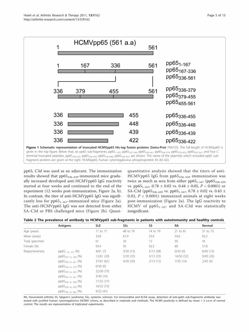
ResultsThe pp65336-439 sub-fragment of HCMV contains a B cellepitope(s) targeted by IgG from SLE patientsTo verify the existence of B-cell epitope(s), HCMVpp65tegument protein (pp65) was cloned, truncated andexpressed as his-tagged fragments (pp651-167, pp65167-336and pp65336-561) that covered the entire antigen (Figure1). Results showed that HCMV-seropositive SLE patientsresponded strongly to pp65336-561 (61%, 37/61) comparedto pp651-167 (7%, 4/61) or pp65167-336 (20%, 12/61). Theelevated positive rate to pp65336-561 by SLE patients’ serawas not found on either healthy or other disease controls(Table 2). In order to reveal the dominant epitope(s)within pp65336-561, pp65336-561 was sub-cloned into threefragments, expressed and re-screened (Figure 1). Of theoriginal 37 pp65336-561-positive sera, 7 were removedfrom subsequent tests due to various availability issues.Of the rest of the 30 pp65336-561-positive sera, 22 werepositive to pp65379-455 (73%, 22/30), 3 were positive topp65455-561 (10%, 3/30) and 0 was positive to pp65336-379(Table 2). Subsequently, pp65336-448, pp65336-439 andpp65336-422 fragments were created by partial deletionfrom the C-terminus of pp65336-561 (Figure 1). Of 22pp65379-455-positive sera, 17 were positive to pp65336-448(77%, 17/22), 16 were positive to pp65336-439 (73%, 16/22)and 9 were positive to pp65336-422 (41%, 9/22). The sero-reactivity to three fragments (pp65336-422, pp65336-439 orpp65336-448) by pp65379-455 positive sera was listed inTable 3.
Induction of anti-HCMV antibody and anti-HeLa proteinantibodyAlthough pp65 antigen immunized BALB/c animals pos-sessed anti-pp65 antibodies and enhanced autoantibodyactivities, the titers were reduced in a few weeks afterimmunization [15]. To improve the immunogenicity of
Hsieh et al. Arthritis Research & Therapy 2011, 13:R162http://arthritis-research.com/content/13/5/R162
Page 4 of 15
pp65, C3d was used as an adjuvant. The immunizationresults showed that pp65336-439-immunized mice gradu-ally increased developed anti-HCMVpp65 IgG reactivitystarted at four weeks and continued to the end of theexperiment (12 weeks post-immunization, Figure 2a, b).In contrast, the titer of anti-HCMVpp65 IgG was signifi-cantly less for pp651-167-immunized mice (Figure 2a).The anti-HCMVpp65 IgG was not detected from eitherSA-C3d or PBS challenged mice (Figure 2b). Quasi-
quantitative analysis showed that the titers of anti-HCMVpp65 IgG from pp65336-439 immunization wastwice as much as sera from either pp651-167 (pp65336-439vs. pp651-167, 0.78 ± 0.02 vs. 0.44 ± 0.05, P < 0.0001) orSA-C3d (pp65336-439 vs. pp651-167, 0.78 ± 0.02 vs. 0.43 ±0.02, P < 0.0001) immunized animals at eight weekspost-immunization (Figure 2a). The IgG reactivity toHCMV of pp651-167 and SA-C3d was statisticallyinsignificant.
Figure 1 Schematic representation of truncated HCMVpp65 His-tag fusion proteins (Swiss-Prot: P06725). The full-length of HCMVpp65 isgiven in the top figure. Below that, six pp65 sub-fragments, pp651-167, pp65167-336, pp65336-561, pp65336-379, pp65379-455, pp65455-561, and four Cterminal truncated peptides, pp65336-422, pp65336-439, pp65336-448, pp65336-455, are shown. The name of the plasmids which encoded pp65 sub-fragment proteins are given at the right. HCMVpp65, human cytomegalovirus phosphoprotein 65 (65 kD).
Table 2 The prevalence of antibody to HCMVpp65 sub-fragments in patients with autoimmunity and healthy controlsAntigens SLE SSc SS RA Normal
Age (years) 11 to 77 48 to 79 14 to 79 21 to 81 31 to 73
Mean (years) 33.8 61.9 53.4 54.6 63.2
Total specimen 61 20 13 50 45
Female (%) 93.4 95 92.3 80 57.8
Responsiveness pp651 to 167 (%) 4/61 (7) 3/20 (15) 5/13 (38) 0/50 (0) 6/45 (13)
pp65167 to 336 (%) 12/61 (20) 5/20 (25) 3/13 (23) 16/50 (32) 9/45 (20)
pp65336 to 561 (%) 37/61 (61) 4/20 (20) 2/13 (15) 7/50 (14) 2/45 (4)
pp65336 to 379 (%) 0/30 (0)
pp65379 to 455 (%) 22/30 (73)
pp65455 to 561 (%) 3/30 (10)
pp65336 to 448 (%) 17/22 (77)
pp65336 to 439 (%) 16/22 (73)
pp65336 to 422 (%) 9/22 (41)
RA, rheumatoid arthritis; SS, Sjögren’s syndrome; SSc, systemic sclerosis. For immunoblot and ELISA assay, detection of anti-pp65 sub-fragments antibody wastested with purified human cytomegalovirus (HCMV) virions, as described in materials and methods. The HCMV positivity is defined by mean + 3 s.e.m of normalcontrol. The results are representative of triplicated experiments.
Hsieh et al. Arthritis Research & Therapy 2011, 13:R162http://arthritis-research.com/content/13/5/R162
Page 5 of 15

In order to demonstrate that the immunization ofpp65336-439 could lead to the development of cross-reac-tive autoantibodies, total HeLa lysate was prepared asthe substrate for the detection of anti-HeLa antibodies(Figure 2c). Although immunization of pp65336-439 andpp651-167 induced anti-HeLa IgG at 4 weeks and contin-ued to 12 weeks post-immunization, pp65336-439 immu-nization exhibited significantly higher anti-HeLa IgGactivity than pp651-167 immunization (pp65336-439 vs.pp651-167, 0.50 ± 0.03 vs. 0.38 ± 0.02, P = 0.0191) at 8weeks post-immunization. To exclude the possibility ofHCMV contamination, HeLa lysate were immunoblottedby pp65 sub-fragment immunized sera (Figure 2d). Theresults showed that of eight anti-pp65 positive sera, onlyone strongly and another weakly react to HeLa antigensat 65 kD position.
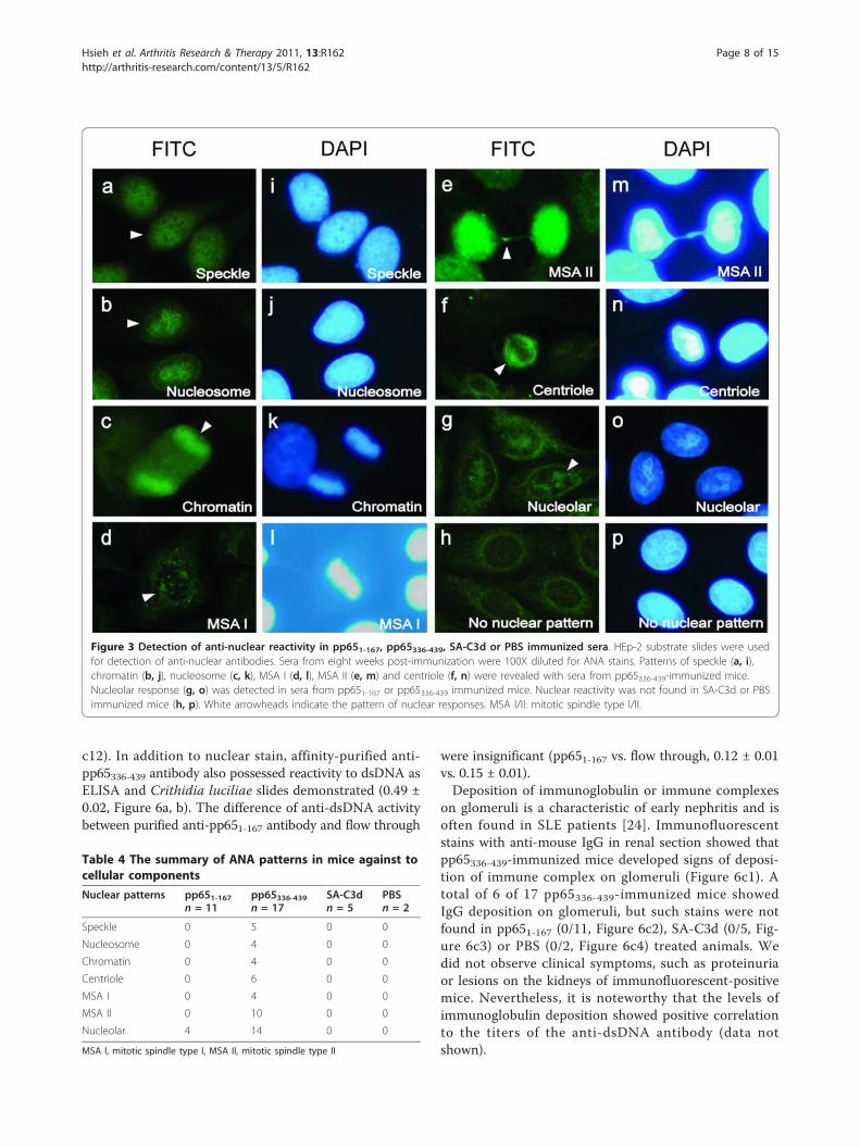
Induction of anti-nuclear antibody (ANA) by pp65336-439-immunizationTo determine if pp65336-439 immunization could induceantibodies against nuclear components from HeLa cells,the anti-nuclear antibody (ANA) test was performed. Theresults showed that pp65336-439 immunization inducedmultiple ANA staining patterns, including speckled (5/17,Figure 3a, i), nucleosome (4/17, Figure 3b, j), chromatin(4/17, Figure 3c, k), mitotic spindle type I (MSA I, 4/17,Figure 3d, l), mitotic spindle type II (MSA II, 10/17, Figure3e, m) centriole (6/17, Figure 3f, n) and nucleolar (14/17,Figure 3g, o) stains at 1:100 dilution at 8 weeks and con-tinued to 12 weeks post-immunization. In several occa-sions, ANA patterns were detected at dilution as much as500-fold. Nuclear stains, however, were not detected fromeither SA-C3d or PBS-immunized animals (0/5, 0/2, Fig-ure 3h, p). Four pp651-167-immunized mice developedweak anti-nucleolar reactivity (4/11, Figure 3g, o) detect-able at 1:40 dilution. Taken together, pp65336-439 immuni-zation could induce cross-reactive antibodies to multiplenucleus components (Table 4).
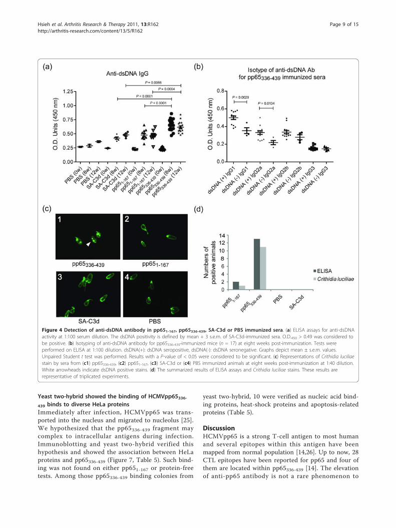
Induction of anti-dsDNA antibody by pp65336-439immunizationAnti-dsDNA antibody is a feature and a disease indicatorfor SLE patients [20-22]. ELISA assays showed thatpp65336-439-immunized sera exhibited significantlyenhanced anti-dsDNA antibody activity compared to ani-mals immunized with pp651-167 (pp65336-439 vs. pp651-167, 0.66 ± 0.02 vs. 0.48 ± 0.03, P < 0.0001), or SA-C3d(pp65336-439 vs. SA-C3d, 0.66 ± 0.02 vs.0.42 ± 0.02, P <0.0001) at 8 weeks and continued to 12 weeks post-immunization (Figure 4a). The differences of anti-dsDNAantibody between pp651-167 and SA-C3d immunizedmice were insignificant. The IgG2a to dsDNA is thedominant isotype to SLE nephritis [23]. ELISA-basedassays showed that 13 of 17 pp65336-439 immunized micewere positive to dsDNA. Isotyping showed that enhancedIgG1 (dsDNA (+) IgG1 vs. dsDNA (-) IgG1, 0.50 ± 0.02vs. 0.35 ± 0.03, P = 0.0029) and IgG2a isotypes (dsDNA(+) IgG2a vs. dsDNA (-) IgG2a, 0.33 ± 0.02 vs. 0.22 ±0.02, P = 0.0134) were the contributors of anti-dsDNAactivity (Figure 4b). To confirm the ELISA-based anti-dsDNA analysis, the Crithidia luciliae stains were per-formed. Of 17 pp65336-439-immunized animals, 11 werepositive for anti-dsDNA antibody (1:40 dilution) at 8weeks and continued to 12 weeks post-immunization,compared to 2 of 11 pp651-167-immunized mice (Figure4c). All Crithidia luciliae-positive sera were positive atELISA tests. Only one pp651-167-immunized mouse waspositive for Crithidia luciliae at 12 weeks post-immuni-zation (Figure 4d).
The elevated anti-HCMV pp65336-439 antibody is cross-reactive to dsDNA and nucleus componentsTo elucidate the relation between pp65336-439 immuniza-tion and anti-nuclear antibody found in animals, antibo-dies to either pp65336-439 or pp651-167 were affinitypurified from pooled pp65336-439 or pp651-167 immunizedmouse sera. The results showed that affinity-purifiedpp65336-439-specific IgG exhibiting significantly enhancedanti-HCMV activity compare to pp651-167 specific IgG(pp65336-439 vs. pp651-167, 1.08 ± 0.05 vs. 0.27 ± 0.01, P <0.0001, Figure 5a). Unbound fractions (flow through) frompurification processes remain anti-HCMV positive, but thetiter reduced significantly (pp65336-439 vs. flow through,1.08 ± 0.05 vs. 0.41 ± 0.02, P = 0.0003, Figure 5a, b). Asimmunofluorescent stains performed in Figure 3, affinity-purified anti-pp65336-439 antibodies reproduced all ANAstains found in direct serum-staining (Figure 3), includingspeckled (Figure 5c1,c7), chromatin (Figure 5c2,c8), cen-triole (Figure 5c3,c9) or MSA II (Figure 5c4,c10) stains.Antibodies purified from flow through or anti-pp651-167immunized sera, however, did not produce noticeablenuclear staining patterns (Figure 5c5,c11 and Figure 5c6,
Table 3 The sero-reactivity to pp65 sub-fragments bypp65379-455 positive sera
Sero-reactivity to antigens Patient number
pp65336-422 pp65336-439 pp65336-448 n = 22
+ + + 1, 4, 5, 9, 13, 17, 18, 19, 22
- + + 7, 10, 11, 12, 14, 16, 20
+ - +
+ + -
- - + 21
- + -
+ - -
- - - 2, 3, 6, 8, 15
+, positive; -, negative
Hsieh et al. Arthritis Research & Therapy 2011, 13:R162http://arthritis-research.com/content/13/5/R162
Page 6 of 15
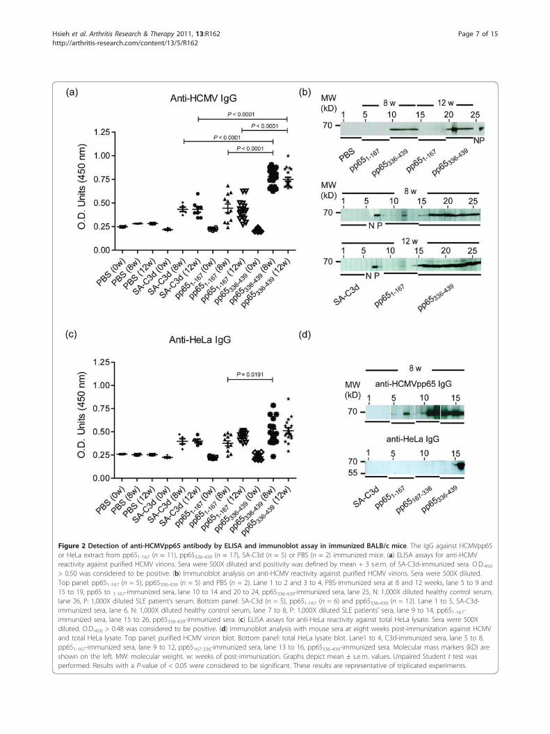
Figure 2 Detection of anti-HCMVpp65 antibody by ELISA and immunoblot assay in immunized BALB/c mice. The IgG against HCMVpp65or HeLa extract from pp651-167 (n = 11), pp65336-439 (n = 17), SA-C3d (n = 5) or PBS (n = 2) immunized mice. (a) ELISA assays for anti-HCMVreactivity against purified HCMV virions. Sera were 500X diluted and positivity was defined by mean + 3 s.e.m. of SA-C3d-immunized sera. O.D.450> 0.50 was considered to be positive. (b) Immunoblot analysis on anti-HCMV reactivity against purified HCMV virions. Sera were 500X diluted.Top panel: pp651-167 (n = 5), pp65336-439 (n = 5) and PBS (n = 2). Lane 1 to 2 and 3 to 4, PBS-immunized sera at 8 and 12 weeks, lane 5 to 9 and15 to 19, pp65 to 1-167-immunized sera, lane 10 to 14 and 20 to 24, pp65336-439-immunized sera, lane 25, N: 1,000X diluted healthy control serum,lane 26, P: 1,000X diluted SLE patient’s serum. Bottom panel: SA-C3d (n = 5), pp651-167 (n = 6) and pp65336-439 (n = 12). Lane 1 to 5, SA-C3d-immunized sera, lane 6, N: 1,000X diluted healthy control serum, lane 7 to 8, P: 1,000X diluted SLE patients’ sera, lane 9 to 14, pp651-167-immunized sera, lane 15 to 26, pp65336-439-immunized sera. (c) ELISA assays for anti-HeLa reactivity against total HeLa lysate. Sera were 500Xdiluted. O.D.450 > 0.48 was considered to be positive. (d) Immunoblot analysis with mouse sera at eight weeks post-immunization against HCMVand total HeLa lysate. Top panel: purified HCMV virion blot. Bottom panel: total HeLa lysate blot. Lane1 to 4, C3d-immunized sera, lane 5 to 8,pp651-167-immunized sera, lane 9 to 12, pp65167-336-immunized sera, lane 13 to 16, pp65336-439-immunized sera. Molecular mass markers (kD) areshown on the left. MW: molecular weight. w: weeks of post-immunization. Graphs depict mean ± s.e.m. values. Unpaired Student t test wasperformed. Results with a P-value of < 0.05 were considered to be significant. These results are representative of triplicated experiments.
Hsieh et al. Arthritis Research & Therapy 2011, 13:R162http://arthritis-research.com/content/13/5/R162
Page 7 of 15
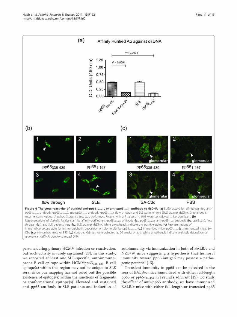
c12). In addition to nuclear stain, affinity-purified anti-pp65336-439 antibody also possessed reactivity to dsDNA asELISA and Crithidia luciliae slides demonstrated (0.49 ±0.02, Figure 6a, b). The difference of anti-dsDNA activitybetween purified anti-pp651-167 antibody and flow through
were insignificant (pp651-167 vs. flow through, 0.12 ± 0.01vs. 0.15 ± 0.01).Deposition of immunoglobulin or immune complexes
on glomeruli is a characteristic of early nephritis and isoften found in SLE patients [24]. Immunofluorescentstains with anti-mouse IgG in renal section showed thatpp65336-439-immunized mice developed signs of deposi-tion of immune complex on glomeruli (Figure 6c1). Atotal of 6 of 17 pp65336-439-immunized mice showedIgG deposition on glomeruli, but such stains were notfound in pp651-167 (0/11, Figure 6c2), SA-C3d (0/5, Fig-ure 6c3) or PBS (0/2, Figure 6c4) treated animals. Wedid not observe clinical symptoms, such as proteinuriaor lesions on the kidneys of immunofluorescent-positivemice. Nevertheless, it is noteworthy that the levels ofimmunoglobulin deposition showed positive correlationto the titers of the anti-dsDNA antibody (data notshown).
Figure 3 Detection of anti-nuclear reactivity in pp651-167, pp65336-439, SA-C3d or PBS immunized sera. HEp-2 substrate slides were usedfor detection of anti-nuclear antibodies. Sera from eight weeks post-immunization were 100X diluted for ANA stains. Patterns of speckle (a, i),chromatin (b, j), nucleosome (c, k), MSA I (d, l), MSA II (e, m) and centriole (f, n) were revealed with sera from pp65336-439-immunized mice.Nucleolar response (g, o) was detected in sera from pp651-167 or pp65336-439 immunized mice. Nuclear reactivity was not found in SA-C3d or PBSimmunized mice (h, p). White arrowheads indicate the pattern of nuclear responses. MSA I/II: mitotic spindle type I/II.
Table 4 The summary of ANA patterns in mice against tocellular componentsNuclear patterns pp651-167
n = 11pp65336-439n = 17
SA-C3dn = 5
PBSn = 2
Speckle 0 5 0 0
Nucleosome 0 4 0 0
Chromatin 0 4 0 0
Centriole 0 6 0 0
MSA I 0 4 0 0
MSA II 0 10 0 0
Nucleolar 4 14 0 0
MSA I, mitotic spindle type I, MSA II, mitotic spindle type II
Hsieh et al. Arthritis Research & Therapy 2011, 13:R162http://arthritis-research.com/content/13/5/R162
Page 8 of 15
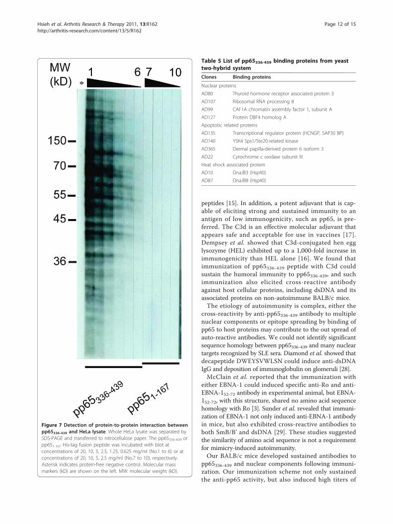
Yeast two-hybrid showed the binding of HCMVpp65336-439 binds to diverse HeLa proteinsImmediately after infection, HCMVpp65 was trans-ported into the nucleus and migrated to nucleolus [25].We hypothesized that the pp65336-439 fragment maycomplex to intracellular antigens during infection.Immunoblotting and yeast two-hybrid verified thishypothesis and showed the association between HeLaproteins and pp65336-439 (Figure 7, Table 5). Such bind-ing was not found on either pp651-167 or protein-freetests. Among those pp65336-439 binding colonies from
yeast two-hybrid, 10 were verified as nucleic acid bind-ing proteins, heat-shock proteins and apoptosis-relatedproteins (Table 5).
DiscussionHCMVpp65 is a strong T-cell antigen to most humanand several epitopes within this antigen have beenmapped from normal population [14,26]. Up to now, 28CTL epitopes have been reported for pp65 and four ofthem are located within pp65336-439 [14]. The elevationof anti-pp65 antibody is not a rare phenomenon to
Figure 4 Detection of anti-dsDNA antibody in pp651-167, pp65336-439, SA-C3d or PBS immunized sera. (a) ELISA assays for anti-dsDNAactivity at 1:100 serum dilution. The dsDNA positivity is defined by mean + 3 s.e.m. of SA-C3d-immunized sera. O.D.450 > 0.49 was considered tobe positive. (b) Isotyping of anti-dsDNA antibody for pp65336-439-immunized mice (n = 17) at eight weeks post-immunization. Tests wereperformed on ELISA at 1:100 dilution. dsDNA(+): dsDNA seropositive, dsDNA(-): dsDNA seronegative. Graphs depict mean ± s.e.m. values.Unpaired Student t test was performed. Results with a P-value of < 0.05 were considered to be significant. (c) Representations of Crithidia luciliaestain by sera from (c1) pp65336-439, (c2) pp651-167, (c3) SA-C3d or (c4) PBS immunized animals at eight weeks post-immunization at 1:40 dilution.White arrowheads indicate dsDNA positive stains. (d) The summarized results of ELISA assays and Crithidia luciliae stains. These results arerepresentative of triplicated experiments.
Hsieh et al. Arthritis Research & Therapy 2011, 13:R162http://arthritis-research.com/content/13/5/R162
Page 9 of 15
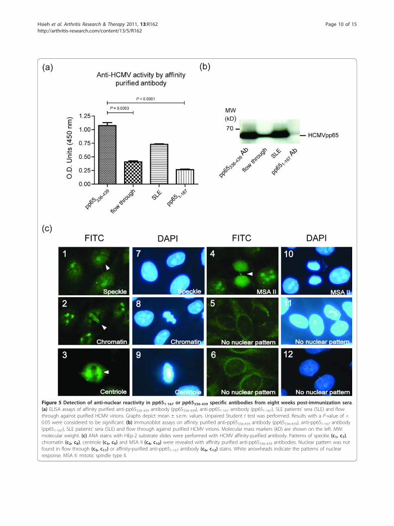
Figure 5 Detection of anti-nuclear reactivity in pp651-167 or pp65336-439 specific antibodies from eight weeks post-immunization sera.(a) ELISA assays of affinity purified anti-pp65336-439 antibody (pp65336-439), anti-pp651-167 antibody (pp651-167), SLE patients’ sera (SLE) and flowthrough against purified HCMV virions. Graphs depict mean ± s.e.m. values. Unpaired Student t test was performed. Results with a P-value of <0.05 were considered to be significant. (b) Immunoblot assays on affinity purified anti-pp65336-439 antibody (pp65336-439), anti-pp651-167 antibody(pp651-167), SLE patients’ sera (SLE) and flow through against purified HCMV virions. Molecular mass markers (kD) are shown on the left. MW:molecular weight. (c) ANA stains with HEp-2 substrate slides were performed with HCMV affinity-purified antibody. Patterns of speckle (c1, c7),chromatin (c2, c8), centriole (c3, c9) and MSA II (c4, c10) were revealed with affinity purified anti-pp65336-439 antibodies. Nuclear pattern was notfound in flow through (c5, c11) or affinity-purified anti-pp651-167 antibody (c6, c12) stains. White arrowheads indicate the patterns of nuclearresponse. MSA II: mitotic spindle type II.
Hsieh et al. Arthritis Research & Therapy 2011, 13:R162http://arthritis-research.com/content/13/5/R162
Page 10 of 15
persons during primary HCMV infection or reactivation,but such activity is rarely sustained [27]. In this study,we reported at least one SLE-specific, autoimmune-prone B-cell epitope within HCMVpp65336-439. B-cellepitope(s) within this region may not be unique to SLEsera, since our mapping has not ruled out the possibleexistence of epitope(s) within the junctions of fragmentsor conformational epitope(s). Elevated and sustainedanti-pp65 antibody in SLE patients and induction of
autoimmunity via immunization in both of BALB/c andNZB/W mice suggesting a hypothesis that humoralimmunity toward pp65 antigen may possess a patho-genic potential [15].Transient immunity to pp65 can be detected in the
sera of BALB/c mice immunized with either full-lengthpp65 or pp65336-439 in Freund’s adjuvant [15]. To studythe effect of anti-pp65 antibody, we have immunizedBALB/c mice with either full-length or truncated pp65
Figure 6 The cross-reactivity of purified anti-pp65336-439 or anti-pp651-167 antibody to dsDNA. (a) ELISA assays for affinity-purified anti-pp65336-439 antibody (pp65336-439), anti-pp651-167 antibody (pp651-167), flow through and SLE patients’ sera (SLE) against dsDNA. Graphs depictmean ± s.e.m. values. Unpaired Student t test was performed. Results with a P-value of < 0.05 were considered to be significant. (b)Representations of Crithidia luciliae stain by affinity-purified anti-pp65336-439 antibody (b1, pp65336-439), anti-pp651-167 antibody (b2, pp651-167), flowthrough (b3) and SLE patients’ sera (b4, SLE) against dsDNA. White arrowheads indicate the positive stains. (c) Representations ofImmunofluorescent stain for immunoglobulin deposition on glomerular by pp65336-439 (c1) immunized mice, pp651-167 (c2) immunized mice, SA-C3d (c3) immunized mice or PBS (c4) controls. Kidneys were collected at 20 weeks of age. White arrowheads indicate antibody deposition onglomerular. dsDNA: double-stranded DNA.
Hsieh et al. Arthritis Research & Therapy 2011, 13:R162http://arthritis-research.com/content/13/5/R162
Page 11 of 15
peptides [15]. In addition, a potent adjuvant that is cap-able of eliciting strong and sustained immunity to anantigen of low immunogenicity, such as pp65, is pre-ferred. The C3d is an effective molecular adjuvant thatappears safe and acceptable for use in vaccines [17].Dempsey et al. showed that C3d-conjugated hen egglysozyme (HEL) exhibited up to a 1,000-fold increase inimmunogenicity than HEL alone [16]. We found thatimmunization of pp65336-439 peptide with C3d couldsustain the humoral immunity to pp65336-439, and suchimmunization also elicited cross-reactive antibodyagainst host cellular proteins, including dsDNA and itsassociated proteins on non-autoimmune BALB/c mice.The etiology of autoimmunity is complex, either the
cross-reactivity by anti-pp65336-439 antibody to multiplenuclear components or epitope spreading by binding ofpp65 to host proteins may contribute to the out spread ofauto-reactive antibodies. We could not identify significantsequence homology between pp65336-439 and many nucleartargets recognized by SLE sera. Diamond et al. showed thatdecapeptide DWEYSVWLSN could induce anti-dsDNAIgG and deposition of immunoglobulin on glomeruli [28].McClain et al. reported that the immunization with
either EBNA-1 could induced specific anti-Ro and anti-EBNA-152-72 antibody in experimental animal, but EBNA-152-72, with this structure, shared no amino acid sequencehomology with Ro [3]. Sunder et al. revealed that immuni-zation of EBNA-1 not only induced anti-EBNA-1 antibodyin mice, but also exhibited cross-reactive antibodies toboth SmB/B’ and dsDNA [29]. These studies suggestedthe similarity of amino acid sequence is not a requirementfor mimicry-induced autoimmunity.Our BALB/c mice developed sustained antibodies to
pp65336-439 and nuclear components following immuni-zation. Our immunization scheme not only sustainedthe anti-pp65 activity, but also induced high titers of
Figure 7 Detection of protein-to-protein interaction betweenpp65336-439 and HeLa lysate. Whole HeLa lysate was separated bySDS-PAGE and transferred to nitrocellulose paper. The pp65336-439 orpp651-167 His-tag fusion peptide was incubated with blot atconcentrations of 20, 10, 5, 2.5, 1.25, 0.625 mg/ml (No.1 to 6) or atconcentrations of 20, 10, 5, 2.5 mg/ml (No.7 to 10), respectively.Asterisk indicates protein-free negative control. Molecular massmarkers (kD) are shown on the left. MW: molecular weight (kD).
Table 5 List of pp65336-439 binding proteins from yeasttwo-hybrid systemClones Binding proteins
Nuclear proteins
AD80 Thyroid hormone receptor associated protein 3
AD107 Ribosomal RNA processing 8
AD99 CAF1A chromatin assembly factor 1, subunit A
AD127 Protein DBF4 homolog A
Apoptotic related proteins
AD135 Transcriptional regulator protein (HCNGP, SAP30 BP)
AD140 YSK4 Sps1/Ste20-related kinase
AD365 Dermal papilla-derived protein 6 isoform 3
AD22 Cytochrome c oxidase subunit III
Heat shock associated protein
AD10 DnaJB3 (Hsp40)
AD87 DnaJB8 (Hsp40)
Hsieh et al. Arthritis Research & Therapy 2011, 13:R162http://arthritis-research.com/content/13/5/R162
Page 12 of 15
antibodies to nuclear components, including the nucleo-some, centriole and chromatin. Such weak anti-nuclearresponses were found on few animals never exposed topp65336-439. This weak anti-nuclear activity is likely dueto adjuvant-induced polyclonal activation becauseneither ANA activity nor the Crithidia luciliae stain wasfound from affinity-purified anti-pp651-167 antibody.Cross-reactivity between a foreign antigen and an auto-antigen is a characteristic of mimicry-induced autoimmu-nity [30]. Our affinity purification results demonstratedthat the anti-pp65336-439 antibodies cross-react to severalnuclear antigens, including dsDNA, suggesting thatmimicry could play a part in the pp65-induced tolerancebreak. The anti-dsDNA antibody, particularly IgG2a, wasreported to SLE nephritis and also identified from ourpp65336-439-immunized BALB/c [31,32]. Nevertheless, thedirect relation to nephritis by anti-pp65336-439 initiatedanti-dsDNA antibody of IgG2 isotype was not studied inthis work. The anti-dsDNA antibody from pp65336-439-immunized mice was detected as early as four weeks,suggesting that pp65336-439 is a potent inductor of cross-reactivity. The Crithidia luciliae stain has been thegolden standard to anti-dsDNA antibody. Of 17 pp65336-439-immunized mice, 11 and 13 mice were positive forthe Crithidia luciliae stains and ELISA assays, respec-tively. All Crithidia luciliae-positive mouse sera also pos-sessed high titers of anti-dsDNA activity by ELISAassays, confirming the significant elevated anti-dsDNAreactivity in pp65336-439-immunized animals.The peptide-induced immunity, which cross-reacts
with both dsDNA and a-actinin, has been described andits pathogenesis was illustrated [33]. Similar to anti-DWEYSVWLSN antibody, affinity-purified anti-pp65336-439 antibody from pp65336-439-immunized animals cross-reacted with dsDNA on both Crithidia luciliae stains andELISA assays. Such anti-dsDNA reactivity was not foundin pp651-167 or adjuvant immunized animals. To the bestof our knowledge, HCMV has not been reported toinduce the anti-dsDNA antibody. The pathogenicity ofpp65336-439-induced anti-dsDNA antibody on BALB/canimals was not fully examined by this study. Neverthe-less, we found precipitation of immune complexes onglomeruli at 12 weeks post-immunization (20 weeks ofage), and noticed positive correlation of anti-dsDNAtiters to the complex precipitation (data not shown). Thisfinding implies that an early stage of renal pathogenesisthat resembles SLE nephritis may have been induced bypp65336-439-mediated cross-reactive antibody. Arbuckle etal. have revealed that anti-dsDNA antibody could befound as early as nine years before the diagnosis of SLE[22]. The asymptomatic existence of anti-dsDNA activityin our animals suggests an extended observation isrequired to demonstrate the clinical consequences bypp65 immunization. Genetics plays an essential role on
pathogenesis of autoimmunity that might also explainthe lack of clinical symptoms on our animals followingimmunization [34].In addition to mimicry, epitope spreading could be
another driving force to pp65336-439-induced autoimmu-nity. The T-antigen of human polyomaviruses has beenshown to complex with nucleosomes of infected cellsduring viral replication. These nucleosomes/T-antigencomplexes are subsequently targeted by immuneresponses and become a catalyst for cross-reactive anti-bodies against both virus and host [35]. At HCMV infec-tion, pp65 is imported to the nucleus immediately viatwo nuclear localization sequences: pp65418-438 andpp65537-561 [36]. The pp65 has been demonstrated tobind to metaphase-arrested chromosomes in the pp65-expressing fibroblasts during productive virus infection[37]. These findings prompted us to hypothesize thatpp65 may not only bind to cellular proteins, but alsoform immune-complexes to DNA or other nuclear com-ponents. As expected, pp65336-439 bound multiple cellularproteins including nucleic acid binding proteins, nuclearproteins, apoptosis-related proteins and heat-shock pro-teins (Table 5). It is noteworthy that nuclear proteins andnucleic acid binding proteins are common targets toautoimmunity, and abnormal apoptosis has been asso-ciated with autoimmunity [38]. These findings suggestthat antigen-bound cellular proteins shared high prob-ability of becoming immunogenic and provide a mechan-ism for subsequent development of autoimmunity.Therefore, binding to intracellular proteins by full-lengthor fragmented pp65 may not only generate immune-complexes (virus/host) that are subsequently targeted byantiviral antibodies but also increase the chance of epi-tope spreading and lead to autoimmunity in persons withsusceptible genetic backgrounds.
ConclusionsThe antibody against HCMVpp65380-439 antibody is rarein healthy populations but is a common feature amongSLE sera. Through immunization of pp65336-439 withC3d as adjuvant, we were able to sustain the antibodytiters to pp65336-439 peptide and demonstrate cross-reac-tivity of anti-pp65 antibody to nuclear components,including dsDNA on BALB/c mice. Yeast two-hybridanalysis revealed that pp65336-439 could bind to nuclearproteins, suggesting the immune-complexes of pp65 andnuclear proteins may be part of the trigger to autoim-munity. Although none of the experimental animalsdeveloped SLE-like clinical symptom, deposition ofimmunoglobulin was identified from pp65336-439-immu-nized animals at 12 weeks post-immunization. There-fore, a sustained humoral immunity to pp65 maypresent a risk to individuals with a background predis-posed to SLE.
Hsieh et al. Arthritis Research & Therapy 2011, 13:R162http://arthritis-research.com/content/13/5/R162
Page 13 of 15
AbbreviationsANAs: anti-nuclear antibodies; C3d: complement 3d; CFA: complete Freund’sadjuvant; CnBr: cyanogen bromide; CTD: connective tissue disease; dsDNA:double-stranded DNA; EBNA-1: Epstein-Barr virus nuclear antigen 1; EBV:Epstein-Barr virus; ELISA: Enzyme Linked Immunosorbent Assay; FITC:fluorescein isothiocyanate; HCMV: human cytomegalovirus; HEL: hen egglysozyme; HRP: horseradish peroxidase; IFA: incomplete Freund’s adjuvant;MSA-I/II: mitotic spindle type I/II; OPD: o-phenylenediamine dihydrochloride;PBS: phosphate-buffered saline; pp65: phosphoprotein 65; RA: rheumatoidarthritis; RT: room temperature; SA: streptavidin; SLE: systemic lupuserythematosus; SmB/B’: Smith antigen B/B’; snRNP: small nuclearribonucleoprotein; SS: Sjögren’s syndrome; SSc: systemic sclerosis; YNB: yeastnitrogen base.
AcknowledgementsWe thank Dr. YL Juang for technical suggestions. The yeast strains and therelated materials in the yeast two-hybrid experiment were supported by Dr.YL Juang. This work was undertaken at Tzu-Chi University and supported byGrant TCIRP 95002-03 from Tzu-Chi University.
Author details1Institute of Medical Science, Tzu-Chi University, No. 701, Sec. 3, ZhongyangRd., Hualien City, Hualien County 970, Taiwan. 2Institute of MicrobiologyImmunology and Biochemistry, Tzu-Chi University, No. 701, Sec. 3,Zhongyang Rd., Hualien City, Hualien County 970, Taiwan. 3The NationalLaboratory Animal Center (NLAC), No. 128, Sec. 2, Academia Rd., NangangDist., Taipei City 115, Taiwan. 4Development Center of Biotechnology,No.101, Ln. 169, Kangning St., Xizhi Dist, New Taipei City 221, Taiwan.
Authors’ contributionsMC, SLW and AHH jointly contributed to the design of the study. AHHperformed ELISA, Western blot and immunofluorescence. YJJ carried out theyeast two-hybrid. CTL participated in the tissue stains. AHH, MC and SLWwere responsible for data analysis and interpretations. AHH and MC wrotethe manuscript. All authors read and approved the final manuscript.
Competing interestsThe authors declare that they have no competing interests.
Received: 23 March 2011 Revised: 6 September 2011Accepted: 11 October 2011 Published: 11 October 2011
References1. Poole BD, Scofield RH, Harley JB, James JA: Epstein-Barr virus and
molecular mimicry in systemic lupus erythematosus. Autoimmunity 2006,39:63-70.
2. Poole BD, Gross T, Maier S, Harley JB, James JA: Lupus-like autoantibodydevelopment in rabbits and mice after immunization with EBNA-1fragments. J Autoimmun 2008, 31:362-371.
3. McClain MT, Heinlen LD, Dennis GJ, Roebuck J, Harley JB, James JA: Earlyevents in lupus humoral autoimmunity suggest initiation throughmolecular mimicry. Nat Med 2005, 11:85-89.
4. Khoshnevis M, Tyring SK: Cytomegalovirus infections. Dermatol Clin 2002,20:291-299, vii.
5. Oldstone MB, Nerenberg M, Southern P, Price J, Lewicki H: Virus infectiontriggers insulin-dependent diabetes mellitus in a transgenic model: roleof anti-self (virus) immune response. Cell 1991, 65:319-331.
6. Osame K, Takahashi Y, Takasawa H, Watanabe S, Kishimoto M, Yasuda K,Kaburagi Y, Nakanishi K, Kajio H, Noda M: Rapid-onset type 1 diabetesassociated with cytomegalovirus infection and islet autoantibodysynthesis. Intern Med 2007, 46:873-877.
7. Cheng HM, Khairullah NS: Induction of antiphospholipid autoantibodyduring cytomegalovirus infection. Clin Infect Dis 1997, 25:1493-1494.
8. Zhu J: Cytomegalovirus infection induces expression of 60 KD/Roantigen on human keratinocytes. Lupus 1995, 4:396-406.
9. Newkirk MM, van Venrooij WJ, Marshall GS: Autoimmune response to U1small nuclear ribonucleoprotein (U1 snRNP) associated withcytomegalovirus infection. Arthritis Res 2001, 3:253-258.
10. Curtis HA, Singh T, Newkirk MM: Recombinant cytomegalovirusglycoprotein gB (UL55) induces an autoantibody response to the U1-70kDa small nuclear ribonucleoprotein. Eur J Immunol 1999, 29:3643-3653.
11. McLaughlin-Taylor E, Pande H, Forman SJ, Tanamachi B, Li CR, Zaia JA,Greenberg PD, Riddell SR: Identification of the major late humancytomegalovirus matrix protein pp65 as a target antigen for CD8+ virus-specific cytotoxic T lymphocytes. J Med Virol 1994, 43:103-110.
12. Tanaka N, Kimura H, Hoshino Y, Nishikawa K, Kojima S, Nishiyama Y,Morishima T: Expression of tegument protein pp65 of humancytomegalovirus (CMV) and its application to the analysis of viral-specific cellular immunity in CMV-infected individuals. Arch Virol 2002,147:2405-2417.
13. Khattab BA, Lindenmaier W, Frank R, Link H: Three T-cell epitopes withinthe C-terminal 265 amino acids of the matrix protein pp65 of humancytomegalovirus recognized by human lymphocytes. J Med Virol 1997,52:68-76.
14. Kondo E, Akatsuka Y, Kuzushima K, Tsujimura K, Asakura S, Tajima K,Kagami Y, Kodera Y, Tanimoto M, Morishima Y, Takahashi T: Identificationof novel CTL epitopes of CMV-pp65 presented by a variety of HLAalleles. Blood 2004, 103:630-638.
15. Chang M, Pan MR, Chen DY, Lan JL: Human cytomegalovirus pp65 lowermatrix protein: a humoral immunogen for systemic lupus erythematosuspatients and autoantibody accelerator for NZB/W F1 mice. Clin ExpImmunol 2006, 143:167-179.
16. Dempsey PW, Allison ME, Akkaraju S, Goodnow CC, Fearon DT: C3d ofcomplement as a molecular adjuvant: bridging innate and acquiredimmunity. Science 1996, 271:348-350.
17. Green TD, Montefiori DC, Ross TM: Enhancement of antibodies to thehuman immunodeficiency virus type 1 envelope by using the molecularadjuvant C3d. J Virol 2003, 77:2046-2055.
18. Yu M, Li da J, Wang XL, Yuan MM, Zhu Y, Yao XY, Li HP: [Molecularadjuvant C3d up-regulates both B7-1 and B7-2 expression on Raji cells].Fen Zi Xi Bao Sheng Wu Xue Bao 2006, 39:77-82.
19. Olmsted JB: Affinity purification of antibodies from diazotized paperblots of heterogeneous protein samples. J Biol Chem 1981,256:11955-11957.
20. Linnik MD, Hu JZ, Heilbrunn KR, Strand V, Hurley FL, Joh T: Relationshipbetween anti-double-stranded DNA antibodies and exacerbation ofrenal disease in patients with systemic lupus erythematosus. ArthritisRheum 2005, 52:1129-1137.
21. Arbuckle MR, James JA, Kohlhase KF, Rubertone MV, Dennis GJ, Harley JB:Development of anti-dsDNA autoantibodies prior to clinical diagnosis ofsystemic lupus erythematosus. Scand J Immunol 2001, 54:211-219.
22. Arbuckle MR, McClain MT, Rubertone MV, Scofield RH, Dennis GJ, James JA,Harley JB: Development of autoantibodies before the clinical onset ofsystemic lupus erythematosus. N Engl J Med 2003, 349:1526-1533.
23. Bijl M, Dijstelbloem HM, Oost WW, Bootsma H, Derksen RH, Aten J,Limburg PC, Kallenberg CG: IgG subclass distribution of autoantibodiesdiffers between renal and extra-renal relapses in patients with systemiclupus erythematosus. Rheumatology (Oxford) 2002, 41:62-67.
24. Hurd ER, Ziff M: Quantitative studies of immunoglobulin deposition inthe kidney, glomerular cell proliferation and glomerulosclerosis in NZB/NZW F1 hybrid mice. Clin Exp Immunol 1977, 27:261-268.
25. Cui Z, Zhang K, Zhang Z, Liu Y, Zhou Y, Wei H, Zhang XE: Visualization ofthe dynamic multimerization of human Cytomegalovirus pp65 inpunctuate nuclear foci. Virology 2009, 392:169-177.
26. Provenzano M, Sais G, Bracci L, Egli A, Anselmi M, Viehl CT, Schaub S,Hirsch HH, Stroncek DF, Marincola FM, Spagnoli GC: A HCMV pp65polypeptide promotes the expansion of CD4+ and CD8+ T cells across awide range of HLA specificities. J Cell Mol Med 2009, 13:2131-2147.
27. Landini MP, Lazzarotto T, Xu J, Geballe AP, Mocarski ES: Humoral immuneresponse to proteins of human cytomegalovirus latency-associatedtranscripts. Biol Blood Marrow Transplant 2000, 6:100-108.
28. Putterman C, Diamond B: Immunization with a peptide surrogate fordouble-stranded DNA (dsDNA) induces autoantibody production andrenal immunoglobulin deposition. J Exp Med 1998, 188:29-38.
29. Sundar K, Jacques S, Gottlieb P, Villars R, Benito ME, Taylor DK, Spatz LA:Expression of the Epstein-Barr virus nuclear antigen-1 (EBNA-1) in themouse can elicit the production of anti-dsDNA and anti-Sm antibodies. JAutoimmun 2004, 23:127-140.
30. Deshmukh US, Kannapell CC, Fu SM: Immune responses to small nuclearribonucleoproteins: antigen-dependent distinct B cell epitope spreadingpatterns in mice immunized with recombinant polypeptides of smallnuclear ribonucleoproteins. J Immunol 2002, 168:5326-5332.
Hsieh et al. Arthritis Research & Therapy 2011, 13:R162http://arthritis-research.com/content/13/5/R162
Page 14 of 15
31. Garcia CO, Molina JF, Gutierrez-Urena S, Scopelitis E, Wilson WA, Gharavi AE,Espinoza LR: Autoantibody profile in African-American patients withlupus nephritis. Lupus 1996, 5:602-605.
32. Winkler TH, Henschel TA, Kalies I, Baenkler HW, Skvaril F, Kalden JR:Constant isotype pattern of anti-dsDNA antibodies in patients withsystemic lupus erythematosus. Clin Exp Immunol 1988, 72:434-439.
33. Zhao Z, Weinstein E, Tuzova M, Davidson A, Mundel P, Marambio P,Putterman C: Cross-reactivity of human lupus anti-DNA antibodies withalpha-actinin and nephritogenic potential. Arthritis Rheum 2005,52:522-530.
34. Mendlovic S, Brocke S, Fricke H, Shoenfeld Y, Bakimer R, Mozes E: Thegenetic regulation of the induction of experimental SLE. Immunology1990, 69:228-236.
35. Andreassen K, Bredholt G, Moens U, Bendiksen S, Kauric G, Rekvig OP: T celllines specific for polyomavirus T-antigen recognize T-antigen complexedwith nucleosomes: a molecular basis for anti-DNA antibody production.Eur J Immunol 1999, 29:2715-2728.
36. Schmolke S, Drescher P, Jahn G, Plachter B: Nuclear targeting of thetegument protein pp65 (UL83) of human cytomegalovirus: an unusualbipartite nuclear localization signal functions with other portions of theprotein to mediate its efficient nuclear transport. J Virol 1995,69:1071-1078.
37. Dal Monte P, Bessia C, Landini MP, Michelson S: Expression of humancytomegalovirus ppUL83 (pp65) in a stable cell line and its associationwith metaphase chromosomes. J Gen Virol 1996, 77:2591-2596.
38. Maniati E, Potter P, Rogers NJ, Morley BJ: Control of apoptosis inautoimmunity. J Pathol 2008, 214:190-198.
doi:10.1186/ar3481Cite this article as: Hsieh et al.: Fragment of tegument protein pp65 ofhuman cytomegalovirus induces autoantibodies in BALB/c mice. ArthritisResearch & Therapy 2011 13:R162.
Submit your next manuscript to BioMed Centraland take full advantage of:
• Convenient online submission
• Thorough peer review
• No space constraints or color figure charges
• Immediate publication on acceptance
• Inclusion in PubMed, CAS, Scopus and Google Scholar
• Research which is freely available for redistribution
Submit your manuscript at www.biomedcentral.com/submit
Hsieh et al. Arthritis Research & Therapy 2011, 13:R162http://arthritis-research.com/content/13/5/R162
Page 15 of 15


