Embed Size (px)
Citation preview

Co-localization of nestin and insulin and expression of islet cellmarkers in long-term human pancreatic nestin-positive cell cultures
Silvya Stuchi Maria-Engler1, Maria Lúcia C Corrêa-Giannella2,
Letícia Labriola1, Karin Krogh1, Christian Colin1,
Fernando Henrique Lojudice1, Carlos Alberto Mayora Aita1,
Elizabeth Maria Costa de Oliveira1, Tatiana C Silveira Corrêa1,
Irenice Cairo da Silva1, Tercio Genzini3, Marcelo Perosa de Miranda3,
Irene Lourdes Noronha3, Luciano Vilela4, Cassio Negro Coimbra2,
Renato A Mortara5, Marcos Mares Guia†4,
Freddy Goldberg Eliaschewitz3 and Mari Cleide Sogayar1
1Instituto de Química, Universidade de São Paulo, Departamento de Bioquímica CP26077, São Paulo 05513-970 SP, Brazil
2Laboratório de Nutrição Humana e Doenças Metabólicas LIM-25, Hospital das Clínicas da Faculdade de Medicina da Universidade de São Paulo, Sao Paulo, Brazil
3Albert Einstein Hospital, São Paulo, Brazil
4 Biomm S/A, Montes Claros, Minas Gerais, Brazil
5Departamento de Micro, Imuno e Parasitologia, Escola Paulista de Medicina-UNIFESP, São Paulo, Brazil
(Requests for offprints should be addressed to M C Sogayar; Email: [email protected])
(S S Maria Engler is now at Faculdade de Ciencias Farmaceuticas, Departamento de Analises Clınicas e Toxıcologicas, Universidade de Sao Paulo, Sao Paulo, Brazil)
†In memoriam
Abstract
Strategies to differentiate progenitor cells into � cellsin vitro have been considered as an alternative to increase� cell availability prior to transplantation. It has recentlybeen suggested that nestin-positive cells could be multipo-tential stem cells capable of expressing endocrine markersupon specific stimulation; however, this issue still remainscontroversial. Here, we characterized short- and long-term islet cell cultures derived from three different humanislet preparations, with respect to expression of nestin andislet cell markers, using confocal microscopy and semi-quantitative RT-PCR. The number of nestin-positivecells was found to be strikingly high in long-term cultures.In addition, a large proportion (49·7%) of these nestin-positive cells, present in long-term culture, are shown tobe proliferative, as judged by BrdU incorporation. Theproportion of insulin-positive cells was found to be high inshort-term (up to 28 days) cultures and declined there-after, when cells were maintained in the presence of10% serum, concomitantly with the decrease in insulinand PDX-1 expression. Interestingly, insulin and nestinco-expression was observed as a rare event in a small
proportion of cells present in freshly isolated human isletsas well as in purified islet cells cultured in vitro for longperiods of time. In addition, upon long-term subculturingof nestin-positive cells in 10% serum, we observed reap-pearance of insulin expression at the mRNA level; whenthese cultures were shifted to 1% serum for a month,expression of insulin, glucagon and somatostatin was alsodetected, indicating that manipulating the culture con-ditions can be used to modulate the nestin-positive cell’sfate. Attempts to induce cell differentiation by platingnestin-positive cells onto Matrigel revealed that these cellstend to aggregate to form islet-like clusters, but this is notsufficient to increase insulin expression upon short-term culture. Our data corroborate previous findingsindicating that, at least in vitro, nestin-positive cells mayundergo the early stages of differentiation to an islet cellphenotype and that long-term cultures of nestin-positivehuman islet cells may be considered as a potentialsource of precursor cells to generate fully differentiated/functional � cells.Journal of Endocrinology (2004) 183, 455–467
Introduction
Islet transplantation has the potential to become a widelyapplicable treatment for type 1 diabetes mellitus. Success-
ful islet transplantation, accompanied by an alternativeimmunosuppressive scheme, has recently been reported(Shapiro et al. 2000). However, one of the most severelimitations of this approach is the scarcity of transplantation
455
Journal of Endocrinology (2004) 183, 455–4670022–0795/04/0183–455 � 2004 Society for Endocrinology Printed in Great Britain
DOI: 10.1677/joe.1.05703Online version via http://www.endocrinology-journals.org

material, namely, purified islets. Therefore, strategies toselect for cells capable of differentiating in vitro into � cellshave been viewed as attractive alternatives to increase �cell availability prior to transplantation.
Understanding the mechanisms of islet cell differentia-tion, together with a more detailed knowledge of thesignals driving later steps in � cell differentiation, consti-tutes an important step towards in vitro growth anddifferentiation of precursor cells. However, the exactnature of the pancreatic stem cell is still not well defined(Berná et al. 2001). It has recently been suggested thatnestin-positive cells present in rodent and human isletscould constitute multipotential stem cells (Hunziker &Stein 2000, Zulewski et al. 2001, Abraham et al. 2002,Esni et al. 2004). However, this issue is still controversial,since several recent publications have correlated nestinexpression with endothelial cells (Lardon et al. 2002,Humphrey et al. 2003, Klien et al. 2003, Treutelaar et al.2003). Human and mouse pancreatic progenitor cells donot express nestin during development (Piper et al. 2002,Selander & Edlund 2002), whereas nestin was shown to beexpressed in mesenchyma but not in epithelial cells of thedeveloping pancreas (Selander & Edlund 2002, Humphreyet al. 2003). It has also been demonstrated that nestin isexpressed in reactive stellate cells in both normal andregenerating rat pancreas (Lardon et al. 2002). Differen-tiation of nestin-positive cells is also controversial, withZulewski et al. (2001), Abraham et al. (2002) and Huang& Tang (2003) showing expression of endocrine markersby these cells, whereas Humphrey et al. (2003) did not findthem to be able to differentiate in vitro or in vivo.
Here we analyzed the expression of � cell markers duringlong term culturing of nestin-positive cell populations ex-panded from primary cultures of purified human pancreaticislets by confocal microcoscopy and RT-PCR. We foundco-localization of nestin and insulin as a rare event, both infreshly isolated islets and in long-term cultures enrichedin nestin-positive cells. Low mRNA levels of insulin,glucagon and somatostatin were detected after prolongedsubculturing in low serum medium without addition ofdifferentiation-inducing factors. The attempt to inducedifferentiation of nestin-positive cells by subculturing ontoMatrigel did not promote any increase in insulin expres-sion, in spite of the formation of islet-like clusters. Theexpression of islet cell markers observed in long-termcultures corroborates previous findings that, at least in vitro,nestin-positive cells may undergo the early stages of differ-entiation to an islet phenotype, deserving further studies toaddress their potential role as an alternative � cell source.
Materials and Methods
Islet isolation and purification
In order to obtain highly purified human islet preparations,we followed the procedure based on that described byShapiro et al. (2000), with minor modifications. Briefly,
upon informed consent from the donors’ relatives, pan-creata were removed from cadaveric donors in a multi-organ procurement program after in situ vascular perfusionwith cold University of Wisconsin (UW) solution.Pancreata were immediately transported to the HumanPancreatic Islet Unit for processing. Pancreatic islets wereisolated by perfusion and digestion with Liberase-HIpurified enzyme blend (Roche, Indianapolis, IN, USA)via the Wirsung duct. Digestion was monitored through-out the digestion procedure by staining samples of pancre-atic tissue with dithizone (diphenylthiocarbazone, Sigma),which specifically stains Zn2+-containing insulin granulespresent in insulin producing � cells. Digestion was inter-rupted when the majority of the islets were free from theexocrine tissue and pancreatic islets were purified incontinuous Ficoll gradients using an aphaeresis/cell sepa-rator system (Cobe 2991 cell processor, Gambro BCT,Inc, Lakewood, CO, USA). Islet-enriched fractions werepooled, stained with dithizone and quantified by countingunder the microscope with the help of an optical net andthen converted to the standard number of islet equivalents(IEQ), which represents the number of 150 µm diameterislets present in the sample. Islet cell viability, assessed bya fluorimetric cell viability assay (Live/Dead) accordingto Bank (1987), was usually greater than 80%. The purityof each preparation was assessed by comparing the amountof dithizone-stained endocrine tissue with the unstainedexocrine (acinar) tissue.
The most purified islet fractions were pooled andcultured in plastic tissue culture flasks in the presence ofCMRL 1066 (GIBCO) medium supplemented with 10%FCS (Cultilab, Campinas, São Paulo, Brazil). Adherentcells could be observed after 12 h in culture and, in a fewdays, cells formed a continuous monolayer, which wassubcultured weekly at a 1:4 ratio.
To observe the effects of serum deprivation on theexpression of islet cell markers, confluent cells grown onuncoated plastic maintained in CMRL 1066 mediumsupplemented with 10% FCS for 12 passages were washedwith PBS and maintained for an additional 28 days inCMRL 1066 medium containing 1% FCS. After thisperiod, insulin, somatostatin, glucagon, PDX-1 and nestinexpression were evaluated by RT-PCR.
Immunocytochemistry
Cells were fixed and permeabilized with methanol for10 min at 20 �C, followed by washing (three times) withcold PBS. After fixation, cells were incubated for 1 h atroom temperature in blocking solution (2% BSA in PBS).After incubation with the primary antibodies, cultureswere washed (four times) with PBS, incubated for 1 h atroom temperature with the secondary antibody, and thenwashed (four times) with PBS. Coverslips were mountedusing the Prolong Antifade Kit (Molecular Probes, Eugene,OR, USA).
S S MARIA-ENGLER and others · Human pancreatic nestin-positive cultures456
www.endocrinology-journals.orgJournal of Endocrinology (2004) 183, 455–467

Antibodies
The rabbit anti-human nestin polyclonal antiserum was agenerous gift from Dr Conrad A Messam (NationalInstitute of Neurological Disorders and Stroke, NationalInstitute of Health, Bethesda, MD, USA) and used at a1:200 dilution, as described in Messan et al. (2000). Themouse monoclonal antibody against human cytokeratin19 (CK19) and the mouse monoclonal antibody againsthuman insulin were obtained from ICN (Costa Mesa, CA,USA) and rabbit affinity purified polyclonal antibodies toinsulin were obtained from Zymed (San Francisco, CA,USA) and diluted at 1:20 (ICN) and 1:50 (Zymed).Antibody against human vimentin (Amersham-Pharmacia)was diluted 1:50. Secondary antibodies were conjugated toeither Texas Red or FITC (Molecular Probes) and used at1:200 dilution.
Culture of nestin-positive cells on Matrigel substrate
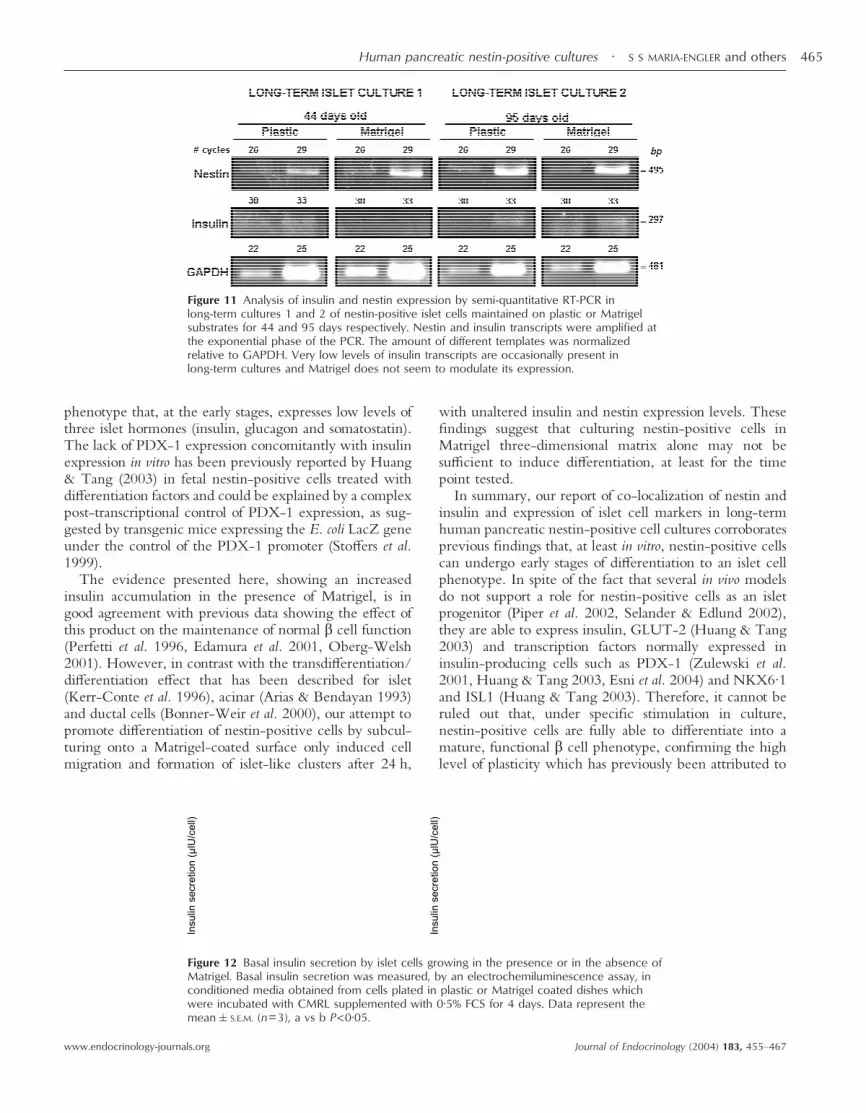
To test the differentiation-inducing ability of Matrigel,nestin-positive cells originating from different islet prepa-rations obtained from different pancreas donors weremaintained in culture dishes coated with Matrigel (50 µlsolution/cm2) for 3 to 120 h. Matrigel is a preparation ofmurine sarcoma basement membrane (Collaborative Bio-medical Products, Bedford, MA, USA). For RT-PCRanalysis, cells cultured for 44 and 95 days were subculturedin Matrigel for 48 h before RNA purification. Controlcultures were kept in uncoated plastic plates.
Confocal analysis
Coverslips were observed under a BioRad 1024-UVconfocal system attached to a Zeiss Axiovert 100 micro-scope, using a × 40 NA 1·2 plan apochromatic waterimmersion objective. All confocal immunofluorescencemicroscopy images presented here correspond to singleoptical sections.
DNA synthesis assay
To assess proliferation of nestin-positive cells, BrdU in-corporation was measured. Cells (5·0 × 104) were sub-cultured in the presence of 10% FCS onto coverslips in60 mm plates. After incubation for 12 h with 10 µMBrdU, cells were fixed in cold methanol for 10 min andwashed twice in cold PBS. BrdU incorporation wasrevealed with an anti-BrdU antibody, using theAmersham-Pharmacia Kit. Triple staining was achievedby staining with the anti-BrdU and anti-human nestinantibody plus 0·5 µg/ml DAPI (4,6-diamidino-2-phenylindole; Sigma); an anti-mouse antibody conjugatedto Texas Red for the anti-BrdU and an anti-rabbitantibody conjugated to FITC for the anti-nestin were
used. The percentage of BrdU-positive nuclei and BrdU/nestin-positive cells was determined by counting at leastsix different microscope fields.
Basal insulin secretion
Cells were plated into 24-well plastic dishes or in dishescoated with CMRL 1066 medium:Matrigel solution/cm2
(50:1) at a density of 104 cells/well and cultured for 48 hin CMRL 1066 (Mediatech Cellgro, Miami, FL, USA)supplemented with 100 U/ml penicillin and 5% FCS. Onthe following day, the cells were incubated with CMRLsupplemented with 0·5% FCS for 4 days. Supernatantswere collected and stored at �20 �C until assayed forinsulin. Secreted insulin was quantified in conditionedmedia from each well by electrochemiluminescence assayELECSYS (Roche). The reagent is highly specific for thedetection of human insulin and cross reaction to proinsulinor C-peptide is <0·01%. Nondetectable insulin levels wereverified in the control samples. Results were normalized tocell number. Data are presented as the mean�S.E.M. Eachexperiment was repeated with triplicate values withineach group. The statistical differences between groupmeans were tested by an unpaired two tailed t-test. A Pvalue <0·05 was considered statistically significant. Thecalculations were performed using the Prism softwareversion 3·03 (Graph Pad Software Inc., San Diego, CA,USA).
RT-PCR
Total RNA was prepared from cell cultures maintaineduntil the 12th passage in CMRL 1066 medium supple-mented with 10% FCS and then shifted to low (1%)FCS concentration for an additional 28 days, and alsofrom freshly isolated islets, by guanidine thiocyanate/mercaptoethanol lysis followed by ultra centrifugation onCsCl cushions (Chirgwin et al. 1979). Reverse transcrip-tion was carried out using SuperScript II� RT and oligodT
Figure 1 Purified islets (A, B) stained with dithizone, whichspecifically stains insulin granules present in � cells. Magnification× 20 and × 40 respectively.
Human pancreatic nestin-positive cultures · S S MARIA-ENGLER and others 457
www.endocrinology-journals.org Journal of Endocrinology (2004) 183, 455–467

priming and 5 µg of total RNA, according to manufac-turer’s instructions (Invitrogen). First strand cDNA prod-ucts were treated with RNase H and diluted 1:20 inTris-EDTA buffer. PCR amplification for each genestudied was performed under the following cycling con-ditions: initial denaturation at 95 �C for 1 min, denatura-tion at 95 �C for 30 s, annealing at 57–66·5 �C for 30 s andextension at 72 �C for 1 min, followed by a final extensionstep at 72 �C for 5 min. Reactions were carried out in20 µl final volume, containing 1U of Taq polymerase(Biolase, Bioline Inc., Reno, NV, USA), 1 × PCRreaction buffer, 1·5 mM MgCl2, 0·2 mM dNTPs and0·4 µM primers. Betaine was added to PCRs for nestinand PDX-1 cDNAs to 0·5 and 0·75 M final concen-trations respectively, in order to improve amplification ofthese GC-rich templates. The number of cycles for theexponential phase of PCR amplification were determined
to be 34 for PDX-1, 30 for nestin and 20 for GAPDH (Fig8), and both 18 and 32 cycles were used in order to analyzeinsulin mRNA expression in samples with very high orvery low levels of this transcript. Annealing temperatureswere 66·5, 57, 65 and 60 �C respectively. GAPDH wasused as an internal control to normalize for the amount oftemplate used in the amplification reaction. The oligosused for nestin were based on Zulewski et al. (2001)(HNF: 5�-AGAGGGGAATTCCTGGAG-3� and HNR:5�-CTGAGGACCAGGACTCTCTA-3�). For GAPDHthe oligos were those from the PCR-Select� kitfrom Clontech. Primers INSF: 5�-CATCACTGTCCTTCTGCCAT-3� and INSR: 5�-TCCACCACCCTGTTGCTGTA-3� were used for insulin, PDX1F:5�-CATGAACGGCGAGGAGCAGTA-3� and PDX1R:5�-GTT GAAGCCCCTCAGCCAGG-3� for PDX-1,SMTF: 5�-ACTCTCCAGCTCGGCTTTC-3� and
Figure 2 Confocal microscopy of triple-labeled immunofluorescence of freshly isolated human pancreaticislet cells cultured for 24 h, depicited for (A) insulin (red), (B) cytokeratin 19 (green) and (C) DAPI stainingfor nuclei (blue). The superposition of A, B and C is shown in D. Note the strong staining for insulin in thecenter of the islet (A) and the presence of ductal cells restricted to the islet periphery (B). Scale bar in �m.
S S MARIA-ENGLER and others · Human pancreatic nestin-positive cultures458
www.endocrinology-journals.orgJournal of Endocrinology (2004) 183, 455–467

SMTR: 5�-TCAGAGGTCTGATATGGACAATAC-3�for somatostatin and GLUCF: 5�-GCACACTACCAGAAGACA GCA-3� and GLUCR: 5�-AAGCAATGTGGCCTCAGAAT-3� for glucagon. Densitometry of atleast three independent experiments was made by Image-Quant� software (Amersham), normalized and plotted intoXY graphs as mean�S.E. To analyze gene expression inconfluent cells maintained in CMRL 1066 medium sup-plemented with 1% FCS for 28 days, the primers forPDX-1 were changed as follows: PDX1F: 5�-GCCTTTCCCATGGATGAAGTCT-3� and PDX1R: 5�-AAGTTCAACATGACAGCCAGCT-3�. In these samples, thenumber of cycles were 20 for GAPDH, 35 for glucagon,somatostatin, PDX-1 and nestin and 39 for insulin.
To analyze gene expression in Matrigel compared withcontrol (uncoated surfaces), the PCR cycling was at 95 � C
for 2 min followed by 95 � C for 30 s, 60 � C for 30 s, 72 �C for 1 min (aliquots were removed every three cyclesbetween cycles 23 and 29 for nestin, 30 and 39 for insulinand between 22 and 28 for GAPDH), with a finalextension step of 72 � C for 5 min. The amount offirst strand template for PCR used for all reactions wasnormalized relative to GAPDH.
Results
Insulin and nestin expression in freshly isolated humanpancreatic islets
The human islet preparations, obtained from cadavericdonors as previously described (Shapiro et al. 2000), werewell preserved, as judged by their high viability (greater
Figure 3 Confocal microscopy of triple-labeled immunofluorescence of freshly isolated islets cultured for96 h depicted for (A) insulin (green), (B) nestin (red) and (C) DAPI labeling for nuclei (blue). Thesuperposition of A, B and C is shown in D. Note the strong staining for insulin in the center of the islet (A),the presence of nestin-positive cells among the insulin-positive cells and of regions of co-localization of bothinsulin and nestin (in yellow). Scale bar in �m.
Human pancreatic nestin-positive cultures · S S MARIA-ENGLER and others 459
www.endocrinology-journals.org Journal of Endocrinology (2004) 183, 455–467
than 85%) via assay by the Live/Dead method (data notshown), and the high degree of enrichment for dithizone-positive islet cells in culture, as shown in Fig. 1A and B.
When examined by confocal microscopy, freshly iso-lated islets cultured for 24 or 96 h (Figs 2 and 3 respec-tively) display a reasonably high insulin staining in the core(Figs 2A, D and 3A) with CK19-positive cells on theperiphery (Fig. 2B and D). A large proportion of cells werenestin-positive (Fig. 3B) and regions of co-localizationof both insulin and nestin can be seen as yellow spots(Fig. 3D).
Characterization of long-term cultures of human pancreaticislets enriched for nestin-positive cells
Due to the observation of co-expression of the precursorcell marker nestin and the � cell marker insulin in a smallpopulation of cells from freshly purified islet preparations,we investigated whether co-expression of these markerswould persist upon prolonged sub-culturing.
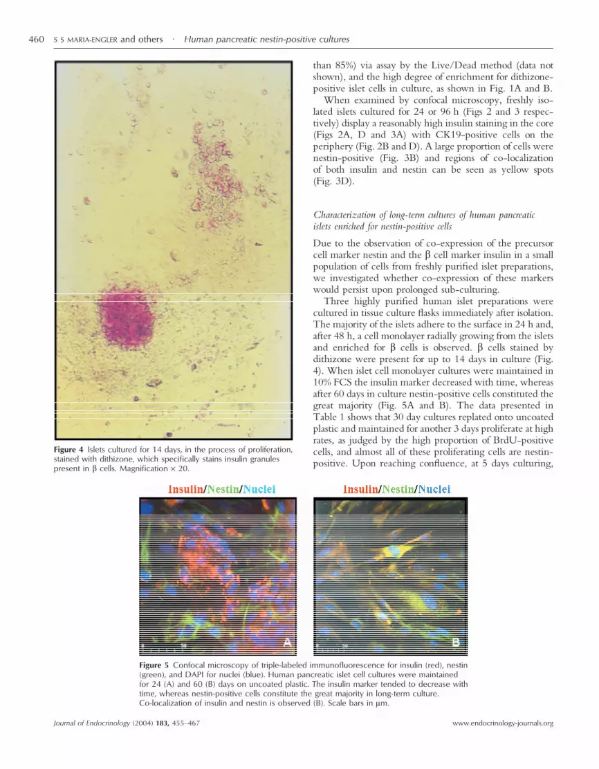
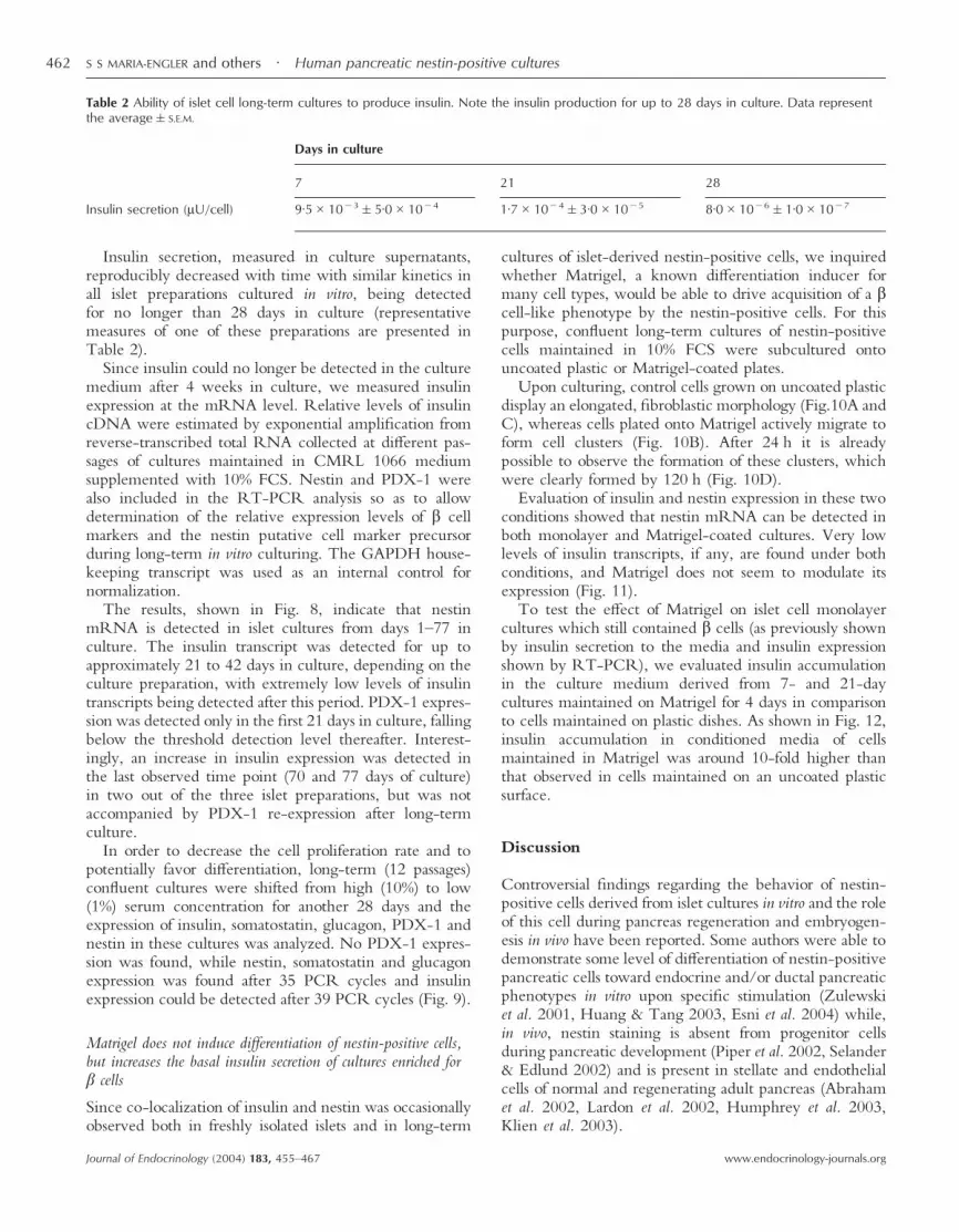
Three highly purified human islet preparations werecultured in tissue culture flasks immediately after isolation.The majority of the islets adhere to the surface in 24 h and,after 48 h, a cell monolayer radially growing from the isletsand enriched for � cells is observed. � cells stained bydithizone were present for up to 14 days in culture (Fig.4). When islet cell monolayer cultures were maintained in10% FCS the insulin marker decreased with time, whereasafter 60 days in culture nestin-positive cells constituted thegreat majority (Fig. 5A and B). The data presented inTable 1 shows that 30 day cultures replated onto uncoatedplastic and maintained for another 3 days proliferate at highrates, as judged by the high proportion of BrdU-positivecells, and almost all of these proliferating cells are nestin-positive. Upon reaching confluence, at 5 days culturing,
Figure 4 Islets cultured for 14 days, in the process of proliferation,stained with dithizone, which specifically stains insulin granulespresent in � cells. Magnification × 20.
Figure 5 Confocal microscopy of triple-labeled immunofluorescence for insulin (red), nestin(green), and DAPI for nuclei (blue). Human pancreatic islet cell cultures were maintainedfor 24 (A) and 60 (B) days on uncoated plastic. The insulin marker tended to decrease withtime, whereas nestin-positive cells constitute the great majority in long-term culture.Co-localization of insulin and nestin is observed (B). Scale bars in �m.
S S MARIA-ENGLER and others · Human pancreatic nestin-positive cultures460
www.endocrinology-journals.orgJournal of Endocrinology (2004) 183, 455–467

the overall proliferative index is lower, but nestin-positivecells are still proliferating.
In long-term cultures, where nestin-positive cells pre-dominate, some rare cells exhibited co-localization ofinsulin and nestin (Fig. 5B). In spite of the high proportionof ductal cells found around fresh islets (Fig. 2), very fewCK19 positive cells were observed even in short-term(Fig. 6) or in long-term cultures. Vimentin immunostain-ing, which labels mainly cells of mesenchymal origin suchas fibroblasts and endothelial cells, was rarely observedduring the entire observation period (data not shown).
After 12 to 14 passages (depending on the islet prep-aration), confluent cells grown on uncoated plastic andmaintained in 10% FCS, formed islet-like structures (Fig.7A and B). The islet cell cultures established from all threehuman islet preparations behaved similarly in terms ofadherence to plastic substrate, optical morphology andgrowth in culture.
Islet cell marker expression in long-term cultured human isletsenriched for nestin-positive cells
We set up to systematically analyze how subculturingwould affect the expression of insulin and nestin markersin these primary cultures of human pancreatic isletsenriched for nestin-positive cells.
Table 1 Percentage of BrdU labeling and BrdU/nestin-positive cells in monolayer culturesobtained from adult islet cells, maintained for 30 days and subcultured for 3 and 5 days.Data represent the average�S.D. of six different microscopic fields
BrdU/nestin-positive cells(%)
BrdU labeling index(%)
Days in culture3 49·7�0·4 57·1�1·45 31·6�0·8 42·9�9·6
Figure 6 Confocal microscopy of triple-labeledimmunofluorescence of human pancreatic islet cell culturesmaintained for 14 days on uncoated plastic, stained for nestin(green), CK19 (red), and DAPI for nuclei (blue). Scale bar in �m.
Figure 7 Phase contrast microscopy of nestin-positive cellsmaintained in culture in uncoated plastic for 12 to 14 passagesshows spontaneously formed islet-like structures.
Human pancreatic nestin-positive cultures · S S MARIA-ENGLER and others 461
www.endocrinology-journals.org Journal of Endocrinology (2004) 183, 455–467
Insulin secretion, measured in culture supernatants,reproducibly decreased with time with similar kinetics inall islet preparations cultured in vitro, being detectedfor no longer than 28 days in culture (representativemeasures of one of these preparations are presented inTable 2).
Since insulin could no longer be detected in the culturemedium after 4 weeks in culture, we measured insulinexpression at the mRNA level. Relative levels of insulincDNA were estimated by exponential amplification fromreverse-transcribed total RNA collected at different pas-sages of cultures maintained in CMRL 1066 mediumsupplemented with 10% FCS. Nestin and PDX-1 werealso included in the RT-PCR analysis so as to allowdetermination of the relative expression levels of � cellmarkers and the nestin putative cell marker precursorduring long-term in vitro culturing. The GAPDH house-keeping transcript was used as an internal control fornormalization.
The results, shown in Fig. 8, indicate that nestinmRNA is detected in islet cultures from days 1–77 inculture. The insulin transcript was detected for up toapproximately 21 to 42 days in culture, depending on theculture preparation, with extremely low levels of insulintranscripts being detected after this period. PDX-1 expres-sion was detected only in the first 21 days in culture, fallingbelow the threshold detection level thereafter. Interest-ingly, an increase in insulin expression was detected inthe last observed time point (70 and 77 days of culture)in two out of the three islet preparations, but was notaccompanied by PDX-1 re-expression after long-termculture.
In order to decrease the cell proliferation rate and topotentially favor differentiation, long-term (12 passages)confluent cultures were shifted from high (10%) to low(1%) serum concentration for another 28 days and theexpression of insulin, somatostatin, glucagon, PDX-1 andnestin in these cultures was analyzed. No PDX-1 expres-sion was found, while nestin, somatostatin and glucagonexpression was found after 35 PCR cycles and insulinexpression could be detected after 39 PCR cycles (Fig. 9).
Matrigel does not induce differentiation of nestin-positive cells,but increases the basal insulin secretion of cultures enriched for� cells
Since co-localization of insulin and nestin was occasionallyobserved both in freshly isolated islets and in long-term
cultures of islet-derived nestin-positive cells, we inquiredwhether Matrigel, a known differentiation inducer formany cell types, would be able to drive acquisition of a �cell-like phenotype by the nestin-positive cells. For thispurpose, confluent long-term cultures of nestin-positivecells maintained in 10% FCS were subcultured ontouncoated plastic or Matrigel-coated plates.
Upon culturing, control cells grown on uncoated plasticdisplay an elongated, fibroblastic morphology (Fig.10A andC), whereas cells plated onto Matrigel actively migrate toform cell clusters (Fig. 10B). After 24 h it is alreadypossible to observe the formation of these clusters, whichwere clearly formed by 120 h (Fig. 10D).
Evaluation of insulin and nestin expression in these twoconditions showed that nestin mRNA can be detected inboth monolayer and Matrigel-coated cultures. Very lowlevels of insulin transcripts, if any, are found under bothconditions, and Matrigel does not seem to modulate itsexpression (Fig. 11).
To test the effect of Matrigel on islet cell monolayercultures which still contained � cells (as previously shownby insulin secretion to the media and insulin expressionshown by RT-PCR), we evaluated insulin accumulationin the culture medium derived from 7- and 21-daycultures maintained on Matrigel for 4 days in comparisonto cells maintained on plastic dishes. As shown in Fig. 12,insulin accumulation in conditioned media of cellsmaintained in Matrigel was around 10-fold higher thanthat observed in cells maintained on an uncoated plasticsurface.
Discussion
Controversial findings regarding the behavior of nestin-positive cells derived from islet cultures in vitro and the roleof this cell during pancreas regeneration and embryogen-esis in vivo have been reported. Some authors were able todemonstrate some level of differentiation of nestin-positivepancreatic cells toward endocrine and/or ductal pancreaticphenotypes in vitro upon specific stimulation (Zulewskiet al. 2001, Huang & Tang 2003, Esni et al. 2004) while,in vivo, nestin staining is absent from progenitor cellsduring pancreatic development (Piper et al. 2002, Selander& Edlund 2002) and is present in stellate and endothelialcells of normal and regenerating adult pancreas (Abrahamet al. 2002, Lardon et al. 2002, Humphrey et al. 2003,Klien et al. 2003).
Table 2 Ability of islet cell long-term cultures to produce insulin. Note the insulin production for up to 28 days in culture. Data representthe average�S.E.M.
Days in culture
7 21 28
Insulin secretion (�U/cell) 9·5�10�3�5·0�10�4 1·7�10�4�3·0�10�5 8·0�10�6�1·0�10�7
S S MARIA-ENGLER and others · Human pancreatic nestin-positive cultures462
www.endocrinology-journals.orgJournal of Endocrinology (2004) 183, 455–467

Figure 8 Fractionation in agarose gels (A) and densitometric data (B) of insulin, nestin, PDX-1 and theGAPDH housekeeping gene amplification products at the exponential range of RT-PCR. Long-term culturesof human pancreatic islets, obtained from three different donors, cultured for different periods of time ontouncoated plastic, showing that nestin-positive cells are present for long culture periods, but insulin isdetectable only in the first month of culture. Insulin and PDX-1 were detected for up to 28 to 42 days inculture, with very low levels of transcripts being present after this period. Insulinoma and bladder carcinomaRNA were used as, respectively, positive and negative controls, in addition to the PCR control (notemplate).
Human pancreatic nestin-positive cultures · S S MARIA-ENGLER and others 463
www.endocrinology-journals.org Journal of Endocrinology (2004) 183, 455–467

Here we describe that both freshly isolated and culturedhuman islet preparations display co-localization of nestinand insulin as a rare event, suggesting that these cells couldbe undergoing early stages of differentiation to a � cellphenotype. This finding is in agreement with evidencerecently described by Street et al. (2004), who showedco-expression of insulin and nestin in cells of some smallislets in one pancreatic biopsy. In spite of the rarity of this
co-expression in their study, these authors do not rule outan involvement of nestin in islet neogenesis. Moreover,Yashpal et al. (2004) have also demonstrated co-expressionof nestin and insulin in a large proportion of cells in theprenatal rat pancreas and in rare cells in the postnatalpancreas. These authors suggest that mature endocrinecells derive from nestin-positive cells and lose theirprogenitor marker expression as they fully differentiateinto the postnatal life.
We rarely observed co-localization of nestin and insulinin our long-term cultures. The scarcity of this co-localization probably reflects the culture conditions weused, which favored proliferation of the nestin-positivecells (as shown by the BrdU assay) instead of differentia-tion, since cells were maintained in 10% FCS with nodifferentiation factor added to the medium. In our long-term cultures, ductal cells and fibroblasts were poorlyrepresented, insulin-positive cells decreased with time,while nestin-positive cells predominated and formed islet-like structures, as previously described (Selander & Edlund2002, Esni et al. 2004).
During subculturing in the presence of 10% serum, lowlevels of insulin expression were detected and graduallydecreased to become undetectable. However, after pro-longed subculturing, low levels of insulin expression couldbe observed in two out of three monolayer cultures grownto confluency. In addition, long-term subculturing ofthese cells under low (1%) serum conditions led todetection of low levels of insulin mRNA, somatostatin andglucagon, at the limit of sensitivity of the RT-PCRtechnique, whereas no PDX-1 expression could be de-tected. We hypothesize that culture conditions that inducegrowth arrest or replicative senescence, and thus cell cycleexit, might favor entry of these cells into the early stages ofpancreatic islet cell differentiation pathway. It is known,for other cell types, that cell senescence alters the expres-sion of growth and differentiation-specific genes, probablydue to an altered profile of transcription factors (Dimri &Campisi 1994). It has been demonstrated in Schwann cellsthat the signaling events associated with the expression ofmyelin-associated proteins are closely related with cessa-tion of mitosis. Besides, growth arrest induction in animmortalized Schwann cell line initiates a sequence ofchanges that ultimately leads to terminal differentiationand the appearance of several myelin-related markers(Rushton et al. 1999). In fact, promoting in vitro terminaldifferentiation of progenitors of diverse tissues such asneuronal (Galderisi et al. 2003), hematopoietic (Taniguchiet al. 1999) and myogenic (Walsh & Perman 1997) tissuesinto fully differentiated and functional cells invariablyrequires induction of cell cycle exit, mostly by deprivationof growth factors and serum, and/or concomitant additionof differentiation-inducing factors. Presumably, pancreaticislet cell progenitors would be no exception and, therefore,growth arrest may be essential, although not sufficient,to promote differentiation toward a pancreatic islet cell
Figure 9 Analysis of insulin, PDX-1, glucagon, nestin, somatostatinand GAPDH expression by semi-quantitative RT-PCR in confluentcultures maintained until the 12th passage in CMRL 1066 mediumcontaining 10% FCS and then shifted to low (1%) FCS for another28 days. Note the expression of nestin, somatostatin, glucagonand insulin. RNA from freshly isolated islets was used as thepositive control and no DNA template as the negative control.
Figure 10 Phase contrast microscopy of nestin-positive cellscultured onto plastic (A, C,) and Matrigel-coated surfaces (B, D).Note the tridimensional cell reorganization and islet-like structureformation upon culturing in Matrigel for 3 h and 120 hrespectively.
S S MARIA-ENGLER and others · Human pancreatic nestin-positive cultures464
www.endocrinology-journals.orgJournal of Endocrinology (2004) 183, 455–467
phenotype that, at the early stages, expresses low levels ofthree islet hormones (insulin, glucagon and somatostatin).The lack of PDX-1 expression concomitantly with insulinexpression in vitro has been previously reported by Huang& Tang (2003) in fetal nestin-positive cells treated withdifferentiation factors and could be explained by a complexpost-transcriptional control of PDX-1 expression, as sug-gested by transgenic mice expressing the E. coli LacZ geneunder the control of the PDX-1 promoter (Stoffers et al.1999).
The evidence presented here, showing an increasedinsulin accumulation in the presence of Matrigel, is ingood agreement with previous data showing the effect ofthis product on the maintenance of normal � cell function(Perfetti et al. 1996, Edamura et al. 2001, Oberg-Welsh2001). However, in contrast with the transdifferentiation/differentiation effect that has been described for islet(Kerr-Conte et al. 1996), acinar (Arias & Bendayan 1993)and ductal cells (Bonner-Weir et al. 2000), our attempt topromote differentiation of nestin-positive cells by subcul-turing onto a Matrigel-coated surface only induced cellmigration and formation of islet-like clusters after 24 h,
with unaltered insulin and nestin expression levels. Thesefindings suggest that culturing nestin-positive cells inMatrigel three-dimensional matrix alone may not besufficient to induce differentiation, at least for the timepoint tested.
In summary, our report of co-localization of nestin andinsulin and expression of islet cell markers in long-termhuman pancreatic nestin-positive cell cultures corroboratesprevious findings that, at least in vitro, nestin-positive cellscan undergo early stages of differentiation to an islet cellphenotype. In spite of the fact that several in vivo modelsdo not support a role for nestin-positive cells as an isletprogenitor (Piper et al. 2002, Selander & Edlund 2002),they are able to express insulin, GLUT-2 (Huang & Tang2003) and transcription factors normally expressed ininsulin-producing cells such as PDX-1 (Zulewski et al.2001, Huang & Tang 2003, Esni et al. 2004) and NKX6·1and ISL1 (Huang & Tang 2003). Therefore, it cannot beruled out that, under specific stimulation in culture,nestin-positive cells are fully able to differentiate into amature, functional � cell phenotype, confirming the highlevel of plasticity which has previously been attributed to
Figure 11 Analysis of insulin and nestin expression by semi-quantitative RT-PCR inlong-term cultures 1 and 2 of nestin-positive islet cells maintained on plastic or Matrigelsubstrates for 44 and 95 days respectively. Nestin and insulin transcripts were amplified atthe exponential phase of the PCR. The amount of different templates was normalizedrelative to GAPDH. Very low levels of insulin transcripts are occasionally present inlong-term cultures and Matrigel does not seem to modulate its expression.
Figure 12 Basal insulin secretion by islet cells growing in the presence or in the absence ofMatrigel. Basal insulin secretion was measured, by an electrochemiluminescence assay, inconditioned media obtained from cells plated in plastic or Matrigel coated dishes whichwere incubated with CMRL supplemented with 0·5% FCS for 4 days. Data represent themean�S.E.M. (n=3), a vs b P<0·05.
Human pancreatic nestin-positive cultures · S S MARIA-ENGLER and others 465
www.endocrinology-journals.org Journal of Endocrinology (2004) 183, 455–467

these cells (Blyszczuk et al. 2003), since under the influ-ence of various factors, they were able to assume featuresof neural (Cattaneo & McKay 1990), hepatic and pancre-atic cells (Lumelsky et al. 2001, Esni et al. 2004).
Recently, Seaberg et al. (2004) described that multi-potential precursor cells (named PMP) from adult mousepancreas were able to differentiate into neurons, endocrineand exocrine cells and stellate cells. Nestin expression didnot seem to predict the identity of these cells, but all of thecolonies derived from these cells expressed nestin and didnot co-express endothelial and epithelial cell markers.Based on these findings, the authors suggested the exist-ence of a different nestin-positive progenitor cell otherthan those pancreatic epithelial cell progenitors previouslydescribed in vivo (Esni et al. 2004, Klein et al. 2003,Selander & Edlund 2002). It still remains to be elucidatedwhether different populations of nestin-positive cells com-mitted to different cell fates exist in different stages ofpancreatic development.
Since the strategies used so far were not able to pro-mote complete differentiation to a � cell phenotype ofductal, acinar or embryonic stem cells, further studiesare necessary to establish the actual potential of nestin-positive culture cells as an alternative source of � cells fortransplantation.
Acknowledgements
We are specially grateful to Drs M C Messam andM Hoffman (NINDS–National Institutes of Health,Bethesda, MD, USA) for generously providing respect-ively, the nestin antibody and the Matrigel used in thesestudies. We also thank Dr Alberto Hayek (University ofCalifornia, San Diego, La Jolla, CA, USA) for criticismsand suggestions and Zizi de Mendonça, Sandra ReginaSouza and Débora Cristina Costa for excellent technicalassistance. This work was supported by: FAPESP, CNPq,ICGEB, FINEP, Biobrás S/A and PRP–USP. Theauthors declare that there is no conflict of interest thatwould prejudice the impartiality of this scientific work.
References
Abraham EJ, Leech CA, Lin JC, Zulewski H & Habener JF 2002Insulinotropic hormone glucagon-like peptide-1 differentiation ofhuman pancreatic islet-derived progenitor cells intoinsulin-producing cells. Endocrinology 143 3152–3161.
Arias AE & Benadayan M 1993 Differentiation of pancreatic acinarcells into duct-like cells in vitro. Laboratory Investigation 69 518–530.
Bank HL 1987 Assessment of islet cell viability using fluorescent dyes.Diabetologia 30 812–816.
Berná G, Léon-Quinto T, Ensenãt-Waser R, Montanya E, Martín F& Soria B 2001 Stem cells and diabetes. Biomedecine andPharmacotherapy 55 206–212.
Blyszczuk P, Czyz J, Kania G, Wagner M, Roll U, St-Onge L &Wobus AM 2003 Expression of Pax4 in embryonic stem cellspromotes differentiation of nestin-positive progenitor andinsulin-producing cells. PNAS 100 998–1003.
Bonner-Weir S, Taneja M, Weir GC, Tatarkiewicz K, Song KH,Sharma A & O’Neil JJ 2000 In vitro cultivation of human isletsfrom expanded ductal tissue. PNAS 97 7999–8004.
Cattaneo E & McKay R 1990 Proliferation and differentiation ofneuronal stem cells regulated by nerve growth factor. Nature 347762–765.
Chirgwin JM, Przybyla AE, Macdonald RJ & Rutter WJ 1979Isolation of biologically active ribonucleic acid from sourcesenriched in ribonuclease. Biochemistry 18 5294–5299.
Dimri GP & Campisi J 1994 Altered profile of transcription factorbinding activities in senescent human fibroblasts. Experimental CellResearch 212 132–140.
Edamura K, Ohgawara H, Nasu K, Iwami Y, Sato A, Ishikawa S,Matsuki N, Ono K, Ogawa H & Sasaki N 2001 Effect of theextracellular matrix on pancreatic endocrine cell function and itsbiocompatibility in dogs. Cell Transplant 10 493–498.
Esni F, Stoffers DA, Takeuchi T & Leach SD 2004 Origin ofexocrine pancreatic cells from nestin-positive precursors indeveloping mouse pancreas. Mechanisms of Development 121 15–25.
Galderisi U, Jori FP & Giordano A 2003 Cell cycle regulation andneural differentiation. Oncogene 22 5208–5219.
Huang H & Tang X 2003 Phenotypic determination andcharacterization of nestinpositive precursors derived from humanfetal pancreas. Laboratory Investigation 83 539–547.
Humphrey RK, Bucay N, Beattie GM, Lopez A, Messan CA, CirulliV & Hayek A 2003 Characterization and isolation ofpromoter-defined nestin-positive cells from the human fetalpancreas. Diabetes 52 2519–2525.
Hunziker E & Stein M 2000 Nestin-expressing cells in the pancreaticislets of Langerhans. Biochemical and Biophysical ResearchCommunications 271 116–119.
Kerr-Conte J, Pattou F, Lecomte-Houcke M, Xia Y, Boilly B, ProyeC & Lefebvre J 1996 Ductal cyst formation in collagen-embeddedadult human islet preparation. A means to the reproduction ofnesidioblastosis in vitro. Diabetes 45 1108–1114.
Klein T, Ling Z, Heimberg H, Madsen OD, Heller RS & Serup P2003 Nestin is expressed in vascular endothelial cells in the adulthuman pancreas. Journal of Histochemistry and Cytochemistry 51697–706.
Lardon J, Rooman I & Bouwens L 2002 Nestin expression inpancreatic stellate cells and angiogenic endothelial cells.Histochemistry and Cell Biology 117 535–540.
Lumelsky N, Blondel O, Laeng P, Velasco I, Ravin R & McKay R2001 Differentiation of embryonic stem cells to insulin-secretingstructures similar to pancreatic islets. Science 292 1389–1394.
Messan CA, Hou J & Major EO 2000. Coexpression of nestin inneural and glial cells in the developing human CNS defined by ahuman-specific anti-nestin antibody. Experimental Neurology 161585–596.
Oberg-Welsh C 2001 Long-term culture in matrigel enhances theinsulin secretion of fetal porcine islet-like cell clusters in vitro.Pancreas 22 157–163.
Perfetti R, Henderson TE, Wang Y, Montrose-Rafizadeh C & EganJM 1996. Insulin release and insulin mRNA levels in rat islets ofLangerhans cultured on extracellular matrix. Pancreas 13 47–54.
Piper K, Ball SG, Tumpenny LW, Brickwood DI & Hanley NA 2002Beta-cell differentiation during human development does not relyon nestin-positive precursors implications for stem cell-derivedreplacement therapy. Diabetologia 45 1045–1047.
Rushton JA, Schmitz S, Gunn-Moore F, Sherman D, Pappas CA,Ritchie JM & Haynes LW 1999 Growth arrest and spontaneousdifferentiation are initiated through an autocrine loop in clonallyderived Schwann cells by �1-procollagen I C-propeptide. Journal ofNeurochemistry 73 1816–1827.
Seaberg RM, Smukler SR, Kieffer TJ, Enikolopov G, Asghar Z,Wheeler MB, Korbutt G and Van Der Kooy D 2004 Clonal
S S MARIA-ENGLER and others · Human pancreatic nestin-positive cultures466
www.endocrinology-journals.orgJournal of Endocrinology (2004) 183, 455–467

identification of multipotent precursors from adult mouse pancreasthat generate neural and pancreatic lineages. Nature Biotechnology 221115–1124.
Selander L & Edlund H 2002 Nestin is expressed in mesenchymal andnot epithelial cells of the developing mouse pancreas. Mechanisms ofDevelopment 113 189–192.
Shapiro AM, Lakey JRT, Ryan EA, Korbutt GS, Toth E, WarnockGL, Kneteman NM & Rajotte RV 2000 Islet transplantation inseven patients with type I diabetes mellitus using aglucocorticoid-free immunosuppressive regimen. New EnglandJournal of Medicine 343 230–238.
Stoffers DA, Heller RS, Miller CP & Habener JF 1999Developmental expression of the homedomain protein IDX-1 inmice transgenic for an IDX-1 promoter/lacZ transcriptionalreporter. Endocrinology 140 5374–5381.
Street CN, Lakey JRT, Seeberger K, Helms L, Rajotte RV, ShapiroAMF & Korbutt AG 2004 Heterogenous expression of nestin inhuman pancreatic tissue precludes its use as an islet precursormarker. Journal of Endocrinology 180 213–225.
Taniguchi T, Endo H, Chikatsu N, Uchimaru K, Asano S, Fujita T,Nakahata T & Motokura T 1999. Expression of
p21(Cip1/Waf1/Sdi1) and p27(Kip1) cyclin-dependent kinaseinhibitors during human hematopoiesis. Blood 93 4167–4178.
Treutelaar MK, Skidmore JM, Dias-Leme CL, Hara M, Zhang L,Simeone D, Martin DM & Burant CF 2003 Nestin-lineage cellscontribute to the microvasculature but not endocrine cells of theislet. Diabetes 52 2503–2512.
Walsh K & Perlman H 1997. Cell cycle exit upon myogenicdifferentiation. Current Opinion in Genetics and Development 7597–602.
Yashpal NK, Li J & Wang R 2004 Characterization of c-Kit andnestin expression during islet cell development in the prenatal andpostnatal rat pancreas. Developmental Dynamics 229 813–825.
Zulewski H, Abraham EJ, Gerlach MJ, Daniel PB, Moritz W, MüllerB, Vallejo M, Thomas MK & Habener JF 2001 Multipotentialnestin-positive stem cells isolated from adult pancreatic isletsdifferentiate ex vivo into pancreatic endocrine, exocrine and hepaticphenotypes. Diabetes 50 521–533.
Received 27 August 2004Accepted 14 September 2004
Human pancreatic nestin-positive cultures · S S MARIA-ENGLER and others 467
www.endocrinology-journals.org Journal of Endocrinology (2004) 183, 455–467


