Embed Size (px)
Citation preview

THROMBOSIS AND HEMOSTASIS
Coagulation, an ancestral serine protease cascade, exerts a novel function in earlyimmune defenseTorsten G. Loof,1 Matthias Morgelin,1 Linda Johansson,2 Sonja Oehmcke,1 Anders I. Olin,1 Gerhard Dickneite,3
Anna Norrby-Teglund,2 Ulrich Theopold,4 and Heiko Herwald1
1Department of Clinical Sciences, Biomedical Center, Lund University, Lund, Sweden; 2Center for Infectious Medicine, Karolinska Institutet, Department ofMedicine, Karolinska University Hospital Huddinge, Stockholm, Sweden; 3Department of Preclinical Research and Development, CSL Behring GmbH, Marburg,Germany; and 4Department of Molecular Biology and Functional Genomics, Stockholm University, Stockholm, Sweden
Phylogenetically conserved serine pro-tease cascades play an important role ininvertebrate and vertebrate immunity. Themammalian coagulation system can betraced back some 400 million years andshares homology with ancestral serineproteinase cascades that are involved in,for example, Toll receptor signaling ininsects and release of antimicrobial pep-tides during hemolymph clotting. In the
present study, we show that the inductionof coagulation by bacteria leads to immo-bilization and killing of Streptococcuspyogenes bacteria inside the clot. Theentrapment is mediated via cross-linkingof bacteria to fibrin fibers by the action ofcoagulation factor XIII (fXIII), an evolution-arily conserved transglutaminase. In astreptococcal skin infection model, fXIII�/�
mice developed severe signs of patho-
logic inflammation at the local site ofinfection, and fXIII treatment of wild-typeanimals dampened bacterial dissemina-tion during early infection. Bacterial kill-ing and cross-linking to fibrin networkswas also detected in tissue biopsies frompatients with streptococcal necrotizingfasciitis, supporting the concept that co-agulation is part of the early innate immunesystem. (Blood. 2011;118(9):2589-2598)
Introduction
Serine protease cascades play an important role in many pathophysi-ologic processes, including hemostasis, immune response, andwound healing.1 Their activation normally occurs by limitedproteolysis, and coagulation and complement are probably thebest-characterized serine proteinase cascades in humans. Phyloge-netic studies have shown that the 2 systems developed � 400 mil-lion years ago,2,3 and it has been proposed that they evolved from acommon ancestral origin in eukaryotes.4 Coagulation and comple-ment cascades share a remarkable degree of convergent evolutionwith other serine protease cascades such as those regulatingdorsal-ventral polarity in Drosophila (leading to an activation ofSpatzle, the ligand of the Toll receptor) and the hemolymph clottingsystem in the horseshoe crab.4,5 These findings suggest that thebasic motifs of some proteolytic cascades existed long before thedivergence of protostomes and deuterostomes.6 Both activation ofSpatzle and the horseshoe crab hemolymph clotting system are keycomponents in ancestral immunity, which relies largely on theinnate immune system. Whereas the complement system has beenconsidered to be part of the innate immune system for � 30 years,it has only recently been determined that coagulation also partakesin inflammation and the early immune defense.7,8 In the latterstudies, a major focus has been on the ability of the clotting cascadeto trigger pro- and anti-inflammatory reactions, such as the releaseof cytokines and the activation of protease-activated receptors.However, it is unknown to what extent coagulation can activelycontribute to the elimination of an invading microorganism.
The present study was undertaken to investigate whetheractivation of the coagulation system in response to bacterialinfection contributes to the innate immune system and to elimina-
tion of the invading pathogen. Special focus was placed on the roleof coagulation factor XIII (fXIII), for which the insect homolog(transglutaminase) was recently found to play a protective role inthe immune response against bacterial pathogens in a Drosophilainfection model.9 Streptococcus pyogenes was used in the presentstudy, because this bacterium is considered to be one of the mostimportant human bacterial pathogens, being responsible for at least18 million cases of severe infection worldwide (1.78 million newcases each year) and � 500 000 deaths yearly, as estimated by theWorld Health Organization.10 Infections with S pyogenes arenormally superficial and self-limiting, but can develop into seriousand life-threatening conditions such as necrotizing fasciitis andstreptococcal toxic shock syndrome, which are associated withhigh morbidity and mortality.11 The fact that S pyogenes can causelocal and systemic infections in the same infection model made itan ideal pathogen to be studied in the present investigation.
Methods
Bacterial strains and culture conditions
The S pyogenes strains AP1 (40/58) and KTL3 of serotype M1 have beendescribed previously.12,13 Bacteria were grown overnight in Todd-Hewittbroth (THB; GIBCO) at 37°C and 5% CO2.
Human plasma
Plasma obtained from healthy donors was purchased from Lund UniversityHospital (Lund, Sweden) and plasma kallikrein–, (PK-), thrombin-, fXII-,and fXIII-deficient plasmas were purchased from George King Bio-Medical.
Submitted February 16, 2011; accepted May 10, 2011. Prepublished online asBlood First Edition paper, May 25, 2011; DOI 10.1182/blood-2011-02-337568.
An Inside Blood analysis of this article appears at the front of this issue.
The online version of this article contains a data supplement.
The publication costs of this article were defrayed in part by page chargepayment. Therefore, and solely to indicate this fact, this article is herebymarked ‘‘advertisement’’ in accordance with 18 USC section 1734.
© 2011 by The American Society of Hematology
2589BLOOD, 1 SEPTEMBER 2011 � VOLUME 118, NUMBER 9
For personal use only.on August 9, 2016. by guest www.bloodjournal.orgFrom

Measurement of coagulation parameters
Activation of the intrinsic and extrinsic pathway of coagulation wasdetermined by measuring activated partial thromboplastin time (aPTT) andprothrombin time (PT) in human or murine plasma using a coagulometer(Amelung), as described previously.12
Substrate assays
PK activity on the bacterial surface after exposure to normal, PK-, orfXIII-deficient plasma was measured using the chromogenic substrateS-2302 (Chromogenix), as described previously.12 To measure thrombinactivity, normal, thrombin-, fXII-, or fXIII-deficient plasma was incubatedwith 2 � 1010 CFU of S pyogenes in 50mM Tris supplemented with 50�MZnCl2, 2mM CaCl2, and 1�M phospholipids (Rossix) for 30 minutes at37°C. The tetrapeptide Gly-Pro-Arg-Pro (Bachem) was added to preventclotting (1.5 mg/mL final concentration). Samples were washed with Trisand resuspended in Tris/ZnCl2 and 2mM of the chromogenic substrateS-2238. After incubation at 37°C and centrifugation, the absorbance of thesupernatants was determined at 405 nm. For fXIII-activity measurements,bacteria were added to normal, thrombin-, fXII-, or fXIII-deficient plasma(diluted 1:100 in sodium citrate), supplemented with zinc, calcium, andphospholipids. After incubation for 15 minutes at 37°C in the presence of agold-labeled N-�-�-glutamyl-lysine antibody (153-81D4; GeneTex), sampleswere subjected to negative-staining electron microscopy.
Bacterial growth in human plasma
Normal and fXIII-deficient plasma was diluted 1:100 in 12.9mM sodiumcitrate, mixed with 2.5 � 105 CFU of S pyogenes, and 0.5 U of thrombinfrom human plasma (Sigma) was added before incubation at 37°C. At theindicated time points, 50 �L of the mixture was plated onto blood agar in10-fold serial dilutions and the number of bacteria was determined bycounting colonies after 18 hours of incubation at 37°C; alternatively, themixture was analyzed by negative-staining electron microscopy.
Generation of plasma clots
Clots from human or murine plasma (normal and fXIII deficient) forelectron microscopic analysis were prepared as described previously.9
Cross-linking and immobilization of bacteria within the clot
Human fibrinogen (ICN Biomedicals) was incubated with M1 protein in theabsence or presence of thrombin-activated human fXIII (Enzyme ResearchLaboratories) for 30 minutes at 37°C. For visualization by electronmicroscopy, the gold-labeled N-�-�-glutamyl-lysine antibody was added tothe reaction mixture.
To analyze bacterial immobilization, clots were generated from normaland fXIII-deficient plasma, washed with PBS, and covered with THBmedium. At the indicated time points, 50 �L of the supernatant was platedonto blood agar.
Electron microscopy
Samples for scanning electron microscopy were processed as describedpreviously.14 Transmission electron microscopy and immunostaining usingthe gold-labeled N-�-�-glutamyl-lysine antibody were performed as de-scribed previously.15 For negative-staining electron microscopy, sampleswere adsorbed to 400-mesh carbon-coated copper grids for 1 minute,washed with 2 drops of water, and stained with 2 drops of 0.75% uranylformate. The grids were rendered hydrophilic by glow discharge at lowpressure in air. Samples were observed in a 1200 EX transmission electronmicroscope (JEOL) operated at a 60-kV accelerating voltage.
Animal infection model
CBA/CaOlaHsd wild-type and fXIII�/� mice were from Harlan and CSLBehring, respectively. All animal experiments were approved by theregional ethical committee for animal experimentation (the Malmo/Lunddjurforsoksetiska namnd, Lund District Court, Lund, Sweden; permit
M220/08). Mice were subcutaneously infected with 2.5 � 108 CFU ofS pyogenes KTL3 as described previously.16 After 24 hours of infection,mice were killed. Skin samples were collected from the local focus ofinfection and fixed in 3.7% formaldehyde. For plasma analysis, citratedblood was taken from the heart at the time of killing, centrifuged at 2655gfor 10 minutes, and frozen at �80°C until use. To determine bacterial loads,blood and homogenates from liver and spleen were plated as described in“Bacterial growth in human plasma.” In some experiments, mice weretreated with 200 U/kg body weight of a human fXIII concentrate(FibrogamminP; CSL Behring) subcutaneously at the site of infection3 hours after bacterial inoculation.
Examination of murine skin samples
Fixed tissue samples were dehydrated in ethanol, embedded in paraffin, andthen cut into 3-�m-thick sections. After deparaffination, samples wereprepared for scanning electron microscopy or stained with H&E (Histolab)or with Giemsa stain (Merck) for histologic analysis using an Eclipse 80imicroscope (Nikon).
Examination of human tissue biopsies
Snap-frozen tissue biopsies collected from the epicenter of infection in2 patients with necrotizing fasciitis caused by S pyogenes of the M1T1serotype were stained and compared with biopsies taken from healthyvolunteers. The human subjects review committees of the University ofToronto (Toronto, ON) and Karolinska University Hospital (Stockholm,Sweden) approved the studies, and informed consent was obtained from thepatients and the volunteers. The biopsies were prepared and immunofluores-cent stainings of serial sections were conducted as described previously.17,18
The following antibodies were used in dilutions ranging from 1:250 to1:10 000: anti N-�-�-glutamyl-lysine, antifactor XIIIa (Acris), a polyclonalrabbit antiserum specific for the Lancefield group A carbohydrate (Difco),and polyclonal rabbit antiserum against M1 protein. The immunohistochemi-cal stainings were conducted in an RXM microscope with a 25�/0.55numeric aperture oil objective lens and immunofluorescent stainings in aconfocal scanner TCS SP II coupled to a DMR microscope (all from Leica).
Statistical analysis
Data were analyzed using Prism 5 software (GraphPad). The significancebetween the values of an experimental group was determined by use of avariance analysis (t test).
Results
Contact activation at the surface of S pyogenes leads to aninduction of fXIII
Previous studies have shown that the presence of S pyogenes inplasma leads to an assembly and activation of the contact system atthe bacterial surface.14 However, those experiments were per-formed in the absence of calcium and phospholipids, 2 indispens-able clotting cofactors required for the activation of coagulationfactors upstream of the contact system.19 We therefore wonderedwhether calcium and phospholipid reconstitution triggers an induc-tion of the remaining clotting cascade at the streptococcal surface.To confirm the previously reported findings, we first measured thePK activity on AP1 bacteria after incubation with normal humanplasma supplemented with zinc. As depicted in supplementalFigure 1A (available on the Blood Web site; see the SupplementalMaterials link at the top of the online article), substrate hydrolysiswas monitored when bacteria were incubated with normal andfXIII-deficient plasma, but not when plasma lacked PK. We thenset up experiments to monitor whether bacteria-induced contactactivation leads to an induction of the entire coagulation cascade bymeasuring thrombin activity, the activator of fXIII. Normal plasma
2590 LOOF et al BLOOD, 1 SEPTEMBER 2011 � VOLUME 118, NUMBER 9
For personal use only.on August 9, 2016. by guest www.bloodjournal.orgFrom
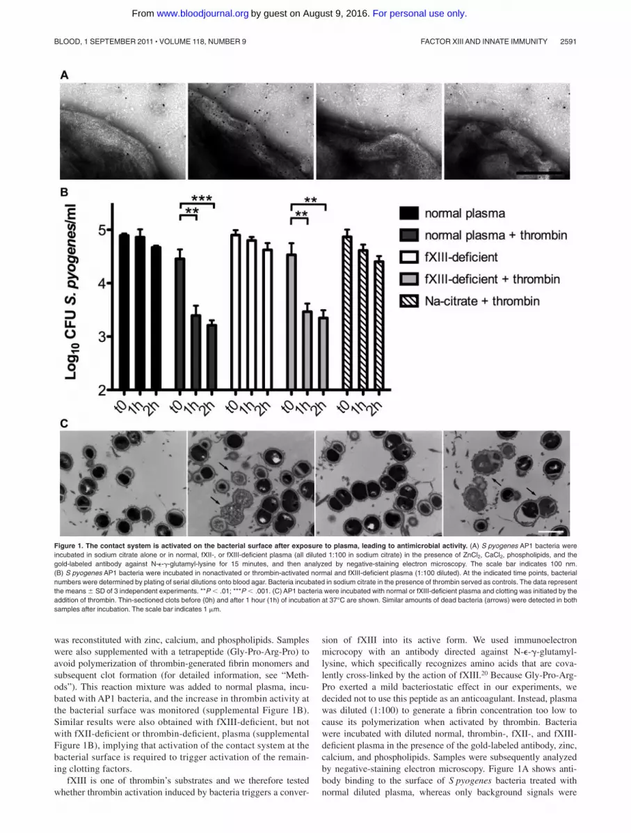
was reconstituted with zinc, calcium, and phospholipids. Sampleswere also supplemented with a tetrapeptide (Gly-Pro-Arg-Pro) toavoid polymerization of thrombin-generated fibrin monomers andsubsequent clot formation (for detailed information, see “Meth-ods”). This reaction mixture was added to normal plasma, incu-bated with AP1 bacteria, and the increase in thrombin activity atthe bacterial surface was monitored (supplemental Figure 1B).Similar results were also obtained with fXIII-deficient, but notwith fXII-deficient or thrombin-deficient, plasma (supplementalFigure 1B), implying that activation of the contact system at thebacterial surface is required to trigger activation of the remain-ing clotting factors.
fXIII is one of thrombin’s substrates and we therefore testedwhether thrombin activation induced by bacteria triggers a conver-
sion of fXIII into its active form. We used immunoelectronmicrocopy with an antibody directed against N-�-�-glutamyl-lysine, which specifically recognizes amino acids that are cova-lently cross-linked by the action of fXIII.20 Because Gly-Pro-Arg-Pro exerted a mild bacteriostatic effect in our experiments, wedecided not to use this peptide as an anticoagulant. Instead, plasmawas diluted (1:100) to generate a fibrin concentration too low tocause its polymerization when activated by thrombin. Bacteriawere incubated with diluted normal, thrombin-, fXII-, and fXIII-deficient plasma in the presence of the gold-labeled antibody, zinc,calcium, and phospholipids. Samples were subsequently analyzedby negative-staining electron microscopy. Figure 1A shows anti-body binding to the surface of S pyogenes bacteria treated withnormal diluted plasma, whereas only background signals were
Figure 1. The contact system is activated on the bacterial surface after exposure to plasma, leading to antimicrobial activity. (A) S pyogenes AP1 bacteria wereincubated in sodium citrate alone or in normal, fXII-, or fXIII-deficient plasma (all diluted 1:100 in sodium citrate) in the presence of ZnCl2, CaCl2, phospholipids, and thegold-labeled antibody against N-�-�-glutamyl-lysine for 15 minutes, and then analyzed by negative-staining electron microscopy. The scale bar indicates 100 nm.(B) S pyogenes AP1 bacteria were incubated in nonactivated or thrombin-activated normal and fXIII-deficient plasma (1:100 diluted). At the indicated time points, bacterialnumbers were determined by plating of serial dilutions onto blood agar. Bacteria incubated in sodium citrate in the presence of thrombin served as controls. The data representthe means � SD of 3 independent experiments. **P � .01; ***P � .001. (C) AP1 bacteria were incubated with normal or fXIII-deficient plasma and clotting was initiated by theaddition of thrombin. Thin-sectioned clots before (0h) and after 1 hour (1h) of incubation at 37°C are shown. Similar amounts of dead bacteria (arrows) were detected in bothsamples after incubation. The scale bar indicates 1 �m.
FACTOR XIII AND INNATE IMMUNITY 2591BLOOD, 1 SEPTEMBER 2011 � VOLUME 118, NUMBER 9
For personal use only.on August 9, 2016. by guest www.bloodjournal.orgFrom

detected when bacteria were incubated with fXII- or fXIII-deficientplasma (Figure 1A). Similar results were obtained with thrombin-deficient plasma (data not shown). These results suggest thatcontact activation at the bacterial surface can evoke an induction ofthe entire coagulation cascade, eventually enabling fXIII to act onS pyogenes surface proteins.
Streptococci are killed in thrombin-activated plasma
It has been shown that contact activation on the surface ofS pyogenes leads to the generation of antimicrobial peptides.21
Therefore, we investigated the fate of cross-linked bacteria inactivated, but nonclotted, normal and fXIII-deficient plasma. Ourresults show that bacterial growth was significantly impaired inthrombin-activated normal and fXIII-deficient plasma (diluted1:100). This effect was dependent on both time and plasmaactivation (Figure 1B). To determine whether these results weredue to an induction of antimicrobial activity, plasma-treatedbacteria were subjected to negative-staining electron microscopy.Supplemental Figure 1C shows intact bacteria that were incubatedwith nonactivated normal plasma. Similar findings were observedwhen bacteria were incubated with nonactivated fXIII-deficientplasma (supplemental Figure 1C). In the presence of thrombin,however, incubation with normal or fXIII-deficient plasma causedmultiple disruptions of the bacterial cell wall and triggered anefflux of cytosolic content (supplemental Figure 1C), a sign ofbacterial killing.17 Incubation of S pyogenes with thrombin in theabsence of plasma neither impaired bacterial growth (Figure 1B)nor caused cytosolic leakage (data not shown).
To determine whether bacterial killing also occurs within aformed clot, AP1 bacteria and undiluted plasma were mixed andthrombin was added. The clots formed were incubated for 1 hour,thin-sectioned, and analyzed by transmission electron microscopy.Figure 1C shows that a significant number of bacteria in clotsgenerated from normal and fXIII-deficient plasma were devoid ofcytosolic content, suggesting a substantial disruption of the cellmembrane and bacterial killing. In contrast, only a few dead
bacteria were seen when clots were thin-sectioned directly after theaddition of thrombin. Statistical analysis revealed an efficientkilling of bacteria within the clot regardless of whether the clot wasformed from normal or fXIII-deficient plasma (6% at time 0 hoursand 35% after 1 hour for normal plasma and 5% at time 0 hours and36% after 1 hour for fXIII-deficient plasma). These data demon-strate that activation of the coagulation cascade on the surface ofS pyogenes leads to an fXIII-independent induction of antimicro-bial activity.
Bacterial entrapment within a plasma clot is fXIII dependent
It was recently shown that human fXIII cross-links and immobi-lizes bacteria of the species Staphylococcus aureus and Escherichiacoli inside a plasma clot.9 To determine whether this also applies toS pyogenes, bacteria of strain AP1 were incubated with normal andfXIII-deficient plasma. After activation with thrombin, clots wereanalyzed by scanning electron microscopy. Figure 2A-B showsclots formed from normal and fXIII-deficient plasma in the absenceof bacteria. The micrographs reveal that the clots have a similarmorphology, although clots generated from fXIII-deficient plasmaappear less dense. However, dramatic changes were observed whenclots were formed in the presence of S pyogenes AP1 bacteria.Whereas massive loads of bacteria were entrapped in clots derivedfrom normal plasma (Figure 2C), only a few bacteria were foundattached to clots when fXIII-deficient plasma was used (Figure2D). In addition, fibrin network formation was reduced whenbacteria were incubated with normal plasma, which was not seen infXIII-deficient plasma (Figure 2C-D). At higher resolution, it isnoticeable that fibrin fibers and bacteria are in close proximity inthe clots generated from normal plasma, and it even appears thatthe fibers originate from the bacterial surface (Figure 2E). Incontrast, bacteria are loosely assembled in clots from fXIII-deficient plasma and no direct interaction with fibrin fibers isvisible (Figure 2F). To confirm these findings, clots from normalplasma were thin-sectioned and subjected to transmission electronmicroscopy, which allows analysis at higher resolution. Figure
Figure 2. Entrapment and immobilization of S pyo-genes inside the clot. Scanning electron micrographsshowing the structure of clots generated from normalplasma (A,C,E) or fXIII-deficient plasma (B,D,F) in theabsence (A-B) or presence (C-F) of bacteria. The scalebars indicate 10 �m in panels A through D and 1 �m inpanels E and F. The transmission electron micrographsdepict S pyogenes alone (G), after exposure to thrombin-activated plasma (H), and after exposure to plasmafollowed by immunostaining with a gold-labeled N-�-�-glutamyl-lysine antibody recognizing the fXIII cross-linking site (I). Scale bars indicate 1 �m in panels G andH and 100 nm in panel I.
2592 LOOF et al BLOOD, 1 SEPTEMBER 2011 � VOLUME 118, NUMBER 9
For personal use only.on August 9, 2016. by guest www.bloodjournal.orgFrom

2G-I depicts thin-sectioned S pyogenes AP1 bacteria before (Figure2G) and directly after incubation with normal plasma and subse-quent thrombin activation (Figure 2H). Within the clot, bacteria arestrung along fibrin fibers and it appears that they have multipleinteraction sites. Additional immunostaining with the gold-labeledantibody against N-�-�-glutamyl-lysine was used to study themode of interaction between bacteria and fibrin fibers. As expected,numerous cross-linking events within the fibrin fibers were de-tected. The electron microscopic analysis also revealed that fibrinfibers are avidly cross-linked to the bacterial surface (Figure 2I).Cross-linking activity was not recorded when bacteria wereincubated with fXIII-deficient plasma (data not shown).
Most streptococcal serotypes have a high affinity for fibrinogen,and the M1 protein has been reported to be the most importantfibrinogen receptor of the S pyogenes AP1 strain.22 The respectivebinding sites were mapped to the aminoterminal region of M1protein and fragment D, which is part of the terminal globulardomain of fibrinogen.22 Negative-staining electron microscopy wasused to study the interaction between M1 protein and fibrinogen atthe molecular level. The results demonstrate that one terminalregion of the streptococcal surface protein is in complex with aglobular domain of fibrinogen (Figure 3A top panel), which is ingood agreement with the mapping study. The nature of thiscomplex was not altered when activated fXIII was coincubatedwith the 2 proteins (Figure 3A middle panel). Indeed, additionalimmunodetection with the gold-labeled antibody against N-�-�-glutamyl-lysine revealed that the interaction site was covalently
cross-linked by fXIII (Figure 3A bottom panel). M proteins are themost abundant surface proteins of streptococci and it is thereforeplausible that the M1 protein of S pyogenes AP1 bacteria is one ofthe major interaction partners that is covalently attached to fibrinfibers by the action of fXIII. However, it cannot be excluded thatother streptococcal surface proteins are also targeted by fXIII.
Whether cross-linking of bacteria by fXIII has a pathophysi-ologic function inside the clot was investigated by measuring theescape of S pyogenes AP1 bacteria from clots generated fromnormal and fXIII-deficient plasma. Streptococci were mixed withundiluted normal or fXIII-deficient plasma and clotting wasinduced by the addition of thrombin. Clots were then washed withPBS and covered with growth medium. At different time points,samples were collected from the supernatant and the bacterial loadwas determined. As seen in Figure 3B, fXIII-induced cross-linkingsignificantly reduced the release of bacteria from the clot, suggest-ing that they are immobilized and killed within the clot. Theseresults show that S pyogenes bacteria are covalently woven into afibrin network by the action of fXIII and therefore their dissemina-tion from the clot is prevented.
S pyogenes–infected fXIII�/� mice show more signs ofinflammation than wild-type animals at the local focus ofinfection
In vitro data suggest that coagulation is part of the early innate immuneresponse, which in a concerted action triggers the immobilization
Figure 3. fXIII cross-links the streptococcal M1 protein with fibrinogen, leading to immobilization of bacteria within the clot. (A) The electron micrographs shownegatively stained human fibrinogen (characterized by 3 domains) in complex with M1-protein (elongated) before (top) and after fXIII cross-linking (middle). Cross-linking wasdetected by immunostaining the fibrinogen M1 protein complex with the gold-labeled antibody against N-�-�-glutamyl-lysine (bottom). A schematic drawing of the fibrinogen(gray) and M1 protein (black) is included to highlight the interaction between fibrinogen and M1 protein. The scale bars indicate 25 nm. (B) Bacteria were incubated with normalor fXIII-deficient plasma and clotting was initiated by the addition of thrombin. Clots were washed briefly, covered with THB medium, and further incubated at 37°C. At theindicated time points, bacterial numbers were determined by plating of serial dilutions of the supernatant onto blood agar. The data represent the means � SD of 3 independentexperiments. *P � .05; ***P � .001.
FACTOR XIII AND INNATE IMMUNITY 2593BLOOD, 1 SEPTEMBER 2011 � VOLUME 118, NUMBER 9
For personal use only.on August 9, 2016. by guest www.bloodjournal.orgFrom

and killing of S pyogenes inside a clot. We therefore hypothesizedthat the prevention of bacterial dissemination and their clearancemay dampen the inflammatory response at the site of infection. Toinvestigate this, we took advantage of a skin infection model that wasestablished with KTL3, another S pyogenes strain of the M1serotype.16 Challenge with S pyogenes KTL3 normally causes localinfections that eventually disseminate from the infection focus and leadto systemic infection.16 Using scanning electron microscopic analysis,we found that incubation of S pyogenes KTL3 with thrombin-activated human normal or fXIII-deficient plasma in vitro gener-ates clots with a morphology similar to those generated withS pyogenes AP1 bacteria (data not shown). Similar results werealso obtained when murine plasma (normal and fXIII-deficient)was incubated with S pyogenes KTL3 bacteria (supplementalFigure 2).
To study the inflammatory response to local infection withS pyogenes, wild-type and fXIII�/� mice were subcutaneouslyinfected with S pyogenes KTL3. Twenty-four hours after infection,mice were killed and the skin from the local focus of infection wassurgically removed and stained for histopathologic analysis. Micro-scopic examination of H&E–stained skin biopsies from nonin-fected wild-type and fXIII�/� mice revealed no signs of inflamma-tion (Figure 4A-B), whereas cell invasion and tissue damage wereseen in biopsies from infected wild-type animals (Figure 4C). Thelesions were far more severe in biopsies from infected fXIII�/�
mice (Figure 4D). Biopsies were also stained with Giemsa to detectinfiltrating cells. Analysis of tissue samples from wild-type animalsshowed that bacteria were found in clustered patches and someneutrophils had been recruited (Figure 4E). In biopsies fromfXIII�/� mice, bacteria were scattered throughout the wholeinfection site and an increased number of neutrophils was detected(Figure 4F).
Further electron microscopy examination of the tissue biopsiesrevealed severe bleeding across the infected site in both wild-typeand fXIII�/� mice (data not shown). Bacteria were found entrappedand clustered within the fibrin meshwork of infected wild-typemice (Figure 4E), whereas bacteria were distributed throughoutthe whole infection site in skin biopsies from infected fXIII�/�
mice (Figure 4F). Additional statistical analysis revealed approxi-mately 8 bacterial clusters per 100 �m2 in the fibrin network ofwild-type animals (Figure 4G), whereas streptococci were mostlyseen as single bacteria or small chains at a density of 41 bacteriachains/100 �m2 in fXIII�/� animals (Figure 4H). At highermagnification it appears that bacteria are an integral part of thefibrin network from infected wild-type mice (Figure 4G inset). Thiswas not observed in biopsies from fXIII�/� mice, in whichstreptococci were found to be associated with, but not a constituentpart of, the network (Figure 4H inset). To assess the contribution ofimmobilization of bacteria to their dissemination, clotting times ofthe intrinsic pathway of coagulation (aPTTs) were measured.Increased aPTTs are a sign of a systemic response to the infection.12
Plasma samples were recovered 24 hours after infection andclotting times of the intrinsic pathway of coagulation weredetermined. Figure 4I shows that the aPTTs of plasma samplesfrom infected wild-type mice were moderately but significantlyincreased, whereas clotting times were extremely high in plasmasamples from fXIII�/� mice. The PT remained unaltered after24 hours of infection in both groups of mice (data not shown).These results demonstrate that the lack of fXIII leads to anincreased inflammatory response at the infectious site combinedwith an induction of systemic reactions.
fXIII cross-linking in patients with necrotizing fasciitis causedby S pyogenes
To determine whether the results obtained from the animal studiesalso apply to the clinical situation, biopsies from patients with
Figure 4. Subcutaneous infection of wild-type and fXIII�/� mice with S pyo-genes. H&E–stained representative tissue sections from noninfected (A-B) andinfected (24 hours after infection; C-D) wild-type (A,C) and fXIII�/� (B,D) mice areshown. Infected animals show signs of inflammation (white arrows) and tissuedamage (black arrows). The scale bar indicates 500 �m. Giemsa-stained biopsiesfrom the inflamed area of wild-type (E) and fXIII�/� mice (F) are shown. Arrowheadspoint to infiltrating inflammatory cells (macrophages/neutrophils). The scale barindicates 25 �m. Scanning electron micrographs depict biopsies from wild-type(G) and fXIII�/� (H) mice. Arrows indicate bacteria entrapped and clustered within thefibrin meshwork in wild-type mice (G) and scattered throughout the infection area infXIII�/� animals (H). Scale bars indicate 10 �m in the figure and 1 �m in the insets.(I) aPTT measured in plasma from noninfected and infected wild-type and fXIII�/�
mice (24 hours after infection). Data are presented as mean values of plasmasamples obtained from 3 or 5 noninfected and 9 infected animals obtained from3 independent experiments. *P � .05; **P � .01.
2594 LOOF et al BLOOD, 1 SEPTEMBER 2011 � VOLUME 118, NUMBER 9
For personal use only.on August 9, 2016. by guest www.bloodjournal.orgFrom

necrotizing fasciitis caused by S pyogenes were analyzed byimmunohistology and electron microscopy. Figure 5 depicts mas-sive tissue necrosis at the site of infection, and subsequentimmunodetection in serial tissue sections showed positive stainingfor the M1 protein and fXIII at these sites. This suggests an influxof plasma to the infected focus, and indeed cross-linking activity atthe same location was recorded (Figure 5 top lane). As controls,biopsies from healthy persons were used, in which no signal wasseen when subjected to the same experimental protocol (Figure 5bottom lane). Tissue sections were further analyzed by confocalimmunofluorescence microscopy using antibodies against M1protein and N-�-�-glutamyl-lysine. Figure 6A shows positivestaining for M1 protein and widespread positive staining forN-�-�-glutamyl-lysine. In addition, we found a striking colocaliza-tion of the 2 antibodies (Figure 6A), suggesting bacterial cross-linking at the infected site. When the biopsies were analyzed byscanning electron microscopy, massive bleeding at the infected sitewas recorded (data not shown), and bacteria were found to beclustered and entrapped inside the fibrin network (Figure 6B).Specimens were also thin-sectioned and studied by immunotrans-mission electron microscopy using the gold-labeled antibodyagainst N-�-�-glutamyl-lysine. Figure 6C shows immunostainingat the bacterial surface in regions in contact with fibrin fibers. Themicrographs also reveal that a significant portion (31%) of theentrapped bacteria were not viable, as shown in Figure 6D. Thesefindings are in agreement with the in vitro and in vivo experiments,and illustrate that immobilization of bacteria and generation ofantimicrobial activity is seen within the fibrin network in patientswith severe and invasive infections with S pyogenes.
Local treatment with fXIII dampens systemic bacterialspreading in infected mice
To determine whether treatment with fXIII is able to preventbacterial spreading in an animal model of infection, wild-type micewere subcutaneously infected with S pyogenes. Three hours afterchallenge, half of the mice were treated with FibrogamminP, ahuman plasma fXIII concentrate that was injected into the site of
infection. A dose of 200 U/kg body weight was chosen, which iswell tolerated in mice and gives rise to approximately 250% of totalfXIII activity when injected intravenously.23 Mice infected withS pyogenes but without FibrogamminP treatment served as con-trols. Infected animals were killed after 24 hours of infection, andbacterial loads in the blood, liver, and spleen were determined. Asdepicted in Figure 7, FibrogamminP treatment resulted in de-creased bacterial loads in the blood, liver, and spleen of the treatedmice, suggesting that fXIII dampens the systemic dissemination ofS pyogenes in the infected animals.
The results presented in this study support the concept that fXIIIhas an important role in the early immune response to bacterialinfections. We have shown that fXIII triggers an immobilization ofbacteria within the fibrin network at the local focus of infection,which is combined with an induction of plasma-derived antimicro-bial activity and subsequent bacterial killing. The 2 mechanismswork in concert and may together diminish early bacterial dissemi-nation and down-regulate the inflammatory response.
Discussion
Sensing the first signs of inflammation and rapid elimination of aninvading microorganism are key features of the early immune responseto infection. In particular, potential ports of microbial entry are at greatrisk and therefore need special protection. Therefore, the immunesystem has developed mechanisms that allow an efficient clearanceof inhaled (eg, with the help of mannose-binding lectin24) orswallowed (eg, by the action of intestinal mucins25) pathogens.Wounds present another port of entry and are associated with agreat risk of promoting infections and allowing microorganisms toenter the circulatory system. To prevent their dissemination andeventual systemic complications, it is of great importance that thehost’s defense system is activated as soon as wound sealing begins.It therefore appears likely that coagulation plays an important rolein these very early processes. However, the extent and underlyingmechanisms of this contribution to immunity are poorly understood.
Figure 5. Immunohistochemical analysis of human biopsies. Tissue biopsies were obtained from patients with necrotizing fasciitis caused by S pyogenes (top) and fromhealthy volunteers (bottom). The biopsies were sectioned and immunohistochemically stained for streptococcal M1 protein, fXIII, and N-�-�-glutamyl-lysine. Stainings withoutprimary antibodies were negative (data not shown). The scale bars indicate 50 �m.
FACTOR XIII AND INNATE IMMUNITY 2595BLOOD, 1 SEPTEMBER 2011 � VOLUME 118, NUMBER 9
For personal use only.on August 9, 2016. by guest www.bloodjournal.orgFrom

In the present study, we show for the first time that, in additionto its proinflammatory role, coagulation plays an active role in thecontainment and elimination of bacteria in infections caused byS pyogenes. Our data support a model based on 2 separatemechanisms involving a fXIII-triggered covalent immobilizationof microorganisms inside the fibrin network and the generation ofantimicrobial activity. We found that clotting is activated at thebacterial surface via the intrinsic pathway of coagulation alsoreferred to as the contact system or kallikrein/kinin system. Apartfrom bacteria,26 fungi27 and viruses28 have also been reported tointeract with the contact system, supporting the notion that contactactivation is subject to the principles of pattern recognition.29 Thesystem is activated within seconds and leads to the release ofantimicrobial peptides21,30 and inflammatory mediators,31 furthersupporting its role in early innate immunity. In addition to the generationof antimicrobial peptides due to activation of the intrinsic pathwayof coagulation, processing of thrombin has recently been shown torelease host defense peptides with a broad specificity.32 However,
the extent to which theses peptides contribute to the antimicrobialactivity seen in the present study and the ability of these peptides tokill other bacterial species need to be clarified.
The in vivo data presented herein show that the lack of fXIIIevokes pathologic inflammatory reactions, which is illustrated by amassive neutrophil influx to the site of infection and subsequenttissue destruction as seen in the infected mice. The inability toimmobilize bacteria in a fibrin network leads to a dramatic increaseof the intrinsic-driven clotting time in these animals, which is anindication of more severe systemic infection in the knockoutcompared with wild-type mice. These findings are in agreementwith a recent report by Sun et al,33 who used mice deficient in factorV or fibrinogen to show that local thrombosis/fibrin depositionlimits the survival and dissemination of group A streptococci.33
Many bacterial pathogens are able to activate plasminogen at theirsurface using different modes of action.34 This mechanism wouldallow bacteria to escape their entrapment in a fibrin network. Forexample, it has been reported for Yersinia pestis that mutant
Figure 6. Colocalization of M1 protein and the fXIII cross-linking site in human biopsies. (A) Tissue biopsies from patients with streptococcal necrotizing fasciitis weresectioned and immunofluorescently stained for M1 protein (green) in combination with anti–N-�-�-glutamyl-lysine (red). Confocal microscopy revealed colocalization of bothantibodies, seen at higher magnification in the inset figure. Cell nuclei are stained in blue with DAPI. (B) Scanning electron microscopy showing bacteria entrapped in the fibrinnetwork (arrows) in a biopsy from a patient with streptococcal necrotizing fasciitis. Scale bar indicates 5 �m. (C) Transmission electron micrograph displaying fXIII-mediatedcross-linking of bacterial surface proteins to fibrin by detection of the gold-labeled antibody against N-�-�-glutamyl-lysine. The scale bar indicates 100 nm. (D) Transmissionelectron microscopy shows dead bacteria inside the fibrin network in a biopsy from a patient with streptococcal necrotizing fasciitis. The scale bar indicates 0.5 �m.
2596 LOOF et al BLOOD, 1 SEPTEMBER 2011 � VOLUME 118, NUMBER 9
For personal use only.on August 9, 2016. by guest www.bloodjournal.orgFrom

strains lacking the plasminogen activator Pla failed to cause anotherwise systemic infection when tested in an subcutaneousmurine infection model.35
Human plasma fXIII is fully active in mice,23 and as a proof ofconcept we administered the human protein in a murine infectionmodel. When wild-type mice were treated with human plasmafXIII, dissemination of S pyogenes was significantly reducedcompared with nontreated mice. Recent findings showing that fXIIIalso cross-links surface proteins from other bacterial species, suchas E coli and S aureus bacteria,9 imply that the mechanismdescribed herein is an important part of the early immune response.Our results underline the importance of fXIII in the early defenseagainst S pyogenes and suggest that fXIII is an interesting target forthe development of novel antimicrobial therapies.
Clotting has been previously implicated in immunity in inverte-brate models, where its immune function is more visible because ofthe lack of redundancy with adaptive effector mechanisms. One ofthe best-studied examples is the clotting system of the horseshoecrab, which is triggered by minute amounts of bacterial elicitorssuch as lipopolysaccharide. This leads to the production ofantimicrobial activity and communication with other effectorsystems. Similarly, there may be cross-talk between complementand blood clotting, for example, via the binding of ficolin tofibrin/fibrinogen.36 The picture that emerges from evolutionarycomparisons is that proteolytic cascades and their constituentproteases are used as flexible modules that can be triggered byendogenous and exogenous microbial elicitors.37 Even the sameproteolytic event can be activated by distinct elicitors in differentcontexts. One such example is the cleavage of the Drosophilaprotein Spatzle, which can act as a key signal both duringdevelopment and in the immune system. In both cases, cleavedSpatzle binds to Toll, the founding member of the TLR family. Weshow here that blood clotting, which has so far been mostly beenstudied in the context of its physiologic hemostatic function, playsa key role in immunity both as an effector mechanism and bycommunicating with other branches of the immune system. Thisleads to fast and efficient instant immune protection, which keepsinfections localized and leaves additional time for other effectormechanisms to be activated.5
Acknowledgments
The authors thank Monica Heidenholm and Maria Baumgarten forexcellent technical assistance and Rita Wallen and Eric Hallberg forhelp with electron microscopy.
This work was supported in part by the foundations of AlfredOsterlund, Crafoord, Greta and Johan Kock, the German ResearchFoundation (DFG grant LO1620/1-1 to T.G.L.), King Gustav V’s80th Anniversary Foundation, Hansa Medical AB, the MedicalFaculty at Lund University, and the Swedish Research Council(project 7480). L.J. was supported by the Swedish Society forMedical Research. The funders had no role in study design, datacollection and analysis, decision to publish, or preparation of themanuscript.
Authorship
Contribution: T.G.L. performed the research, analyzed the data, andwrote the paper; M.M. contributed analytic tools and performed the
Figure 7. fXIII-Treatment of wild-type mice and bacterial dissemination. Micereceived a subcutaneous injection of S pyogenes and were treated with Fibrogam-minP 3 hours after infection. Nontreated mice served as controls. Twenty-four hoursafter infection, mice were killed and bacterial loads in the blood (A), liver (B), andspleen (C) were determined. Data are presented as means of 10 mice per group andwere obtained from 3 independent experiments. *P � .05; **P � .01.
FACTOR XIII AND INNATE IMMUNITY 2597BLOOD, 1 SEPTEMBER 2011 � VOLUME 118, NUMBER 9
For personal use only.on August 9, 2016. by guest www.bloodjournal.orgFrom

research; L.J., S.O., and A.I.O. performed the research; G.D. andA.N.-T. contributed analytic tools; and U.T. and H.H. designed theresearch and wrote the paper.
Conflict-of-interest disclosure: CSL Behring GmbH (Marburg,Germany) and Hansa Medical AB (Lund, Sweden) are in theprocess of filing a patent application on fXIII, for which G.D.,
H.H., M.M., T.G.L., and U.T. are listed as inventors. The remainingauthors declare no competing financial interests.
Correspondence: Heiko Herwald, Department of Clinical Sci-ences, Section for Clinical and Experimental Infection Medicine,Lund University, BMC B14, Tornavagen 10, S-221 84 Lund,Sweden; e-mail: [email protected].
References
1. Page MJ, Di Cera E. Serine peptidases: classifi-cation, structure and function. Cell Mol Life Sci.2008;65(7-8):1220-1236.
2. Davidson CJ, Tuddenham EG, McVey JH. 450million years of hemostasis. J Thromb Haemost.2003;1(7):1487-1494.
3. Nonaka M, Kimura A. Genomic view of the evolu-tion of the complement system. Immunogenetics.2006;58(9):701-713.
4. Krem MM, Di Cera E. Evolution of enzyme cas-cades from embryonic development to blood co-agulation. Trends Biochem Sci. 2002;27(2):67-74.
5. Loof TG, Schmidt O, Herwald H, Theopold U.Coagulation systems of invertebrates and verte-brates and their roles in innate immunity: thesame side of two coins? J Innate Immun. 2011;3(1):34-40.
6. Krem MM, Di Cera E. Molecular markers of ser-ine protease evolution. EMBO J. 2001;20(12):3036-3045.
7. Delvaeye M, Conway EM. Coagulation and in-nate immune responses: can we view them sepa-rately? Blood. 2009;114(12):2367-2374.
8. Doolittle RF. Coagulation in vertebrates with afocus on evolution and inflammation. J Innate Im-mun. 2011;3(1):9-16.
9. Wang Z, Wilhelmsson C, Hyrsl P, et al. Pathogenentrapment by transglutaminase–a conservedearly innate immune mechanism. PLoS Pathog.2010;6(2):e1000763.
10. Carapetis JR, Steer AC, Mulholland EK, Weber M.The global burden of group A streptococcal dis-eases. Lancet Infect Dis. 2005;5(11):685-694.
11. Cunningham MW. Pathogenesis of group A strep-tococcal infections. Clin Microbiol Rev. 2000;13(3):470-511.
12. Oehmcke S, Shannon O, von Kockritz-Blickwede M,et al. Treatment of invasive streptococcal infec-tion with a peptide derived from human high-molecular weight kininogen. Blood. 2009;114(2):444-451.
13. Rasmussen M, Muller HP, Bjorck L. ProteinGRAB of streptococcus pyogenes regulates pro-teolysis at the bacterial surface by binding al-pha2-macroglobulin. J Biol Chem. 1999;274(22):15336-15344.
14. Herwald H, Morgelin M, Dahlback B, Bjorck L.Interactions between surface proteins of Strepto-coccus pyogenes and coagulation factors modu-
late clotting of human plasma. J Thromb Hae-most. 2003;1(2):284-291.
15. Bengtson SH, Sanden C, Morgelin M, et al. Acti-vation of TAFI on the surface of Streptococcuspyogenes evokes inflammatory reactions bymodulating the kallikrein/kinin system. J InnateImmun. 2008;1(1):18-28.
16. Toppel AW, Rasmussen M, Rohde M, Medina E,Chhatwal GS. Contribution of protein G-relatedalpha2-macroglobulin-binding protein to bacterialvirulence in a mouse skin model of group A strep-tococcal infection. J Infect Dis. 2003;187(11):1694-1703.
17. Malmstrom E, Morgelin M, Malmsten M, et al.Protein C inhibitor–a novel antimicrobial agent.PLoS Pathog. 2009;5(12):e1000698.
18. Thulin P, Johansson L, Low DE, et al. Viablegroup A streptococci in macrophages duringacute soft tissue infection. PLoS Med. 2006;3(3):e53.
19. Hoffman M, Monroe DM 3rd. A cell-based modelof hemostasis. Thromb Haemost. 2001;85(6):958-965.
20. el Alaoui S, Legastelois S, Roch AM, Chantepie J,Quash G. Transglutaminase activity and N epsi-lon (gamma glutamyl) lysine isopeptide levelsduring cell growth: an enzymic and immunologi-cal study. Int J Cancer. 1991;48(2):221-226.
21. Frick IM, Akesson P, Herwald H, et al. The con-tact system–a novel branch of innate immunitygenerating antibacterial peptides. EMBO J. 2006;25(23):5569-5578.
22. Akesson P, Schmidt KH, Cooney J, Bjorck L. M1protein and protein H: IgGFc- and albumin-bind-ing streptococcal surface proteins encoded byadjacent genes. Biochem J. 1994;300(pt 3):877-886.
23. Lauer P, Metzner HJ, Zettlmeissl G, et al. Tar-geted inactivation of the mouse locus encodingcoagulation factor XIII-A: hemostatic abnormali-ties in mutant mice and characterization of thecoagulation deficit. Thromb Haemost. 2002;88(6):967-974.
24. Eisen DP. Mannose-binding lectin deficiency andrespiratory tract infection. J Innate Immun. 2010;2(2):114-122.
25. Dharmani P, Srivastava V, Kissoon-Singh V,Chadee K. Role of intestinal mucins in innate hostdefense mechanisms against pathogens. J In-nate Immun. 2009;1(2):123-135.
26. Frick IM, Bjorck L, Herwald H. The dual role of thecontact system in bacterial infectious disease.Thromb Haemost. 2007;98(3):497-502.
27. Rapala-Kozik M, Karkowska J, Jacher A, et al.Kininogen adsorption to the cell surface of Can-dida spp. Int Immunopharmacol. 2008;8(2):237-241.
28. Gershom ES, Sutherland MR, Lollar P, Pryzdial EL.Involvement of the contact phase and intrinsicpathway in herpes simplex virus-initiated plasmacoagulation. J Thromb Haemost. 2010;8(5):1037-1043.
29. Opal SM, Esmon CT. Bench-to-bedside review:functional relationships between coagulation andthe innate immune response and their respectiveroles in the pathogenesis of sepsis. Crit Care.2003;7(1):23-38.
30. Nordahl EA, Rydengård V, Morgelin M,Schmidtchen A. Domain 5 of high molecularweight kininogen is antibacterial. J Biol Chem.2005;280(41):34832-34839.
31. Leeb-Lundberg LMF, Marceau F, Muller-Esterl W,Pettibone DJ, Zuraw BL. International union ofpharmacology. XLV. Classification of the kinin re-ceptor family: from molecular mechanisms topathophysiological consequences. PharmacolRev. 2005;57(1):27-77.
32. Papareddy P, Rydengard V, Pasupuleti M, et al.Proteolysis of human thrombin generates novelhost defense peptides. PLoS Pathog. 2010;6(4):e1000857.
33. Sun H, Wang X, Degen JL, Ginsburg D. Reducedthrombin generation increases host susceptibilityto group A streptococcal infection. Blood. 2009;113(6):1358-1364.
34. Tapper H, Herwald H. Modulation of hemostaticmechanisms in bacterial infectious diseases.Blood. 2000;96(7):2329-2337.
35. Sodeinde OA, Subrahmanyam YV, Stark K, Quan T,Bao Y, Goguen JD. A surface protease and theinvasive character of plague. Science. 1992;258(5084):1004-1007.
36. Endo Y, Nakazawa N, Iwaki D, Takahashi M,Matsushita M, Fujita T. Interactions of ficolin andmannose-binding lectin with fibrinogen/fibrin aug-ment the lectin complement pathway. J InnateImmun. 2009;2(1):33-42.
37. Bidla G, Hauling T, Dushay MS, Theopold U. Acti-vation of insect phenoloxidase after injury: endog-enous versus foreign elicitors. J Innate Immun.2009;1(4):301-308.
2598 LOOF et al BLOOD, 1 SEPTEMBER 2011 � VOLUME 118, NUMBER 9
For personal use only.on August 9, 2016. by guest www.bloodjournal.orgFrom

online May 25, 2011 originally publisheddoi:10.1182/blood-2011-02-337568
2011 118: 2589-2598
Dickneite, Anna Norrby-Teglund, Ulrich Theopold and Heiko HerwaldTorsten G. Loof, Matthias Mörgelin, Linda Johansson, Sonja Oehmcke, Anders I. Olin, Gerhard function in early immune defenseCoagulation, an ancestral serine protease cascade, exerts a novel
http://www.bloodjournal.org/content/118/9/2589.full.htmlUpdated information and services can be found at:
(995 articles)Thrombosis and Hemostasis Articles on similar topics can be found in the following Blood collections
http://www.bloodjournal.org/site/misc/rights.xhtml#repub_requestsInformation about reproducing this article in parts or in its entirety may be found online at:
http://www.bloodjournal.org/site/misc/rights.xhtml#reprintsInformation about ordering reprints may be found online at:
http://www.bloodjournal.org/site/subscriptions/index.xhtmlInformation about subscriptions and ASH membership may be found online at:
Copyright 2011 by The American Society of Hematology; all rights reserved.of Hematology, 2021 L St, NW, Suite 900, Washington DC 20036.Blood (print ISSN 0006-4971, online ISSN 1528-0020), is published weekly by the American Society
For personal use only.on August 9, 2016. by guest www.bloodjournal.orgFrom


