Embed Size (px)
Citation preview

BioMed CentralJournal of Translational Medicine
ss
Open AcceResearchCombined IL-21 and Low-Dose IL-2 therapy induces anti-tumor immunity and long-term curative effects in a murine melanoma tumor modelHong He1, Preya Wisner1, Guojun Yang1, Hong-Ming Hu1, Dan Haley1, William Miller1, Aisling O'Hara1, W Gregory Alvord2, Christopher H Clegg3, Bernard A Fox1, Walter J Urba1 and Edwin B Walker*1Address: 1Robert W. Franz Cancer Research Center, Earle A. Chiles Research Institute, Providence Portland Medical Center, Portland OR, USA, 2DMS-National Cancer Institute, Frederick MD, USA and 3Immunology Research, ZymoGenetics, Seattle WA, USA
Email: Hong He - [email protected]; Preya Wisner - [email protected]; Guojun Yang - [email protected]; Hong-Ming Hu - [email protected]; Dan Haley - [email protected]; William Miller - [email protected]; Aisling O'Hara - [email protected]; W Gregory Alvord - [email protected]; Christopher H Clegg - [email protected]; Bernard A Fox - [email protected]; Walter J Urba - [email protected]; Edwin B Walker* - [email protected]
* Corresponding author
AbstractBackground: In vivo studies have recently demonstrated that interleukin 21 (IL-21) enhances theanti-tumor function of T-cells and NK cells in murine tumor models, and the combined use of IL-21 and IL-15 has resulted in prolonged tumor regression and survival in mice with previouslyestablished tumors. However, the combined anti-tumor effects of IL-21 and low dose IL-2 have notbeen studied even though IL-2 has been approved for human use, and, at low dose administration,stimulates the proliferation of memory T cells, and does not significantly increase antigen-inducedapoptosis or regulatory T cell (Treg) expansion. This study examined whether recombinant IL-21alone or in combination with low-dose IL-2 could improve the in vivo anti-tumor function of naïve,tumor-antigen specific CD8+ T cells in a gp10025–33 T cell receptor transgenic pmel murinemelanoma model.
Methods: Congenic C57BL/6 (Ly5.2) mice bearing subcutaneous B16F10 melanoma tumors weresublethally irradiated to induce lymphopenia. After irradiation naive pmel splenocytes wereadoptively transferred, and mice were immunized with bone marrow-derived dendritic cells pulsedwith human gp10025–33 (hgp10025–33). Seven days after vaccination groups of mice received 5consecutive days of intraperitoneal administration of IL-2 alone (20 × 103 IU), IL-21 alone (20 µg)or IL-21 and IL-2. Control animals received no cytokine therapy.
Results: IL-21 alone and IL-2 alone both delayed tumor progression, but only IL-21 significantlyaugmented long-term survival (20%) compared to the control group. However, combinationtherapy with IL-21 and IL-2 resulted in the highest long-term (>150 days) tumor-free survivalfrequency of 46%. Animals that were tumor-free for > 150 days demonstrated tumor-specificprotection after rechallenge with B16F10 melanoma cells. At peak expansion (21 days postvaccination), the combination of IL-21 plus IL-2 resulted in a 2- to 3-fold higher absolute numberof circulating tumor antigen-specific pmel CD8+ T cells than was stimulated by IL-2 or IL-21 alone.
Published: 13 June 2006
Journal of Translational Medicine 2006, 4:24 doi:10.1186/1479-5876-4-24
Received: 10 April 2006Accepted: 13 June 2006
This article is available from: http://www.translational-medicine.com/content/4/1/24
© 2006 He et al; licensee BioMed Central Ltd.This is an Open Access article distributed under the terms of the Creative Commons Attribution License (http://creativecommons.org/licenses/by/2.0), which permits unrestricted use, distribution, and reproduction in any medium, provided the original work is properly cited.
Page 1 of 16(page number not for citation purposes)

Journal of Translational Medicine 2006, 4:24 http://www.translational-medicine.com/content/4/1/24
Pmel CD8+ T cells were predominantly partitioned into central memory (CD62L+/CD127+) oreffector-memory (CD62L-/CD127+) phenotypes by day 28-post vaccination in IL-21 + IL-2 treatedmice.
Conclusion: These observations support the potential use of IL-21 and low-dose IL-2 therapy incombination with a tumor-antigen vaccine and lymphopenic conditioning in future cancer clinicaltrials to maintain high numbers of anti-tumor memory CD8+ T cells with the potential to sustainlong term tumor regression and survival.
BackgroundIL-21 has potent immunomodulatory effects on T cellsand NK cells [1]. However, the current understanding ofthe effects of IL-21 on the regulation of both antigen-inde-pendent homeostatic and antigen-stimulated prolifera-tion and activation of naive and memory CD8+ T cells isconfounded by conflicting data. IL-21 has been reportedboth to synergize with IL-15 to increase homeostatic anti-gen-independent proliferation of naive and memoryCD8+ T cells [2,3], and to inhibit IL-15 induced homeo-static proliferation of memory T cells [4]. While evidencesuggests IL-21 enhances primary antigen-stimulated T-cellproliferation and functional activation [3-6], there is disa-greement over whether this effect occurs during priming[5], or during the late expansion phase of antigen-specificproliferation [6]. Recent data suggests IL-21 primarilyincreases the frequency and absolute number of in vitrostimulated antigen-specific CD8+ T cells by enhancing therate of proliferation rather than by reducing apoptosis [5].Alternatively, other studies indicate IL-21 maintainsincreased numbers of antigen-stimulated effector andlong-term memory CD8+ T cells by reducing apoptosiswhile maintaining a low rate of cell division [6]. Theseconflicting data may be attributed to differences in the invitro and in vivo models employed in the individual stud-ies. However, they suggest that much work remains in theeffort to elucidate the mechanism of IL-21 regulation of T-cell immune responses, including any potential role itmay play in the regulation of T-cell mediated anti-tumorimmunity through the induction of functionally activetumor-specific effector and memory T cells (Reviewed in[1]).
In vivo data demonstrate that IL-21 enhances the anti-tumor function of T-cells and NK cells. Several studieshave shown that genetically engineered IL-21-secretingmurine tumors activate potent NK and CD8+ T cell medi-ated anti-tumor responses [7-9]. IL-21 was also comparedto IL-2 and IL-15 injected intraperitoneally (IP) subse-quent to challenge with the ova-expressing E.G7 thy-moma in either naive C57BL/6 mice, or in mice infusedwith OT-1 transgenic CD8+ T cells [6]. In both models, IL-2 and IL-15 delayed tumor growth, but only IL-21 resultedin significant prevention of tumor progression andimproved survival beyond 50 days from tumor challenge.
The combination of IL-21 and IL-15 was also adminis-tered IP subsequent to human gp10025–33 (hgp10025–33)peptide vaccination of lymphopenic, tumor-bearing(B16F10 melanoma) C57BL/6 mice [3]. Before vaccina-tion mice received in vitro stimulated (IVS) pmel trans-genic (Tg) CD8+ T cells, which have a T-cell receptorspecific for the murine H-2Db restricted gp10025–33 pep-tide of the B16F10 melanoma associated gp100 protein,and strongly cross react with hgp10025–33 [10]. The com-bination of IL-21 + IL-15 resulted in prolonged tumorregression and survival out to 32 days. Mice treated withthe vaccine plus IL-15 or IL-21 alone all died of tumorwithin 32 days of treatment. No data was presented dem-onstrating long-term survival (>60 days) of mice treatedwith IL-21 + IL-15. Notably, IL-2 was not tested in vivo incombination with IL-21 in this study primarily becausethe combination of these two cytokines failed to driveantigen-independent homeostatic proliferation of murineCD8+ splenocytes in vitro [3], and the concern that high-dose IL-2 has been shown to decrease memory CD8+ T-cellfunction by inducing regulatory T-cells and increasingantigen-driven apoptosis [11-14]. However, other datahave demonstrated that the in vitro maintenance of anti-gen-stimulated murine T cells in low-dose IL-2 resulted inthe proliferative expansion of cells with a memory pheno-type similar to that induced by IL-15; these cells werecapable of long-term survival in vivo, and potent prolifer-ative and functional responses after secondary in vivochallenge [15]. Thus, treatment with IL-21 + low-dose IL-2 after the adoptive transfer of pmel Tg CD8+ T cells intomice with established B16 melanoma tumors (pmel Tg/B16 model) might yield anti-tumor effects comparable toor greater than those observed with IL-21+IL-15.
The pmel Tg/B16 melanoma model has been employedrepeatedly in recent studies to model immunotherapeuticstrategies for treatment of pre-existing disease. Most ofthese experiments have focused on the adoptive transferof antigen-educated pmel CD8+ splenocytes stimulated invitro with hgp10025–33 peptide and IL-2, and the subse-quent in vivo expansion of these highly activated cells intumor-bearing mice with hgp10025–33 vaccination andfollow-on cytokine therapy [3,16-18]. While this strategyhas resulted in significant tumor regression, little data hasbeen presented demonstrating long-term curative effects
Page 2 of 16(page number not for citation purposes)

Journal of Translational Medicine 2006, 4:24 http://www.translational-medicine.com/content/4/1/24
(≥ 100 days) with IL-2 alone [16,17], IL-15 alone [18], orthe combination of IL-15 and IL-21 [3] – except in micewith small tumors (≤ 10 mm2) at the time of treatment, orafter the adoptive transfer of very large numbers (107) ofIVS pmel CD8+ T cells [16]. The adoptive transfer of highlyactivated IVS T-cells may not produce long-term anti-tumor curative effects due to functional and proliferativeexhaustion [19-21]. This conclusion is supported by aseries of provocative experiments using the pmel Tg/B16model in which the adoptive transfer of naive or "early"IVS-effector pmel CD8+ T cells into lymphopenic tumor-bearing recipient mice resulted in much more durabletumor regression following hgp10025–33 vaccination andIL-2 therapy compared to the poor therapeutic effectsobserved using IVS expanded, highly stimulated interme-diate or late-stage effector T cells [22]. Thus, as describedherein, the adoptive transfer of naïve, tumor antigen-spe-cific CD8+ T cells in this model prior to vaccine and IL-21+ low-dose IL-2 therapy may provide a better opportunityto study methods of generating both effector and long-term anti-tumor memory T-cell function than can beachieved with highly activated IVS T cells.
There have been few published studies describing the anti-tumor effects of recombinant IL-21 in conjunction withhgp10025–33 vaccination in B16F10 tumor-bearing lym-phopenic mice receiving naive pmel Tg CD8+ T cells. Therehave, to our knowledge, been no reports describing thepotential synergy of IL-21 + low-dose IL-2 in this or othertumor models. Herein we describe the long-term curativeeffect of combined IL-21 + low-dose IL-2 cytokine therapyin lymphopenic, tumor-bearing C57BL/6 mice infusedwith naive pmel splenocytes and vaccinated with thehgp10025–33 melanoma peptide.
Materials and methodsMiceCongenic C57BL/6 (Ly5.2) mice (Charles River Laborato-ries, Inc. NCI-Frederick) bred at the Earle A. ChilesResearch Institute (EACRI) served as recipient mice fortumor inoculation and pmel Tg CD8+ T-cell adoptivetransfer in all experiments. Pmel Tg mice express a TCRspecific for an H-2Db epitope (gp10025–33) of themelanoma-associated gp100 protein on the C57BL/6(Ly5.1) background [10,16]. Virtually all (>90%) CD8+Tcells in pmel Tg mice were Vβ13+, and were also distin-guishable by a monoclonal antibody specific for a pointmutation of the CD45 epitope (CD45.2). Pmel Tg micewere the generous gift of Dr. Nicholas Restifo (SurgeryBranch, NCI-NIH) and were bred at EACRI. Male orfemale mice (10–16 weeks) were used in separate experi-ments. All mice were treated according to the regulationsand guidelines of the Institutional Animal Care and UseCommittee.
Tumor cell lines, tumor inoculation and in vivo measurementThe B16 F10 melanoma and 3LL (Lewis Lung) cell lineswere obtained from ATCC. Both cell lines were main-tained in complete medium consisting of RPMI 1640 sup-plemented with 10% heat-inactivated fetal bovine serum(Biofluids, Rockville, MD), 0.03% L-glutamine, 100 µg/ml streptomycin, 100 µg/ml penicillin, 50 µg/ml gen-tamicin sulfate and 50 mmol 2-mercaptoethanol. Tumorwas established by injecting mice subcutaneously in theflank with 2 × 10⁵ B16 F10 melanoma cells in 0.1 ml ofphosphate buffered balanced salt solution (PBS). Intumor rechallenge experiments mice were injected subcu-taneously in the opposite flank with 5.0 × 10⁵ B16 F10melanoma cells or 5.0 × 10⁴ 3LL tumor cells in 0.1 ml ofPBS. Tumor growth was monitored three times a week bymeasurement of two perpendicular diameters using a dig-ital caliper. The products of the perpendicular diametersplotted for multiple animals are presented as the meanmm2 ± SEM. Mice were sacrificed when tumors exceeded200 mm2. Survival was analyzed by using Kaplan-Meierstatistics.
Adoptive cell transferSplenocytes from pmel transgenic mice were depleted oferythrocytes with ACK lysing buffer, and 4 × 10⁶ wereadoptively transferred via I.V. injection into C57BL/6(Ly5.2) mice. Pmel spleen cells contained on average20%–25% (~1 × 10⁶) naive CD45.2+ CD8+ T cells.
PeptidesThe H-2Db-restricted hgp10025–33 peptide, (KVPRN-QDWL) was used as the CD8+ immunogen [16], and theI-Ab-restricted epitope of Plasmodium falciparum(NANPNVDPNANP), hereafter referred to as "NV", wasused as a CD4+directed helper peptide [23,24]in all exper-iments. The peptides were made by Invitrogen Inc. andpurified by reverse-phase high-performance liquid chro-matography. Purity of >99% was confirmed by mass spec-trometry.
Preparation of dendritic cellsBone marrow-derived murine dendritic cells (DCs) weregenerated as described previously [25]. Briefly, cells fromthe femur of C57BL/6 mice were grown at 1 × 10⁶ cells/mlin RPMI 1640 complete medium supplemented with 25ng/ml of murine GM-CSF (PeproTech, Rocky Hill, NJ).Fresh medium supplemented with GM-CSF was added onday 3, and all loosely adherent cells were transferred topetri dishes on day 6. Three days later, nonadherent andloosely adherent cells were harvested, washed, and frozenin 10% DMSO and 90% FCS in liquid nitrogen at 107 cellsper vial. Frozen DCs were thawed and pulsed for 2 hoursat 37°C with hgp10025–33 (1 µg/ml), and 1 µg/ml of theNV peptide in complete medium. DCs were washed three
Page 3 of 16(page number not for citation purposes)

Journal of Translational Medicine 2006, 4:24 http://www.translational-medicine.com/content/4/1/24
times with PBS before injection. DC purity and matura-tion were confirmed by flow cytometry analysis afterstaining with antibodies against MHC class I (H-2Db, H-2Kb), class II (I-Ab), CD11c, CD40, CD80, CD86. (BD Bio-sciences PharMingen, San Diego, CA.)
Vaccine and cytokine therapy regimenAfter B16F10 tumor cells were injected subcutaneously onday 0, groups of 5–10 mice per test group were sublethallyirradiated (500 cGy) 7 days later to induce lymphopenia.All animals were then infused IV with 4 × 10⁶ pmel splen-ocytes, which equated to the transfer of 8 × 10⁵ - 1 × 10⁶
naive CD45.2+/CD8+ pmel T cells. Immediately after celltransfer, mice were immunized subcutaneously with 2 ×10⁶ C57BL/6 DCs pulsed with hgp10025–33 (1 µg/ml) andNV peptide (1 µg/ml). The hgp10025–33 and NV peptidepulsed DC immunization was the standard vaccine in allexperiments. Seven days after pmel T cell transfer and vac-cination, mice received 5 consecutive days (days 14–18post tumor inoculation) of IP injection of recombinanthuman IL-2 at 20 × 103 IU/dose (Chiron Corporation,Emoryville, CA), murine IL-21 at 20 µg/dose (ZymoGe-netics Corporation, Seattle, WA), or the combination ofIL-2 and IL-21 at the same dosage used for single cytokineinjections.
Flow cytometryExcept where noted, cell surface staining of peripheralblood lymphocytes and spleen cells was performed usingBD Biosciences PharMingen (San Diego, CA) reagents.Single-cell suspensions were incubated with anti-mouseCD16/32 (eBioscience, San Diego, CA) to block Fc recep-tors. Cells were then stained with allophycocyanin (APC)conjugated anti-CD8 and fluorescein isothiocyanate(FITC)-labeled anti-CD45.2, phycoerythrin (PE) anti-CD62L and PE-Cy7 anti-CD127 (eBiosciences). Data wereacquired on a FACS Calibur (BD Biosciences, San Jose,CA.) and analyzed using Cellquest Pro software (version4.0.1). Peripheral blood and tissue derived-lymphocyteswere analyzed by direct ex vivo interrogation of fresh sam-ples. The frequency of pmel T cells was determined bymeasuring the frequency of total gated CD8+ T cells, whichwere also CD45.2+. CD8+ T cells from recipient Ly5.2 micecould be distinguished from donor pmel CD8+ T-cellssince recipient T cells did not express the CD45.2 pointmutation, and thus were not stained with the anti-CD45.2antibody. "Flow-Count" fluorospheres (Beckman-Coul-ter; Miami, Fl.) were added to each sample before eventacquisition to determine the absolute cell count accordingto the manufacturer's instructions. A minimum of 8000"Flow Count" beads were counted for each sample col-lected. The absolute number of pmel CD8+ T cells per µlof blood was determined by first calculating the ratio ofbeads counted by flow divided by the total number ofbeads added to the sample, and multiplying this ratio
times the known volume of the test sample of blood; thisdetermined the fraction of the total test sample volume(in µls) analyzed. The total sample volume analyzed (inµls) was then divided into the total analysis pmel cellcount to give pmel+CD8+ T cells/µl.
Cytokine flow cytometry (CFC) analysis was performedby stimulating 106 spleen cells suspended in 250 ul ofcomplete medium in a microtiter well (96 well plate) withhgp10025–33 peptide (1 ug/ml). Splenocytes were culturedwith antigen and brefeldin A (10 ug/ml) for 5 hours at37°C. Cells were washed twice in PBS and incubated withanti-mouse CD16/32 (FcR-blocking) monoclonal anti-body (eBiosciences). Cells were then stained with APC-conjugated CD8 and FITC-labeled CD45.2 (eBiosciences)antibodies for 30 minutes at 4°C, washed 2× in PBS, andfixed and permeablized with 100 ul Cytofix/Cytoperm™buffer/well (BD Biosciences Pharmingen) for15 minutesat room temperature. Cells were washed 2× in Per-mWash™ buffer (BD Biosciences Pharmingen) and resus-pended in 100 ul of PermWash™ buffer. PE-conjugatedanti-IL-2, IFNγ or TNFγ specific antibodies (BD Bio-sciences Pharmingen) were added at optimal dilution andincubated for one hour at 4°C; PE-conjugated isotypecontrols were used to determine non-specific cytoplasmicbackground staining. Cells were washed 2× with PBS andanalyzed by flow cytometry. Cytokine positive cell fre-quencies were determined for pre-gated CD8+/CD45.2+pmel T cells.
Statistical analysisPhenotype data in this study were analyzed using repeatedmeasures analysis of variance (ANOVA), analysis of covar-iance (ANCOVA), linear hierarchical mixed-effects regres-sion models, nonparametric (distribution free) tumorgrowth analyses, and simple and advanced graphical tech-niques [26-28]. Animal survival data were analyzed withstandard log-rank statistics and Kaplan-Meier plots. Pairwise a posteriori comparisons among treatment conditionswere performed with standard post hoc tests (i.e. Tukey'stest). Interpretations of tumor growth results from follow-up tests were consistent with those obtained from globalanalyses. Hence, for simplicity we report probability (p)values obtained from follow-up nonparametric Wilcoxontests at specific time points. All tests were two-sided; prob-ability values less than 0.05 were considered significant.
ResultsCombined IL-21 + IL-2 therapy significantly enhanced anti-tumor immunitySeven days after subcutaneous injection of 2 × 10⁵ B16F10melanoma cells C57BL/6 mice were sublethally irradi-ated, infused with naive pmel splenocytes and vaccinatedwith hgp10025–33 pulsed DCs. Seven days later therapywas initiated with 5 daily IP injections (days 14–18 post
Page 4 of 16(page number not for citation purposes)

Journal of Translational Medicine 2006, 4:24 http://www.translational-medicine.com/content/4/1/24
tumor inoculation) of IL-2 alone, IL-21 alone or bothcytokines. The optimal dose of IL-21 (20 µg/dose) wasestablished previously [6]; and the optimal IL-2 dose (20× 103 IU/injection) was established by titration (data notshown). Figure 1 shows the cumulative mean tumor sizefrom three separate experiments during the first fourweeks following tumor inoculation (≥ 15 mice/group).On day 14, just prior to cytokine administration, themean tumor size for all vaccinated mice (N = 73) was 26mm2 (± 2.4 mm). Thus, all vaccinated mice had estab-lished disease prior to cytokine therapy. Global analysisshowed that each therapy, including the vaccine alone,reduced the rate of tumor growth compared to the irradi-ation only control. By day 21, vaccination combined with
IL-21 + IL-2 treatment inhibited tumor growth signifi-cantly better than vaccination alone (p = 0.0013) and vac-cination combined with IL-2 (p = 0.0044) or with IL-21 (p= 0.044). By day 28, vaccination combined with IL-21+ IL-2 again inhibited tumor growth significantly better thanvaccination alone (p < 0.0001) and vaccination combinedwith IL-2 (p = 0.0006) or with IL-21 (p = 0.0033). By day30, vaccination combined with IL-21 + IL-2 similarlyinhibited tumor growth significantly better than vaccina-tion alone (P < 0.0001), but produced higher statisticallysignificant inhibition than vaccination combined with IL-2 (p = 0.0002) or with IL-21 (p = 0.0024) when comparedto day 28. Mice in the irradiation only (lymphopenic)control group exhibited rapid tumor growth; the mean
B16F10 melanoma tumor growth in hgp10025–33 vaccinated and cytokine treated lymphopenic miceFigure 1B16F10 melanoma tumor growth in hgp10025–33 vaccinated and cytokine treated lymphopenic mice. Tumor size is presented as the mean (mm2) ± SEM of the cumulative analysis of replicate mice (≥ 15) for each test group from 3 separate experiments. Cytokines were administered for 5 consecutive days beginning on day 14 when the average tumor size for vaccinated mice from all test groups in 3 experiments was 26 mm2 ± 2.4 mm (N = 73 mice). By day 30 IL-21 + low-dose IL-2 treated mice showed significant inhibition of tumor growth compared to the vaccine control (p < 0.0001), or compared to vaccinated mice treated with IL-2 (p = 0.0002) or IL-21 only (p = 0.0024).
7 10 13 16 19 22 25 280
50
100
150
200
250
IL21 + IL-2
IL-21
IL-2
Vaccine Control
Irradiation Control
Days Post Tumor Inoculation
Tum
or
Siz
e(m
m2)
Page 5 of 16(page number not for citation purposes)

Journal of Translational Medicine 2006, 4:24 http://www.translational-medicine.com/content/4/1/24
tumor size for all animals reached > 200 mm2 30 daysafter tumor inoculation. Similarly, unvaccinated tumor-bearing control mice which were irradiated and treatedwith IL-21 or IL-21 + IL-2 only had comparable rapidtumor growth (data not shown). Mice treated with IL-21+ IL-2 after vaccination were the only animals that experi-enced no significant increase in tumor size (Figure 1).
IL-21 + IL-2 improved long-term tumor-free survivalHaving shown that treatment with IL-21+ low-dose IL-2significantly delayed early tumor growth, we comparedthe effects of the three different cytokine regimens onlong-term tumor regression and overall survival beyond30 days. Figure 2 shows individual tumor growth curvesand long-term survival for all mice in each test group col-lected from three separate experiments. The large majorityof tumor-bearing mice that received sublethal irradiationonly (Figure 2A) had progressing tumors ≥ 200 mm2 byday 28. Control animals that received sublethal irradia-tion and the hgp10025–33 vaccine exhibited a short delayin tumor progression, but >80% of the mice had tumors ≥
200 mm2 by day 35. There were no long-term survivorsamong mice whose treatment did not include a cytokine(Figure 2A, B). Approximately half of the mice treatedwith low dose IL-2 demonstrated a modest delay of tumorgrowth to ≥ 200 mm2 until approximately day 42, andthere was one long-term survivor (Figure 2C). IL-21 treat-ment resulted in an even longer delay (Figure 2D) intumor progression, with 50% of animals reaching day 49with tumors < 200 mm2, and 3/15 mice from the threeexperiments survived long-term. Mice treated with bothIL-21 and IL-2 exhibited the best anti-tumor response.Among 24 mice from three separate experiments approxi-mately 63% survived to day 49 with tumors < 200 mm2,and 46% (11/24) remained tumor free 63 days aftertumor inoculation (Figure 2E). The cumulative long-termsurvival of mice from all groups from three experimentsfollowed out to 150 days post tumor inoculation showedthat IL-21 + IL-2 treated mice had significantly better sur-vival (46%) compared to all other groups (Figure 2F).Combined therapy with IL-21 and IL-2 resulted inimproved survival compared to vaccination only (p <<
Rate of tumor growth and long-term survival in tumor-bearing mice after hgp10025–33 vaccination and cytokine therapyFigure 2Rate of tumor growth and long-term survival in tumor-bearing mice after hgp10025–33 vaccination and cytokine therapy. Cumu-lative analysis of tumor growth rates are depicted for all replicate mice from 3 separate experiments in which mice were irra-diated only (A), irradiated and vaccinated (B), or irradiated, vaccinated and treated with IL-2 (C), IL-21 (D), or IL-21 + IL-2 (E). Both IL-21 and IL-21 + IL-2 therapy delayed tumor progression in >50% of treated mice until day 42 while tumors in control and IL-2 treated mice all progressed more rapidly. IL-21 + IL-2 treated mice had a significant survival advantage (46%) over IL-21 (p = 0.035), or IL-2 (p << 0.0001) treated mice; and IL-21 therapy alone (20% survival) was significantly better than the vac-cine only (p = 0.03) (F).
Irradiation Control
0 35 700
50
100
150
200
0/18
Vaccine Control
0 35 700
50
100
150
200
0/18
IL-21
0 35 700
50
100
150
200
3/15
IL-2
0 35 700
50
100
150
200
1/16
IL-21 + IL-2
0 35 700
50
100
150
200
11/24
Days Post Tumor Inoculation
Tum
or
Siz
e(m
m2)
B CA
D E F
0 25 50 75 1000
25
50
75
100 Irradiation CntrlVaccine CntrlIL-2IL-21IL-21+IL-2
Pe
rce
nt
Su
rviv
al
//150
Page 6 of 16(page number not for citation purposes)

Journal of Translational Medicine 2006, 4:24 http://www.translational-medicine.com/content/4/1/24
0.0001), vaccination plus IL-2 (p << 0.0001), or vaccina-tion plus IL-21 (p = 0.035). The IL-21 only effect was sig-nificantly better than the vaccine only control (p = 0.030),but there was no significant difference between the long-term survival response to IL-21 alone and IL-2 alone (p =0.80). All surviving mice in the IL-21 + IL-2 test groupwere tumor-free at day 150-post tumor inoculation.
Tumor-Specific Protective Immunity in Long-Term Survivor MiceTo determine whether long-term tumor-specific protec-tive immunity developed after treatment with IL-21 + IL-2, mice surviving tumor free for 150 days were subse-quently rechallenged subcutaneously with 5 × 10⁵ B16F10tumor cells or 5 × 10⁴ 3LL tumor cells. Tumor size wasmonitored three times per week out to 39 days (Figure 3).All 4 mice injected with control 3LL cells developed tumor(mean size >150 mm2) by day 32, while all 5 mice chal-
lenged with B16F10 were tumor-free to day 39, andremained so for another 150 days (data not shown). Addi-tional control groups included naive congenic C57BL/6mice inoculated with B16F10 (5 mice) or 3LL (5 mice)tumor cells. The mean tumor size for both tumors in naivecontrol mice reached 200 mm2 before day 25. Thus, theregimen of adoptive cell transfer with lymphopenic con-ditioning and tumor antigen vaccination followed by IL-21 + IL-2 therapy was curative in mice with establishedtumors, and produced a long-term tumor-specific mem-ory response capable of protecting against a secondarychallenge with the original tumor.
IL-21 + IL-2 therapy induced high numbers of circulating pmel CD8+ T cellsRepresentative data from one of three experiments dem-onstrated that IL-21 + IL-2 therapy in the pmel Tg/B16F10model induces higher absolute numbers of circulating
Tumor-specific protective immunity in long-term survivor miceFigure 3Tumor-specific protective immunity in long-term survivor mice. IL-21 + IL-2 treated mice that survived disease free for 150 days were rechallenged with B16F10 (5 × 10⁵) or 3LL(5 × 10⁴) tumor cells. All 3LL challenged mice (4/4) exhibited mean tumor growth to ≥ 200 mm2 by day 32; all B16F10 challenged mice (5/5) were protected. Naïve mice challenged with the same B16F10 (5/5) or 3LL (5/5) tumor cells had rapid mean tumor growth to ≥ 200 mm2 by day 25.
0 7 14 21 28 35 420
50
100
150
200
Survivor+ 3LL
Survivor + F10
Naive + F10
Naive + 3LL
Days Post Rechallenge
Tum
or
Siz
e (
mm
2)
Page 7 of 16(page number not for citation purposes)

Journal of Translational Medicine 2006, 4:24 http://www.translational-medicine.com/content/4/1/24
pmel CD8+ T cells than either IL-2 or IL-21 therapy alone(Figure 4). Pooled blood from 5–10 mice in each cytokinegroup and the vaccine only control were stained for CD8/CD127/CD62L/CD45.2 expression, and the absolutenumber of circulating pmel CD8+ T cells was determinedusing counting beads as described. Analysis of variance(ANOVA) was performed on cell counts made at weeklyintervals from 7–28 days post vaccine administrationfrom pooled samples for each group; there was a signifi-cant difference between test groups (p = 0.029). A follow-up (post hoc) Tukey test showed that the absolute numberof circulating pmel CD8+ T cells in peripheral blood wassignificantly higher after combined cytokine therapy thanfollowing IL-2 (p < 0.05), or IL-21 (p < 0.05) therapyalone at 14, 21, and 28 days after vaccination. The expan-
sion of circulating pmel cells appeared to peak at day 21and go through a contraction phase from day 21 to day 28in mice treated with IL-21+ IL-2, and for mice in the vac-cine only control group. Cell numbers remained relativelyconstant from day 14–28 for mice treated with IL-21 or IL-2 alone.
Cell surface phenotype of pmel CD8+ T cells from cytokine-treated miceDetailed pmel CD8+ T cell subset analysis (CD8/CD127/CD62L/CD45.2) was performed on pooled peripheralblood samples from mice used in the experimentdescribed in Figure 4. Recent reports have identifiedCD127 (IL-7α receptor) expression as an importantdescriptor for long-lived memory T cells which may arise
Quantitation of absolute numbers of pmel CD8+ T cellsFigure 4Quantitation of absolute numbers of pmel CD8+ T cells. Pooled blood from mice in the vaccine control and all three cytokine experimental groups (≥ 5 mice/group) was analyzed by flow cytometry to determine the absolute number of circulating pmel-1 CD8+ T cells/µL of blood. Vaccinated mice treated with IL-21+ IL-2 had the highest total pmel CD8+ cell numbers at all time points. Data are shown from a representative experiment and show that IL-21 + IL-2 resulted in significantly higher circulating pmel CD8+ T cell numbers than was produced by IL-2 (p = 0.05), or IL-21 (p = 0.05) alone.
0 5 10 15 20 25 300
25
50
75
100 Vaccine Control
IL-2
IL-21
IL-2 + IL-21
Days Post hgp100 Vaccine
Pm
el C
ount
/uL
Page 8 of 16(page number not for citation purposes)
Journal of Translational Medicine 2006, 4:24 http://www.translational-medicine.com/content/4/1/24
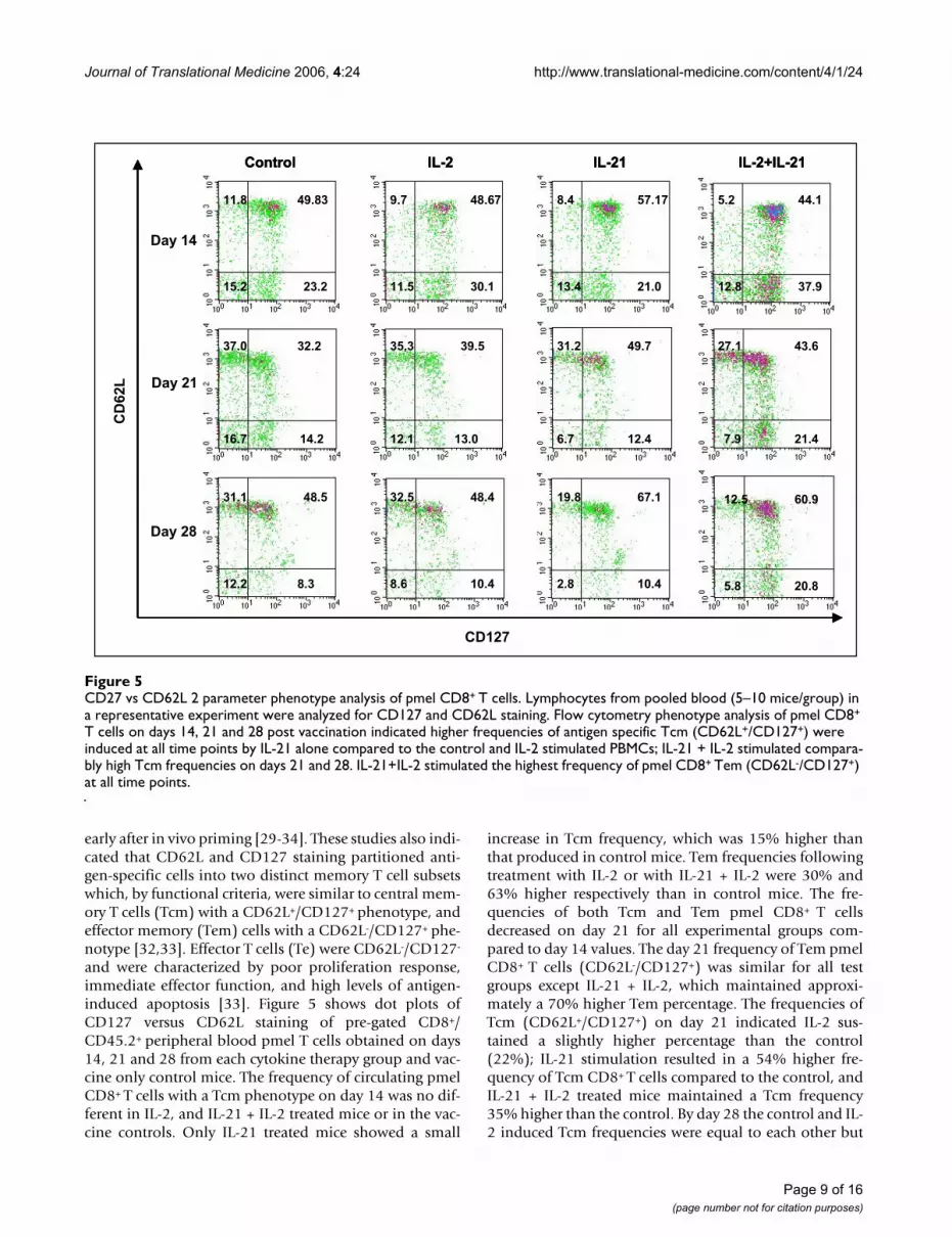
early after in vivo priming [29-34]. These studies also indi-cated that CD62L and CD127 staining partitioned anti-gen-specific cells into two distinct memory T cell subsetswhich, by functional criteria, were similar to central mem-ory T cells (Tcm) with a CD62L+/CD127+ phenotype, andeffector memory (Tem) cells with a CD62L-/CD127+ phe-notype [32,33]. Effector T cells (Te) were CD62L-/CD127-
and were characterized by poor proliferation response,immediate effector function, and high levels of antigen-induced apoptosis [33]. Figure 5 shows dot plots ofCD127 versus CD62L staining of pre-gated CD8+/CD45.2+ peripheral blood pmel T cells obtained on days14, 21 and 28 from each cytokine therapy group and vac-cine only control mice. The frequency of circulating pmelCD8+ T cells with a Tcm phenotype on day 14 was no dif-ferent in IL-2, and IL-21 + IL-2 treated mice or in the vac-cine controls. Only IL-21 treated mice showed a small
increase in Tcm frequency, which was 15% higher thanthat produced in control mice. Tem frequencies followingtreatment with IL-2 or with IL-21 + IL-2 were 30% and63% higher respectively than in control mice. The fre-quencies of both Tcm and Tem pmel CD8+ T cellsdecreased on day 21 for all experimental groups com-pared to day 14 values. The day 21 frequency of Tem pmelCD8+ T cells (CD62L-/CD127+) was similar for all testgroups except IL-21 + IL-2, which maintained approxi-mately a 70% higher Tem percentage. The frequencies ofTcm (CD62L+/CD127+) on day 21 indicated IL-2 sus-tained a slightly higher percentage than the control(22%); IL-21 stimulation resulted in a 54% higher fre-quency of Tcm CD8+ T cells compared to the control, andIL-21 + IL-2 treated mice maintained a Tcm frequency35% higher than the control. By day 28 the control and IL-2 induced Tcm frequencies were equal to each other but
CD27 vs CD62L 2 parameter phenotype analysis of pmel CD8+ T cellsFigure 5CD27 vs CD62L 2 parameter phenotype analysis of pmel CD8+ T cells. Lymphocytes from pooled blood (5–10 mice/group) in a representative experiment were analyzed for CD127 and CD62L staining. Flow cytometry phenotype analysis of pmel CD8+
T cells on days 14, 21 and 28 post vaccination indicated higher frequencies of antigen specific Tcm (CD62L+/CD127+) were induced at all time points by IL-21 alone compared to the control and IL-2 stimulated PBMCs; IL-21 + IL-2 stimulated compara-bly high Tcm frequencies on days 21 and 28. IL-21+IL-2 stimulated the highest frequency of pmel CD8+ Tem (CD62L-/CD127+) at all time points.
Day 21
27.1 43.6
7.9 21.4
37.0 32.2
14.216.7
39.535.3
12.1 13.0
31.2 49.7
6.7 12.4
Day 14
30.111.5
11.8 48.679.7
23.215.2
49.83 44.1
37.912.821.013.4
57.178.4 5.2
Day 28
12.5 60.9
5.8 20.810.42.8
67.119.848.432.5
8.6 10.48.312.2
48.531.1
Control IL-2 IL-21 IL-2+IL-21
CD
62L
CD127
Day 21
27.1 43.6
7.9 21.4
37.0 32.2
14.216.7
39.535.3
12.1 13.0
31.2 49.7
6.7 12.4
Day 21
27.1 43.6
7.9 21.4
37.0 32.2
14.216.7
39.535.3
12.1 13.0
31.2 49.7
6.7 12.4
Day 14
30.111.5
11.8 48.679.7
23.215.2
49.83 44.1
37.912.821.013.4
57.178.4 5.2
Day 14
30.111.5
11.8 48.679.7
23.215.2
49.83 44.1
37.912.821.013.4
57.178.4 5.2
Day 28
12.5 60.9
5.8 20.810.42.8
67.119.848.432.5
8.6 10.48.312.2
48.531.1
Day 28
12.5 60.9
5.8 20.810.42.8
67.119.848.432.5
8.6 10.48.312.2
48.531.1
Control IL-2 IL-21 IL-2+IL-21
CD
62L
CD127
Control IL-2 IL-21 IL-2+IL-21
CD
62L
CD127
Page 9 of 16(page number not for citation purposes)

Journal of Translational Medicine 2006, 4:24 http://www.translational-medicine.com/content/4/1/24
higher than on day 21, while the IL-21 effect (38%increase) and the IL-21 + IL-2 effect (25% increase) onTcm expression were both similarly elevated compared tothe control and IL-2 induced percentages. Combined IL-21 and low-dose IL-2 therapy sustained the circulatingTem frequency at a level that was 2-fold higher than thecontrol group or the effect induced by IL-2 alone or IL-21alone. The cell density plots for IL-21+IL-2 on all three
days also suggested an overall higher absolute number ofcirculating Tcm and Tem CD8+/CD45.2+ T cells than wasinduced by any other treatment. Generally, lower percent-ages of circulating effector cells (CD62L-/CD127-) werepresent at day 21 and day 28 for IL-21 and IL-21+IL-2treated mice compared to both IL-2 treated animals andcontrol mice. The upper left quadrant of each 2-parameterhistogram contained CD62L+/CD127- T cells, and their
Quantitation of absolute numbers of Tem and Tcm CD8+ T cellsFigure 6Quantitation of absolute numbers of Tem and Tcm CD8+ T cells. PBMCs were examined by flow cytometry to determine the absolute number of Tem (CD62L-/CD127+) pmel CD8+ T cells (A), and Tcm (CD62L+/CD127+) pmel CD8+ T cells (B) on days 14, 21, and 28 after vaccination and cytokine treatment. IL-21 + IL-2 supported the highest level of circulating Tem cells on all three days compared to all other experimental groups (p = 0.011), and stimulated the greatest increase of Tcm cells on days 21 and 28 (p = 0.029). Data was collected from pooled blood from ≥ 5 mice in each test group.
0
5
10
15
20
25
30
14 21 28
Tem
Cells
/uL
(CD
62L-/
CD
127+
)Vaccine Control IL-2
IL-21 IL-2 + IL-21
0
10
20
30
40
50
60
14 21 28
Tcm
Ce
lls/u
L
(CD
62
L+
/CD
12
7+
)A
B
Days Post hgp100 Vaccine
0
5
10
15
20
25
30
14 21 28
Tem
Cells
/uL
(CD
62L-/
CD
127+
)Vaccine Control IL-2
IL-21 IL-2 + IL-21
0
10
20
30
40
50
60
14 21 28
Tcm
Ce
lls/u
L
(CD
62
L+
/CD
12
7+
)A
B
Days Post hgp100 Vaccine
Page 10 of 16(page number not for citation purposes)

Journal of Translational Medicine 2006, 4:24 http://www.translational-medicine.com/content/4/1/24
frequency was generally lower in the IL-21 and IL-21+IL-2treated animals. Presently there is no correlated functionaldata to determine the lineage relationship of this popula-tion to Tcm or Tem T cells or their functional properties.Overall the data in Figure 5 suggest IL-21+IL-2 inducedhigher frequencies of Tem cells compared to IL-2 or IL-21alone on all three days; and by the early contraction phaseof the pmel CD8+ T cell response (day 28) IL-21 and IL-21+ low-dose IL-2 induced equally higher percentages oftumor-specific Tcm cells than were stimulated in the con-trol or IL-2 treated mice.
IL-21+ IL-2 induced proliferation of Tem and Tcm pmel CD8+ T cellsThe frequencies of Tcm and Tem T cells on days 14, 21 and28 (Figure 5), and the total number of circulating pmelcells determined for each group (Figure 4) were subse-quently used to calculate the absolute number of Tem(Figure 6A) and Tcm (Figure 6B) pmel CD8+ T cells foreach experimental group. Tem numbers on each day weresignificantly higher for IL-21+ IL-2 treated mice than forall other groups (p = 0.011) as determined by ANCOVA,and declined from day 14 to day 28 (Figure 6A). By con-trast, the absolute number of Tcm cells for all groups wasequally low at day 14 and increased incrementally foreach group over time (Figure 6B). IL-21 + IL-2 inducedTcm cell numbers increased at a significantly higher ratecompared to all other test groups (p = 0.029) as deter-mined by ANCOVA. Thus, IL-21+ IL-2 treatment main-tained Tem absolute numbers at all time points that were
2- to 4-fold greater than those produced by the vaccinecontrol or by IL-2 or IL-21 therapy alone. Similarly, Tcmabsolute numbers induced by IL-21+ IL-2 were 2- to 4-fold higher than those stimulated in any other test groupby day 28. Recent data have demonstrated that the adop-tive transfer of pmel CD8+ Tcm (characterized as CD62L+/CCR7+), or "early effector" T cells (CD62Ldim/CD127+/CCR7+) produced a potent anti-tumor response subse-quent to tumor-antigen hgp10025–33 vaccination andhigh-dose IL-2 therapy in the pmel Tg/B16 therapeuticmodel [22,35]. The data in Figure 6B suggest IL-21+ low-dose IL-2 in vivo therapy promotes the long-term mainte-nance of CD8+ T cells with a similar Tcm (CD62L+/CD127+) or "early effector" – like phenotype (CD62Ldim/-
/CD127+).
Functional phenotype of CD8+ T cells from IL-21 + IL-2 treated tumor regressor miceBy day 42 previously regressing tumors in a subset of IL-21 + IL-2 treated mice began to grow again, reaching amean tumor size of 25–50 mm2. All tumors of this size onday 42–49 continued to progress to ≥ 200 mm2 by day 63,whereupon the mice were sacrificed. However, approxi-mately 50% of all IL-21+ IL-2 treated mice in three exper-iments did not exhibit tumor growth, and by day 42–49manifested complete regression of the tumor. These ani-mals remained tumor free out to >150 days. Day 42 splen-ocytes from two "progressor", and from two "regressor"mice treated with IL-21 + IL-2 were pooled separately, andwere stimulated in vitro with hgp10025–33; cells were then
Cytokine flow cytometry analysis of the functional phenotype of pmel CD8+ splenocytes from IL-21 + IL-2 treated miceFigure 7Cytokine flow cytometry analysis of the functional phenotype of pmel CD8+ splenocytes from IL-21 + IL-2 treated mice. Spleen cells were pooled from 2 mice with progressing tumors (≥ 50 mm2) and from 2 mice with regressing tumors (<10 mm2) 42 days after vaccination. Cytokine flow cytometry analysis shows the absolute number of pmel CD8+ T cells per 106 splenocytes (A), and the frequency of pmel CD8+ cells (B) that are positive for IL-2, IFNγ, and TNFγ production. Data show mice with regressing tumors had 4.7-fold, 4.8-fold and 2.5-fold more IL-2+, IFN-γ+, and TNF-α+ pmel CD8+ T cells respectively, and higher frequencies of Tc1 cytokine+ cells than progressor mice.
Absolute # of Cytokine+ Pmel CD8
+T cells
per 1 x10⁶ Splenocytes
0
2
4
6
8
10
12
IL-2 IFN-gamma TNF-alpha
Cyto
kin
e+/p
me
l+ c
ells
(x1
03) Progressor
Regressor
% of Cytokine+ Pmel-1 CD8 T Cells
0
10
20
30
40
50
60
IL-2 IFN-gamma TNF-alpha
% o
f C
yto
kin
e+ p
me
l-1
CD
8 T
Ce
lls
Progressor
Regressor
A BAbsolute # of Cytokine
+ Pmel CD8
+T cells
per 1 x10⁶ Splenocytes
0
2
4
6
8
10
12
IL-2 IFN-gamma TNF-alpha
Cyto
kin
e+/p
me
l+ c
ells
(x1
03) Progressor
Regressor
% of Cytokine+ Pmel-1 CD8 T Cells
0
10
20
30
40
50
60
IL-2 IFN-gamma TNF-alpha
% o
f C
yto
kin
e+ p
me
l-1
CD
8 T
Ce
lls
Progressor
Regressor
A B
Page 11 of 16(page number not for citation purposes)

Journal of Translational Medicine 2006, 4:24 http://www.translational-medicine.com/content/4/1/24
analyzed for IFNγ, IL-2 and TNF-α production using astandard five-hour CFC assay. After IVS cells were fixed,permeablized, and stained with CD8, CD45.2 andcytokine-specific antibodies. Spleens from regressor micehad more IFNγ, IL-2 and TNF-α positive CD8+/CD45.2+
pmel T cells than spleens from progressor animals (Figure7A). Regressor mice had 4.7-fold, 4.8-fold, and 2.5-foldmore IL-2+, IFNγ+ and TNF-α+ pmel CD8+ T cells, respec-tively, than progressor mice with rapidly growing tumors.The data in Figure 7A indicates that tumor regression inmice receiving IL-21+ IL-2 therapy was associated with anincrease in the absolute number of pmel CD8+ T cells pro-ducing Tc1 cytokines compared to the numbers found inmice with growing tumors. The presence of higher num-bers of Tc1 cytokine+ CD8+ T cells in regressor comparedto progressor mice was attributable to both a 2-foldgreater absolute number of pmel CD8+ T cells in regressormice than were measured in progressor animals (data notshown) and to higher frequencies of pmel CD8+ T cellswhich were positive for Tc1 cytokines in regressor mice at42 days (Figure 7B).
Discussion and conclusionsWe report for the first time the anti-tumor effects of atreatment strategy that combines lymphopenic condition-ing, adoptive transfer of naive antigen-specific CD8+ Tcells, tumor antigen-specific vaccination, and cytokinetreatment with IL-21 + low-dose IL-2. Using a well estab-lished murine model of pre-existing disease, the pmel Tg/B16 model, our results show that combined IL-21 + low-dose IL-2 therapy delayed B16F10 melanoma growth inthe majority of mice, and resulted in a significant increasein tumor-free survival (46%) out to ≥ 150 days comparedto mice treated with IL-21 or IL-2 alone. Surviving miceexhibited tumor-specific protective immunity since theyresisted B16F10 rechallenge but succumbed to the unre-lated 3LL tumor. These data are in contrast to other studiesin the pmel Tg/B16 model, which employed the adoptivetransfer of highly activated IVS pmel CD8+ T cells (stimu-lated with hgp10025–33 peptide and IL-2 or IL-15) ratherthan naive pmel T cells [3,17,18]. After lymphopenic con-ditioning and T-cell transfer to tumor-bearing mice, ther-apy was administered in these studies using differentcombinations of hgp10025–33 vaccination and cytokinetreatment. Thus, Lou et al [17] vaccinated with hgp10025–
33 peptide-pulsed DCs, and administered high-dose IL-2(1.2 × 10⁶ IU/day) for three days beginning the day of vac-cination. Klebanoff, et al [18] vaccinated with a recom-binant fowl pox virus encoding hgp10025–33 (rFPhgp100)after adoptive transfer of IVS pmel CD8+ T cells grown inIL-2 or in IL-15 cultures. Follow-on cytokine therapy con-sisted of high-dose IL-2 (1.2 × 10⁶ IU/day) for three days.In the only published study using IL-21 in the pmel Tg/B16 model Zeng and coworkers [3] also vaccinated withrFPhgp10025–33 following adoptive transfer of IL-2 cul-
tured pmel T cells, and animals were treated over threedays with IL-15 (20 µg/day), IL-21 (20 µg/day), or bothcytokines at the same doses. None of these treatment strat-egies resulted in long-term inhibition of tumor growthbeyond 30–35 days after treatment unless very large num-bers (4–6 × 10⁶) of IVS pmel CD8+ T cells were adoptivelytransferred [17]. Furthermore, in contrast to our results,none of these studies presented data describing long-termtumor-free survival ≥ 150 days with any combination oftransferred T cells, vaccine administration and cytokinetherapy. Another group compared the ability of IL-2, IL-15or IL-21 to augment anti-tumor immunity in C57BL/6mice challenged with the OVA-expressing E.G7 thymoma[6]. Cytokine therapy was initiated 48 hours after tumorinoculation – well before the establishment of vascular-ized tumors, and was administered every other day fortwo weeks thereafter. In this model IL-21 (20 µg/day) didresult in prolonged tumor-free survival out to 100 days inapproximately 25–30% of treated mice. Neither low-doseIL-2 (2 × 10³ IU and 20 × 103 IU/day) nor IL-15 (5 µg and50 µg/day) produced a survival frequency that was ashigh. All animals that survived beyond 100 days were pro-tected from rechallenge with E.G7 tumor cells. Impor-tantly, this report also showed that the 2-week cytokineadministration could begin as late as day 12 followingtumor inoculation and still produce a therapeuticresponse [6]. This study also demonstrated the ability ofIL-21 to augment immunity to the OVA-expressing E.G7tumor in the absence of previously antigen-educatedCD8+ T cells. However, the experimental design did nottest the anti-tumor effects of IL-21 in a model of estab-lished bulky disease such as the pmel Tg/B16 system, inwhich the cognate tumor antigen is a relatively weak self-tumor antigen rather than a strongly immunogenic for-eign protein. To date only the combined therapeuticeffects of IL-21+ low-dose IL-2 reported here have resultedin long-term survival of tumor-free mice in the pmel Tg/B16 model. As noted, in most of the previous studiesusing the pmel Tg/B16 melanoma model very high doses(1.2 × 10⁶ IU/day) of IL-2 were used in combination withthe adoptive transfer of large numbers (up to 107) ofhighly activated IVS pmel CD8+ T cells [16-18] However,treatment was initiated after very large, bulky (50–100mm2) tumors were established. By contrast, cytokine ther-apy was initiated in our experiments when vascularizedtumors were smaller (26 mm2). Long-term survival usingour current IL-21 + IL-2 therapeutic model may decreasewith larger established tumors; experiments designed totest and optimize IL-21 +low-dose IL-2 therapy in micewith larger vascularized tumors are ongoing.
The H-2Db-restricted NV peptide was used in all experi-ments as a putative CD4 "helper" antigen source [23,24]However, our recent experiments suggest NV peptide vac-cination produces only a modest increase in circulating
Page 12 of 16(page number not for citation purposes)

Journal of Translational Medicine 2006, 4:24 http://www.translational-medicine.com/content/4/1/24
pmel CD8+ T cell numbers over that produced byhgp10025–33 alone (unpublished data – H.M. Hu, EACRI).CD4 helper T cell function in our DC-based vaccine sys-tem may have been provided by fetal calf serum (FCS)proteins/peptides associated with the DCs in the vaccine– both DCs and the B16F10 melanoma cells used in thetumor inoculum were cultured in medium containingFCS. The potential for FCS antigen-induced CD4 T cellactivation is also suggested by other hgp10025–33-pulsedDC vaccine studies in the pmel Tg/B16 model in whichanti-tumor therapeutic effect was achieved in the absenceof any known source of CD4-specific antigen [17].
There is limited information on IL-21-mediated changesin the cell surface and functional phenotype of CD8+ T-cells associated with tumor immunity. In this report, weshow that IL-21 alone and IL-21+ low-dose IL-2 increasedthe in vivo frequency of Tcm T cells as defined by concom-itant CD62L+/CD127+ staining[29], compared to the vac-cine only control or to cells from IL-2-treated mice (Figure5). IL-21 + IL-2 treatment also sustained the highest per-centage of Tem (CD62L-/CD127+) pmel CD8+ T cells [29]compared to any other test group. This observation corre-lated with the result that IL-21+ IL-2 therapy also pro-duced the highest absolute number of circulating pmelCD8+ T cells during the beginning (day 14), peak (day 21)and end (day 28) of the hgp10025–33 induced expansionof pmel CD8+ T cells (Figure 4). As a consequence, IL-21 +IL-2 therapy produced the highest absolute number of cir-culating pmel CD8+ Tem cells at each time point of theexpansion and early contraction phases of the anti-hgp10025–33 immune response, and also resulted in thehighest absolute number of Tcm T cells in the peripheralblood (Figure 6). Although Tem absolute numbersdecreased from day 14 to day 28, and Tcm numbersincreased over this same period the data do not indicate ifthis is attributable in anyway to innate differences in IL-21+ IL-2 induced Tem vs Tcm proliferation. Future in vitroand in vivo studies will examine the relative proliferativepotential of cognate antigen-driven purified populationsof pmel CD8+ Tcm and Tem cells. Notably, the tumor-spe-cific protective memory response observed in long-termsurvivor mice was also associated with high frequencies ofTcm in the spleen and lymph nodes. Polychromatic (8color) flow cytometry analysis of pmel CD8+ splenocytesfrom IL-21 + IL-2 treated long-term survivor mice (> 150days) indicated approximately 10%–12% of all CD8+ Tcells in the spleen and lymph nodes were pmel CD8+ Tcells, and 43% and 70% of these T cells in the lymphnodes and spleen respectively expressed a Tcm phenotype(CD62L+/CD127+/CD27+/CD28+)(unpublished data).Recent data have demonstrated that enriched pmel CD8+
Tcm T cells conferred a potent in vivo anti-tumor recallresponse upon adoptive transfer – leading to the eradica-tion of large established tumors in the pmel Tg/B16
model [35]. Thus, as described herein, IL-21 + IL-2 ther-apy favored the in vivo expansion and maintenance ofsuch tumor antigen-specific Tcm T cells from naive pmelCD8+ T cell precursors which were capable of providinglong-term protective immunity. Preliminary phenotypeanalysis of IL-21 + low-dose IL-2 and hgp10025–33 IVSpmel splenocytes similarly indicates the combination ofboth cytokines results in the proliferation of pmel TgCD8+ T cells with a Tcm (CD62L+/CD127+/CD27+/CD28+) phenotype (unpublished data – EACRI). This fur-ther suggests that IL-21 + low-dose IL-2 might be effectivein supporting the cognate antigen-driven in vitro expan-sion of rare self-tumor antigen Tcm CD8+ T cells fromautologous PBMCs – perhaps in concert with proceduresto remove tumor antigen-specific regulatory T cell effects.In addition to increasing tumor-antigen directed expan-sion and maintenance of Tcm CD8+ T cells, data presentedhere demonstrates that IL-21 + IL-2 therapy also stimu-lated the expression of anti-tumor memory CD8+ T cellswith a Tc1 cytokine functional phenotype in mice withregressing tumors. By day 42 post-tumor inoculation IL-21 + IL-2 treated tumor-regressor mice had much higherabsolute numbers of IL-2+, IFNγ+ and TNF-α+ pmel CD8+
splenocytes than IL-21 + IL-2 treated tumor-progressormice with rapidly growing tumors (Figure 7). The relativeabsence of CD8+ splenocytes with a Tc1 cytokine func-tional phenotype in tumor progressor mice may be attrib-utable in part to a simple lack of sustained B16F10 tumorantigen stimulation. Previous published data from ourinstitute using the pmel Tg/B16 model has demonstratedloss of MHC class I and gp100 expression in a high per-centage of tumors in experimental animals with progres-sive disease due to immunoediting[36]. Overall our datasuggest that IL-21 + low-dose IL-2-mediated tumor regres-sion and long-term survival was associated with elevatedabsolute numbers of tumor antigen-specific Tem and TcmCD8+ T cells with a Tc1 functional phenotype.
IL-21 + low-dose IL-2 induction of increased in vivoexpression of Tc1 cytokine+ Tcm and Tem CD8+ T cells is acentral observation for any putative mechanistic explana-tion of the anti-tumor therapeutic synergy produced usingboth cytokines. This observation examined in the contextof recent studies describing the broad inhibitory effects ofIL-21 on B lymphocytes, NK cells and DCs (Reviewed in[37]) suggests a possible mechanism for the IL-21+ low-dose IL-2 induced expansion of Tcm CD8+ T cells similarto what may occur during the late contraction phase of anongoing immune response. Thus, CD4 T cell (Th2)-derived IL-21[38] inhibits anti-IgM and IL-4-mediated Bcell proliferation [2] and enhances apoptosis of activatedB cells [39,40], inhibits DC maturation and the ability ofDCs to prime T cells [41,42]; and increases NK maturationand cytolytic function while inhibiting NK proliferation[43]. Similarly, while IL-21 has generally been described
Page 13 of 16(page number not for citation purposes)

Journal of Translational Medicine 2006, 4:24 http://www.translational-medicine.com/content/4/1/24
as an inducer of antigen stimulated IFNγ and CTL func-tion in CD8+ T cells [4,8,44,45], our unpublished data inboth murine and human in vitro experiments indicate IL-21 alone does not drive high levels of cognate antigenstimulated CTL proliferation. In vivo data presented heresuggest that IL-21 works best in concert with low-dose IL-2 to support both optimal anti-tumor effector CTL func-tion and expansion of CD8+ memory T cells. Paradoxi-cally, IL-21 + low-dose IL-2 treatment producedunexpectedly low frequencies of circulating effector pmelCD8+ T cells (Figure 4) – perhaps due to cell trafficking tothe primary tumor and sites of metastatic disease. Ongo-ing experiments are directed at the in situ analysis of thephenotype, cell number, and functional properties oftumor invasive pmel CD8+ T cells in IL-21 + low dose IL-2 treated mice. Such studies should shed light on whethercombined cytokine therapy induces trafficking ofincreased numbers of antigen-specific cytolytic CD8+ Tcells to the tumor site. Taken together, these observationssuggest a dual role for IL-21 in dampening ongoing innateand adaptive immune responses (as might occur duringthe contraction phase of antigen-specific immunity),while concomitantly augmenting the expansion of anti-gen-driven long-term memory T cells. Recently publisheddata demonstrate IL-21 treated cultures of antigen-stimu-lated cells produce CD8+ T cells with increased TCR bind-ing affinity [5]. Other data show the IL-21 receptor (IL-21R) is upregulated upon TCR engagement [46,47]. Thus,Th2-derived IL-21 may "rescue" memory T cells by block-ing further antigen-driven effector T cell differentiationthrough presently unknown mechanisms, while simulta-neously increasing antigen binding affinity. The resultingincrease in binding of low-levels of residual antigen(present during the contraction phase) could increase IL-21R expression and up-regulate increased IL-21 bindingto memory T cells – thus further skewing the shift to mem-ory T cells. Memory CD8+ T cells in turn express amplifiedlevels of CD122 [48], which may facilitate increased highaffinity IL-2R formation if other IL-2R chains (CD25 andCD132) are available, and potentially increase binding ofthe low levels of IL-2 which may be present during immu-nological contraction. The combination of the "arrested"Tcm memory phenotype (maintained by IL-21), and lowdose IL-2-induced proliferation of these cells could resultin increased Tcm cell numbers. Thus, IL-21 + low-dose IL-2 therapy may favor the enhanced expansion of long-termmemory (Tcm) CD8+ T cells through augmented memoryT cell "rescue" and expansion mechanisms similar tothose which may normally be present during the late con-traction phase of an ongoing immune response. This con-cept is supported by the results of our study and otherreports [6,8], which suggest IL-21 induced tumor immu-nity is most effective when IL-21 is administered severaldays (4–12 days) after initial tumor-antigen activation ofT cells. The late acting anti-tumor effects of IL-21, and, in
our study, IL-21 + IL-2 therapy suggests IL-21 may be act-ing directly on tumor antigen educated CD8+ T cells ratherthan modulating early APC and/or T cell function associ-ated with primary tumor antigen stimulation. Preliminaryin vitro studies with purified naïve and hgp10025–33 stim-ulated pmel CD8+ T cells support this conclusion (datanot shown).
In summary, the results of this study suggest that IL-21 +low-dose IL-2 cytokine therapy may "rescue" and aug-ment proliferation of tumor antigen-specific memoryCD8+ T cells, and thus provide an important new strategiccomponent for effective cancer immunotherapy. Thecombined use of both cytokines may be most effective inthe context of lymphopenic conditioning, tumor antigen-specific vaccination, and the adoptive transfer of autolo-gous tumor-specific Tcm or "early" effector T cells. Ongo-ing work at our institute continues to focus on developingprocedures for the IL-21 + low-dose IL-2-directed in vivoand in vitro expansion and maintenance of such Tcm and"early" effector subpopulations from small numbers ofnormally tolerized autologous self-tumor antigen-specificprecursors.
Competing interestsChristopher Clegg is a scientist employed by Zymogenet-ics – all other authors declare that they have no competinginterests.
Authors' contributionsHH designed the in vivo experiments and worked withPW and DH to perform the flow cytometry analysis ofPBMC and splenocyte samples. GY assisted in all aspectsof tumor inoculation, lymphopenic conditioning, tumormeasurement and tissue harvest. HMH, CHC, and AOHprovided key reagents and assisted in the design of con-trols which utilized these reagents in the experiments.WM was directly involved in drafting and revising themanuscript, and WGA performed all the statistical analy-sis. EBW designed and supervised all aspects of the exper-imental strategy and acquisition of data, and, with BAFand WJU, was involved in data interpretation and the crit-ical review and revision of the manuscript.
AcknowledgementsThis study was supported in part by NIH grant 1 R21CA101325-02 and by grants from the Providence Portland Medical Foundation, and the M.J. Mur-dock Charitable Trust, and the Chiles Foundation.
References1. Leonard WJ, Spolski R: Interleukin-21: a modulator of lymphoid
proliferation, apoptosis and differentiation. Nat Rev Immunol2005, 5(9):688-698.
2. Parrish-Novak J, Dillon SR, Nelson A, Hammond A, Sprecher C,Gross JA, Johnston J, Madden K, Xu W, West J, Schrader S, BurkheadS, Heipel M, Brandt C, Kuijper JL, Kramer J, Conklin D, Presnell SR,Berry J, Shiota F, Bort S, Hambly K, Mudri S, Clegg C, Moore M, GrantFJ, Lofton-Day C, Gilbert T, Rayond F, Ching A, Yao L, Smith D, Web-
Page 14 of 16(page number not for citation purposes)

Journal of Translational Medicine 2006, 4:24 http://www.translational-medicine.com/content/4/1/24
ster P, Whitmore T, Maurer M, Kaushansky K, Holly RD, Foster D:Interleukin 21 and its receptor are involved in NK cell expan-sion and regulation of lymphocyte function. Nature 2000,408(6808):57-63.
3. Zeng R, Spolski R, Finkelstein SE, Oh S, Kovanen PE, Hinrichs CS,Pise-Masison CA, Radonovich MF, Brady JN, Restifo NP, Berzofsky JA,Leonard WJ: Synergy of IL-21 and IL-15 in regulating CD8+ Tcell expansion and function. J Exp Med 2005, 201(1):139-148.
4. Kasaian MT, Whitters MJ, Carter LL, Lowe LD, Jussif JM, Deng B,Johnson KA, Witek JS, Senices M, Konz RF, Wurster AL, DonaldsonDD, Collins M, Young DA, Grusby MJ: IL-21 limits NK cellresponses and promotes antigen-specific T cell activation: amediator of the transition from innate to adaptive immu-nity. Immunity 2002, 16(4):559-569.
5. Li Y, Bleakley M, Yee C: IL-21 influences the frequency, pheno-type, and affinity of the antigen-specific CD8 T cell response.J Immunol 2005, 175(4):2261-2269.
6. Moroz A, Eppolito C, Li Q, Tao J, Clegg CH, Shrikant PA: IL-21enhances and sustains CD8+ T cell responses to achievedurable tumor immunity: comparative evaluation of IL-2, IL-15, and IL-21. J Immunol 2004, 173(2):900-909.
7. Ugai S, Shimozato O, Kawamura K, Wang YQ, Yamaguchi T, SaishoH, Sakiyama S, Tagawa M: Expression of the interleukin-21 genein murine colon carcinoma cells generates systemic immu-nity in the inoculated hosts. Cancer Gene Ther 2003,10(3):187-192.
8. Ma HL, Whitters MJ, Konz RF, Senices M, Young DA, Grusby MJ, Col-lins M, Dunussi-Joannopoulos K: IL-21 activates both innate andadaptive immunity to generate potent antitumor responsesthat require perforin but are independent of IFN-gamma. JImmunol 2003, 171(2):608-615.
9. Kishida T, Asada H, Itokawa Y, Cui FD, Shin-Ya M, Gojo S, YasutomiK, Ueda Y, Yamagishi H, Imanishi J, Mazda O: Interleukin (IL)-21and IL-15 genetic transfer synergistically augments thera-peutic antitumor immunity and promotes regression ofmetastatic lymphoma. Mol Ther 2003, 8(4):552-558.
10. Overwijk WW, Tsung A, Irvine KR, Parkhurst MR, Goletz TJ, TsungK, Carroll MW, Liu C, Moss B, Rosenberg SA, Restifo NP: gp100/pmel 17 is a murine tumor rejection antigen: induction of"self"-reactive, tumoricidal T cells using high-affinity, alteredpeptide ligand. J Exp Med 1998, 188(2):277-286.
11. Ku CC, Murakami M, Sakamoto A, Kappler J, Marrack P: Control ofhomeostasis of CD8+ memory T cells by opposing cytokines.Science 2000, 288(5466):675-678.
12. Waldmann TA, Dubois S, Tagaya Y: Contrasting roles of IL-2 andIL-15 in the life and death of lymphocytes: implications forimmunotherapy. Immunity 2001, 14(2):105-110.
13. Murakami M, Sakamoto A, Bender J, Kappler J, Marrack P:CD25+CD4+ T cells contribute to the control of memoryCD8+ T cells. Proc Natl Acad Sci USA 2002, 99(13):8832-8837.
14. Refaeli Y, Van Parijs L, London CA, Tschopp J, Abbas AK: Biochem-ical mechanisms of IL-2-regulated Fas-mediated T cell apop-tosis. Immunity 1998, 8(5):615-623.
15. Manjunath N, Shankar P, Wan J, Weninger W, Crowley MA, HieshimaK, Springer TA, Fan X, Shen H, Lieberman J, von Andrian UH: Effec-tor differentiation is not prerequisite for generation of mem-ory cytotoxic T lymphocytes. J Clin Invest 2001, 108(6):871-878.
16. Overwijk WW, Theoret MR, Finkelstein SE, Surman DR, de Jong LA,Vyth-Dreese FA, Dellemijn TA, Antony PA, Spiess PJ, Palmer DC,Heimann DM, Klebanoff CA, Yu Z, Hwang LN, Feigenbaum L, Kruis-beek AM, Rosenberg SA, Restifo NP: Tumor regression andautoimmunity after reversal of a functionally tolerant stateof self-reactive CD8+ T cells. J Exp Med 2003, 198(4):569-580.
17. Lou Y, Wang G, Lizee G, Kim GJ, Finkelstein SE, Feng C, Restifo NP,Hwu P: Dendritic cells strongly boost the antitumor activityof adoptively transferred T cells in vivo. Cancer Res 2004,64(18):6783-6790.
18. Klebanoff CA, Finkelstein SE, Surman DR, Lichtman MK, Gattinoni L,Theoret MR, Grewal N, Spiess PJ, Antony PA, Palmer DC, Tagaya Y,Rosenberg SA, Waldmann TA, Restifo NP: IL-15 enhances the invivo antitumor activity of tumor-reactive CD8+ T cells. ProcNatl Acad Sci USA 2004, 101(7):1969-1974.
19. Yee C, Thompson JA, Byrd D, Riddell SR, Roche P, Celis E, GreenbergPD: Adoptive T cell therapy using antigen-specific CD8+ Tcell clones for the treatment of patients with metastaticmelanoma: in vivo persistence, migration, and antitumor
effect of transferred T cells. Proc Natl Acad Sci USA 2002,99(25):16168-16173.
20. Dudley ME, Wunderlich J, Nishimura MI, Yu D, Yang JC, Topalian SL,Schwartzentruber DJ, Hwu P, Marincola FM, Sherry R, Leitman SF,Rosenberg SA: Adoptive transfer of cloned melanoma-reac-tive T lymphocytes for the treatment of patients with meta-static melanoma. J Immunother 2001, 24(4):363-373.
21. Dudley ME, Wunderlich JR, Yang JC, Hwu P, Schwartzentruber DJ,Topalian SL, Sherry RM, Marincola FM, Leitman SF, Seipp CA, Rogers-Freezer L, Morton KE, Nahvi A, Mavroukakis SA, White DE, Rosen-berg SA: A phase I study of nonmyeloablative chemotherapyand adoptive transfer of autologous tumor antigen-specific Tlymphocytes in patients with metastatic melanoma. J Immu-nother 2002, 25(3):243-251.
22. Gattinoni L, Klebanoff CA, Palmer DC, Wrzesinski C, Kerstann K, YuZ, Finkelstein SE, Theoret MR, Rosenberg SA, Restifo NP: Acquisi-tion of full effector function in vitro paradoxically impairs thein vivo antitumor efficacy of adoptively transferred CD8+ Tcells. J Clin Invest 2005, 115(6):1616-1626.
23. Nardin EH, Nussenzweig RS: T cell responses to pre-erythro-cytic stages of malaria: role in protection and vaccine devel-opment against pre-erythrocytic stages. Annu Rev Immunol1993, 11:687-727.
24. Munesinghe DY, Clavijo P, Calle MC, Nussenzweig RS, Nardin E:Immunogenicity of multiple antigen peptides (MAP) con-taining T and B cell epitopes of the repeat region of the P.falciparum circumsporozoite protein. Eur J Immunol 1991,21(12):3015-3020.
25. Lutz MB, Kukutsch N, Ogilvie AL, Rossner S, Koch F, Romani N,Schuler G: An advanced culture method for generating largequantities of highly pure dendritic cells from mouse bonemarrow. J Immunol Methods 1999, 223(1):77-92.
26. Crepeau H, Koziol J, Reid N, Yuh YS: Analysis of incomplete mul-tivariate data from repeated measurement experiments.Biometrics 1985, 41:505-514.
27. Kozial J, Maxwell DA, Fukushima M, Colmeraurer M, Pilch YH: A dis-tribution-free test fro tumor-growth curve analyses withapplication to an animal tumor immuntherapy experiment.Biometrics 1981, 37:383-390.
28. Milliken G, Johnson DE: Analysis of Messy Data. Volume I.Designed Experiments edn. New York; 1991.
29. Powell DJ Jr, Dudley ME, Robbins PF, Rosenberg SA: Transition oflate-stage effector T cells to CD27+ CD28+ tumor-reactiveeffector memory T cells in humans after adoptive cell trans-fer therapy. Blood 2005, 105(1):241-250.
30. Schluns KS, Kieper WC, Jameson SC, Lefrancois L: Interleukin-7mediates the homeostasis of naive and memory CD8 T cellsin vivo. Nat Immunol 2000, 1(5):426-432.
31. Bradley LM, Haynes L, Swain SL: IL-7: maintaining T-cell memoryand achieving homeostasis. Trends Immunol 2005, 26(3):172-176.
32. Huster KM, Busch V, Schiemann M, Linkemann K, Kerksiek KM, Wag-ner H, Busch DH: Selective expression of IL-7 receptor onmemory T cells identifies early CD40L-dependent genera-tion of distinct CD8+ memory T cell subsets. Proc Natl Acad SciUSA 2004, 101(15):5610-5615.
33. Bachmann MF, Wolint P, Schwarz K, Jager P, Oxenius A: Functionalproperties and lineage relationship of CD8+ T cell subsetsidentified by expression of IL-7 receptor alpha and CD62L. JImmunol 2005, 175(7):4686-4696.
34. Kaech SM, Tan JT, Wherry EJ, Konieczny BT, Surh CD, Ahmed R:Selective expression of the interleukin 7 receptor identifieseffector CD8 T cells that give rise to long-lived memorycells. Nat Immunol 2003, 4(12):1191-1198.
35. Klebanoff CA, Gattinoni L, Torabi-Parizi P, Kerstann K, Cardones AR,Finkelstein SE, Palmer DC, Antony PA, Hwang ST, Rosenberg SA,Waldmann TA, Restifo NP: Central memory self/tumor-reac-tive CD8+ T cells confer superior antitumor immunity com-pared with effector memory T cells. Proc Natl Acad Sci USA 2005,102(27):9571-9576.
36. Wang LX, Li R, Yang G, Lim M, O'Hara A, Chu Y, Fox BA, Restifo NP,Urba WJ, Hu HM: Interleukin-7-dependent expansion and per-sistence of melanoma-specific T cells in lymphodepletedmice lead to tumor regression and editing. Cancer Res 2005,65(22):10569-10577.
37. Mehta DS, Wurster AL, Grusby MJ: Biology of IL-21 and the IL-21 receptor. Immunol Rev 2004, 202:84-95.
Page 15 of 16(page number not for citation purposes)

Journal of Translational Medicine 2006, 4:24 http://www.translational-medicine.com/content/4/1/24
Publish with BioMed Central and every scientist can read your work free of charge
"BioMed Central will be the most significant development for disseminating the results of biomedical research in our lifetime."
Sir Paul Nurse, Cancer Research UK
Your research papers will be:
available free of charge to the entire biomedical community
peer reviewed and published immediately upon acceptance
cited in PubMed and archived on PubMed Central
yours — you keep the copyright
Submit your manuscript here:http://www.biomedcentral.com/info/publishing_adv.asp
BioMedcentral
38. Wurster AL, Rodgers VL, Satoskar AR, Whitters MJ, Young DA, Col-lins M, Grusby MJ: Interleukin 21 is a T helper (Th) cell 2cytokine that specifically inhibits the differentiation of naiveTh cells into interferon gamma-producing Th1 cells. J ExpMed 2002, 196(7):969-977.
39. Mehta DS, Wurster AL, Whitters MJ, Young DA, Collins M, GrusbyMJ: IL-21 induces the apoptosis of resting and activated pri-mary B cells. J Immunol 2003, 170(8):4111-4118.
40. Suto A, Nakajima H, Hirose K, Suzuki K, Kagami S, Seto Y, HoshimotoA, Saito Y, Foster DC, Iwamoto I: Interleukin 21 prevents anti-gen-induced IgE production by inhibiting germ line C(epsi-lon) transcription of IL-4-stimulated B cells. Blood 2002,100(13):4565-4573.
41. Brandt K, Bulfone-Paus S, Jenckel A, Foster DC, Paus R, Ruckert R:Interleukin-21 inhibits dendritic cell-mediated T cell activa-tion and induction of contact hypersensitivity in vivo. J InvestDermatol 2003, 121(6):1379-1382.
42. Brandt K, Bulfone-Paus S, Foster DC, Ruckert R: Interleukin-21inhibits dendritic cell activation and maturation. Blood 2003,102(12):4090-4098.
43. Brady J, Hayakawa Y, Smyth MJ, Nutt SL: IL-21 induces the func-tional maturation of murine NK cells. J Immunol 2004,172(4):2048-2058.
44. van Leeuwen EM, Gamadia LE, Baars PA, Remmerswaal EB, ten BergeIJ, van Lier RA: Proliferation requirements of cytomegalovirus-specific, effector-type human CD8+ T cells. J Immunol 2002,169(10):5838-5843.
45. Di Carlo E, Comes A, Orengo AM, Rosso O, Meazza R, Musiani P,Colombo MP, Ferrini S: IL-21 induces tumor rejection by spe-cific CTL and IFN-gamma-dependent CXC chemokines insyngeneic mice. J Immunol 2004, 172(3):1540-1547.
46. Parrish-Novak J, Foster DC, Holly RD, Clegg CH: Interleukin-21and the IL-21 receptor: novel effectors of NK and T cellresponses. J Leukoc Biol 2002, 72(5):856-863.
47. Collins M, Whitters MJ, Young DA: IL-21 and IL-21 receptor: anew cytokine pathway modulates innate and adaptive immu-nity. Immunol Res 2003, 28(2):131-140.
48. Goldrath AW, Bogatzki LY, Bevan MJ: Naive T cells transientlyacquire a memory-like phenotype during homeostasis-driven proliferation. J Exp Med 2000, 192(4):557-564.
Page 16 of 16(page number not for citation purposes)


