Embed Size (px)
Citation preview

Int J ThermophysDOI 10.1007/s10765-014-1773-3
Combined Photoacoustic Ultrasound and BeamDeflection Signal Monitoring of Gold NanoparticleAgglomerate Concentrations in Tissue Phantoms Usinga Pulsed Nd:YAG Laser
Mohammad E. Khosroshahi · Andreas Mandelis
Received: 2 December 2013 / Accepted: 17 October 2014© Springer Science+Business Media New York 2014
Abstract The purpose of this paper is to show and discuss the effects of the goldnanoparticle (Au-NPs) concentration inside a tissue phantom using a combined sys-tem of photoacoustics (PA) and optical beam deflection and their applications par-ticularly to photoacoustic imaging. It was found that the PA signal from aggregatedAu nanoparticles is significantly enhanced. The stock concentration of 100 nm Au-NPs was 3.8 × 10⁹ particles/mL from which three samples with 30 %, 70 %, and90 % concentration were prepared using polyvinyl chloride-plastisol. Each samplewas then irradiated across a line scan using a 10 ns pulsed Q-switched Nd:YAG laserat a 1 Hz repetition rate and 5 W · cm−2 so that no physical ablation was observed.The corresponding photoacoustic pressure was found to approximately cover a rangebetween 10 kPa and 51 kPa. This corresponds to approximately 130 pJ to 315 pJof acoustic energy radiated by Au-NPs into the tissue. The maximal efficacy of thetransformation of optical energy into thermal energy was ∼29 %. Time-resolved pho-toacoustic deflection was also used to monitor the laser-interaction process. The resultsclearly indicated that (i) the photoacoustic signal amplitude varies in a given sampleas a result of the non-uniform concentration distribution of embedded Au-NPs; (ii) anincrease of the concentration increased the signal amplitude linearly; and (iii) at highernanoparticle concentrations, the probe deflection was found to increase due to a steeperthermoelastic gradient as a result of a higher absorption by particle agglomerates andparticle size-dependent dispersions.
M. E. Khosroshahi · A. MandelisCenter for Advanced Diffusion-Wave Technologies, Department of Mechanicaland Industrial Engineering, University of Toronto, Toronto, ON M5S 3G8, Canada
M. E. Khosroshahi (B)Faculty of Biomedical Engineering, Biomaterial Group, Laser & NanobiophotonicsLaboratory, Amirkabir University of Technology, Tehran, Irane-mail: [email protected]
123

Int J Thermophys
Keywords Biomedical photoacoustics · Gold nanoparticles · Nd:YAG laser ·Photoacoustic beam deflection
1 Introduction
In recent years growing interest has been shown in developing new techniques for thenon-invasive monitoring and imaging of biomedical structures and tissues. Opticalscattering in soft tissue degrades the resolution significantly with depth while ultra-sound can provide a better resolution than optical means for depths greater than about1 mm. Thus, the combination of high optical absorption contrast and high ultrasonicspatial resolution (low scattering) makes it a very useful imaging technique. Basically,photoacoustics (PA) is a material probing modality in which the absorption of inci-dent pulsed laser radiation leads to impulsive heating of the irradiated tissue volume,followed by rapid thermoelastic expansion and subsequent generation of broadbandultrasonic thermoelastic waves [1]. Equally, the photothermal deflection (PTD) isbased on the localized heating of a sample by a focused laser source acting as a “ther-mal piston.” Rapid heating is then transferred to air molecules in the vicinity of thesurrounding gas producing a temperature gradient field which is effective when thebeam passing through this heated region is deflected by the thermally modulated indexof the refraction gradient. The amplitude and phase of the deflected beam carry someinformation about optical and thermophysical properties of the solid or liquid, therebyenabling a number of biomedical applications by both techniques [2,3]. In this manner,one can obtain a better contrast and spatial resolution of tissue images [4]. Despitemuch valuable research work regarding PA imaging [5–7], a further enhancement ofphotoacoustic (or optoacoustic) imaging contrast would be necessary for the earlydetection of cancer at deep subsurface locations. Gold nanoparticles (Au-NPs) exhibitunique optical properties, namely, strong localized surface plasmon resonance (LSPR)which is defined as a collective and coherent oscillation of conduction electrons whenexcited by an external source of an electromagnetic field. As a consequence of the plas-mon oscillation, a dipolar is generated with a huge enhancement of the local electricfield at the nanoparticles surface. This electric field leads to strong light absorption andscattering at the SPR frequency by the particle [8–10], and their major advantages arebiocompatibility due to their inert surface, nontoxicity, surface conjugation chemistry,and lack of photobleaching or blinking as with quantum dots [11,12]. Besides, Au-NPsare relatively simple to synthesize, and they are photostable and can be easily conju-gated with proteins, antibodies, and specific cancer ligands [13,14]. Thus, they havebeen chosen for bioimaging [15,16], mainly due to their ability to convert absorbedlight into heat (i.e., PA efficiency) [17], drug delivery [18,19], cancer cell diagnosisand therapeutics [20,21], and laser tissue welding and soldering [22,23]. Above all,since cellular uptake and endocytosis of particles results in their aggregation, it also hasa significant impact on the application of plasmonic metal nanoparticles for molecularimaging. The goal of this paper is to study the effects of the gold NP concentration onPA signals using a Q-switched pulsed Nd:YAG laser and simultaneously monitoringthe interaction process based on photoacoustic deflection signals.
123

Int J Thermophys
Fig. 1 Experimental setup for simultaneous PA ultrasound and laser beam deflection measurements
2 Experimental
2.1 Materials
To study the effects of the Au-NP concentration on photoacoustic signals, a tissuephantom made of polyvinyl chloride-plastisol (PVCP), a non-toxic plastic synthesizedfrom chloride monomers and soluble in water was purchased from M-F ManufacturingCo., Fort Worth, TX, USA. PVCP is a viscoelastic material and its creep deformation isvery low compared with other plastics due to its limited molecular motion at ordinarytemperatures. For these types of materials, the relationship between stress and straindepends on time and the stiffness will depend on the rate of the applied load. Inaddition, the mechanical energy is dissipated by conversion of heat in the deformationof viscoelastic materials. The solution is an oil-based liquid and was uniformly heatedand stirred continuously using a magnetic stirrer up to ≈200 ◦C in order to avoidstructural and optical inhomogeneities and then allowed to cool. It has no or verynegligible optical absorption at a 1.06 µm wavelength of an Nd:YAG laser and hasa similar speed of sound (1400 m · s−1) and density as tissue and makes a suitablecandidate for modeling tissue biomedical applications [24]. A 25 mL gold nanoparticle(Au-NP) source with 100 nm diameter and a concentration of 3.8 × 10⁹ particles/mL(i.e., 9.5 × 10¹⁰ particles) stabilized as a suspension in a citrate buffer was purchasedfrom Sigma-Aldrich. Three samples were prepared by injecting 0.3 mL, 0.7 mL, and0.9 mL of Au-NPs in 1 cm3 of PVCP solution. Upon cooling, the solution was solidifiedand was easily removed from the container.
2.2 Method
The experimental setup is shown in Fig. 1. Each sample was irradiated using a 10 nspulsed Q-switched Nd:YAG laser (Continum-Surelite) at a 1.064 µm wavelengthwith a 1 mm collimated pulse at 1 Hz and 5 W · cm−2 intensity. The thermoelasticsignals were detected by a 2.2 MHz focused transducer (V305,Olympus NDT Inc.,Panametrics) with 18.8 mm diameter and 25 mm focal length, and were then recordedwith a fast digital oscilloscope (Tektronix-DPO 7104-1 GHz).
123

Int J Thermophys
A 2 mW He-Ne laser (632 nm) was used as a probe beam for photoacoustic deflec-tion measurements. The beam was focused with a lens of 100 mm focal length to adiameter of about 0.5 mm. The dependence of the photodiode response �V on thebeam deflection is [25]
�V = V0 erf[21/2ϕ/θ
](1)
with ϕ and θ being the beam angular deflection due to changes in refraction andangular divergence, respectively. Also, the thermally or PA pressure-induced opticaldeflection, ϕ, is directly related to the rate of change of the refractive index and thetemperature
ϕ = 1
n
∂n
∂T
∂T
∂zL , (2)
where n is the refractive index, T is the temperature, and L is the probe beam path.It is interesting to note that since pure plastisol (i.e., without impurities) acts as aweakly absorbing material for an Nd:YAG laser, the temperature in Eq. 1 is mainlydue to absorption by Au-NPs. The temperature of a single NP is given by Eq. 3where it increases linearly with absorbed power but is inversely related to the mediumthermal conductivity, Kp(=Dpρpcp) with ρp, cp, and Dp being the density, specificheat capacity, and the thermal diffusivity of the NP, respectively [26].
�T = P
4π Rp Kp(3)
where P is the laser power and Rp is the NP radius. Thus, the larger is the sphere,the longer it takes for heat to diffuse or transfer to the surrounding medium (i.e., itcools slowly at a longer time). In fact, both PA and PTD are observable with the samesetup except that PA deflections occur on a much earlier time scale. The output signalwas then registered using a Si-based photodiode (Thorlabs-DET10A) with a spectralsensitivity between 200 nm and 1100 nm. In combining PA ultrasound detection with aconventional transducer and PA beam deflection, some additional sample informationcan be obtained, such as the sound velocity, elasticity, temperature, flow velocity, ther-mal diffusivity, and thickness. In the case of viscoelasticity, if we assume a knowledgeof the tensile stress, σ , and differentiating it with respect to x ,
σ = Ey[ε0 − βT (x, t)]∂σ
∂x= Ey
[∂ε
∂x− β
∂T
∂t(x, t)
], (4)
where Ey = σ/ε0 is the Young’s modulus, ε0 = ∂u/∂x is the strain or the displacement
of a particle in the x-direction, β = �PCp
c2a αF
represents the volumetric thermal expansion,
�P is the pressure increase due to volume expansion, Cp is the specific heat capacity,
123

Int J Thermophys
ca is the acoustic velocity in the material, α is the material absorption coefficient, andF is the laser fluence. Since T (x, t) = α
∫ t0
I dtρCp
∂T
∂x= α
ρCp
∂∫ t
0 I dt
∂x= 1
Cp∂W/∂x (5)
where W = αρCp
∫ t0 I dt is the absorbed energy per unit volume. Using Newton’s
second law of motion, we obtain
∂2u
∂t2 = 1
ρ
∂σ
∂x(6)
Substituting Eqs. 4 and 5 into Eq. 6 and simplifying,
∂2u
∂x2 − 1
ca
∂2u
∂t2 = β
Cp
∂W
∂x
∂2u
∂x2 − 1
ca
∂2u
∂t2 = �P
c2a
1
αF
∂W
∂x. (7)
It can be seen from Eqs. 5 and 7 that the rate of change of temperature is directlyrelated to the absorption coefficient of the material and the laser intensity and, hence,to the optical deflection, ϕ. Secondly, for an unknown material, the value of (Ey/ρ)1/2
can be deduced using the experimental value of the acoustic propagation velocity (i.e.,ca = (Ey/ρ)1/2).
3 Results and Discussion
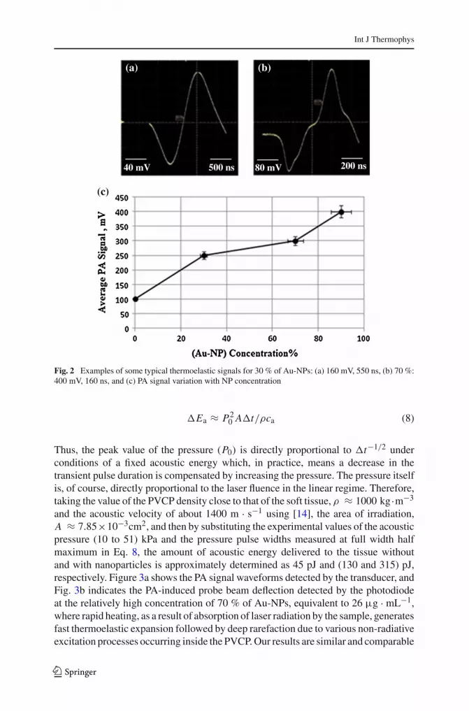
The amplitude of our bipolar thermoelastic signals increased approximately linearlywith increasing Au-NP concentration, as expected from linear photoacoustic theory.Some examples of PA thermoelastic responses are shown in Fig. 2a and b. The increas-ing trend of the average PA amplitude with Au concentration at a constant powerdensity is illustrated in Fig. 2c. The peak output voltage from the transducer can beconverted to a corresponding normal force and hence to a pressure (= F/A, Pa) if theirradiated area, A, is known. From the known voltage amplitude, V , and other con-stants of the PZT transducer, the corresponding values of the average photoacousticpressure can be found using P = CV (t)/dt A where C = (Cl + Cd ≈ 10−9F) isthe sum of load and transducer capacitances and dt ≈ 10−12 pC · N−1 is the strainconstant. Thus, it was found from the measured amplitudes that the correspondingcalculated acoustic pressure covered the range between 10 kPa and 51 kPa.
The narrowing of the pressure transient FWHM,�t, with concentration at a constantpower density, Fig. 2b, can be explained by considering the simple relation (Eq. 8)below which relates the acoustic energy, �Ea, delivered to the tissue to the pulse peakpressure, P0, and �t through [27]
123
Int J Thermophys
Fig. 2 Examples of some typical thermoelastic signals for 30 % of Au-NPs: (a) 160 mV, 550 ns, (b) 70 %:400 mV, 160 ns, and (c) PA signal variation with NP concentration
�Ea ≈ P20 A�t/ρca (8)
Thus, the peak value of the pressure (P0) is directly proportional to �t−1/2 underconditions of a fixed acoustic energy which, in practice, means a decrease in thetransient pulse duration is compensated by increasing the pressure. The pressure itselfis, of course, directly proportional to the laser fluence in the linear regime. Therefore,taking the value of the PVCP density close to that of the soft tissue, ρ ≈ 1000 kg·m−3
and the acoustic velocity of about 1400 m · s−1 using [14], the area of irradiation,A ≈ 7.85×10⁻³cm2, and then by substituting the experimental values of the acousticpressure (10 to 51) kPa and the pressure pulse widths measured at full width halfmaximum in Eq. 8, the amount of acoustic energy delivered to the tissue withoutand with nanoparticles is approximately determined as 45 pJ and (130 and 315) pJ,respectively. Figure 3a shows the PA signal waveforms detected by the transducer, andFig. 3b indicates the PA-induced probe beam deflection detected by the photodiodeat the relatively high concentration of 70 % of Au-NPs, equivalent to 26 µg · mL−1,where rapid heating, as a result of absorption of laser radiation by the sample, generatesfast thermoelastic expansion followed by deep rarefaction due to various non-radiativeexcitation processes occurring inside the PVCP. Our results are similar and comparable
123

Int J Thermophys
Fig. 3 (a) Typical photoacoustic signal detected by transducer and (b) photoacoustic probe beam deflectionwaveforms detected from the surface of the tissue phantom
with those of Sell et al. [28], and they suggested that polarities of the deflection signalare consistent with the evolution of a shock wave from a sound wave. When this occurs,the negative leading edge tends to shorten and steepen, while the positive shock’s waveedge broadens. Although there is no agreed-upon value for a safety threshold (it variescase by case), the concentration used in this experiment is almost half the amount(56 µg · mL−1) used by Bayer et al. [29] and Sun et al. [30] for PA imaging of drugrelease.
Using the expression for the nanoparticle thermal diffusion length, XT = (Dpτp)1/2
where τp is the laser pulse duration, we can assume the NP volume was heated duringthe laser pulse action because Rp (50 nm) << XT ≈ 1 µm, and for gold NPs,Dp ≈ 1.2 × 10⁻⁴ m2 · s−1 [31]. Similarly, the thermal diffusion length delivered bythe NPs in tissue would be (Dtτp)
1/2 ≈ 4 µm, taking Dt ≈ 1.3×10⁻³ cm2 ·s−1 [32].Now, the following equation is used to determine the characteristic thermal relaxationtime for nanoparticles with a radius, Rp [33]:
τr = ρpcp R2p/3Kp (9)
With ρp ≈ 19.3 g · cm−3 and cp ≈ 0.13 J · g−1 · ◦C−1, Eq. 9 yields τr ≈ 7 ps <<
τp ≈ 10 ns [34,35]. Therefore, in our case, τp >> τr, and we have a non-adiabaticsituation where no thermal confinement is achieved within a nanoparticle and thereis heat exchange between NP and tissue. Our findings are in close agreement withBayer et al. [29] where PA signals from an agglomeration were stronger than frommonodisperse NPs. This is so because the PA signal is sensitive to the heat transferproperties of embedded nanoparticles relative to their surroundings; therefore, it isexpected that changes to the temporal and spatial characteristics of heat transfer dueto aggregation lead to a signal increase which is linked to the thermal propertiesand thermodynamics of the nanoparticle-surroundings system [29,36]. In terms ofenergy, the PA signal is insensitive to the scattering effect because the PA signal isdetermined by the absorbed fraction of the incident optical energy that is converted toheat. However, the photon density distribution of light changes when it is scattered.
123

Int J Thermophys
This causes a change in the heated region and introduces a change in the shape of thesound source.
While optical absorption depends on the material type, scattering is caused by theinhomogeneity in the refractive index of a medium and the spatial distribution of thescattering depends on the size and shape of the inhomogeneity relative to the sourcewavelength. It is known that for a turbid medium, the reduced scattering coefficient,β ′ = β(1 − g), where β is the scattering coefficient and g is the anisotropy factoror the mean cosine of the scattering which varies between −1 and 1. Since, in ourcase, Rp ≈ 50 nm ≤ λ/20 ≈ 53 nm and x = 2π Rp/λ ≈ 0.3 < 1; thus,Rayleigh scattering can be assumed when g = 0. However, when the particle sizeincreases due to, for example, NP clustering, the intensity distribution increases inthe forward direction, g = 1, and the scattering phase function, p(a, s′) for smallangles becomes much higher than for all other angles. The minimum value of g = −1indicates backward scattering. p(s, s′) describes the fraction of light energy incidenton the scatterer from the s′ direction that gets scattered in the new direction s.
However, it must be emphasized that the concept, and hence the effects of agglomer-ation or clustering under optical interaction irradiation, are different from the situationwhere high numbers of single particle dispersions exist within the medium. This canfurther be understood and clarified by noting that basically, the agglomeration processfor colloidal particles results from the coupling between two main interactions: (1)particle-fluid interactions, which play a role in the motion of particles within a flowand govern the number of particle–particle encounters, and (2) particle–particle inter-actions, which control whether colliding particles will adhere (adhesion or attractiveinteraction) or simply bounce (repulsive interaction). The second process, as in thiscase, is described by the DLVO (Derjaguin, Landau, Verwey, and Overbeek) theory[37,38] which defines inter-particle forces as the sum of van der Waals and double-layer electrostatic contributions. Taking this idea into consideration, it then can beassumed that the number of spherical solid particles (NNP) dispersed in a medium(analogous to the Gibbs energy) is proportional to the change of average particlediameter (D), equivalent to the coordination number, at any time (t),
NNP = P (Dmax − D) , (10)
where Dmax is the maximum diameter that particles can reach when a minimumnumber of particles remain in the dispersion and P is a proportionality constant thattakes into account the shape factor of the particles. The variation of the number ofparticles with respect to time due to agglomeration is [39]
− dNNP/dt = k N n . (11)
Here, k is the agglomeration rate coefficient, and n is the reaction order and derivingEq. 10:
dNNP = −Pd (D) . (12)
123

Int J Thermophys
Substituting Eq. 10 into Eqs. 12 and 11,
Pd(D)
dt= k [P (Dmax − D)]n . (13)
Considering n = 1 [40],
d(D)
dt= k (Dmax − D) . (14)
If at t = 0, D = D0, then Eq. 14 becomes
D = Dmax − exp (−kt) (Dmax − D0) , (15)
where D0 is the initial particle diameter at t = 0.Dividing the equation by D0 and rearranging, we obtain
d = deq − exp (−kt)(deq − 1
), (16)
where d = D/D0 and deq = Dmax/D0. Equation16 represents the behavior of theparticle diameter as a function of time for n = 1. The agglomeration rate, k, is a func-tion of temperature. The calculation of the activation energy is necessary to determinethe nature of the agglomeration process. Now, it is well known that quantitative PAimaging in the presence of nanoparticles is based on the linearity of the PA signal(maximum signal voltage, Vmax), and on the number of nanoparticles (NNP) with awavelength-dependent optical absorption cross section, σ(λ), in the illuminated vol-ume with fluence F , and on the deposited energy (σF). This relationship is givenas
Vmax (F) − V0 (F) ∝ Γeffσ (λ) NNP F, (17)
where Γeff is the effective Grüneisen constant for a given NP in a non-absorbingsolvent and V0 is the PA signal from any endogenous absorbers. This relation holdsas long as the NP absorption cross section and environment are constant, and particle-to-particle thermal and electromagnetic coupling can be neglected. If V0 is negligible,thenVmax results from the NPs only and Γeffσ(λ) is a constant that can be measuredindependently. Based on Eq. 17, the PA signal was increased by increasing the Au-NPconcentration.
4 Conclusion
PA and photoacoustic beam deflection (PABD) were applied simultaneously for thefirst time as a combined modality for monitoring the distribution of the nanoparticleconcentration within a tissue phantom and also for the study of nanoparticle effectson the ultrasound signal. The increase in the PA signal amplitude in relation to theconcentration of Au-NPs was quantitatively demonstrated which in turn has a direct
123

Int J Thermophys
effect on the imaging quality. The effect of 100 nm Au nanoparticle concentration onthe pulsed Nd:YAG laser-induced PA ultrasonic and PA deflection signals was stud-ied using PVCP as a phantom tissue. A non-adiabatic condition was obtained wherea thermal exchange took place between the tissue phantom and Au-NPs. Based onEq. 17, the PA signal was increased by increasing the Au-NP concentration and at aconstant Au concentration. PABD which is based on density variations in the vicinityof the solid surface followed by a refractive-index gradient in the medium indicatedan optoacoustic wave propagation and acoustic density gradients due to absorption byNPs. The results further imply the potential for improved biomedical PA imaging con-trast using nanoparticle agglomerates or a high number of monodispersions. Becausecellular uptake and endocytosis of particles results in their aggregation, it also hassignificant impact on the application of plasmonic metal nanoparticles for molecularimaging. Also, capitalizing on the Doppler shift in the plasmon resonance frequency ofAu-NPs due to specific molecular aggregation, PA can be used for selective imaging.A multi-wavelength PA imaging system is thought to quantitatively indicate spectralvariations in the optical absorption properties of tissue; therefore, it can differentiatebetween the distribution of endogenous and exogenous contrast agents.
References
1. A. Tam, Rev. Mod. Phys. 58, 381 (1986)2. P.E. Dyer, in Photoacoustic and Photothermal Phenomena, Springer Series in Optical Sciences, ed.
by P. Hess, J. Pelzl, vol. 58 (Springer-Verlag, Berlin, 1988), p. 1643. M.E. Khosroshahi, A. Ghasemi, Lasers Med. Sci. 18, 196 (2004)4. D. O’Neal, L. Hirch, N.J. Halas, Cancer Lett. 209, 171 (2004)5. R. Esenaliev, A. Karabutov, A. Oraevsky, IEEE J. Quan. Electron. 5, 186 (1999)6. P.C. Beard, Proc. SPIE 4618, 54 (2002)7. S. Telenkov, A. Mandelis, J. Biomed. Opt. 14, 044025 (2009)8. V.K. Pustovalov, V. Babenkov, Laser Phys. Lett. 10, 516 (2004)9. O. Govorov, H.H. Richardson, Nano Today 1, 30 (2007)
10. Z. Hasannejad, M.E. Khosroshahi, Opt. Mater. 35, 644 (2013)11. C. Weibo, G. Ting, H. Hao, S. Jiangto, Nanotech. Sci. Technol. 1, 17 (2008)12. M.E. Khosroshahi, M. Nourbakhsh, Lasers Med. Sci. 26, 49 (2011)13. R. Sinha, G.J. Kim, S. Nie, D.M. Shin, Mol. Cancer Ther. 5, 1909 (2006)14. M.E. Khosroshahi, L. Ghazanfari, Mater. Chem. Phys. 133, 55 (2012)15. Q. Zhang, N. Iwakuma, B. Moudgil, C. Wu, Nanotechnology 20, 1 (2009)16. A. Sajjadi, A. Suratkar, K. Mitra, J. Nanotech, Eng. Med. (ASME) 3, 1 (2012)17. V.K. Pustovalov, L.G. Astafyeva, E. Galanzha, V.P. Zharov, Cancer Nanotechnol. 1, 35 (2010)18. A.A. Kuznetsov, V.I. Filippov, R.N. Alyautdin, N.L. Torshina, O.A. Kuznetsov, J. Magn. Magn. Mater.
225, 95 (2001)19. M. Mahmoodi, M.E. Khosroshahi, F. Atyabi, J. Biophotonics 6, 403 (2011)20. J.B. Wolinsky, M.W. Grinstaff, Adv. Drug Del. Rev. 60, 1037 (2008)21. Ch. Patra, R. Bhttacharya, D. Mukhopadhyay, P. Mukherjee, Adv. Drug. Del. Rev. 62, 346 (2010)22. A.M. Gobin, D.P. O’Neal, D.M. Watkins, N.J. Halas, R.A. Drezek, J.L. West, Lasers Surg. Med. 37,
123 (2005)23. M.E. Khosroshahi, M. Nourbakhsh, J. Biomed. Opt. 16, 088002 (2011)24. G. Spirou, A. Oraevsky, I. Vitkin, W. Whelan, Phys. Med. Biol. 50, N142 (2005)25. J. Diaci, J. Mozina, Appl. Phys. A 55, 84 (1992)26. B. Falsa, A.R. Senoudi, A. Boussaid, M. Benmouna, R. Benmouna, J. Biomater. Nanobiotechnol. 2,
49 (2011)27. P. Dyer, M.E. Khosroshahi, S. Tuft, Appl. Phys. B. 56, 84 (1993)28. J.A. Sell, D. Heffelfinger, P. Ventzek, R. Gilgenbach, J. Appl. Phys. 69, 1330 (1991)
123

Int J Thermophys
29. C. Bayer, S. Yun Nam, Y. Chen, J. Biomed. Opt. 18, 016001 (2013)30. Y. Sun, K.C.P. King, B. O’Neill, Proc. SPIE 8581, 85813F-1 (2013)31. G. Lopez-Munoz, J. Pescador-Rojas, Nanoscale Res. Lett. 7, 423 (2012)32. J.T. Walsh Jr, J.P. Cummings, Proc. SPIE 1202, 12 (1990)33. V. Pustovalov, L. Astafyevea, E. Galanzha, Cancer Nano. 1, 35 (2010)34. American Institute of Physics, American Institute of Physics Handbook (McGraw-Hill Press, New
York, 1972)35. V.K. Pustovalov, Chem. Phys. 308, 103 (2005)36. J. Shah, S. Park, S. Aglyamov, T. Larson, L. Ma, K. Sokolov, K. Johnston, T. Milner, S. Emelianov,
Proc. SPIE 6856, 68560U–1 (2008)37. B. Derjaguin, L. Landau, Acta Physicochim. URSS 14, 633 (1941)38. E. Verwey, J. Overbeek, Theory of Stability of Lyophobic Colloides (Elsevier Press, Amesterdam,
Netherlands, 1948)39. H. Loria, P. Pereira-Almao, C. Scott, Ind. Eng. Chem. Res. 50, 8529 (2011)40. J. Thompson, J. Vasquez, J. Hill, P. Pereira-Almao, Ind. Eng. Chem. Fundam. 123, 16 (2008)
123


