Embed Size (px)
Citation preview

ICANCERRESEARCH57, 2116-2120. June I, 19971
Advances in Brief
Comparative Genomic Hybridization Analysis Detects Frequent, Often High-Level,
Overrepresentation of DNA Sequences at 3q, Sp, 7p, and 8q in Human
Non-Small Cell Lung Carcinomas'
Binaifer R. Balsara, Gonosuke Sonoda, Stanislas du Manoir, Jill M. Siegfried, Edward Gabrielson, andJoseph R. Testa2
Department of Medical Oncology, Fox C'hase Cancer C'enter. Philadelphia, Pennsylvania 19111 (B. R. B., G. S., J. R. TI: National Center for Human Genome Research, NIH,Bethesda, Maryland 20892 (S. d. MI: Department of Pharmacology, University of Pittsburgh, Pittsburgh, Pennsylvania 15261 (J. M. S.]; and Department of Pathology, JohnsHopkins University School of Medicine. Baltimore, Maryland 21231 (E. G.J
Abstract
Comparative genomichybridization analysiswasused to identifychromosomal imbalances in 20 non-small cell lung carcinoma (NSCLC) biopsiesand celllines.The chromosomearms mostoftenoverrepresentedwere3q (85%), Sp (70%), 7p (65%), and Sq (65%), which were observed athigh copy numbers in many cases, Other common overrepresented sites
were lq, 2p, and 20p. DNA sequence amplification was often observed,with the most frequent site being 3q26 (six Cases).Other recurrent sites ofamplification included 8q24, 3q13, 3q28—qter, 7q11.2, Spll—l2, l2pl2,
and 19q13.1—13.2.The most frequent underrepresented segment was 3p2l(50%); other recurrent sites of autosomal loss included 8p21—pter,15q11.2—13,5q11.2—15,9p, 13q12—14,tip, and 18q21—qter.These regionsof copy number decreases are also common sites of allelic loss, furtherimplicating these sites as locations of tumor suppressor genes. Althoughsome of the overrepresented segments harbor known or suspected oncogen&growth-regulatory genes, we have identified 3q and Sp as new sitesthat are very frequently overrepresented in NSCLC. These findings couldrepresent entry points for the Identification of novel amplified DNAsequences that may contribute to the development or progression ofNSCLC.
Introduction
Lung carcinomas represent the leading cause of cancer mortalityamong both men and women in the United States, accounting forapproximately 159,000 deaths in 1996 (1). Based on their biology,therapy, and prognosis, lung cancers are divided into two majorclasses: NSCLC3 (75—80% of all lung cancers) and SCLC. NSCLCs
consist of three major types: adenocarcinoma, squamous cell carci
noma, and large cell carcinoma; however, many NSCLCs exhibit twoor more histological patterns (2). Due to the inadequacies of currenttherapeutic protocols, less than 15% of NSCLC patients will be alive5 years or more after diagnosis (1). Improvement in the efficacy ofNSCLC therapy is a major public health goal. Thus, the identificationof specific genetic alterations in a given tumor could provide moleculartargetsforindividuallytailoredtherapy.
NSCLC specimens often have a low mitotic index, and the rate of
Received 3/5/97: accepted 4/I 8/97.The costs of publication of this article were defrayed in part by the payment of page
charges. This article must therefore be hereby marked advertisement in accordance with18 U.S.C. Section 1734 solely to indicate this fact.
I Supported by NIH Grants CA-58 I 84, CA-50674, and CA-06927; by an appropriation
from the Commonwealth of Pennsylvania; and by a gift from the Ann Ricci MemorialFund.
2 To whom correspondence should be addressed, at Department of Medical Oncology.
Fox Chase Cancer Center, 7701 Burbolme Avenue, Philadelphia, PA 19111. Phone: (215)728-2610;Fax:(215)728-2741.
3 The abbreviations used are: NSCLC, non-small cell lung carcinoma; SCLC, small
cell lung carcinoma; LOH. loss of heterozygosity; CGH, comparative genomic hybridization; CNA, copy number aberration; HLG, high level gain; IGF, insulin-like growthfactor; PDGF, platelet-derived growth factor; EGFR, epidermal growth factor receptor,TGF-a, transforming growth factor a.
successful cytogenetic analysis is frequently less than 50% (reviewedin Ref. 3). Furthermore, the karyotypes are often very complex withwidespread alterations, complicating efforts to identify consistentcopy number changes having potential diagnostic or prognostic utility.Despite this fact, deletions of 3p, 9p, and l'7p and gain of chromosome7 have been found to be recurrent karyotypic alterations in NSCLC.LOH studies of NSCLCs have shown frequent allelic losses fromvarious chromosome arms including 3p, 5q, 8p, 9p, 1lp, l3q, l7p, andI8q (reviewed in Ref. 4). These sites harbor known or suspectedtumor suppressor genes whose inactivation may play a critical role inlung tumorigenesis. Amplification and/or overexpression of certainproto-oncogenes (e.g., MYC and EGFR) have also been implicated inNSCLCs (3, 4).
CGH is a valuable procedure for whole-genome scanning. It identifies chromosomal imbalances (gains, losses, or amplification ofDNA sequences) in entire tumor genomes (5). Because such copynumber changes are detected by CGH only if present in at least 50%of the cells, this method has the potential to identify consistent clonalaberrations associated with tumor development or progression. Todate, there have been no reports of CGH analysis of NSCLCs. However, CGH has proven useful in identifying genomic alterations inSCLCs (6, 7). In addition to the expected frequent chromosome losses(i.e., 3p-, 13q-, and l7p-) previously recognizedin karyotypic andLOH studies of SCLC (3, 4), CGH analysis identified other recurrentabnormalities including frequent overrepresentation of 3q. Furthermore, several new recurrent amplification sites were described by thisapproach.
In this investigation, CGH analysis was carried out to identifygenomic imbalances in tumor biopsies and early-passage cell linesfrom 20 NSCLC patients. One of the peculiarities of NSCLC is thatcontaminating normal stroma and infiltrating lymphocytes are a common feature. Therefore, we microdissected all tumor biopsies toenrich the samples for tumor cells, permitting an accurate assessmentof CNAs. Previous CGH analyses carried out on microdissectedcervical tumors have shown a highly consistent pattern of CNAs (8).In agreement with karyotypic and LOH studies of NSCLC, losses
from 3p, 5q, 8p, 9p, l3q, lip, and l8q were recurrent changes in thepresent study. In addition, CGH analysis discovered several sites ofcopy number increases whose high frequency had not been previouslyrecognized. Prominent among these was overrepresentation of 3q (17of 20 cases, 85%), suggesting a pivotal role of this aberration in mostNSCLCs. Extra copies of part or all of 5p, ‘lp,and 8q were also verycommon, and in many instances, these overrepresented regions werepresent at high copy numbers. Eight recurrent sites of DNA sequenceamplification were delineated, only some of which are at locations ofknown oncogenes. The remaining sites could represent entry points
2116
on June 15, 2015. © 1997 American Association for Cancer Research. cancerres.aacrjournals.org Downloaded from
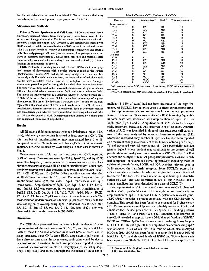
TabIc I Clinical and CGH findings in 20NSCLCsCase
no.SexHistologic type°Grade― Total no.imbalancesTumor
specimensTU-lTU-2TU-3TU-4TU-5TU-6TU-7TU-8TU-9TU-lO
Cell linesCL-ICL-2CL-3CL-4CL-SCL-6CL-7CL-8CL-9CL-bM
MMMFMMMFM
FFFMMMMMMF5CC
SCC5CCACACAC5CC5CC5CC5CC
5CC5CC5CCSCC5CCACACACASCCASCCPD
MDPDPDMDWDMDWDMDPD
PDMDWDMDPDPDPDPDMDMDIS
1898
18111769
15
141823202126196
2020
squamous cell carcinoma; ASCC, adenosquamous cell
CGH ANALYSIS OF LUNG TUMORS
for the identification of novel amplified DNA sequences that maycontribute to the development or progression of NSCLC.
Materials and Methods
Primary Tumor Specimens and Cell Lines. All 20 cases were newly
diagnosed, untreated patients from whom primary tumor tissue was collectedat the time of surgical resection. Ten frozen tumor specimens were microdissected by a single pathologist (E. G.). Frozen 12-@xmsections were stained withH&E, visualized while immersed in drops of 80% ethanol, and microdissectedwith a 26-gauge needle to remove contaminating lymphocytes and stromal
cells. Ten early-passage cell lines (median number, five passages) were prepared as described elsewhere (3). DNAs from cell lines and microdissectedtumor samples were extracted according to our standard method (9). Clinicalfindings are summarized in Table 1.
CGH. Protocolsfor labeling tumorand referenceDNAs, captureof graylevel images of fluorescence with a cooled charge-coupled device camera(Photometrics, Tucson, AZ), and digital image analysis were as describedpreviously (10). For each tumor specimen, the mean values of individual ratioprofiles were calculated from at least seven metaphase spreads. Averagedvalues were plotted as profiles alongside individual chromosome ideograms.Thethreevertical linesnextto theindividualchromosomeideogramsindicatedifferent threshold values between tumor DNA and normal reference DNA.The line on the left corresponds to a threshold value of 0.75, which would existif 50% of the cells from a near diploid tumor had monosomy of a givenchromosome. The center line indicates a balanced state. The line on the rightrepresents a threshold value of 1.25, which would occur if 50% of the cellpopulation exhibited trisomy for that chromosome. Such an overrepresentationwas considered a gain, whereas overrepresentation exceeding a threshold valueof 1.50 was designated a HLG. Overrepresentation defined by a sharp peakwas considered indicative of amplification.
Results
All 20 cases exhibited numerous genomic imbalances (mean, 15.6/case), with every chromosome involved at least once in a CNA. Thetotal number of imbalances/tumor specimen ranged from 6 to I8,compared to 6 to 26 in tumor cell lines (Table 1). A schematicsummary of CNAs detected by CGH analysis in each case is shown inFig.1.
Overrepresentation of 3q was the most consistent CNA observed(85%ofcases).ChromosomearmsSp(70%),7p(65%),and8q(65%)were also frequently overrepresented. In many instances, these fourchromosome arms displayed HLGs and/or amplifications. Other cornmonly overrepresented sites included lq (55%), 2p (50%), 20p (45%),12ql4—21 (45%), and l2p (40%). DNA amplification was identifiedat 19 different locations in 13 cases. The most frequent sites ofamplification were 3q26 (six cases), 8q24 (four cases), and 3q13(three cases). Amplification of 3q28—qter, 7ql I .2, 8pl 1—12,l2pl2,and l9ql3.l—l3.2 was observed in two cases each. Amplification of2q32.2-.32.3,3p2526, 5pl3l4, 7pl2,7q3l,9p24,lOpl4,10q22,12ql4—l5,l3ql4, and 20p13 was detected in a single tumor each. Themost common underrepresented site was 3p (10 cases; 50%), with thesmallest region of overlap being 3p2l. Autosornal loss at 8p21—pter,l5qll.2—l3,Sqll.2—l5,9p, l3ql2—l4,Yip,and 18q2l—qterwasobserved in four to six cases each (20—30%).
Discussion
The CGH data presented here indicate a high incidence of overrepresentation of chromosome arms 3q, Sp, 7p, and 8q in NSCLCs.Each of these CNAs was observed in at least 65% of cases, and inmany instances, these CNAs were HLGs suggestive of polysomy ofthese chromosome arms. In some cases, such gains could be due toisochromosome formation. In fact, we previously reported severalrecurrent isochromosomes in NSCLC karyotypes (3), including i(5p),i(8q), i(lq), i(3q), and i(7p), although the incidence of these abnor
“AC, adenocarcinoma; 5CC,carcinoma.
h WD, well differentiated; MD. moderately differentiated: PD, poorly differentiated.
malities (8—14%of cases) had not been indicative of the high frequency of NSCLCs having extra copies of these chromosome arms.
Overrepresentation of chromosome arm 3q was the most prominentfeature in this series. Nine cases exhibited a HLG involving 3q, which
in some cases was associated with amplification of 3q26, 3q13, or3q28—qter (Figs. I and 2). Amplification of 3q26 seems to be especially important, because it was observed in 6 of 20 cases. Amplifi
cation of 3q26 was identified in three of nine squamous cell carcinomas of the lung analyzed by reverse chromosome painting ( 11).Moreover, increased copy number at 3q26—27has also been reportedas a recurrent change in a variety of other tumors including SCLCs (6,
7) and advanced cervical carcinomas (8). One potentially relevantgene at 3q26.3 whose product may contribute to the control of cellproliferation and malignant transformation is PIK3CA (12). PIK3CAencodes the catalytic subunit of phosphatidylinositol-3 kinase, a critical component of several cell signaling pathways including those ofepidermal growth factor, PDGF, and IGF. Another relevant gene at3q26 encodes the transferrin receptor. Some NSCLCs express in
creased numbers of surface transferrin receptor and elevated levels oftransferrin,4 the locus for which is also in 3q at band q2 1. Amplification of 3q28—qter was identified in two of our NSCLCs, and asimilar amplicon has been described in a case of SCLC (6).
Overrepresentation of 5p, the second most common CNA observedin this series, presented as a HLG in eight of our cases and asamplification of 5pl3—14 in case CL-6. A potentially relevant gene,
SKP2 (Spl3), encodes a protein associated with the CDK2/cyclin Acomplex. This protein has been found to be essential for S-phase entry(13). Overrepresentation of 7p was also a highly consistent CNA, andcandidate loci include genes for EGFR (7pl2), IGF binding proteinsI and 3 (7pl2—l4), and PDGF-a (7p22). Southern blot analysis ofcase CL-9 revealed an approximately 20-fold amplification of EGFR.5EGFR and TGF-a (2pl3) form an autocrine growth-regulatory loop inNSCLC (14). Interestingly, HLG or amplification encompassing 7pl2was observed in six of our NSCLCs, four of which also displayedHLGs at 2pl3. EGFR has been found to be amplified in about 10% ofNSCLCs (3, 4), and elevated levels of EGFR and TGF-a have eachbeen reported in 50—60%of NSCLCs (14). PDGF-a is expressed in
4 F. Cuttitta and J. M. Siegfried, unpublished observations.
5 J. R. Tests, unpublished data.
2117
on June 15, 2015. © 1997 American Association for Cancer Research. cancerres.aacrjournals.org Downloaded from

CGH ANALYSIS OF LUNG TUMORS
5I
ft@II I
I I16
@Il III@I@I'x
more than 80% of NSCLC cell lines (15). Overrepresentation of 8qwas observed in 13 of our cases, 4 of which showed amplification of8q24, the location of the MYC oncogene. In case CL-9, Southernanalysis revealed a 6-fold level of amplification of MYC.5 MYCamplification has been reported in approximately 10% of NSCLCs(reviewed in Ref. 3). The smallest region of overlap of lq gainsobserved in our investigation was lq25—3l (Fig. 1). Breakage at or
i'HCL-I
near the centromere of lq and gains of lq are often observed inkaryotypic studies of NSCLCs (3). Candidate loci in lq25—3l includethe Abelson-related oncogene ARG and the protein tyrosine phosphatase receptor type c polypeptide gene (PTPRC).
The CGH findings described here also suggest an associationbetween overrepresented chromosomal regions and the sites of genesencoding other growth-regulatory proteins that have been implicatedin the pathogenesis of NSCLC (16). Representative profiles depictingoverrepresentation at such sites are shown in Fig. 3. Overrepresentation at 7p, mentioned earlier, is a prominent example. In addition toEGFR, this region harbors genes for the IGF binding proteins 1 and 3(IGFBPJ and IGFBP3) at 7pl2—l4, which was overrepresented infive cases. The region 2q33—34,which encompasses another IGFbinding protein gene, IGFBP2, was also overrepresented in threecases. IGF binding proteins have been found to be secreted in largeexcess of the endogenously secreted IGFs in NSCLCs and maypreferentially deliver synthesized IGF to its receptor (17). Interestingly, the region harboring the IGF receptor type 1 gene, located atl5q25—qter,was overrepresented in 7 of 12 cases exhibiting copynumber increases at lpl2—l4.Overrepresentation at l2p was observedin eight cases, three of which displayed a HLG or amplification. Theparathyroid hormone-related peptide gene, PTHLH, located atl2pl 1.2—12.1, has been implicated in NSCLC (I 8, 19). PTHLH wasfound to be coamplified with the KRAS2oncogene in a human lungcancer cell line (18). Because PTHLH was present at a higher level ofamplification than KRAS2, it was thought to be the target gene in this
2118
I@@@@@@@ ‘@ @I
I‘¶‘ IfflMA@i1t@@hoftI I@ lU,14
IIII111as
@‘‘
£g@@@@@@@
‘III'' @‘@ DJ@@@@ Iii l@@ @l
III@III$Yi@il0 O II
I@ @c1iU@@hi!i@iciI @l@ I@@@ II 12
‘IH
@ 13
II I L@h3Mli
!@II!i@1I1III@@@@
Fig. 1. Summary of CGH imbalances detected in 20 NSCLC tumor specimens and early-passage cell lines. Vertical lines on the left of each chromosome ideogram represent lossof genetic material in a given tumor, whereas those on the right correspond to gains. Thicklines, HLGs; squares, amplification. For each chromosome, smaller CNASare placed furthestfrom the ideogram for ease of identification of minimal regions of overlap, e.g., overrepresentation at lq2S—3I,2p16—2l,3q26—qter,8q24, and I2pll.2—l2.
III I II1
17
I@‘!@iii‘‘‘@
TU-2 CL-b CL-6
CL-5
I@t-.—%'>I¶@I@N.)lu-i CL-4 CL-i
Fig. 2. Ratio profiles of chromosome 3 from eight selected NSCLCs, depicting 3qwhole-armgainsas wellas amplificationat 3q26—qter,3q13,and/or3q28—qter.
on June 15, 2015. © 1997 American Association for Cancer Research. cancerres.aacrjournals.org Downloaded from

CGH ANALYSISOF LUNG TUMORS
2 7
TGFa-
CL-5
ILGFBP2
TGFa@
Fig. 3. Selected ratio profiles ofchromosomes 2,7, 12, 15, and 20 illustrating recurrent DNA gains,HLGs, or amplifications, along with candidategenes at these sites that have relevance to NSCLC.Included are genes encoding growth factors(PDGF-a, and TGF-a), growth factor receptors(EGFR, IGFIR, MET/HGFR), oncogene/cell cycleregulatory proteins (MDM2 and CDC25B), andother peptides (ILGFBP and PTHLH). TU-2
amplicon. PTHLH encodes a potent osteoclast-activating factorthought to play an important role in bone metastasis (19). PTHLH hasalso been associated with hypercalcemia and leukocytosis in lungcancer patients (19, 20).
Other overrepresented regions highlighted by our CGH analysisseem to be sites of genes encoding phosphatases, some of which havebeen shown to be involved in human malignancy. Contrary to theirputative role as tumor suppressors, in some instances, phosphataseshave been found to be amplified and overexpressed and, thus, behavelike oncogenes (21, 22). An example is the CDC25B tyrosine phosphatase gene located at 20pl3, a frequent site of overrepresentation inour series. The human CDC25 tyrosine phosphatases trigger activation of CDC2 by removing inhibitory phosphates and have beensuspected as potential oncogenes due to their role in promoting celldivision (23). Besides 20pl3, four other commonly overrepresentedsites harbor genes for protein phosphatases. One of these, l0q22,represents the location of genes encoding the catalytic and regulatorysubunits of protein phosphatase 3. The others, 7pl5.l—lS.2, l2pl2—13, and 20ql3.l—l3.2, harbor genes for phosphoserine phosphatase,protein tyrosine phosphatase nonreceptor type 6, and protein tyrosinephosphatase nonreceptor type 1, respectively.
Other amplified regions observed in our series involved sites ofknown oncogenes. One such site, l9q13.l—13.2, was amplified in twocases. CGH evidence for amplification at 19ql3 has also been reported in several SCLCs (6). A possible target is the AKT2 oncogene,which has been shown to be amplified and overexpressed in 10—20%
of ovarian and pancreatic carcinomas (9). The most striking illustration of amplification seen in our series was at 7q31 (Fig. 3). The METoncogene, which encodes the hepatocyte growth factor receptor, islocated at 7q3l, and MET has been found to be expressed at highlevels in a majority of primary NSCLCs (24). Another site in thischromosome, 7ql I .2, was amplified in two cases, and a candidategene at this location (SKPJA) encodes a protein associated with theCDK2/cyclin A complex. The region l2q14—l5 was amplified in oneadenocarcinoma case (CL-6) and overrepresented in another eightcases. The MDM2 oncogene, located at 12ql4—l5, has been shown tobe amplified and overexpressed in several lung adenocarcinomas (25).
Overall, loss of chromosomal regions was less frequent than gainsin this CGH analysis. However, underrepresentation of 3p was acommon occurrence, being observed in five tumor specimens and fivecell lines, with the smallest region of overlap being 3p2 1. PreviousLOH analyses have revealed that allelic loss from 3p2l is a frequentearly change in NSCLC, and this chromosomal region is thought toharbor one or more tumor suppressor genes that may play an important role in the pathogenesis of this malignancy (4). Other recurrentunderrepresented sites observed in this CGH study, i.e., 8p2l—pter,l5qll.2—l3,Sqll.2—l5,9p, l3ql2—14,Yip,and l8q21—qter,have
also been reported to be common regions of allelic loss in NSCLC (4).Each of these segments is a known or suspected site of a tumorsuppressor gene(s), e.g., TP53 at Ylpl3 and CDKN2A (p16) at 9p2l,whose alteration may contribute to the development or progression ofNSCLCs.
2119
12 15 20
PTHLH-I@@ IGF1R4 @@f'I@ CDC25B] k—―@'
CL-iCL-4 CL-3
PTHLH -t@ [@@@@_// IGF1R -[ @f@1@—@LCDC25B -@
GL-3CL-6CL-3
MDM2-[@ j@-F@ IGF1R.jttl CDC25B-@
PTHLI I
CL-bCL-b
Ct--aPDGFa -
ILGFgg@@@@
CL-9IGF1R -[@ ¶\@%\ CDC25B-@ @t:-1
TU-2TU-5
CDC25B -@@
TU-5
TGFa -@@ METII-IGFR -
PDGFa -
@ f:@:::::::@,
CL-7 CL-b
on June 15, 2015. © 1997 American Association for Cancer Research. cancerres.aacrjournals.org Downloaded from

CGH ANALYSIS OF LUNG TUMORS
The consistency of genomic imbalances documented here in tumorspecimens as well as in early-passage cell lines may be attributed tothe use of microdissected specimens. Such manual microdissectionseems to be an essential step in maximizing the sensitivity of CGH todetect CNAs in NSCLC. However, this procedure is very timeconsuming, thus limiting the number of cases that can be studied.However, a recent technical innovation, laser capture microdissection(26), may greatly facilitate the rapid procurement of selected cellpopulations from heterogeneous tissue sections. This methodologyholds considerable promise for enhancing the sensitivity and speed ofCGH analysis in the future.
In summary, the recurrent copy number decreases identified inNSCLC by CGH analysis are supportive of previous allelic loss data,further implicating these sites as locations of tumor suppressor genes.In addition, many of the overrepresented chromosome segments observed in NSCLC are sites of known oncogenes/growth-regulatorygenes implicated in lung tumorigenesis. Other overrepresented regions could be sites of novel amplified DNA sequences whose overexpression may provide a proliferative growth advantage in theseaggressive neoplasms.
References
I. Parker, S. L.. Tong, T.. Bolden, S., and Wingo, P. A. Cancer statistics, 1996. CACancer J. Clin. 46: 5—27,1996.
2. Ginsberg, R. J., Kris, M. G.. and Armstrong, J. G. Cancer ofthe lung. In: V. T. Devita,Jr., S. Hellman, and S. A. Rosenberg (eds.), Cancer Principles and Practice ofOncology, 4th Edition, pp. 673—722.Philadelphia: J. B. Lippincott Co., 1993.
3. Tests, J. R., Liu, Z., Feder, M., Bell, D. W., Balsam, B., Cheng, J. Q., and Taguchi.T. Advances in the analysis of chromosome alterations in human lung carcinomas.Cancer Genet. Cytogenet., in press, 1997.
4. Minna, J. D. Molecular biology overview. In: H. I. Pass, J. B. Mitchell, D. H.Johnson, and A. T. Turrisi (eds.), Lung Cancer—Principlesand Practice, pp. 143—148. Philadelphia: Lippincots-Raven, 1996.
5. Kallioniemi. 0. P., Kallioniemi, A., Sudar, D.. Rutovitz, D., Gray. J. W., Waldman,F., and Pinkel, D. Comparative genomic hybridization: a rapid new method fordetecting and mapping DNA amplification in tumors. Semin. Cancer Biol., 4: 41—46,1993.
6. Ried, T., Petersen, I., Holtgreve-Grez, H., Speicher, M. R., Schrock, E., du Manoir,S., and Cremer, T. Mapping of multiple DNA gains and losses in primary small celllung carcinomas by comparative genomic hybridization. Cancer Res., 54: 1801—1806,1994.
7. Levin, N. A., Brzoska, P., Gupta. N., Minna, J. D., Gray, J. W., and Christman, M. F.Identification of frequent novel genetic alterations in small cell lung carcinoma.Cancer Res.. 54: 5086—5091, 1994.
8. Heselmeyer, K., Schrock, E., du Manoir, S., Blegen, H., Shah, K., Steinbeck, R.,Auer, G., and Ried, T. Gain of chromosome 3q defines the transition from severedysplasia to invasive carcinoma of the uterine cervix. Proc. Nail. Acad. Sci. USA, 93:479—484,1996.
9. Cheng, J. Q., Ruggeri, B., Klein, W. M., Sonoda, G., Altomare, D. A., Watson, D. K.,and Testa, J. R. Amplification of AKT2 in human pancreatic cancer cells andinhibition of AK12 expression and tumorigenicity by antisense RNA. Proc. NatI.Acad. Sci. USA, 93: 3636—3641,1996.
10. du Manoir, S., Schrock, E., Bentz, M., Speicher, M. R., Joos, S., Ried, T., Lichter, P.,and Cremer, T. Quantitative analysis of comparative genomic hybridization. CytomcOy, 19: 27—41,1995.
I 1. Brass, N., Ukena, I., Remberger, K., Mack, U., Sybrecht, G. W., and Meese, E. U.DNA amplification on chromosome 3q26.l—q26.3in squamous cell carcinoma of thelung detected by reverse chromosome painting. Eur. J. Cancer, 32A: 1205—1208,1996.
12. Gray, J., Daneshvar, I., Shoenberg-Fejzo, M., Hwang, S., Godfrey, T., Palazzolo,M. C., Kowbel, D., Tanner, M., Kallioniemi, 0. P., Romans, J., Pinnacle, D., andCollins, C. Genomic analysis of three regions of increased copy number discoveredusing CGH. Seventh International Workshop on Chromosomes in Solid Tumors,Tucson,AZ, 1997.
13. Zhang, H., Kobayashi, R., Galaktionov, K., and Beach, D. pl9Skpl and p45Skp2are essential elements of the cyclin A-CDK2 S-phase kinase. Cell, 82: 915—925,1995.
14. Rusch, V., Baselga, J.. Cordon-Cardo, C., Orazem, J., Zaman, M., Hoda, S.,McIntosh, J., Kurie, J., and Dmitrovsky, E. Differential expression of the epidermalgrowth factor receptor and its ligands in primary non-small cell lung cancers andadjacent benign lung. Cancer Res., 53: 2379—2385, 1993.
15. HoIm, C., Gineitis, D., McConville, G. S., and Kazlauskas, A. Expression of PDGF,VEGF, and their receptors in non-small cell lung tumor cell lines. Int. J. Oncol., 9:1077—1086,1996.
16. Woll, P. J. Growth factors and lung cancer. In: H. I. Pass, J. B. Mitchell, D. H.Johnson, and A. T. Turrisi (eds.), Lung Cancer—Principles and Practice, pp. 123—131. Philadelphia: Lippincott-Raven, 1996.
17. Quinn, K. A., Treston, A. M., Unsworth, E. J., Miller, M. J., Vos, M., Grimley, C.,Battey, J., Mulshine, J. L., and Cuttitta, F. Insulin-like growth factor expression inhuman cancer cell lines. J. Biol. Chem., 271: 11477—11483, 1996.
18. Rudduck, C., Duncan, L., Center, R., and Garson, 0. M. Co-amplification of the genefor parathyroid hormone-related protein (PTHRP) and KRAS2in a human lung cancercell line. Genes Chromosomes & Cancer, 7: 213—218,1993.
19. Iguchi, H., Tanaka, S., Ozawa, Y., Kashiwakuma, 1., Kimura, T., Hiraga, T., Ozawa.H., and Kono, A. An experimental model of bone metastasis by human lung cancercells: the role of parathyroid hormone-related protein in bone metastasis. Cancer Res.,56: 4040—4043, 1996.
20. Sakamoto, A., Katakami, H., Mukae, H., Taniguchi, H., Maid, H., Ashitani, J., ilsi,T.,Dohtsu, Y., and Matsukura, S. Simultaneous production of parathyroid hormonerelated protein (PTHrP) and granulocyte colony-stimulating factor (G-CSF) in lungcancer patients with hypercalcemia and leukocytosis. Jpn. J. Thorac. Dis., 33: 34—38,1995.
21 . Harder, K. W., Saw, J., Mild, N., and Jirik, F. Coexisting amplifications of thechromosome lp32 genes (PTPRF and MYCLI)encoding protein tyrosine phosphataseLARandL-mycin a smallcell lungcancerline.Genomics,27: 552—553,1995.
22. Zheng, X. M., Wang, Y., and Pallen, C. J. Cell transfonnation and activation ofppbOc-srcby overexpression of a protein tyrosine phosphatase. Nature (Lond.), 359:336—339,1992.
23. Galaktionov, K., Lee, A. K., Eckstein, J., Draeua, G., Meckler, J., Loda, M., andBeach, D. CDC25 phosphatases as potential human oncogenes. Science (WashingtonDC), 269: 1575—1577,1995.
24. Tsao, M. S., Thu. H., Giaid, A., Viallet, J., Nakamura, T.. and Park, M. Hepatocytegrowth factor/scatter factor is an autocrine factor for human normal bronchial epithelial and lung carcinoma cells. Cell Growth & Differ., 4: 571—579,1993.
25. Marchetti, A., Buttitta, F., Pellegrini, S., Merlo, G., Chella, A., Angeletti, C. A., andBevilacqua. G. mdm2 gene amplification and overexpression in non-small cell lungcarcinomas with accumulation of the p53 protein in the absence of p53 genemutations. Diagn. Mol. Pathol., 4: 93—97,1995.
26. Emmert-Buck, M. R., Bonner, R. F., Smith, P. D., Chuaqui, R. F., Thuang, Z.,Goldstein, S. R., Weiss, R. A., and Liorta, L. A. Laser capture microdissection.Science (Washington DC), 274: 998—1001, 1996.
2120
on June 15, 2015. © 1997 American Association for Cancer Research. cancerres.aacrjournals.org Downloaded from

1997;57:2116-2120. Cancer Res Binaifer R. Balsara, Gonosuke Sonoda, Stanislas du Manoir, et al. 5p, 7p, and 8q in Human Non-Small Cell Lung CarcinomasOften High-Level, Overrepresentation of DNA Sequences at 3q, Comparative Genomic Hybridization Analysis Detects Frequent,
Updated version
http://cancerres.aacrjournals.org/content/57/11/2116
Access the most recent version of this article at:
E-mail alerts related to this article or journal.Sign up to receive free email-alerts
Subscriptions
Reprints and
To order reprints of this article or to subscribe to the journal, contact the AACR Publications
Permissions
To request permission to re-use all or part of this article, contact the AACR Publications
on June 15, 2015. © 1997 American Association for Cancer Research. cancerres.aacrjournals.org Downloaded from


