Embed Size (px)
Citation preview
COMPUTED TOMOGRAPHY
GROUP THREE PRESENTATION

MEMBERS Daniel Nkrumah Aidoo Rosemary Serwaah Boateng Gbogbotsi Richard Amoako Jefferson Frimpong
QUESTIONExamine design features and various generations of CT scans emphasizing on mode of operation, special features for enhancement of image quality and patient safety. Give also considerations for quality assurance (QA) issues.
INTRODUCTION-THE CT-SCAN Computed Tomography (CT), sometimes called computed axial tomography is a non-invasive radiological procedure for producing images of cross-sections of the human body using specialized x-ray equipment.
It can be performed for a variety of reasons which includes diagnostic, treatment planning, interventional or screening.
It utilizes a mathematical technique called reconstruction to accomplish this task.
INTRODUCTION-CT SCAN CONT.
Computed tomography was introduced into clinical practice in 1972.
The CT scanner has since undergone a number of changes which is termed generations.
Each generation was intended to:o Increase the scanner’s efficiencyo Improve safety o Improve the quality of its image
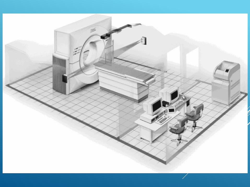
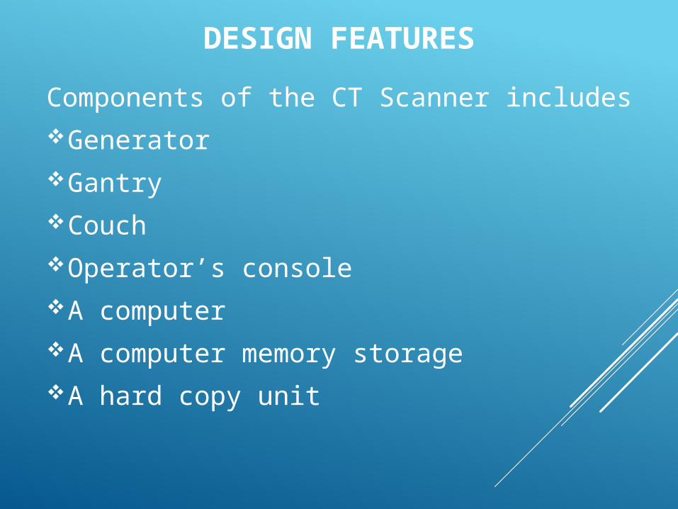
DESIGN FEATURESComponents of the CT Scanner includesGeneratorGantryCouchOperator’s consoleA computerA computer memory storageA hard copy unit
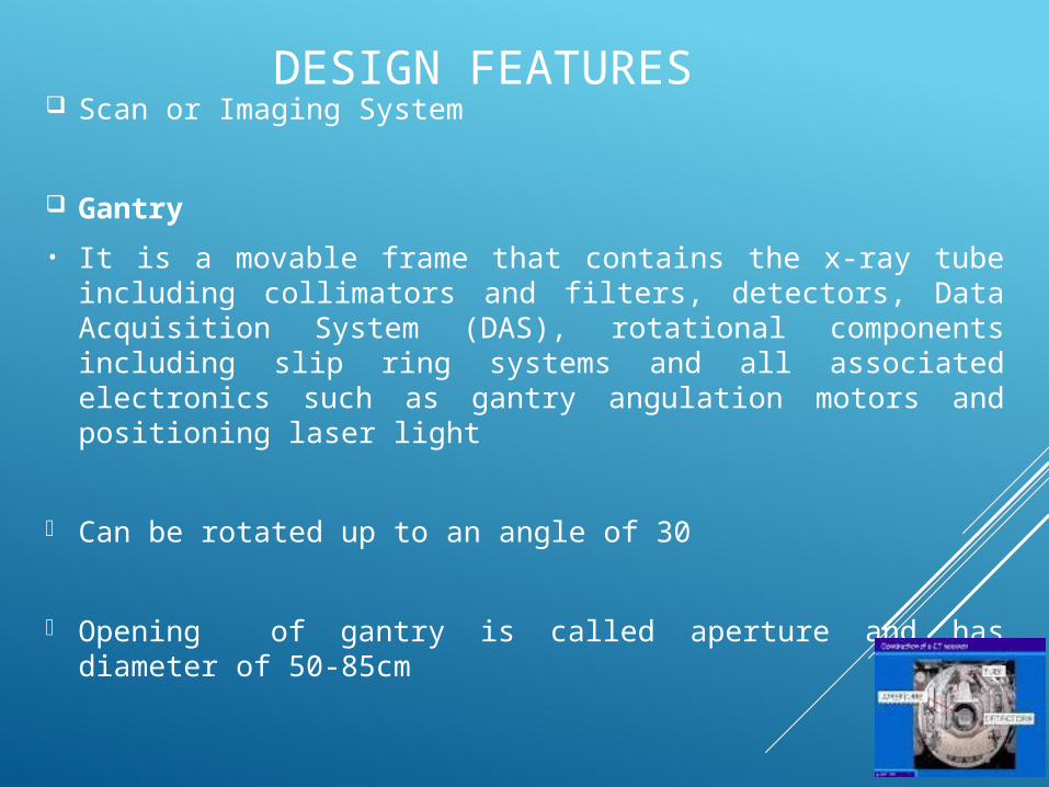
DESIGN FEATURES Scan or Imaging System
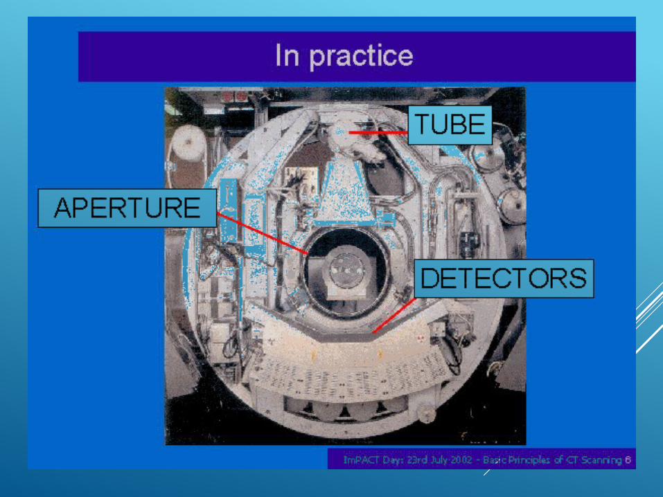
Gantry• It is a movable frame that contains the x-ray tube including collimators and filters, detectors, Data Acquisition System (DAS), rotational components including slip ring systems and all associated electronics such as gantry angulation motors and positioning laser light
Can be rotated up to an angle of 30
Opening of gantry is called aperture and has diameter of 50-85cm
DESIGN FEATURES CON’T.
Larger apertures are used to do large volume of biopsy procedures
Lasers or high intensity lights within or mounted on the gantry which serves as an anatomical positioning guide
Couch It uses material that will not cause artifacts when scanned. e.g. carbon fibre.
The table movement is referred to as incrementation or indexing and it’s quantified in mm/s
All couch have weight limit.
DESIGN FEATURES CON’T.
X-RAY TUBE
It facilitates the use of large exposure time and short exposure time.
It usually uses high frequency, rotating anode and a dual focal spot sizes (0.8-1.4mm).
CT tube has high heat capacity expressed in heat unit
Modern CT has heat capacity of app. 3.5-5MHU
DESIGN FEATURES CON’T.
Possesses high heat dissipation rate.
Uses combination of oil and air cooling system to dissipate heat.
Anode has large diameter with graphite backing enabling the anode absorb and dissipate large amount of heat.
Focal spot size is determined by size of filament and cathode.
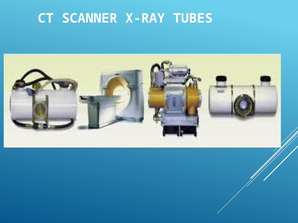
CT SCANNER X-RAY TUBES
DESIGN FEATURES CON’T COLLIMATORS Pre-patient(source) collimator and post-patient(detector) collimator are used
Filters: 2 types used – normally made of Al or Teflon.
Special filter called bow-tie
DETECTORS Image receptors used in CTTypes of detectors• Scintillation crystals- Bismuth germanate crystals coupled with photomultiplier or sodium iodide (NaI) crystals, are more efficient and has less afterglow
DESIGN FEATURES CON’T
• Ionization chamber containing xenon gas under pressure, they have no afterglow but are slightly less efficient.
Data Acquisition System (DAS) Basically amplifies signals from the detectors
Converts analogue signals into digital information through complex mathematical calculation
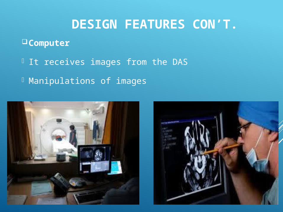
DESIGN FEATURES CON’T.Computer It receives images from the DAS Manipulations of images
GENERATIONS OF CT-SCAN The term generation has been applied because of the order in which the CT scanner designs have been introduced, and each has a number associated with it.
Progress was rapid so the fourth-generating CT imagers appeared in 1978, just 6 year after the first CT imager.
Unlike Hounsfield's early experiments, the patient does not move during CT, except for spiral CT rather, the x-ray source and the image receptor move.
GENERATIONS OF CT-SCAN CON’TFirst Generation:
Finely collimated x-ray beam (pencil beam) was used in first-generation CT images.
Single radiation detector.
Translate-rotate motion.
180 translations with 1degree rotation between translates.
• Single image projection per translation.
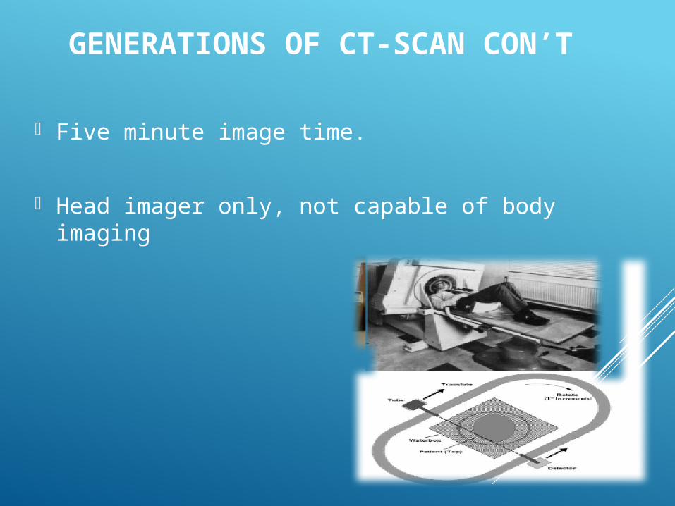
GENERATIONS OF CT-SCAN CON’T
Five minute image time.
Head imager only, not capable of body imaging
GENERATIONS OF CT-SCAN CON’TSecond Generation
Fan –shaped x-ray beam.
Multiple radiation detectors . A detector array.
Translate-rotate motion.
Usually 180 translations with 10 degree rotations between translations.
Multiple image projections per translations.
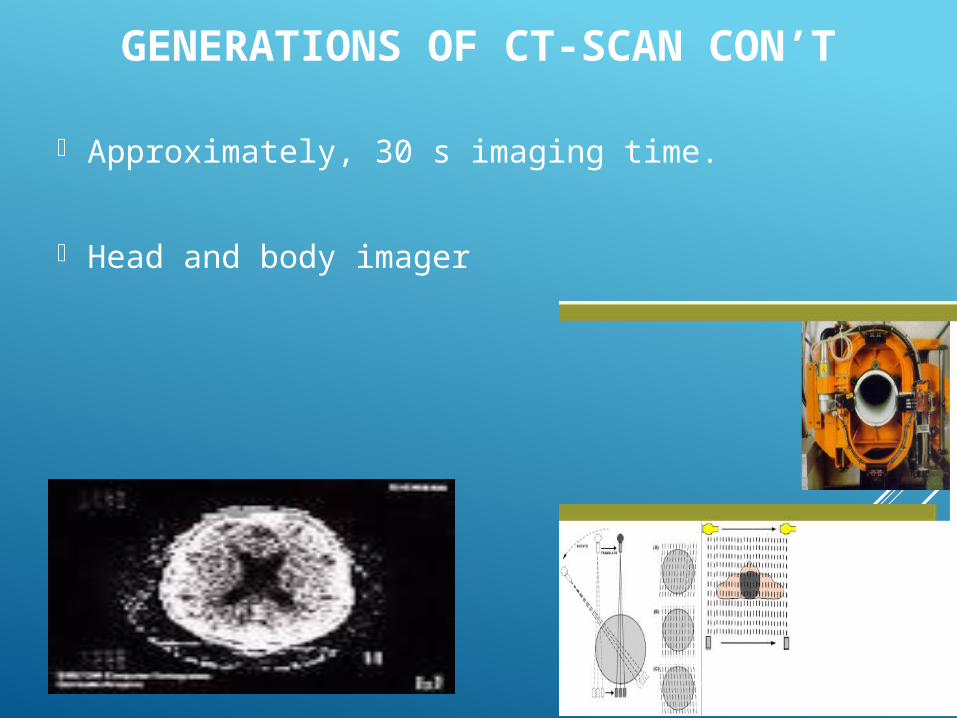
GENERATIONS OF CT-SCAN CON’T
Approximately, 30 s imaging time.
Head and body imager
GENERATION OF CT-SCAN CON’T.
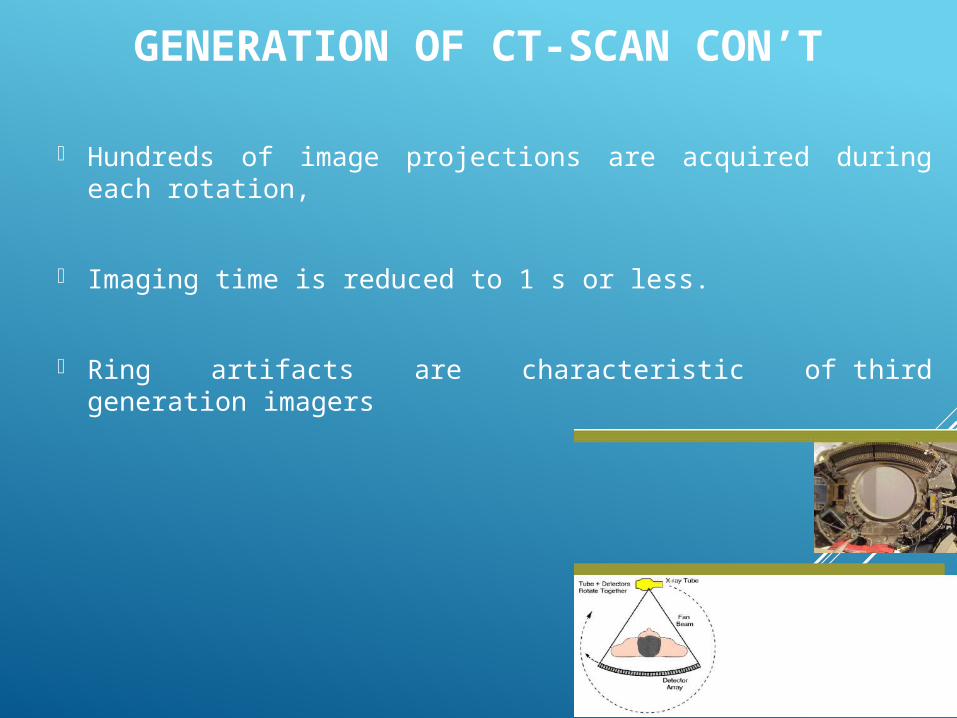
Third Generation
A fan beam X-ray source is used
several hundred radiation detector
The curvilinear detector array provides constant distance between source and each detector resulting in good image reconstruction.
360 degree rotate-rotate mention.
GENERATION OF CT-SCAN CON’T
Hundreds of image projections are acquired during each rotation,
Imaging time is reduced to 1 s or less.
Ring artifacts are characteristic of third generation imagers
GENERATIONS OF CT-SCAN CON’T
Fourth Generation Fourth generation was developed principally to suppress ring artifacts.
Several thousand individual detectors.
The mechanical motion is rotation of the x-ray source around a fixed detector array (rotate stationary).
As the fan beam passes across each detector, and image projection is acquired.
Imaging time is 1 s or less.
GENERATIONS OF CT-SCAN CON’T
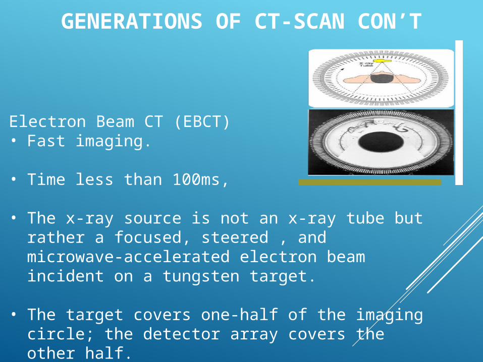
Electron Beam CT (EBCT)• Fast imaging.
• Time less than 100ms,
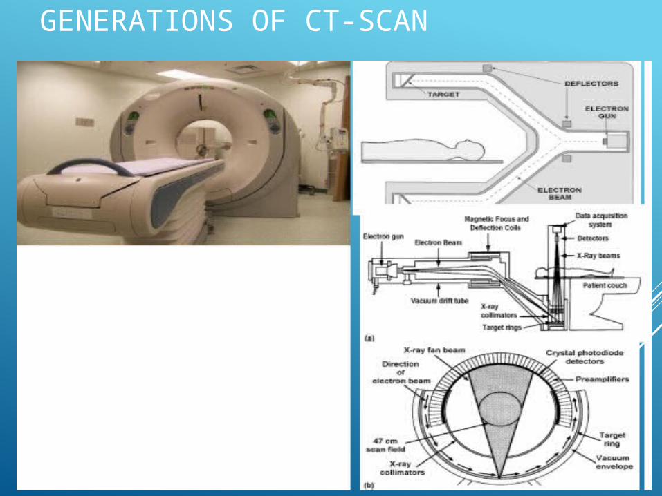
• The x-ray source is not an x-ray tube but rather a focused, steered , and microwave-accelerated electron beam incident on a tungsten target.
• The target covers one-half of the imaging circle; the detector array covers the other half.
GENERATIONS OF CT-SCAN CON’T
The electron beam is steered along the curved tungsten target creating moving source.
EBCT is principally applied to cardiac imaging .
Heat dissipation no problem in EBCT.
EBCT scan time as short as 50ms
GENERATIONS OF CT-SCAN
MODE OF OPERATION
Image is first converted to digital image then to conventional gray scale image
Image of the section is broken down in tiny picture elements called pixels,
Projection of solid cores of tissues called voxels.
Computer counts the light flashes in solid detectors & ions in ionization detectors and gives them a CT number according to attenuation coefficients
MODE OF OPERATION
Complicated reconstruction process of whole image section done by multiple projection on computer. One of the methods is back-projection.
Contrast can be adjusted by operators
SAFETY ISSUES
CT scan imparts a relatively high radiation dose to patients as compared with conventional x-ray examination
The high radiation dose imparted by CT scan exposure could lead to an increase in radiation-induced cancers. In view that, only the patient has to be in the room when the radiation is on. In case there is the need for another person to be in, a lead apron should be worn.
Also, the medical benefits should outweigh any possible radiation damage before the examination should performed i.e. CT scan should be justified .
SAFETY ISSUES CON’T.
The radiation dose should be kept as low as reasonably achievable (ALARA) i.e. CT scan should be optimised.
Indication lights should be installed on the doors for warning when the radiation is on.
EUROPEAN COMMISSION (1999).GUIDANCE ON DIAGNOSTIC REFERENCE LEVELS (DRLS) FOR MEDICAL EXPOSURES. REPORT NO 109. LUXEMBOURG: EUROPEAN COMMISSION &THE OFFICE FOR OFFICIAL PUBLICATIONS OF THE EUROPEAN COMMUNITIES.
EUROPEAN COMMISSION (2000). EUROPEAN GUIDELINES ON QUALITY CRITERIA FOR COMPUTED TOMOGRAPHY. REPORT NO EUR 16262 EN. LUXEMBOURG: EUROPEAN COMMISSION &THE OFFICE FOR OFFICIAL PUBLICATIONS OF THE EUROPEAN COMMUNITIES.
DEAK D., SMAL Y., KALENDER A. (2010), MULTISECTION CT PROTOCOL: SEX AND AGE SPECIFIC CONVERSION FACTORS USED TO DETERMINE EFFECTIVE DOSE FROM DOSE-LENGTH PRODUCT. MEDICAL PHYSICS JOURNAL, RADIOLOGY: 257(1).
DONNELLY F.L., EMERY H.K., BRODY S.A., LAOR T., GYLYS-MORIN V.M., ANTON C.G., THOMAS S.R., FRUSH D.P. (2001), MINIMIZING RADIATION DOSE FOR PEDIATRIC BODY APPLICATIONS OF SINGLE-DETECTOR HELICAL CT: STRATEGIES AT A LARGE CHILDREN’S HOSPITAL. AJR; 176: 303-306.

THANK YOU
YOUR QUESTIONS ARE WELCOMED


