Embed Size (px)
Citation preview
fpls-08-00333 March 10, 2017 Time: 16:36 # 1
ORIGINAL RESEARCHpublished: 14 March 2017
doi: 10.3389/fpls.2017.00333
Edited by:Olivier Lamotte,
CNRS, UMR Agroécologie, France
Reviewed by:Serge Delrot,
University of Bordeaux, FranceBiswapriya Biswavas Misra,
Texas Biomedical Research Institute,USA
*Correspondence:Muthappa [email protected]
Specialty section:This article was submitted to
Plant Physiology,a section of the journal
Frontiers in Plant Science
Received: 21 November 2016Accepted: 27 February 2017
Published: 14 March 2017
Citation:Sinha R, Gupta A and
Senthil-Kumar M (2017) ConcurrentDrought Stress and Vascular
Pathogen Infection Induce Commonand Distinct Transcriptomic
Responses in Chickpea.Front. Plant Sci. 8:333.
doi: 10.3389/fpls.2017.00333
Concurrent Drought Stress andVascular Pathogen Infection InduceCommon and Distinct TranscriptomicResponses in ChickpeaRanjita Sinha, Aarti Gupta and Muthappa Senthil-Kumar*
National Institute of Plant Genome Research, New Delhi, India
Chickpea (Cicer arietinum); the second largest legume grown worldwide is proneto drought and various pathogen infections. These drought and pathogen stressesoften occur concurrently in the field conditions. However, the molecular events inresponse to that are largely unknown. The present study examines the transcriptomedynamics in chickpea plants exposed to a combination of water-deficit stressand Ralstonia solanacearum infection. R. solanacearum is a potential wilt diseasecausing pathogen in chickpea. Drought stressed chickpea plants were infectedwith this pathogen and the plants were allowed to experience progressive droughtwith 2 and 4 days of R. solanacearum infection called short duration stress (SDstresses) and long duration stress (LD stresses), respectively. Our study showed thatR. solanacearum multiplication decreased under SD-combined stress compared to SD-pathogen but there was no significant change in LD-combined stress compared toLD-pathogen. The microarray analysis during these conditions showed that 821 and1039 differentially expressed genes (DEGs) were unique to SD- and LD-combinedstresses, respectively, when compared with individual stress conditions. Three andfifteen genes were common among all the SD-stress treatments and LD-stresstreatments, respectively. Genes involved in secondary cell wall biosynthesis, alkaloidbiosynthesis, defense related proteins, and osmo-protectants were up-regulated duringcombined stress. The expression of genes involved in lignin and cellulose biosynthesiswere specifically up-regulated in SD-combined, LD-combined, and LD-pathogen stress.A close transcriptomic association of LD-pathogen stress with SD-combined stresswas observed in this study which indicates that R. solanacearum infection alsoexerts drought stress along with pathogen stress thus mimics combined stress effect.Furthermore the expression profiling of candidate genes using real-time quantitativePCR validated the microarray data. The study showed that down-regulation ofdefense-related genes during LD-combined stress resulted in an increased bacterialmultiplication as compared to SD-combined stress. Overall, our study highlights asub-set of DEGs uniquely expressed in response to combined stress, which serve aspotential candidates for further functional characterization to delineate the molecularresponse of the plant to concurrent drought-pathogen stress.
Keywords: Ralstonia solanacearum, microarray, unique response, cellulose and lignin biosynthesis, combinedstress, drought-pathogen stress, molecular response
Frontiers in Plant Science | www.frontiersin.org 1 March 2017 | Volume 8 | Article 333
fpls-08-00333 March 10, 2017 Time: 16:36 # 2
Sinha et al. Interaction of Chickpea to Ralstonia solanacearum under Drought
INTRODUCTION
Chickpea (Cicer arietinum) is second largest cultivated legumecrop in world. The crop is vulnerable to drought stress (Gauret al., 2012) as well as to wilt diseases like Fusarium wiltcaused by Fusarium oxysporum f. sp. ciceris (Nene et al., 2012).Under field conditions drought and pathogen stress often occurssimultaneously. Moreover, the combination of drought andpathogen stress has been noted to be devastating for growth andyield of crop plants (Olson et al., 1990; McElrone et al., 2001).These two stressors are noted for their influence on each other’sinteraction with plant, which might result in either negative orpositive impact on the plants. Several studies in Arabidopsis,bean, grapevine have shown that drought stress makes the plantvulnerable to pathogen infection (McElrone et al., 2001; Mayek-Péreza et al., 2002; Mohr and Cahill, 2003; Prasch and Sonnewald,2013). Conversely, reports also indicate that drought stressenhances the defense response of plants against pathogen (Achuoet al., 2006; Ramegowda et al., 2013; Hatmi et al., 2015). Pathogeninfection has also been shown to alter the response of plantsto water-deficit conditions. For example, wilt causing pathogensinhabit the xylem tissues, resulting in vascular dysfunction, andconsequently causes a drought-like effect in plants (Douglas andMacHardy, 1981; Genin, 2010).
Overall, it is observed that the water deprivation inducedduring combined occurrence of drought and vascular pathogeninfection increases the susceptibility of plants against pathogenas well as drought stress (McElrone et al., 2001; Kavak andBoydak, 2011; Choi et al., 2013; Ochola et al., 2015). Thereforeit is important to understand the impact of combined stress andthe cognate defense strategies adopted by plants to circumventthe concurrent onslaught of drought and vascular pathogen.However, the studies to understand the underlying molecularresponses to combined drought and vascular pathogen arelimited (Choi et al., 2013). At this juncture, in our presentstudy we tried to dissect the molecular response of chickpeaunder combined drought and Ralstonia solanacearum infection.R. solanacearum is one of the most devastating wilt causingvascular pathogen having a broad host range and known toinvade more than 200 different plants species (Genin, 2010).R. solanacearum colonizes xylem tissue and secretes massiveamount of exopolysaccharides, which subsequently interruptsxylem function and leads to the wilting of the plant (Genin,2010). R. solanacearum infection in chickpea was observed toresult in a mild to severe disease symptoms which includeyellowing, wilting and cell death; however, it was also observedthat R. solanacearum does not provoke sudden plant deathin leaf-inoculated plants (Sinha et al., 2016). The molecularresponses of plants against R. solanacearum infection has beenextensively studied (Hwang et al., 2011; Ishihara et al., 2012; Chenet al., 2014; Prasath et al., 2014; Zuluaga et al., 2015); however, sofar no attempt has been made to understand the transcriptomicresponses of plant to combined drought and R. solanacearuminfection.
Recently, Sinha et al. (2016) showed that drought restricts themultiplication of R. solanacearum in chickpea which suggests thatcombined stress can induce robust defense responses in chickpea,
thus proposes the need to explore transcriptomic responsesof chickpea under combined drought and R. solanacearuminfection. Since R. solanacearum infection in chickpea causesdisease symptoms after 6 days post infection (dpi) (Sinhaet al., 2016), it has been hypothesized that R. solanacearumprobably induces early and late defense responses at 2 and 4 dpi,respectively.
In the present study, transcriptomic responses of chickpeatoward combined and individual R. solanacearum infection anddrought stress was investigated using microarray at two timepoints namely short duration (SD) and long duration (LD). Thetranscriptomic changes induced during LD and SD combinedstresses were categorized as unique (responses observed onlyduring combined stress) and common (responses overlappingbetween individual and combined stresses) responses.
MATERIALS AND METHODS
Plant Material and Growth ConditionsChickpea plants (variety ICC 4958) were grown in pots (5 inchesdiameter and 5 inches height) containing 500 gm of 3:1 mixture(vol/vol) of air dried peat (Prakruthi Agri Coco peat Industries,Karnataka, India) and vermiculite (Keltech Energies Pvt Ltd.,Maharashtra, India) in an environmentally controlled growthchamber (PGR15, Conviron, Winnipeg, MB, Canada) withdiurnal cycle of 12-h-light/12-h-dark, 200 µEm−2s−1 photonflux intensity, 22◦C temperature and 75% relative humidity. Potswere bottom irrigated every 2 days with half strength Hoagland’smedium (TS1094, Hi-media Laboratories, Mumbai, India).
Inoculum Preparation for PathogenInfectionRalstonia solanacearum procured from Indian Type CultureCollection (ITCC# BI0001), IARI, New Delhi, India was grownin LB medium (without antibiotic) at 28◦C to OD600 = 0.6 witha continuous shaking of 200 rpm for 2.5 h. Culture was pelletdown by centrifuging at 3500× g for 10 min, washed thrice withsterile distilled water, and re-suspended in sterile distilled waterto OD600 = 0.005 corresponding to 7× 10⁵ colony forming units(cfu)/ml.
Stress TreatmentsPathogen stresses namely SD-pathogen (2 days postR. solanacearum infection) and LD-pathogen (4 days postR. solanacearum infection) and drought stress namely SD-drought, SD or fast drought imposition and LD-drought, LDor slow drought imposition were used for this study. Thetwo drought methods were included with incite that durationof drought imposition determines the plant’s biochemical andmolecular responses and therefore may affect the combined stressoutcome. With SD- and LD-drought and SD- and LD-pathogenstresses, two combinations of combined stress treatments namelySD-combined (fast drought with 2 days of R. solanacearuminfection) and LD-combined stress (slow drought with 4 daysof R. solanacearum infection) were considered. Altogether, six
Frontiers in Plant Science | www.frontiersin.org 2 March 2017 | Volume 8 | Article 333
fpls-08-00333 March 10, 2017 Time: 16:36 # 3
Sinha et al. Interaction of Chickpea to Ralstonia solanacearum under Drought
stress treatments were SD-combined (SD drought stress with2 days of R. solanacearum infection), SD-pathogen (2 days ofR. solanacearum infection), SD-drought [SD drought stress,35% field capacity (FC)], LD-combined (LD drought with4 days of R. solanacearum infection), LD-pathogen (4 daysof R. solanacearum infection) and LD-drought (30% FC).Four plants were maintained for each stress treatment, alongwith absolute control and mock control and were placedin growth chamber using completely randomized design(CRD).
Drought stress was imposed by withholding the water,and stress level was assessed by measuring pot mix usinggravimetric method following Sinha et al. (2016). For SD-and LD-drought stress treatments, water was withheld for10 and 15 days for 29- and 24-day-old plants to attain35% (SD-drought) and 30% FC (LD-drought), respectively,on 39th day of plant growth. R. solanacearum inoculationwas done by vacuum infiltration, in which the plants wereupturned in a beaker placed in vacuum chamber containingR. solanacearum culture (7 × 10⁵ cfu/ml) with 0.02% SilwetL77 (Lehle seeds, Fisher Scientific, Waltham, MA, USA) andvacuum of 8.7 psi was applied for 10 min. Plants were brieflyrinsed instantly after infiltration. To avoid the entry of bacterialsuspension into the pot mix, the pot surface was coveredwith polythene wrap prior to infiltration. R. solanacearumwas infiltrated into 35- and 37-day-old plants and sampleswere collected after 4 and 2 days of infiltration for LD- andSD-pathogen stress treatments, respectively. In case of LD-and SD-combined stress treatments, water was withheld for8 and 11 days for 29- and 24-day-old plants to attain 40and 35% FC, respectively. Following this, plants were vacuuminfiltrated with 7 × 10⁵ cfu/ml of R. solanacearum cultureand were allowed for 2 and 4 days of progressive droughtstress post R. solanacearum infection. After 2 and 4 daysof progressive drought stress, FC of SD- and LD-combinedstressed plants were 35 and 30%, respectively. Since plants at60% FC showed better growth than 100% FC because of thehigh water holding capacity of pot mix, absolute and mockcontrols were maintained at 60% FC. For mock control, plantswere infiltrated with water containing 0.02% Silwet L77. Leafsamples for all the treatment were collected from 39 daysold plant for microarray analysis. The methodology of stressimposition is diagrammatically illustrated in SupplementaryFigure S2.
Assessment of In planta BacterialNumbersTotal in planta colony forming units (cfu) of R. solanacearumwere counted at 0 and 2 days post treatment (dpt) for SD-pathogen and SD-combined stress, and at 0, 2, and 4 dptfor LD-pathogen and LD-combined stress experiments. Theleaflets harvested from infiltrated plants were weighed andsurface sterilized with 0.01% H2O2 for 5 s and subsequentlyhomogenized in 100 µl of sterile water. After serial dilutionin sterile water, leaflet homogenate was plated on LB mediumwithout antibiotics. Since there was no significant difference in
dry weight (DW) as well as fresh weight (FW) ratio for individualpathogen and combined stressed plants, FW was accounted incalculating bacterial numbers. Bacterial count was expressed aslog transformed values of cfu/mg FW of leaflet.
Cfu/mg was calculated using the following formula:
CFU/mg =Number of colonies× volume of homogenate (ml)×dilution factor
volume plated (ml)
weight of the leaflet (mg)
Relative Water ContentFor all the six stress treatments and controls, relative watercontent (RWC) was measured at the end of the experiment (39-day-old plant). Moreover, RWC was also measured for SD- andLD-drought at 8 and 11 days after start of drought imposition,respectively. To measure the RWC, FW of leaflet samples wasmeasured immediately after sample collection and samples werehydrated by floating on de-ionized water. Turgid weight (TW)was noted once leaflets attended full turgidity after 6 h at 22◦Ctemperature. Samples were then oven dried at 60◦C until theyreach constant weight after 3 days, and DW was measured. RWCwas calculated using the formula:
RWC(%) =FW− DWTW− DW
∗100
Microarray AnalysisMicroarray analysis for all treatments was performed in twobiological replicates after taking clue from previous studies withtwo biological replicates (Yang et al., 2012; Liu et al., 2014;Chan et al., 2016). Leaf samples from two plants were pooledto make one biological replicate following recommendationby Kendziorski et al. (2005). The pooled leaves were mixedproperly and a part of it was used for RNA isolation. Customizedchickpea microarray chip with 60 mer oligonucleotide probe(Agilent_AMADID – 037094, Agilent technologies, Palo Alto,CA, USA) was used for the study. Entire experiment wasperformed once. The sampling method is thoroughly describedin Supplementary Figure S3 and a flow chart describing the stepsin the microarray analysis is shown in Supplementary Figure S4.
RNA IsolationTotal RNA from leaf tissue for all the treatments and controlswas isolated using TRIzol reagent (Cat# 15596026, Invitrogen,Carlsbad, CA, USA) following manufacturers’ protocol. Further,RNA was purified using RNeasy minikit (Cat# 74104, Qiagen,Hilden, Germany) and quantified using NanoDrop ND-1000spectrophotometer (Thermo Scientific, Waltham, MA, USA).Quality of RNA was assessed on a Bioanalyzer (Agilenttechnologies, Palo Alto, CA, USA).
LabelingSamples for transcriptome analysis were labeled using AgilentQuick-Amp labeling Kit (Cat# 5190-0442, Agilent Technologies,Palo Alto, CA, USA). Total RNA (RIN numbers 6–8, 500 ng)was reverse transcribed at 40◦C using oligo(dT) primer taggedto a T7 polymerase promoter and converted to double strandedcDNA to be used as template for cRNA generation. cRNA was
Frontiers in Plant Science | www.frontiersin.org 3 March 2017 | Volume 8 | Article 333
fpls-08-00333 March 10, 2017 Time: 16:36 # 4
Sinha et al. Interaction of Chickpea to Ralstonia solanacearum under Drought
generated by in vitro transcription and the dye Cy3 CTP wasincorporated during this step. The cDNA synthesis and in vitrotranscription procedures were carried out at 40◦C. Labeled cRNAwas purified using Qiagen RNeasy columns (Cat# 74106, Qiagen,Hilden, Germany) and quality was assessed for yields and qualityusing the Nanodrop ND-1000.
Hybridization and ScanningLabeled cRNA (600 ng) was fragmented at 60◦C and hybridizedon to a Chickpea GXP_8X60K (AMADID: 037094) microarraychip. Fragmentation of labeled cRNA and hybridization weredone using Gene Expression Hybridization Kit (Cat# 5190-0404, Agilent Technologies, California, USA). Hybridization wascarried out in SureHyb Microarray Hybridization Chamber (Cat#G2534A, Agilent Technologies, Palo Alto, CA, USA) at 65◦Cfor 16 h. The hybridized slides were washed with Agilent GeneExpression Wash Buffers (Cat# 5188-5327, Agilent technologies,Palo Alto, CA, USA) and scanned using the Agilent microarrayscanner (Model# G2600D, Agilent Technologies, Palo Alto, CA,USA).
Data AnalysisScanned images were quantified using Feature ExtractionSoftware (Version-11.5 Agilent technologies, Palo Alto, CA,USA). Feature extracted raw data was analyzed using GeneSpringGX software (Version 12.1, Agilent technologies, Palo Alto, CA,USA). Normalization of the data was done in GeneSpring GXusing the 50th percentile shift (where n has a range from 0to 100 and n = 50 is the median) and differential expressionpatterns were identified for each sample. Fold expression valuesfor combined stresses and pathogen stresses were obtained withrespect to mock control and for drought stresses were obtainedwith respect to absolute control samples. Statistical unpairedstudent’s t-test was applied among the replicates and p-valuewas calculated based on volcano plot algorithm (GeneSpringGX, Agilent technologies, Palo Alto, CA, USA). Microarraydataset was submitted to Gene Expression Omnibus (GSE89228).Differentially expressed genes (DEGs) with p-value < 0.05 wereselected. Different fold change cut off values ranging from 0.5to 1.5 were tried with intension of including genes with highfold change expression. Also we looked for the fold changevalue which did not eliminate genes related to hormone, bioticstress, abiotic stress and xylem modification. Consequently, up-regulated genes with fold > 1 (log base2) and down-regulatedgenes with fold >−1 (log base2) were selected for further studies.
Annotation of Transcripts and In silicoExpression ProfilingGene annotation data was retrieved from chickpea transcriptomedatabase1 (Verma et al., 2015). Further, annotation of someof the genes was updated by performing BLASTN searchagainst chickpea database (taxid: 3827) available in NCBI.Differentially regulated genes were clustered using hierarchicalclustering based on Pearson coefficient correlation algorithm2.
1http://www.nipgr.res.in/ctdb.html2https://software.broadinstitute.org/morpheus/
Venn diagrams were generated to view the common and uniquegenes in various conditions (Venny 2.03). Orthologous genes ofArabidopsis thaliana were obtained from chickpea transcriptomedatabase4 (Verma et al., 2015) and their GO enrichment wasperformed using AgriGO singular enrichment analysis5 withdefault setting of Arabidopsis gene model (TAIR10) backgroundand other parameters (statistical test method: Fisher; multi-test adjustment method: Yekutieli FDR under dependency;significance level: 0.055). Finally, heat maps were generated usingGENE-E software6.
Real-Time PCR AnalysisRNA from leaf tissue (100 mg FW) was isolated by TRIzolreagent (Cat# 15596018, Thermo Fisher Scientific, Waltham,MA, USA) following manufacturer’s guidelines. Quality, quantityand integrity of RNA were verified by agarose gel electrophoresisand NanoDrop (Thermo Scientific, Waltham, MA, USA). TheRNA samples with O.D. ratios at 260/280 nm in the range of1.9–2.1, and at 260/230 nm in the range of 2.0–2.3 were usedfor RT-qPCR. First strand cDNA was synthesized from 5 µg ofDNase treated total RNA in a reaction volume of 50 µl usingVerso cDNA synthesis kit (Cat# K1621, Thermo Fisher Scientific,Waltham, MA, USA). Primers were obtained from Sigma–Aldrich, St. Louis, MO, USA. Details of gene-specific primersused for the RT-qPCR are provided in Supplementary Table S1.Reaction mix was prepared by adding 1 µl of two fold-dilutedcDNA and 1 µl of each of the specific primers (10 µM/µl) to 5 µlof SYBR Green PCR master mix (Cat# 4309155, Thermo FisherScientific, Waltham, MA, USA) and final volume was made to10 µl. The reaction was run in ABI Prism 7000 sequence detectionsystem (Applied Biosystems, Foster City, CA, USA). Relative foldchange in gene expression was quantified using 2−11Ct method(Livak and Schmittgen, 2001) using CaActin1 (EU529707.1) asendogenous control to normalize the data. For all the RT-qPCRexperiments, three independent biological replicates and twotechnical replicates were included. For statistical analysis, therelative quantification value (RQ) was transformed to log2 valueand test of significance was performed by one sample t-test.
RESULTS
Differential Transcriptomic Response ofChickpea under Combined andIndividual Drought and Ralstoniasolanacearum StressesTo understand the transcriptomic responses of chickpeato combined drought and vascular pathogen Ralstoniasolanacearum, combined stress experiments were conductedby imposing two levels of drought and pathogen stress. Thestudy was conducted with two types of drought stress namely
3http://bioinfogp.cnb.csic.es/tools/venny/4http://www.nipgr.res.in/ctdb.html5http://bioinfo.cau.edu.cn/agriGO/analysis.php6http://www.broadinstitute.org/cancer/software/GENE-E/
Frontiers in Plant Science | www.frontiersin.org 4 March 2017 | Volume 8 | Article 333
fpls-08-00333 March 10, 2017 Time: 16:36 # 5
Sinha et al. Interaction of Chickpea to Ralstonia solanacearum under Drought
SD drought or fast drought (SD-drought) and LD drought orslow drought (LD-drought) with the premise that duration andseverity of drought stress determines the plants biochemical andmolecular responses and therefore may differentially influencethe combined stress response in chickpea. Two time points inR. solanacearum growth, i.e., 2 days post infiltration (dpi) (SD-pathogen stress) and 4 dpi (LD-pathogen stress) were selectedfor the evaluation of transcriptomic response of chickpea againstR. solanacearum. With the two types of drought stress and twotime points of R. solanacearum multiplication, we imposed twotypes of combined stress namely SD (SD-combined stress, fastdrought with 2 days post combined stress treatment, dpt) and LD(LD-combined stress, slow drought with 4 dpt) (SupplementaryFigure S2A). Before initiating the combined stress study,R. solanacearum was first assessed for its pathogenicity inchickpea. It was found that at an infiltrated concentration of7 × 105 cfu/ml, R. solanacearum was multiplying in planta andcaused disease symptoms varying from mild water soaked lesionsand yellowing to severe cell death and wilting. Also, bacterialooze was noticed from base of the leaflet (SupplementaryFigure S1). In the present study, drought stress was observedto induce reduction in leaf RWC. LD-drought stress showedmore reduction in leaf water, i.e., 64% RWC after 11 days ofwater-withholding compared to SD-drought stress after 8 daysof water withholding, i.e., 75% RWC (Supplementary FigureS2D) indicating an increased severity of drought stress withincreased drought duration. Plants exposed to SD drought(FC-35%) and LD drought (FC-30%) showed 73 and 52% RWC,respectively, compared to 86% RWC in control plants after 10and 15 days of drought treatment, respectively. R. solanacearuminfiltration resulted in water soaking leading to 86% RWC inSD-combined stressed and 87% RWC in LD-combined stressedplants which were close to those of SD-pathogen (86%) andLD-pathogen (83%) stressed plants after 2 and 4 days postcombined stress treatment (Supplementary Figure S2E). Notablereduction in planta multiplication of R. solanacearum wasobserved under SD-combined stress compared to SD-pathogenstress (Supplementary Figure S2B). However, in planta bacterialcount was unchanged under LD-combined stress compared toLD-pathogen stress treatments (Supplementary Figure S2C).
Transcriptomic alterations in chickpea plants challenged withcombined drought and R. solanacearum stress (SD- and LD-combined) and individual drought (SD- and LD-drought) andR. solanacearum stress (SD- and LD-pathogen) were studiedby microarray analysis. Microarray data was submitted toGene Expression Omnibus (GEO# GSE89228). The DEGs (Log2FC ≥ ± 1) under drought stress compared to absolute control,and under combined and pathogen stresses compared to mockcontrol were screened using unpaired t-test (p ≤ 0.05). Putativeannotation of DEGs were obtained from chickpea transcriptomedatabase7 (Verma et al., 2015) and also by homolog searchusing nucleotide BLAST8 against chickpea (NCBI taxid: 3827)database. The DEGs for which putative annotation could not beobtained by BLASTN or BLASTX were termed as unannotated
7http://www.nipgr.res.in/ctdb.html8https://blast.ncbi.nlm.nih.gov/Blast.cgi
DEGs (Supplementary Table S2). Under both SD-pathogenand LD-pathogen stress treatments, many genes involved indefense response (WRKY33, MAP KINASE 11, and DEFENSIN)were up-regulated. Similarly, under SD-drought and LD-droughtstress treatments, genes involved in signaling, biosynthesis ofabscisic acid (ABA) and osmo-protectants namely the genesencoding for LATE EMBRYOGENESIS ABUNDANT 5 (LEA5),LOW-TEMPERATURE-INDUCED 65 KDA PROTEIN were up-regulated. Concordant with these observations, genes involvedin both defense responses and abiotic stress tolerance (genesencoding for LEA and RESPIRATORY BURST OXIDASEHOMOLOG B) were differentially expressed in SD-combinedand LD-combined stresses, which conforms to the nature of thestressors (Supplementary Figure S5 and File S1). The majority oftop most up-regulated genes were belonging to stress responsiveand cell wall modification categories under combined stress(Supplementary Figure S5 and File S1).
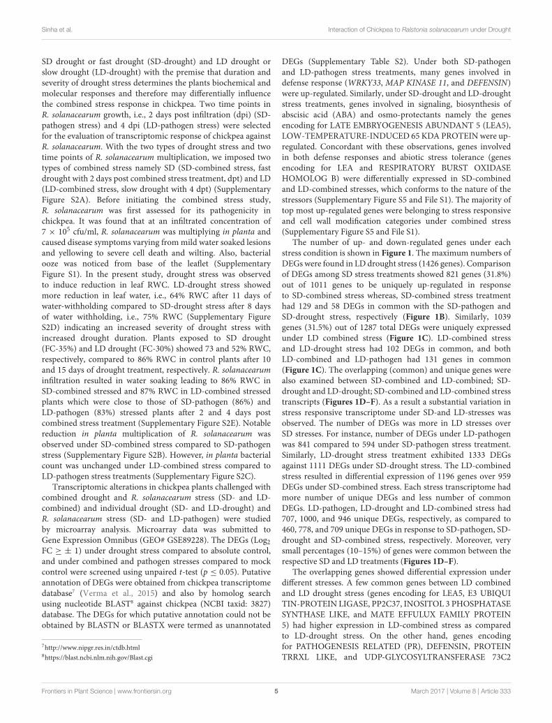
The number of up- and down-regulated genes under eachstress condition is shown in Figure 1. The maximum numbers ofDEGs were found in LD drought stress (1426 genes). Comparisonof DEGs among SD stress treatments showed 821 genes (31.8%)out of 1011 genes to be uniquely up-regulated in responseto SD-combined stress whereas, SD-combined stress treatmenthad 129 and 58 DEGs in common with the SD-pathogen andSD-drought stress, respectively (Figure 1B). Similarly, 1039genes (31.5%) out of 1287 total DEGs were uniquely expressedunder LD combined stress (Figure 1C). LD-combined stressand LD-drought stress had 102 DEGs in common, and bothLD-combined and LD-pathogen had 131 genes in common(Figure 1C). The overlapping (common) and unique genes werealso examined between SD-combined and LD-combined; SD-drought and LD-drought; SD-combined and LD-combined stresstranscripts (Figures 1D–F). As a result a substantial variation instress responsive transcriptome under SD-and LD-stresses wasobserved. The number of DEGs was more in LD stresses overSD stresses. For instance, number of DEGs under LD-pathogenwas 841 compared to 594 under SD-pathogen stress treatment.Similarly, LD-drought stress treatment exhibited 1333 DEGsagainst 1111 DEGs under SD-drought stress. The LD-combinedstress resulted in differential expression of 1196 genes over 959DEGs under SD-combined stress. Each stress transcriptome hadmore number of unique DEGs and less number of commonDEGs. LD-pathogen, LD-drought and LD-combined stress had707, 1000, and 946 unique DEGs, respectively, as compared to460, 778, and 709 unique DEGs in response to SD-pathogen, SD-drought and SD-combined stress, respectively. Moreover, verysmall percentages (10–15%) of genes were common between therespective SD and LD treatments (Figures 1D–F).
The overlapping genes showed differential expression underdifferent stresses. A few common genes between LD combinedand LD drought stress (genes encoding for LEA5, E3 UBIQUITIN-PROTEIN LIGASE, PP2C37, INOSITOL 3 PHOSPHATASESYNTHASE LIKE, and MATE EFFULUX FAMILY PROTEIN5) had higher expression in LD-combined stress as comparedto LD-drought stress. On the other hand, genes encodingfor PATHOGENESIS RELATED (PR), DEFENSIN, PROTEINTRRXL LIKE, and UDP-GLYCOSYLTRANSFERASE 73C2
Frontiers in Plant Science | www.frontiersin.org 5 March 2017 | Volume 8 | Article 333
fpls-08-00333 March 10, 2017 Time: 16:36 # 6
Sinha et al. Interaction of Chickpea to Ralstonia solanacearum under Drought
FIGURE 1 | Overview of differentially expressed genes (DEGs) in chickpea transcriptome in response to combined and individual stresses. To studythe transcriptomic changes in chickpea ICC4958 in response to pathogen (Ralstonia solanacearum), drought and combination of R. solanacearum and droughtstress, chickpea plants were imposed with drought alone, pathogen alone and combined stress for short duration (SD; 2 days) and long duration (LD; 4 days) andnamed as SD-stresses and LD-stresses, respectively. DEGs over control in drought only treated plant and over mock in pathogen treated and combined stressesplants were obtained by microarray analysis. Total number of DEGs having a minimum fold change of 1 (Log2 transformed) and p < 0.05 under all six stresstreatments, SD stresses (SD-pathogen, SD-drought, SD-combined) and LD stresses (LD-pathogen, LD-drought, LD-combined) are represented in graph (A).Positive values in chart shows number of up-regulated and negative values represents number of down-regulated DEGs. The number of common and unique genesamong different SD stresses and LD stresses are shown in Venn diagram (B,C), respectively. Number of DEGs unique and common in SD- and LD-pathogen (D),SD- and LD-drought (E) and SD- and LD-combined stress (F) are shown.
Frontiers in Plant Science | www.frontiersin.org 6 March 2017 | Volume 8 | Article 333
fpls-08-00333 March 10, 2017 Time: 16:36 # 7
Sinha et al. Interaction of Chickpea to Ralstonia solanacearum under Drought
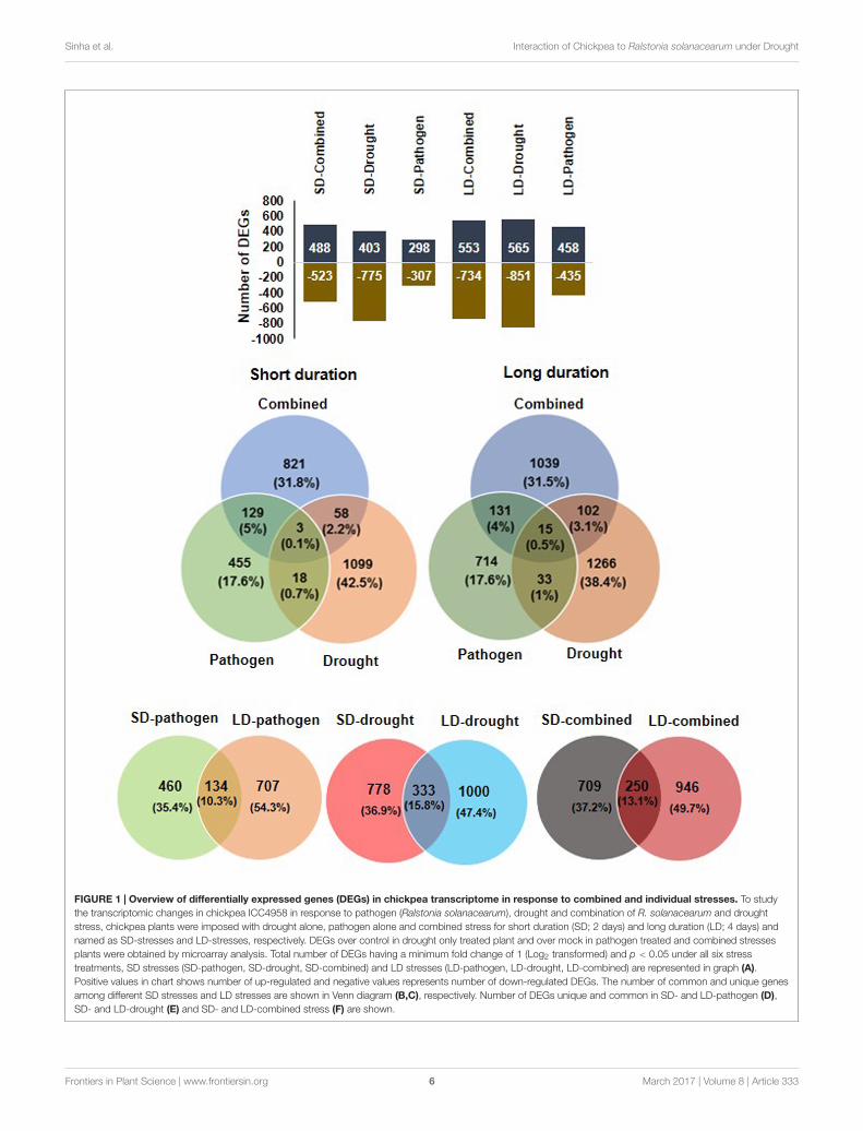
(UGT73C2) proteins showed down-regulation in LD-combinedstress as compared to their up-regulation in LD-pathogen stress.However, some of the down-regulated genes encoding forproteins in SD-pathogen stress like CYTOCHROME P450 734A1(CYP734A1) and GTP BINDING PROTEIN 1 were induced inSD-combined stress (Figure 2). All the LD stress treatments(combined and individual) had 15 genes in common wherefour genes exhibited similar expression and 11 others exhibitedtailored expression (Figures 1B, 2 and Supplementary File S5).
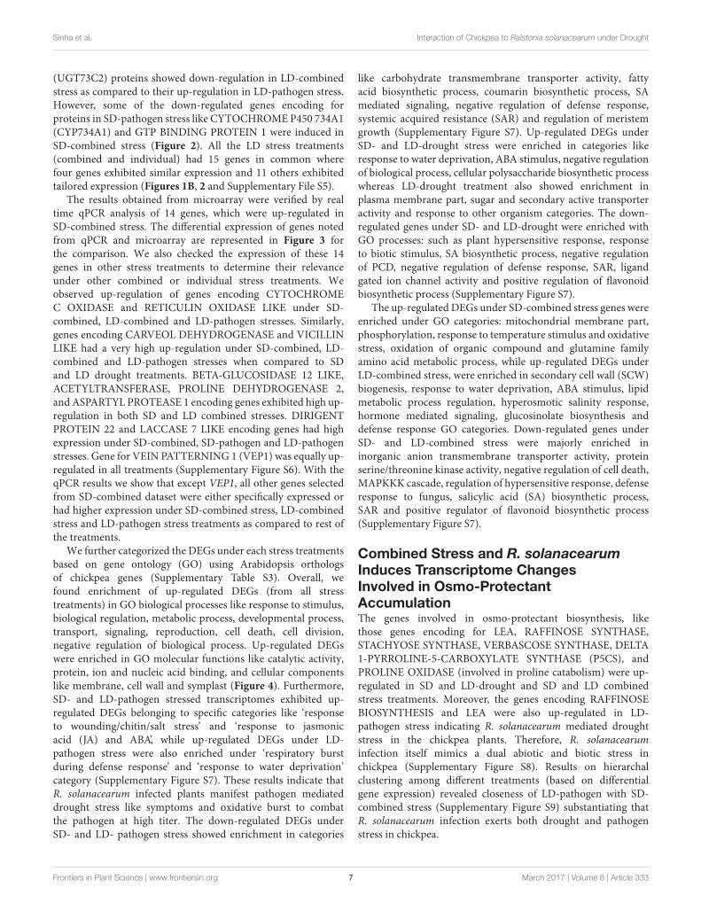
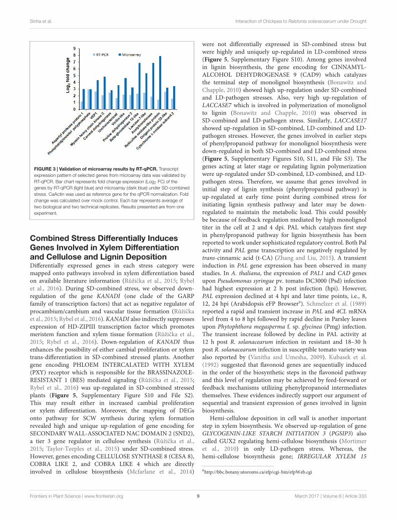
The results obtained from microarray were verified by realtime qPCR analysis of 14 genes, which were up-regulated inSD-combined stress. The differential expression of genes notedfrom qPCR and microarray are represented in Figure 3 forthe comparison. We also checked the expression of these 14genes in other stress treatments to determine their relevanceunder other combined or individual stress treatments. Weobserved up-regulation of genes encoding CYTOCHROMEC OXIDASE and RETICULIN OXIDASE LIKE under SD-combined, LD-combined and LD-pathogen stresses. Similarly,genes encoding CARVEOL DEHYDROGENASE and VICILLINLIKE had a very high up-regulation under SD-combined, LD-combined and LD-pathogen stresses when compared to SDand LD drought treatments. BETA-GLUCOSIDASE 12 LIKE,ACETYLTRANSFERASE, PROLINE DEHYDROGENASE 2,and ASPARTYL PROTEASE 1 encoding genes exhibited high up-regulation in both SD and LD combined stresses. DIRIGENTPROTEIN 22 and LACCASE 7 LIKE encoding genes had highexpression under SD-combined, SD-pathogen and LD-pathogenstresses. Gene for VEIN PATTERNING 1 (VEP1) was equally up-regulated in all treatments (Supplementary Figure S6). With theqPCR results we show that except VEP1, all other genes selectedfrom SD-combined dataset were either specifically expressed orhad higher expression under SD-combined stress, LD-combinedstress and LD-pathogen stress treatments as compared to rest ofthe treatments.
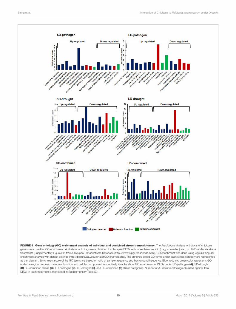
We further categorized the DEGs under each stress treatmentsbased on gene ontology (GO) using Arabidopsis orthologsof chickpea genes (Supplementary Table S3). Overall, wefound enrichment of up-regulated DEGs (from all stresstreatments) in GO biological processes like response to stimulus,biological regulation, metabolic process, developmental process,transport, signaling, reproduction, cell death, cell division,negative regulation of biological process. Up-regulated DEGswere enriched in GO molecular functions like catalytic activity,protein, ion and nucleic acid binding, and cellular componentslike membrane, cell wall and symplast (Figure 4). Furthermore,SD- and LD-pathogen stressed transcriptomes exhibited up-regulated DEGs belonging to specific categories like ‘responseto wounding/chitin/salt stress’ and ‘response to jasmonicacid (JA) and ABA’, while up-regulated DEGs under LD-pathogen stress were also enriched under ‘respiratory burstduring defense response’ and ‘response to water deprivation’category (Supplementary Figure S7). These results indicate thatR. solanacearum infected plants manifest pathogen mediateddrought stress like symptoms and oxidative burst to combatthe pathogen at high titer. The down-regulated DEGs underSD- and LD- pathogen stress showed enrichment in categories
like carbohydrate transmembrane transporter activity, fattyacid biosynthetic process, coumarin biosynthetic process, SAmediated signaling, negative regulation of defense response,systemic acquired resistance (SAR) and regulation of meristemgrowth (Supplementary Figure S7). Up-regulated DEGs underSD- and LD-drought stress were enriched in categories likeresponse to water deprivation, ABA stimulus, negative regulationof biological process, cellular polysaccharide biosynthetic processwhereas LD-drought treatment also showed enrichment inplasma membrane part, sugar and secondary active transporteractivity and response to other organism categories. The down-regulated genes under SD- and LD-drought were enriched withGO processes: such as plant hypersensitive response, responseto biotic stimulus, SA biosynthetic process, negative regulationof PCD, negative regulation of defense response, SAR, ligandgated ion channel activity and positive regulation of flavonoidbiosynthetic process (Supplementary Figure S7).
The up-regulated DEGs under SD-combined stress genes wereenriched under GO categories: mitochondrial membrane part,phosphorylation, response to temperature stimulus and oxidativestress, oxidation of organic compound and glutamine familyamino acid metabolic process, while up-regulated DEGs underLD-combined stress, were enriched in secondary cell wall (SCW)biogenesis, response to water deprivation, ABA stimulus, lipidmetabolic process regulation, hyperosmotic salinity response,hormone mediated signaling, glucosinolate biosynthesis anddefense response GO categories. Down-regulated genes underSD- and LD-combined stress were majorly enriched ininorganic anion transmembrane transporter activity, proteinserine/threonine kinase activity, negative regulation of cell death,MAPKKK cascade, regulation of hypersensitive response, defenseresponse to fungus, salicylic acid (SA) biosynthetic process,SAR and positive regulator of flavonoid biosynthetic process(Supplementary Figure S7).
Combined Stress and R. solanacearumInduces Transcriptome ChangesInvolved in Osmo-ProtectantAccumulationThe genes involved in osmo-protectant biosynthesis, likethose genes encoding for LEA, RAFFINOSE SYNTHASE,STACHYOSE SYNTHASE, VERBASCOSE SYNTHASE, DELTA1-PYRROLINE-5-CARBOXYLATE SYNTHASE (P5CS), andPROLINE OXIDASE (involved in proline catabolism) were up-regulated in SD and LD-drought and SD and LD combinedstress treatments. Moreover, the genes encoding RAFFINOSEBIOSYNTHESIS and LEA were also up-regulated in LD-pathogen stress indicating R. solanacearum mediated droughtstress in the chickpea plants. Therefore, R. solanacearuminfection itself mimics a dual abiotic and biotic stress inchickpea (Supplementary Figure S8). Results on hierarchalclustering among different treatments (based on differentialgene expression) revealed closeness of LD-pathogen with SD-combined stress (Supplementary Figure S9) substantiating thatR. solanacearum infection exerts both drought and pathogenstress in chickpea.
Frontiers in Plant Science | www.frontiersin.org 7 March 2017 | Volume 8 | Article 333
fpls-08-00333 March 10, 2017 Time: 16:36 # 8
Sinha et al. Interaction of Chickpea to Ralstonia solanacearum under Drought
FIGURE 2 | Expression profile of DEGs common between combined and individual stresses. The DEGs with more than one fold expression and p < 0.05under SD treatments (SD-pathogen, SD-drought, SD-combined stress) and LD treatments (LD-pathogen, LD-drought, LD-combined) were compared and DEGscommon between combined and individual stresses were selected. The heat maps represent DEGs which are shared between combined and individual stresses buthave at least one fold difference in their expression between two treatments. Heat map (A) represents 12 shared genes with differential expression out of total 129common genes between SD-pathogen and SD-combined and (B) represents 16 out of total 58 shared genes between SD-drought and SD-combined stress withdifferent expression. Similarly, heat map (C) represents 40 genes with differential expression out of total 131 common genes between LD-pathogen andLD-combined and (D) represents 27 out of total 102 common genes between LD-drought and LD-combined stress. Expression level of DEGs common among allLD stresses are represented in heat map (E). Color scale shows gene expression range where color bar in red and blue represents up- and down-regulated genes,respectively. Details of the genes shown in heat maps are available in Supplementary File S5.
Frontiers in Plant Science | www.frontiersin.org 8 March 2017 | Volume 8 | Article 333
fpls-08-00333 March 10, 2017 Time: 16:36 # 9
Sinha et al. Interaction of Chickpea to Ralstonia solanacearum under Drought
FIGURE 3 | Validation of microarray results by RT-qPCR. Transcriptexpression pattern of selected genes from microarray data was validated byRT-qPCR. Bar chart represents fold change expression (Log2 FC) of thegenes by RT-qPCR (light blue) and microarray (dark blue) under SD-combinedstress. CaActin was used as reference gene for the qPCR normalization. Foldchange was calculated over mock control. Each bar represents average oftwo biological and two technical replicates. Results presented are from oneexperiment.
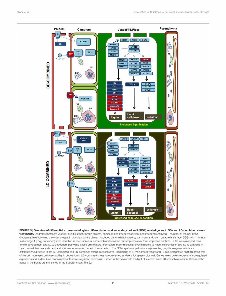
Combined Stress Differentially InducesGenes Involved in Xylem Differentiationand Cellulose and Lignin DepositionDifferentially expressed genes in each stress category weremapped onto pathways involved in xylem differentiation basedon available literature information (Ružicka et al., 2015; Rybelet al., 2016). During SD-combined stress, we observed down-regulation of the gene KANADI (one clade of the GARPfamily of transcription factors) that act as negative regulator ofprocambium/cambium and vascular tissue formation (Ružickaet al., 2015; Rybel et al., 2016). KANADI also indirectly suppressesexpression of HD-ZIPIII transcription factor which promotesmeristem function and xylem tissue formation (Ružicka et al.,2015; Rybel et al., 2016). Down-regulation of KANADI thusenhances the possibility of either cambial proliferation or xylemtrans-differentiation in SD-combined stressed plants. Anothergene encoding PHLOEM INTERCALATED WITH XYLEM(PXY) receptor which is responsible for the BRASSINAZOLE-RESISTANT 1 (BES) mediated signaling (Ružicka et al., 2015;Rybel et al., 2016) was up-regulated in SD-combined stressedplants (Figure 5, Supplementary Figure S10 and File S2).This may result either in increased cambial proliferationor xylem differentiation. Moreover, the mapping of DEGsonto pathway for SCW synthesis during xylem formationrevealed high and unique up-regulation of gene encoding forSECONDARY WALL-ASSOCIATED NAC DOMAIN 2 (SND2),a tier 3 gene regulator in cellulose synthesis (Ružicka et al.,2015; Taylor-Teeples et al., 2015) under SD-combined stress.However, genes encoding CELLULOSE SYNTHASE 8 (CESA 8),COBRA LIKE 2, and COBRA LIKE 4 which are directlyinvolved in cellulose biosynthesis (Mcfarlane et al., 2014)
were not differentially expressed in SD-combined stress butwere highly and uniquely up-regulated in LD-combined stress(Figure 5, Supplementary Figure S10). Among genes involvedin lignin biosynthesis, the gene encoding for CINNAMYL-ALCOHOL DEHYDROGENASE 9 (CAD9) which catalyzesthe terminal step of monolignol biosynthesis (Bonawitz andChapple, 2010) showed high up-regulation under SD-combinedand LD-pathogen stresses. Also, very high up-regulation ofLACCASE7 which is involved in polymerization of monolignolto lignin (Bonawitz and Chapple, 2010) was observed inSD-combined and LD-pathogen stress. Similarly, LACCASE17showed up-regulation in SD-combined, LD-combined and LD-pathogen stresses. However, the genes involved in earlier stepsof phenylpropanoid pathway for monolignol biosynthesis weredown-regulated in both SD-combined and LD-combined stress(Figure 5, Supplementary Figures S10, S11, and File S3). Thegenes acting at later stage or regulating lignin polymerizationwere up-regulated under SD-combined, LD-combined, and LD-pathogen stress. Therefore, we assume that genes involved ininitial step of lignin synthesis (phenylpropanoid pathway) isup-regulated at early time point during combined stress forinitiating lignin synthesis pathway and later may be down-regulated to maintain the metabolic load. This could possiblybe because of feedback regulation mediated by high monolignoltiter in the cell at 2 and 4 dpi. PAL which catalyzes first stepin phenylpropanoid pathway for lignin biosynthesis has beenreported to work under sophisticated regulatory control. Both Palactivity and PAL gene transcription are negatively regulated bytrans-cinnamic acid (t-CA) (Zhang and Liu, 2015). A transientinduction in PAL gene expression has been observed in manystudies. In A. thaliana, the expression of PAL1 and CAD genesupon Pseudomonas syringae pv. tomato DC3000 (Psd) infectionhad highest expression at 2 h post infection (hpi). However,PAL expression declined at 4 hpi and later time points, i.e., 8,12, 24 hpi (Arabidopsis eFP Browser9). Schmelzer et al. (1989)reported a rapid and transient increase in PAL and 4CL mRNAlevel from 4 to 8 hpi followed by rapid decline in Parsley leavesupon Phytophthora megasperma f. sp. glycinea (Pmg) infection.The transient increase followed by decline in PAL activity at12 h post R. solanacearum infection in resistant and 18–30 hpost R. solanacearum infection in susceptible tomato variety wasalso reported by (Vanitha and Umesha, 2009). Kubasek et al.(1992) suggested that flavonoid genes are sequentially inducedin the order of the biosynthetic steps in the flavonoid pathwayand this level of regulation may be achieved by feed-forward orfeedback mechanisms utilizing phenylpropanoid intermediatesthemselves. These evidences indirectly support our argument ofsequential and transient expression of genes involved in ligninbiosynthesis.
Hemi-cellulose deposition in cell wall is another importantstep in xylem biosynthesis. We observed up-regulation of geneGLYCOGENIN-LIKE STARCH INITIATION 3 (PGSIP3) alsocalled GUX2 regulating hemi-cellulose biosynthesis (Mortimeret al., 2010) in only LD-pathogen stress. Whereas, thehemi-cellulose biosynthesis gene; IRREGULAR XYLEM 15
9http://bbc.botany.utoronto.ca/efp/cgi-bin/efpWeb.cgi
Frontiers in Plant Science | www.frontiersin.org 9 March 2017 | Volume 8 | Article 333
fpls-08-00333 March 10, 2017 Time: 16:36 # 10
Sinha et al. Interaction of Chickpea to Ralstonia solanacearum under Drought
FIGURE 4 | Gene ontology (GO) enrichment analysis of individual and combined stress transcriptomes. The Arabidopsis thaliana orthologs of chickpeagenes were used for GO enrichment. A. thaliana orthologs were obtained for chickpea DEGs with more than one fold (Log2 converted) and p < 0.05 under six stresstreatments (Supplementary Figure S2) from Chickpea Transcriptome Database (http://www.nipgr.res.in/ctdb.html). GO enrichment was done using AgriGO singularenrichment analysis with default settings (http://bioinfo.cau.edu.cn/agriGO/analysis.php). The enriched broad GO terms under each stress category are representedas bar diagram. Enrichment score of the GO terms are based on ratio of sample frequency and background frequency. Blue, red, and green color represents GOunder biological process, molecular function and cellular component, respectively. Graphs show GO enrichment of DEGs under SD-pathogen (A), SD-drought(B) SD-combined stress (C), LD-pathogen (D), LD-drought (E), and LD-combined (F) stress categories. Number of A. thaliana orthologs obtained against totalDEGs in each treatment is mentioned in Supplementary Table S2.
Frontiers in Plant Science | www.frontiersin.org 10 March 2017 | Volume 8 | Article 333
fpls-08-00333 March 10, 2017 Time: 16:36 # 11
Sinha et al. Interaction of Chickpea to Ralstonia solanacearum under Drought
FIGURE 5 | Overview of differential expression of xylem differentiation and secondary cell wall (SCW) related genes in SD- and LD-combined stresstreatments. Diagrams represent vascular bundle structure with phloem, cambium and xylem vessel/fiber and xylem parenchyma. The order of the cell in thediagram is likely following the order present in dicot leaf where phloem is placed on abaxial followed by cambium and xylem on adaxial surface. DEGs with minimumfold change 1 (Log2 converted) were identified in each individual and combined stressed transcriptome over their respective controls. DEGs were mapped onto‘xylem development and SCW deposition’ pathways based on literature information. Major molecular events related to xylem differentiation and SCW synthesis inxylem vessel, tracheary element and fiber are represented once in the same box. The SCW synthesis pathway is representing only those genes which aredifferentially expressed in the SD-combined and LD-combined stress transcriptome. Thickening of SCW in xylem vessel and TE are represented as thick green wallof the cell. Increased cellulose and lignin deposition in LD-combined stress is represented as dark thick green color wall. Genes in red boxes represents up-regulatedexpression and in dark blue boxes represents down-regulated expression. Genes in the boxes with the light blue color has no differential expression. Details of thegenes in the boxes are mentioned in the Supplementary File S2.
Frontiers in Plant Science | www.frontiersin.org 11 March 2017 | Volume 8 | Article 333
fpls-08-00333 March 10, 2017 Time: 16:36 # 12
Sinha et al. Interaction of Chickpea to Ralstonia solanacearum under Drought
(IRX15-like) (Brown et al., 2011) showed unique and highup-regulation in SD-combined stress only. Two hemicellulosebiosynthetic genes encoding for PURVUS/GLZ1 and PECTINMETHYLESTERASE (PME) (Scheller and Ulvskov, 2010) wereup-regulated during SD- and LD-drought stress, respectively(Figure 5, Supplementary Figures S10, S11 and Files S2, S3). Thisstrongly suggests that combined stress results in induction oflignin biosynthesis and modification in xylem SCW.
Furthermore, we wanted to explore if up-regulation of genesinvolved in SCW formation is a general consensus undercombined stress. Therefore we looked for the expression of genesencoding for CAFFEIC ACID O-METHYLTRANSFERASE(COAMT), LACCASE 7 and 17 and CESA7 and 8 genes incombined drought and Psd transcriptomic data from Guptaet al. (2016). We found absence of those genes in differentiallyexpressed transcriptome of both drought first Psd later (DP)and Psd first and drought later (PD) treatments (Gupta et al.,2016) (Supplementary Figure S12). This indicates that combineddrought and foliar pathogen stress may not employ SCWmodification for combined stress tolerance.
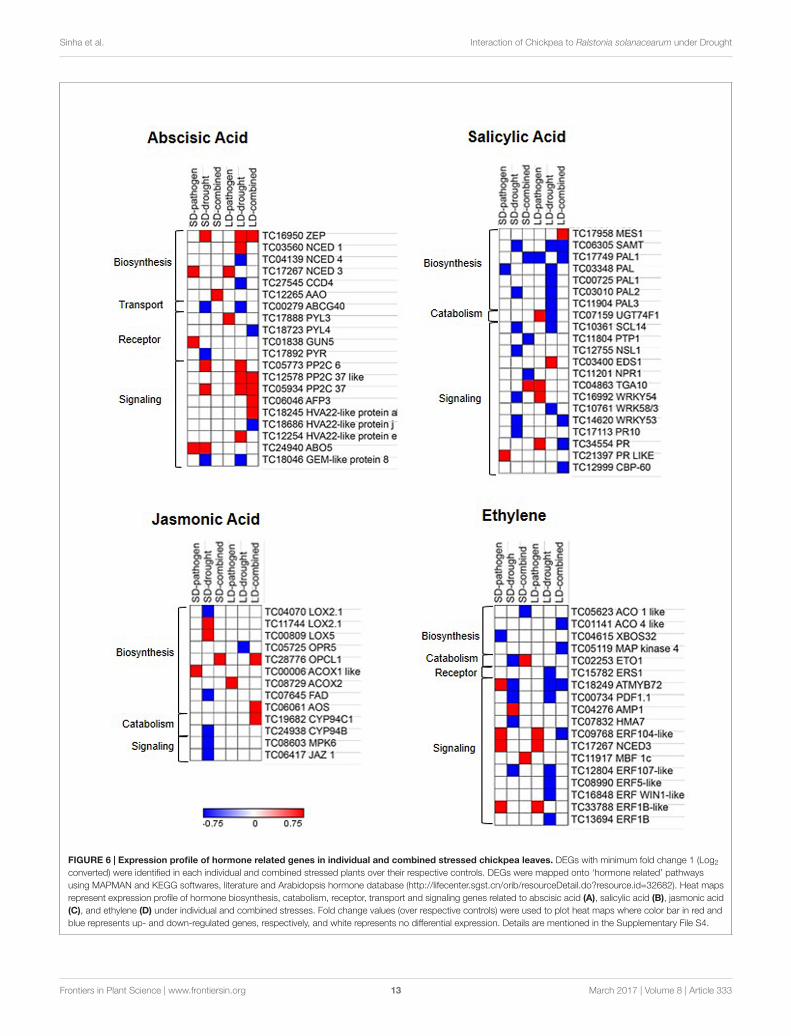
Combined Drought and R. solanacearumStress Differentially InducesPhytohormone Biosynthesis andSignaling GenesThe combined stress mediated alteration in hormonebiosynthesis, catabolism, transport, and signaling were studiedby mapping the DEGs onto hormone pathways based onavailable literature information. We found up-regulation ofABA biosynthesis genes in all stress treatments. We observedthe stress treatment specific differential expression of ABAbiosynthetic genes. ABA biosynthesis gene encoding 9-CIS-EPOXYCAROTENOID DIOXYGENASE 3 (NCED3) wasspecifically induced during SD- and LD-pathogen stress whereasduring SD- and LD-drought stress, ABA biosynthetic genesencoding ZEAXANTHIN EPOXIDASE (ZEP) and NCED1 wereinduced. During SD-combined stress, ALDEHYDE OXIDASE(AAO) was specifically up-regulated, similarly in LD-combinedstress only ZEP is found to be up-regulated. Down-regulation ofgene encoding for ABA transporter ATP-BINDING CASSETTEG40 (ABCG40) was observed only during SD- and LD-drought.ABA receptor encoding genes were up-regulated in SD- andLD-pathogen stress and signaling genes PP2C6 and PP2C37 wereup-regulated in SD-pathogen, SD-drought, LD-drought, andLD-combined stressed plants (Figure 6A and SupplementaryFile S4). PP2C is negative regulator of ABA signaling, however,has been shown to confer abiotic stress tolerance in manyplants in ABA insensitive manner (Bhaskara et al., 2012; Zhanget al., 2013; Singh et al., 2015). Another negative regulatorof ABA signaling geneABI5-interacting protein (AFP3) wasalso specifically up-regulated in LD-combined stress. Theup-regulation of PP2C and AFP3 under drought stress hasbeen previously reported (Garcia et al., 2008) but its role undercombined stress has not been seen yet. These results indicatethat ABA signaling plays a major part in plant response underdrought, R. solanacearum and combined stress.
Short duration- and long duration-pathogen stresses inchickpea induced up-regulation of SA and ethylene (ET)signaling genes encoding for TGACG (TGA) MOTIF-BINDINGPROTEIN 10 (TGA10), PATHOGENESIS-RELATED GENE,WRKY54, ETHYLENE RESPONSE FACTOR104 LIKE (ERF104), and ERF1B (Figures 6B,D and SupplementaryFile S4). Contrastingly, we also encountered up-regu-lation of SA catabolism gene encoding for UDP-GLYCO-SYLTRANSFERASE 74 F1 (UGT74F1) in LD-pathogen stress.SD- and LD-pathogen stress in chickpea also induced up-regulation of JA biosynthetic genes encoding ACYL-COAOXIDASE 1 and 2, respectively, but signaling genes wereun-induced. SD-combined stress showed up-regulation of JAbiosynthesis gene (OPCL1), down-regulation of SA repressor(PROTEIN TYROSINE PHOSPHATASE1 (PTP1), Bartels et al.,2009) and up-regulation of SA and ET signaling genes PR andMULTIPROTEIN BRIDGING FACTOR-1c (MBF1c), respectively(Figure 6C and Supplementary File S4). LD-combined stresstreatment exhibited up-regulation of both JA biosyntheticgenes OPCL1, AOS, and catabolism gene CYTOCHROMP450 94C1 (Figure 6C). While, LD-combined stress had up-regulation of SA biosynthetic gene MES1 (METHYLESTERASE 1), which converts methyl salicylate (MeSA) to SA(Dempsey et al., 2011), it showed down-regulation of SAsignaling genes WRKY53, PR like, CBP60 and ET signalinggenes MYB72 and ERF104 (Figures 6B,D). Altogether, theyindicate toward suppression of immunity in LD combinedstress (Supplementary Figure S13C). Collectively, our results ontranscriptome analysis of phytohormone related genes suggestan involvement of ABA, SA, and ET mediated signaling inmodulating combined stress response in these plants.
Both SD- and LD-combined stress showed up-regulationof brassinosteroid (BR) inactivator CYP734A1 (Vriet et al.,2013). Genes encoding for BR receptors BRASSINOSTEROIDINSENSITIVE 1 (BRI1) and BRASSINOSTEROID INSENSITIVE 1 LIKE 2 (BRI1like 2) were up-regulated under LD-combi-ned and SD-pathogen stresses, respectively (SupplementaryFigure S13B). LD-combined stress also showed up-regulationof gibberellin (GA) biosynthesis gene GIBBERELLIN 3-BETA-DIOXYGENASE 1 (GA1), catabolism gene CYTOCHROMEP450 714ALIKE (CYP714Alike) and negative regulator of GAsignaling gene; GIBBERELLIC ACID INSENSITIVE (GAI)indicating a loss of GA signaling in LD-combined stress(Supplementary Figure S13A). SD-combined stress resultedin up-regulation of genes encoding Auxin receptor; TOLL-INTERLEUKIN-RESISTANCE (TIR) and auxin transporter;ATP-BINDING CASSETTE B4 (ABCB4) (SupplementaryFigures S13D and File S4).
Differential Expression of DefenseRelated Genes in SD- and LD-CombinedStress Influences R. solanacearumMultiplicationWe looked for the differential expression of biotic stressresponsive genes in the all the six treatments. We found up-regulated expression of genes encoding PLEIOTROPIC DRUG
Frontiers in Plant Science | www.frontiersin.org 12 March 2017 | Volume 8 | Article 333
fpls-08-00333 March 10, 2017 Time: 16:36 # 13
Sinha et al. Interaction of Chickpea to Ralstonia solanacearum under Drought
FIGURE 6 | Expression profile of hormone related genes in individual and combined stressed chickpea leaves. DEGs with minimum fold change 1 (Log2
converted) were identified in each individual and combined stressed plants over their respective controls. DEGs were mapped onto ‘hormone related’ pathwaysusing MAPMAN and KEGG softwares, literature and Arabidopsis hormone database (http://lifecenter.sgst.cn/orib/resourceDetail.do?resource.id=32682). Heat mapsrepresent expression profile of hormone biosynthesis, catabolism, receptor, transport and signaling genes related to abscisic acid (A), salicylic acid (B), jasmonic acid(C), and ethylene (D) under individual and combined stresses. Fold change values (over respective controls) were used to plot heat maps where color bar in red andblue represents up- and down-regulated genes, respectively, and white represents no differential expression. Details are mentioned in the Supplementary File S4.
Frontiers in Plant Science | www.frontiersin.org 13 March 2017 | Volume 8 | Article 333
fpls-08-00333 March 10, 2017 Time: 16:36 # 14
Sinha et al. Interaction of Chickpea to Ralstonia solanacearum under Drought
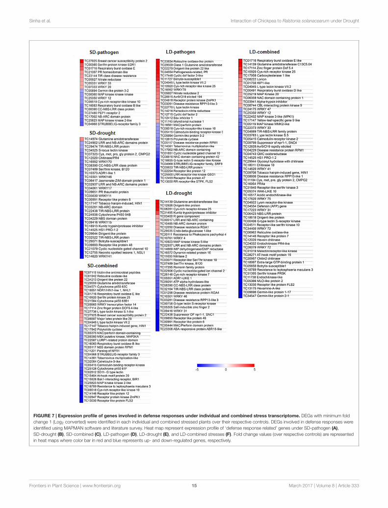
RESISTANCE 3, ZINC FINGER PROTEIN, DOF ZINC FINGERPROTEIN DOF1.1, SER/THR-PROTEIN KINASE EDR1,(STPKEDR1) RESPIRATORY BURST OXIDASE HOMOLOG B(RBOHB), TIR CLASS DISEASE RESISTANCE, and NITRATEREDUCTASE under SD-pathogen stress. LD-pathogen stressshowed up-regulation of defense related genes encoding RETICULIN OXIDASE LIKE PROTEIN, DISEASE RESISTANCE-RESPONSIVE (dirigent-like protein), GLUTAMINE AMIDOTRANSFERASE, PR, BOTRYTIS SUSCEPTIBLE 1, WRKY70,and RPP13. SD-combined stressed plants exhibited high up-regulation of genes encoding RETICULIN OXIDASE LIKEPROTEIN (also called BERBERINE BRIDGE ENZYME)involved in alkaloid biosynthesis, DISEASE RESISTANCE-RESPONSIVE (DIRIGENT-LIKE PROTEIN), LACCASE 7involved in lignan and antioxidant synthesis to confer defenseresponse in plant (Figures 5, 7) (Dittrich and Kutchan, 1991;Zhao et al., 2013). SD-combined stress also exhibited highup-regulation of defense related genes encoding GLUTAMINEAMIDO TRANSFERASE, RBOHE like, STPK25, WRKY4,DOF5.4, and MAJOR LATEX PROTEIN LIKE 28 (MLP28)(Figure 7). SD-combined stressed transcriptome exhibitedmore number of up-regulated genes involved in defenseresponse with high amplitude of expression as compared toSD-pathogen stress. This justifies the activated defense leading todecreased bacterial growth in SD-combined stress as comparedto SD-pathogen. In LD-combined stress, we observed up-regulation of genes encoding for RBOHE like, GLUTAMINEAMIDOTRANSFERASE C13C5.04, ZINC FINGER PROTEINDOF5.4, CYS-RICH RECEPTOR KINASE 25, however, severaldefense related genes such as genes encoding DEFENSIN,MLO LIKE PROTEIN, BOTRYTIS-SUSCEPTIBLE1, PR5,CHITINASE, RETICULINE OXIDASE-LIKE, DIRIGENT-LIKE PROTEIN, WRKY12/47/33/31/35/75, RESISTANCE TOLEPTOSPHAERIA MACULANS 3 (RLM3), TIR-NBS-LRRFAMILY PROTEIN, SUPPRESSOR OF NPR1-1 (SNC4), andDISEASE RESISTANCE PROTEIN RPM1 were down-regulatedwhich otherwise were up-regulated under either SD-pathogen,LD-pathogen or SD-combined stress (Figure 7). Therefore, weconclude that the imposition of slow drought has a differentimpact on defense related transcriptome and disease resistancecapacity of plant when compared to fast drought.
DISCUSSION
Xylem invading pathogens induce physiological drought stressin plants by blocking xylem and resultantly induce wilt (Genin,2010). When wilt disease co-occurs with drought, plants areeither resistant (Pennypacker et al., 1991; Sinha et al., 2016)or susceptible to the wilt pathogen (Abd El-Rahim et al., 1998;Choi et al., 2013). The combined occurrence of drought andvascular pathogen is often reported to reduce the plant height,total leaf area and decrease the hydraulic conductance, RWCand transpiration (Pennypacker et al., 1991; Abd El-Rahim et al.,1998; Choi et al., 2013). In our previous study (Sinha et al., 2016),the vascular pathogen Ralstonia solanacearum multiplication wasshown to be significantly decreased after 6 days of infection
under severe drought stress when compared to R. solanacearuminfection alone in chickpea. In the present study, LD-combinedstress did not change bacterial multiplication, however, SD-combined stress lead to decreased bacterial multiplication.The transcriptomic study under both SD- an LD-combinedstresses unraveled responses unique to combined stress aswell as responses common to both combined and individualstresses. Also SD- and LD-combined stress displayed verylittle overlap in transcriptomic responses between them whichindicated that different durations of drought imposition incombined stress induces different transcriptomic changes inchickpea, consequently changing the overall effect on bacterialmultiplication. Also, with increasing severity, the transcriptomecomplexity increased as reflected in more number of DEGs inLD-combined stress compared to SD-combined stress. Earlier,Gupta et al. (2016) reported increased resistance of A. thalianato Psd under combined drought and Psd stress and they alsoreported that combined stress response differs with order ofcombined stress imposition (Gupta et al., 2016). However,Bidzinski et al. (2016) reported an increased susceptibility of riceplants toward Magnaporthe oryzae under intermittent droughtand M. oryzae combined stress. Together these studies indicatethat the plant’s response toward combined stress varies withthe severity and order of stresses and the plant’s transcriptomicresponse also varies with continuous or intermittent droughtstress. We also looked for the expression of certain unique genesfrom our study in transcriptomic data under combined droughtand Psd (DPsd stress) stress in A. thaliana (Gupta et al., 2016)to compare if response to drought–foliar pathogen combinationdiffers with drought–wilt pathogen combination. We could notfind the differential expression of genes like LACCASE involvedin lignin modification and flavonoid formation (Zhao et al., 2013)and CELLULOSE SYNTHASE involved in cellulose synthesisin DPsd transcriptome data. This suggests that unlike plant’sresponse toward combined drought and wilt pathogen, combinedstress with drought and foliar pathogen does not involve SCWmodification. Gupta et al. (2016) suggested the priming ofbasal defenses due to interaction of drought and pathogenderived responses in combined stressed plants as a contributoryfactor for the resistance response observed under combinedstress.
In the present study, we observed that R. solanacearuminfection induces expression of genes involved in SA and ETsignaling, biotic stress response and cell wall modification inchickpea. Earlier R. solanacearum infection to potato (Solanumcommersonii) was also found to induce genes related to SA, ET,biotic stress and cell wall modification (Zuluaga et al., 2015).Narancio et al. (2013) also reported the R. solanacearum defensein potato (S. commersonii Dun) to be mediated by ET and SAmediated responses. They also reported up-regulation of ERF,PR, and WRKY genes. It was evident through transcriptomeof LD-pathogen that R. solanacearum alone induces droughtlike symptoms. LD-pathogen stress showed highly up-regulatedexpression of genes encoding RAFFINOSE SYNTHASE, LEA14,MYOINOSITOL OXYGENASE, and CPRF2. The transcriptomicresponses were close to SD-combined stress transcriptomeindicating that chickpea upon R. solanacearum infection feels
Frontiers in Plant Science | www.frontiersin.org 14 March 2017 | Volume 8 | Article 333
fpls-08-00333 March 10, 2017 Time: 16:36 # 15
Sinha et al. Interaction of Chickpea to Ralstonia solanacearum under Drought
FIGURE 7 | Expression profile of genes involved in defense responses under individual and combined stress transcriptome. DEGs with minimum foldchange 1 (Log2 converted) were identified in each individual and combined stressed plants over their respective controls. DEGs involved in defense responses wereidentified using MAPMAN software and literature survey. Heat map represent expression profile of ‘defense response related’ genes under SD-pathogen (A),SD-drought (B), SD-combined (C), LD-pathogen (D), LD-drought (E), and LD-combined stresses (F). Fold change values (over respective controls) are representedin heat maps where color bar in red and blue represents up- and down-regulated genes, respectively.
Frontiers in Plant Science | www.frontiersin.org 15 March 2017 | Volume 8 | Article 333
fpls-08-00333 March 10, 2017 Time: 16:36 # 16
Sinha et al. Interaction of Chickpea to Ralstonia solanacearum under Drought
drought stress like effect. Xylem invading Xylella fastidiosawas also found to invoke drought like response as it up-regulated expression ABA biosynthesis genes and two galactinolsynthase genes involved in synthesis of galactinol and raffinoseosmoprotectants (Choi et al., 2013).
The transcriptomic changes under SD-combined stress in thisstudy directed toward defense response mediated SA and ETsignaling and lignin and flavonoid accumulation. Contrastingly,LD-combined stress showed repressed expression of SA and ETsignaling genes along with various defense related genes andthus induced the susceptibility of plant. We noted differentialexpression of various genes unique to SD- and LD-combinedstresses. One of the unique responses under SD-combined stresswas specific up-regulation of ET signaling gene MultiproteinBridging Factor-1c (MBF1c). MBF1 is a DNA-binding proteintranscriptional coactivator which is involved in regulatingmetabolic and development pathways (Suzuki et al., 2011).Earlier, up-regulation of MBF1c was noticed in A. thalianaunder pathogen infection, salinity, drought, heat, hydrogenperoxide, ABA, and SA application (Rizhsky et al., 2004; Tsudaand Yamazaki, 2004). Moreover, its constitutive expression wasreported to increase the tolerance of transgenic plants to bacterialinfection, salinity, heat, and osmotic stress and combined heatand osmotic stress (Suzuki et al., 2005). Similarly, genes encodingfor auxin receptor TIR, transporters ABCB4, HP4, ARR17,PXY, and SND2 showed up-regulation only under SD-combinedstress.
Xylem being conductor of water in plant is the mostaffected tissue under drought (De Souza et al., 2013) andwilt diseases (Yadeta and Thomma, 2013). While inhabitingthe xylem, vascular pathogen exploits all inorganic andsugar resources present in the xylem (Yadeta and Thomma,2013). Also, it enzymatically digests xylem cell wall tofulfill its nutritional needs (Yadeta and Thomma, 2013). Theplant in turn, induces vascular coating as well as metabolicchanges like secretion of PR proteins, peroxidases, proteases,xyloglucan-endotransglycosylase (XET), and xyloglucan-specificendoglucanase inhibitor protein (XEGIP), phenols, phytoalexins,and lignin-like compounds as a part of the plant defense towardthe pathogen (Yadeta and Thomma, 2013). Primary and SCWalso modulate ET, JA, SA, and ABA hormone signaling andthus have role in drought stress tolerance and regulation ofdefense response (Schulze-Lefert, 2004; Somerville et al., 2004;Hernández-Blanco et al., 2007; Denancé et al., 2013). In thisregard, we looked for the expression of genes involved in xylemdifferentiation and SCW modification to understand if plantis utilizing xylem modification or re-differentiation as defensemechanism against combined stress. We observed that SD-combined stress had up-regulated expression of genes involvedin xylem differentiation and SCW modification especially genesinvolved in lignin biosynthesis. During LD-combined stressthe genes involved in cellulose biosynthesis were up-regulated.Increased lignin accumulation has been shown to increase theplants defense mechanism (Bhuiyan et al., 2009; Xu et al., 2011).However, on contrary increased cellulose synthesis is known to
increase the susceptibility of plants toward pathogen. Hernández-Blanco et al. (2007) showed that mutation in cellulose synthaseencoding genes (CeSA4, CeSA7, and CeSA8) for cellulosedeposition in SCW enhanced resistance of A. thaliana towardR. solanacearum. In our study, we could partly correlate increasedlignification during SD-combined stress with the enhanceddefense under this stress and up-regulation of CeSA7 with thecompromised disease resistance in LD-combined stressed plants.
CONCLUSION
The study highlights that combined drought and R. solanacearumstress invokes transcriptome changes unique to combinedstress and also transcriptome common between combined andindividual stresses in chickpea. SD-combined stress in chickpeacauses up-regulation of genes involved in SA, ET signaling,and lignin biosynthesis. LD-combined stress down-regulates theexpression of defense related genes and increases expression ofgenes involved in cellulose biosynthesis resulting in susceptibilityof chickpea toward R. solanacearum. Transcriptome underR. solanacearum infection exhibit up-regulated expression ofvarious abiotic stress related genes and displays closeness with theSD-combined stress transcriptome.
AUTHOR CONTRIBUTIONS
MS-K conceived the idea. MS-K and RS designed the study. RSand AG performed the experiments. RS analyzed the data withthe input from MS-K. MS-K and RS wrote the manuscript.
FUNDING
We thank Science and Engineering Research Board (SERB),Department of Science and Technology (DST) for providinggrant and fellowship for this study (SB/YS/LS-237/2013) to RS.Projects at MS-K lab are supported by National Institute ofPlant Genome Research core funding and DBT-Ramalingaswamire-entry fellowship grant (BT/RLF/re-entry/23/2012).
ACKNOWLEDGMENTS
Authors thank Dr. Mehanathan Muthamilarasan and Mr. JoelL. Fernandes for critical reading of the manuscript. Microarrayanalysis was outsourced to Genotypic India Pvt. Ltd, Bangalore,India.
SUPPLEMENTARY MATERIAL
The Supplementary Material for this article can be found onlineat: http://journal.frontiersin.org/article/10.3389/fpls.2017.00333/full#supplementary-material
Frontiers in Plant Science | www.frontiersin.org 16 March 2017 | Volume 8 | Article 333
fpls-08-00333 March 10, 2017 Time: 16:36 # 17
Sinha et al. Interaction of Chickpea to Ralstonia solanacearum under Drought
REFERENCESAbd El-Rahim, M. F., Fahmy, G. M., and Fahmy, Z. M. (1998). Alterations in
transpiration and stem vascular tissues of two maize cultivars under conditionsof water stress and late wilt disease. Plant Pathol. 47, 216–223. doi: 10.1046/j.1365-3059.1998.00211.x
Achuo, E. A., Prinsen, E., and Hofte, M. (2006). Influence of drought, salt stressand abscisic acid on the resistance of tomato to Botrytis cinerea and Oidiumneolycopersici. Plant Pathol. 55, 178–186. doi: 10.1111/j.1365-3059.2005.01340.x
Bartels, S., Jeffrey, C. A., Marina, A. G., Scott, C. P., Alessandro, C., Heribert, H.,et al. (2009). MAP KINASE PHOSPHATASE1 and PROTEIN TYROSINEPHOSPHATASE1 are repressors of salicylic acid synthesis and SNC1-mediatedresponses in Arabidopsis. Plant Cell 21, 2884–2897. doi: 10.1105/tpc.109.067678
Bhaskara, G. B., Thao, T. N., and Paul, E. V. (2012). Unique drought resistancefunctions of the highly ABA-induced clade a protein phosphatase 2Cs. PlantPhysiol. 160, 379–395. doi: 10.1104/pp.112.202408
Bhuiyan, N. H., Selvaraj, G., Wei, Y., and King, J. (2009). Role of lignification inplant defense. Plant Signal. Behav. 4, 158–159. doi: 10.4161/psb.4.2.7688
Bidzinski, P., Ballini, E., Ducasse, A., Michel, C., Zuluaga, P., Genga, A., et al.(2016). Transcriptional basis of drought-induced susceptibility to the rice blastfungus Magnaporthe oryzae. Front. Plant Sci. 7:1558. doi: 10.3389/fpls.2016.01558
Bonawitz, N. D., and Chapple, C. (2010). The genetics of lignin biosynthesis?:connecting genotype to phenotype. Annu. Rev. Genet. 44, 337–363. doi: 10.1146/annurev-genet-102209-163508
Brown, D., Wightman, R., Zhang, Z., Gomez, L. D., Atanassov, I., Bukowski,J.-P., et al. (2011). Arabidopsis genes IRREGULAR XYLEM (IRX15) andIRX15L encode DUF579-containing proteins that are essential for normal xylandeposition in the secondary cell wall. Plant J. 66, 401–413. doi: 10.1111/j.1365-313X.2011.04501.x
Chan, Z., Wang, Y., Cao, M., Gong, Y., Mu, Z., Wang, H., et al. (2016). RDM4modulates cold stress resistance in Arabidopsis partially through the CBF-mediated pathway. New Phytol. 209, 1527–1539. doi: 10.1111/nph.13727
Chen, Y., Ren, X., Zhou, X., Huang, L., Yan, L., Lei, Y., et al. (2014). Dynamics inthe resistant and susceptible peanut (Arachis hypogaea L.) root transcriptomeon infection with the Ralstonia solanacearum. BMC Genomics 15:1078.doi: 10.1186/1471-2164-15-1078
Choi, H. K., Alberto, I., Francisco, G. S., and Douglas, C. (2013). Water deficitmodulates the response of Vitis vinifera to the Pierce’s disease pathogen Xylellafastidiosa. Mol. Plant Microbe Interact. 26, 1–46. doi: 10.1094/MPMI-09-12-0217-R
De Souza, T. C., de Castro, E. M., Magalhães, P. C., Lino, L. D. O., Alves, E. T., andAlbuquerque, P. E. P. (2013). Morphophysiology, morphoanatomy, and grainyield under field conditions for two maize hybrids with contrasting responseto drought stress. Acta Physiol. Plant. 35, 3201–3211. doi: 10.1007/s11738-013-1355-1
Dempsey, D. A., Vlot, A. C., Wildermuth, M. C., and Klessig, D. F. (2011). Salicylicacid biosynthesis and metabolism. Arabidopsis Book 9:e0156. doi: 10.1199/tab.0156
Denancé, N., Ranocha, P., Oria, N., Barlet, X., Rivière, M.-P., Yadeta, K. A., et al.(2013). Arabidopsis wat1 (walls are thin1)-mediated resistance to the bacterialvascular pathogen, Ralstonia solanacearum, is accompanied by cross-regulationof salicylic acid and tryptophan metabolism. Plant J. 73, 225–239. doi: 10.1111/tpj.12027
Dittrich, H., and Kutchan, T. M. (1991). Molecular cloning, expression, andinduction of berberine bridge enzyme, an enzyme essential to the formationof benzophenanthridine alkaloids in the response of plants to pathogenicattack. Proc. Natl. Acad. Sci. U.S.A. 88, 9969–9973. doi: 10.1073/pnas.88.22.9969
Douglas, S. M., and MacHardy, W. E. (1981). The relationship betweenvascular alterations and symptom development in Verticillium wilt ofChrysanthemum. Physiol. Plant Pathol. 19, 31–39. doi: 10.1016/S0048-4059(81)80005-7
Garcia, M. E., Lynch, T., Peeters, J., Snowden, C., and Ruth, F. (2008). A smallplant-specific protein family of ABI five binding proteins (AFPs) regulates stressresponse in germinating Arabidopsis seeds and seedlings. Plant Mol. Biol. 67,643–658. doi: 10.1007/s11103-008-9344-2
Gaur, P. M., Jukanti, A. K., and Varshney, R. K. (2012). Impact of genomictechnologies on chickpea breeding strategies. Agronomy 2, 199–221. doi: 10.3390/agronomy2030199
Genin, S. (2010). Molecular traits controlling host range and adaptation to plantsin Ralstonia solanacearum. New Phytol. 187, 920–928. doi: 10.1111/j.1469-8137.2010.03397.x
Gupta, A., Sarkar, A. K., and Senthil-Kumar, M. (2016). Global transcriptionalanalysis reveals unique and shared responses in Arabidopsis thaliana exposedto combined drought and pathogen stress. Front. Plant Sci. 7:686. doi: 10.3389/fpls.2016.00686
Hatmi, S., Charlotte, G., Patricia, T. A., Sandra, V., Fanja, R., Fabienne, B., et al.(2015). Drought stress tolerance in grapevine involves activation of polyamineoxidation contributing to improved immune response and low susceptibility toBotrytis cinerea. J. Exp. Bot. 66, 775–787. doi: 10.1093/jxb/eru436
Hernández-Blanco, C., Feng, D. X., Hu, J., Sánchez-Vallet, A., Deslandes, L.,Llorente, F., et al. (2007). Impairment of cellulose synthases required forArabidopsis secondary cell wall formation enhances disease resistance. PlantCell 19, 890–903. doi: 10.1105/tpc.106.048058
Hwang, J., Choi, Y., Kang, J., Kim, S., Cho, M., Mihalte, L., et al. (2011). Microarrayanalysis of the transcriptome for bacterial wilt resistance in pepper (Capsicumannuum L.). Notulae Botanicae Horti Agrobotanici Cluj Napoca 39, 49–57.doi: 10.15835/nbha3926820
Ishihara, T., Mitsuhara, I., Takahashi, H., and Nakaho, K. (2012).Transcriptome analysis of quantitative resistance-specific responseupon Ralstonia solanacearum infection in tomato. PLoS ONE 7:e46763.doi: 10.1371/journal.pone.0046763
Kavak, H., and Boydak, E. (2011). Trends of sudden wilt syndrome in sesameplots irrigated with delayed intervals. Afr. J. Microbiol. Res. 5, 1837–1841.doi: 10.5897/AJMR11.289
Kendziorski, C., Irizarry, R. A., Chen, K. S., Haag, J. D., and Gould, M. N. (2005).On the utility of pooling biological samples in microarray experiments. PNAS102, 4252–4257. doi: 10.1073/pnas.0500607102
Kubasek, W., Shirley, B., McKillop, A., Goodman, H., Briggs, W., and Ausubel, F.(1992). Regulation of flavonoid biosynthetic genes in germinating Arabidopsisseedlings. Plant Cell 4, 1229–1236. doi: 10.1105/tpc.4.10.1229
Liu, W., Tai, H., Li, S., Gao, W., Zhao, M., Xie, C., et al. (2014). bHLH122 isimportant for drought and osmotic stress resistance in Arabidopsis and in therepression of ABA catabolism. New Phytol. 201, 1192–1204. doi: 10.1111/nph.12607
Livak, K. J., and Schmittgen, T. D. (2001). Analysis of relative gene expressiondata using real-time quantitative PCR and the 2 (-Delta Delta C(T)) method.Methods 25, 402–408. doi: 10.1006/meth.2001.1262
Mayek-Péreza, N., GarcÍa-Espinosa, R., López-Castañeda, C., Acosta-Gallegos, J.,and Simpson, J. (2002). Water relations, histopathology and growth of commonbean (Phaseolus vulgaris l.) during pathogenesis of Macrophomina phaseolinaunder drought stress. Physiol. Mol. Plant Pathol. 60, 185–195. doi: 10.1006/pmpp.2001.0388
McElrone, A. J., Sherald, J. L., and Forseth, L. N. (2001). Effects of water stress onsymptomatology and growth of Parthenocissus quinquefolia infected by Xylellafastidiosa. Plant Dis. 85, 1160–1164. doi: 10.1094/PDIS.2001.85.11.1160
Mcfarlane, H. E., Doring, A., and Persson, S. (2014). The cell biology of cellulosesynthesis. Annu. Rev. Plant Biol. 65, 69–94. doi: 10.1146/annurev-arplant-050213040240
Mohr, P. G., and Cahill, D. M. (2003). Abscisic acid influences the susceptibilityof Arabidopsis thaliana to Pseudomonas syringae pv.tomato and Peronosporaparasitica. Funct. Plant Biol. 30, 461–469. doi: 10.1071/FP02231
Mortimer, J. C., Miles, G. P., Brown, D. M., Zhang, Z., Segura, M. P., Weimar, T.,et al. (2010). Absence of branches from xylan in Arabidopsis gux mutantsreveals potential for simplification of lignocellulosic biomass. Proc. Natl. Acad.Sci. U.S.A 107, 17409–17414. doi: 10.1073/pnas.1005456107
Narancio, R., Zorrilla, P., Robello, C., Gonzalez, M., Vilaro, F., Pritsch, C., et al.(2013). Insights on gene expression response of a characterized resistantgenotype of Solanum commersonii Dun against Ralstonia solanacearum. J. PlantPathol. 136, 823–835. doi: 10.1007/s10658-013-0210-y
Nene, Y. L., Reddy, M. V., Haware, M. P., Ghanekar, A. M., Amin, K. S., Pande, S.,et al. (2012). Field Diagnosis of Chickpea Diseases and Their Control. InformationBulletin No. 28 (revised). Technical Report. Patancheru: International CropsResearch Institute for the Semi-Arid Tropics.
Frontiers in Plant Science | www.frontiersin.org 17 March 2017 | Volume 8 | Article 333
fpls-08-00333 March 10, 2017 Time: 16:36 # 18
Sinha et al. Interaction of Chickpea to Ralstonia solanacearum under Drought
Ochola, D., Ocimati, W., Tinzaara, W., Blomme, G., and Karamura, E. B. (2015).Effects of water stress on the development of banana Xanthomonas wilt disease.Plant Pathol. 64, 552–558. doi: 10.1111/ppa.12281
Olson, A. J., Pataky, J. K., D’Arcy, C. J., and Ford, R. E. (1990). Effects of droughtstress and infection by maize dwarf mosaic virus on sweet corn. Plant Dis. 74,147–151. doi: 10.1094/PD-74-0147
Pennypacker, B. W., Leath, K. T., and Hill, R. R. Jr. (1991). Impact of droughtstress on the expression of resistance to Verticillium albo-atrum in Alfalfa.Phytopathology 81, 1014–1024. doi: 10.1094/Phyto-81-1014
Prasath, D., Karthika, R., Habeeba, N. T., Suraby, E. J., Rosana, O. B., Shaji, A., et al.(2014). Comparison of the transcriptomes of ginger (Zingiber officinale Rosc.)and mango ginger (Curcuma amada Roxb.) in response to the bacterial wiltinfection. PLoS ONE 9:e99731. doi: 10.1371/journal.pone.0099731
Prasch, C. M., and Sonnewald, U. (2013). Simultaneous application of heat,drought, and virus to Arabidopsis plants reveals significant shifts in signalingnetworks. Plant Physiol. 162, 1849–1866. doi: 10.1104/pp.113.221044
Ramegowda, V., Senthil-Kumar, M., Ishiga, Y., Kaundal, A., Udayakumar, M.,and Mysore, K. S. (2013). Drought stress acclimation imparts tolerance toSclerotinia sclerotiorum and Pseudomonas syringae in Nicotiana benthamiana.Int. J. Mol. Sci. 14, 9497–9513. doi: 10.3390/ijms14059497
Rizhsky, L., Liang, H., Shuman, J., Shulaev, V., Davletova, S., and Mittler, R. (2004).When defense pathways collide. The response of Arabidopsis to a combinationof drought and heat stress. Plant Physiol. 134, 1683–1696. doi: 10.1104/pp.103.033431.1
Ružicka, K., Ursache, R., Hejátko, J., and Helariutta, Y. (2015). Xylemdevelopment – from the cradle to the grave. New Phytol. 207, 519–535.doi: 10.1111/nph.13383
Rybel, B. D., Mähönen, A. P., Helariutta, Y., and Weijers, D. (2016). Plant vasculardevelopment?: from early specification to differentiation. Nat. Rev. Mol. CellBiol. 17, 30–40. doi: 10.1038/nrm.2015.6
Scheller, H. V., and Ulvskov, P. (2010). Hemicelluloses. Annu. Rev. Plant Biol. 61,263–289. doi: 10.1146/annurev-arplant-042809-112315
Schmelzer, E., Kruger-Lebus, S., and Hahlbrock, K. (1989). Temporal and spatialpatterns of gene expression around sites of attempted fungal infection in parsleyleaves. Plant Cell 1, 993–1001. doi: 10.1105/tpc.1.10.993
Schulze-Lefert, P. (2004). Knocking on the heaven’s wall: pathogenesis of andresistance to biotrophic fungi at the cell wall. Curr. Opin. Plant Biol. 7, 377–383.doi: 10.1016/j.pbi.2004.05.004
Singh, A., Saroj, K. J., Jayram, B., and Pandey, G. K. (2015). ABA inducible riceprotein phosphatase 2C confers ABA insensitivity and abiotic stress tolerancein Arabidopsis. PLoS ONE 10:e0125168. doi: 10.1371/journal.pone.0125168
Sinha, R., Gupta, A., and Senthil-Kumar, M. (2016). Understanding the impactof drought on foliar and xylem invading bacterial pathogen stress in chickpea.Front. Plant Sci. 7:902. doi: 10.3389/fpls.2016.00902
Somerville, C., Bauer, S., Brininstool, G., Facette, M., Hamann, T., Milne, J., et al.(2004). Toward a systems approach to understanding plant cell walls. Curr.Opin. Plant Biol. 7, 2206–2212. doi: 10.1016/j.pbi.2004.05.004
Suzuki, N., Rizhsky, L., Liang, H., Shuman, J., Shulaev, V., and Mittler, R. (2005).Enhanced tolerance to environmental stress in transgenic plants expressing thetranscriptional coactivator multiprotein bridging factor 1c. Plant Physiol. 139,1313–1322. doi: 10.1104/pp.105.070110
Suzuki, N., Sejima, H., Tam, R., Schlauch, K., and Mittler, R. (2011). Identificationof the MBF1 heat-response regulon of Arabidopsis thaliana. Plant J. 66,844–851. doi: 10.1111/j.1365-313X.2011.04550.x
Taylor-Teeples, M., Lin, L., de Lucas, M., Turco, G., Toal, T. W.,Gaudinier, A., et al. (2015). An Arabidopsis gene regulatory network forsecondary cell wall synthesis. Nature 517, 571–575. doi: 10.1038/nature14099
Tsuda, K., and Yamazaki, K. (2004). Structure and expression analysis of threesubtypes of Arabidopsis MBF1 genes. Biochim. Biophys. Acta 1680, 1–10.doi: 10.1016/j.bbaexp.2004.08.004
Vanitha, S. C., and Umesha, S. (2009). Variations in defense related enzymeactivities in tomato during the infection with bacterial wilt pathogen. J. PlantInteract. 3, 245–253. doi: 10.1080/17429140802032863
Verma, M., Kumar, V., Patel, R. K., Garg, R., and Jain, M. (2015).CTDB: an integrated chickpea transcriptome database for functional andapplied genomics. PLoS ONE 10:e0136880. doi: 10.1371/journal.pone.0136880
Vriet, C., Russinova, E., and Reuzeau, C. (2013). From squalene to brassinolide: thesteroid metabolic and signaling pathways across the plant kingdom. Mol. Plant6, 1738–1757. doi: 10.1093/mp/sst096
Xu, L., Zhu, L., Tu, L., Liu, L., Yuan, D., Jin, L., et al. (2011). Lignin metabolism hasa central role in the resistance of cotton to the wilt fungus Verticillium dahliaeas revealed by RNA-Seq-dependent transcriptional analysis and histochemistry.J. Exp. Bot. 62, 5607–5621. doi: 10.1093/jxb/err245
Yadeta, K. A., and Thomma, B. P. H. J. (2013). The xylem as battleground for planthosts and vascular wilt pathogens. Front. Plant Sci. 4:97. doi: 10.3389/fpls.2013.00097
Yang, A., Dai, X., and Zhang, W. H. (2012). A R2R3-type MYB gene, OsMYB2,is involved in salt, cold, and dehydration tolerance in rice. J. Exp. Bot. 63,2541–2556. doi: 10.1093/jxb/err431
Zhang, X., and Liu, C. J. (2015). Multifaceted regulations of gateway enzymephenylalanine ammonia-lyase in the biosynthesis of phenylpropanoids. Mol.Plant 8, 17–27. doi: 10.1016/j.molp.2014.11.001
Zhang, Y., Wang, X., Li, Y., Wu, L., Zhou, H., Zhang, G., et al. (2013).Ectopic expression of a novel ser/thr protein kinase from cotton (Gossypiumbarbadense), enhances resistance to Verticillium dahliae infection and oxidativestress in Arabidopsis. Plant Cell Rep. 32, 1703–1713. doi: 10.1007/s00299-013-1481-7
Zhao, Q., Nakashima, J., Chen, F., Yin, Y., Fu, C., Yun, J., et al. (2013). Laccase isnecessary and nonredundant with peroxidase for lignin polymerization duringvascular development in Arabidopsis. Plant Cell 25, 3976–3987. doi: 10.1105/tpc.113.117770
Zuluaga, A. P., Solé, M., Lu, H., Góngora-Castillo, E., Vaillancourt, B., Coll, N.,et al. (2015). Transcriptome responses to Ralstonia solanacearum infection inthe roots of the wild potato Solanum commersonii. BMC Genomics 16:246.doi: 10.1186/s12864-015-1460-1
Conflict of Interest Statement: The authors declare that the research wasconducted in the absence of any commercial or financial relationships that couldbe construed as a potential conflict of interest.
Copyright © 2017 Sinha, Gupta and Senthil-Kumar. This is an open-access articledistributed under the terms of the Creative Commons Attribution License (CC BY).The use, distribution or reproduction in other forums is permitted, provided theoriginal author(s) or licensor are credited and that the original publication in thisjournal is cited, in accordance with accepted academic practice. No use, distributionor reproduction is permitted which does not comply with these terms.
Frontiers in Plant Science | www.frontiersin.org 18 March 2017 | Volume 8 | Article 333


