Embed Size (px)
Citation preview
THE IMPACT OF PATHOGEN-PATHOGEN AND
HOST-PATHOGEN SIGNALING ON THE VIRULENCE
OF VIBRIO HARVEYI TOWARDS GNOTOBIOTIC
BRINE SHRIMP (ARTEMIA FRANCISCANA) LARVAE
YANG QIAN
Thesis submitted in fulfillment of the requirements for the degree of Doctor (PhD) in Applied Biological Sciences
2015
Dutch translation of the title: De impact van pathogeen-pathogeen en gastheer-pathogeen communicatie op de virulentie van Vibrio harveyi tegenover gnotobiotische pekelkreeftjes (Artemia franciscana)
Cover picture: The double helix structure of DNA. Male brine shrimp adult, citing from http://www.warrenphotographic.co.uk/05377-brine-shrimp-adult-male. Vibrio harveyi cells, citing from Lambert M. Surhone et al., Vibrio harveyi. Betascript Publishing (2010-10-01) -ISBN-13: 978-613-3-19426-7. Bioluminescence of V. harveyi, taking from Lab of Aquaculture and Artemia Reference Center.
To cite this work: Q. Yang (2015) The impact of pathogen-pathogen and host-pathogen signaling on the virulence of Vibrio harveyi towards gnotobiotic brine shrimp (Artemia franciscana) larvae. PhD thesis, Ghent University, Belgium. ISBN: 978-90-5989-844-8 This work was funded by the China Scholarship Council (CSC) and the Bijzonder Onderzoeksfonds (BOF) from Ghent University. The author and the promoters give the authorization to consult and copy parts of this work for personal use only. Every other use is subject to the copyright laws. Permission to reproduce any material contained in this work should be obtained from the author.
Promoters: Prof.dr.ir.PeterBossier
LaboratoryofAquaculture&ArtemiaReferenceCenter,
DepartmentofAnimalProduction
FacultyofBioscienceEngineering
GhentUniversity,Ghent,Belgium
Dr.ir.TomDefoirdt
LaboratoryofAquaculture&ArtemiaReferenceCenter
DepartmentofAnimalProduction
FacultyofBioscienceEngineering
GhentUniversity,Ghent,Belgium
Dean Prof.dr.ir.MarcvanMeirvenne(Actingdean)
Rector: Prof.dr.AnneDePaepe
Members of the Examination and Reading (*) Committee:
Prof. dr. ir. Peter Bossier Department of Animal Production Faculty of Bioscience Engineering, Ghent University Dr. ir. Tom Defoirdt Department of Animal Production Faculty of Bioscience Engineering, Ghent University Em. Prof. dr. Patrick Sorgeloos Department of Animal Production Faculty of Bioscience Engineering, Ghent University Prof. dr. ir. Sven Mangelinckx * Department of Sustainable Organic Chemistry and Technology Faculty of Bioscience Engineering, Ghent University Prof. dr. Tom Coenye * Department of Pharmaceutical Analysis Faculty of Pharmaceutical Sciences, Ghent University Prof. dr. Ana Maria Otero Casal * Department of Microbiology Faculty of Biology, University of Santiago de Compostela Prof. dr. ir. Tom Van de Wiele (secretary) Department of Biochemical and Microbial Technology Faculty of Bioscience Engineering, Ghent University Prof. dr. ir. Monica Höfte (chair) Department of Crop Protection Faculty of Bioscience Engineering, Ghent University
Dedication
To my beloved parents, for your endless love and constant support
And
To my dearest friends, for all care, patience and encouragement
谨以此文献给我亲爱的家 及所有关心我的朋友们

TABLE OF CONTENTS
LIST OF ABBREVIATIONS AND UNITS .................................................................... I
LIST OF FIGURES ..................................................................................................... III
LIST OF TABLES ....................................................................................................... V
CHAPTER I INTRODUCTION AND THESIS OUTLINE
1 General review of aquaculture ........................................................................... 1
2 Thesis outline .................................................................................................... 8
CHAPTER II LITERATURE REVIEW
Abstract .............................................................................................................. 13
2.1 Vibrio harveyi ................................................................................................ 13
2.2 Viulence regulatory mechanisms .................................................................. 18
2.3 Antivirulence therapy .................................................................................... 33
2.4 Research gaps ............................................................................................. 45
CHAPTER III QUORUM SENSING POSITIVELY REGULATES
FLAGELLAR MOTILITY IN PATHOGENIC VIBRIO HARVEYI
Abstract .............................................................................................................. 49
3.1 Introduction ................................................................................................... 50
3.2 Results and discussion ................................................................................. 54
3.3 Conclusions .................................................................................................. 61
3.4 Experimental procedures .............................................................................. 62
CHAPTER IV SPECIFIC QUORUM SENSING-DISRUPTING
ACTIVITY (AQSI) OF THIOPHENONES AND THEIR THERAPEUTIC
POTENTIAL IN A GNOTOBIOTIC BRINE SHRIMP - VIBRIO
HARVEYI MODEL SYSTEM
Abstract .............................................................................................................. 69
4.1 Introduction ................................................................................................... 70
4.2 Results ......................................................................................................... 74
4.3 Discussion .................................................................................................... 84
4.4 Methods ........................................................................................................ 87
CHAPTER V INDOLE AND INDOLE ANALOGUES PRODUCED BY
MICRO-ALGAE DECREASE THE VIRULENCE OF LUMINESCENT
VIBRIOS, MAJOR PATHOGENS OF AQUATIC ORGANISMS
Abstract .............................................................................................................. 91
5.1 Introduction ................................................................................................... 92
5.2 Results ......................................................................................................... 94
5.3 Discussion .................................................................................................. 109
5.4 Materials and methods ............................................................................... 113
CHAPTER VI NOREPINEPHRINE AND DOPAMINE INCREASE
MOTILITY, BIOFILM FORMATION AND VIRULENCE OF VIBRIO
HARVEYI
Abstract ............................................................................................................ 123
6.1 Introduction ................................................................................................. 124
6.2 Results ....................................................................................................... 126
6.3 Discussion .................................................................................................. 137
6.4 Conclusions ................................................................................................ 141
6.5 Materials and methods ............................................................................... 141
CHAPTER VII GERNERAL DISCUSSION, CONCLUSION AND
FUTURE PERSPECTIVES
7.1 Introduction ................................................................................................. 149
7.2 The impact of quorum sensing on flagellar motility – an essential virulence
factor in V. harveyi ................................................................................................... 150
7.3 Application of quorum sensing inhibitors to protect aquaculture animals from
disease .................................................................................................................... 153
7.4 Indole signaling in V. harveyi ...................................................................... 158
7.5 Sensing of catecholamine stress hormones produced by the host increases
the virulence of V. harveyi ....................................................................................... 160
7.6 Conclusions ................................................................................................ 163
7.7 Future perspectives .................................................................................... 164
APPENDICES
Appendix A: References ....................................................................................... 167
Appendix B: Summary/Samenvatting ................................................................. 205
Appendix C: Acknowledgements ........................................................................ 211
Appendix D: Curriculum vitae .............................................................................. 213

I
LIST OF ABBREVIATIONS AND UNITS
°C Degree Celsius
% Percentage
μg Microgram
μl Microliter
g Relative centrifugal force for G force
± Approximately
/ Per
AHL Acyl-homoserine lactone
AI-2 Autoinducer 2
CAI-1 Cholera autoinducer 1
ANOVA Analysis of variance
cDNA Complementary deoxyribonucleic acid
CFU Colony forming unit
DNA Deoxyribonucleic acid
EPSs Exopolysaccharide production
FAO Food and Agricultural Organization of the United Nations
FASW Filtered and autoclaved seawater
g Gram
g/L Gram per liter
II
GenBank Genetic sequence database of the National Institute of Health, USA
GRAS Generally recognized as safe
h Hour
HAI-1 Harveyi autoinducer 1
L Liter
LB Luria-Bertani
LPS Lipopolysaccharides
LSI Larval stage index
MA Marine agar 2216
MB Marine broth
mg Milligram
ml Milliliter
mRNA Messenger RNA
OD Optical density
P Statistical p-value
PCR Polymerase chain reaction
QS Quorum sensing
QSI Quorum sensing inhibitor
RT-PCR Real time-polymerase chain reaction
rpm Rotations per minute
III
LIST OF FIGURES
Figure 1.1. World capture fisheries and aquaculture production (Source: FAO, 2012)___________________2Figure 2.1. The three types of autoinducers produced by Vibrio harveyi._________________________________23Figure 2.2. Quorum sensing in Vibrio harveyi.________________________________________________________________24Figure 2.3. Chemical structures of the catecholamine stress hormones dopamine, norepinephrine and
epinephrine.__________________________________________________________________________________________30Figure 3.1. Schematic diagram of the flagellum._____________________________________________________________52Figure 3.2. The V. harveyi quorum sensing circuit.__________________________________________________________53Figure 3.3. Swimming motility of Vibrio harveyi wild type and QS mutants on soft agar after 24h of
incubation at 28°C.__________________________________________________________________________________56Figure 3.4. Effects of phenamil methanesulfonate on the swimming motility and growth rate of V.
harveyi BB120._______________________________________________________________________________________60Figure 4.1. Structures of the thiophenones used in this study and TF310._______________________________73Figure 4.2. Bioluminescence of wild type V. harveyi and V. harveyi JAF548 pAKlux1 in Luria-Bertani
medium containing 35g/l of sodium chloride with and without the thiophenones added. __75Figure 4.3. Growth of wild type V. harveyi in Luria-Bertani medium containing 35g/l of sodium chloride
with and without the thiophenones._______________________________________________________________76Figure 4.4. Relative percentage survival of brine shrimp larvae (average ± standard deviation of three
replicates) after 2 days of challenge with wild type V. harveyi, without and with the
thiophenones added to the rearing water.________________________________________________________80Figure 4.5. Relative percentage survival of axenic brine shrimp larvae (average ± standard deviation of
three replicates) after 2 days without and with the thiophenones added to the rearing water.
_________________________________________________________________________________________________________81Figure 4.6. Scatter plots showing specific quorum sensing-inhibitory activity (AQSI) and protection of
brine shrimp larvae from V. harveyi for the different thiophenones. __________________________82Figure 4.7. Correlations between toxicity towards V. harveyi and toxicity towards brine shrimp for the
different thiophenones. _____________________________________________________________________________83Figure 4.8�Proposed reaction mechanism of the thiophenones._________________________________________86
IV
Figure 5.1. Growth of wild type V. harveyi in LB35 medium, with and without indole.____________________96Figure 5.2. Density of wild type V. harveyi, either or not pretreated with indole prior to inoculation, in the
brine shrimp rearing water after 24h and 48h of challenge.____________________________________96Figure 5.3. Impact of indole and indole analogues on (a) biofilm formation and (b) exopolysaccharide
production of V. harveyi.____________________________________________________________________________98Figure 5.4. Impact of indole on swimming motility of V. harveyi.__________________________________________101Figure 5.5. Indole production in V. harveyi spontaneous streptomycin resistant mutant (STR) and the
rpoS1/rpoS2 double deletion mutant derived from STR (rpoS1/rpoS2).____________________103Figure 5.6. Impact of indole and indole analogues on bioluminescence of V. harveyi. ________________106Figure 5.7. Quorum sensing in Vibrio harveyi._______________________________________________________________107Figure 5.8. Indole production levels (bars) and cell density (lines) in V. harveyi wild type and different
quorum sensing mutant strains at 24h.__________________________________________________________108Figure 5.9.Structures of compounds used in this study.___________________________________________________113
Figure 6.1. Impact of norepinephrine and receptor antagonists on swimming motility of V. harveyi._128Figure 6.2. Impact of dopamine and its receptor antagonists on swimming motility of V. harveyi.___129Figure 6.3. Impact of catecholamines and catecholamine receptor antagonists on the growth of V.
harveyi in LB35 broth containing 30% (v/v) serum._____________________________________________131Figure 6.4. Impact of catecholamines and catecholamine receptor antagonists on siderophore
production by V. harveyi.__________________________________________________________________________132Figure 6.5. Impact of catecholamines and catecholamine receptor antagonists on biofilm formation by
V. harveyi.___________________________________________________________________________________________133Figure 6.6. Impact of catecholamines and catecholamine receptor antagonists on exopolysaccharide
production of V. harveyi.___________________________________________________________________________134Figure 7.1. Overview of the effect of quorum sensing on virulence factor production in V. harveyi. _151Figure 7.2. Schematic overview of different strategies to prevent and control bacterial disease in
aquaculture._________________________________________________________________________________________154Figure 7.3. Overview of bacterial processes controlled by indole_________________________________________158
V
LIST OF TABLES
Table 1.1. The different classes of antibiotics used in aquaculture, their importance for human medicine
and examples of (multi) resistant pathogenic bacteria isolated from aquaculture settings __6
Table 2.1. Diseases of vertebrates and invertebrates associated with V. harveyi._______________________13Table 2.2. Overview of virulence factors produced by Vibrio harveyi._____________________________________15Table 2.3. Bacterial pathogens in aquaculture and the link between virulence and quorum sensing._20Table 2.4. Phenotypic changes affected by indole (or TnaA) in microorganisms. _______________________27Table 2.5. Representative examples of published data on claimed quorum sensing (QS) inhibitors._37
Table 3.1. Relative expression of motility-related genes relative to rpoA mRNA in V. harveyi wild type
and quorum sensing mutants______________________________________________________________________58Table 3.2. Survival of brine shrimp larvae challenged with V. harveyi BB120 in the absence and
presence of 50 µM phenamil methanesulfonate after 48 hours. ______________________________61Table 3.3. Vibrio harveyi strains used in this study.__________________________________________________________63Table 3.4. Specific primers used for reverse transcriptase real-time PCR._______________________________65
Table 4.1. Comparison of the specific quorum sensing inhibitory activity (AQSI) and the therapeutic
potential of the thiophenones used in this study.________________________________________________78
Table 5.1. Survival of gnotobiotic brine shrimp larvae after 2 days of challenge with V. harveyi ATCC
BAA-1116, either untreated or pretreated with indole or indole analogues prior to inoculation
into the brine shrimp rearing water________________________________________________________________95Table 5.2. Survival of gnotobiotic conventionally reared giant river prawn larvae after 6 days of
challenge with V. harveyi ATCC BAA-1116, either untreated or pretreated with indole prior
to inoculation into the giant river prawn rearing water__________________________________________97Table 5.3. Relative expression of Vibrio polysaccharide production regulators, motility-related genes
and the stationary phase sigma factors in V. harveyi after 24h of incubation in the absence
and presence of 100 μM indole___________________________________________________________________100

VI
Table 5.4. Specific primers used for reverse transcriptase qPCR.________________________________________117
Table 6.1. Impact of catecholamines on the expression of flagellar genes in V. harveyi.______________127Table 6.2. Impact of pretreatment of V. harveyi with catecholamines and on virulence of the bacterium
towards gnotobiotic brine shrimp larvae at different time points._____________________________136Table 6.3. Impact of pretreatment of V. harveyi with catecholamines and catecholamine receptor
antagonists on virulence of the bacterium towards gnotobiotic brine shrimp larvae.______137Table 6.4. Catecholamine receptor antagonists used in this study._______________________________________142

CHAPTER I INTRODUCTION AND THESIS OUTLINE

Chapter I
1
1. GENERAL REVIEW OF AQUACULTURE
1.1 The state of the world aquaculture production
The global human population continues to expand at a high rate and is expected to reach between 8.3 and 10.9 billion by 2050 (UN News Center, 2013), which is placing the huge challenge of feeding our planet while protecting its natural resources for future generations. FAO estimates that fish provides approximately 2.9 billion people worldwide with almost 20% of their intake of animal protein, and 4.3 billion people with about 15% of this protein (FAO, 2014). The global consumption of fish has been reported to reach a record high in 2011 (an average of 17kg per person), and is expected to reach 19.6kg per person in 2021 (FAO, 2014). However, global capture fishery production has been leveling off since the mid-1980s (Duarte et al., 2009). FAO concludes that the maximum wild capture fisheries potential of most of the global oceans has been reached, and it is not likely to recover without adequate conservation strategies.
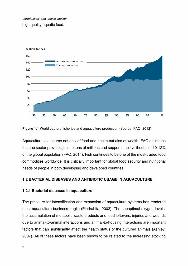
On the other hand, to meet the ever-rising demand for fish, aquaculture has become the fastest growing food-producing sector in the world, and now provides almost half of the global fish consumption (Figure 1.1). According to the latest statistics of FAO (2014), world aquaculture production has attained 90.4 million tonnes in 2012, including 23.8 million tonnes of aquatic algae and 66.6 million tonnes of food fish. The food fish includes finfishes, crustaceans, molluscs, amphibians, freshwater turtles and other aquatic animals (such as sea cucumbers, sea urchins, sea squirts and edible jellyfishes) produced as food for human consumption. World food fish aquaculture production increased at an average annual rate of 6.2% during 2000-2012, while world aquaculture production volume expanded at an average rate of 8.6% per year between 1980 and 2012 (FAO, 2014). Meanwhile, the diversity of aquaculture species has also increased. In 2012, the number of species registered in FAO statistics was 567, including finfishes (345 species, with 5 hybrids), molluscs (102), crustaceans (59), amphibians and reptiles (6), aquatic invertebreates (9), and marine and freshwater algae (37). Therefore, it is now believed that aquaculture has the potential to make a significant contribution to the rising food demand and to provide safe and
Introduction and thesis outline
2
high quality aquatic food.
Figure 1.1 World capture fisheries and aquaculture production (Source: FAO, 2012)
Aquaculture is a source not only of food and health but also of wealth. FAO estimates that the sector provides jobs to tens of millions and supports the livelihoods of 10-12% of the global population (FAO, 2014). Fish continues to be one of the most-traded food commodities worldwide. It is critically important for global food security and nutritional needs of people in both developing and developed countries.
1.2 BACTERIAL DISEASES AND ANTIBIOTIC USAGE IN AQUACULTURE
1.2.1 Bacterial diseases in aquaculture
The pressure for intensification and expansion of aquaculture systems has rendered most aquaculture business fragile (Piedrahita, 2003). The suboptimal oxygen levels, the accumulation of metabolic waste products and feed leftovers, injuries and wounds due to animal-to-animal interactions and animal-to-housing interactions are important factors that can significantly affect the health status of the cultured animals (Ashley, 2007). All of these factors have been shown to be related to the increasing stocking
Chapter I
3
density of the animals. When being reared in intensive systems, aquatic animals are exposed to stressful environmental conditions, which leads to decreased growth performance, reduced immune responses and increased disease susceptibility (Ashley, 2007).
Disease outbreaks have become a primary constraint to aquaculture growth and have a severe impact on both the economic and socio-economic development in many countries worldwide (Bondad-Reantaso et al., 2005). Economic losses in the aquaculture industry resulting from disease outbreaks have been estimated by the FAO to be in excess of US$9 billion per year, which is roughly 15% of the value of world farmed fish and shellfish production. Bacteria and viruses are the main etiological agents in aquaculture, while bacterial pathogens have been reported to cause more disease problems than all other causes combined (Meyer, 1991). Vibriosis, disease caused by bacteria belonging to the genus Vibrio, has become the economically most important disease in marine fish culture, affecting a large number of species (Haenen et al., 2014). The development and spread of bacterial diseases is the result of a complex interaction among pathogen, host and environment (Ashley, 2007). In many cases, vibrios are found to cause disease when the host organism is immune-suppressed or otherwise physiologically stressed (Peddie and Wardle, 2005). Among vibrios, strains belonging to the Harveyi clade (including the species Vibrio harveyi, V. campbellii, V. parahaemolyticus and V. alginolyticus) are widely accepted to be major pathogens, causing massive mortalities of cultured aquatic animals worldwide (Defoirdt et al., 2007; Austin and Zhang, 2006; Karunasagar et al., 1994).
1.2.2 Antibiotic use in aquaculture
Antibiotics are still critically important as the most popular and intensively used chemotherapeutic agents in the treatment of infectious diseases of humans, animals and plants. In a study by Holmström et al., (2003), around 74% (56 of 76) shrimp farmers being interviewed used antibiotics, and most of the farmers used the antibiotics prophylactically, even on a daily basis. All the antibiotics legally used in aquaculture must be authorized by the government agency that is responsible for veterinary medicine, such as the Food and Drug Administration (FDA) in the USA.
Introduction and thesis outline
4
These regulatory agencies also set different rules for antibiotic use, including permissible routes of delivery, dose forms, withdrawal times and tolerances (Burridge et al., 2010). The growing awareness that antibiotics should be used with more care has prompted more strict regulations on the use of antibiotics in aquaculture and on the presence of antibiotic residues in aquaculture products (Romero et al., 2014). One notable example is the ban on the use of antibiotics as growth promoters in animal production in Europe in 2006 (European Parliament and Council Regulation No 1831/2003). An overview of common antibiotics used in aquaculture is shown in Table 1.1.
The use of antibiotics in aquaculture is decreasing in some countries, generally those of Northern Europe, North America and Japan, which have implied strict regulations on the use of antimicrobial agents in animal production. However, the use of antibiotics for preventive purposes is still significant in those countries with insufficient guidance. Moreover, it is difficult to determine the current levels of antibiotic use in aquaculture worldwide because different countries have different distribution and registration systems. To make things even more complicated, the data of the quantity of antibiotic usage are inadequate. As mentioned by Defoirdt et al., (2011), many countries lack sufficient documentation on the quantity of antibiotics used in animal production, including aquaculture industry. Nevertheless, some early studies provided estimations of the antibiotics used in aquaculture. For example, Moriarty (1999) suggested that the quantity of antibiotics used for shrimp farming in Thailand was around 500-600 tonnes in 1994. Due to the differences of regulations regarding the use of antibiotics, the usage of antibiotics may significantly vary between countries (Burridge et al., 2010). For example, the study of Smith (2008) reported that the use of antibiotics could range from 1 g per tonne in Norway to 700 g per tonne in Vietnam.
The massive use of antibiotics in the past has led to development of multiple antibiotic resistances in pathogens (Table 1.1). As a consequence, antibiotic treatments are becoming less effective in controlling bacterial infections in some cases at this moment. For instance, mass mortality in Penaeus monodon larvae caused by Vibrio harveyi strains with multiple resistance to cotrimoxazole, chloramphenicol, erythromycin and streptomycin has been demonstrated (Karunasagar et al., 1994).
Chapter I
5
Moreover, the excessive use of antibiotics in aquaculture also presents a risk to the environment and human health, as the antibiotic resistance determinants have been found located on mobile genetic elements (Alderman and Hastings, 1998; Cabello, 2006). Therefore, these resistance determinants can be horizontally transferred to other (antibiotic-sensitive) bacteria, not only in the aquatic environment but even to bacteria from the terrestrial environment, including animal and human pathogens (Cabello et al., 2013). Additionally, the application of disinfection strategies with a broad spectrum targets not only the pathogenic bacteria, but also leads to an alternation of the normal host microflora, and this might render the situation even more problematic as in this way, bacteria that compete with the pathogens for resources are killed as well (De Schryver et al., 2014). Therefore, for more effective treatment of bacterial diseases, global efforts are needed to promote more judicious use of antibiotics in aquaculture and novel alternative strategies are also urgently needed for the sustainable development of aquaculture, there is an urgent need for alternatives to antibiotics in aquaculture.
Introduction and thesis outline
6
Table 1.1 The different classes of antibiotics used in aquaculture, their importance for human medicine and examples of (multi) resistant
pathogenic bacteria isolated from aquaculture settings (adapted from Bondad-Reantaso et al., 2005).
Drug classes Importance
for human
medicinea
Example Resistant bacteria Multipleb
resistance
Isolated from Reference
Aminoglycosides Critically
important
Streptomycin Edwardsiella ictulari Yes Diseased striped catfish
(Pangasianodon hypophthalmus),
Vietnam
Dung et al., 2008
Amphenicols Important Florfenicol Enterobacter spp. and
Pseudomonas spp.
Yes Freshwater salmon farms, Chile Fernández Alarcón
et al., 2010
Beta-lactams Critically
important
Amoxicillin Vibrio spp.,
Aeromonas spp.
and Edwardsiella tarda
Yes Different aquaculture settings,
Australia
Akinbowale et al.,
2006
Beta-lactams Critically
important
Ampicillin Vibrio harveyi Yes Shrimp farms and coastal waters,
Indonesia
Teo et al., 2000
Fluoroquinolones Critically
important
Enrofloxacin Tenacibaculum
maritimum
Yes Diseased turbot (Scophthalmus
maximus) and sole (Solea
senegalensis), Spain and Portugal
Avendaño-Herrera
et al., 2008
Macrolides Critically
important
Erythromycin Salmonella spp Yes Marketed fish, China Broughton and
Walker, 2009
Chapter I
7
Nitrofurans Critically
important
Furazolidone Vibrio anguillarum Yes Diseased sea bass and sea bream,
Greece
Smith and
Christofilogiannis,
2007
Nitrofurans Important Nitrofurantoin Vibrio harveyi Yes Diseased penaeid shrimp, Taiwan Liu et al., 1997
Quinolones Critically
important
Oxolinic acid Aeromonas spp.,
Pseudomonas spp.
and Vibrio spp.
Yes Pond water, pond sediment and tiger
shrimp (Penaeus monodon),
Philippines
Tendencia and la
Peña, 2001
Sulphonamides Important Sulphadiazine Aeromonas spp. Yes Diseased katla (Catla catla), mrigel
(Cirrhinus mrigala) and punti (Puntius
spp.), India
Das et al., 2009
Tetracyclines Highly
important
Tetracycline Aeromonas
Hydrophila
Yes Water from mullet and tilapia farms,
Egypt
Ishida et al., 2010
Tetracyclines Highly
important
Oxytetracycline Aeromonas
Salmonicida
Yes Atlantic salmon (Salmo salar)
culture facilities, Canada
McIntosh et al.,
2008
Introduction and thesis outline
8
2 THESIS OUTLINE
The general objective of this study was to evaluate the impact of pathogen-pathogen
signaling and sensing of host factors on the virulence of V. harveyi in a model system
with gnotobiotic brine shrimp larvae. The specific objectives and the thesis outline are
as following:
! Chapter 2 (Literature review) gives an overview of the current knowledge on V.
harveyi, including the virulence, pathogenesis, and the regulatory mechanisms of
virulence factors. This chapter also discusses antivirulence therapy as a strategy
for the future treatment of bacterial infections.
! Chapter 3 (Quorum sensing positively regulates flagellar motility in pathogenic Vibrio harveyi) aims at verifying the effect of quorum sensing on
swimming motility in V. harveyi and on the expression of selected genes involved
in flagellar motility. We further investigated the importance of flagellar motility for
the virulence of V. harveyi by applying a motility inhibitor.
! Chapter 4 (Specific quorum sensing-disrupting activity (Aqsi) of thiophenones and their therapeutic potential in a gnotobiotic brine shrimp - Vibrio harveyi model system) aims at determining quorum sensing-disrupting
activity, protective effect and toxicity of 20 novel thiophenone compounds.
Furthermore, we propose a new parameter to describe specific quorum
sensing-inhibitory activity, AQSI. The use of the proposed parameter AQSI is a
straightforward and elegant way to exclude false positives by taking into account
side effects related to the use of quorum sensing molecule reporters.
! Chapter 5 (Indole and indole analogues produced by micro-algae decrease the virulence of luminescent vibrios, major pathogens of aquatic organisms) aims at determining the impact of indole signaling on the virulence of
V. harveyi, and at investigating whether indole analogues produced by
micro-algae induce a similar response as indole.
! Chapter 6 (Norepinephrine and dopamine increase motility, biofilm formation and virulence of Vibrio harveyi) aims at investigating the impact of
the catecholamines norepinephrine and dopamine (neurotransmitters produced

Chapter I
9
by higher organisms) on the growth of V. harveyi in serum-based medium, on the
expression of various virulence-related characteristics and on virulence towards
gnotobiotic brine shrimp (Artemia franciscana) larvae.
! Chapter 7 summarizes the overall findings obtained in this thesis. Conclusions
are drawn and possibilities for future research are proposed.

Introduction and thesis outline
10

CHAPTER II LITERATURE REVIEW

Chapter II
11
ABSTRACT
Vibrio harveyi is amongst the most significant pathogens in the larviculture and
aquaculture industry. It is able to infect a wide range of marine vertebrates and
invertebrates, causing significant losses to the aquaculture industry worldwide. The
pathogenicity mechanism of V. harveyi is not yet completely understood and is
thought to involve attachment to host surfaces, biofilm formation and the production of
various extracellular products. The inhibition of the production of virulence factors that
are required to cause disease, i.e. antivirulence therapy, has been proposed as a
novel strategy to control bacterial infections. Ideally, such a strategy does not harm
the harmless and beneficial microbiota associated with the host, and is also thought to
apply less selective pressure for the development of resistance than conventional
antibiotics. The production of virulence factors often is under strict regulatory control,
and one of the regulatory mechanisms is quorum sensing, bacterial cell-to-cell
communication. Disruption of quorum sensing is the most intensively studied strategy
to inhibit virulence factor production. In this review, we discuss the current knowledge
with respect to pathogenicity mechanisms by which V. harveyi causes infection.
Furthermore, an overview is given about the present knowledge with respect to
antivirulence therapy.
2.1 Vibrio harveyi
2.1.1 Characteristics
Vibrio harveyi has been considered to be amongst the most significant pathogens in
the larviculture and aquaculture industry (Austin and Zhang, 2006). V. harveyi was
originally named as Achromobacter harveyi (Johnson and Shunk, 1936). Afterwards,
it has been called Lucibacterium harveyi, Beneckea harveyi at various times (Farmer
et al. 2005). Finally, the organism is regarded as one of the core species of the genus
Vibrio according to the result of 16S rRNA sequence analysis (Dorsch et al., 1992). V.
harveyi is a marine Gram-negative luminous organism that is ubiquitous in the marine
environment, either free-living in the sea or associated with marine organisms (Austin
Literature Review
12
and Zhang, 2006). It is rod-shaped, motile (via polar and lateral flagella), facultatively anaerobic, halophilic, and competent for both fermentative and respiratory metabolism. It does not grow below 4°C or above 35°C (Owens et al., 2006).
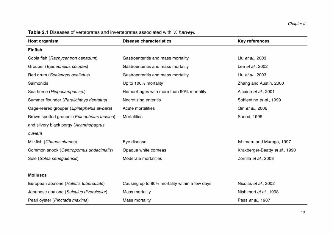
2.1.2 The diseases caused by V. harveyi
V. harveyi has been regarded as both a primary and opportunistic pathogen of a wide range of marine vertebrates and invertebrates, including corals, oysters, prawns, lobsters, and finfish (Austin and Zhang, 2006). Diseases caused by V. harveyi include eye-lesions, gastro-enteritis, vasculitis, and luminous vibriosis (Table 2.1). Luminous vibriosis has been reported to cause severe losses in aquaculture worldwide, particularly in shrimp culture, and it has been considered to be a major constraint to shrimp production in South America and Asia (Austin and Zhang, 2006). Previous work in our laboratory has shown that the pathogenicity of V. harveyi is a strain characteristic rather than a species characteristic as some strains can be highly virulent, whereas other strains are avirulent (Ruwandeepika et al., 2012). The relationship between the presence of virulence genes and the pathogenicity of bacteria is not always evident (CanoGomez et al. 2009). For example, it has been recently found that both highly virulent and non-virulent V. harveyi strains contain all virulence genes tested, including haemolysin and proteases. The regulation of virulence gene expression is critical for the virulence of these bacteria. Indeed, virulent strains showed significantly higher expression levels of virulence-related genes than non-virulent strains (Ruwandeepika et al. 2011).
Chapter II
13
Table 2.1 Diseases of vertebrates and invertebrates associated with V. harveyi. Host organism Disease characteristics Key references
Finfish
Cobia fish (Rachycentron canadum) Gastroenteritis and mass mortality Liu et al., 2003
Grouper (Epinephelus coiodes) Gastroenteritis and mass mortality Lee et al., 2002
Red drum (Scaienops ocellatus) Gastroenteritis and mass mortality Liu et al., 2003
Salmonids Up to 100% mortality Zhang and Austin, 2000
Sea horse (Hippocampus sp.) Hemorrhages with more than 90% mortality Alcaide et al., 2001
Summer flounder (Paralichthys dentatus) Necrotizing enteritis Soffientino et al., 1999
Cage-reared grouper (Epinephelus awoara) Acute mortalities Qin et al., 2006
Brown spotted grouper (Epinephelus tauvina)
and silvery black porgy (Acanthopagrus
cuvieri)
Mortalities Saeed, 1995
Milkfish (Chanos chanos) Eye disease Ishimaru and Muroga, 1997
Common snook (Centropomus undecimalis) Opaque white corneas Kraxberger-Beatty et al., 1990
Sole (Solea senegalensis)
Moderate mortalities Zorrilla et al., 2003
Molluscs
European abalone (Haliotis tuberculate) Causing up to 80% mortality within a few days Nicolas et al., 2002
Japanese abalone (Sulculus diversicolor) Mass mortality Nishimori et al., 1998
Pearl oyster (Pinctada maxima) Mass mortality Pass et al., 1987
Literature Review
14
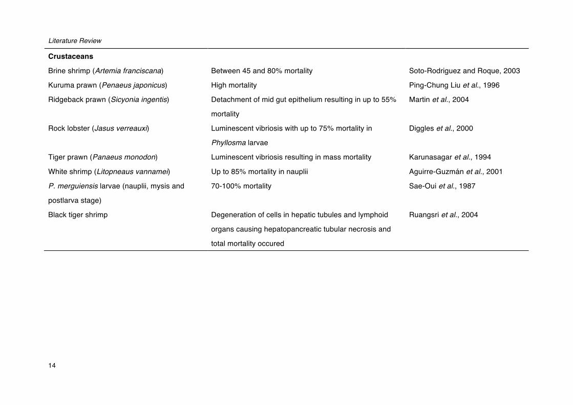
Crustaceans
Brine shrimp (Artemia franciscana) Between 45 and 80% mortality Soto-Rodriguez and Roque, 2003
Kuruma prawn (Penaeus japonicus) High mortality Ping-Chung Liu et al., 1996
Ridgeback prawn (Sicyonia ingentis) Detachment of mid gut epithelium resulting in up to 55%
mortality
Martin et al., 2004
Rock lobster (Jasus verreauxi) Luminescent vibriosis with up to 75% mortality in
Phyllosma larvae
Diggles et al., 2000
Tiger prawn (Panaeus monodon) Luminescent vibriosis resulting in mass mortality Karunasagar et al., 1994
White shrimp (Litopneaus vannamei) Up to 85% mortality in nauplii Aguirre-Guzmán et al., 2001
P. merguiensis larvae (nauplii, mysis and
postlarva stage)
70-100% mortality Sae-Oui et al., 1987
Black tiger shrimp Degeneration of cells in hepatic tubules and lymphoid
organs causing hepatopancreatic tubular necrosis and
total mortality occured
Ruangsri et al., 2004
Chapter II
15
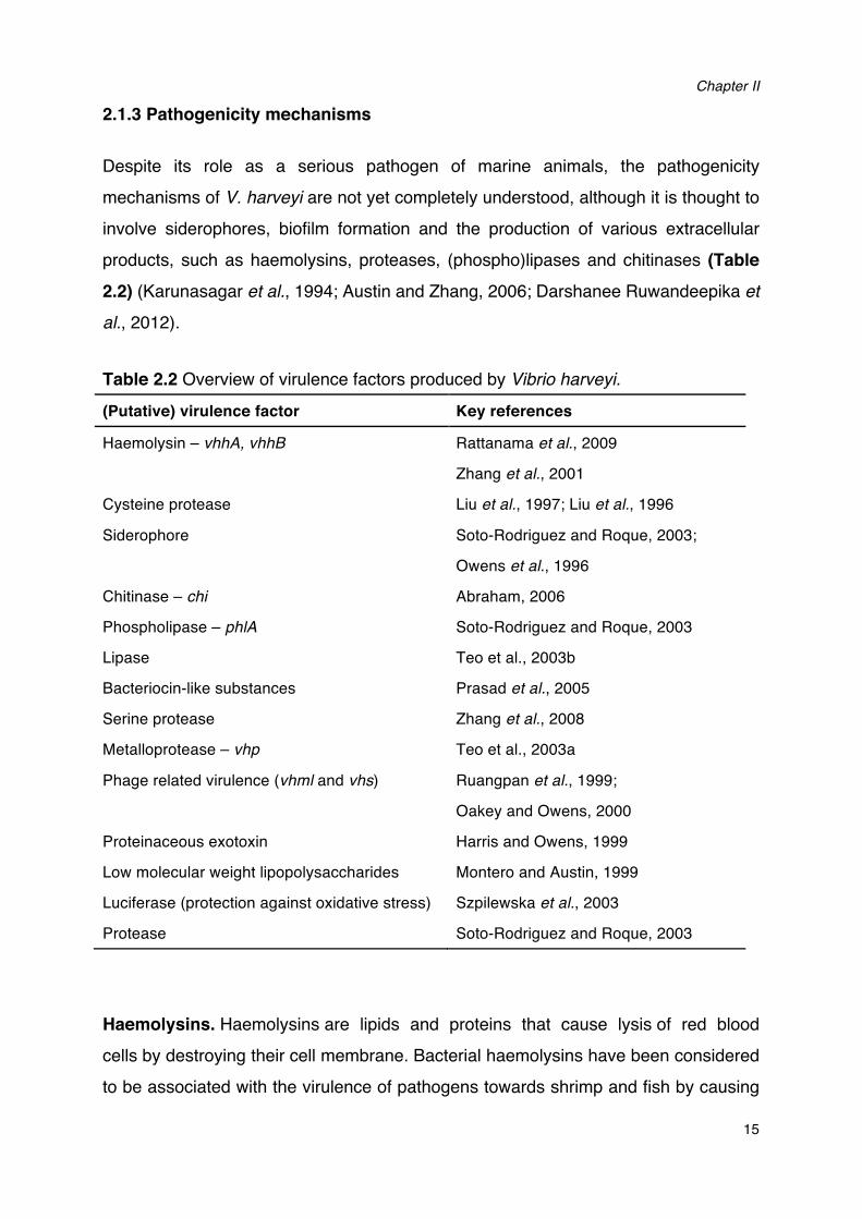
2.1.3 Pathogenicity mechanisms
Despite its role as a serious pathogen of marine animals, the pathogenicity mechanisms of V. harveyi are not yet completely understood, although it is thought to involve siderophores, biofilm formation and the production of various extracellular products, such as haemolysins, proteases, (phospho)lipases and chitinases (Table 2.2) (Karunasagar et al., 1994; Austin and Zhang, 2006; Darshanee Ruwandeepika et
al., 2012).
Table 2.2 Overview of virulence factors produced by Vibrio harveyi. (Putative) virulence factor Key references
Haemolysin – vhhA, vhhB Rattanama et al., 2009
Zhang et al., 2001
Cysteine protease Liu et al., 1997; Liu et al., 1996
Siderophore Soto-Rodriguez and Roque, 2003;
Owens et al., 1996
Chitinase – chi Abraham, 2006
Phospholipase – phlA Soto-Rodriguez and Roque, 2003
Lipase Teo et al., 2003b
Bacteriocin-like substances Prasad et al., 2005
Serine protease Zhang et al., 2008
Metalloprotease – vhp Teo et al., 2003a
Phage related virulence (vhml and vhs) Ruangpan et al., 1999;
Oakey and Owens, 2000
Proteinaceous exotoxin Harris and Owens, 1999
Low molecular weight lipopolysaccharides Montero and Austin, 1999
Luciferase (protection against oxidative stress) Szpilewska et al., 2003
Protease Soto-Rodriguez and Roque, 2003
Haemolysins. Haemolysins are lipids and proteins that cause lysis of red blood cells by destroying their cell membrane. Bacterial haemolysins have been considered to be associated with the virulence of pathogens towards shrimp and fish by causing
Literature Review
16
hemorrhagic septicemia and diarrhea in the host (Liu et al., 1996; Sun et al., 2007).
Haemolysin is one of the major virulence factors in V. harveyi. V. harveyi VIB 645,
which is known to be pathogenic towards salmonids, was found to produce
extracellular products with a high hemolytic activity towards fish erythrocytes, and two
closely related haemolysin genes (designated vhhA and vhhB) were identified in this
strain (Zhang et al., 2001). Furthermore, a single residue change in V. harveyi
haemolysin showed a loss of haemolytic activity and pathogenicity to turbot (Sun et al.,
2007).
Proteases. Proteases are enzymes that hydrolyse the peptide bonds that link amino
acids together in proteins, and different classes of protease can perform the same
reaction by completely different catalytic mechanisms. Proteases have been reported
in different aquaculture pathogens to be linked with virulence towards both shrimp and
fish (Teo et al., 2003a). This group of enzymes includes metalloproteases, cysteine
proteases, serine proteases, collagenases, caseinases and gelatinases (Defoirdt,
2013). Among the proteases, metalloprotease has been regarded as a virulence
factor in V. harveyi. Teo et al. (2003) found that a novel metalloprotease Pap6, which
was able to digest various of host proteins, including gelatin, fibronectin and type IV
collagen, could play a potential role in the pathogenesis of V. harveyi strain AP6.
Additionally, cysteine protease, lipase, phospholipase and chitinase were also
considered to be major virulence factors of V. harveyi (Teo et al., 2003b; Lee et al.,
1999; Soto-Rodriguez and Roque, 2003; Abraham, 2006).
Iron acquisition and siderophores. Iron is a vital nutrient required for several
important biological cellular processes, ranging from growth and DNA replication to
oxygen transport and protection against oxidative stress (Sandy and Butler, 2009).
Iron is also an essential element for bacterial pathogens, as these organisms have to
acquire iron within their vertebrate hosts in order to replicate and cause infection
(Skaar, 2010). However, the bioavailability of iron is limited in the host, since the
majority of vertebrate iron is intracellular and sequestered by lactoferrin, transferrin,
and ferritin as a primary defense mechanism (Brooks et al., 2004). Further, the
physiological pH of serum and the aerobic environment ensures that extracellular iron
is insoluble and is difficult to access by invading pathogens (Sandy and Butler, 2009).
Chapter II
17
In order to thrive within vertebrates, many pathogens have evolved iron uptake mechanisms to extract iron from their surrounding environments (Naka, 2011). These uptake systems can be divided into three main categories: siderophore-mediated iron uptake, heme-mediated iron uptake, transferrin and lactoferrin-mediated iron uptake (Sandy and Butler, 2009). V. harveyi can acquire iron by means of siderophores, which are low-molecular-weight iron (III) chelators secreted by the bacteria. The siderophore-iron complex is bound by a receptor at the bacterial surface. Then it is internalized into the cell and the iron is released as a nutrient source (Skaar, 2010). V.
harveyi can produce amphiphilic enterobactin-like siderophores. Eight amphi-enterobactins have been identified in V. harveyi with various fatty acid appendages ranging in length (C10-C14), degree of unsaturation and hydroxylation (Zane et al., 2014). Siderophores contribute to competitiveness in environmental bacteria and serve as virulence determinants in many vibrios such as V. harveyi, V.
cholerae, V. parahaemolyticus and V. anguillarum (Andrus et al., 1983; Owens et al., 1996; Amaro et al., 1990; Pybus et al., 1994).
Production of extracellular polysaccharide and biofilm formation. Surface molecules are playing a major role during bacterial infection by enabling a complex interaction between the pathogen and its host, and these molecules include extracellular polysaccharides (EPS) and lipopolysaccharides (LPS). EPS are secreted around the cell as a capsule or as a loose slime, while LPS are components of the outer membrane in most of the Gram-negative bacteria (Costerton et al., 1981). EPS are important for adhesion, nutrient sequestration, chelation of heavy metals, detoxification of toxic compounds and protection against osmotic shock (Hoagland et
al., 1993; Decho, 1990). Production of both EPS and LPS have been reported in V.
harveyi as virulence factors (Bramhachari and Dubey, 2006; Montero and Austin, 1999). Capsular polysaccharides can form a capsule surrounding the bacterial cells, which is involved in attachment to host cells and play an important role in immune evasion (Chen et al., 2010; Hsieh et al., 2003). Another group of extracellular polysaccharides, the exopolysaccharides are able to form a loose slime outside the cell, which can form an intercellular matrix in biofilms (Mah and O'Toole, 2001). Biofilm formation is one of the important adaptive mechanisms of microorganisms to survive in the environment and within the host. The biofilm matrix enhances the
Literature Review
18
growth and survival of microorganisms by providing access to nutrients and protection from detergents and antimicrobials (Donlan and Costerton, 2002; Mah and O'Toole, 2001). The persistence and survival of V. harveyi in shrimp hatcheries has been attributed to the bacterium’s biofilm formation ability, which is governing resistance to antibiotics and disinfectants (Karunasagar et al., 1994).
Other virulence factors. In V. harveyi, it has been reported that bacteriophages are important elements in transferring virulence. A bacteriophage identified as V. harveyi
siphoviridae-like phage (VHS1) was reported to enhance the virulence of V. harveyi
towards black tiger shrimp (Khemayan et al., 2006). Later, Munro et al. (2003) demonstrated that another bacteriophage, V. harveyi myovirus-like phage (VHML), could increase the haemolysin activity, protein secretion, and virulence towards P.
monodon larvae. Further, Harris and Owens (1999) suggested that the protenaceaous exotoxins known as T1 and T2 encoded by a phage have a negative effect on Penaeus monodon.
2.2 VIRULENCE REGULATORY MECHANISMS
As virulence factors are often costly metabolic products, it should not be surprising that their production is under strict regulatory control. Various virulence regulatory mechanisms have been documented in pathogenic bacteria. In the following paragraphs, we will focus on quorum sensing and sensing of host stress hormones.
2.2.1 Quorum sensing
Quorum sensing is a cell-to-cell communication process in which bacteria control the expression of certain genes by producing, detecting and responding to small extracellular signal molecules named autoinducers (Ng and Bassler, 2009). Autoinducers are synthesized intracellularly and then passively released or actively secreted outside of the cells. The extracellular autoinducers accumulate as the bacterial population increases. When the concentration reaches the threshold level required for detection, specific cognate receptors bind the autoinducers and trigger signal transduction cascades, resulting in activation or repression of various target genes (Waters and Bassler, 2007; Ng and Bassler, 2009). This kind of mechanism
Chapter II
19
was first described in the marine bacterium Vibrio fisheri, and was found to exist in many other bacteria later on (Nealson et al., 1970).
Originally, three main types of autoinducers were described. Acyl-homoserine lactones (AHLs) are a major class of autoinducers used by Gram-negative proteobacteria for intraspecies communication, and they also have been documented in some bacteriodetes, cyanobacteria and archaea (Sharif et al., 2008; Huang et al., 2008; Zhang et al., 2012). AHLs consist of conserved homoserine lactone rings coupled to an acyl side chain of variable length (from C4 to C18), which can have a modification (oxo or hydroxyl) at the C3 position (Fuqua and Greenberg, 2002). The second type of autoinducers is oligopeptides, which are used as signal molecules by Gram-positive bacteria. Unlike AHLs, peptide autoinducers are genetically encoded, thus each species of bacteria is capable of producing a peptide autoinducer with a unique sequence (Ng et al., 2010). A third major signal molecule is autoinducer-2 (AI-2), which is synthesized by the LuxS enzyme that has been identified in over 70 species of Gram-negative and Gram-positive bacteria. AI-2 has several chemical forms derived from 4,5-dihydroxy-2,3-pentanedione (DPD), and it is proposed to allow interspecies communication (Xavier and Bassler, 2005; De Keersmaecker et al., 2006; Jayaraman and Wood, 2008). However, the AI-2 biosynthesis enzyme LuxS also has an important metabolic function, as it is a key factor in the activated methyl cycle. Therefore, the role of LuxS in quorum sensing in most species needs to be further elucidated (Platt and Fuqua, 2010). In addition to these three types of signal molecules, an increasing number of autoinducers belonging to various chemical classes are still being discovered (and more are probably awaiting discovery) (for a review, see LaSarre and Federle, 2013).
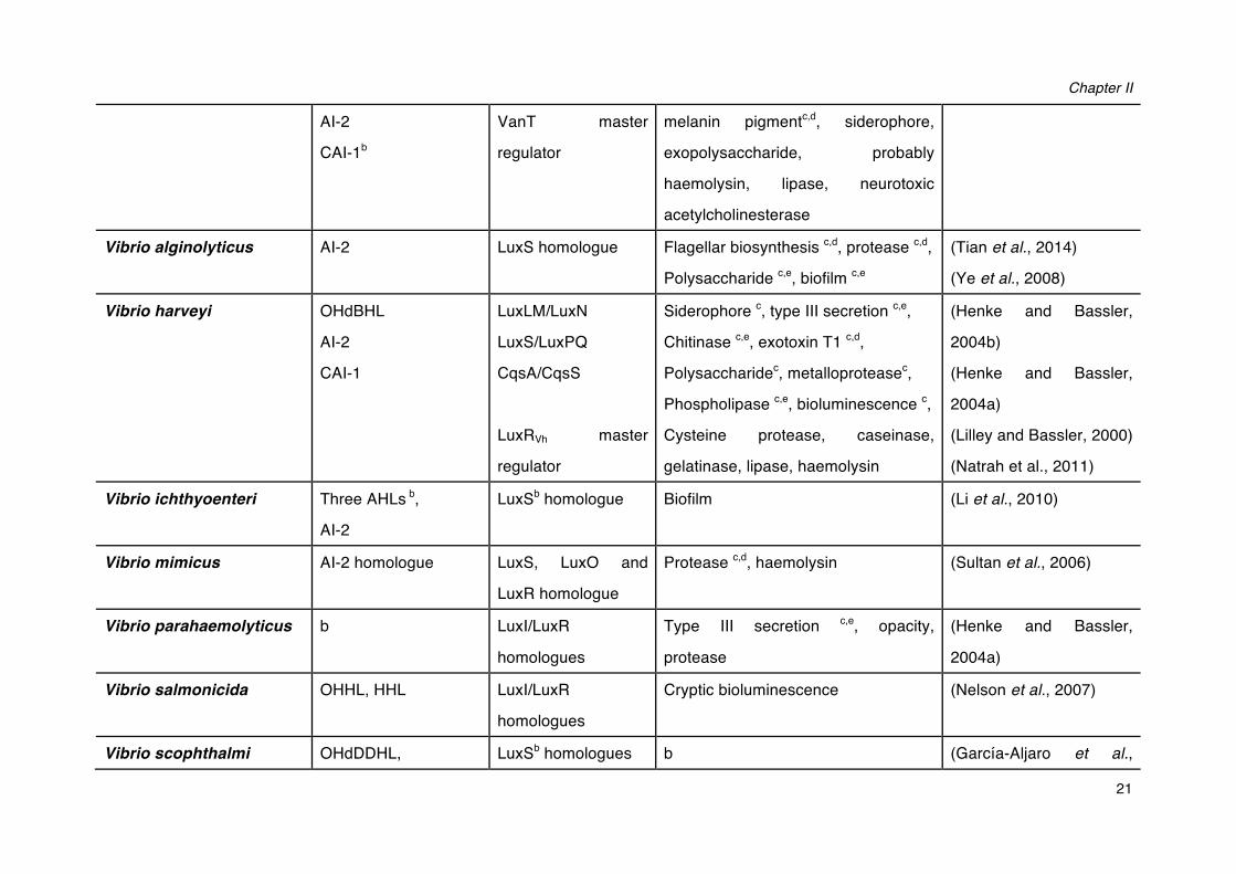
Quorum sensing systems regulate a wide variety of phenotypes in bacteria, including bioluminescence, conjugal plasmid transfer, nodulation, sporulation, antibiotic resistance, antibiotic production, swarming motility and most importantly biofilm formation and virulence (Table 2.3; Diggle et al., 2007; Fuqua and Greenberg, 2002; Miller and Bassler, 2003; Whitehead et al., 2001; De Kievit and Iglewski, 2000).
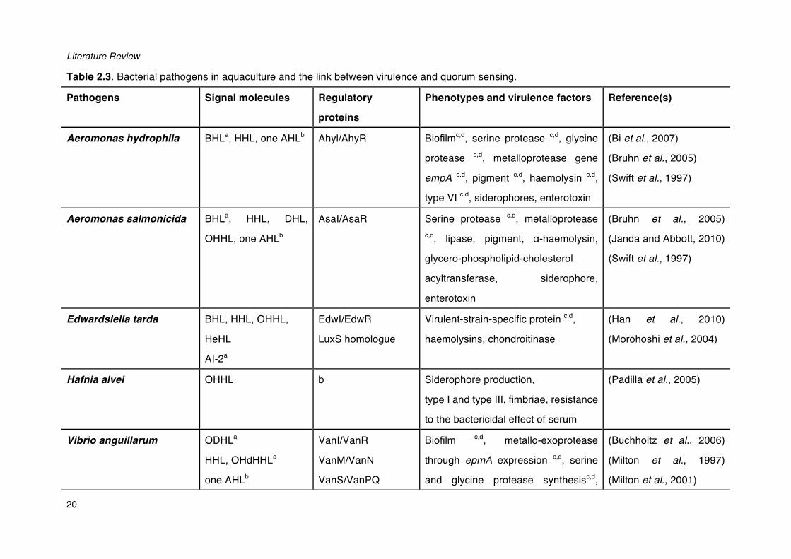
Literature Review
20
Table 2.3. Bacterial pathogens in aquaculture and the link between virulence and quorum sensing.
Pathogens Signal molecules Regulatory
proteins
Phenotypes and virulence factors Reference(s)
Aeromonas hydrophila BHLa, HHL, one AHLb Ahyl/AhyR Biofilmc,d, serine protease c,d, glycine
protease c,d, metalloprotease gene
empA c,d, pigment c,d, haemolysin c,d,
type VI c,d, siderophores, enterotoxin
(Bi et al., 2007)
(Bruhn et al., 2005)
(Swift et al., 1997)
Aeromonas salmonicida BHLa, HHL, DHL,
OHHL, one AHLb
AsaI/AsaR Serine protease c,d, metalloprotease c,d, lipase, pigment, ɑ-haemolysin,
glycero-phospholipid-cholesterol
acyltransferase, siderophore,
enterotoxin
(Bruhn et al., 2005)
(Janda and Abbott, 2010)
(Swift et al., 1997)
Edwardsiella tarda BHL, HHL, OHHL,
HeHL
AI-2a
EdwI/EdwR
LuxS homologue
Virulent-strain-specific protein c,d,
haemolysins, chondroitinase
(Han et al., 2010)
(Morohoshi et al., 2004)
Hafnia alvei OHHL b Siderophore production,
type I and type III, fimbriae, resistance
to the bactericidal effect of serum
(Padilla et al., 2005)
Vibrio anguillarum ODHLa
HHL, OHdHHLa
one AHLb
VanI/VanR
VanM/VanN
VanS/VanPQ
Biofilm c,d, metallo-exoprotease
through epmA expression c,d, serine
and glycine protease synthesisc,d,
(Buchholtz et al., 2006)
(Milton et al., 1997)
(Milton et al., 2001)
Chapter II
21
AI-2
CAI-1b
VanT master
regulator
melanin pigmentc,d, siderophore,
exopolysaccharide, probably
haemolysin, lipase, neurotoxic
acetylcholinesterase
Vibrio alginolyticus AI-2 LuxS homologue Flagellar biosynthesis c,d, protease c,d,
Polysaccharide c,e, biofilm c,e
(Tian et al., 2014)
(Ye et al., 2008)
Vibrio harveyi OHdBHL
AI-2
CAI-1
LuxLM/LuxN
LuxS/LuxPQ
CqsA/CqsS
LuxRVh master
regulator
Siderophore c, type III secretion c,e,
Chitinase c,e, exotoxin T1 c,d,
Polysaccharidec, metalloproteasec,
Phospholipase c,e, bioluminescence c,
Cysteine protease, caseinase,
gelatinase, lipase, haemolysin
(Henke and Bassler,
2004b)
(Henke and Bassler,
2004a)
(Lilley and Bassler, 2000)
(Natrah et al., 2011)
Vibrio ichthyoenteri Three AHLs b,
AI-2
LuxSb homologue Biofilm (Li et al., 2010)
Vibrio mimicus AI-2 homologue LuxS, LuxO and
LuxR homologue
Protease c,d, haemolysin (Sultan et al., 2006)
Vibrio parahaemolyticus b LuxI/LuxR
homologues
Type III secretion c,e, opacity,
protease
(Henke and Bassler,
2004a)
Vibrio salmonicida OHHL, HHL LuxI/LuxR
homologues
Cryptic bioluminescence (Nelson et al., 2007)
Vibrio scophthalmi OHdDDHL, LuxSb homologues b (García-Aljaro et al.,
Literature Review
22
Two AHLsb
AI-2
2008)
Vibrio vulnificus BHL, ODHL, ODDHL,
minor HHL, OHL,
OTHL
AI-2
LuxU, LuxO,
SmcR transcriptional
regulator
LuxS/LuxPQ
Metalloprotease c,d, cytolysin c,e,
haemolysin c, extracellular capsular
polysaccharide, siderophore, toxin
RTX
(Bruhn et al., 2005)
(Kim et al., 2003)
Yersinia ruckeri OOHLa, HHL, OHHL,
OHeHL, OHL, ONHL,
ODHL, ODDHL
YenI/YenR Metalloprotease, protein secretion,
Siderophores, heat sensitive factors
(Bruhn et al., 2005)
(Kastbjerg et al., 2007)
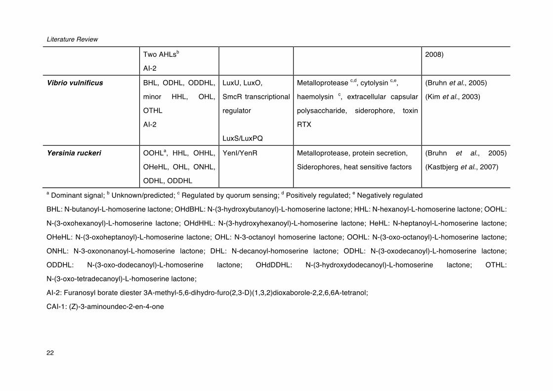
a Dominant signal; b Unknown/predicted; c Regulated by quorum sensing; d Positively regulated; e Negatively regulated
BHL: N-butanoyl-L-homoserine lactone; OHdBHL: N-(3-hydroxybutanoyl)-L-homoserine lactone; HHL: N-hexanoyl-L-homoserine lactone; OOHL:
N-(3-oxohexanoyl)-L-homoserine lactone; OHdHHL: N-(3-hydroxyhexanoyl)-L-homoserine lactone; HeHL: N-heptanoyl-L-homoserine lactone;
OHeHL: N-(3-oxoheptanoyl)-L-homoserine lactone; OHL: N-3-octanoyl homoserine lactone; OOHL: N-(3-oxo-octanoyl)-L-homoserine lactone;
ONHL: N-3-oxononanoyl-L-homoserine lactone; DHL: N-decanoyl-homoserine lactone; ODHL: N-(3-oxodecanoyl)-L-homoserine lactone;
ODDHL: N-(3-oxo-dodecanoyl)-L-homoserine lactone; OHdDDHL: N-(3-hydroxydodecanoyl)-L-homoserine lactone; OTHL:
N-(3-oxo-tetradecanoyl)-L-homoserine lactone;
AI-2: Furanosyl borate diester 3A-methyl-5,6-dihydro-furo(2,3-D)(1,3,2)dioxaborole-2,2,6,6A-tetranol;
CAI-1: (Z)-3-aminoundec-2-en-4-one
Chapter II
23
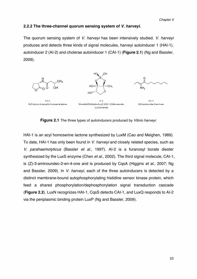
2.2.2 The three-channel quorum sensing system of V. harveyi.
The quorum sensing system of V. harveyi has been intensively studied. V. harveyi
produces and detects three kinds of signal molecules, harveyi autoinducer 1 (HAI-1),
autoinducer 2 (AI-2) and cholerae autoinducer 1 (CAI-1) (Figure 2.1) (Ng and Bassler,
2009).
Figure 2.1 The three types of autoinducers produced by Vibrio harveyi.
HAI-1 is an acyl homoserine lactone synthesized by LuxM (Cao and Meighen, 1989).
To date, HAI-1 has only been found in V. harveyi and closely related species, such as
V. parahaemolyticus (Bassler et al., 1997). AI-2 is a furanosyl borate diester
synthesized by the LuxS enzyme (Chen et al., 2002). The third signal molecule, CAI-1,
is (Z)-3-aminoundec-2-en-4-one and is produced by CqsA (Higgins et al., 2007; Ng
and Bassler, 2009). In V. harveyi, each of the three autoinducers is detected by a
distinct membrane-bound autophosphorylating histidine sensor kinase protein, which
feed a shared phosphorylation/dephosphorylation signal transduction cascade
(Figure 2.2). LuxN recognizes HAI-1, CqsS detects CAI-1, and LuxQ responds to AI-2
via the periplasmic binding protein LuxP (Ng and Bassler, 2009).
Literature Review
24
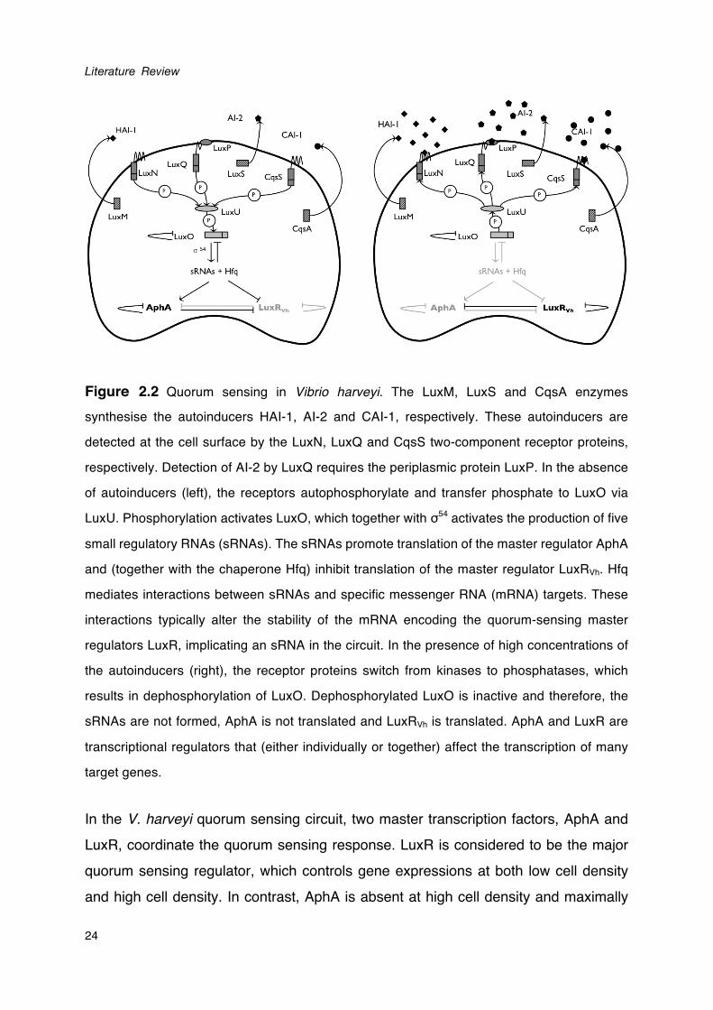
Figure 2.2 Quorum sensing in Vibrio harveyi. The LuxM, LuxS and CqsA enzymes
synthesise the autoinducers HAI-1, AI-2 and CAI-1, respectively. These autoinducers are
detected at the cell surface by the LuxN, LuxQ and CqsS two-component receptor proteins,
respectively. Detection of AI-2 by LuxQ requires the periplasmic protein LuxP. In the absence
of autoinducers (left), the receptors autophosphorylate and transfer phosphate to LuxO via
LuxU. Phosphorylation activates LuxO, which together with σ54 activates the production of five
small regulatory RNAs (sRNAs). The sRNAs promote translation of the master regulator AphA
and (together with the chaperone Hfq) inhibit translation of the master regulator LuxRVh. Hfq
mediates interactions between sRNAs and specific messenger RNA (mRNA) targets. These
interactions typically alter the stability of the mRNA encoding the quorum-sensing master
regulators LuxR, implicating an sRNA in the circuit. In the presence of high concentrations of
the autoinducers (right), the receptor proteins switch from kinases to phosphatases, which
results in dephosphorylation of LuxO. Dephosphorylated LuxO is inactive and therefore, the
sRNAs are not formed, AphA is not translated and LuxRVh is translated. AphA and LuxR are
transcriptional regulators that (either individually or together) affect the transcription of many
target genes.
In the V. harveyi quorum sensing circuit, two master transcription factors, AphA and LuxR, coordinate the quorum sensing response. LuxR is considered to be the major quorum sensing regulator, which controls gene expressions at both low cell density and high cell density. In contrast, AphA is absent at high cell density and maximally
Chapter II
25
produced at low cell density (Rutherford et al., 2011). AphA and LuxR directly regulate expression of genes encoding the quorum sensing regulatory small RNAs, which leads to a feedback loop ensuring a well-timed transition between the individual and the group life-styles (van Kessel et al., 2013). Depending on the target gene, AphA and LuxR can either activate or repress the expression to different extents. Therefore, the production of AphA and LuxR coupled with differences between target genes with respect to the sensitivity of the respective promoters for these regulators enables a myriad of gene expression patterns (van Kessel et al., 2013).
A number of virulence factors has been found to be regulated by quorum sensing in V.
harveyi, including biofilm formation (Anetzberger et al., 2009), type III secretion (Henke and Bassler, 2004), production of a siderophore (Lilley and Bassler, 2000), the Vhp metalloprotease (Mok et al., 2003; Ruwandeepika et al., 2011), chitinase A (Defoirdt et al., 2009) and three phospholipase genes (Natrah et al., 2011). Interestingly, some virulence factors were found to be controlled by a subset of the autoinducers. For instance, vhh heamolysin gene in V. harveyi shows a constantly low expression in AI-2-negative mutant, however, it shows no difference in expression levels between the mutant with inactive quorum sensing system and the mutant with maximally activated quorum sensing system. This suggests that vhh heamolysin expression is regulated by AI-2 quorum sensing through a mechanism that is different from the currently known three-channel signal transduction pathway (Ruwandeepika et al., 2011). Other virulence factors are either not controlled by quorum sensing (e.g. serine protease; Ruwandeepika et al., 2011), or the link with quorum sensing has not yet been investigated (e.g. motility).
2.2.3 Indole signaling
A variety of both Gram-positive and Gram-negative bacteria (more than 85 species) are known to produce indole. These include many pathogenic species such as V.
vulnificus, Proteus vulgaris, Pasteurella multocida and Haemophilus influenzae (Dalsgaard et al., 1999; DeMoss and Moser, 1969; Clemons and Gadberry, 1982; Stull et al., 1995). Indole is synthesized from tryptophan by tryptophanase (TnaA) through a reversible reaction, with pyruvate and ammonia as by-products (Newton
Literature Review
26
and Snell, 1965). Recently, increasing evidence suggests that indole is also used as a
signaling molecule in some bacteria (Lee and Lee, 2010).
Although the indole synthesis pathway has been extensively studied, the biological
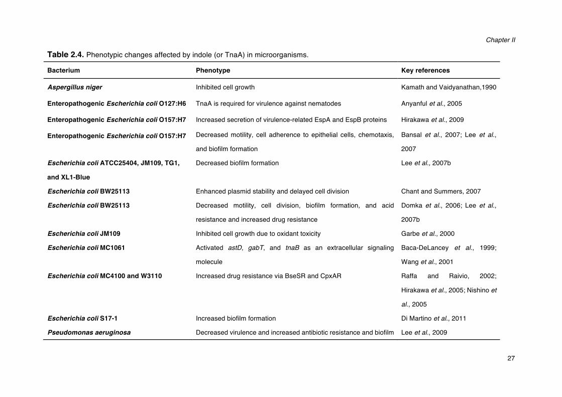
functions of indole are not yet fully known. Till now, several responses to indole have
been revealed (Table 2.4). Further, indole has also been reported to interfere with
AHL-based quorum sensing in a number of Gram-negative bacteria (Hidalgo-Romano
et al., 2014). Some bacteria that lack tryptophanase, and therefore do not produce
indole, e.g. Pseudomonas aeruginosa, also respond to the presence of extracellular
indole (Melander et al., 2014). Many non-indole-producing bacteria, such as
Pseudomonas putida (Ensley et al., 1983), Ralstonia pickettii (Fishman et al., 2005)
and Pseudomonas mendocina (Tao et al., 2004), can oxidize indole, and some of
them utilize indole as a carbon source (Doukyu and Aono, 1997). The oxidized indole
derivatives have been reported to show significant effects on gene expression, motility
and biofilm formation in E. coli and P. aeruginosa (Lee et al., 2007; Bansal et al.,
2007).
A quorum sensing property of indole was first reported in Stigmatella aurantiaca
(Gerth et al., 1993). To date, a signaling role of indole in vibrios only has been
documented in V. cholerae and very recently in V. anguillarum. In V. cholerae, indole
activated Vibrio polysaccharide (VPS) production and biofilm formation, whereas it
decreased motility (Beyhan et al., 2009). In V. anguillarum, by contrast,
exopolysaccharide production was decreased by indole, whereas motility was not
affected (Li et al., 2014). Further, in V. anguillarum, the stationary phase sigma factor
(RpoS) is involved in the production of indole as an rpoS deletion mutant showed
elevated indole levels and increased expression of the indole synthase tnaA (Li et al.,
2014). Finally, the indole receptor has not yet been identified in any bacterium,
although the transcriptional regulator SdiA has been suggested to be central in the
indole signalling cascade in E. coli (Lee et al, 2007a) and the DnaK suppressor
protein DksA has been hypothesised to be involved in the indole signaling cascade in
V. cholerae (Beyhan et al., 2009). In summary, indole represents a new class of
signaling molecules that play an important role in interspecies communication and
various biological functions.
Chapter II
27
Table 2.4. Phenotypic changes affected by indole (or TnaA) in microorganisms.
Bacterium Phenotype Key references
Aspergillus niger Inhibited cell growth Kamath and Vaidyanathan,1990
Enteropathogenic Escherichia coli O127:H6 TnaA is required for virulence against nematodes Anyanful et al., 2005
Enteropathogenic Escherichia coli O157:H7 Increased secretion of virulence-related EspA and EspB proteins Hirakawa et al., 2009
Enteropathogenic Escherichia coli O157:H7 Decreased motility, cell adherence to epithelial cells, chemotaxis,
and biofilm formation
Bansal et al., 2007; Lee et al.,
2007
Escherichia coli ATCC25404, JM109, TG1,
and XL1-Blue
Decreased biofilm formation Lee et al., 2007b
Escherichia coli BW25113 Enhanced plasmid stability and delayed cell division Chant and Summers, 2007
Escherichia coli BW25113 Decreased motility, cell division, biofilm formation, and acid
resistance and increased drug resistance
Domka et al., 2006; Lee et al.,
2007b
Escherichia coli JM109 Inhibited cell growth due to oxidant toxicity Garbe et al., 2000
Escherichia coli MC1061 Activated astD, gabT, and tnaB as an extracellular signaling
molecule
Baca-DeLancey et al., 1999;
Wang et al., 2001
Escherichia coli MC4100 and W3110 Increased drug resistance via BseSR and CpxAR Raffa and Raivio, 2002;
Hirakawa et al., 2005; Nishino et
al., 2005
Escherichia coli S17-1 Increased biofilm formation Di Martino et al., 2011
Pseudomonas aeruginosa Decreased virulence and increased antibiotic resistance and biofilm Lee et al., 2009
Literature Review
28
formation
Salmonella enterica Enhanced drug resistance via RamA Nikaido et al., 2008
Stigmatella aurantiaca Induced a spore formation Stamm et al., 2005
Vibrio cholerae Activated genes involved in polysaccharide production, increased
biofilm formation and grazing resistance to phagocytic eukaryote
Mueller et al., 2007; Mueller et
al., 2009
Vibrio anguillarum Decreased virulence towards gnotobiotic see bass (Dicentrarchus
labrax) larvea, biofilm formation, exopolysaccharide production and
expression of the exopolysacchride synthesis gene wbfD
Li et al., 2014
Chapter II
29
2.2.4 Sensing of host factors
The metabolic condition of the host and some metabolic products has a significant
impact on the success of bacterial infection. Bacteria continuously monitor the
environmental cues in the host during colonization and infection, and subsequently
coordinate the expression of virulence determinants at an appropriate timing
(Mekalanos, 1992). Therefore, it is not surprising that bacteria have evolved a
sophisticated signal transduction system to sense the presence of host cues such as
mucus (Hsiao et al., 2006), bile (Gotoh et al., 2010), bicarbonate (Abuaita and Withey,
2009), catecholamine hormones and lipid hormones (Hughes and Sperandio, 2008).
For example, mucus and bile have been reported to increase production of several
virulence factors in V. anguillarum, including protease activity, flagellar motility, biofilm
formation and exopolysaccharide production (Li et al., 2014). In addition, bile has also
been found to induce production of virulence factors such as type III secretion
system-related proteins, haemolysins, and capsular polysaccharide in V.
parahaemolyticus (Hsieh et al., 2003). In the following paragraphs, we will focus on
the bacterial sensing of host stress hormones.
Catecholamine stress hormones
Stress is essential for the survival of organisms, as it is the basis of a fundamental
survival mechanism, the innate fight-or-flight response, which can prepare the
organism to either challenge or to flee from a threat (Dhabhar, 2009). This response
results in the release of several neurotransmitters, cytokines, hormones, peptides and
other factors into the circulation or locally within tissues (Verbrugghe et al., 2012).
Among them, the major mediators of the stress response include the catecholamines
norepinephrine and epinephrine, which are fast-acting mediators secreted by the
sympathetic nervous system; and cortisol and corticosterone, which are slow-acting
glucocorticoids released by the adrenal gland after activation of the hypothalamic–
pituitary–adrenal (HPA) axis (Lundberg, 2005). Catecholamines are effector
compounds derived from tyrosine and characterized by having a catecholate moiety
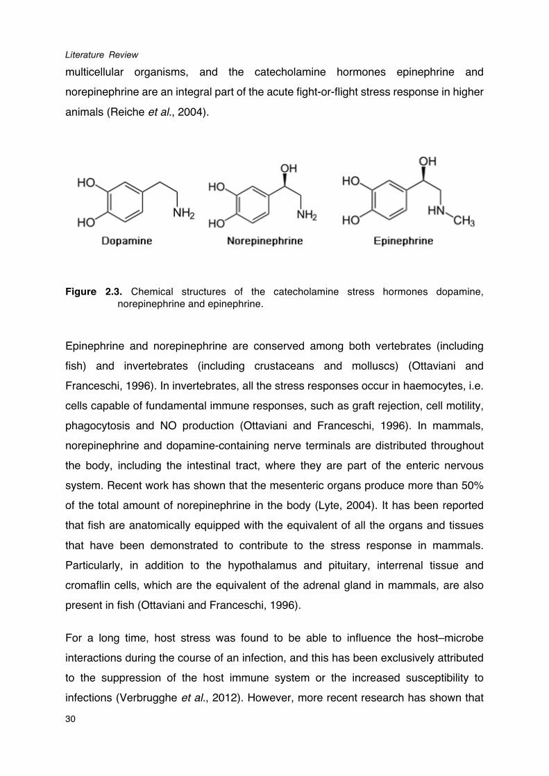
(a benzene ring with two adjacent hydroxyl groups) and an opposing amine side chain
(Figure 2.3). Catecholamines affect several neuroendocrine signaling pathways in
Literature Review
30
multicellular organisms, and the catecholamine hormones epinephrine and norepinephrine are an integral part of the acute fight-or-flight stress response in higher animals (Reiche et al., 2004).
Figure 2.3. Chemical structures of the catecholamine stress hormones dopamine, norepinephrine and epinephrine.
Epinephrine and norepinephrine are conserved among both vertebrates (including fish) and invertebrates (including crustaceans and molluscs) (Ottaviani and Franceschi, 1996). In invertebrates, all the stress responses occur in haemocytes, i.e. cells capable of fundamental immune responses, such as graft rejection, cell motility, phagocytosis and NO production (Ottaviani and Franceschi, 1996). In mammals, norepinephrine and dopamine-containing nerve terminals are distributed throughout the body, including the intestinal tract, where they are part of the enteric nervous system. Recent work has shown that the mesenteric organs produce more than 50% of the total amount of norepinephrine in the body (Lyte, 2004). It has been reported that fish are anatomically equipped with the equivalent of all the organs and tissues that have been demonstrated to contribute to the stress response in mammals. Particularly, in addition to the hypothalamus and pituitary, interrenal tissue and cromaflin cells, which are the equivalent of the adrenal gland in mammals, are also present in fish (Ottaviani and Franceschi, 1996).
For a long time, host stress was found to be able to influence the host–microbe interactions during the course of an infection, and this has been exclusively attributed to the suppression of the host immune system or the increased susceptibility to infections (Verbrugghe et al., 2012). However, more recent research has shown that
Chapter II
31
infectious bacteria have evolved specific sensing systems for detecting the stress
hormones released by the host and the detection of these stress hormones can
directly affect the growth and virulence of the pathogens (Lyte, 2004). These new
findings led to the development of a research area named microbial endocrinology,
which provides evidences of understanding the interaction between microbes and
their animal host during episodes of stress.
Catecholamines and virulence
Most research on sensing of host stress hormones by bacteria has been conducted
on the effects of the catecholamine stress hormones epinephrine (adrenaline),
norepinephrine (noradrenaline) and dopamine on intestinal bacteria such as
Escherichia coli and Salmonella spp. (Freestone et al., 2008). Catecholamines can
stimulate the growth of many Gram-negative and Gram-positive bacteria, including
Escherichia coli, Yersinia enterocolitica, Listeria monocytogenes, Aeromonas
hydrophila, Pseudomonas aeruginosa and Vibrio parahaemolyticus, in minimal
medium containing serum (Lyte and Ernst, 1992; Coulanges et al., 1997; Kinney et al.,
1999; Belay et al., 2003; Nakano et al., 2007). Such media are iron-limited because of
chelation of free iron by the high-affinity iron-binding proteins such as transferrin (Tf)
and lactoferrin (Lf) that are present in serum. Sandrini et al. (2010) demonstrated that
catecholamines can remove iron from host iron-binding proteins via direct binding of
Tf/Lf-complexed iron, with a resultant reduction of the Tf/Lf-coordinated Fe (III) to Fe
(II), an iron valency for which these iron-sequestering proteins have much reduced
binding affinity. Subsequently, catecholamines can deliver the host-sequestered iron
to bacteria through iron uptake systems, and the increased availability of iron
enhances bacterial growth (Sandrini et al., 2010; Sharaff and Freestone, 2011a).
In addition to the growth-stimulatory effect, catecholamines have also been reported
to increase the production of virulence factors in pathogenic bacteria, including the
production of Shiga toxin, chemotaxis, biofilm formation, and attachment and
colonization to epithelial cells in pathogenic E. coli, motility and invasiveness of
Campylobacter jejuni, motility and type III secretion in Salmonella typhimurium, and
cytotoxic activity in V. parahaemolyticus (Bansal et al., 2007; Cogan et al., 2007;
Literature Review
32
Nakano et al., 2007; Lyte et al., 2011; Sharaff and Freestone, 2011b). Possibly, bacteria detect the increased concentrations of stress hormones and respond with an increased growth and enhanced potential to cause disease when the host is weakened (Freestone et al., 2008).
Specificity and receptors
In animals, catecholamines exert their effects by binding to specific adrenergic and dopaminergic receptors. In eukaryotes, epinephrine and norepinephrine bind to adrenergic receptors, which are divided into two major families (α and β), each with a number of receptor subtypes, while dopamine binds to dopaminergic receptors with at least 5 receptor subtypes. Catecholamine binding can be prevented by antagonists specific to the catecholamine receptors (Freestone et al., 2007). Interestingly, antagonists of eukaryotic adrenergic and dopaminergic receptors can also inhibit catecholamine-induced effects in bacteria (Sharaff and Freestone, 2011a). Epinephrine and norepinephrine have been found to bind to the two-component regulator sensor kinase QseC in Escherichia coli O157:H7, leading to the proposal that this could be a prokaryotic adrenergic receptor for these catecholamines (Clarke et al., 2006). The inner membrane localized QseC receptor can autophosphorylate and transfer phosphate to the intracellular response regulator QseB in the presence of catecholamines. The roles of QseBC have been well documented in various aquaculture pathogens, such as Aeromonas hydrophila (Khajanchi et al., 2011) and Edwardsiella tarda (Wang et al., 2011).
Furthermore, there are increasing reports of alternative receptors for catecholamines, including QseE of the QseEF two-component signal transduction system in enterohemorrhagic E. coli (Reading et al., 2007), BasS of the BasSR system mediating the antimicrobial peptide response in Salmonella typhimurium (Karavolos et
al., 2008), and CpxA of the CpxAR system mediating haemolytic phenotype in S.
typhimurium (Karavolos et al., 2011). However, direct binding of epinephrine or norepinephrine to BasS and CpxA remains to be demonstrated (Karavolos et al., 2013).
Chapter II
33
2.3 ANTIVIRULENCE THERAPY
Antibiotic resistance is one of the greatest challenges of aquaculture in the 21-century.
Based on the increasing knowledge of bacterial pathogenesis and intercellular
communication, many potential alternative strategies have been developed to treat
bacterial disease. Interference with bacterial virulence factor production, i.e.
antivirulence therapy, is an especially compelling approach, as it is thought to apply
less selective pressure for the development of resistance comparing with traditional
antibiotic treatment (Clatworthy et al., 2007). Such antivirulence therapy can consist of
either specifically inhibiting a certain virulence factor or blocking several virulence
factors at once through interfering with the regulation of virulence factor expression
(Defoirdt, 2013).
2.3.1 Interfering with virulence factor regulation: quorum sensing disruption
It has been revealed that inactivating the quorum sensing system in several
pathogens can result in a decreased virulence factor production and a decreased
virulence (Jones et al., 1993; Swift et al., 1999; Wu et al., 2001). Therefore, disruption
of quorum sensing has been suggested as a new antivirulence strategy (Finch et al.,
1998). Quorum sensing systems generally offer three points of attack: the signal
generator, the signal molecules and the signal receptor (Kalia, 2013; Defoirdt, 2014).
Inhibition of quorum sensing signal production
To date, inhibition of AHLs production is the least investigated strategy to interfere
with quorum sensing, because of the fact that inhibition is difficult to measure,
particularly in cell-based assays. Most work with respect to this has been conducted
by application of several substrate analogues, including holo-ACP, sinefungin,
L/D-S-adenosylhomocysterine and butyryl-S-adenosylmethionine, all of which were
found to be able to block AHLs production in vitro (Parsek et al., 1999). Recently,
Christensen and coworkers (2013) developed and executed an enzyme-coupled
high-throughput cell-free screen to discover AHLs synthase inhibitors. By screening
over 12,000 compounds, they identified three strongest inhibitors, two of which
showed activity in whole cells, while another one behaved as a noncompetitive
Literature Review
34
inhibitor and showed activity in a cell-based assay (Christensen et al., 2013).
However, none of these compounds have been tested on bacteria in vivo. The
mechanisms by which these analogues block the AHLs production need to be further
investigated in order to determine if they can affect other vital processes in prokaryotic
and eukaryotic organisms.
Enzymatic inactivation of quorum sensing signal molecules
A second strategy to disrupt quorum sensing system is the inactivation of signal
molecules through chemical inactivation or biodegradation. AHLs are chemically
inactivated via alkaline hydrolysis at high pH (pH 7 or higher) (Yates et al., 2002).
Further, it has been demonstrated that 3-oxo-substituted AHLs were rapidly
inactivated upon exposure to oxidized halogens, but the 3-oxo-unsubstituted ones
retained activity (Butler and Sandy, 2009). A number of higher organisms employ this
strategy to defend against invading (quorum sensing) pathogens by producing
halogens, e.g. the benthic diatom Nitzschia cf pellucida possesses a natural
haloperoxidase system that is capable of mediating the inactivation of AHLs (Syrpas
et al., 2014).
The inactivation of signal molecules can also be accomplished by enzymatic activity,
such as AHLs biodegradation ability, which is widely distributed in the bacterial
kingdom (Dong et al., 2007). Four types of enzymes have been shown to be able to
mediate the degradation of AHLs; i.e. AHL lactonase and decarboxylases that can
hydrolyse lactone ring, while AHL acylase and deaminase that can cleave the acyl
side chain (Kalia, 2013). AHL lactonases are by far the most intensively studied group
of AHL degradation enzymes, and inactivate AHLs by catalysing the ring-opening
reaction (Dong et al., 2007). The production of the AHL lactonases named AiiA is
widespread in different Bacillus species, and these AiiA homologues share 90%
sequence identity at the peptide level (Dong et al., 2002). However, the AHL
lactonases production is not limited to Bacillus species. Several bacteria include
Pseudomonas aeruginosa PAI-A, Klebsiella pneumoniae, Agrobacterium tumefaciens,
Arthrobacter sp., and Rhodococcus sp. have been found to produce AiiA homologues
(Uroz et al., 2003; Carlier et al., 2003; Park et al., 2003; Huang et al., 2003). Further,
Chapter II
35
AHL lactonases are considered to be the most specific degradation enzyme as they can hydrolyse both short- and long-chain AHLs with similar efficiency, but show no or little activity to other chemicals such as non-acyl lactones and aromatic carboxylic acid esters (Wang et al., 2004).
AHL-acylases inactivate AHLs by hydrolysing the amide bond and producing corresponding fatty acids and homoserine lactone (Kalia, 2013). Furthermore, there are notable differences in the substrate specificities among AHL-acylases, such as AiiD from Ralstonia strain XJ12B effectively degrades long-chain AHLs and also short-chain AHLs with less efficiency (Lin et al., 2003), PvdQ from P. aeruginosa
PAO1 is unable to degrade AHLs with acyl chains shorter than eight carbons (Huang et al., 2003), while AhlM produced by Streptomyces sp. shows only residue activity in degrading AHLs with acyl chains shorter than eight carbons (Park et al., 2005).
Paraoxonases (PONs) are a group of enzymes produced by mammals that are involved in the hydrolysis of organophosphates and lactones (Eckerson et al., 1983). The paraoxonase enzyme family comprises three members, PON1, PON2, and PON3, whose genes are located adjacent to each other on the long arm of chromosome 7 in humans (Bergmeier et al., 2004; Li et al., 2003). PON1 has PON, arylesterase and lactonase activities (Billecke et al., 2000) and is involved in the protection against xenobiotic toxicity (Costa et al., 2005). PON2 and PON3 are not active against organophosphate substrates, but have lactonase activity (Draganov et al., 2005). PON1 has been reported to degrade 3-oxo-C12-AHL and decrease biofilm formation in P. aeruginosa (Ozer et al., 2005). Furthermore, PON2 and PON3 are also able to degrade 3-oxo-C12-AHL, with PON2 being the most efficient with respect to this function (Ozer et al., 2005). Research from other groups has confirmed the fact that PONs can degrade AHLs and that PON2 has the strongest lactonase activity of the three enzymes (Draganov et al., 2005; Yang et al., 2005).
AHL-degrading enrichment cultures have been isolated from the intestinal tract of healthy shrimp and fish, and they could increase the survival of different aquaculture species, such as turbot (Scophthalmus maximus) larvae and giant freshwater prawn (Macrobrachium rosenbergii) larvae (Tinh et al., 2008; Nhan et al., 2010). More
Literature Review
36
interestingly, pure strains of AHL-degrading Bacillus sp. have recently been isolated
from these enrichment cultures (Defoirdt et al., 2011), which indicates that bacteria
that can degrade quorum sensing signals show promise as novel bio control agents
for aquaculture.
Interference with the quorum sensing signal molecule detection and signal
transduction
A third approach to disrupt bacterial quorum sensing is to interfere with signal
molecule detection by either blockage or destruction of the receptor protein, using
quorum sensing inhibitors (QSIs) (Table 2.5). QSIs can be isolated from natural
sources such as plants and fungi. Fungi have long been known to produce secondary
metabolites which can effectively control bacterial infections, such as penicillin
produced by Penicillium species. Recently, around 33 Penicillium species were found
to produce QSIs, such as penicillic acid (PA) and patulin, produced by Pe. radicicola
and Pe. coprobium, respectively (Rasmussen et al., 2005a).
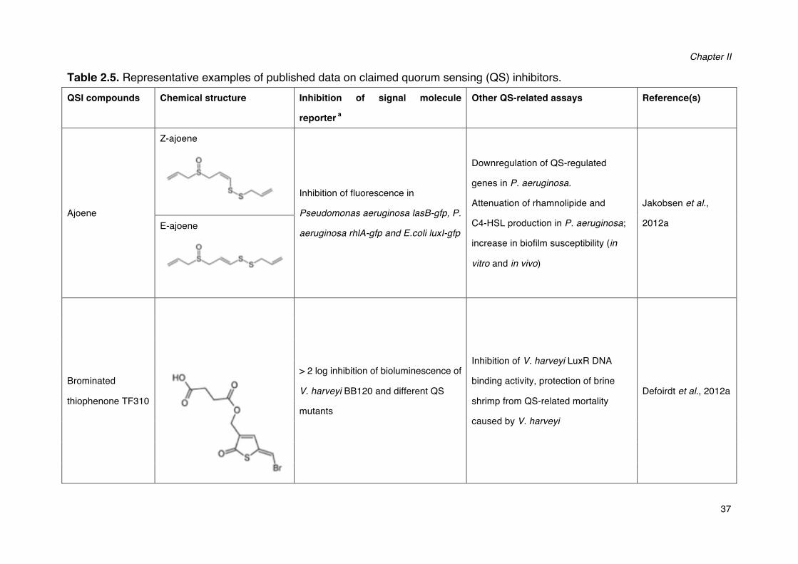
Chapter II
37
Table 2.5. Representative examples of published data on claimed quorum sensing (QS) inhibitors. QSI compounds Chemical structure Inhibition of signal molecule
reporter a
Other QS-related assays Reference(s)
Ajoene
Z-ajoene
Inhibition of fluorescence in
Pseudomonas aeruginosa lasB-gfp, P.
aeruginosa rhlA-gfp and E.coli luxI-gfp
Downregulation of QS-regulated
genes in P. aeruginosa.
Attenuation of rhamnolipide and
C4-HSL production in P. aeruginosa;
increase in biofilm susceptibility (in
vitro and in vivo)
Jakobsen et al.,
2012a E-ajoene
Brominated
thiophenone TF310
�
> 2 log inhibition of bioluminescence of
V. harveyi BB120 and different QS
mutants
Inhibition of V. harveyi LuxR DNA
binding activity, protection of brine
shrimp from QS-related mortality
caused by V. harveyi
Defoirdt et al., 2012a
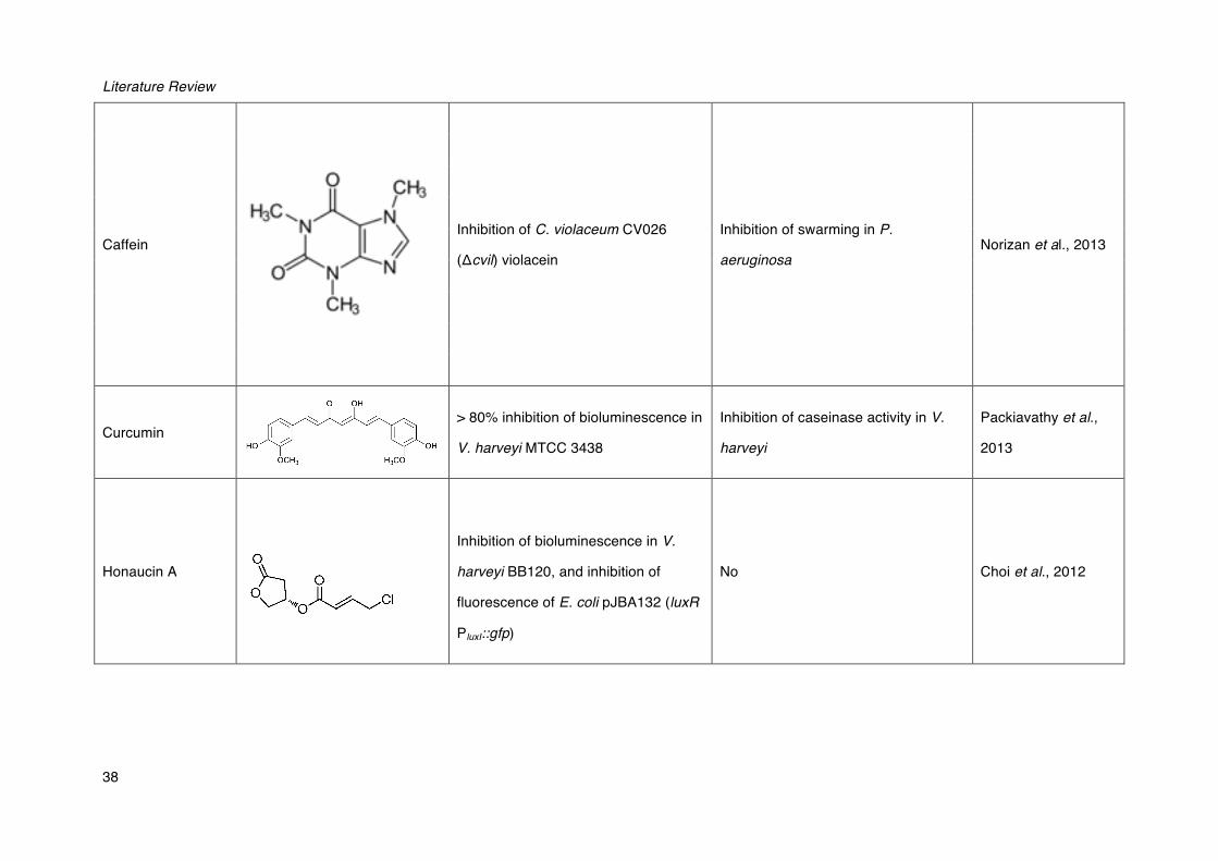
Literature Review
38
Caffein
�
Inhibition of C. violaceum CV026
(Δcvil) violacein
Inhibition of swarming in P.
aeruginosa Norizan et al., 2013
Curcumin
�
> 80% inhibition of bioluminescence in
V. harveyi MTCC 3438
Inhibition of caseinase activity in V.
harveyi
Packiavathy et al.,
2013
Honaucin A
�
�
�
�� �
Inhibition of bioluminescence in V.
harveyi BB120, and inhibition of
fluorescence of E. coli pJBA132 (luxR
PluxI::gfp)
No Choi et al., 2012
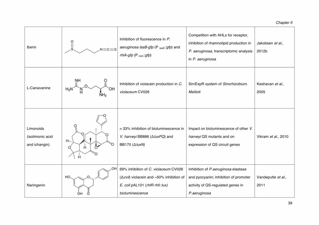
Chapter II
39
Iberin �
Inhibition of fluorescence in P.
aeruginosa lasB-gfp (P lasB::gfp) and
rhlA-gfp (P rhlA::gfp)
Competition with AHLs for receptor,
inhibition of rhamnolipid production in
P. aeruginosa, transcriptomic analysis
in P. aeruginosa
Jakobsen et al.,
2012b
L-Canavanine
�
Inhibition of violacein production in C.
violaceum CV026
Sin/ExpR system of Sinorhizobium.
Meliloti
Keshavan et al.,
2005
Limonoids
(isolimonic acid
and ichangin)
> 33% inhibition of bioluminescence in
V. harveyi BB886 (ΔluxPQ) and
BB170 (ΔluxN)
Impact on bioluminescence of other V.
harveyi QS mutants and on
expression of QS circuit genes
Vikram et al., 2010
�
Naringenin
�
69% inhibition of C. violaceum CV026
(Δcvil) violacein and ~50% inhibition of
E. coli pAL101 (rhlR rhll::lux)
bioluminescence
Inhibition of P.aeruginosa elastase
and pyocyanin; inhibition of promoter
activity of QS-regulated genes in
P.aeruginosa
Vandeputte et al.,
2011
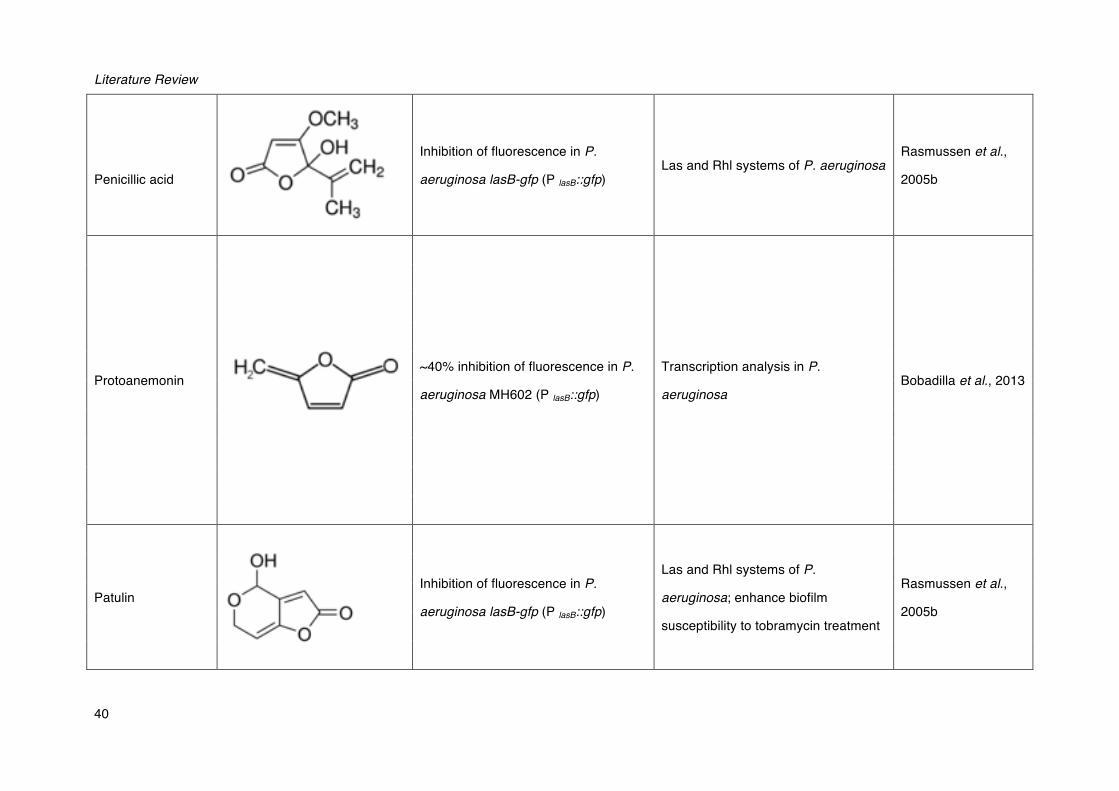
Literature Review
40
Penicillic acid
Inhibition of fluorescence in P.
aeruginosa lasB-gfp (P lasB::gfp) Las and Rhl systems of P. aeruginosa
Rasmussen et al.,
2005b
Protoanemonin
~40% inhibition of fluorescence in P.
aeruginosa MH602 (P lasB::gfp)
Transcription analysis in P.
aeruginosa Bobadilla et al., 2013
Patulin�
Inhibition of fluorescence in P.
aeruginosa lasB-gfp (P lasB::gfp)
Las and Rhl systems of P.
aeruginosa; enhance biofilm
susceptibility to tobramycin treatment
Rasmussen et al.,
2005b
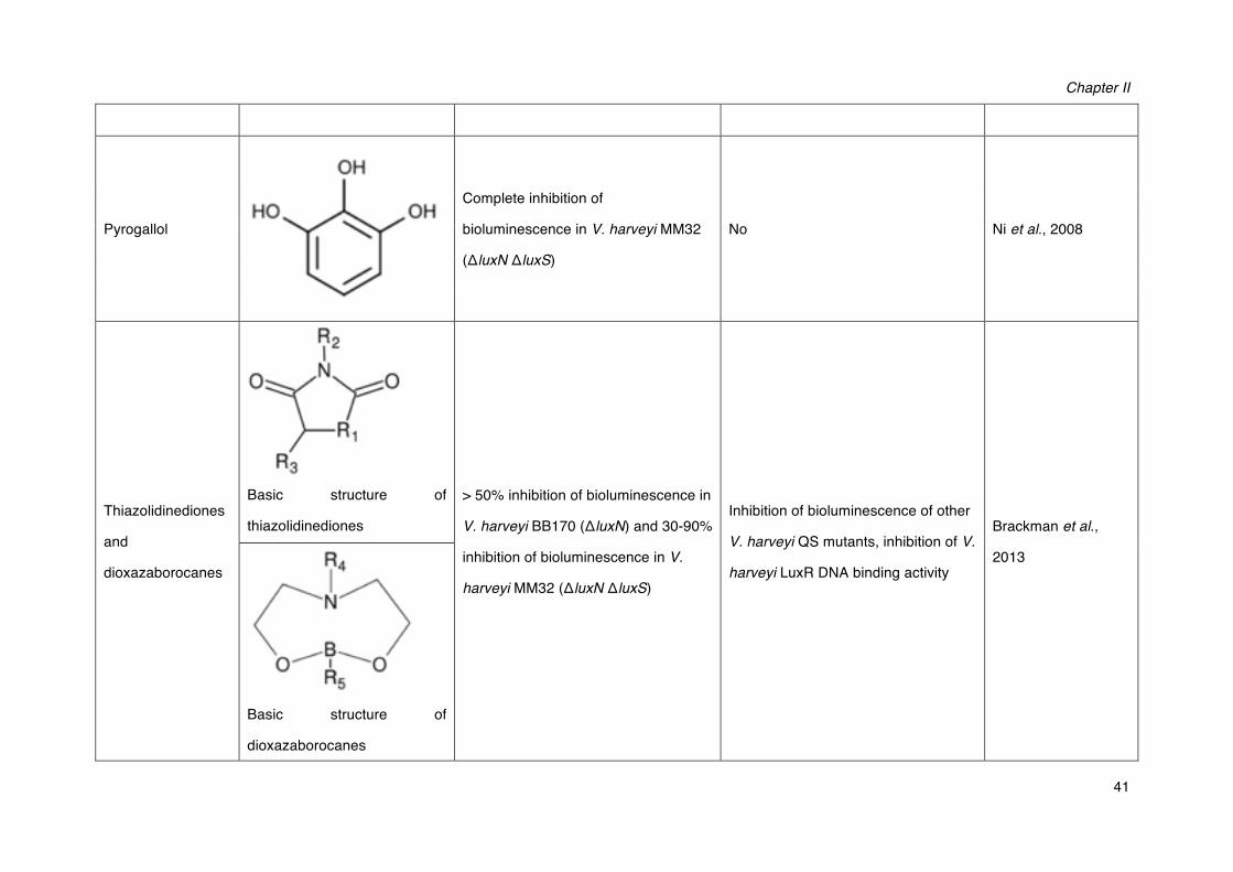
Chapter II
41
Pyrogallol�
Complete inhibition of
bioluminescence in V. harveyi MM32
(ΔluxN ΔluxS)
No Ni et al., 2008
Thiazolidinediones
and
dioxazaborocanes
Basic structure of
thiazolidinediones
> 50% inhibition of bioluminescence in
V. harveyi BB170 (ΔluxN) and 30-90%
inhibition of bioluminescence in V.
harveyi MM32 (ΔluxN ΔluxS)�
Inhibition of bioluminescence of other
V. harveyi QS mutants, inhibition of V.
harveyi LuxR DNA binding activity
Brackman et al.,
2013
Basic structure of
dioxazaborocanes
Literature Review
42
a ahyR and ahyl, Aeromonas hydrophila AHL receptor and synthase; Chromobacterium violaceum AHL synthase; lasB,
Pseudomonas aeruginosa QS-regulated elastase; lasR and lasI, P. aeruginosa receptor and synthase of
N-3-oxododecanoyl-L-homoserine lactone; rhlA, P. aeruginosa QS-regulated rhamnolipid synthase; rhlR and rhlI, P. aeruginosa
receptor and synthase of N-butanoyl-L-homoserine lactone; luxR and luxI, Vibrio fischeri AHL receptor and synthase; luxN, Vibrio
harveyi AHL receptor; luxS and lux PQ, V. harveyi AI-2 receptor and synthase.
Chapter II
43
Additionally, a number of plants, including crown vetch, carrot, soybean, water lily, tomato, pea seedlings, habanero and garlic, have been found to produce QSIs (Teplitski, 2004; Rasmussen et al., 2005).
Many plant extracts are believed to interfere with AHL signal detection by competing with them due to the similarity of their structural and/or by accelerating the degradation of the LuxR/LasR receptors of the AHL molecules (Lyon and Muir, 2003; Vattem et al., 2007). For example, halogenated furanones, including both natural compounds produced by macro alga Delisea pulchra and synthetic derivatives, are one of the most intensively studied groups of QSIs (Janssens et al., 2008). It had been assumed that furanones act by competing with AHLs for binding to LuxR-type proteins (Manefield et al., 1999). The mechanism of this inhibition has received renewed attention in light of studies suggesting that, rather than displacing the AHLs from LuxR, the furanones function by accelerating LuxR turnover (Manefield et al., 2002). However, the precise structural mechanism by which furanones stimulate LuxR turnover remains to be characterized (Manefield et al., 2002).
Halogenated furanones were found to disrupt quorum sensing-regulated gene expression both in AHL-mediated quorum sensing systems (e.g. interfere with the AHLs receptor LuxR in V. fischeri ) and in multi-channel quorum sensing systems of vibrios by decreasing the DNA-binding activity of the master regulator LuxRvh (not homologuous to V. fischeri LuxR) (Manefield et al., 2002; Defoirdt et al., 2007). It has been demonstrated that halogenated furanones can protect both fish and crustaceans against vibriosis (Rasch et al., 2004; Defoirdt et al., 2006). However, these furanones are toxic to higher organisms; especially fish larvae (Natrah et al., 2012), which means that they will not be safe for practical applications in aquaculture. More recently, brominated thiophenones, sulphur analogues of the brominated furanones, have been synthesized (Benneche et al., 2011), and these compounds have the same effect on the quorum sensing system as brominated furanones, by decreasing the DNA-binding activity of LuxR (Defoirdt et al., 2012). Thiophenones were found to be more active than the corresponding furanones (Benneche et al., 2011; Lönn-Stensrud and Benneche, 2011). One of these compounds, (Z)-4-((5-(bromomethylene)-2-oxo-2,5-dihydrothiophen-3-yl)methoxy)-4-oxobutanoic
Literature Review
44
acid (TF310) has been reported to show a promising therapeutic potential in the V.
harveyi – brine shrimp model, with complete protection against the pathogen at 2.5
µM and severe toxicity only being observed at 250 µM (Defoirdt et al., 2012).
Interestingly, the food additive cinnamaldehyde, a non-toxic synthetic flavouring
substance that is generally recognized as safe (GRAS), has been reported to have a
similar activity as brominated furanones and thiophenones (Brackman et al., 2008).
Furthermore, it has been found that cinnamaldehyde can protect giant fresh water
prawn larvae and gnotobiotic brine shrimp larvae against pathogenic V. harveyi
(Pande et al., 2013; Brackman et al., 2008), and blocked the virulence of both
Aeromonas hydrophila and Aeromonas salmonicida towards burbot larvae (Natrah et
al., 2012).
In addition to D. pulchra, production of QSIs has also been found in other macro-algae,
micro-algae, corals and sponges (Skindersoe et al., 2008). Marine bacteria can also
produce secondary metabolites with quorum sensing-inhibitory activity, such as two
phenetylamide metabolites from Halobacillus salinu, which are able to block quorum
sensing-mediated phenotypes in serveral bacteria, including V. harveyi (Teasdale et
al., 2009).
2.3.1 Antivirulence compounds targeting other regulatory mechanisms
In addition to quorum sensing inhibition, inhibitors targeting other virulence regulatory
pathways have also been demonstrated more recently. For instance, the
membrane-bound QseC histidine sensor kinase, which is responsible for bacterial
detection of catecholamine host stress hormones (Rasko et al., 2008). QseC was
shown to be present in at least 25 important human and plant pathogens, and can
initiate a complex phosphorylation cascade in the bacterial cells that regulates the
expression of virulence genes. It has been reported that a compound,
N-phenyl-4-[[(phenylamino)thioxomethyl]amino]- benzenesulphonamide (LED209), is
able to block the binding of signaling molecules to QseC, and consequently inhibit
QseC-mediated virulence gene expression (Rasko et al., 2008). LED209 was found to
be able to protect mice and rabbits from mammalian pathogens, such as Salmonella
typhimurium and enterohaemorrhagic Escherichia coli (Rasko et al., 2008). In addition,
Chapter II
45
the QseC-dependent signaling system shows no direct effect on bacterial growth in
vitro, therefore, inhibition of this signaling pathway probably will not exert strong
selective pressure towards development of drug resistance. These results indicate
that QseC is an attractive target for novel antivirulence therapies.
Another class of virulence inhibitors interferes with the ToxR regulon in vibrios. The
ToxR regulon has been studied extensively in V. cholerae, in which ToxR regulon was
found to be responsible for expression of two crucial virlence factors: the cholera toxin
and the toxin co-regulated pilus (TCP) (Matson et al., 2007). Recently, one small
molecule, 4-[N-(1,8- naphthalimide)]-n-butyric acid (also known as virstatin), was
shown to directly inhibit the activity of transcriptional regulator ToxT and subsequent
expression of virulence factors in V. cholerae (Hung et al., 2005). In the same report,
in vivo characterization in an infant-mouse model of infection showed that virstatin
could reduce cholerae infectivity at 50 μM, which suggests that virstatin provides
promise for the treatment of cholerae infection.
2.3.3 Antivirulence compounds targeting specific virulence factors
Apart from antivirulence compounds that interfere with regulatory mechanisms,
antivirulence therapies could also aim at directly blocking specific virulence factors,
such as toxins, adhesion factors and secretory systems, without killing or inhibiting
bacterial growth (Defoirdt, 2013). Clatworthy et al. (2007) reported a group of
compounds named pilicides that can block a specific virulence factor, i.e. the
formation of pili. Pilicides (e.g. bicyclic 2-pyridones) might have broad-spectrum
activity due to the conservation of the pathways they are targeting (Clatworthy et al.
2007). Another group of specific virulence factor-inhibiting compounds also show
broad spectrum antivirulence activity, since they are able to block bacterial secretion
systems in different bacteria. For instance, several inhibitors of type III secretion have
been reported, including acylated salicylaldehyde hydrazones and thiazolidinones
(Baron, 2010).
2.4 RESEARCH GAPS
The massive misuse of antibiotics to control infections in aquaculture has resulted in
Literature Review
46
severe problems, particularly the development of antibiotic resistance strains, which
have rendered the traditional antibiotic treatment no longer effective. Therefore,
alternative strategies to combat bacterial diseases are urgently needed for a
sustainable further development of the aquaculture industry (Defoirdt et al., 2007).
Antivirulence therapy is one of the alternative strategies, aiming at specifically
inhibiting the function required during an infection (Clatworthy et al., 2007).
Antivirulence therapy is based on a thorough understanding of the mechanisms by
which bacterial pathogens cause disease.
To date, the pathogenicity mechanisms of V. harveyi are not yet completely
understood. To our knowledge, the pathogen produces a variety of virulence factors
that are regulated by quorum sensing in V. harveyi, including biofilm formation
(Anetzberger et al., 2009), type III secretion (Henke and Bassler, 2004), production of
a siderophore (Lilley and Bassler, 2000), the Vhp metalloprotease (Mok et al., 2003;
Ruwandeepika et al., 2011), chitinase A (Defoirdt et al., 2010) and three
phospholipase genes (Natrah et al., 2011). Most of these virulence factors, except
metalloprotease, are negatively controlled by quorum sensing. However, according to
previous research that quorum sensing is required for full virulence of V. harveyi
towards different hosts (Defoirdt and Sorgeloos, 2012; Pande et al., 2013), there
should be some other important virulence factors that are positively regulated by
quorum sensing in V. harveyi. Therefore, the link between quorum sensing and
virulence in V. harveyi warrants further investigation.
The inhibition of quorum sensing is a promising alternative strategy to control disease.
Comparing with strategies that specifically block a certain virulence factor, disruption
of quorum sensing can interfere with several virulence factors at once. To date, a
variety of quorum sensing inhibitors has been described or claimed (Kalia, 2013;
Defoirdt et al., 2013). Brominated furanones are the most intensively studied quorum
sensing inhibitors, which have been reported to disrupt quorum sensing in various
Gram-negative bacteria (Manefield et al., 2002; Kalia, 2013). Unfortunately, these
furanones are toxic to higher organisms (Natrah et al., 2012), which means that they
will not be safe for practical applications. Therefore, finding novel quorum
sensing-disrupting agents with less negative side effects is crucially important.
Chapter II
47
Most research efforts with respect to exploring new antivirulence strategies for
combating bacterial infection have focused on the disruption of quorum sensing.
However, this mechanism also has its limitations (e.g. technology for practical
applications is not yet available). Therefore, it is important to further explore more
mechanisms that might be potential targets for antivirulence therapy for aquaculture,
including other signaling mechanisms (e.g. indole signaling) and sensing of host
factors (e.g. catecholamine stress hormones). Different antivirulence therapies could
be used synergistically to maximize the chance of success. Additionally, with less of
the therapeutic agents there will be less of an opportunity for resistance development.

Literature Review
48
CHAPTER III QUORUM SENSING POSITIVELY REGULATES FLAGELLAR MOTILITY IN PATHOGENIC VIBRIO HARVEYI
Qian Yang and Tom Defoirdt (2015) Environmental microbiology 17: 960-968

Chapter III
49
ABSTRACT
Vibrios belonging to the Harveyi clade are among the major pathogens of aquatic organisms. Quorum sensing (QS) is essential for virulence of V. harveyi towards different hosts. However, most virulence factors reported to be controlled by QS to date are negatively regulated by QS, therefore suggesting that their impact on virulence is limited. In this study, we report that QS positively regulates flagellar motility. We found that autoinducer synthase mutants showed significantly lower swimming motility than the wild type, and the swimming motility could be restored by adding synthetic signal molecules. Further, motility of a luxO mutant with inactive QS (LuxO D47E) was significantly lower than that of the wild type and of a luxO mutant with constitutively maximal QS activity (LuxO D47A). Furthermore, we found that the expression of flagellar genes (both early, middle and late genes) was significantly lower in the luxO mutant with inactive QS when compared to wild type and the luxO mutant with maximal QS activity. Motility assays and gene expression also revealed the involvement of the quorum sensing master regulator LuxR in the QS regulation of motility. Finally, the motility inhibitor phenamil significantly decreased the virulence of V. harveyi towards gnotobiotic brine shrimp larvae.
Keywords: Quorum sensing; swimming motility; flagellum; virulence
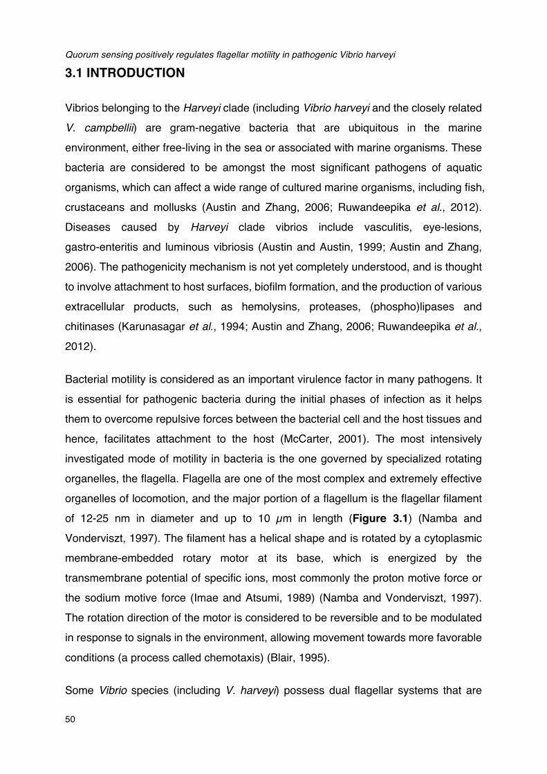
Quorum sensing positively regulates flagellar motility in pathogenic Vibrio harveyi
50
3.1 INTRODUCTION
Vibrios belonging to the Harveyi clade (including Vibrio harveyi and the closely related
V. campbellii) are gram-negative bacteria that are ubiquitous in the marine
environment, either free-living in the sea or associated with marine organisms. These
bacteria are considered to be amongst the most significant pathogens of aquatic
organisms, which can affect a wide range of cultured marine organisms, including fish,
crustaceans and mollusks (Austin and Zhang, 2006; Ruwandeepika et al., 2012).
Diseases caused by Harveyi clade vibrios include vasculitis, eye-lesions,
gastro-enteritis and luminous vibriosis (Austin and Austin, 1999; Austin and Zhang,
2006). The pathogenicity mechanism is not yet completely understood, and is thought
to involve attachment to host surfaces, biofilm formation, and the production of various
extracellular products, such as hemolysins, proteases, (phospho)lipases and
chitinases (Karunasagar et al., 1994; Austin and Zhang, 2006; Ruwandeepika et al.,
2012).
Bacterial motility is considered as an important virulence factor in many pathogens. It
is essential for pathogenic bacteria during the initial phases of infection as it helps
them to overcome repulsive forces between the bacterial cell and the host tissues and
hence, facilitates attachment to the host (McCarter, 2001). The most intensively
investigated mode of motility in bacteria is the one governed by specialized rotating
organelles, the flagella. Flagella are one of the most complex and extremely effective
organelles of locomotion, and the major portion of a flagellum is the flagellar filament
of 12-25 nm in diameter and up to 10 µm in length (Figure 3.1) (Namba and
Vonderviszt, 1997). The filament has a helical shape and is rotated by a cytoplasmic
membrane-embedded rotary motor at its base, which is energized by the
transmembrane potential of specific ions, most commonly the proton motive force or
the sodium motive force (Imae and Atsumi, 1989) (Namba and Vonderviszt, 1997).
The rotation direction of the motor is considered to be reversible and to be modulated
in response to signals in the environment, allowing movement towards more favorable
conditions (a process called chemotaxis) (Blair, 1995).
Some Vibrio species (including V. harveyi) possess dual flagellar systems that are

Chapter III
51
suited for movement under different conditions (Hendrie et al., 1970; McCarter, 2004).
A single polar flagellum is involved in swimming in liquid environments, while the
numerous lateral flagella enable swarming over surfaces or movement in more
viscous environments. In the whole genome sequence of V. harveyi BB120 (ATCC
BAA-1116; recently reclassified as Vibrio campbellii; Lin et al., 2010), approximately
50 polar and 30 lateral flagellar genes have been identified to encode components of
the flagellum and its export structure in Vibrio harveyi. However, the actual
contribution of motility to virulence of V. harveyi thus far is unknown.
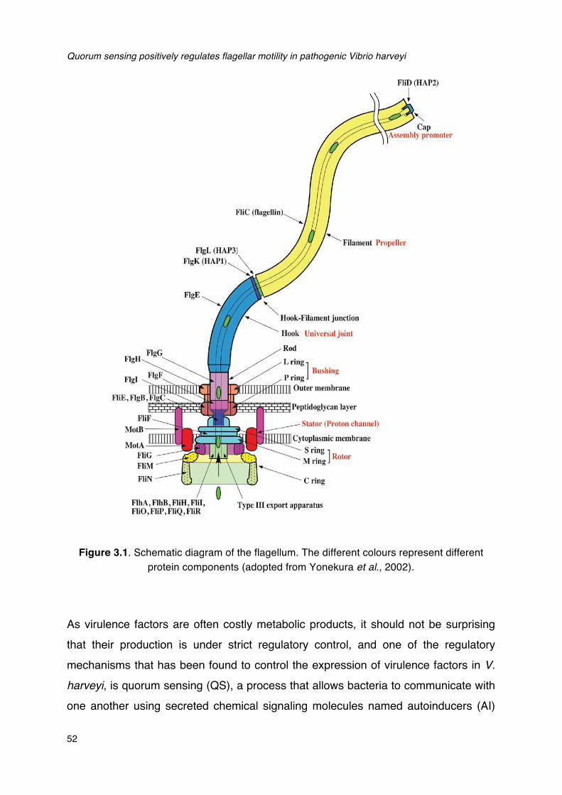
Quorum sensing positively regulates flagellar motility in pathogenic Vibrio harveyi
52
Figure 3.1. Schematic diagram of the flagellum. The different colours represent different protein components (adopted from Yonekura et al., 2002).
As virulence factors are often costly metabolic products, it should not be surprising
that their production is under strict regulatory control, and one of the regulatory
mechanisms that has been found to control the expression of virulence factors in V.
harveyi, is quorum sensing (QS), a process that allows bacteria to communicate with
one another using secreted chemical signaling molecules named autoinducers (AI)
Chapter III
53
(Miller and Bassler, 2001). The QS system of V. harveyi BB120 has been extensively studied. The strain produces, detects and responds to three kinds of QS signal molecules, harveyi autoinducer 1 (HAI-1; Cao and Meighen, 1989), autoinducer 2 (AI-2; Chen et al., 2002) and cholerae autoinducer 1 (CAI-1; Higgins et al., 2007). These three signal molecules are detected at the cell surface by their respective two-component receptors, which feed a shared phosphorylation/ dephosphorylation signal transduction cascade (Figure 3.2).
Figure 3.2. The V. harveyi quorum sensing circuit. The LuxM, LuxS and CqsA enzymes synthesize the autoinducer HAI-1, AI-2 and CAI-1, respectively. These autoinducers are detected at the cell surface by the LuxN, LuxQ and CqsS two-component receptor proteins respectively. Detection of AI-2 by LuxQ requires the periplasmic protein LuxP.
A. At low signal molecule levels the LuxN, LuxPQ and CqsS receptors function as kinases. LuxO is phosphorylated, the Qrr1-5 sRNAs are transcribed and LuxR protein is not produced.
B. At high signal molecule levels, the LuxN, LuxPQ and CqsS receptors function as phosphatases. LuxO is unphosphorylated, Qrr1-5 sRNAs are not transcribed and LuxR protein is produced. LuxR is the quorum sensing master regulator, and controls the expression of target genes by binding to their promoter regions. Solid and dotted lines denote regulatory factors that are produced and not produced respectively.
A variety of virulence factors has been found to be regulated by QS in V. harveyi,
Quorum sensing positively regulates flagellar motility in pathogenic Vibrio harveyi
54
including biofilm formation (Anetzberger et al., 2009), type III secretion (Henke and Bassler, 2004a), production of a siderophore (Lilley and Bassler, 2000), the Vhp metalloprotease (Mok et al., 2003; Ruwandeepika et al., 2011a), chitinase A (Defoirdt et al., 2010), and three phospholipase genes (Natrah et al., 2011). However, with the exception of the metalloprotease (which probably is not an essential virulence factor; Ruwandeepika et al., 2011b), these virulence factors are negatively regulated by QS, and this is in contrast with the fact that QS is required for full virulence of these bacteria towards different hosts (Defoirdt and Sorgeloos, 2012; Pande et al., 2013). Hence, in addition to the Vhp metalloprotease, there should be at least another important virulence factor that is positively regulated by QS in V. harveyi. Since a link between QS and motility in V. harveyi has thus far not been investigated, in this study, we investigated the effect of QS on swimming motility of V. harveyi and on the expression of selected genes involved in flagellar motility. We further investigated the importance of flagellar motility for virulence of V. harveyi by applying a motility inhibitor in a model system with gnotobiotic brine shrimp (Artemia franciscana) larvae.
3.2 RESULTS AND DISCUSSION
3.2.1 Impact of QS on the motility of V. harveyi
The impact of QS on the motility of V. harveyi was investigated using wild type V.
harveyi BB120 and various QS mutants. The strains were spotted on soft agar plates, and zones of motility were measured after 24h of incubation. In a first experiment, we determined the impact of inactivation of signal molecule synthases and found that inactivation of all three synthases in triple mutant JMH634 resulted in a significantly decreased motility when compared to the wild type (Figure 3.3A). Furthermore, we investigated the impact of inactivation of single synthase mutants and found that all mutants showed lower motility than the wild type. The swimming motility of HAI-1 synthase mutant BB152 could be restored to the same level of BB120 by adding synthetic D-HAI-1. We used the synthase deletion mutants rather than testing receptor deletion mutants because the deletion of a receptor results in a condition similar to that of the receptor in the presence of its cognate signal molecule (no phosphorylation of LuxO by the deleted receptor) (Freeman and Bassler, 1999).
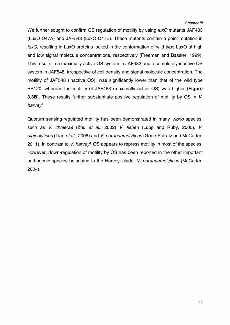
Chapter III
55
We further sought to confirm QS regulation of motility by using luxO mutants JAF483 (LuxO D47A) and JAF548 (LuxO D47E). These mutants contain a point mutation in luxO, resulting in LuxO proteins locked in the conformation of wild type LuxO at high and low signal molecule concentrations, respectively (Freeman and Bassler, 1999). This results in a maximally active QS system in JAF483 and a completely inactive QS system in JAF548, irrespective of cell density and signal molecule concentration. The motility of JAF548 (inactive QS), was significantly lower than that of the wild type BB120, whereas the motility of JAF483 (maximally active QS) was higher (Figure
3.3B). These results further substantiate positive regulation of motility by QS in V. harveyi.
Quorum sensing-regulated motility has been demonstrated in many Vibrio species, such as V. cholerae (Zhu et al., 2002) V. fisheri (Lupp and Ruby, 2005), V. alginolyticus (Tian et al., 2008) and V. parahaemolyticus (Gode-Potratz and McCarter, 2011). In contrast to V. harveyi, QS appears to repress motility in most of the species. However, down-regulation of motility by QS has been reported in the other important pathogenic species belonging to the Harveyi clade, V. parahaemolyticus (McCarter, 2004).
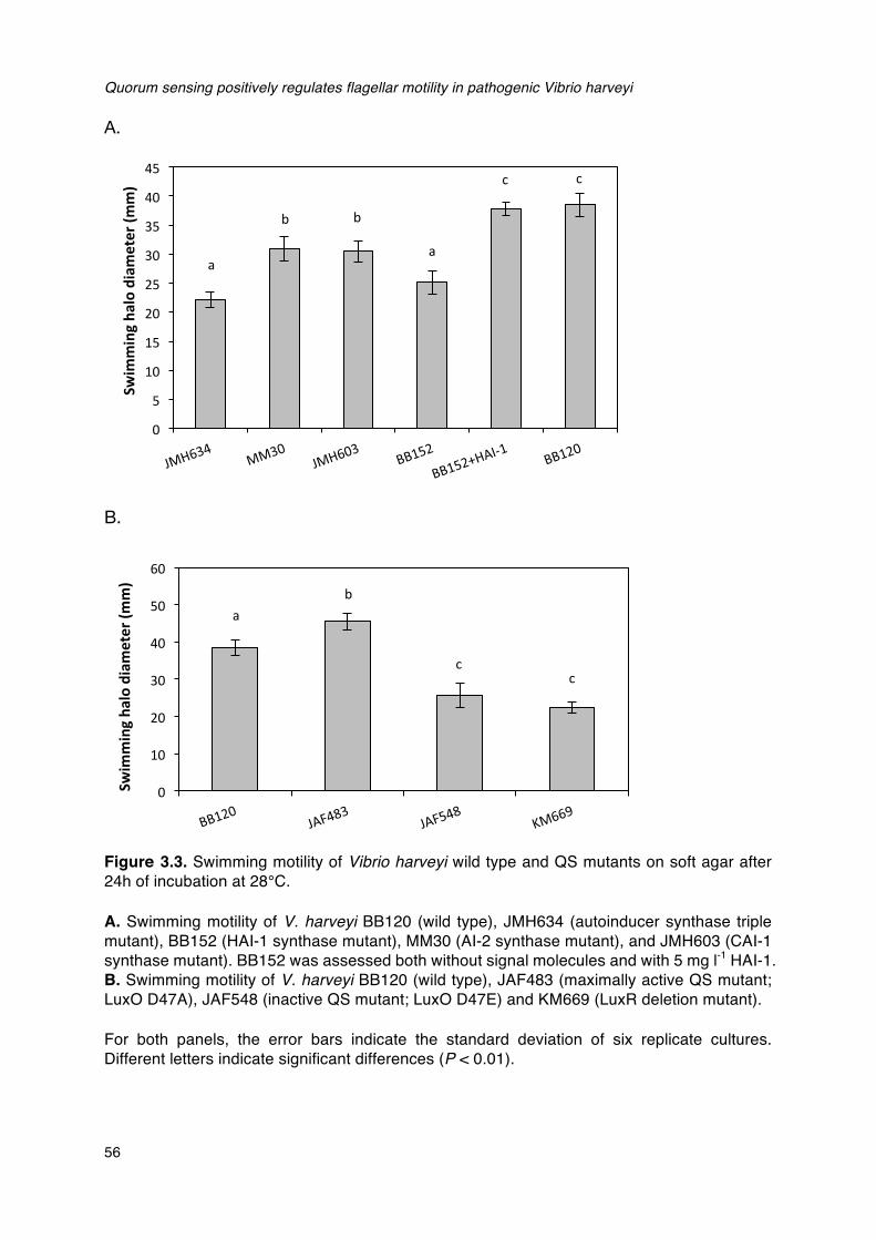
Quorum sensing positively regulates flagellar motility in pathogenic Vibrio harveyi
56
A.
B.
Figure 3.3. Swimming motility of Vibrio harveyi wild type and QS mutants on soft agar after 24h of incubation at 28°C.
A. Swimming motility of V. harveyi BB120 (wild type), JMH634 (autoinducer synthase triple mutant), BB152 (HAI-1 synthase mutant), MM30 (AI-2 synthase mutant), and JMH603 (CAI-1 synthase mutant). BB152 was assessed both without signal molecules and with 5 mg l-1 HAI-1. B. Swimming motility of V. harveyi BB120 (wild type), JAF483 (maximally active QS mutant; LuxO D47A), JAF548 (inactive QS mutant; LuxO D47E) and KM669 (LuxR deletion mutant).
For both panels, the error bars indicate the standard deviation of six replicate cultures. Different letters indicate significant differences (P < 0.01).
a�
b� b
a
c� c�
0
5
10
15
20
25
30
35
40
45
Swim
mingha
lodiameter(m
m)
a�b�
cc
0
10
20
30
40
50
60
Swim
mingha
lodiameter(m
m)
Chapter III
57
3.2.2 Impact of QS on the expression of motility-related genes in V. harveyi
In order to further characterize the relation between QS and motility, we investigated
the effect of QS on motility at the transcriptional level. For this experiment, we
selected 10 genes involved in flagellar motility. This selection was based on the
knowledge on flagellar motility in Vibrio parahaemolyticus, the paradigm of flagellar
motility in vibrios (McCarter, 2004) and on previous reports on QS regulation of
motility in other vibrios (Zhu et al., 2002; Lupp and Ruby, 2005; Tian et al., 2008). The
selected genes included 6 genes involved in the synthesis of the polar flagellum (both
regulators and structural genes), 2 genes involved in the synthesis of lateral flagella,
and 2 genes involved in chemotaxis, which is strongly linked to flagellar motility
(McCarter, 2004). The results showed that on soft agar the expression of the polar
flagellum and chemotaxis genes was significantly higher in JAF483 (maximal QS
activity) when compared to JAF548 (inactive QS) (Table 3.1). The expression levels
in BB120 were intermediate between these two strains. For the two genes involved in
lateral flagella synthesis, the comparison among these three strains was opposite on
soft agar. In fact, the expression of lateral flagellar genes was in general low on soft
agar plates, which was expected as lateral flagella are activated in more viscous
environments (McCarter, 2004). If the bacteria were grown in a more viscous
environment (plates with 1.5% agar), the expression levels of the two lateral flagellar
genes increased and showed the same trend as observed for the polar flagellar genes
in soft agar (i.e. positive regulation by QS). These findings confirmed that QS
positively regulates flagellar motility.
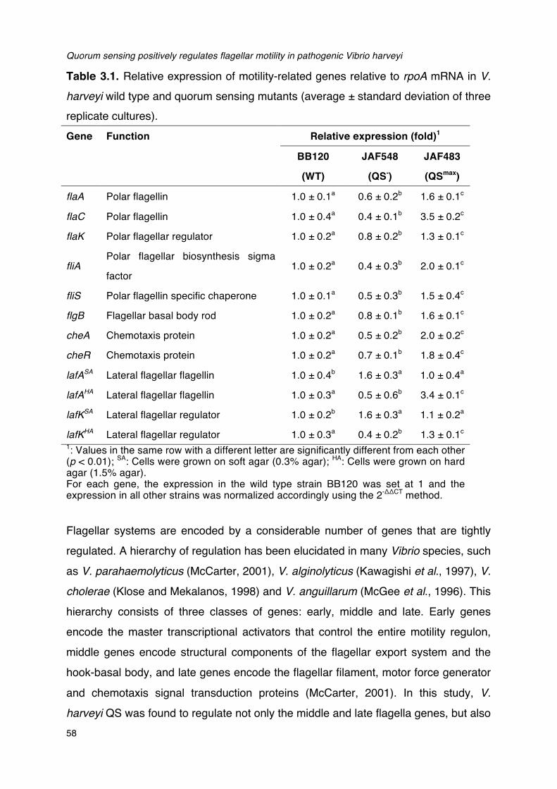
Quorum sensing positively regulates flagellar motility in pathogenic Vibrio harveyi
58
Table 3.1. Relative expression of motility-related genes relative to rpoA mRNA in V. harveyi wild type and quorum sensing mutants (average ± standard deviation of three replicate cultures). Gene Function Relative expression (fold)1
BB120
(WT)
JAF548
(QS-)
JAF483
(QSmax)
flaA Polar flagellin 1.0 ± 0.1a 0.6 ± 0.2b 1.6 ± 0.1c
flaC Polar flagellin 1.0 ± 0.4a 0.4 ± 0.1b 3.5 ± 0.2c
flaK Polar flagellar regulator 1.0 ± 0.2a 0.8 ± 0.2b 1.3 ± 0.1c
fliA Polar flagellar biosynthesis sigma
factor 1.0 ± 0.2a 0.4 ± 0.3b 2.0 ± 0.1c
fliS Polar flagellin specific chaperone 1.0 ± 0.1a 0.5 ± 0.3b 1.5 ± 0.4c
flgB Flagellar basal body rod 1.0 ± 0.2a 0.8 ± 0.1b 1.6 ± 0.1c
cheA Chemotaxis protein 1.0 ± 0.2a 0.5 ± 0.2b 2.0 ± 0.2c
cheR Chemotaxis protein 1.0 ± 0.2a 0.7 ± 0.1b 1.8 ± 0.4c
lafASA Lateral flagellar flagellin 1.0 ± 0.4b 1.6 ± 0.3a 1.0 ± 0.4a
lafAHA Lateral flagellar flagellin 1.0 ± 0.3a 0.5 ± 0.6b 3.4 ± 0.1c
lafKSA Lateral flagellar regulator 1.0 ± 0.2b 1.6 ± 0.3a 1.1 ± 0.2a
lafKHA Lateral flagellar regulator 1.0 ± 0.3a 0.4 ± 0.2b 1.3 ± 0.1c 1: Values in the same row with a different letter are significantly different from each other (p < 0.01); SA: Cells were grown on soft agar (0.3% agar); HA: Cells were grown on hard agar (1.5% agar). For each gene, the expression in the wild type strain BB120 was set at 1 and the expression in all other strains was normalized accordingly using the 2-ΔΔCT method. Flagellar systems are encoded by a considerable number of genes that are tightly
regulated. A hierarchy of regulation has been elucidated in many Vibrio species, such
as V. parahaemolyticus (McCarter, 2001), V. alginolyticus (Kawagishi et al., 1997), V.
cholerae (Klose and Mekalanos, 1998) and V. anguillarum (McGee et al., 1996). This
hierarchy consists of three classes of genes: early, middle and late. Early genes
encode the master transcriptional activators that control the entire motility regulon,
middle genes encode structural components of the flagellar export system and the
hook-basal body, and late genes encode the flagellar filament, motor force generator
and chemotaxis signal transduction proteins (McCarter, 2001). In this study, V.
harveyi QS was found to regulate not only the middle and late flagella genes, but also
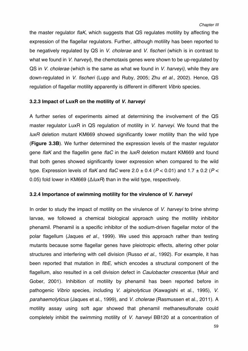
Chapter III
59
the master regulator flaK, which suggests that QS regulates motility by affecting the
expression of the flagellar regulators. Further, although motility has been reported to
be negatively regulated by QS in V. cholerae and V. fischeri (which is in contrast to
what we found in V. harveyi), the chemotaxis genes were shown to be up-regulated by
QS in V. cholerae (which is the same as what we found in V. harveyi), while they are
down-regulated in V. fischeri (Lupp and Ruby, 2005; Zhu et al., 2002). Hence, QS
regulation of flagellar motility apparently is different in different Vibrio species.
3.2.3 Impact of LuxR on the motility of V. harveyi
A further series of experiments aimed at determining the involvement of the QS
master regulator LuxR in QS regulation of motility in V. harveyi. We found that the
luxR deletion mutant KM669 showed significantly lower motility than the wild type
(Figure 3.3B). We further determined the expression levels of the master regulator
gene flaK and the flagellin gene flaC in the luxR deletion mutant KM669 and found
that both genes showed significantly lower expression when compared to the wild
type. Expression levels of flaK and flaC were 2.0 ± 0.4 (P < 0.01) and 1.7 ± 0.2 (P <
0.05) fold lower in KM669 (ΔluxR) than in the wild type, respectively.
3.2.4 Importance of swimming motility for the virulence of V. harveyi
In order to study the impact of motility on the virulence of V. harveyi to brine shrimp
larvae, we followed a chemical biological approach using the motility inhibitor
phenamil. Phenamil is a specific inhibitor of the sodium-driven flagellar motor of the
polar flagellum (Jaques et al., 1999). We used this approach rather than testing
mutants because some flagellar genes have pleiotropic effects, altering other polar
structures and interfering with cell division (Russo et al., 1992). For example, it has
been reported that mutation in flbE, which encodes a structural component of the
flagellum, also resulted in a cell division defect in Caulobacter crescentus (Muir and
Gober, 2001). Inhibition of motility by phenamil has been reported before in
pathogenic Vibrio species, including V. alginolyticus (Kawagishi et al., 1995), V.
parahaemolyticus (Jaques et al., 1999), and V. cholerae (Rasmussen et al., 2011). A
motility assay using soft agar showed that phenamil methanesulfonate could
completely inhibit the swimming motility of V. harveyi BB120 at a concentration of
Quorum sensing positively regulates flagellar motility in pathogenic Vibrio harveyi
60
50µM (Figure 3.4a), but had no significant effect on growth rate at this concentration (Figure 3.4b).
Figure 3.4. Effects of phenamil methanesulfonate on the swimming motility and growth rate of V. harveyi BB120. (a) Swimming motility of V. harveyi BB120 on soft agar after 24h of incubation at 28°C without (left) and with 50µM phenamil methanesulfonate (right). (b) Growth of V. harveyi BB120 in marine broth, in the absence and presence of phenamil methanesulfonate (50µM). As a control, the same volume of DMSO as added together with phenamil methanesulfonate was tested.
Chapter III
61
The importance of swimming motility for the virulence of V. harveyi was determined by measuring the effect of phenamil on the survival of brine shrimp larvae in a standardized challenge test (Defoirdt et al., 2005; Defoirdt and Sorgeloos, 2012). Interestingly, the addition of 50 µM phenamil methanesulfonate to the culture water significantly increased the survival of brine shrimp larvae challenged with V. harveyi
(Table 3.2). Furthermore, we verified that phenamil was not toxic to the vibrios in the more harsh environmental conditions of the brine shrimp culture water (when compared to nutrient rich growth medium) by spread-plating the brine shrimp rearing water after 24h and 48h of challenge. At both time points, there were no differences in V. harveyi counts between control and phenamil treatment (data not shown), indicating that phenamil did not inhibit growth of V. harveyi. These data confirm that swimming motility plays an important role in the virulence of V. harveyi in the brine shrimp model. Motility has been reported to be involved in the virulence in other vibrios as well, including Vibrio anguillarum (O’ Toole et al., 1996) and Vibrio cholerae
(Merrell et al., 2002).
Table 3.2. Survival of brine shrimp larvae challenged with V. harveyi BB120 in the absence and presence of 50 µM phenamil methanesulfonate after 48 hours (average ± standard error of four replicate brine shrimp cultures). BB120 was added to the culture water at 105 CFU/ml. Brine shrimp were fed with autoclaved LVS3 bacteria at 107 cells ml-1. Artemia cultures to which only autoclaved LVS3 bacteria were added as feed and that were otherwise treated in the same way as for the other treatments, were used as controls. Treatment Survival (%)1
BB120 50 ± 5b BB120 + phenamil 70 ± 5a Phenamil 98 ± 3c Control 100 ± 0c 1: Values with a different superscript letter are significantly different from each other (p < 0.01).
3.3 CONCLUSIONS
Although QS has been reported to be required for full virulence of V. harveyi towards different hosts (Defoirdt and Sorgeloos, 2012; Pande et al., 2013), until now, most of the virulence factors that were reported to be controlled by QS are negatively
Quorum sensing positively regulates flagellar motility in pathogenic Vibrio harveyi
62
regulated by QS. These virulence factors include chitinase A (Defoirdt et al., 2010),
phospholipase (Natrah et al., 2011), siderophore (Lilley and Bassler, 2000), and
components of a type III secretion system (Henke and Bassler, 2004b). As QS is
essential for virulence and these virulence factors are negatively regulated by QS,
their impact on virulence probably is rather limited. The only virulence factor thus far
known to be positively regulated by QS in V. harveyi was the Vhp metalloprotease
(Mok et al., 2003). However, this metalloprotease probably is not essential as the V.
harveyi genome encodes several proteases and as we previously found no correlation
between the expression level of vhp and virulence towards brine shrimp larvae
(Ruwandeepika et al., 2011b). In this study, we further unravelled the mechanism by
which QS controls the virulence of V. harveyi by demonstrating (1) that QS positively
regulates motility by affecting flagellar biosynthesis, (2) that LuxR is involved in the
regulation of motility and (3) that flagellar motility significantly affects virulence of V.
harveyi in our gnotobiotic brine shrimp model.
3.4 EXPERIMENTAL PROCEDURES
3.4.1 Bacterial strains and growth conditions
The bacterial strains used in this study are listed in Table 3.3. All V. harveyi strains
were grown at 28°C in Marine Broth (Difco) under constant agitation (100 min-1) and
the densities were measured spectrophotometrically at 600nm. Antibiotics were
added to the media at the following concentrations when necessary: 20 mg/l of
kanamycin (Sigma-Aldrich, Bornem, Belgium) for JAF483 and JAF548; 20 mg/l of
chloramphenicol (Sigma-Aldrich) for JMH603 and JMH634.
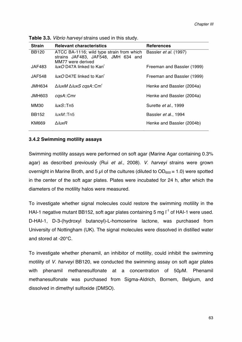
Chapter III
63
Table 3.3. Vibrio harveyi strains used in this study. Strain Relevant characteristics References BB120 ATCC BA-1116; wild type strain from which
strains JAF483, JAF548, JMH 634 and MM77 were derived
Bassler et al. (1997)
JAF483 luxO D47A linked to Kanr Freeman and Bassler (1999)
JAF548 luxO D47E linked to Kanr Freeman and Bassler (1999)
JMH634 ΔluxM ΔluxS cqsA::Cmr Henke and Bassler (2004a)
JMH603 cqsA::Cmr Henke and Bassler (2004a)
MM30 luxS::Tn5 Surette et al., 1999
BB152 luxM::Tn5 Bassler et al., 1994
KM669 ΔluxR Henke and Bassler (2004b)
3.4.2 Swimming motility assays
Swimming motility assays were performed on soft agar (Marine Agar containing 0.3% agar) as described previously (Rui et al., 2008). V. harveyi strains were grown overnight in Marine Broth, and 5 µl of the cultures (diluted to OD600 = 1.0) were spotted in the center of the soft agar plates. Plates were incubated for 24 h, after which the diameters of the motility halos were measured.
To investigate whether signal molecules could restore the swimming motility in the HAI-1 negative mutant BB152, soft agar plates containing 5 mg l-1 of HAI-1 were used. D-HAI-1, D-3-(hydroxyl butanoyl)-L-homoserine lactone, was purchased from University of Nottingham (UK). The signal molecules were dissolved in distilled water and stored at -20°C.
To investigate whether phenamil, an inhibitor of motility, could inhibit the swimming motility of V. harveyi BB120, we conducted the swimming assay on soft agar plates with phenamil methanesulfonate at a concentration of 50µM. Phenamil methanesulfonate was purchased from Sigma-Aldrich, Bornem, Belgium, and dissolved in dimethyl sulfoxide (DMSO).
Quorum sensing positively regulates flagellar motility in pathogenic Vibrio harveyi
64
3.4.3 RNA extraction
Three V. harveyi strains BB120, JAF483 and JAF548 were grown overnight in
triplicate on soft agar plates (0.3 % agar) and hard agar plates (1.5% agar),
respectively. Cells were harvested and RNA was extracted with the SV Total RNA
Isolation System (Promega, USA) according to the manufacturer’s instructions. The
RNA quantity was measured spectrophotometrically (NanoDrop Technologies) and
adjusted to 200 ng µl-1 in all samples. The RNA integrity was checked by Agarose Gel
Electrophoresis and the RNA samples were stored in -80°C for subsequent use.
3.4.4 Primers
The genome sequence of V. harveyi (Genbank) was screened for flagella-related
genes and 10 genes (including regulators, structural genes and chemotaxis genes)
were selected. Specific primers (Table 3.4) were designed using the software Primer
Premier version 5.00 (Premier Biosoft International, Palo Alto, CA), with predicted
product sizes in the 100 to 200 bp range. The RNA polymerase A submit (rpoA)
mRNA was used as an endogenous control, and expression of the genes was
calculated relative to the rpoA mRNA levels (Defoirdt et al., 2007).
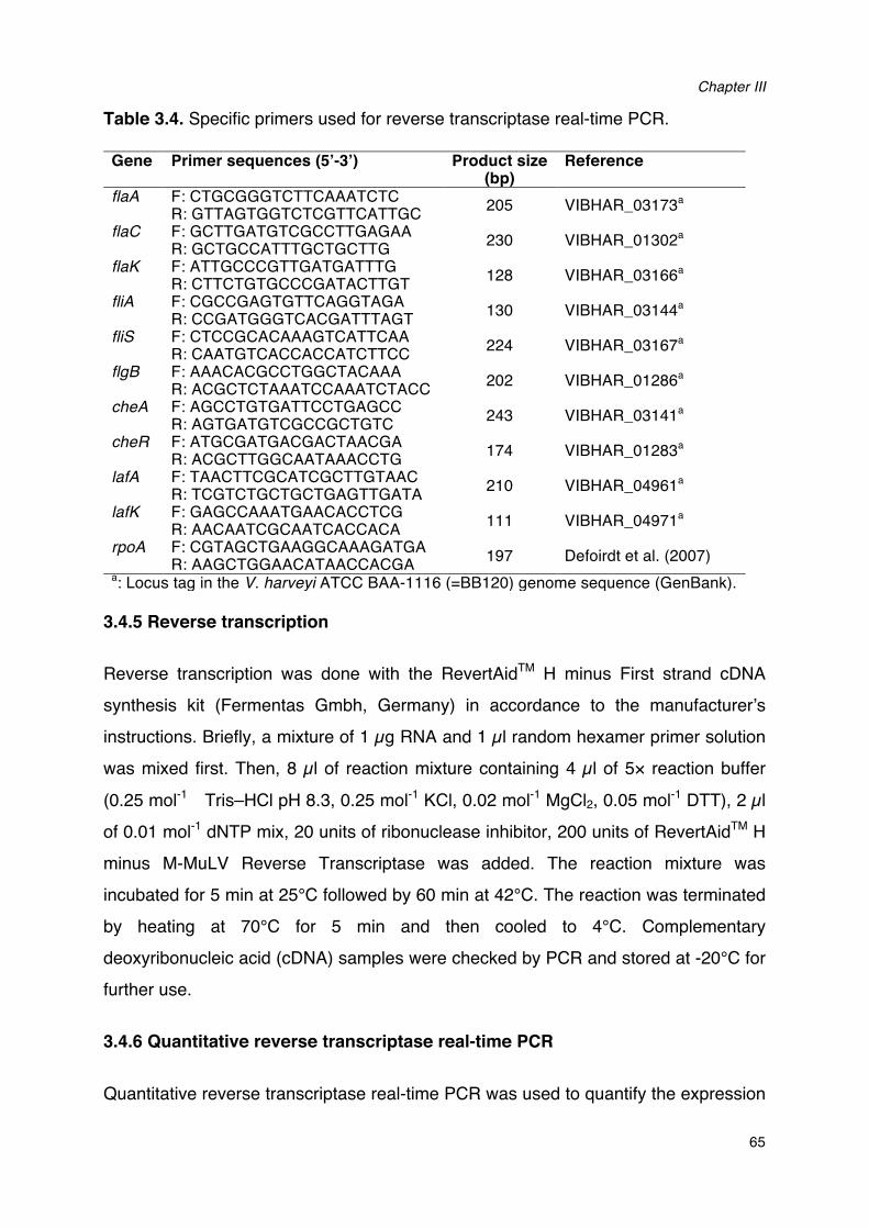
Chapter III
65
Table 3.4. Specific primers used for reverse transcriptase real-time PCR. Gene Primer sequences (5’-3’) Product size
(bp) Reference
flaA F: CTGCGGGTCTTCAAATCTC R: GTTAGTGGTCTCGTTCATTGC 205 VIBHAR_03173a
flaC F: GCTTGATGTCGCCTTGAGAA R: GCTGCCATTTGCTGCTTG 230 VIBHAR_01302a
flaK F: ATTGCCCGTTGATGATTTG R: CTTCTGTGCCCGATACTTGT 128 VIBHAR_03166a
fliA F: CGCCGAGTGTTCAGGTAGA R: CCGATGGGTCACGATTTAGT 130 VIBHAR_03144a
fliS F: CTCCGCACAAAGTCATTCAA R: CAATGTCACCACCATCTTCC 224 VIBHAR_03167a
flgB F: AAACACGCCTGGCTACAAA R: ACGCTCTAAATCCAAATCTACC 202 VIBHAR_01286a
cheA F: AGCCTGTGATTCCTGAGCC R: AGTGATGTCGCCGCTGTC 243 VIBHAR_03141a
cheR F: ATGCGATGACGACTAACGA R: ACGCTTGGCAATAAACCTG 174 VIBHAR_01283a
lafA F: TAACTTCGCATCGCTTGTAAC R: TCGTCTGCTGCTGAGTTGATA 210 VIBHAR_04961a
lafK F: GAGCCAAATGAACACCTCG R: AACAATCGCAATCACCACA 111 VIBHAR_04971a
rpoA F: CGTAGCTGAAGGCAAAGATGA R: AAGCTGGAACATAACCACGA 197 Defoirdt et al. (2007)
a: Locus tag in the V. harveyi ATCC BAA-1116 (=BB120) genome sequence (GenBank).
3.4.5 Reverse transcription
Reverse transcription was done with the RevertAidTM H minus First strand cDNA
synthesis kit (Fermentas Gmbh, Germany) in accordance to the manufacturer’s
instructions. Briefly, a mixture of 1 µg RNA and 1 µl random hexamer primer solution
was mixed first. Then, 8 µl of reaction mixture containing 4 µl of 5× reaction buffer
(0.25 mol-1 Tris–HCl pH 8.3, 0.25 mol-1 KCl, 0.02 mol-1 MgCl2, 0.05 mol-1 DTT), 2 µl
of 0.01 mol-1 dNTP mix, 20 units of ribonuclease inhibitor, 200 units of RevertAidTM H
minus M-MuLV Reverse Transcriptase was added. The reaction mixture was
incubated for 5 min at 25°C followed by 60 min at 42°C. The reaction was terminated
by heating at 70°C for 5 min and then cooled to 4°C. Complementary
deoxyribonucleic acid (cDNA) samples were checked by PCR and stored at -20°C for
further use.
3.4.6 Quantitative reverse transcriptase real-time PCR
Quantitative reverse transcriptase real-time PCR was used to quantify the expression
Quorum sensing positively regulates flagellar motility in pathogenic Vibrio harveyi
66
level of all the flagella-related genes and was performed with Maxima® SYBR Green/ROX qPCR Master Mix (Fermentas, Cambridgeshire). The reaction was performed in an StepOne™ Real-Time PCR System thermal cycler (Applied Biosystems) in a total volume of 25 µl, containing 12.5 µl of 2× SYBR green master mix, 300 nM of forward and reverse primers and 2 µl of template cDNA. The thermal cycling consisted an initial denaturation at 95°C for 10 min followed by 40 cycles of denaturation at 95°C for 15s and primer annealing and elongation at 60°C for 1min. Dissociation curve analysis was performed to check for the amplification of untargeted fragments. Data acquisition was performed with the StepOne™ Software.
3.4.7 Quantitative reverse transcriptase real-time PCR data analysis (2-∆∆CT method)
The quantitative reverse transcriptase real-time PCR was validated by amplifying serial dilutions of cDNA synthesized from 1 µg of RNA isolated from bacterial samples. Serial dilutions of cDNA were amplified by real time PCR using gene specific primers. ∆CT (average CT value of target-average CT value of rpoA) was calculated for the different dilutions and plotted against the cDNA concentration. The slope of the graph was almost equal to 0 for all of the target nine genes. Therefore, the amplification efficiency of reference and the target genes was considered to be equal. Based on this precondition, real-time PCR data were analyzed using the 2-∆∆CT method (Livak and Schmittgen, 2001). The expression of the target genes was normalized to the endogenous control (rpoA) by calculating ∆CT :
∆Ct = Ct target � Ct rpoA
and expressed relative to a calibrator strain by calculating ∆∆Ct:
∆∆Ct = ∆Ct � Ct calibratior
Strain JAF548 (in which QS is inactive) was used as acalibrator (Ruwandeepika et al., 2011b). The relative expression was then calculated as
Relative expression = 2-∆∆Ct
Chapter III
67
3.4.8 Axenic hatching of brine shrimp larvae
Two hundred milligrams of high-quality hatching cysts of Artemia franciscana cysts (EG® Type; INVE Aquaculture, Baasrode, Belgium) were hydrated in 18 ml of filtersterilized tap water for 1 h. Sterile cysts were obtained by decapsulation based on the method described by Marques et al. (2004). Briefly, 660 µl of NaOH (32%) and 10 ml of NaOCl (50%) were added to the hydrated cyst suspension to facilitate decapsulation. The process was stopped after 2 min by adding 14 ml of Na2S2O3 (10 g L-1). Filtered (0.22 µm) aeration was provided during the reaction. The decapsulated cysts were washed with filtered (passed through 0.22-µm membrane filter) and autoclaved (moist heat at 121°C for 15 min) artificle seawater (containing 35 g l-1 of instant ocean synthetic sea salt, Aquarium Systems, Sarrebourg, France). The cysts were resuspended in a 50-ml tube containing 30 ml of filtered, autoclaved seawater and hatched for 28 h on a rotor (4 min-1) at 28°C with constant illumination (c. 2000 lux). The axenity of cysts was verified by inoculating one ml of culture water into 9 ml of Marine broth and incubating at 28°C for 24 h. After 28 h of hatching, batches of 30 nauplii were counted and transferred to fresh, sterile 50-ml tubes containing 30 ml of filtered and autoclaved seawater. Finally, the tubes were returned to the rotor and kept at 28°C. All manipulations were performed in a laminar flow to maintain sterility of the cysts and nauplii.
3.4.9 Brine shrimp challenge test
The effect of swimming motility inhibitor on the virulence of V. harveyi was determined by a standardized challenge test of Artemia. Challenge tests were performed as described by Defoirdt et al. (2005) with some modifications. The animals were challenged with 105 CFU ml-1 of the vibrios per ml Artemia culture water. A suspension of autoclaved LVS3 bacteria (Verschuere et al. 1999) in filtered and autoclaved seawater was added as feed at the start of the challenge test equivalent to 107 cells ml-1 culture water. The inhibitor phenamil methanesulfonate (dissolved in DMSO at a concentration of 1mM) was added directly into the culture water at the same time at a final concentration of 50 µM. Artemia cultures to which only autoclaved LVS3 bacteria were added as feed, were used as controls. The survival of the larvae
Quorum sensing positively regulates flagellar motility in pathogenic Vibrio harveyi
68
was counted 48 h after the addition of the pathogens. Each treatment was carried out in quadruplicate and each experiment was repeated twice to verify the reproducibility. In each test, the sterility of the control treatments were checked at the end of the challenge by inoculating 1 ml of Artemia culture water to 9 ml of Marine Broth and incubating the mixture for 2 days at 28°C.
3.4.10 Statistical analysis
Data analysis was carried out using the SPSS statistical software (version 15). All data were compared with one-way ANOVA, followed by a Tukey’s post hoc test.
ACKNOWLEDGEMENT
We thank Bonnie Bassler for kindly providing us with the V. harveyi wild type and QS mutants. This work was funded by the China Scholarship Council, the Special Research Fund of Ghent University (BOF-UGent) and the Fund for Scientific Research Flanders (FWO project n° 1.5.013.12N). Tom Defoirdt is a postdoctoral fellow of FWO. The authors declare that no conflict of interest exists.
CHAPTER IV SPECIFIC QUORUM SENSING-DISRUPTING ACTIVITY (AQSI) OF THIOPHENONES AND THEIR THERAPEUTIC POTENTIAL IN A GNOTOBIOTIC BRINE SHRIMP - VIBRIO HARVEYI MODEL SYSTEM
Qian Yang, Anne Aamdal-Scheie, Tore Benneche, Tom Defoirdt (2015) Scientific
Reports. In press.

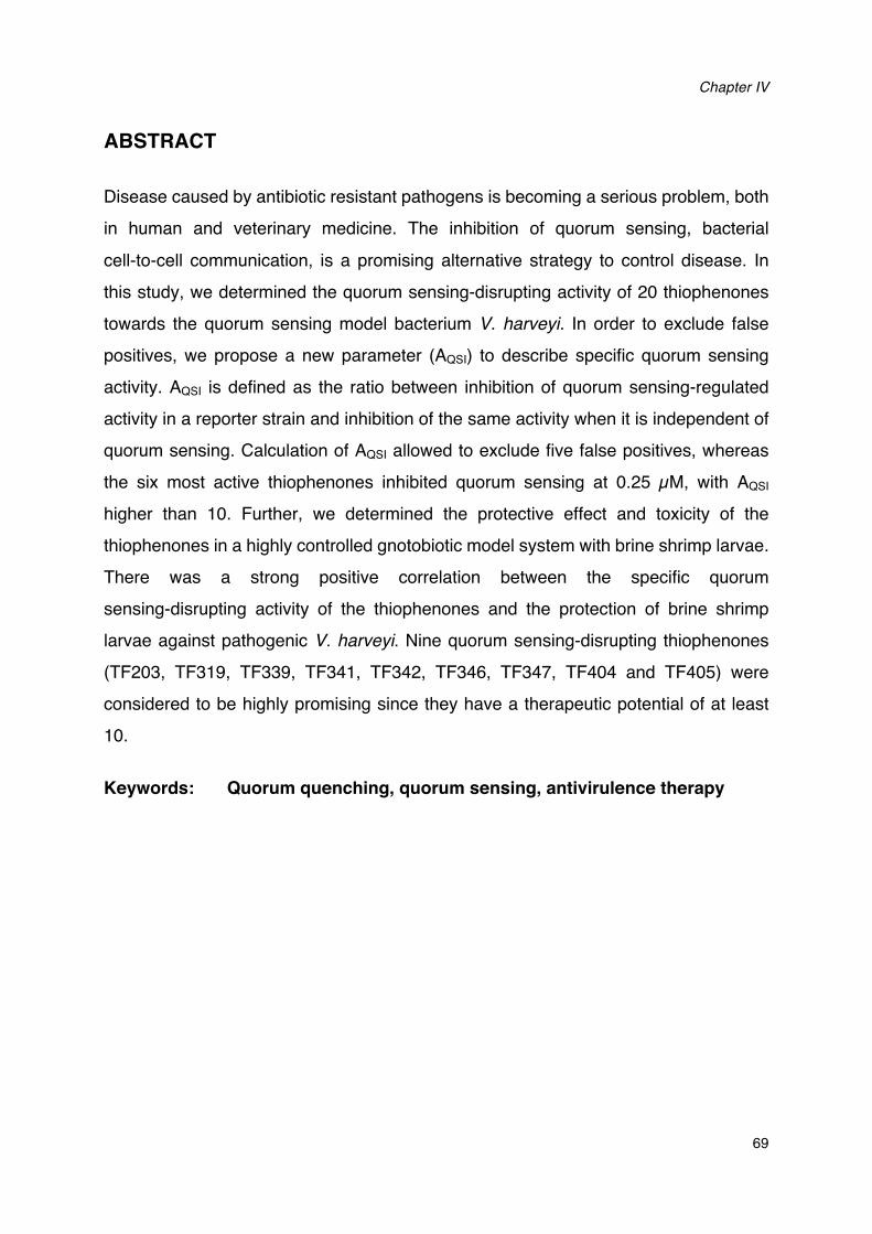
Chapter IV
69
ABSTRACT
Disease caused by antibiotic resistant pathogens is becoming a serious problem, both
in human and veterinary medicine. The inhibition of quorum sensing, bacterial
cell-to-cell communication, is a promising alternative strategy to control disease. In
this study, we determined the quorum sensing-disrupting activity of 20 thiophenones
towards the quorum sensing model bacterium V. harveyi. In order to exclude false
positives, we propose a new parameter (AQSI) to describe specific quorum sensing
activity. AQSI is defined as the ratio between inhibition of quorum sensing-regulated
activity in a reporter strain and inhibition of the same activity when it is independent of
quorum sensing. Calculation of AQSI allowed to exclude five false positives, whereas
the six most active thiophenones inhibited quorum sensing at 0.25 µM, with AQSI
higher than 10. Further, we determined the protective effect and toxicity of the
thiophenones in a highly controlled gnotobiotic model system with brine shrimp larvae.
There was a strong positive correlation between the specific quorum
sensing-disrupting activity of the thiophenones and the protection of brine shrimp
larvae against pathogenic V. harveyi. Nine quorum sensing-disrupting thiophenones
(TF203, TF319, TF339, TF341, TF342, TF346, TF347, TF404 and TF405) were
considered to be highly promising since they have a therapeutic potential of at least
10.
Keywords: Quorum quenching, quorum sensing, antivirulence therapy
Specific quorum sensing-disrupting activity (Aqsi) of thiophenones and their therapeutic potential
70
4.1 INTRODUCTION
The discovery of antibiotics brought great relief from a large number of deadly illnesses in the 20th century, and until now, these kinds of compounds are still used as a first line therapy to treat bacterial infections in the clinic (Cheema et al., 2007; Defoirdt, 2013). However, excessive and non-judicious use of antibiotics has resulted in the evolution of multiple drug resistant bacterial strains. Antibiotic treatments are no longer effective in some cases at this moment, and diseases caused by antibiotic resistant bacteria are currently the second leading cause of death worldwide (WHO report, 2014). Additionally, massive use of antibiotics in animal production also constitutes a direct threat to human health and to the environment (Cabello, 2006), and this has resulted in more strict regulations with respect to antibiotic use. One notable example is the ban on the use of antibiotics as growth promoters in animal production in Europe in 2006 (European Parliament and Council Regulation No 1831/2003). However, there are good indications that this ban could result in a higher frequency of pathogenic bacteria (such as Salmonella spp.), which in turn could lead to a higher frequency of infections (in animals as well as consumers). As a consequence, the development of novel strategies to control bacterial diseases, both in human and veterinary medicine will be critically important in order to ensure public health in the future.
An alternative strategy to combat bacterial infections is the specific inhibition of functions required to infect the host, which has been termed antivirulence therapy (Clatworthy et al., 2007). This therapy consists of either inhibiting a specific virulence factor, or interfering with the regulation of virulence factor expression (Defoirdt et al., 2011). Quorum sensing (QS) is a mechanism by which bacteria co-ordinate the expression of certain genes in response to small signal molecules. Quorum sensing has been shown to control expression of virulence-related genes in many different pathogens, making quorum sensing-disruption an interesting strategy to control bacterial disease (Kalia, 2013; Defoirdt et al., 2013). Vibrio harveyi is one of the major pathogens of aquatic organisms, affecting a wide range of cultured marine animals, and causes significant losses in the aquaculture industry worldwide (Austin and Zhang, 2006; Defoirdt et al., 2007). The species is also one of the model organisms in studies
Chapter IV
71
on QS in bacteria. V. harveyi contains a three-channel QS system, which is mediated by three types of signal molecules including HAI-1, AI-2 and CAI-1, respectively (Ng and Bassler, 2009). This QS system has been shown to be required for full virulence of the bacterium towards several aquatic hosts, including a highly controlled model system with gnotobiotic brine shrimp (Artemia franciscana) larvae (Defoirdt and Sorgeloos, 2012; Pande et al., 2013).
To date, a variety of QS inhibitors have been described or claimed (Kalia, 2013; Defoirdt et al., 2013). Brominated furanones are the most intensively studied QS inhibitors, and these compounds have been reported to disrupt QS in various Gram-negative bacteria (Manefield et al., 2002; Kalia, 2013). These compounds inhibit QS in V. harveyi by decreasing the DNA-binding activity of the quorum sensing master regulator LuxR (Defoirdt et al., 2007b). Unfortunately, these brominated furanones are toxic to higher organisms (Natrah et al., 2012), which means that they will not be safe for practical applications. More recently, brominated thiophenones, sulphur analogues of the brominated furanones with the same mode of action, have been synthesized (Benneche et al., 2011), and these compounds were found to be more active than the corresponding furanones (Benneche et al., 2011; Lönn-Stensrud and Benneche, 2011). One of these compounds, (Z)-4-((5-(bromomethylene)-2-oxo-2, 5-dihydrothiophen-3-yl) methoxy)-4-oxobutanoic acid (TF310, Figure 4.1) has been reported to show the highest therapeutic index of all QS-disrupting compounds tested in our V. harveyi – brine shrimp model thus far, with complete protection against the pathogen at 2.5 µM and severe toxicity only being observed at 250 µM (Defoirdt et al., 2012). Based on these promising results, in the present study, we aimed at determining quorum sensing-disrupting activity, protective effect and toxicity of 20 novel thiophenones (Figure 4.1). Furthermore, we propose a new parameter to describe specific quorum sensing-inhibitory activity, AQSI, defined as the ratio between inhibition of quorum sensing-regulated activity and inhibition of the same activity when independent of quorum sensing. Most claims with respect to quorum sensing inhibition are based on experiments with quorum sensing signal molecule reporter strains. We recently argued that these experiments are prone to bias due to other effects compounds may have on reporter strains, and therefore, that good control experiments are required in order to exclude false positives (Defoirdt et al., 2013). The

Specific quorum sensing-disrupting activity (Aqsi) of thiophenones and their therapeutic potential
72
use of the proposed parameter AQSI is a straightforward and elegant way to exclude
false positives by taking into account (potential) bias related to the use of quorum
sensing molecule reporters.
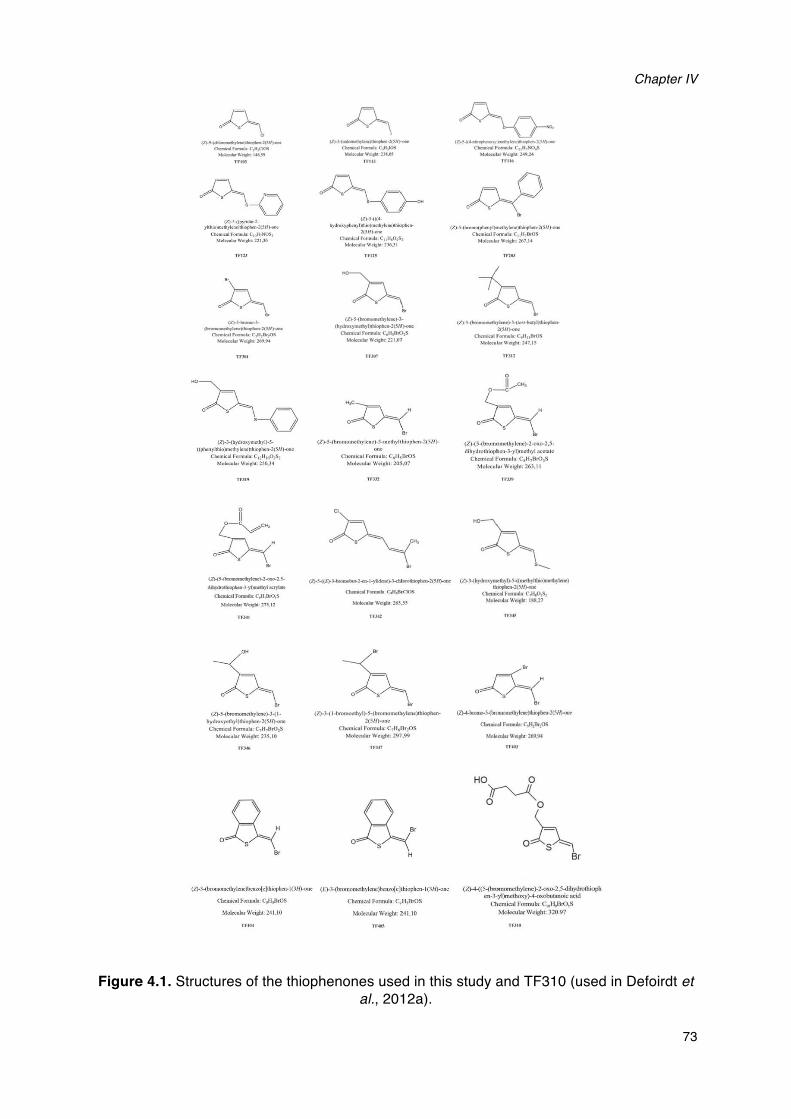
Chapter IV
73
Figure 4.1. Structures of the thiophenones used in this study and TF310 (used in Defoirdt et al., 2012a).
~~~ ......... l('JH.._ ~r..-c~pos
~."~,...."
0~-o-• \ j
(Z .. S.((pyndol-2· )'lllUO)mc1b)kfle)dooopbrft·l(SII>-c
Clotmll"al ~·omou.ll. C.JI7NOS1 Mol«toa..We.alu 221,J.O
(Z).J...--..j..
('"-doylc.c,......_l(SH>-~f'__."C,U11k,M
M~o~cna.V.'ftlk;:%69,,..
~Ä-o s \ j
(1)-J-{hydtO!Iytnelhyi .. S· ((ph"'yhhio)mtlhykne)lhiophcn-2(SII)-mc
CMmic:al Form~la: C 11111.0:SJ MoiC'ailar Wcoa.tu. 2SOJ4
0
11
5?;:( ..
(Z){~)-2~2.S.
~J..)'t)a:lc*)110)1aotc
C1laxal FormalL c;.H,.BrO.S
'-~'4'ftliii27S.I2
Á "'
(l}-S-{~Ihykne)-l-(l-
h)'droxydhyl)th!Ophm·2(Sif)-«w:
Chnn~eal Formula C7111Br01S MolcculatWctJhl:lJS,IO
(l).J.(......,...~l(SH,___
o-.c.~ r .... c,tt110S MokonolwV.fllklU~
0~~ ·~""
h)'dRIA)',MI)'1)lh~)nwthykM)thoopht-n•
1($11>--Chcmoool fom~ulf,;C1 111101S1
Mok.:ulw WctPI 236JI
(Z).$-{~ket)-J,
(h)'ihA,._.),,..,._.2(S//~
CWnucal f"-" CtfiJ8rO:S MolmiWWc.p;221.07
~:J:k" "'
(l).S..(bromooncthylcnc)-l-mcthylthlophen-2(S//)-ooo
Chemocal Fonnul1: C111.t-DrOS
Molttullar We~KfoL 205,07
0~ fb~C~_.,._....,_..~(".._
C~~nwca~F....,..c,1tt,"K>.s Moknol.lr wcop... 249,24
p=\() oA,~ ..
(Z .. S-(brolno(pttmyl)mclhylmc)thiopbco·2(51/)-one Chtmw:al r-ula C11ll1ü oOS Mo~u'-Wc!lht 267,14
Ä . (Z)-.5-(.,.-..yket)-J-(,..,.,-tNI}I)Ih ......
l($H.,_ Oonn.:.a F-ubo C.,ll 111h0S
Molen a.W ....... ; 247,1$
TfJU
0
.~· "' (Z)-(S-(bromomcthylene)-2-oxo-l • .S·
d ihydrothîophcn-l-yl)mclhyl acctate
Chcmiul Fonnula : C 111,Dr0)S MolccularWci&Jit;263.11
(Z)-S-{(l}J~l-1-)we.}-Jd~SH>-
n-.c:.J FOI'Dib C.H.BrQOS
{l}-3-{ll)"lhx)-..c111)·1)-s..((mdbyltt.o)lndil)·~)
-""~ Cbco.cal Fonuu&l C,ttp,S1
A "'
(l)-3-{ 1-bromo<-thyi)-S-(bromomcthyle~)lhtophen-
2(S/I)-onc:
Chcmteal Fonnula: c,u.orpS MoleeuW Wcaaht; 297.99
Mokalla Wnp! IU.27
o~" "' (2)-4..._5-(bo_, __ l(lll).ooo
C'hnulul FonnuL. C,II:Brps
Mokaàr W. 269.9-t
HO 0
'/r'--Zo
0~ Br
(~J-(br"omomctbylmc)bmzo(c)thropbnl·l(l/{)-otw. (E)-3-(bromomcthylme)bmzo(c}thlophm· I(JH}-one (Z)-4-((5-(bromomcthylcne}-2~o-2_5.-dihydrothioph cn-3-yl)mclhoxy)-4-oxobut.."UUo;c acid
Chemica! Fonnub C101 t.DrO,S MolecularWeiaht· 320.97
Clxnu<:al Fonnula Cti i,BrOS
MoltcularWtlgbl 241,10
Chmucal Fomml11 C.tt,DrOS
MoktularWe•a.tn 241 ,10
Specific quorum sensing-disrupting activity (Aqsi) of thiophenones and their therapeutic potential
74
4.2 RESULTS
4.2.1. Impact of the thiophenones on quorum sensing-regulated
bioluminescence of V. harveyi.
In a first experiment, we determined the impact of the thiophenones on QS-controlled
bioluminescence of V. harveyi. Wild type V. harveyi was grown to high cell density in
order to activate QS-controlled bioluminescence, after which the thiophenones were
added at 0.25, 1, 5 and 10 µM, respectively. Bioluminescence was measured 1 h after
the addition of the thiophenones and our results revealed that most of the compounds
were able to block bioluminescence in wild type V. harveyi in a
concentration-dependent way. Fifteen of the 20 compounds (TF103, TF113, TF116,
TF125, TF203, TF307, TF312, TF319, TF332, TF339, TF341, TF342, TF346, TF347
and TF403) were found to inhibit bioluminescence at a concentration of 0.25 µM and
higher, while TF123 and TF301 significantly reduced the bioluminescence from 5 µM
onwards. Additionally, TF203 could completely inhibit the QS-regulated
bioluminescence at 5 μM, and TF301, TF332 and TF341 completely blocked the
bioluminescence at 10 μM. Finally, TF345, TF404 and TF405 showed no effect on the
bioluminescence even at the highest concentration tested (Figure 4.2). Importantly,
the compounds had no effect on the growth of V. harveyi at the concentrations used
(Figure 4.3).
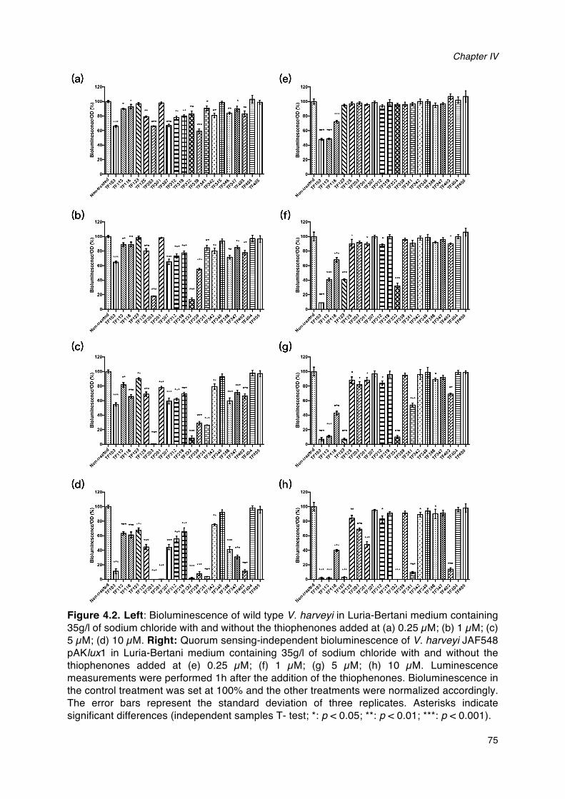
Chapter IV
75
Figure 4.2. Left: Bioluminescence of wild type V. harveyi in Luria-Bertani medium containing 35g/l of sodium chloride with and without the thiophenones added at (a) 0.25 µM; (b) 1 µM; (c) 5 µM; (d) 10 µM. Right: Quorum sensing-independent bioluminescence of V. harveyi JAF548 pAKlux1 in Luria-Bertani medium containing 35g/l of sodium chloride with and without the thiophenones added at (e) 0.25 µM; (f) 1 µM; (g) 5 µM; (h) 10 µM. Luminescence measurements were performed 1h after the addition of the thiophenones. Bioluminescence in the control treatment was set at 100% and the other treatments were normalized accordingly. The error bars represent the standard deviation of three replicates. Asterisks indicate significant differences (independent samples T- test; *: p < 0.05; **: p < 0.01; ***: p < 0.001).
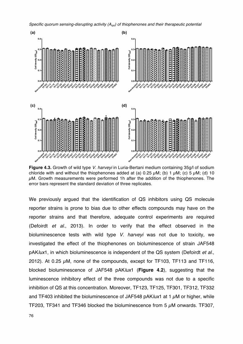
Specific quorum sensing-disrupting activity (Aqsi) of thiophenones and their therapeutic potential
76
Figure 4.3. Growth of wild type V. harveyi in Luria-Bertani medium containing 35g/l of sodium chloride with and without the thiophenones added at (a) 0.25 µM; (b) 1 µM; (c) 5 µM; (d) 10 µM. Growth measurements were performed 1h after the addition of the thiophenones. The error bars represent the standard deviation of three replicates.
We previously argued that the identification of QS inhibitors using QS molecule
reporter strains is prone to bias due to other effects compounds may have on the
reporter strains and that therefore, adequate control experiments are required
(Defoirdt et al., 2013). In order to verify that the effect observed in the
bioluminescence tests with wild type V. harveyi was not due to toxicity, we
investigated the effect of the thiophenones on bioluminescence of strain JAF548
pAKlux1, in which bioluminescence is independent of the QS system (Defoirdt et al.,
2012). At 0.25 µM, none of the compounds, except for TF103, TF113 and TF116,
blocked bioluminescence of JAF548 pAKlux1 (Figure 4.2), suggesting that the
luminescence inhibitory effect of the three compounds was not due to a specific
inhibition of QS at this concentration. Moreover, TF123, TF125, TF301, TF312, TF332
and TF403 inhibited the bioluminescence of JAF548 pAKlux1 at 1 µM or higher, while
TF203, TF341 and TF346 blocked the bioluminescence from 5 µM onwards. TF307,
Chapter IV
77
TF319, TF339, TF345, TF347, TF404 and TF405 showed no effect on the
bioluminescence of JAF548 pAKlux1 at the highest concentration tested in this study
(Figure 4.2), indicating that they have low toxicity.
For the compounds that showed significant inhibition of quorum sensing-regulated
bioluminescence, we further calculated the specific quorum sensing-inhibitory activity
AQSI as the ratio between the percentage inhibition of quorum sensing-regulated
bioluminescence (in wild type V. harveyi) and the percentage inhibition of quorum
sensing-independent bioluminescence (in JAF548 pAKlux1). The specific quorum
sensing-inhibitory activity of compounds TF103, TF113, TF116, TF123 and TF301
was smaller than 2 at any of the concentrations tested (Table 4.1), and these
compounds were therefore considered as false positives. Six compounds showed a
high specific quorum sensing activity (>10) at least one of the concentrations tested:
TF203, TF307, TF319, TF339, TF342 and TF403.
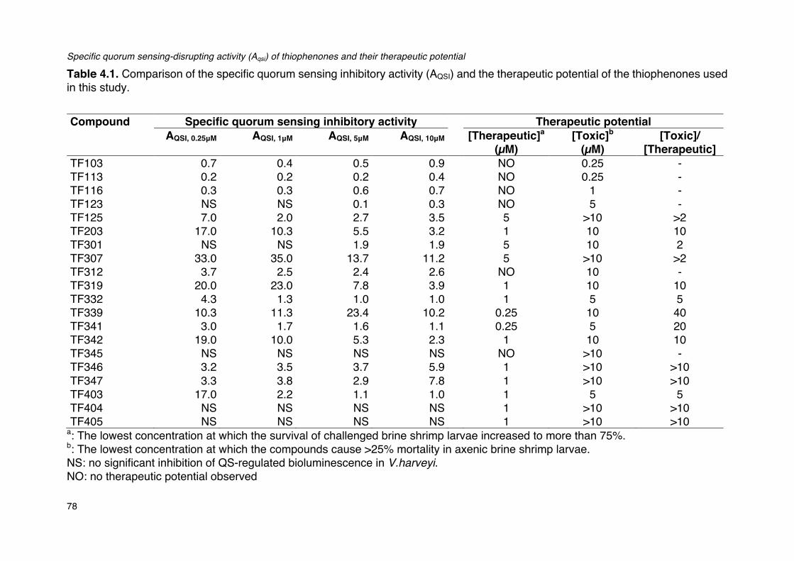
Specific quorum sensing-disrupting activity (Aqsi) of thiophenones and their therapeutic potential
78
Table 4.1. Comparison of the specific quorum sensing inhibitory activity (AQSI) and the therapeutic potential of the thiophenones used in this study. Compound Specific quorum sensing inhibitory activity Therapeutic potential
AQSI, 0.25μM AQSI, 1μM AQSI, 5μM AQSI, 10μM [Therapeutic]a (µM)
[Toxic]b (µM)
[Toxic]/ [Therapeutic]
TF103 0.7 0.4 0.5 0.9 NO 0.25 - TF113 0.2 0.2 0.2 0.4 NO 0.25 - TF116 0.3 0.3 0.6 0.7 NO 1 - TF123 NS NS 0.1 0.3 NO 5 - TF125 7.0 2.0 2.7 3.5 5 >10 >2 TF203 17.0 10.3 5.5 3.2 1 10 10 TF301 NS NS 1.9 1.9 5 10 2 TF307 33.0 35.0 13.7 11.2 5 >10 >2 TF312 3.7 2.5 2.4 2.6 NO 10 - TF319 20.0 23.0 7.8 3.9 1 10 10 TF332 4.3 1.3 1.0 1.0 1 5 5 TF339 10.3 11.3 23.4 10.2 0.25 10 40 TF341 3.0 1.7 1.6 1.1 0.25 5 20 TF342 19.0 10.0 5.3 2.3 1 10 10 TF345 NS NS NS NS NO >10 - TF346 3.2 3.5 3.7 5.9 1 >10 >10 TF347 3.3 3.8 2.9 7.8 1 >10 >10 TF403 17.0 2.2 1.1 1.0 1 5 5 TF404 NS NS NS NS 1 >10 >10 TF405 NS NS NS NS 1 >10 >10
a: The lowest concentration at which the survival of challenged brine shrimp larvae increased to more than 75%. b: The lowest concentration at which the compounds cause >25% mortality in axenic brine shrimp larvae. NS: no significant inhibition of QS-regulated bioluminescence in V.harveyi. NO: no therapeutic potential observed
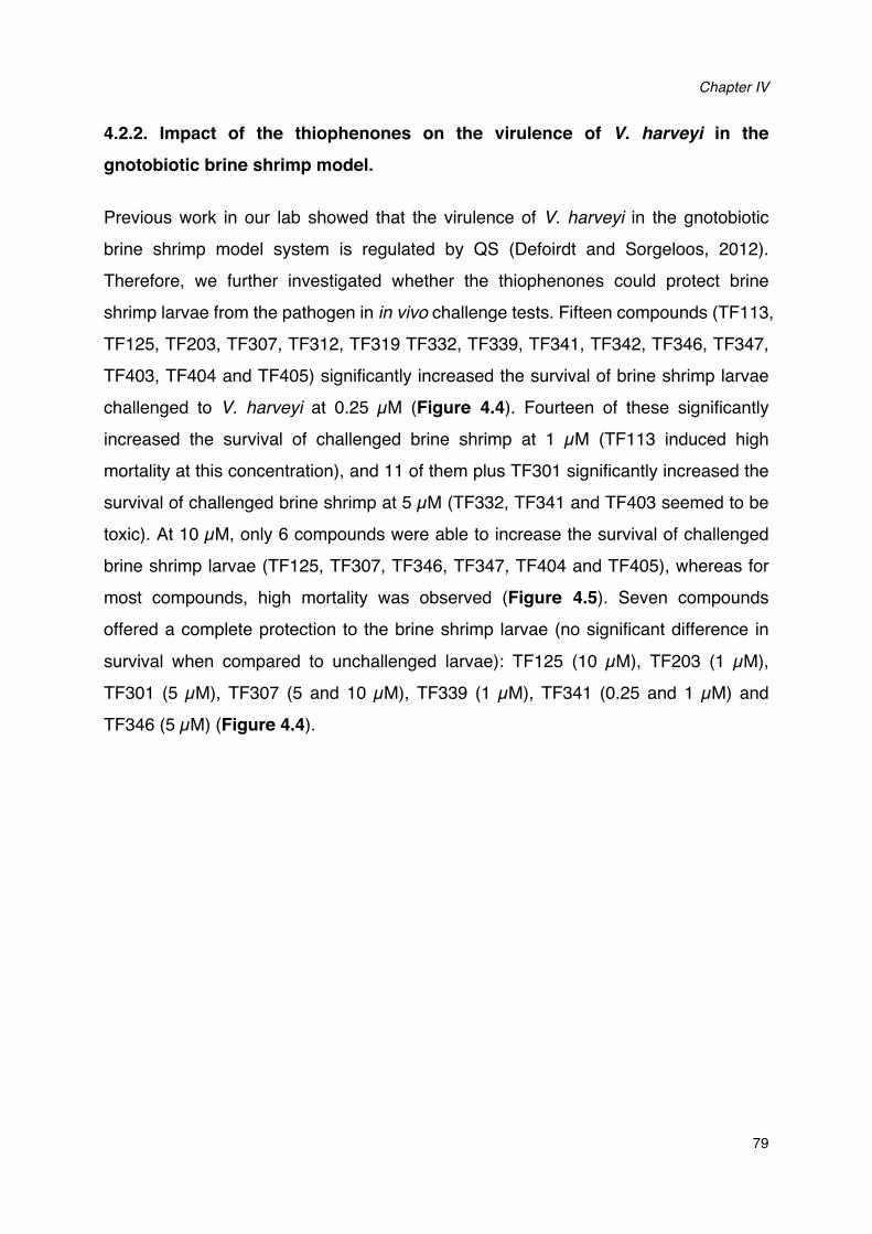
Chapter IV
79
4.2.2. Impact of the thiophenones on the virulence of V. harveyi in the gnotobiotic brine shrimp model.
Previous work in our lab showed that the virulence of V. harveyi in the gnotobiotic
brine shrimp model system is regulated by QS (Defoirdt and Sorgeloos, 2012).
Therefore, we further investigated whether the thiophenones could protect brine
shrimp larvae from the pathogen in in vivo challenge tests. Fifteen compounds (TF113,
TF125, TF203, TF307, TF312, TF319 TF332, TF339, TF341, TF342, TF346, TF347,
TF403, TF404 and TF405) significantly increased the survival of brine shrimp larvae
challenged to V. harveyi at 0.25 µM (Figure 4.4). Fourteen of these significantly
increased the survival of challenged brine shrimp at 1 µM (TF113 induced high
mortality at this concentration), and 11 of them plus TF301 significantly increased the
survival of challenged brine shrimp at 5 µM (TF332, TF341 and TF403 seemed to be
toxic). At 10 µM, only 6 compounds were able to increase the survival of challenged
brine shrimp larvae (TF125, TF307, TF346, TF347, TF404 and TF405), whereas for
most compounds, high mortality was observed (Figure 4.5). Seven compounds
offered a complete protection to the brine shrimp larvae (no significant difference in
survival when compared to unchallenged larvae): TF125 (10 µM), TF203 (1 µM),
TF301 (5 µM), TF307 (5 and 10 µM), TF339 (1 µM), TF341 (0.25 and 1 µM) and
TF346 (5 µM) (Figure 4.4).
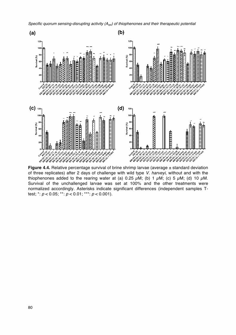
Specific quorum sensing-disrupting activity (Aqsi) of thiophenones and their therapeutic potential
80
Figure 4.4. Relative percentage survival of brine shrimp larvae (average ± standard deviation of three replicates) after 2 days of challenge with wild type V. harveyi, without and with the thiophenones added to the rearing water at (a) 0.25 µM; (b) 1 µM; (c) 5 µM; (d) 10 µM. Survival of the unchallenged larvae was set at 100% and the other treatments were normalized accordingly. Asterisks indicate significant differences (independent samples T- test; *: p < 0.05; **: p < 0.01; ***: p < 0.001).
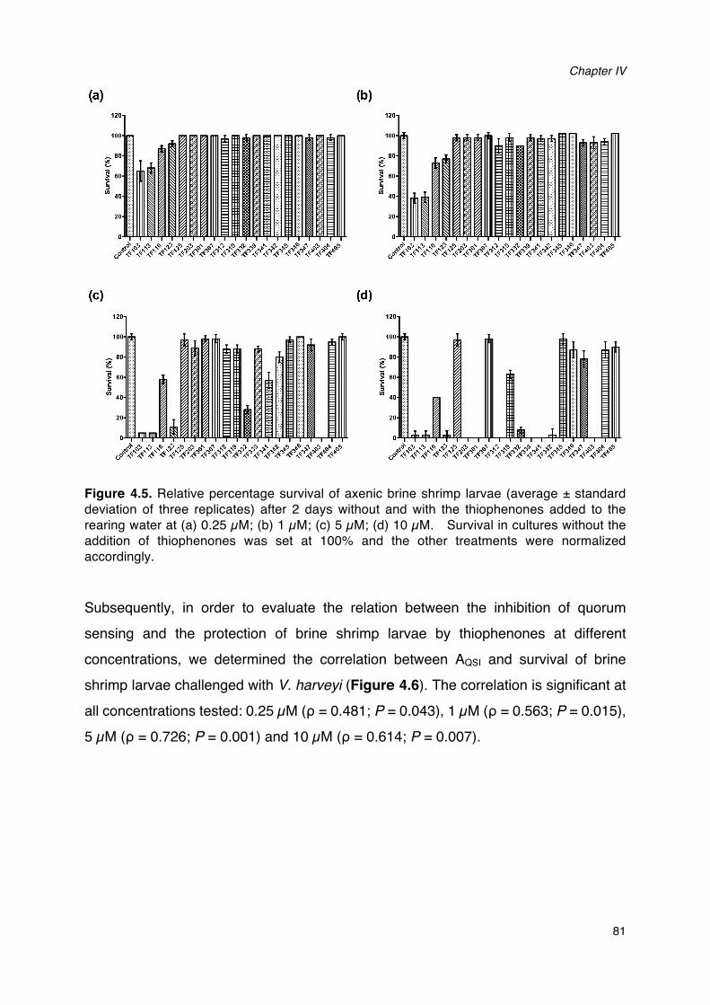
Chapter IV
81
Figure 4.5. Relative percentage survival of axenic brine shrimp larvae (average ± standard deviation of three replicates) after 2 days without and with the thiophenones added to the rearing water at (a) 0.25 µM; (b) 1 µM; (c) 5 µM; (d) 10 µM. Survival in cultures without the addition of thiophenones was set at 100% and the other treatments were normalized accordingly. Subsequently, in order to evaluate the relation between the inhibition of quorum
sensing and the protection of brine shrimp larvae by thiophenones at different
concentrations, we determined the correlation between AQSI and survival of brine
shrimp larvae challenged with V. harveyi (Figure 4.6). The correlation is significant at
all concentrations tested: 0.25 µM (ρ = 0.481; P = 0.043), 1 µM (ρ = 0.563; P = 0.015),
5 µM (ρ = 0.726; P = 0.001) and 10 µM (ρ = 0.614; P = 0.007).
Specific quorum sensing-disrupting activity (Aqsi) of thiophenones and their therapeutic potential
82
Figure 4.6. Scatter plots showing specific quorum sensing-inhibitory activity (AQSI) and protection of brine shrimp larvae from V. harveyi for the different thiophenones at (a) 0.25 μM; (b) 1 μM; (c) 5 μM; (d) 10 μM. 4.2.3 Toxicity of the thiophenones to axenic brine shrimp larvae.
We determined toxicity to axenic brine shrimp larvae at 0.25, 1, 5 and 10 µM. At 0.25
µM, TF103 and TF113 showed appreciable toxicity (causing > 25 % mortality). At 1
µM, 4 compounds induced appreciable mortality, including TF103, TF113, TF116 and
TF123. At 5 µM, TF332, TF341 and TF403 also started to induce appreciable
mortality, and at 10 µM, only 7 compounds showed no toxic effect (TF125, TF307,
TF345, TF346, TF347, TF404 and TF405), whereas all other compounds except for
TF116 and TF319 induced (almost) complete mortality (Figure 4.5).
In addition, we also calculated correlations between inhibition of bioluminescence in
JAF548 pAKlux1 and survival of brine shrimp larvae treated with thiophenones
(Figure 4.7). Results showed that the correlation is significant at all the
concentrations we tested in this study: 0.25 µM (ρ = –0.676; P = 0.001), 5 µM (ρ = –
Chapter IV
83
0.762; P = 0.000), 5 µM (ρ = – 0.761; P = 0.000) and 10 µM (ρ = – 0.635; P = 0.002),
indicating that the toxicity of thiophenones to pathogens is significantly related with the
toxicity to brine shrimp larvae.
Figure 4.7. Correlations between toxicity towards V. harveyi (as determinied by inhibition of quorum sensing-independent bioluminescence in V. harveyi JAF548 pAKlux1) and toxicity towards brine shrimp (as determined by survival of brine shrimp larvae) for the different thiophenones at (a) 0.25 μM; (b) 1 μM; (c) 5 μM; (d) 10 μM.
4.2.4 Therapeutic potential of the thiophenones.
In order to determine the therapeutic potential of each of the compounds, we
calculated the ratio between the lowest concentration at which they increased the
survival of challenged brine shrimp larvae to more than 75% and the lowest
concentration at which they caused more than 25% mortality in axenic brine shrimp.
Nine thiophenones showed a good therapeutic potential (ratio of at least 10): TF203,
TF319, TF339, TF341, TF342, TF346, TF347, TF404 and TF405 (Table 4.1).
Specific quorum sensing-disrupting activity (Aqsi) of thiophenones and their therapeutic potential
84
4.3. DISCUSSION
Due to the rise of antibiotic resistance, a significant research effort currently is devoted to the development of novel methods to control bacterial disease, and quorum sensing inhibition is one of the promising alternatives that are explored. Many compounds have been claimed to be able to inhibit quorum sensing in various pathogens (Kalia, 2013). Brominated furanones are one of the most intensively studied classes of quorum sensing inhibitory compounds with a well-defined mode of action (Defoirdt et
al., 2007; Janssens et al., 2008). However, these compounds are toxic to higher organisms, which hampers their application to control disease. Brominated thiophenones, the sulphur analogues of brominated furanones, were recently reported to be more effective and less toxic (Defoirdt et al., 2012). Given the promising results obtained before with a brominated thiophenones, in this study, we investigated the quorum sensing-inhibitory activity and therapeutic potential of 20 new synthetic thiophenones in a highly controlled model system. Seventeen of the thiophenones included in this study were found to significantly decrease bioluminescence in wild type V. harveyi at one of the concentrations tested, and 12 of them decreased luminescence at the lowest concentration (0.25 µM).
One of the factors that have resulted in a boost of the quorum sensing research is the development of signal molecule reporter strains, which demonstrate a certain phenotype in response to quorum sensing molecules. An important limitation to the use of such reporter strains is that the quorum sensing-regulated phenotypes are often co-dependent on other factors and/or depend on the metabolic activity of the cells, leading to false positives (Defoirdt et al., 2013). In order to solve this problem, we propose the use of the specific quorum sensing-inhibitory activity AQSI, defined as the ratio between the inhibition of a quorum sensing-regulated phenotype (bioluminescence in this study) and the inhibition of the same phenotype in the same bacterium, but independent of quorum sensing, as a new parameter. We calculated the specific quorum sensing-inhibitory activity AQSI for the thiophenones that showed significant inhibition of bioluminescence in wild type V. harveyi, and this allowed us to identify 5 compounds (TF103, TF113, TF116, TF123 and TF301) as false positives. On the other hand, six thiophenones showed a high specific quorum sensing activity
Chapter IV
85
(AQSI > 10) for at least one of the concentrations tested: TF203, TF307, TF319, TF339,
TF342 and TF403.
It has been proposed that the 5-bromomethylene side-chain of quorum
sensing-inhibiting thiophenones enables them to bind to nucleophilic amino acid
residues in LuxR, the quorum sensing master regulator in V. harveyi (Defoirdt et al.,
2007). Candidate nucleophilic amino acid residues include 4 cysteine residues in the
C-terminal dimerisation domain of LuxR (De Silva et al., 2007). Binding to one of these
residues would likely decrease the ability of LuxR to form a dimer, thereby decreasing
the ability to bind to target promoter DNA and to activate quorum sensing target genes
(Figure 4.6; Defoirdt et al., 2007; Benneche et al., 2012). Most of the thiophenones
that inhibited quorum sensing V. harveyi (with AQSI > 2) also possess the
bromomethylene side-chain, except for TF203, TF319 and TF342. These compounds,
however contain a 5-side chain that can have the same function as the
bromomethylene moiety (i.e. binding to nucleophilic amino acid residues). Some of
the compounds with a 5-bromomethylene side chain (or 5-chloromethylene or
5-iodo-methylene, which have the same activity) showed no or a low specific quorum
sensing inhibitory activity (i.e. TF103, TF113 and TF301). This is probably due to a
low specificity of these compounds and the toxic activity that results from this (they did
inhibit quorum sensing-regulated bioluminescence, but also inhibited quorum
sensing-independent bioluminescence). Remarkably, the two compounds with a
benzothiophenone core (TF404 and TF405) did not inhibit quorum sensing-regulated
bioluminescence (despite having a bromomethylene side chain). Although the reason
for this is not yet clear, we hypothesise that it might be caused by either a decreased
reactivity of the bromomethylene moiety due to the presence of the benzene ring
(making it less susceptible to nucleophilic attack), or steric hindrance due to the rigid
benzene moiety attached to the thiophenone ring.
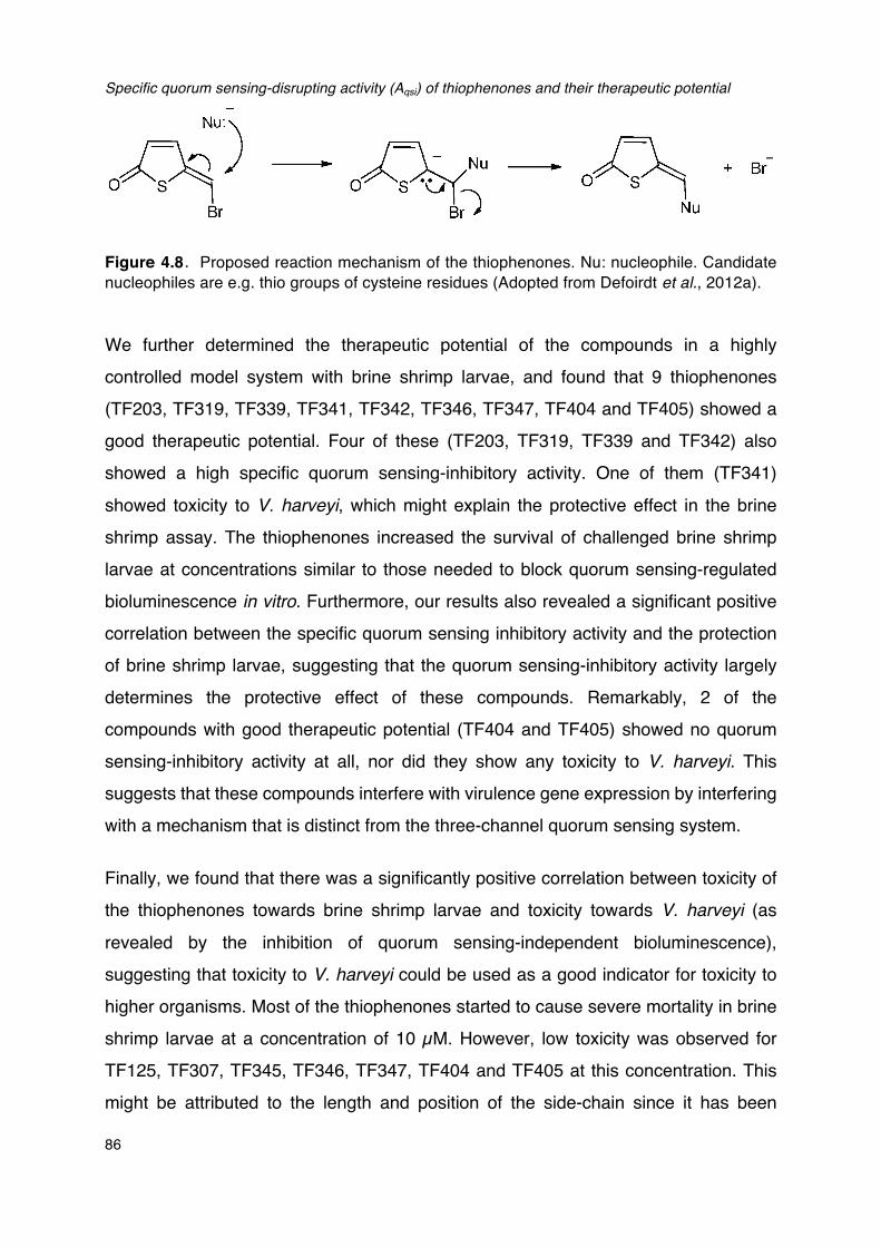
Specific quorum sensing-disrupting activity (Aqsi) of thiophenones and their therapeutic potential
86
Figure 4.8�Proposed reaction mechanism of the thiophenones. Nu: nucleophile. Candidate nucleophiles are e.g. thio groups of cysteine residues (Adopted from Defoirdt et al., 2012a). We further determined the therapeutic potential of the compounds in a highly controlled model system with brine shrimp larvae, and found that 9 thiophenones (TF203, TF319, TF339, TF341, TF342, TF346, TF347, TF404 and TF405) showed a good therapeutic potential. Four of these (TF203, TF319, TF339 and TF342) also showed a high specific quorum sensing-inhibitory activity. One of them (TF341) showed toxicity to V. harveyi, which might explain the protective effect in the brine shrimp assay. The thiophenones increased the survival of challenged brine shrimp larvae at concentrations similar to those needed to block quorum sensing-regulated bioluminescence in vitro. Furthermore, our results also revealed a significant positive correlation between the specific quorum sensing inhibitory activity and the protection of brine shrimp larvae, suggesting that the quorum sensing-inhibitory activity largely determines the protective effect of these compounds. Remarkably, 2 of the compounds with good therapeutic potential (TF404 and TF405) showed no quorum sensing-inhibitory activity at all, nor did they show any toxicity to V. harveyi. This suggests that these compounds interfere with virulence gene expression by interfering with a mechanism that is distinct from the three-channel quorum sensing system.
Finally, we found that there was a significantly positive correlation between toxicity of the thiophenones towards brine shrimp larvae and toxicity towards V. harveyi (as revealed by the inhibition of quorum sensing-independent bioluminescence), suggesting that toxicity to V. harveyi could be used as a good indicator for toxicity to higher organisms. Most of the thiophenones started to cause severe mortality in brine shrimp larvae at a concentration of 10 µM. However, low toxicity was observed for TF125, TF307, TF345, TF346, TF347, TF404 and TF405 at this concentration. This might be attributed to the length and position of the side-chain since it has been
Chapter IV
87
reported that the side-chain could significantly affect the toxicity of thiophenones by
interfering their binding to essential proteins (Steenackers et al., 2010; Defoirdt et al.,
2012). We indeed found that the compounds with the largest side chains at the
3-position of the thiophenone ring (e.g. TF339 and 341) showed the lowest toxicity. In
addition to this, the two compounds with a benzothiophenone core (TF404 and TF405)
also showed low toxicity.
In conclusion, in this study we determined the quorum sensing-inhibiting activities of
20 new synthetic thiophenones towards the quorum sensing model bacterium V.
harveyi. We proposed the new parameter AQSI to determine specific quorum sensing
inhibitory activity based on experiments with a quorum sensing reporter strain. We
used this parameter to analyse data obtained with bioluminescence of V. harveyi as
reporter phenotype, and it can easily be applied to any other signal molecule reporter
strains. The use of AQSI allowed us to exclude 5 false positives out of the 17
compounds that were able to inhibit quorum sensing-regulated bioluminescence in V.
harveyi. We identified 6 thiophenones that were able to inhibit quorum sensing at
submicromolecular levels. Further, we determined the protective effect and toxicity of
the thiophenones in a highly controlled gnotobiotic model system with brine shrimp
larvae. There was a significantly positive correlation between the specific quorum
sensing-disrupting activity of the thiophenones and the protection of brine shrimp
larvae against pathogenic V. harveyi, and 6 quorum sensing-disrupting thiophenones
were considered to be highly promising to control bacterial disease.
4.4 METHODS
4.4.1. Thiophenones
The structures of the thiophenones used in this study are shown in Figure 4.1. The
compounds were synthesised as described before (Benneche et al., 2011; Benneche
et al., submitted). All the thiophenones were dissolved in pure ethanol at 5 mM and
stored at -20 °C.
Specific quorum sensing-disrupting activity (Aqsi) of thiophenones and their therapeutic potential
88
4.4.2 Bacterial strains and growth conditions
V. harveyi wild type strain ATCC BAA-1116 (recently reclassified as V. campbellii; Lin et al., 2010) and mutant strain JAF548 pAKlux1 (Defoirdt et al., 2012) were used in this study. The latter strain is derived from wild type strain ATCC BAA-1116 (BB120). It contains a point mutantion in the luxO gene, rendering the LuxO protein incapable of phosphorelay, and hence the native bioluminescence operon (which is under QS control) is not activated. Then plasmid pAKlux1 was conjugated into this strain. The plasmid contains the Photorhabdus luminescens bioluminescence operon under the control of a constitutive promoter. Hence, strain JAF548 pAKlux1 produces bioluminescence that is independent of the quorum sensing system. This strain is used as a control in order to verify that inhibition of luminescence in V. harveyi is specifically caused by QS inhibition. Both strains were cultured in Luria-Bertani medium containing 35g/L of sodium chloride (LB35) at 28 °C under constant agitation (100 min-1). Cell densities were measured spectrophotometrically at 600 nm.
4.4.3. Bioluminescence assays
V. harveyi wild type and mutant strain were cultured overnight and diluted to an OD600 of 0.1. The thiophenones were added at different concentrations and the cultures were further incubated at 28 °C with shaking. Then bioluminescence was measured after 1h with a Tecan Infinite 200 microplate reader (Tecan, Mechelen, Belgium).
4.4.4. Specific quorum sensing-inhibitory activity AQSI
The specific quorum sensing-inhibitory of the compounds at a given concentration was calculated as follows:
tindependenQS
regulatedQSQSI Inhibition%
Inhibition%A
−
−=
With
% InhibitionQS-regulated: percentage inhibition of QS-regulated bioluminescence in wild
Chapter IV
89
type V. harveyi
% InhibitionQS-independent: percentage inhibition of QS-independent bioluminescence of V. harveyi JAF548 pAKlux1
Compounds were considered as quorum sensing inhibitors if they caused a significant inhibition of quorum sensing-regulated bioluminescence and if AQSI was higher than 2 at one of the concentrations tested.
4.4.5. Axenic hatching of brine shrimp larvae
Two hundred milligrams of high-quality hatching cysts of Artemia franciscana (EG® Type; INVE Aquaculture, Baasrode, Belgium) were hydrated in 18 ml of filter-sterilized tap water for 1 h. Steril cysts and larvae were obtained by decapsulation according to Marques et al. (2004). Briefly, 660 µl of NaOH (32%) and 10 ml of NaOCl (50%) were added to the hydrated cyst suspension to facilitate decapsulation. The process was stopped after 2 min by adding 14 ml of Na2S2O3 (10 g L-1). Filtered (0.22 µm) aeration was provided during the reaction. The decapsulated cysts were washed with filtered (passed through 0.22-µm membrane filter) and autoclaved (moist heat at 121 °C for 15 min) artificial seawater (containing 35 g l-1 of instant ocean synthetic sea salt, Aquarium Systems, Sarrebourg, France). The cysts were resuspended in a 50-ml tube containing 30 ml of filtered, autoclaved seawater and hatched for 28 h on a rotor (4 min-1) at 28 °C with constant illumination (c. 2000 lux). The axenity of cysts was verified by inoculating one ml of culture water into 9 ml of marine broth and incubating at 28 °C for 24 h. After 28 h of hatching, batches of 30 larvae were counted and transferred to fresh, sterile 50-ml tubes containing 30 ml of filtered and autoclaved seawater. Finally, the tubes were returned to the rotor and kept at 28 °C. All manipulations were performed in a laminar flow to maintain sterility of the cysts and larvae.
4.4.6. Brine shrimp challenge tests
The impacts of the thiophenones on the virulence of V. harveyi were determined in a standardized challenge test with gnotobiotic brine shrimp larvae as described by
Specific quorum sensing-disrupting activity (Aqsi) of thiophenones and their therapeutic potential
90
Defoirdt et al. (2005) with some modifications. A suspension of autoclaved LVS3 bacteria (Verschuere et al., 1999) in filtered and autoclaved seawater was added as feed at the start of the challenge test at 107 cells ml-1, and V. harveyi was added at 105 CFU ml-1. The thiophenones were added directly into the brine shrimp rearing water at different concentrations. Brine shrimp cultures, to which only autoclaved LVS3 bacteria were added as feed, were used as controls. The survival of the larvae was counted 48 h after the addition of the pathogens. Each treatment was carried out in triplicate and each experiment was repeated twice to verify the reproducibility. In each test, the sterility of the control treatments were checked at the end of the challenge by inoculating 1 ml of rearing water to 9 ml of marine broth and incubating the mixture for 2 days at 28 °C.
4.4.7. Statistics
Data analysis was carried out using the SPSS statistical software (version 21). Unless stated otherwise, all data were analysed using independent samples t- tests.
Acknowledgements
We thank Bonnie Bassler for providing us with the V. harveyi wild type and mutant JAF548. We also thank Gunnar Herstad for performing the synthesis of the thiophenones. This work was funded by the China Scholarship Council and the Special Research Fund of Ghent University (BOF-UGent).
CHAPTER V
INDOLE AND INDOLE ANALOGUES PRODUCED BY MICRO-ALGAE DECREASE THE VIRULENCE OF LUMINESCENT VIBRIOS, MAJOR PATHOGENS OF AQUATIC ORGANISMS

Chapter V
91
ABSTRACT
Bacterial production of indole has been known for a long time. To date, around 85
species of both Gram-negative and Gram-positive bacteria have been found to
produce large quantities of indole. However, diverse biological responses to indole
have only recently been revealed. Most work in this respect has been conducted on
Escherichia coli, in which indole has been reported to control several virulence-related
phenotypes. To our knowledge, the role of indole in vibrios only has been documented
in V. cholerae and very recently in V. anguillarum. In the present study, we
investigated the impact of indole signaling on the virulence of another important
pathogen V. harveyi, and whether indole analogues produced by micro-algae induce a
similar response as indole. Results showed that indole could decrease the virulence of
V. harveyi towards brine shrimp larvae and giant river prawn larvae. The addition of
exogenous indole was found to down-regulate the production of several virulence
factors, including biofilm formation, exopolysaccharide production and swimming
motility. V. harveyi could produce indole during its growth, and the production was
significantly affected by the alternative sigma factor RpoS. Finally, we found that two
indole analogues, indole-3-acetic acid and indole-3-acetamide, which are produced by
micro-algae, also showed a similar effect as indole except for the swimming motility.
More interestingly, indole and both analogues could interfere the three-channel
quorum sensing system in V. harveyi at different stages. Therefore, they might
potentially be used for antivirulence therapies.
Keywords: indole, auxin, antivirulence therapy, greenwater, alga-bacteria interaction
Indole and indole analogues produced by micro-algae decrease the virulence of luminescent vibrios, major pathogens of aquatic organisms
92
5.1 INTRODUCTION
Vibrios belonging to the Harveyi clade (including Vibrio harveyi) are marine Gram-negative (often luminous) bacteria that can infect a wide range of wild and cultured aquatic organisms (both vertebrates and invertebrates), leading to significant losses in the aquaculture industry worldwide (Austin and Zhang, 2006; Darshanee Ruwandeepika et al., 2012). Despite their role as major pathogens of marine animals, the pathogenicity mechanisms of these bacteria are currently not yet fully understood. To our knowledge, the factors that have been considered to be involved in the pathogenicity include biofilm formation, swimming motility and the production of a variety of extracellular products, including hemolysins, proteases, phospholipase and chitinases (Karunasagar et al., 1994; Austin and Zhang, 2006; Darshanee Ruwandeepika et al., 2012; Yang and Defoirdt, 2014).
Since virulence factors are often costly metabolic products, their expression usually is under strict regulatory control. One of the regulatory mechanisms controlling the production of virulence factors in V. harveyi is quorum sensing (QS), cell-to-cell communication that uses secreted signaling molecules named autoinducers (AI) (Miller and Bassler, 2003). V. harveyi strain ATCC BAA-1116 is a model bacterium in quorum sensing research, and the bacterium has been found to contain a three-channel quorum sensing system, which is mediated by three kinds of signal molecules, harveyi autoinducer 1 (HAI-1; Cao and Meighen, 1989), autoinducer 2 (AI-2; Chen et al., 2002) and cholerae autoinducer 1 (CAI-1; Higgins et al., 2007), respectively. Previous work in our lab has proven that the three-channel quorum sensing system is required for full virulence of V. harveyi towards different hosts (Defoirdt and Sorgeloos, 2012; Pande et al., 2013).
In recent years, more and more compounds are being added to the arsenal of bacterial signaling molecules, and one of these compounds is indole (Lee and Lee, 2010). Indole is produced by tryptophanase (TnaA), which reversibly converts tryptophan into indole, pyruvate and ammonia (Lee et al., 2007a; 2008). A variety of both Gram-positive and Gram-negative bacteria (more than 85 species including many pathogenic bacteria such as V. vulnificus, Proteus vulgaris, Pasteurella

Chapter V
93
multocida and Haemophilus influenzae) have been found to produce indole
(Dalsgaard et al., 1999; DeMoss and Moser, 1969; Clemons and Gadberry, 1982;
Stull et al., 1995). Diverse biological responses to indole have recently been revealed,
including spore formation (Stamm et al., 2005), drug resistance (Hirakawa et al.,
2005), virulence (Hirakawa et al., 2009), plasmid stability (Chant and Summers, 2007),
persister formation (Vega et al., 2012), motility (Bansal et al., 2007),
lipopolysaccharide production (Han et al., 2011) and biofilm formation (Beyhan et al.,
2009). Additionally, indole has been reported to interfere with AHL-based quorum
sensing in a number of Gram-negative bacteria (Hidalgo-Romano et al., 2014). Some
bacteria that lack tryptophanase, and therefore do not produce indole, e.g.
Pseudomonas aeruginosa, also respond to the presence of extracellular indole
(Melander et al., 2014). To date, a signaling role of indole in vibrios only has been
documented in V. cholerae and very recently in V. anguillarum. In V. cholerae, indole
activated Vibrio polysaccharide (VPS) production and biofilm formation, whereas it
decreased motility (Beyhan et al., 2009). In V. anguillarum, by contrast,
exopolysaccharide production was decreased by indole, whereas motility was not
affected (Li et al., 2014). Further, in V. anguillarum, the stationary phase sigma factor
(RpoS) is involved in the production of indole as an rpoS deletion mutant showed
elevated indole levels and increased expression of the indole synthase tnaA (Li et al.,
2014). Finally, the indole receptor has not yet been identified in any bacterium,
although the transcriptional regulator SdiA has been reported to be central in the
indole signalling cascade in E. coli (Lee et al., 2007a) and the DnaK suppressor
protein DksA has been hypothesised to be involved in the indole signaling cascade in
V. cholerae (Beyhan et al., 2009).
In addition to indole, several bacteria have also been reported to respond to natural
indole analogues (Melander et al., 2014). Most notably from an ecological perspective
are the auxin plant hormones, such as indole-3-acetic acid and indole-3-acetamide.
These compounds are also produced by micro-algae (Stirk et al., 2013), which share
the aquatic environment with vibrios. Remarkably, although auxins have been
reported to affect various phenotypes in a number of bacteria (including E.coli and P.
aeruginosa; Spaepen and Vanderleyden, 2011), their impact on gene expression in
vibrios has thus far not been investigated. In addition to the ecological significance,
Indole and indole analogues produced by micro-algae decrease the virulence of luminescent vibrios, major pathogens of aquatic organisms
94
unraveling micro-alga-bacteria interactions is also important from a practical point of
view. Indeed, micro-algae are an important constituent of many aquaculture systems,
especially the so-called greenwater systems, in which the animals are cultured in
water containing high levels of micro-algae (Natrah et al., 2014). Empirical evidence
has suggested that when compared to conventional systems, there is a lower
incidence of disease in greenwater systems (De Schryver et al., 2014). However, the
mechanisms behind this apparent beneficial practice are not yet understood.
Given the ecological and economical importance of V. harveyi as a major pathogen of
aquatic organisms, the previous reports documenting the impact of indole on vibrios,
and the potential impact of micro-algae on this, in the present study, we aimed at
determining the impact of indole signaling on the virulence of V. harveyi, and at
investigating whether indole analogues produced by micro-algae induce a similar
response as indole.
5.2 RESULTS
5.2.1 Impact of indole on the virulence of V. harveyi towards gnotobiotic brine
shrimp larvae and conventionally reared giant river prawn larvae
To examine whether the addition of indole could decrease the virulence of wild type V.
harveyi, we performed a standardized challenge test with gnotobiotic brine shrimp
larvae. In order to exclude any direct effects of indole on the larvae, V. harveyi was
incubated in the presence of indole at 50 μM, 100μM and 200μM, respectively, after
which the cultures were washed to remove the indole prior to inoculation into the brine
shrimp rearing water. A significantly increased survival of brine shrimp larvae
challenged to indole-pretreated V. harveyi was observed, indicating that indole indeed
decreases the virulence of V. harveyi (Table 5.1). Importantly, indole at these
concentrations showed no effect on the growth of V. harveyi (Figure 5.1). Moreover,
all treatments received the same inoculum (105 cells per ml brine shrimp rearing
water), and there were no differences between treatments with respect to the density
of V. harveyi in the brine shrimp rearing water after 1 and 2 days of challenge (Figure 5.2).
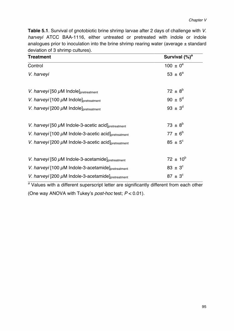
Chapter V
95
Table 5.1. Survival of gnotobiotic brine shrimp larvae after 2 days of challenge with V. harveyi ATCC BAA-1116, either untreated or pretreated with indole or indole analogues prior to inoculation into the brine shrimp rearing water (average ± standard deviation of 3 shrimp cultures). Treatment Survival (%)a
Control 100 ± 0e V. harveyi 53 ± 6a
V. harveyi [50 µM Indole]pretreatment 72 ± 8b
V. harveyi [100 µM Indole]pretreatment 90 ± 5d
V. harveyi [200 µM Indole]pretreatment 93 ± 3d V. harveyi [50 µM Indole-3-acetic acid]pretreatment 73 ± 8b V. harveyi [100 µM Indole-3-acetic acid]pretreatment 77 ± 6b V. harveyi [200 µM Indole-3-acetic acid]pretreatment 85 ± 5c V. harveyi [50 µM Indole-3-acetamide]pretreatment 72 ± 10b V. harveyi [100 µM Indole-3-acetamide]pretreatment 83 ± 3c V. harveyi [200 µM Indole-3-acetamide]pretreatment 87 ± 3c a Values with a different superscript letter are significantly different from each other (One way ANOVA with Tukey’s post-hoc test; P < 0.01).
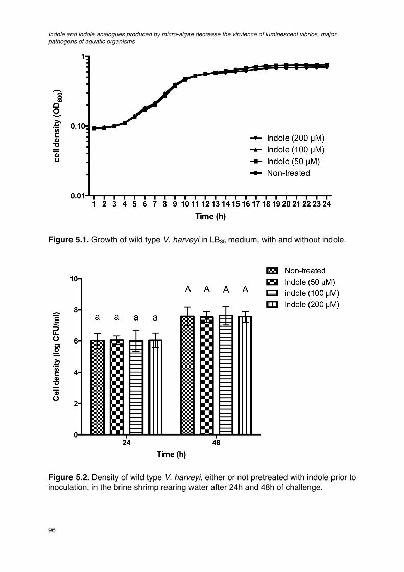
Indole and indole analogues produced by micro-algae decrease the virulence of luminescent vibrios, major pathogens of aquatic organisms
96
Figure 5.1. Growth of wild type V. harveyi in LB35 medium, with and without indole.
Figure 5.2. Density of wild type V. harveyi, either or not pretreated with indole prior to inoculation, in the brine shrimp rearing water after 24h and 48h of challenge.
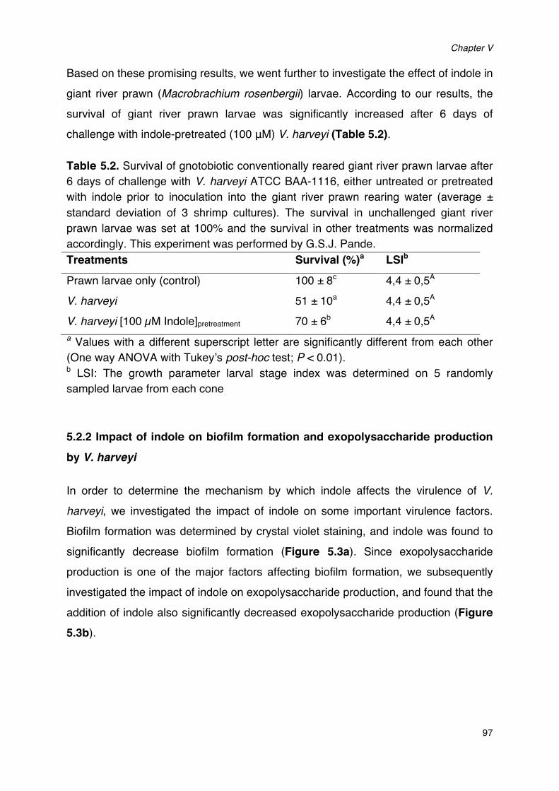
Chapter V
97
Based on these promising results, we went further to investigate the effect of indole in giant river prawn (Macrobrachium rosenbergii) larvae. According to our results, the survival of giant river prawn larvae was significantly increased after 6 days of challenge with indole-pretreated (100 μM) V. harveyi (Table 5.2).
Table 5.2. Survival of gnotobiotic conventionally reared giant river prawn larvae after 6 days of challenge with V. harveyi ATCC BAA-1116, either untreated or pretreated with indole prior to inoculation into the giant river prawn rearing water (average ± standard deviation of 3 shrimp cultures). The survival in unchallenged giant river prawn larvae was set at 100% and the survival in other treatments was normalized accordingly. This experiment was performed by G.S.J. Pande. Treatments Survival (%)a LSIb
Prawn larvae only (control) 100 ± 8c 4,4 ± 0,5A V. harveyi 51 ± 10a 4,4 ± 0,5A V. harveyi [100 µM Indole]pretreatment 70 ± 6b 4,4 ± 0,5A a Values with a different superscript letter are significantly different from each other (One way ANOVA with Tukey’s post-hoc test; P < 0.01). b LSI: The growth parameter larval stage index was determined on 5 randomly sampled larvae from each cone
5.2.2 Impact of indole on biofilm formation and exopolysaccharide production
by V. harveyi
In order to determine the mechanism by which indole affects the virulence of V.
harveyi, we investigated the impact of indole on some important virulence factors. Biofilm formation was determined by crystal violet staining, and indole was found to significantly decrease biofilm formation (Figure 5.3a). Since exopolysaccharide production is one of the major factors affecting biofilm formation, we subsequently investigated the impact of indole on exopolysaccharide production, and found that the addition of indole also significantly decreased exopolysaccharide production (Figure 5.3b).
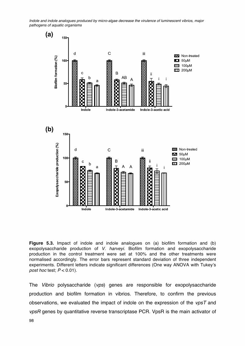
Indole and indole analogues produced by micro-algae decrease the virulence of luminescent vibrios, major pathogens of aquatic organisms
98
Figure 5.3. Impact of indole and indole analogues on (a) biofilm formation and (b) exopolysaccharide production of V. harveyi. Biofilm formation and exopolysaccharide production in the control treatment were set at 100% and the other treatments were normalised accordingly. The error bars represent standard deviation of three independent experiments. Different letters indicate significant differences (One way ANOVA with Tukey’s post hoc test; P < 0.01).
The Vibrio polysaccharide (vps) genes are responsible for exopolysaccharide production and biofilm formation in vibrios. Therefore, to confirm the previous observations, we evaluated the impact of indole on the expression of the vpsT and vpsR genes by quantitative reverse transcriptase PCR. VpsR is the main activator of
Chapter V
99
Vibrio polysaccharide production in V. cholerae, and VpsT is a secondary activator
that acts synergistically with VpsR (Yildiz et al., 2001). The V. harveyi vpsT and vpsR
genes show 73% and 70% nucleotide homology with the V. cholerae analogues,
respectively. We found that the addition of indole resulted in 10-fold and 5-fold
decrease in vpsT and vpsR expression, respectively, in the presence of 100 µM
exogenously added indole (Table 5.3).
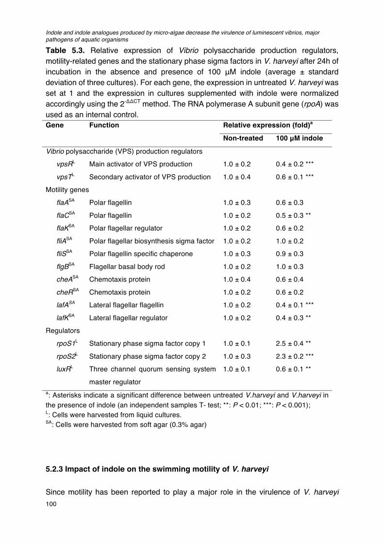
Indole and indole analogues produced by micro-algae decrease the virulence of luminescent vibrios, major pathogens of aquatic organisms
100
Table 5.3. Relative expression of Vibrio polysaccharide production regulators, motility-related genes and the stationary phase sigma factors in V. harveyi after 24h of incubation in the absence and presence of 100 μM indole (average ± standard deviation of three cultures). For each gene, the expression in untreated V. harveyi was set at 1 and the expression in cultures supplemented with indole were normalized accordingly using the 2-ΔΔCT method. The RNA polymerase A subunit gene (rpoA) was used as an internal control. Gene Function Relative expression (fold)a
Non-treated 100 μM indole
Vibrio polysaccharide (VPS) production regulators
vpsRL Main activator of VPS production 1.0 ± 0.2 0.4 ± 0.2 ***
vpsTL Secondary activator of VPS production 1.0 ± 0.4 0.6 ± 0.1 ***
Motility genes
flaASA Polar flagellin 1.0 ± 0.3 0.6 ± 0.3
flaCSA Polar flagellin 1.0 ± 0.2 0.5 ± 0.3 **
flaKSA Polar flagellar regulator 1.0 ± 0.2 0.6 ± 0.2
fliASA Polar flagellar biosynthesis sigma factor 1.0 ± 0.2 1.0 ± 0.2
fliSSA Polar flagellin specific chaperone 1.0 ± 0.3 0.9 ± 0.3
flgBSA Flagellar basal body rod 1.0 ± 0.2 1.0 ± 0.3
cheASA Chemotaxis protein 1.0 ± 0.4 0.6 ± 0.4
cheRSA Chemotaxis protein 1.0 ± 0.2 0.6 ± 0.2
lafASA Lateral flagellar flagellin 1.0 ± 0.2 0.4 ± 0.1 ***
lafKSA Lateral flagellar regulator 1.0 ± 0.2 0.4 ± 0.3 **
Regulators
rpoS1L Stationary phase sigma factor copy 1 1.0 ± 0.1 2.5 ± 0.4 **
rpoS2L Stationary phase sigma factor copy 2 1.0 ± 0.3 2.3 ± 0.2 ***
luxRL Three channel quorum sensing system
master regulator
1.0 ± 0.1 0.6 ± 0.1 **
a: Asterisks indicate a significant difference between untreated V.harveyi and V.harveyi in the presence of indole (an independent samples T- test; **: P < 0.01; ***: P < 0.001); L: Cells were harvested from liquid cultures. SA: Cells were harvested from soft agar (0.3% agar)
5.2.3 Impact of indole on the swimming motility of V. harveyi
Since motility has been reported to play a major role in the virulence of V. harveyi
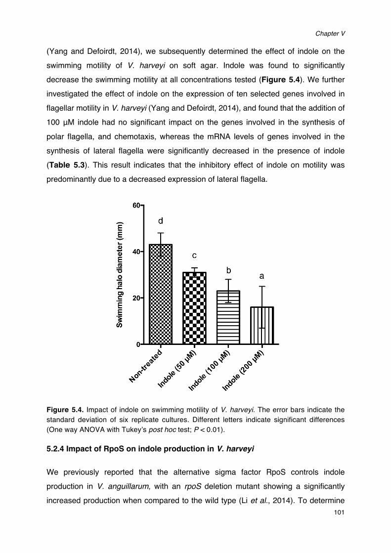
Chapter V
101
(Yang and Defoirdt, 2014), we subsequently determined the effect of indole on the swimming motility of V. harveyi on soft agar. Indole was found to significantly decrease the swimming motility at all concentrations tested (Figure 5.4). We further investigated the effect of indole on the expression of ten selected genes involved in flagellar motility in V. harveyi (Yang and Defoirdt, 2014), and found that the addition of 100 μM indole had no significant impact on the genes involved in the synthesis of polar flagella, and chemotaxis, whereas the mRNA levels of genes involved in the synthesis of lateral flagella were significantly decreased in the presence of indole (Table 5.3). This result indicates that the inhibitory effect of indole on motility was predominantly due to a decreased expression of lateral flagella.
Figure 5.4. Impact of indole on swimming motility of V. harveyi. The error bars indicate the standard deviation of six replicate cultures. Different letters indicate significant differences (One way ANOVA with Tukey’s post hoc test; P < 0.01).
5.2.4 Impact of RpoS on indole production in V. harveyi
We previously reported that the alternative sigma factor RpoS controls indole production in V. anguillarum, with an rpoS deletion mutant showing a significantly increased production when compared to the wild type (Li et al., 2014). To determine
Indole and indole analogues produced by micro-algae decrease the virulence of luminescent vibrios, major pathogens of aquatic organisms
102
whether this regulation also exists in V. harveyi, we investigated the indole production
in V. harveyi wild type and an rpoS1/rpoS2 deletion mutant (the V. harveyi ATCC
BAA-1116 genome contains 2 copies of rpoS). Consistent with what has been
reported for V. anguillarum, indole production was also significantly higher in the
rpoS1/rpos2 mutant than in the wild type at 6h (exponential phase), 12h (early
stationary phase) and 24h (stationary phase) of incubation (Figure 5.5a).
Furthermore, the expression level of the indole synthase gene tnaA was significantly
higher in the rpoS1/rpoS2 mutant than in the wild type at all sampling points (3-fold at
6h; 4-fold at 12h and 3-fold at 24h; Figure 5.5b).
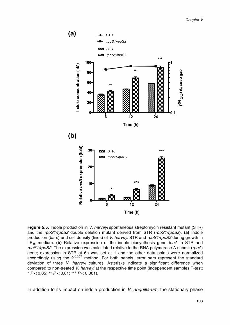
Chapter V
103
Figure 5.5. Indole production in V. harveyi spontaneous streptomycin resistant mutant (STR) and the rpoS1/rpoS2 double deletion mutant derived from STR (rpoS1/rpoS2). (a) Indole production (bars) and cell density (lines) of V. harveyi STR and rpoS1/rpoS2 during growth in LB35 medium. (b) Relative expression of the indole biosynthesis gene tnaA in STR and rpoS1/rpoS2. The expression was calculated relative to the RNA polymerase A submit (rpoA) gene; expression in STR at 6h was set at 1 and the other data points were normalized accordingly using the 2-ΔΔCT method. For both panels, error bars represent the standard deviation of three V. harveyi cultures. Asterisks indicate a significant difference when compared to non-treated V. harveyi at the respective time point (independent samples T-test; * P < 0.05; ** P < 0.01; *** P < 0.001). In addition to its impact on indole production in V. anguillarum, the stationary phase
Indole and indole analogues produced by micro-algae decrease the virulence of luminescent vibrios, major pathogens of aquatic organisms
104
sigma factor RpoS has also been reported before to be (partially) involved in the response to indole in V. cholerae (Mueller et al., 2009), and we previously found that rpoS mRNA levels in V. anguillarum are higher upon exposure to endogenous indole (Li and Defoirdt, unpublished). Consistent with this, we found that the expression levels of both rpoS1 and rpoS2 were also significantly higher in V. harveyi upon exposure to 100 µM indole (Table 5.3).
5.2.5 Impact of indole on the 3-channel quorum sensing system of V. harveyi
Indole has been reported to interfere with other quorum sensing mechanisms in various bacteria (Kim and Park, 2013; Hidalgo-Romano et al., 2014), and therefore, we also evaluated whether indole interfered with the activity of the three-channel quorum sensing system of V. harveyi. We used bioluminescence as readout of quorum sensing activity since it is one of the phenotypes that are regulated by quorum sensing. Wild type V. harveyi was grown to high cell density in order to activate quorum sensing-controlled bioluminescence, after which indole was added at 50 μM, 100 μM and 200 μM, respectively, and bioluminescence was measured after 1h. Indole could indeed inhibit the quorum sensing-regulated bioluminescence in wild type V. harveyi (Figure 5.6a).
We further sought to confirm that the effect of indole on bioluminescence was due to interference with the quorum sensing system by determining the impact of indole on bioluminescence of V. harveyi JAF548 pAKlux1, in which bioluminescence is independent of the three channel quorum sensing system (Defoirdt et al., 2012). Indole had no effect on the bioluminescence of JAF548 pAKlux1 at all the tested concentrations, indicating that the inhibition of bioluminescence in wild type V. harveyi was caused by interference with the three-channel quorum sensing system (Figure
5.6c).
In order to determine at what stage in the quorum sensing signal transduction cascade indole was acting, we used mutants JAF483 (LuxO D47A) and BNL258 (hfq::Tn5lacZ). Mutant JAF483 contains a point mutation in luxO, that renders the LuxO protein locked in the conformation of wild type LuxO at high signal molecule concentrations (Freeman and Bassler, 1999). Strain BNL258 has a transposon
Chapter V
105
insertion in the hfq gene, resulting in a nonfunctional Hfq protein (Lenz et al., 2004).
These mutations result in a maximally active quorum sensing system and constitutive
luminescence in JAF483 and BNL258, irrespective of cell density and signal molecule
concentration. Blocking of luminescence in these mutants would indicate that indole
interacts with a quorum sensing signal transduction component located downstream
of the mutated one (Defoirdt et al., 2007); a scheme of the V. harveyi three-channel
quorum sensing system is presented in Figure 5.7 for further information). Indole was
also found to block luminescence in JAF483 and BNL258 at all the concentrations
tested (Figure 5.6 b & c), which indicates that it interferes with signal transduction (by
interacting with a signal transduction cascade component downstream of Hfq) and not
by interacting with signal molecule detection (by interacting with one of the signal
molecule receptors). Inversely, the three- channel quorum sensing system had no
effect on the production of indole as there was no difference between the strains in
indole levels (Figure 5.8).
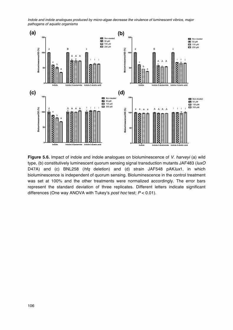
Indole and indole analogues produced by micro-algae decrease the virulence of luminescent vibrios, major pathogens of aquatic organisms
106
Figure 5.6. Impact of indole and indole analogues on bioluminescence of V. harveyi (a) wild type, (b) constitutively luminescent quorum sensing signal transduction mutants JAF483 (luxO D47A) and (c) BNL258 (hfq deletion) and (d) strain JAF548 pAKlux1, in which bioluminescence is independent of quorum sensing. Bioluminescence in the control treatment was set at 100% and the other treatments were normalized accordingly. The error bars represent the standard deviation of three replicates. Different letters indicate significant differences (One way ANOVA with Tukey’s post hoc test; P < 0.01).
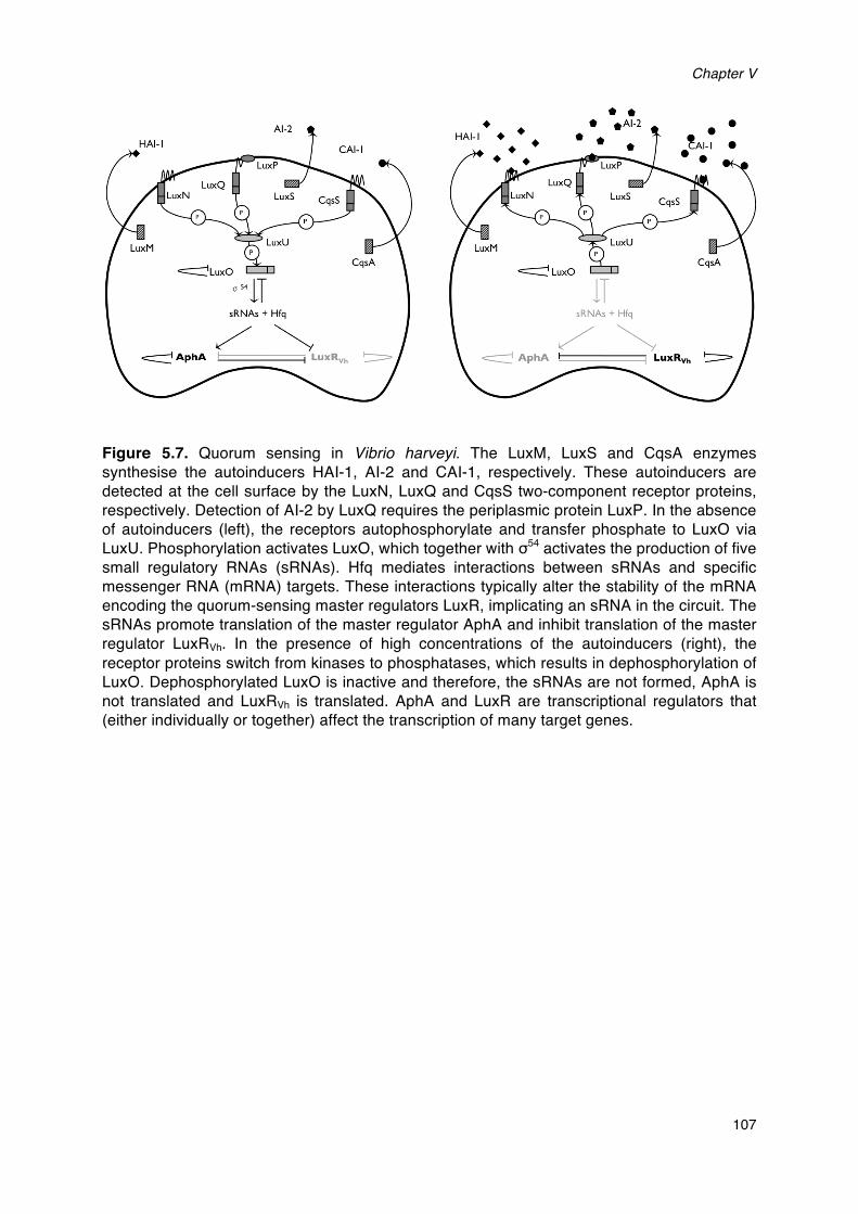
Chapter V
107
Figure 5.7. Quorum sensing in Vibrio harveyi. The LuxM, LuxS and CqsA enzymes synthesise the autoinducers HAI-1, AI-2 and CAI-1, respectively. These autoinducers are detected at the cell surface by the LuxN, LuxQ and CqsS two-component receptor proteins, respectively. Detection of AI-2 by LuxQ requires the periplasmic protein LuxP. In the absence of autoinducers (left), the receptors autophosphorylate and transfer phosphate to LuxO via LuxU. Phosphorylation activates LuxO, which together with σ54 activates the production of five small regulatory RNAs (sRNAs). Hfq mediates interactions between sRNAs and specific messenger RNA (mRNA) targets. These interactions typically alter the stability of the mRNA encoding the quorum-sensing master regulators LuxR, implicating an sRNA in the circuit. The sRNAs promote translation of the master regulator AphA and inhibit translation of the master regulator LuxRVh. In the presence of high concentrations of the autoinducers (right), the receptor proteins switch from kinases to phosphatases, which results in dephosphorylation of LuxO. Dephosphorylated LuxO is inactive and therefore, the sRNAs are not formed, AphA is not translated and LuxRVh is translated. AphA and LuxR are transcriptional regulators that (either individually or together) affect the transcription of many target genes.
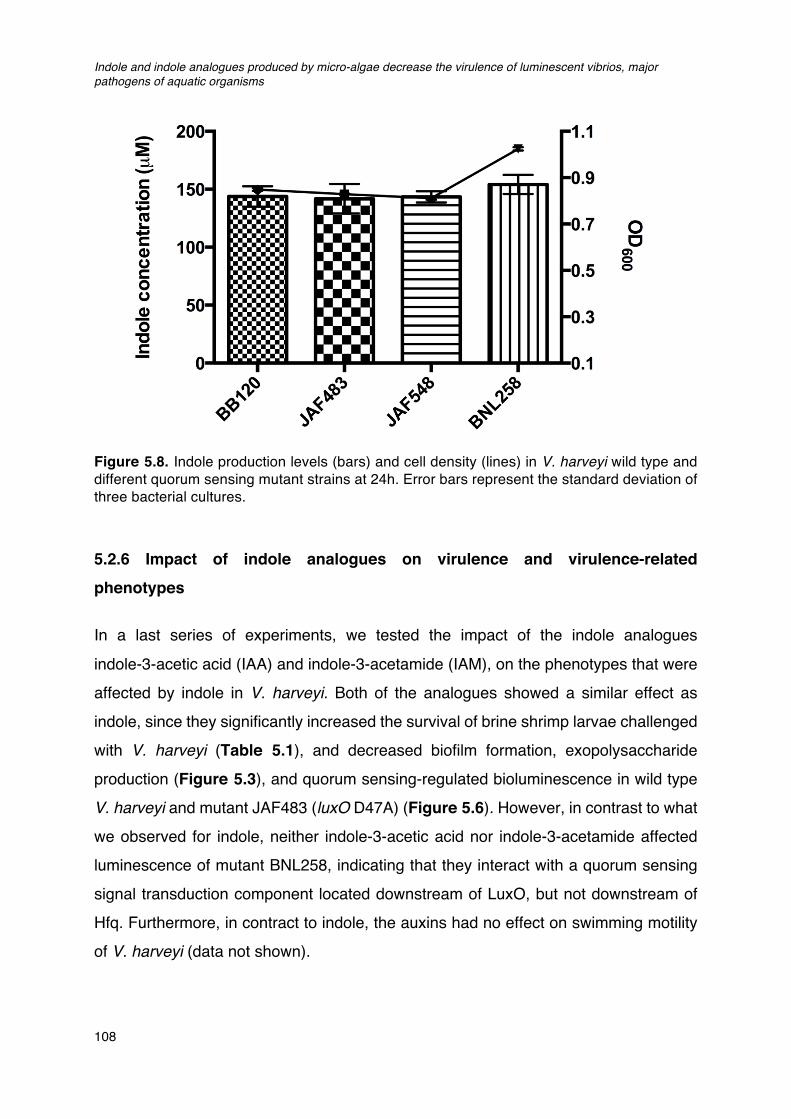
Indole and indole analogues produced by micro-algae decrease the virulence of luminescent vibrios, major pathogens of aquatic organisms
108
Figure 5.8. Indole production levels (bars) and cell density (lines) in V. harveyi wild type and different quorum sensing mutant strains at 24h. Error bars represent the standard deviation of three bacterial cultures.
5.2.6 Impact of indole analogues on virulence and virulence-related
phenotypes
In a last series of experiments, we tested the impact of the indole analogues indole-3-acetic acid (IAA) and indole-3-acetamide (IAM), on the phenotypes that were affected by indole in V. harveyi. Both of the analogues showed a similar effect as indole, since they significantly increased the survival of brine shrimp larvae challenged with V. harveyi (Table 5.1), and decreased biofilm formation, exopolysaccharide production (Figure 5.3), and quorum sensing-regulated bioluminescence in wild type V. harveyi and mutant JAF483 (luxO D47A) (Figure 5.6). However, in contrast to what we observed for indole, neither indole-3-acetic acid nor indole-3-acetamide affected luminescence of mutant BNL258, indicating that they interact with a quorum sensing signal transduction component located downstream of LuxO, but not downstream of Hfq. Furthermore, in contract to indole, the auxins had no effect on swimming motility of V. harveyi (data not shown).
Chapter V
109
5.3 DISCUSSION
Recently, indole has been proposed as an intercellular signal molecule in bacteria, affecting various behaviors (Jin-Hyung Lee and Jintae Lee, 2010; Melander et al., 2014). In this study, we demonstrated that indole controls the virulence of V. harveyi in our highly controlled model system with gnotobiotic brine shrimp larvae. Gnotobiotic conditions are important in this respect since many bacteria are capable of producing high levels of indole (Lee and Lee, 2010) and therefore, the microbiota that is naturally associated with the host might confuse the experiments. We observed significantly decreased virulence of V. harveyi in this model system when the pathogen was pretreated with indole at 50 µM or more. This concentration is similar to the concentration produced by wild type V. harveyi when grown in Luria-Bertani broth (hence, doubling the concentration of extracellular indole results in decreased virulence). The pathogen was pretreated with indole in order to avoid a direct effect of indole on the host, which would confuse interpretation of the results. Indeed, indole has been reported to exert a beneficial effect on higher organisms, e.g. by increasing epithelial-cell tight-junction resistance (Bansal et al., 2010). The results obtained in this study are consistent with our previous observation that indole protects sea bass (Dicentrarchus labrax) larvae from V. anguillarum (Li et al., 2014). Hence, indole-decreasing virulence seems to be conserved among distantly related vibrios that are pathogenic to aquatic organisms.
In order to determine the mechanism(s) by which indole reduces the virulence of V.
harveyi, we evaluated the impact of indole on several important virulence factors that were affected by indole in other vibrios (Beyhan et al., 2009; Li et al., 2014). Indole decreased biofilm formation, exopolysaccharide production and motility of V. harveyi, and these effects were confirmed at the transcriptional level by reverse transcriptase qPCR targeting key genes involved in these phenotypes. Importantly, no effect was observed in the controls which received the same volume of DMSO as added to the other treatments. The effect on biofilm formation and exopolysaccharide production is consistent with what we observed before in V. anguillarum (Li et al., 2014) and with what has been reported for E. coli (Jintae Lee, Jayaraman and Wood, 2007b; Bansal et al., 2007). However, these results are in contrast with what has been reported for V.
Indole and indole analogues produced by micro-algae decrease the virulence of luminescent vibrios, major pathogens of aquatic organisms
110
cholerae, where deletion of the indole synthase gene tnaA resulted in decreased
biofilm formation, and which was restored upon addition of 350 µM indole (Beyhan et
al., 2009). The observation that indole decreased the motility of V. harveyi and the
expression of genes involved in bacterial flagella biosynthesis and chemotaxis, is
consistent with what has been reported for V. cholerae (Beyhan et al., 2009), E. coli
(Lee et al., 2007a), Salmonella enterica serovar Typhimurium (Nikaido et al., 2012)
and P. aeruginosa (Lee et al., 2009). In contrast, indole had no impact on motility of V.
anguillarum in our previous study (Li et al., 2014). The decrease in flagellar motility
could, together with decreased exopolysaccharide production, explain the decrease in
biofilm formation induced by indole. Indeed, both phenotypes play a key role in biofilm
formation in bacteria (Petrova and Sauer, 2012).
We further determined the connection of indole signaling with other key regulatory
mechanisms in V. harveyi. The alternative sigma factor RpoS has previously been
reported to regulate the production of indole in E. coli (Lelong et al., 2007) and V.
anguillarum (Li et al., 2014). In agreement with what has been demonstrated in V.
anguillarum, the V. harveyi rpoS1/rpoS2 double mutant produced higher indole levels
than the wild type (approximately double the amount, note that this is similar to the
extracellular indole levels of the wild type when adding 50 µM exogenous indole), and
this was reflected in a higher expression of the indole synthase tnaA in the
rpoS1/rpoS2 mutant. However, these results are opposite to what has been revealed
in E. coli, in which RpoS enhanced indole production (Lelong et al., 2007). We also
found that there is a feedback loop as addition of exogenous indole resulted in
increased expression of the two rpoS copies in wild type V. harveyi. This is consistent
with what we observed in V. anguillarum (Xuan Li and Tom Defoirdt, unpublished). We
hypothesise that indole induces a stress response (by inducing expression of RpoS,
which is known to be related to stress), and that down-regulation of indole production
by RpoS might be a mechanism to maintain homeostasis. Furthermore, we found that
indole could interfere with the three-channel quorum sensing system of V. harveyi by
interacting with signal transduction. We previously found that motility is controlled by
this three-channel quorum sensing system (Yang and Defoirdt, 2014). Hence, the
decreased motility in the presence of exogenous indole might be explained by the
inhibition of quorum sensing signal transduction. Inhibition of quorum sensing signal
Chapter V
111
transduction by indole will also contribute to the decreased virulence towards brine
shrimp larvae since quorum sensing is required for full virulence of V. harveyi in this
model system (Defoirdt and Sorgeloos, 2012). Inhibition of quorum sensing by indole
apparently is widespread in Gram-negative bacteria, including as Chromobacterium violaceum, Pseudomonas chlororaphis, Serratia marcescens, Pseudomonas aeruginosa and Acinetobacter oleivorans (Chu et al., 2012; Hidalgo-Romano et al., 2014; Attila et al., 2009; Kim and Park, 2013). Finally, the transcriptional regulator
SdiA has been reported to be central in the indole signalling cascade in E.coli (Lee et al., 2009), and the DnaK suppressor protein DksA has been hypothesised to be
involved in the indole signaling cascade in V. cholerae (Beyhan et al., 2009). The V. harveyi ATCC BAA-1116 genome does not contain a homologue of sdiA, but it does
contain a dksA homologue (82% identity at nucleotide level with V. cholerae dksA).
Unfortunately, we were not able to obtain a dksA deletion mutant (which would have
enabled us to determine whether DksA is involved in indole sensing), and this might
indicate that this gene is essential in V. harveyi.
V. harveyi shares the aquatic environment with micro-algae, which like terrestrial
plants are known to produce various indole analogues as auxin hormones (Stirk et al., 2013). Auxins have been reported to be produced by several unicellular and
multicellular algal species, e.g. brown (Macrocystis, Laminaria, Fucus, Ascophyllum),
red (Botryocladia, Prionitis and Porphyra), and green (Enteromorpha, Chlorella,
Cladophora and Caulerpa) algae and also in cyanobacteria (Oscillatoria and
Chlorogloea) (Tarakhovskaya et al., 2007). The auxin concentrations measured in
most of the studies are lower when compared to those in land plants, e.g. the level of
free indole-3-acetic acid was 2.5 ng/g fresh weight in the red algae Prionitis lanceolata, while 20 ng/g fresh weight in angiosperms (Ashen et al., 1999; Basu et al., 2002; Han,
2006; Stirk et al., 2004; 2002). Auxins can affect various phenotypes in terrestrial
bacteria (Spaepen and Vanderleyden, 2011). However, despite the potential
ecological significance as cross-kingdom signals, the impact of auxins produced by
micro-algae on aquatic bacteria has thus far not been investigated. We found that the
auxins indole-3-acetic acid and indole-3-acetamide showed similar effects as indole,
except for motility (which was not affected by the auxins). Indeed, pretreatment of V. harveyi resulted in a similar decrease in mortality of challenged brine shrimp larvae,
Indole and indole analogues produced by micro-algae decrease the virulence of luminescent vibrios, major pathogens of aquatic organisms
112
and biofilm formation and exopolysaccharide production were also decreased to the
same extent as was observed for indole at the same concentrations. The auxins also
inhibited quorum sensing signal transduction, although the effect was slightly less
pronounced than for indole and although they interact with a different three-channel
quorum sensing signal transduction component. Given the fact that quorum sensing
controls swimming motility, this might (in part) explain the fact that motility was not
affected by the auxins.
In addition to the ecological significance of cross-communication between aquatic
bacteria and micro-algae, these observations also offer an explanation for the
beneficial effect that is associated with micro-algae in aquaculture. Indeed,
micro-algae are an important constituent of the so-called greenwater aquaculture
systems, in which the animals are cultured in water containing high levels of
micro-algae (Natrah et al., 2014). Empirical evidence has indicated that there is a
lower incidence of disease in this kind of rearing systems (De Schryver et al., 2014),
which might be explained by the presence of (relatively) high levels of indole
analogues in the digestive tract of animals that have ingested high levels of
micro-algae. Consistent with this are several observations in our lab that feeding
aquatic animals with micro-algae results in an improved survival upon challenge with
a pathogen, even if the micro-algae had been autoclaved before adding them to the
rearing water (e.g.) (Marques et al., 2005; Natrah et al., 2012). This effect was
originally thought to be nutritional (Marques et al., 2005). However, since many
nutrients are heat-labile, this probably is not the major explanation of the beneficial
effect. Given the fact that indoles are heat-stable (even resisting autoclavation), the
auxins that are produced as plant hormones by micro-algae might be better
candidates to explain the beneficial effect. We anticipate that the selection of
micro-algae that are capable of producing high levels of auxins could be an interesting
novel strategy to control bacterial disease in aquaculture. Indeed, not all algae show
the same beneficial effect; there are even differences between strains belonging to
the same species (Marques et al., 2005), and there are also differences between
micro-algae with respect to the produced levels of auxins (Stirk et al., 2013).
In conclusion, our results indicate that V. harveyi produce large quantity of indole
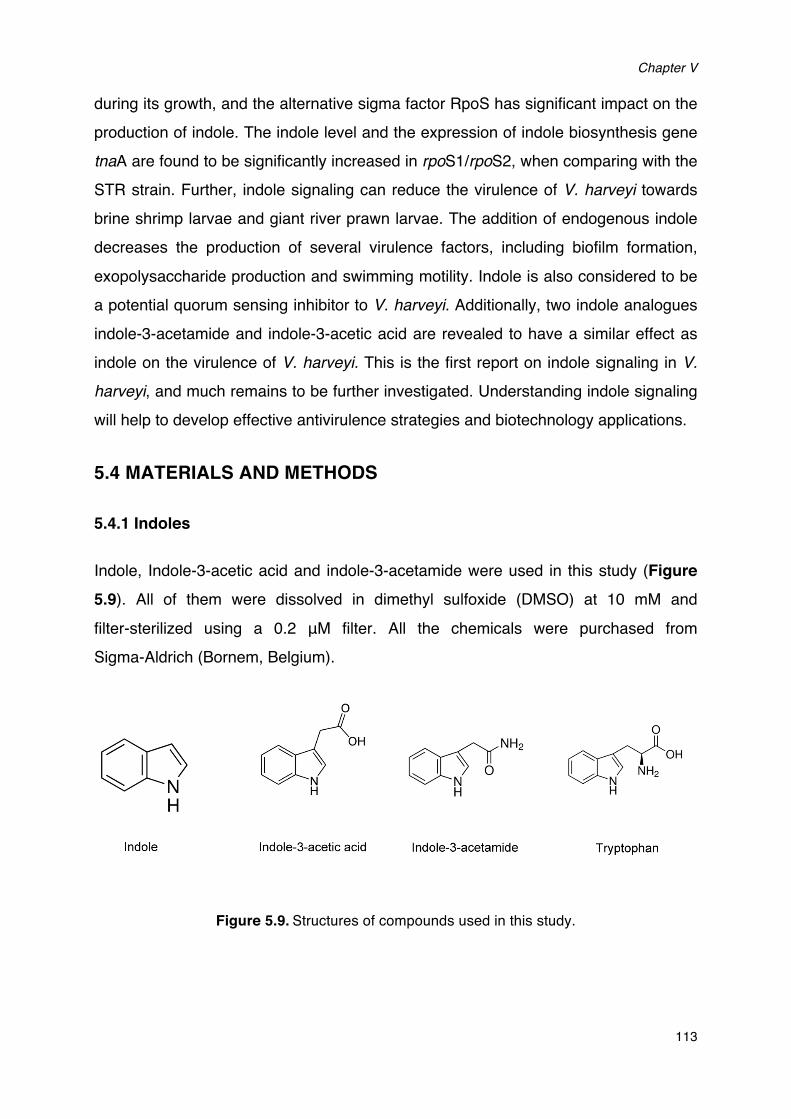
Chapter V
113
during its growth, and the alternative sigma factor RpoS has significant impact on the production of indole. The indole level and the expression of indole biosynthesis gene tnaA are found to be significantly increased in rpoS1/rpoS2, when comparing with the STR strain. Further, indole signaling can reduce the virulence of V. harveyi towards brine shrimp larvae and giant river prawn larvae. The addition of endogenous indole decreases the production of several virulence factors, including biofilm formation, exopolysaccharide production and swimming motility. Indole is also considered to be a potential quorum sensing inhibitor to V. harveyi. Additionally, two indole analogues indole-3-acetamide and indole-3-acetic acid are revealed to have a similar effect as indole on the virulence of V. harveyi. This is the first report on indole signaling in V.
harveyi, and much remains to be further investigated. Understanding indole signaling will help to develop effective antivirulence strategies and biotechnology applications.
5.4 MATERIALS AND METHODS
5.4.1 Indoles
Indole, Indole-3-acetic acid and indole-3-acetamide were used in this study (Figure 5.9). All of them were dissolved in dimethyl sulfoxide (DMSO) at 10 mM and filter-sterilized using a 0.2 μM filter. All the chemicals were purchased from Sigma-Aldrich (Bornem, Belgium).
Figure 5.9.Structures of compounds used in this study.
Indole and indole analogues produced by micro-algae decrease the virulence of luminescent vibrios, major pathogens of aquatic organisms
114
5.4.2 Bacterial strains and growth conditions
Five V. harveyi strains were used in this study, all derived from wild type strain ATCC BAA-1116. Mutants JAF483 (luxO D47A) and mutant BNL258 (hfq::Tn5lacZ), in which the quorum sensing is maximally activated (Freeman and Bassler, 1999; Lenz et al., 2004), and mutant JAF548 (luxO D47E) pAKlux1, in which bioluminescence is independent of quorum sensing (Defoirdt et al., 2012) were used for bioluminescence experiments. Spontaneous streptomycin resistant mutant STR was derived from BB120 (Wang et al., 2012), and the rpoS1/rpoS2 double deletion mutant was derived from this strain (Wang et al., 2012). Unless otherwise stated, all strains were cultured in Luria-Bertani medium containing 35g/L of sodium chloride (LB35) at 28 °C under constant agitation (100 min-1). Cell densities were measured spectrophotometrically at 600 nm.
5.4.3 Bacterial growth assays
For the bacterial growth assays, V. harveyi was grown overnight in LB35 broth at 28°C. After that, the culture was re-inoculated at a concentration of 102 CFU/ml into fresh LB35 broth, with and without indole at 50, 100 and 200 μM, respectively. The cultures were grown in 200 µl volumes in 96-well plates at 28°C for 48 h, and the turbidity at 600 nm was monitored every hour using a Multireader machine (Infinite M200, TECAN, Austria). Growth curves were determined for three independent cultures.
5.4.4 Quantification of indole
V. harveyi cultures grown in LB35 medium were harvested at different time points and centrifuged at 8000 x g for 5 min. The concentration of indole in the supernatants was measured as described previously (Li et al., 2014) by mixing 500 µl of supernatant with 500 µl of Kovac’s reagent. After vortexing, the top 200 µl were removed and the OD571 was measured. The indole concentration in each sample was determined based on a standard curve using synthetic indole (Sigma-Aldrich). At least three different V. harveyi cultures were sampled for each strain at each time point.
Chapter V
115
5.4.5 Swimming motility assay
The swimming motility assay was performed on soft agar (LB35 plates containing 0.2% agar) as described previously (Yang and Defoirdt, 2014). Indole was added to the autoclaved agar. V. harveyi was grown overnight in LB35 broth, and 5 µl aliquots (OD600 = 1.0) were spotted in the center of the soft agar plates. Plates were incubated for 24 h, after which the diameters of the motility halos were measured. All assays were done with freshly prepared media in 6 replicates.
5.4.6 Biofilm formation assay
Biofilm formation was quantified by crystal violet staining, as described previously (Stepanović et al., 2007). In brief, an overnight culture of V. harveyi was diluted to an OD600 of 1.0 in LB35 broth with or without indole and indole analogues, and 200 µl aliquots of these suspensions were pipetted into the wells of a 96 well plate. Then the bacteria were allowed to adhere and grow without agitation for 24h at 28°C. After that, the cultures were removed and the wells were washed three times with 300 μl sterile physiological saline to remove all non-adherent bacteria. The remaining attached bacteria were fixed with 150 μl of 99% methanol per well for 20 min, after which the methanol was removed and plates were air-dried. Then, biofilms were stained for 15 min with 150 μl per well of a 1% crystal violet solution (Pro-lab Diagnostics, Richmond Hill, ON, Canada). Excess stain was rinsed off by placing the plate under running tap water, and washing was continued until the washings were free of the stain. After the plates were air dried, the dye bound to the adherent cells was resolubilized with 150 μl of 95% ethanol per well, and absorbance was measured at 570 nm. Sterile medium served as negative control. For the quantification of exopolysaccharides, Calcofluor white staining (Sigma-Aldrich) was used. In brief, wells were rinsed after 24 h biofilm formation and 100 μl phosphate buffered saline containing 0.5 μl 5 mM Calcofluor white staining dye was added to the wells. After 60 min, fluorescence (excitation 405 nm and emission 500 nm) was measured with a Multi-reader (Infinite M200, TECAN, Austria).
Indole and indole analogues produced by micro-algae decrease the virulence of luminescent vibrios, major pathogens of aquatic organisms
116
5.4.7 Bioluminescence assays
V. harveyi strains were grown overnight and diluted to an OD600 of 0.1. Indole and analogues were added at 50, 100 and 200 μM, respectively. The cultures were further incubated at 28 °C with shaking, and bioluminescence was measured after 1h with a Tecan Infinite 200 microplate reader (Tecan, Mechelen, Belgium).
5.4.8 RNA extraction
V. harveyi strains were grown in triplicate in LB35 broth with or without 100 μM indole at 28°C, and cells were harvested after 24h (unless otherwise indicated). The samples for flagellar gene expression tests were harvested from soft (0.2 % agar) or hard agar plates (1.5% agar) after 24h incubation at 28°C. Then RNA was extracted with the SV Total RNA Isolation System (Promega, Leiden, The Netherlands) according to the manufacturer’s instructions. The RNA quantity was measured spectrophotometrically (NanoDrop Technologies, Wilmington, DE, USA) and adjusted to 200 ng µl-1 in all samples. The RNA integrity was checked by Agarose Gel Electrophoresis and the RNA samples were stored in -80°C.
5.4.9 Primers
The primers used for RT-qPCR analysis are listed in Table 5.4. Specific primers were designed using the software PRIMER PREMIER version 5.00 (Premier Biosoft Inter- national, Palo Alto, CA USA), with predicted product sizes in the 100 to 200 bp range. The RNA polymerase A submit (rpoA) mRNA was used as an endogenous control (Defoirdt et al., 2007).
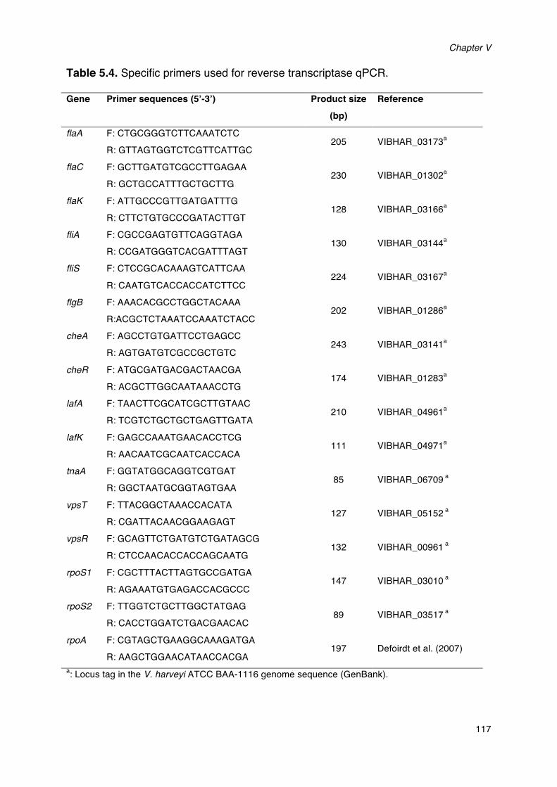
Chapter V
117
Table 5.4. Specific primers used for reverse transcriptase qPCR. Gene Primer sequences (5’-3’) Product size
(bp)
Reference
flaA F: CTGCGGGTCTTCAAATCTC
R: GTTAGTGGTCTCGTTCATTGC 205 VIBHAR_03173a
flaC F: GCTTGATGTCGCCTTGAGAA
R: GCTGCCATTTGCTGCTTG 230 VIBHAR_01302a
flaK F: ATTGCCCGTTGATGATTTG
R: CTTCTGTGCCCGATACTTGT 128 VIBHAR_03166a
fliA F: CGCCGAGTGTTCAGGTAGA
R: CCGATGGGTCACGATTTAGT 130 VIBHAR_03144a
fliS F: CTCCGCACAAAGTCATTCAA
R: CAATGTCACCACCATCTTCC 224 VIBHAR_03167a
flgB F: AAACACGCCTGGCTACAAA
R:ACGCTCTAAATCCAAATCTACC 202 VIBHAR_01286a
cheA F: AGCCTGTGATTCCTGAGCC
R: AGTGATGTCGCCGCTGTC 243 VIBHAR_03141a
cheR F: ATGCGATGACGACTAACGA
R: ACGCTTGGCAATAAACCTG 174 VIBHAR_01283a
lafA F: TAACTTCGCATCGCTTGTAAC
R: TCGTCTGCTGCTGAGTTGATA 210 VIBHAR_04961a
lafK F: GAGCCAAATGAACACCTCG
R: AACAATCGCAATCACCACA 111 VIBHAR_04971a
tnaA F: GGTATGGCAGGTCGTGAT
R: GGCTAATGCGGTAGTGAA 85 VIBHAR_06709 a
vpsT F: TTACGGCTAAACCACATA
R: CGATTACAACGGAAGAGT 127 VIBHAR_05152 a
vpsR F: GCAGTTCTGATGTCTGATAGCG
R: CTCCAACACCACCAGCAATG 132 VIBHAR_00961 a
rpoS1 F: CGCTTTACTTAGTGCCGATGA
R: AGAAATGTGAGACCACGCCC 147 VIBHAR_03010 a
rpoS2 F: TTGGTCTGCTTGGCTATGAG
R: CACCTGGATCTGACGAACAC 89 VIBHAR_03517 a
rpoA F: CGTAGCTGAAGGCAAAGATGA
R: AAGCTGGAACATAACCACGA 197 Defoirdt et al. (2007)
a: Locus tag in the V. harveyi ATCC BAA-1116 genome sequence (GenBank).
Indole and indole analogues produced by micro-algae decrease the virulence of luminescent vibrios, major pathogens of aquatic organisms
118
5.4.10 Reverse transcription
Reverse transcription was performed with the RevertAidTM H minus First strand cDNA synthesis kit (Fermentas Gmbh, Baden-Württemberg, Germany) in accordance to the manufacturer’s instructions. Briefly, a mixture of 1 µg RNA and 1 µl random hexamer primer solution was mixed first. Then, 8 µl of reaction mixture containing 4 µl of 5× reaction buffer (0.25 mol-1 Tris–HCl pH 8.3, 0.25 mol-1 KCl, 0.02 mol-1 MgCl2, 0.05 mol-1 DTT), 2 µl of 0.01 mol-1 dNTP mix, 20 units of ribonuclease inhibitor, 200 units of RevertAidTM H minus M-MuLV Reverse Transcriptase was added. The reaction mixture was incubated for 5 min at 25°C followed by 60 min at 42°C. The reaction was terminated by heating at 70°C for 5 min and then cooled to 4°C. cDNA samples were checked by PCR and stored at -20°C for further use.
5.4.11 Real-time PCR
Real-time PCR was used to quantify the expression level of all the genes and was performed with Maxima® SYBR Green/ROX qPCR Master Mix (Fermentas, Fisher Scientific, Erembodegem, Belgium) as described previously (Yang and Defoirdt, 2014). The reaction was performed in an StepOne™ Real-Time PCR System thermal cycler (Applied Biosystems, Gent, Belgium) in a total volume of 25 µl, containing 12.5 µl of 2× SYBR green master mix, 300 nM of forward and reverse primers and 2 µl of template cDNA. The thermal cycling consisted of an initial denaturation at 95°C for 10 min followed by 40 cycles of denaturation at 95°C for 15 s and primer annealing and elongation at 60°C for 1 min. Dissociation curve analysis was performed to check for the amplification of untargeted fragments. Data acquisition was performed with the StepOne™ Software.
5.4.12 Real-time PCR data analysis (2-∆∆CT method)
The real-time PCR was validated by amplifying serial dilutions of cDNA synthesized from 1 µg of RNA isolated from bacterial samples. Serial dilutions of cDNA were amplified using gene specific primers. ∆CT (average CT value of target-average CT value of rpoA) was calculated for the different dilutions and plotted against the cDNA concentration. The slope of the graph was almost equal to 0 for all of the target nine
Chapter V
119
genes. Therefore, the amplification efficiency of reference and the target genes was considered to be equal. Based on this precondition, real-time PCR data were analyzed using the 2-∆∆CT method (Livak and Schmittgen, 2001). The expression of the target genes was normalized to the endogenous control (rpoA) by calculating ∆CT :
∆Ct = Ct target � Ct rpoA
and expressed relative to a calibrator strain by calculating ∆∆Ct:
∆∆Ct = ∆Ct � Ct calibratior
Unless otherwise indicated, untreated V. harveyi ATCC BAA-1116 was used as a calibrator, and all other treatments were normailsed accordingly. The relative expression was then calculated as
Relative expression = 2-∆∆Ct
5.4.13 Axenic hatching of brine shrimp larvae
Two hundred milligrams of high-quality hatching cysts of Artemia franciscana (EG® Type; INVE Aquaculture, Baasrode, Belgium) were hydrated in 18 ml of filter-sterilized tap water for 1 h. Sterile cysts and larvae were obtained by decapsulation according to Marques et al. (2004). Briefly, 660 µl of NaOH (32%) and 10 ml of NaOCl (50%) were added to the hydrated cyst suspension to facilitate decapsulation. The process was stopped after 2 min by adding 14 ml of Na2S2O3 (10 g L-1). Filtered (0.22 µm) aeration was provided during the reaction. The decapsulated cysts were washed with filtered (passed through 0.22-µm membrane filter) and autoclaved (moist heat at 121°C for 15 min) artificial seawater (containing 35 g l-1 of instant ocean synthetic sea salt, Aquarium Systems, Sarrebourg, France). The cysts were re-suspended in a 50-ml tube containing 30 ml of filtered, autoclaved seawater and hatched for 28 h on a rotor (4 min-1) at 28°C with constant illumination (c. 2000 lux). The axenity of cysts was verified by inoculating one ml of culture water into 9 ml of marine broth and incubating at 28°C for 24 h. After 28 h of hatching, batches of 30 larvae were counted and transferred to fresh, sterile 50-ml tubes containing 30 ml of filtered and autoclaved seawater. Finally, the tubes were returned to the rotor and kept at 28°C. All
Indole and indole analogues produced by micro-algae decrease the virulence of luminescent vibrios, major pathogens of aquatic organisms
120
manipulations were performed in a laminar flow to maintain sterility of the cysts and larvae.
5.4.14 Brine shrimp challenge tests
The impacts of indole and indole analogues on the virulence of V. harveyi were determined in a standardized challenge test with gnotobiotic brine shrimp larvae. V.
harveyi was incubated with or without indole and analogues at 50, 100 and 200 μM respectively, and cultures were washed with phosphate-buffered saline (pH 7.4) prior to inoculation into the brine shrimp rearing water at 105 CFU ml-1. The challenge tests were performed as described by (Defoirdt et al., 2005) with some modifications. A suspension of autoclaved LVS3 bacteria (Verschuere et al., 1999) in filtered and autoclaved seawater was added as feed at the start of the challenge test at 107 cells ml-1. Brine shrimp cultures, to which only autoclaved LVS3 bacteria were added as feed, were used as controls. The survival of the larvae was counted 48 h after the addition of the pathogens. Each treatment was carried out in triplicate and each experiment was repeated twice to verify the reproducibility. In each test, the sterility of the control treatments were checked at the end of the challenge by inoculating 1 ml of rearing water to 9 ml of marine broth and incubating the mixture for 2 days at 28°C.
5.4.15 Giant freshwater prawn experiments
Giant freshwater prawn experiments were performed as described in Pande et al. (2013). Briefly, prawn broodstock maintenance was performed according to Cavalli et al. (2001) and water quality parameters were adjusted according to New (2003). The larvae were obtained from a single oviparous female breeder. A matured female, which had just completed its pre-mating moult, was mated with a hard-shelled male as described before (Baruah et al., 2009). The female with fertilized eggs was then maintained for 20 to 25 days to undergo embryonic development. When fully ripe (indicated by dark grey color of the eggs), the female was transferred to a hatching tank (30 L) containing slightly brackish water (containing 6 g L−1 Instant Ocean synthetic sea salt, Aquarium System Inc., Sarrebourg, France). The water temperature was maintained at 28 °C by a thermostat heater. After hatching, the newly hatched larvae with yolk were left for 24 h in the hatching tank. The next day,
Chapter V
121
prawn larvae with absorbed yolk were distributed in groups of 25 larvae in 200 ml glass cones containing 100 ml fresh autoclaved brackish water (12 g L−1 synthetic sea salts). The glass cones were placed in a rectangular tank containing water maintained at 28 °C and were provided with aeration. The larvae were fed daily with 5 Artemia nauplii/larvae and acclimatized to the experimental conditions for 24 h. During the experiments, water quality parameters were kept at minimum 5 mg L−1 dissolved oxygen, maximum 0.5 mg L−1 ammonium-N and maximum 0.05 mg L−1 nitrite-N.
Prawn larvae were challenged with wild type V. harveyi or pre-treated V. harveyi by adding the strains at 106 CFU.ml-1 to the culture water on the day after first feeding. Survival was counted daily in the treatment challenged to wild type V. harveyi and the challenge test was stopped when more than 50% mortality was achieved in this treatment (in order to have enough larvae remaining for the growth measurement). At this time point, larval survival was determined in all treatments by considering that only those larvae presenting movement of appendages were alive. The growth parameter larval stage index (LSI) was determined according to Maddox and Manzi (1976) on 5 randomly sampled larvae from each cone and calculated as:
LSI = Σ Si/N
Si : stage of the larva (i = 1 to 12)
N : the number of larvae examined
5.4.16 Statistical analyses
Data analysis was carried out using the SPSS statistical software (version 15). Log transformed gene expression data were analysed using independent samples t- tests. Unless stated otherwise, all other data were compared with one-way ANOVA, followed by Tukey’s post hoc test.
Acknowledgements
The authors thank Bonnie Bassler for providing us with the V. harveyi wild type and quorum sensing mutants. This work was funded by the China Scholarship Council and

Indole and indole analogues produced by micro-algae decrease the virulence of luminescent vibrios, major pathogens of aquatic organisms
122
the Special Research Fund of Ghent University (BOF-UGent).
CHAPTER VI
NOREPINEPHRINE AND DOPAMINE INCREASE MOTILITY, BIOFILM FORMATION AND VIRULENCE OF VIBRIO HARVEYI
Qian Yang, Nguyen Duc Qyunh Anh, Peter Bossier, Tom Defoirdt (2014)
Frontiers in Microbiology 5: 584.

Chapter VI
123
ABSTRACT
Vibrio harveyi is one of the major pathogens of aquatic organisms, affecting both
vertebrates and invertebrates, and causes important losses in the aquaculture
industry. In order to develop novel methods to control disease caused by this
pathogen, we need to obtain a better understanding of pathogenicity mechanisms.
Sensing of catecholamines increases both growth and production of virulence-related
factors in pathogens of terrestrial animals and humans. However, at this moment,
knowledge on the impact of catecholamines on the virulence of pathogens of aquatic
organisms is lacking. In the present study, we report that in V. harveyi, norepinephrine
and dopamine increased growth in serum-supplemented medium, siderophore
production, swimming motility and expression of genes involved in flagellar motility,
biofilm formation, and exopolysaccharide production. Consistent with this,
pretreatment of V. harveyi with catecholamines prior to inoculation into the rearing
water resulted in significantly decreased survival of gnotobiotic brine shrimp larvae,
when compared to larvae challenged with untreated V. harveyi. Further,
norepinephrine-induced effects could be neutralized by α-adrenergic antagonists or
by the bacterial catecholamine receptor antagonist LED209, but not by β-adrenergic
or dopaminergic antagonists. Dopamine-induced effects could be neutralized by
dopaminergic antagonists or LED209, but not by adrenergic antagonists. Together,
our results indicate that catecholamine sensing increases the success of transmission
of V. harveyi and that interfering with catecholamine sensing might be an interesting
strategy to control vibriosis in aquaculture. We hypothesise that upon tissue and/or
hemocyte damage during infection, pathogens come into contact with elevated
catecholamine levels, and that this stimulates the expression of virulence factors that
are required to colonize a new host.
Keywords: swimming motility, flagellum, virulence, shrimp, antivirulence therapy, microbial endocrinology
Norepinephrine and dopamine increase motility, biofilm formation and virulence of Vibrio harveyi
124
6.1 INTRODUCTION
Vibrio harveyi is a ubiquitous, bioluminescent marine bacterium which can cause
luminous vibriosis in both marine vertebrates and invertebrates, leading to significant
losses in the global aquaculture industry (Austin and Zhang, 2006; Ruwandeepika et
al., 2012). For example, it has been reported to be associated with high mortalities of
cultured penaeid shrimp larvae and packhorse rock lobster larvae (Soto-Rodriguez et
al., 2003), diseased sea horses (Tendencia, 2004), and skin ulceration in juvenile sea
cucumber (Becket et al. 2004). The pathogenicity of V. harveyi is considered to
involve biofilm formation, swimming motility, and the production of various
extracellular products, such as haemolysins, proteases, (phospho)lipases and
chitinases (Karunasagar et al., 1994; Austin and Zhang, 2006; Ruwandeepika et al.,
2012; Yang and Defoirdt, 2014). Conventional antibiotics are becoming increasingly
ineffective to control bacterial infections in aquaculture, and consequently, alternative
methods to control infections are urgently needed (Defoirdt et al., 2011). In this
respect, inhibiting the production of virulence-related phenotypes, a strategy that has
been termed antivirulence therapy, is an interesting novel approach for controlling
bacterial infections (Clatworthy et al., 2007; Defoirdt, 2013; Defoirdt, 2014). The
inhibition of specific virulence genes is possible (Baron, 2010). However, much more
research effort thus far has been devoted to virulence-regulatory mechanisms
because these mechanisms control the expression of (multiple) virulence factors and
consequently, by targeting these mechanisms it would be possible to block several
virulence factors at once.
Host stress has long been known to influence host-pathogen interactions. For a long
time, the impact of stress on infection has been exclusively associated with the
suppression of the immune system of the host and an increased susceptibility to
infections due to elevated levels of stress hormones (Dhabhar and McEwen, 1997;
Freestone et al., 2008). However, investigations over the past decades have
introduced a new perspective which implies that infectious bacteria also respond to
these stress hormones (Lyte, 2004). The catecholamine stress hormones
norepinephrine and dopamine, which are an integral part of the ‘fight or flight’ stress
Chapter VI
125
response in animals (Reiche et al., 2004), stimulate the growth of several species of bacteria (including Escherichia coli, Yersinia enterocolitica, Listeria monocytogenes, Aeromonas hydrophila, Pseudomonas aeruginosa and Vibrio parahaemolyticus) in serum-based media (Lyte and Ernst, 1992; Coulanges et al., 1997; Kinney et al., 1999; Belay et al., 2003; Nakano et al., 2007a; 2007b). Such media are iron-limited because of chelation of free iron by high-affinity iron-binding proteins such as transferrin. According to Sharaff and Freestone (2011), catecholamines increase the availability of iron through complex formation with the iron-binding proteins, thereby decreasing their affinity for iron to a point of iron loss. Catecholamines have also been reported to increase the production of Shiga toxin, chemotaxis, biofilm formation, and attachment and colonization to epithelial cells in pathogenic E. coli, motility and invasiveness of Campylobacter jejuni, motility and type III secretion in Salmonella typhimurium, and cytotoxic activity in V. parahaemolyticus (Bansal et al., 2007; Cogan et al., 2007; Nakano et al., 2007b; Lyte et al., 2011; Sharaff and Freestone, 2011).
Catecholamines exert their effects by binding to specific receptors. In eukaryotes, epinephrine and norepinephrine bind to adrenergic receptors, which are divided into two major families (α and β), each with a number of receptor subtypes, while dopamine binds to dopaminergic receptors with at least 5 receptor subtypes; and binding of the hormones to the receptors can be prevented by specific antagonists (Freestone et al., 2007). Interestingly, antagonists of eukaryotic adrenergic and dopaminergic receptors can also inhibit catecholamine-induced effects in bacteria (Sharaff and Freestone, 2011). Furthermore, several bacterial catecholamine receptors have been reported as well, including the histidine sensor kinases QseC and QseE (Hughes et al., 2009), for which a highly active antagonist, LED209, has been identified (Rasko et al., 2008).
The production of catecholamines is highly conserved in the animal kingdom, both in vertebrates (including fish) and invertebrates (including mollusks and crustaceans) (Ottaviani and Franceschi, 1996). However, at this moment, knowledge on the impact of catecholamines on the virulence of major pathogens of aquatic organisms, such as V. harveyi, is lacking. In this study, we aimed at investigating the impact of the catecholamines norepinephrine and dopamine on the growth of V. harveyi in
Norepinephrine and dopamine increase motility, biofilm formation and virulence of Vibrio harveyi
126
serum-based medium, on the expression of various virulence-related characteristics
and on virulence towards gnotobiotic brine shrimp (Artemia franciscana) larvae.
6.2 RESULTS
6.2.1 Effects of catecholamines and antagonists on the swimming motility
Motility is required for the virulence of V. harveyi (Yang and Defoirdt, 2014), and since
catecholamines are known to affect the motility of other pathogens (Lyte et al., 2011),
we investigated the effect of catecholamines on the swimming motility of V. harveyi on
soft agar. Both norepinephrine and dopamine could significantly increase the
swimming motility of V. harveyi (Figure 6.1 and Figure 6.2). We investigated the
effects of catecholamines on the expression of ten selected genes involved in flagellar
motility in V. harveyi, including 6 genes involved in the synthesis of the polar flagellum
(both regulators and structural genes), 2 genes involved in the synthesis of lateral
flagella, and 2 genes involved in chemotaxis. Both norepinephrine and dopamine
significantly up-regulated the expressions of all selected genes (Table 6.1), which
confirmed the stimulatory effect of catecholamines on flagellar motility.
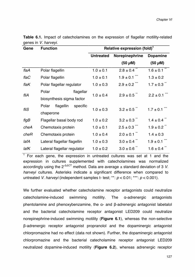
Chapter VI
127
Table 6.1. Impact of catecholamines on the expression of flagellar motility-related genes in V. harveyi. Gene Function Relative expression (fold)1
Untreated Norepinephrine
(50 µM) Dopamine
(50 µM)
flaA Polar flagellin 1.0 ± 0.1 2.8 ± 0.4 ** 1.6 ± 0.1 **
flaC Polar flagellin 1.0 ± 0.1 1.9 ± 0.1 *** 1.3 ± 0.2
flaK Polar flagellar regulator 1.0 ± 0.3 2.9 ± 0.2 *** 1.7 ± 0.3 **
fliA Polar flagellar
biosynthesis sigma factor 1.0 ± 0.4 2.9 ± 0.5 ** 2.2 ± 0.1 ***
fliS Polar flagellin specific
chaperone 1.0 ± 0.3 3.2 ± 0.5 ** 1.7 ± 0.1 ***
flgB Flagellar basal body rod 1.0 ± 0.2 3.2 ± 0.3 ** 1.4 ± 0.4 **
cheA Chemotaxis protein 1.0 ± 0.1 2.5 ± 0.3 *** 1.9 ± 0.2 **
cheR Chemotaxis protein 1.0 ± 0.4 2.0 ± 0.1 ** 1.4 ± 0.3
lafA Lateral flagellar flagellin 1.0 ± 0.3 3.0 ± 0.4 ** 1.9 ± 0.1 ***
lafK Lateral flagellar regulator 1.0 ± 0.2 3.0 ± 0.6 ** 1.6 ± 0.4 **
1 For each gene, the expression in untreated cultures was set at 1 and the expression in cultures supplemented with catecholamines was normalized accordingly using the 2-ΔΔCT method. Data are average ± standard deviation of 3 V. harveyi cultures. Asterisks indicate a significant difference when compared to untreated V. harveyi (independent samples t- test; **: p < 0.01; ***: p < 0.001).
We further evaluated whether catecholamine receptor antagonists could neutralize
catecholamine-induced swimming motility. The α-adrenergic antagonists
phentolamine and phenoxybenzamine, the α- and β-adrenergic antagonist labetalol
and the bacterial catecholamine receptor antagonist LED209 could neutralize
norepinephrine-induced swimming motility (Figure 6.1), whereas the non-selective
β-adrenergic receptor antagonist propranolol and the dopaminergic antagonist
chlorpromazine had no effect (data not shown). Further, the dopaminergic antagonist
chlorpromazine and the bacterial catecholamine receptor antagonist LED209
neutralized dopamine-induced motility (Figure 6.2), whereas adrenergic receptor
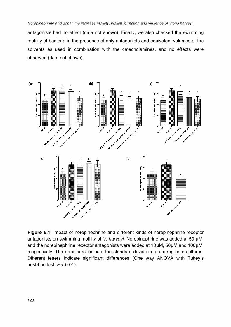
Norepinephrine and dopamine increase motility, biofilm formation and virulence of Vibrio harveyi
128
antagonists had no effect (data not shown). Finally, we also checked the swimming motility of bacteria in the presence of only antagonists and equivalent volumes of the solvents as used in combination with the catecholamines, and no effects were observed (data not shown).
Figure 6.1. Impact of norepinephrine and different kinds of norepinephrine receptor antagonists on swimming motility of V. harveyi. Norepinephrine was added at 50 μM, and the norepinephrine receptor antagonists were added at 10μM, 50μM and 100μM, respectively. The error bars indicate the standard deviation of six replicate cultures. Different letters indicate significant differences (One way ANOVA with Tukey’s post-hoc test; P < 0.01).
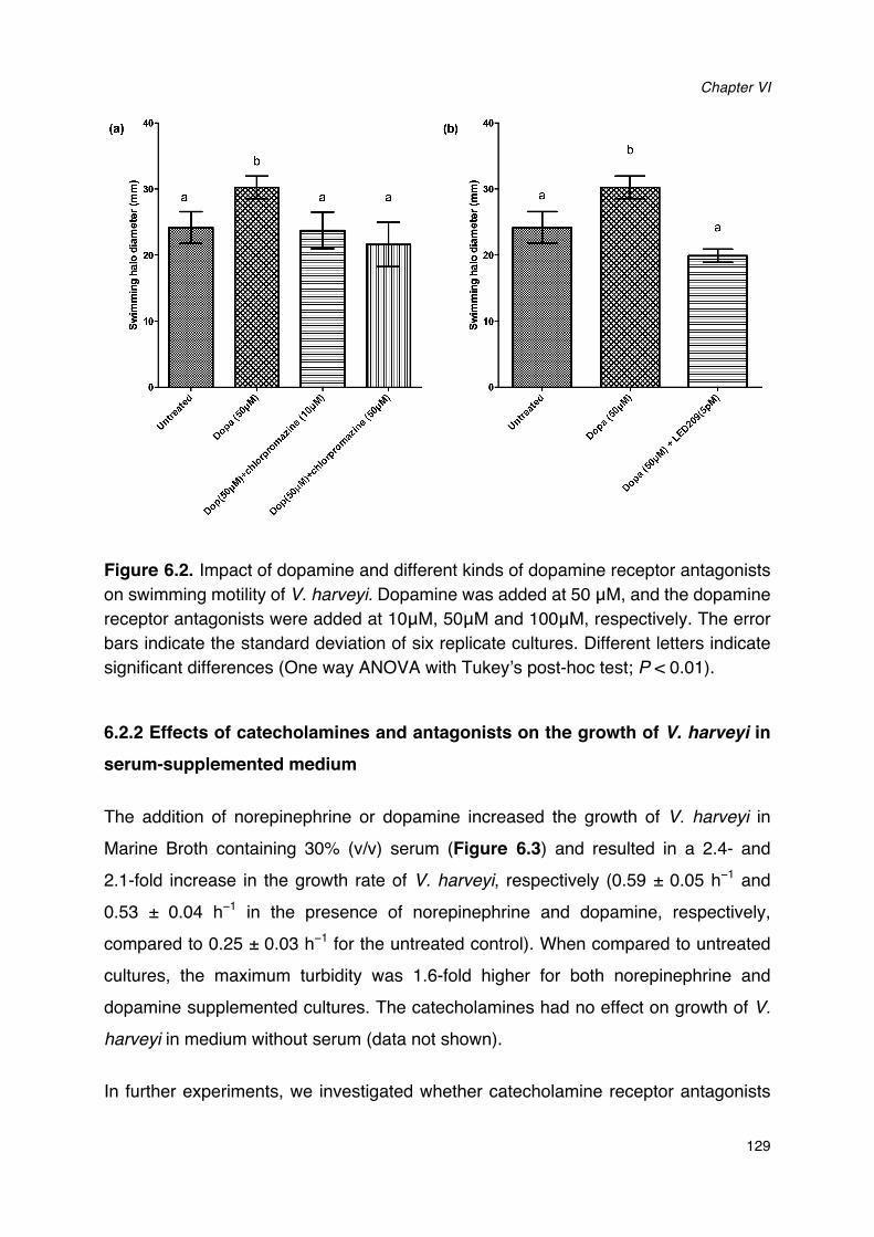
Chapter VI
129
Figure 6.2. Impact of dopamine and different kinds of dopamine receptor antagonists on swimming motility of V. harveyi. Dopamine was added at 50 μM, and the dopamine receptor antagonists were added at 10μM, 50μM and 100μM, respectively. The error bars indicate the standard deviation of six replicate cultures. Different letters indicate significant differences (One way ANOVA with Tukey’s post-hoc test; P < 0.01).
6.2.2 Effects of catecholamines and antagonists on the growth of V. harveyi in
serum-supplemented medium
The addition of norepinephrine or dopamine increased the growth of V. harveyi in Marine Broth containing 30% (v/v) serum (Figure 6.3) and resulted in a 2.4- and 2.1-fold increase in the growth rate of V. harveyi, respectively (0.59 ± 0.05 h−1 and 0.53 ± 0.04 h−1 in the presence of norepinephrine and dopamine, respectively, compared to 0.25 ± 0.03 h−1 for the untreated control). When compared to untreated cultures, the maximum turbidity was 1.6-fold higher for both norepinephrine and dopamine supplemented cultures. The catecholamines had no effect on growth of V.
harveyi in medium without serum (data not shown).
In further experiments, we investigated whether catecholamine receptor antagonists
Norepinephrine and dopamine increase motility, biofilm formation and virulence of Vibrio harveyi
130
could neutralize the growth-stimulatory effects of the catecholamines in
serum-supplemented LB35 broth containing 50μM catecholamines. The results were
consistent with what we observed in the motility assays: antagonists with α-adrenergic
activity (but not antagonists with only β-adrenergic activity) were able to inhibit
norepinephrine-induced growth (Figure 6.3a), and the adrenergic antagonists were
not able to neutralize dopamine-induced growth (data not shown). Further, the
dopaminergic antagonist chlorpromazine neutralized dopamine-induced growth but
had no effect on norepinephrine-induced growth (Figure 6.3b). The prokaryotic
catecholamine receptor antagonist LED209 was able to neutralize the effects of both
norepinephrine and dopamine. None of the antagonists and solvents used to dissolve
the antagonists affected the growth of V. harveyi when tested alone at the same
volumes as used in combination with catecholamines (data not shown), which
indicates that the growth inhibition by the antagonists was not due to toxicity, but a
specific antagonism of the bacterial response to catecholamines.
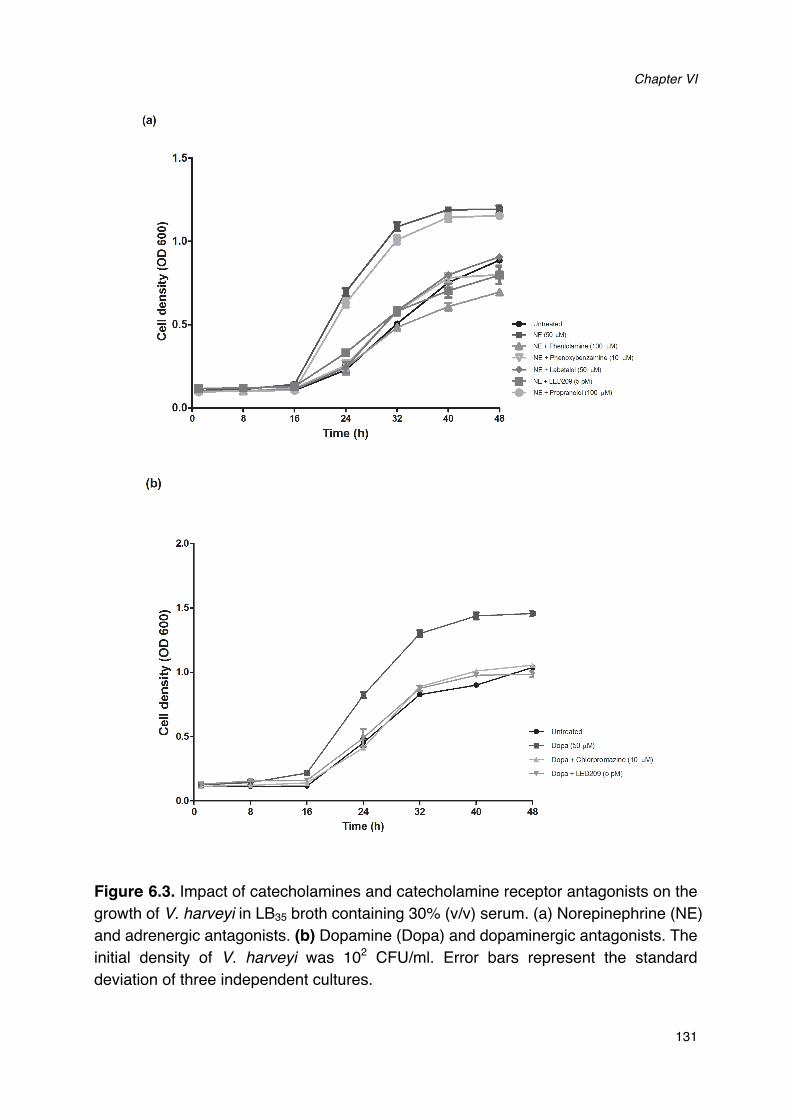
Chapter VI
131
Figure 6.3. Impact of catecholamines and catecholamine receptor antagonists on the growth of V. harveyi in LB35 broth containing 30% (v/v) serum. (a) Norepinephrine (NE) and adrenergic antagonists. (b) Dopamine (Dopa) and dopaminergic antagonists. The initial density of V. harveyi was 102 CFU/ml. Error bars represent the standard deviation of three independent cultures.
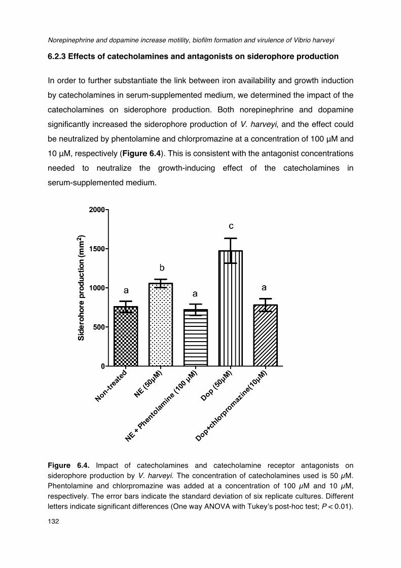
Norepinephrine and dopamine increase motility, biofilm formation and virulence of Vibrio harveyi
132
6.2.3 Effects of catecholamines and antagonists on siderophore production
In order to further substantiate the link between iron availability and growth induction by catecholamines in serum-supplemented medium, we determined the impact of the catecholamines on siderophore production. Both norepinephrine and dopamine significantly increased the siderophore production of V. harveyi, and the effect could be neutralized by phentolamine and chlorpromazine at a concentration of 100 μM and 10 μM, respectively (Figure 6.4). This is consistent with the antagonist concentrations needed to neutralize the growth-inducing effect of the catecholamines in serum-supplemented medium.
Figure 6.4. Impact of catecholamines and catecholamine receptor antagonists on siderophore production by V. harveyi. The concentration of catecholamines used is 50 µM. Phentolamine and chlorpromazine was added at a concentration of 100 µM and 10 µM, respectively. The error bars indicate the standard deviation of six replicate cultures. Different letters indicate significant differences (One way ANOVA with Tukey’s post-hoc test; P < 0.01).
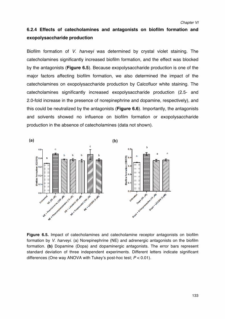
Chapter VI
133
6.2.4 Effects of catecholamines and antagonists on biofilm formation and exopolysaccharide production
Biofilm formation of V. harveyi was determined by crystal violet staining. The catecholamines significantly increased biofilm formation, and the effect was blocked by the antagonists (Figure 6.5). Because exopolysaccharide production is one of the major factors affecting biofilm formation, we also determined the impact of the catecholamines on exopolysaccharide production by Calcofluor white staining. The catecholamines significantly increased exopolysaccharide production (2.5- and 2.0-fold increase in the presence of norepinephrine and dopamine, respectively), and this could be neutralized by the antagonists (Figure 6.6). Importantly, the antagonists and solvents showed no influence on biofilm formation or exopolysaccharide production in the absence of catecholamines (data not shown).
Figure 6.5. Impact of catecholamines and catecholamine receptor antagonists on biofilm formation by V. harveyi. (a) Norepinephrine (NE) and adrenergic antagonists on the biofilm formation. (b) Dopamine (Dopa) and dopaminergic antagonists. The error bars represent standard deviation of three independent experiments. Different letters indicate significant differences (One way ANOVA with Tukey’s post-hoc test; P < 0.01).
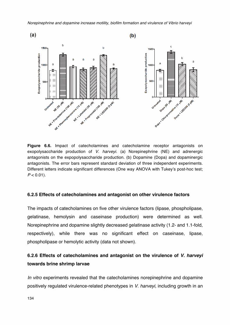
Norepinephrine and dopamine increase motility, biofilm formation and virulence of Vibrio harveyi
134
Figure 6.6. Impact of catecholamines and catecholamine receptor antagonists on exopolysaccharide production of V. harveyi. (a) Norepinephrine (NE) and adrenergic antagonists on the expopolysaccharide production. (b) Dopamine (Dopa) and dopaminergic antagonists. The error bars represent standard deviation of three independent experiments. Different letters indicate significant differences (One way ANOVA with Tukey’s post-hoc test; P < 0.01).
6.2.5 Effects of catecholamines and antagonist on other virulence factors
The impacts of catecholamines on five other virulence factors (lipase, phospholipase, gelatinase, hemolysin and caseinase production) were determined as well. Norepinephrine and dopamine slightly decreased gelatinase activity (1.2- and 1.1-fold, respectively), while there was no significant effect on caseinase, lipase, phospholipase or hemolytic activity (data not shown).
6.2.6 Effects of catecholamines and antagonist on the virulence of V. harveyi towards brine shrimp larvae
In vitro experiments revealed that the catecholamines norepinephrine and dopamine positively regulated virulence-related phenotypes in V. harveyi, including growth in an
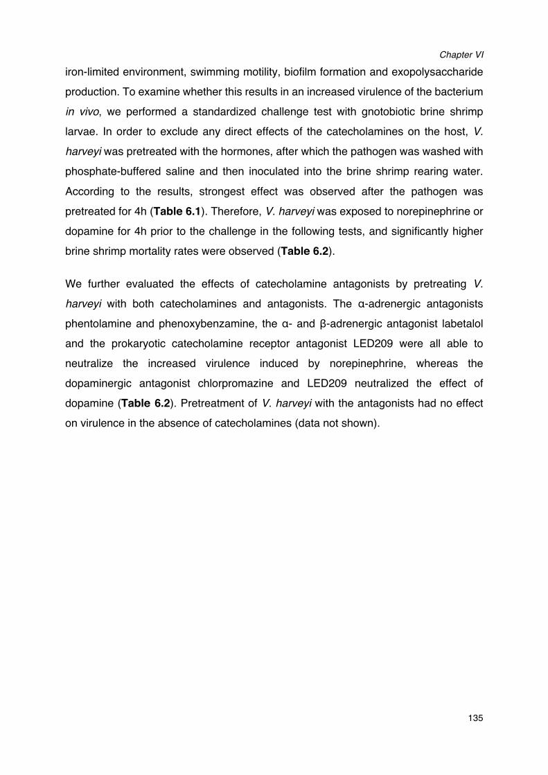
Chapter VI
135
iron-limited environment, swimming motility, biofilm formation and exopolysaccharide
production. To examine whether this results in an increased virulence of the bacterium
in vivo, we performed a standardized challenge test with gnotobiotic brine shrimp
larvae. In order to exclude any direct effects of the catecholamines on the host, V.
harveyi was pretreated with the hormones, after which the pathogen was washed with
phosphate-buffered saline and then inoculated into the brine shrimp rearing water.
According to the results, strongest effect was observed after the pathogen was
pretreated for 4h (Table 6.1). Therefore, V. harveyi was exposed to norepinephrine or
dopamine for 4h prior to the challenge in the following tests, and significantly higher
brine shrimp mortality rates were observed (Table 6.2).
We further evaluated the effects of catecholamine antagonists by pretreating V.
harveyi with both catecholamines and antagonists. The α-adrenergic antagonists
phentolamine and phenoxybenzamine, the α- and β-adrenergic antagonist labetalol
and the prokaryotic catecholamine receptor antagonist LED209 were all able to
neutralize the increased virulence induced by norepinephrine, whereas the
dopaminergic antagonist chlorpromazine and LED209 neutralized the effect of
dopamine (Table 6.2). Pretreatment of V. harveyi with the antagonists had no effect
on virulence in the absence of catecholamines (data not shown).
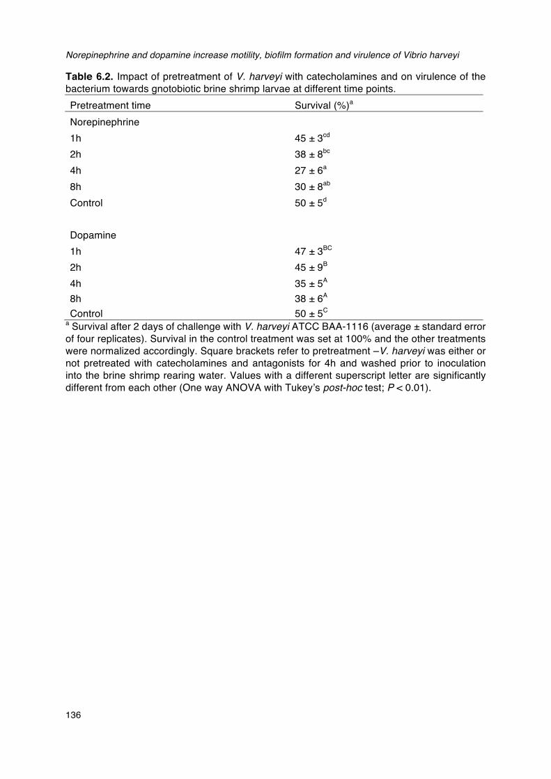
Norepinephrine and dopamine increase motility, biofilm formation and virulence of Vibrio harveyi
136
Table 6.2. Impact of pretreatment of V. harveyi with catecholamines and on virulence of the bacterium towards gnotobiotic brine shrimp larvae at different time points. Pretreatment time Survival (%)a Norepinephrine
1h 45 ± 3cd 2h 38 ± 8bc 4h 27 ± 6a 8h 30 ± 8ab Control 50 ± 5d
� �Dopamine 1h 47 ± 3BC
2h 45 ± 9B 4h 35 ± 5A 8h 38 ± 6A Control 50 ± 5C
a Survival after 2 days of challenge with V. harveyi ATCC BAA-1116 (average ± standard error of four replicates). Survival in the control treatment was set at 100% and the other treatments were normalized accordingly. Square brackets refer to pretreatment –V. harveyi was either or not pretreated with catecholamines and antagonists for 4h and washed prior to inoculation into the brine shrimp rearing water. Values with a different superscript letter are significantly different from each other (One way ANOVA with Tukey’s post-hoc test; P < 0.01).
Chapter VI
137
Table 6.3. Impact of pretreatment of V. harveyi with catecholamines and catecholamine receptor antagonists on virulence of the bacterium towards gnotobiotic brine shrimp larvae. Treatment Survival (%)a
Control 100 ± 0c V. harveyi
50 ± 5b
V. harveyi [50 µM norepinephrine] 27 ± 6a
V. harveyi [50 µM norepinephrine + 100 µM phentolamine] 48 ± 3b
V. harveyi [50 µM norepinephrine + 10 µM phenoxybenzamine] 47 ± 3b V. harveyi [50 µM norepinephrine + 50 µM labetalol] 48 ± 3b V. harveyi [50 µM norepinephrine + 100 µM propanolol] 29 ± 4a
V. harveyi [50 µM norepinephrine + 5 pM LED209]
55 ± 5b
V. harveyi [50 µM dopamine] 35 ± 5a
V. harveyi [50 µM dopamine + 50 µM chlorpromazine] 47 ± 3b V. harveyi [50 µM dopamine + 5 pM LED209] 52 ± 8b
a Survival after 2 days of challenge with V. harveyi ATCC BAA-1116 (average ± standard error of four replicates). Survival in the control treatment was set at 100% and the other treatments were normalized accordingly. Square brackets refer to pretreatment –V. harveyi was either or not pretreated with catecholamines and antagonists for 4h and washed prior to inoculation into the brine shrimp rearing water. Values with a different superscript letter are significantly different from each other (One way ANOVA with Tukey’s post-hoc test; P < 0.01).
6.3 DISCUSSION
The successful interaction between bacteria and their host depends not only on a coordinated response to population density, temperature and pH (Mekalanos, 1992; Winzers et al., 2001), but also on the detection of diverse host cell effector molecules such as catecholamine stress hormones (Burton et al., 2002). Exposure of bacteria to catecholamine stress hormones has been demonstrated to stimulate both growth and production of virulence-related factors in various pathogens of terrestrial animals and humans, such as E. coli (Bansal et al., 2007), Campylobacter jejuni (Cogan et al., 2007), Salmonella typhimurium (Bearson and Bearson, 2008), and V.
Norepinephrine and dopamine increase motility, biofilm formation and virulence of Vibrio harveyi
138
parahaemolyticus (Nakano et al., 2007b). The present study demonstrates that the
catecholamines norepinephrine and dopamine could significantly increase the
production of major virulence factors and growth in serum-supplemented medium of V.
harveyi, a major pathogen of aquatic organisms with a broad host spectrum (both
vertebrates such as fish and invertebrates such as crustaceans and mollusks). This
result is in good agreement with, and considerably extends, the previous reports.
The catecholamines significantly increased the virulence of V. harveyi towards
gnotobiotic brine shrimp larvae. The pathogen was pretreated with catecholamines in
order to avoid a direct effect of catecholamines on the host (e.g. decreased activity of
the defense system) and to ensure that any impact on survival of challenged larvae
was due to increased virulence of the pathogen. This is further substantiated by our
observation that in addition to their effect on growth in serum-supplemented medium
and siderophore production, the catecholamines increased the production of several
other phenotypes that are important for infection. Indeed, both norepinephrine and
dopamine significantly stimulated biofilm formation, exopolysaccharide production,
swimming motility and the expression of genes involved in flagellum synthesis
(structural genes and regulators including the flagellar master regulator) in V. harveyi.
These results agree well with previous reports documenting the impact of
catecholamines on biofilm formation in Staphylococcus epidermidis (Lyte et al., 2003)
and on swimming motility in E. coli, C. jejuni and Edwardsiella tarda (Kendall et al.,
2007; Cogan et al., 2007; Wang et al., 2011). Furthermore, previous work in our
laboratory has also confirmed the increased virulence of V. harveyi by catecholamines
in conventionally reared giant freshwater prawn larvae (Pande et al., 2014).
Catecholamines are produced by hemocytes of invertebrates, and concentrations in
the hemolymph of shrimp and prawn that have been reported range between 10 nM
and ~3 µM (Chen et al., 2003; Hsieh et al., 2006; Pan et al., 2014). These
concentrations are lower than the concentrations used in this study. However, local
concentrations can be considerably higher. For instance, the intrasynaptic
concentration of norepinephrine in the central nervous system of mammals is as high
as 10 mM (versus nM levels in serum) (Lyte, 2004). Hence, upon infection, pathogens
Chapter VI
139
can come into contact with local concentrations that are several orders of magnitude higher than those that are found in hemolymph (or serum in case of vertebrates). This probably is also the case when tissues and/or hemocytes are damaged during infection and hence, elevated catecholamine levels might be a cue informing the pathogen of tissue damage.
Pretreatment of V. harveyi with catecholamines simulated transmission of the pathogen from a host site showing elevated catecholamine levels (e.g. due to cell or tissue damage). In combination with our observation that the catecholamines increased biofilm formation, exopolysaccharide production and swimming motility of V.
harveyi (which are all important during the initial stages of infection), our results suggest that catecholamine sensing increases the success of transmission to a new host. Hence, elevated catecholamine levels might be a cue informing the pathogen that the infection reached a final stage (cell and tissue damage) and that it is time to leave the host.
Catecholamine receptor antagonists have been used extensively to identify and characterize catecholamine receptors in mammals. The effects of various antagonists on growth and production of virulence factors in V. harveyi have been investigated in the present study, and the results demonstrated that ɑ- but not β-adrenergic receptor antagonists could block responses to norepinephrine, but did not show any effect on dopamine responsiveness. On the contrary, dopaminergic receptor antagonists neutralized induction caused by dopamine, but did not neutralize induction by norepinephrine. Similar results have been reported in enteric pathogens of terrestrial animals (Freestone et al., 2007), and suggest that bacterial response systems for catecholamines possess a degree of specificity similar to mammalian catecholamine receptors. In contrast to the wealth of adrenergic and dopaminergic receptors described in eukaryotes, there have been only few reports examining the presence of such receptors in bacteria. Clarke et al. (2006) reported that norepinephrine was able to be recognized by the E. coli O157:H7 two-component regulator sensor kinase QseC in in vitro constructs, leading to the hypothesis that this is the bacterial catecholamine receptor. Later on, a QseC antagonist, LED209, has been identified in a high-throughput screen (Rasko et al., 2008). We found that LED209 was able to
Norepinephrine and dopamine increase motility, biofilm formation and virulence of Vibrio harveyi
140
neutralize the effects of both norepinephrine and dopamine, indicating that a similar response system for catecholamines might exist in V. harveyi. This is also substantiated by the fact that the V. harveyi ATCC BAA-1116 genome contains a QseC homolog. Alternatively, since our experiments with eukaryotic catecholamine receptor antagonists showed that dopaminergic antagonists did not neutralize norepinephrine-induced effects and vice versa, there are probably at least two different catecholamine receptors in V. harveyi, with LED209 being able to bind to both of them. Further research will be needed in order to identify the catecholamine receptors and the signal transduction pathways in V. harveyi.
Both norepinephrine and dopamine significantly stimulated the growth of V. harveyi in serum-supplemented medium, and this effect could be neutralized by eukaryotic catecholamine receptor antagonists. Similar results have been reported in other Gram-negative bacteria such as E. coli and V. parahaemolyticus, and the stimulation of growth in serum-supplemented medium by catecholamines has mainly been attributed to their ability to facilitate iron removal from the host iron-binding proteins transferrin and lactoferrin (Lyte, 2004; Nakano et al., 2007b). We used bovine serum since it was practically not feasible to obtain sufficient amounts of crustacean serum. However, it should be noted that crustaceans produce equivalents of iron-binding proteins produced by mammals (e.g. Toe et al., 2012). Our observations that catecholamine receptor antagonists are able to neutralize the growth-stimulatory effect of catecholamines and that adrenergic receptor antagonists were found to show no effect on dopamine-induced growth and vice versa suggest that a regulatory mechanism involving catecholamine receptors is involved as well, and this has also been reported for enteric pathogens such as E. coli O157:H7, Salmonella enterica
and Yersinia enterocolitica (Freestone et al., 2007). To confirm the hypothesis that in addition to increasing iron uptake in a direct way, catecholamines induced an iron uptake mechanism, we investigated the effect of catecholamines on siderophore production, and found that both norepinephrine and dopamine could significantly induce siderophore production. Siderophores have been reported to be essential for catecholamine-induced growth in other Gram-negative bacteria such as E. coli and Salmonella, where they serve to internalize the iron removed by the catecholamines
Chapter VI
141
(Freestone et al., 2003; Williams et al., 2006). Finally, it should be noted that in addition to limiting iron availability, serum might have other effects on the pathogens as well. Serum e.g. contains proteins (some of which might have antimicrobial activity; Zasloff, 2002). However, in view of the literature that is available on other pathogens and our observation that catecholamines increase siderophore production we think that modulation of iron availability is the most plausible explanation of growth stimulation by catecholamines in the presence of serum.
6.4 CONCLUSIONS
Our study has shown that the catecholamines norepinephrine and dopamine increase the virulence of V. harveyi by increasing swimming motility, biofilm formation, exopolysaccharide production and growth in environments with low iron availability, and the effects could be neutralized by antagonists for eukaryotic catecholamine receptors and the bacterial catecholamine receptor antagonist LED209. Different effects of the adrenergic and dopaminergic receptors antagonists indicate the presence of specific sensing systems for different catecholamines in V. harveyi.
6.5 MATERIALS AND METHODS
6.5.1 Bacterial strains and growth conditions
V. harveyi wild type strain ATCC BAA-1116 (recently reclassified as V. campbellii; Lin et al., 2010) was used in this study. Unless otherwise stated, the strain was cultured at 28°C in Luria broth containing 35 g/L sodium chloride (LB35) under constant agitation (100 min-1). Cell densities were measured spectrophotometrically at 600 nm.
6.5.2 Catecholamines and eukaryotic catecholamine receptor antagonists
Norepinephrine was dissolved in hydrochloride acid (HCl 0.1N) at 10 mM, while dopamine was dissolved in distilled water at 10 mM. The antagonists used in this study are listed in Table 6.3. All the chemicals were purchased from Sigma-Aldrich (Bornem, Belgium). The reagents were sterilized using a 0.22 µm filter and stored at -20°C.

Norepinephrine and dopamine increase motility, biofilm formation and virulence of Vibrio harveyi
142
Table 6.4. Catecholamine receptor antagonists used in this study. Compound Specificity Solvent
Phentolamine hydrochloride reversible α-adrenergic Ethanol Phenoxybenzamine hydrochloride irreversible α-adrenergic DMSO S-propanolol hydrochloride β-adrenergic Ethanol Labetalol hydrochloride α- and β-adrenergic Ethanol Chlorpromazine hydrochloride dopaminergic Distilled water LED2091 bacterial catecholamine
receptor QseC DMSO
All compounds were dissolved at 10mM, except for LED209, which was dissolved at 10 µM. 1 N-phenyl-4-[[(phenylamino)thioxomethyl]amino]-benzenesulfonamide
6.5.3 Bacterial growth assays
For the bacterial growth assays, V. harveyi was grown overnight in LB35 broth at 28°C. After that, the culture was re-inoculated at a concentration of 102 CFU/ml into fresh LB35 broth containing 30% (v/v) adult bovine serum (Sigma-Aldrich), with and without 50 µM norepinephrine or dopamine. Additionally, different concentrations of the catecholamine receptor antagonists were added in conjunction with the catecholamines to determine whether they could neutralize catecholamine-induced growth responses. The cultures were grown in 200 µl volumes in 96-well plates at 28°C for 48 h, and the turbidity at 600 nm was monitored every hour using a Multireader machine (Infinite M200, TECAN, Austria). Growth curves were determined for three independent cultures, and the growth rate of the exponentially growing cultures was calculated. The statistical significance of specific growth rate was determined using an independent samples t-test.
6.5.4 Siderophore activity assay
The siderophore activity was determined by Chrome azurol S (CAS) agar diffusion
Chapter VI
143
assay according to Shin et al. (2001), which was performed as follows. The CAS agar plates were prepared according to Schwyn and Neilands (1987); 60.5 mg Chrome azurol S (CAS)(Sigma-Aldrich) was dissolved in 50 ml deionized water, and mixed with 10 ml iron (III) solution (1 mM FeCl3·6H2O, 10 mM HCl). Under stirring, this solution was slowly added to 72.9 mg hexadecyltrimethylammonium bromide (Sigma-Aldrich), dissolved in 40 ml water. The resultant dark blue solution was autoclaved and stored in a plastic container. Then 100 ml 10× MM9 salts, 15 g agar, 30.24 g Pipes, and 12 g of a 50% (w/v) NaOH solution (to raise the pH to 6.8) were added to 750 ml water. After autoclaving and cooling to 50°C, 30 ml of a sterile 10% casamino acids solution was added as the carbon source. The dye solution was finally added with enough agitation to achieve mixing without generation of foam. Serum (30%, v/v), catecholamines and antagonists were added directly into the agar. V.
harveyi was cultured overnight in LB35 broth containing 30% (v/v) serum, with or without catecholamines and antagonists. Then the CAS agar plates were punched with 5-mm-diameter holes and each hole was filled with 35 µl of a V. harveyi culture (OD600 = 1.0). After incubation at 28°C for 24h, the size of the orange halo formed around each hole was measured. Siderophore activity was expressed as the square value of the halo diameter.
6.5.5 Swimming motility assay
The swimming motility assay was performed on soft agar (LB35 plates containing 0.3% agar) as described previously (Yang and Defoirdt, 2014). The catecholamines and antagonists were added to the autoclaved agar. V. harveyi was grown overnight in LB35 broth, and 5 µl aliquots (OD600 = 1.0) were spotted in the center of the soft agar plates. Plates were incubated for 24 h, after which the diameters of the motility halos were measured. All assays were done with freshly prepared media in 6 replicates.
6.5.6 Biofilm formation assay
Biofilm formation assay was quantified by crystal violet staining, as described previously (Stepanovic´ et al., 2007). In brief, an overnight culture of V. harveyi was diluted to an OD600 of 1.0 in LB35 broth with or without catecholamines and antagonists, and 200 µl aliquots of these suspensions were pipetted into the wells of a 96 well plate.
Norepinephrine and dopamine increase motility, biofilm formation and virulence of Vibrio harveyi
144
Then the bacteria were allowed to adhere and grow without agitation for 24h at 28°C. After that, the cultures were removed and the wells were washed three times with 300 μl sterile physiological saline to remove all non-adherent bacteria. The remaining attached bacteria were fixed with 150 μl of 99% methanol per well for 20 min, after which the methanol was removed and plates were air-dried. Then, biofilms were stained for 15 min with 150 μl of a 1% crystal violet solution (Pro-lab Diagnostics, Richmond Hill, ON, Canada) per well. Excess stain was rinsed off by placing the plate under running tap water, and washing was continued until the washings were free of the stain. After the plates were air dried, the dye bound to the adherent cells was resolubilized with 150 μl of 95% ethanol per well, and absorbance was measured at 570 nm. Sterile medium served as negative control. For the quantification of exopolysaccharides, Calcofluor white staining (Sigma-Aldrich) was used. In brief, wells were rinsed after 24 h biofilm formation and 100 μl phosphate buffered saline containing 0.5 μl 5 mM Calcofluor white staining dye was added to the wells. After 60 min, fluorescence (excitation 405 nm and emission 500 nm) was measured with a Multi-reader (Infinite M200, TECAN, Austria).
6.5.7 Lytic enzyme activity assays
All assays were conducted according to Natrah et al. (2011). For each assay, an overnight culture of V. harveyi was diluted to an OD600 of 0.5 and 5 µl of the diluted culture was spotted in the middle of the test plates. The catecholamines and antagonists were added to the autoclaved agar before pouring. All assays were done at least in triplicate. Lipase and phospholipase activities were assessed on marine agar plates supplemented with 1% Tween 80 (Sigma–Aldrich) and 1% egg yolk emulsion (Sigma–Aldrich), respectively. The development of opalescent zones around the colonies was observed and the diameters of the zones were measured after 2–4 days of incubation at 28°C. Caseinase assay plates were prepared by mixing double strength Marine Agar with a 4% skim milk powder suspension (Oxoid, Basingstoke, Hampshire, UK), sterilized separately at 121°C for 5 min. Clearing zones surrounding the bacterial colonies were measured after 2 days of incubation. Gelatinase assay plates were prepared by mixing 0.5% gelatin (Sigma–Aldrich) into the agar. After
Chapter VI
145
incubation for 7 days, saturated ammonium sulfate (80%) in distilled water was poured over the plates and after 2 min, the diameters of the clearing zones around the colonies were measured. Hemolytic assay plates were prepared by supplementing Marine Agar with 5% defibrinated sheep blood (Oxoid) and clearing zones were measured after 2 days of incubation.
6.5.8 RNA extraction
V. harveyi was grown overnight in triplicate on soft agar plates (0.3 % agar). Cells were harvested and RNA was extracted with the SV Total RNA Isolation System (Promega, Leiden, The Netherlands) according to the manufacturer’s instructions. The RNA quantity was measured spectrophotometrically (NanoDrop Technologies, Wilmington, DE, USA) and adjusted to 200 ng µl-1 in all samples. The RNA integrity was checked by Agarose Gel Electrophoresis and the RNA samples were stored in -80°C for subsequent use.
6.5.9 Primers
Specific primers were used for 10 selected genes involved in motility of V. harveyi
(Yang and Defoirdt, 2014). The RNA polymerase A submit (rpoA) mRNA was used as an endogenous control (Defoirdt et al., 2007).
6.5.10 Reverse transcription
Reverse transcription was performed with the RevertAidTM H minus First strand cDNA synthesis kit (Fermentas Gmbh, Baden-Württemberg, Germany) in accordance to the manufacturer’s instructions. Briefly, a mixture of 1 µg RNA and 1 µl random hexamer primer solution was mixed first. Then, 8 µl of reaction mixture containing 4 µl of 5× reaction buffer (0.25 mol-1 Tris–HCl pH 8.3, 0.25 mol-1 KCl, 0.02 mol-1 MgCl2, 0.05 mol-1 DTT), 2 µl of 0.01 mol-1 dNTP mix, 20 units of ribonuclease inhibitor, 200 units of RevertAidTM H minus M-MuLV Reverse Transcriptase was added. The reaction mixture was incubated for 5 min at 25°C followed by 60 min at 42°C. The reaction was terminated by heating at 70°C for 5 min and then cooled to 4°C. cDNA samples were checked by PCR and stored at -20°C for further use.
Norepinephrine and dopamine increase motility, biofilm formation and virulence of Vibrio harveyi
146
6.5.11 Real-time PCR
Real-time PCR was used to quantify the expression level of all the flagella-related genes and was performed with Maxima® SYBR Green/ROX qPCR Master Mix (Fermentas, Fisher Scientific, Erembodegem, Belgium) as described previously (Yang and Defoirdt, 2014). The reaction was performed in an StepOne™ Real-Time PCR System thermal cycler (Applied Biosystems, Gent, Belgium) in a total volume of 25 µl, containing 12.5 µl of 2× SYBR green master mix, 300 nM of forward and reverse primers and 2 µl of template cDNA. The thermal cycling consisted an initial denaturation at 95°C for 10 min followed by 40 cycles of denaturation at 95°C for 15s and primer annealing and elongation at 60°C for 1 min. Dissociation curve analysis was performed to check for the amplification of untargeted fragments. Data acquisition was performed with the StepOne™ Software.
6.5.12 Real-time PCR data analysis (2-∆∆ct method)
The real-time PCR was validated by amplifying serial dilutions of cDNA synthesized from 1 µg of RNA isolated from bacterial samples. Serial dilutions of cDNA were amplified by real time PCR using gene specific primers. ∆CT (average CT value of target-average CT value of rpoA) was calculated for the different dilutions and plotted against the cDNA concentration. The slope of the graph was almost equal to 0 for all of the target nine genes. Therefore, the amplification efficiency of reference and the target genes was considered to be equal. Based on this precondition, real-time PCR data were analyzed using the 2-∆∆CT method (Schmittgen and Livak, 2008). The expression of the target genes was normalized to the endogenous control (rpoA) by calculating ∆CT :
∆Ct = Ct target � Ct rpoA
and expressed relative to a calibrator strain by calculating ∆∆Ct:
∆∆Ct = ∆Ct � Ct calibratior
Strain BB120 without any treatments was used as acalibrator. The relative expression
Chapter VI
147
was then calculated as
Relative expression = 2-∆∆Ct
6.5.13 Axenic hatching of brine shrimp larvae
Two hundred milligrams of high-quality hatching cysts of Artemia franciscana (EG® Type; INVE Aquaculture, Baasrode, Belgium) were hydrated in 18 ml of filtersterilized tap water for 1 h. Sterile cysts were obtained by decapsulation based on the method described by Marques et al. (2004). Briefly, 660 µl of NaOH (32%) and 10 ml of NaOCl (50%) were added to the hydrated cyst suspension to facilitate decapsulation. The process was stopped after 2 min by adding 14 ml of Na2S2O3 (10 g L-1). Filtered (0.22 µm) aeration was provided during the reaction. The decapsulated cysts were washed with filtered (passed through 0.22-µm membrane filter) and autoclaved (moist heat at 121°C for 15 min) artificle seawater (containing 35 g l-1 of instant ocean synthetic sea salt, Aquarium Systems, Sarrebourg, France). The cysts were resuspended in a 50-ml tube containing 30 ml of filtered, autoclaved seawater and hatched for 28 h on a rotor (4 min-1) at 28°C with constant illumination (c. 2000 lux). The axenity of cysts was verified by inoculating one ml of culture water into 9 ml of Marine broth and incubating at 28°C for 24 h. After 28 h of hatching, batches of 30 larvae were counted and transferred to fresh, sterile 50-ml tubes containing 30 ml of filtered and autoclaved seawater. Finally, the tubes were returned to the rotor and kept at 28°C. All manipulations were performed in a laminar flow to maintain sterility of the cysts and larvae.
6.5.14 Brine shrimp challenge test
The effects of the catecholamines and antagonists on the virulence of V. harveyi were determined in a standardized challenge test with gnotobiotic brine shrimp larvae. V.
harveyi was incubated with or without norepinephrine or dopamine (50 µM) and with or without antagonists, and cultures were washed with phosphate-buffered saline (pH 7.4) prior to inoculation into the brine shrimp rearing water at 105 CFU ml-1. The challenge tests were performed as described by Defoirdt et al. (2005) with some modifications. A suspension of autoclaved LVS3 bacteria (Verschuere et al., 1999) in
Norepinephrine and dopamine increase motility, biofilm formation and virulence of Vibrio harveyi
148
filtered and autoclaved seawater was added as feed at the start of the challenge test at 107 cells ml-1 culture water. Brine shrimp cultures to which only autoclaved LVS3 bacteria were added as feed, were used as controls. The survival of the larvae was counted 48 h after the addition of the pathogens. Each treatment was carried out in quadruplicate and each experiment was repeated twice to verify the reproducibility. In each test, the sterility of the control treatments were checked at the end of the challenge by inoculating 1 ml of rearing water to 9 ml of Marine Broth and incubating the mixture for 2 days at 28°C.
6.5.15 Statistical analyses
Data analysis was carried out using the SPSS statistical software (version 15). Log transformed gene expression data were analysed using independent samples t- tests. Unless stated otherwise, all other data were compared with one-way ANOVA, followed by Tukey’s post hoc test.
ACKNOWLEDGEMENTS
This work was funded by the Research Foundation of Flanders (FWO-Vlaanderen; project no 1.5.013.12N), by the Belgian Science Policy Office (Belspo IUAP project No IUAP/64/Sorgeloos), by the China Scholarship Council (through a doctoral scholarship to Qian Yang) and by the special research fund of Ghent University. Tom Defoirdt is a postdoctoral fellow of FWO-Vlaanderen. The authors have no conflicts of interest to declare.

CHAPTER VII
GENERAL DISCUSSION, CONCLUSION AND FUTURE PERSPECTIVES

Chapter VII
149
7.1 INTRODUCTION
The United Nation’s Food and Agriculture Organization (FAO) considers aquaculture as the fastest-growing food-producing industry worldwide. However, disease outbreaks are considered to be a significant constraint to the development of the sector (FAO, 2014). In this study, we focus on disease caused by Vibrio harveyi, which is an important pathogen in aquaculture that can affect almost all types of cultured animals. Due to the frequent and excessive use of antibiotics, aquaculture pathogens (including V. harveyi) have acquired (multiple) antibiotic resistance, rendering the antibiotic treatments less effective in some cases. Additionally, massive use of antibiotics in animal production also constitutes a threat to human health and to the environment (Cabello et al., 2006), and this has resulted in more strict regulations with respect to antibiotic use. One notable example is the ban on the use of antibiotics as growth promoters in animal production in Europe in 2006 (European Parliament and Council Regulation No 1831/2003). As a consequence, the development of novel strategies to control bacterial diseases, both in human and veterinary medicine will be critically important in order to ensure public health and food security in the future. One of the novel alternative strategies to replace antibiotic use in aquaculture is preventing the pathogenic bacteria from attacking the host without the need to kill them, which is named antivirulence therapy. Consequently, there is a need for thoroughly understanding the pathogen-pathogen and host-pathogen interactions, in order to develop effective antivirulence therapies.
This study addressed the role of cell-to-cell signaling and sensing of host factors on the virulence of V. harveyi in the gnotobiotic brine shrimp larvae model system. We investigated the regulation of virulence-related factors by quorum sensing in V.
harveyi, and also determined the effect of quorum sensing disruption in this host-pathogen system by the application of some novel quorum sensing inhibitors. Furthermore, this study addressed the role of indole signaling on the virulence of V.
harveyi. In addition to bacterial cell-to-cell signaling, the present study also focused on sensing of host factors and its impact on the virulence of V. harveyi. In this chapter, the most important findings of this work are highlighted and discussed, and some
General discussion, conclusion and future perspectives
150
directions for future research are presented.
7.2 THE IMPACT OF QUORUM SENSING ON FLAGELLAR MOTILITY – AN ESSENTIAL VIRULENCE FACTOR IN V. harveyi
V. harveyi is one of the model organisms in studies on bacterial quorum sensing (Ng
and Bassler, 2009). Unlike most other Gram-negative bacteria, V. harveyi has been
found to use a three-channel quorum sensing system, which is mediated by three
different types of signal molecules, including Harveyi Autoinducer 1 (HAI-1),
Autoinducer 2 (AI-2), and Cholerae Autoinducer 1 (CAI-1). These three autoinducers
are detected at the cell surface by their respective receptors that feed a shared
phosphorylation/dephosphorylation signal transduction cascade. V. harveyi quorum
sensing system has been found to control the bioluminescence (Bassler et al., 1997)
and the production of several virulence factors (Figure 7.1). However, most of these
virulence factors have been reported to be negatively regulated by quorum sensing,
i.e. their production decreases with increasing levels of the signal molecules (Natrah
et al., 2011). This appears to be in conflict with the fact that quorum sensing is
required for full virulence of V. harveyi towards different hosts (Defoirdt et al., 2005;
Pande et al., 2013). In order to further characterise why quorum sensing is required
for full virulence of V. harveyi, in Chapter 3, we aimed to identify more virulence
factors that are positively regulated by quorum sensing in V. harveyi.
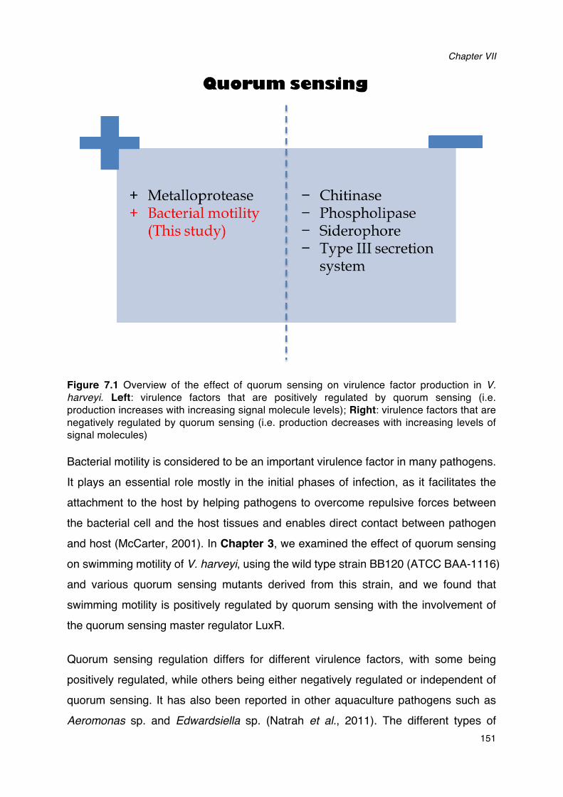
Chapter VII
151
Figure 7.1 Overview of the effect of quorum sensing on virulence factor production in V. harveyi. Left: virulence factors that are positively regulated by quorum sensing (i.e. production increases with increasing signal molecule levels); Right: virulence factors that are negatively regulated by quorum sensing (i.e. production decreases with increasing levels of signal molecules)
Bacterial motility is considered to be an important virulence factor in many pathogens. It plays an essential role mostly in the initial phases of infection, as it facilitates the attachment to the host by helping pathogens to overcome repulsive forces between the bacterial cell and the host tissues and enables direct contact between pathogen and host (McCarter, 2001). In Chapter 3, we examined the effect of quorum sensing on swimming motility of V. harveyi, using the wild type strain BB120 (ATCC BAA-1116) and various quorum sensing mutants derived from this strain, and we found that swimming motility is positively regulated by quorum sensing with the involvement of the quorum sensing master regulator LuxR.
Quorum sensing regulation differs for different virulence factors, with some being positively regulated, while others being either negatively regulated or independent of quorum sensing. It has also been reported in other aquaculture pathogens such as Aeromonas sp. and Edwardsiella sp. (Natrah et al., 2011). The different types of
General discussion, conclusion and future perspectives
152
regulation probably reflect the need to express different virulence factors at different
infectious stages. For example, virulence factors that are negatively regulated by
quorum sensing might be predominantly required during initial infection, whereas
virulence factors that are positively regulated might be also required at later stages.
However, little literature data are available to support this hypothesis.
Through a further series of experiments at the transcriptional level, we confirmed that
quorum sensing positively regulates motility by affecting the expression of flagellar
biosynthesis genes. Flagella are the organelles that mediate bacterial motility, and a
number of tightly regulated genes have been identified to encode flagellar systems
(McCarter, 2001). Quorum sensing in V. harveyi was found to regulate not only the
expression of the structural components of flagella, but also the expression of master
transcriptional activators and regulators. Importantly, the quorum sensing regulation
of flagellar motility apparently differs between bacterial species. For instance, in
contrast to V. harveyi, quorum sensing was found to repress motility in other bacteria
such as V. cholerae (Zhu et al., 2002) V. fisheri (Lupp and Ruby, 2005), and V.
parahaemolyticus (Gode-Potratz and McCarter, 2011). These differences in the
regulation of genes by quorum sensing might reflect differences with respect to the life
styles of these bacteria (Hammer and Bassler, 2003).
Finally, we demonstrated that a flagellar motility inhibitor could completely inhibit the
swimming motility and significantly increased the survival of brine shrimp larvae
challenged with V. harveyi. Further, it has been reported that JAF548 (QS-), which
was found to have a significantly decreased motility in our study, also showed a
significantly decreased virulence towards brine shrimp larvae (Defoirdt et al., 2005).
The relative contribution of the three channels is dependent on the environmental
conditions (Defoirdt et al., 2008). All the single synthase mutants tested in this study
showed a significantly lower motility compared with the wild type, with the strongest
impact being observed in the HAI-1 deficient mutant. This is consistent with the
relative importance of the signal molecules as observed in vitro grown cells (Henke
and Bassler, 2004b). However, the three channels have a different impact on
virulence of V. harveyi towards different hosts. Defoirdt et al. (2005; 2012) reported
that inactivation of AI-2 and CAI-1 signal molecules resulted in reduced virulence of V.
Chapter VII
153
harveyi towards brine shrimp larvae, whereas inactivation of HAI-1 had no effect. A
recent research by Pande et al. (2013) revealed that HAI-1 and AI-2 deficient mutants
were significantly less virulent than the wild type towards commercially important giant
freshwater prawn (Macrobrachium rosenbergii), while CAI-1 deficient mutant has no
effect on this host. This could be explained by the fact that V. harveyi quorum sensing
system has been described as a three-way detector, with the expression of quorum
sensing-regulated genes being proportional to the levels of the three signal molecules
(Henke and Bassler, 2004b). The detection of two types of signal molecules results in
sufficient expression levels of the quorum sensing master regulator LuxR, which
allows production of the virulence factors that are essential to kill the host, whereas
the LuxR expression level in the presence of only one of these two signal molecules is
not (Deforidt et al., 2012).
To our knowledge, this is the first report demonstrating that quorum sensing controls
the virulence of V. harveyi by affecting swimming motility.
7.3 APPLICATION OF QUORUM SENSING INHIBITORS TO PROTECT AQUACULTURE ANIMALS FROM DISEASE
It is proposed that disease is the outcome of an imbalance between host, pathogen
and environment. A holistic strategy, which takes into account the different aspects of
the pathogen-host-environment continuum, will be the preferred approach for
preventing and controlling bacterial disease in aquaculture (Figure 7.2).
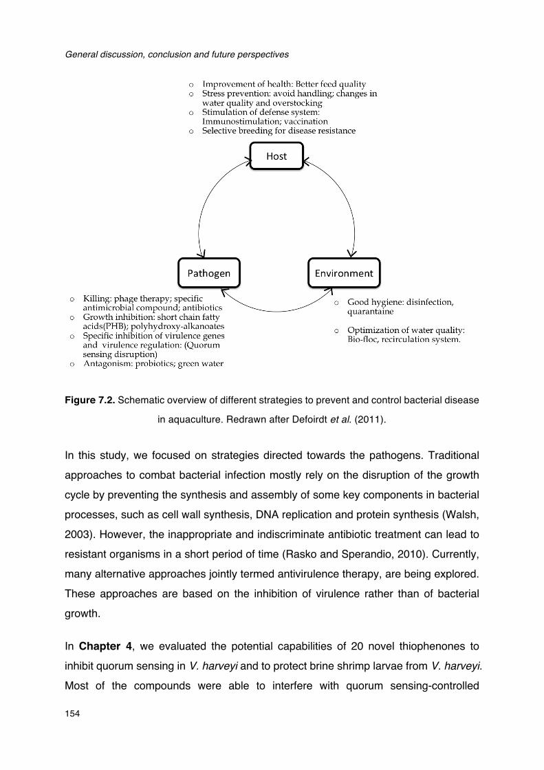
General discussion, conclusion and future perspectives
154
Figure 7.2. Schematic overview of different strategies to prevent and control bacterial disease
in aquaculture. Redrawn after Defoirdt et al. (2011).
In this study, we focused on strategies directed towards the pathogens. Traditional
approaches to combat bacterial infection mostly rely on the disruption of the growth
cycle by preventing the synthesis and assembly of some key components in bacterial
processes, such as cell wall synthesis, DNA replication and protein synthesis (Walsh,
2003). However, the inappropriate and indiscriminate antibiotic treatment can lead to
resistant organisms in a short period of time (Rasko and Sperandio, 2010). Currently,
many alternative approaches jointly termed antivirulence therapy, are being explored.
These approaches are based on the inhibition of virulence rather than of bacterial
growth.
In Chapter 4, we evaluated the potential capabilities of 20 novel thiophenones to
inhibit quorum sensing in V. harveyi and to protect brine shrimp larvae from V. harveyi.
Most of the compounds were able to interfere with quorum sensing-controlled
Chapter VII
155
bioluminescence in V. harveyi. One of the factors that facilitated the development of quorum sensing research is the exploitation of signal molecule reporter strains, which demonstrate a certain phenotype responding to quorum sensing molecules. However, there is an important limitation of the use of such reporter strains, which is that the quorum sensing-controlled phenotypes usually also depend on other factors and/or on the metabolic activity of the cells and this can result in false positives (Defoirdt et al., 2013). For instance, the apparent quorum sensing-inhibitory ability of pyrogallol, which is a compound previously claimed as an AI-2 quorum sensing inhibitor, was recently found to be a side effect of its toxicity rather than true quorum sensing blockage (Defoirdt et al., 2013). Therefore, adequate control experiments are always required. In this study, to solve this problem of false positives, we proposed a new parameter to represent specific quorum sensing-inhibitory activity (AQSI), which is defined as the ratio between inhibition of quorum sensing-inhibitory activity (in this case bioluminescence) and inhibition of the same activity when it is independent of quorum sensing. We set the threshold to exclude compounds at an AQSI of 2 (i.e. if AQSI was below 2, then the activity was considered to be not due to quorum sensing inhibition), and considered compounds to be strong inhibitors if AQSI was above 10. This approach allowed us to identify 5 compounds as false positives in the present study. Several other strategies to strengthen the evidence for quorum sensing inhibition have been proposed, such as (i) verify that the candidate QSI show no effect on the reporter phenotype when it is independent of quorum sensing; (ii) verify the impact of candidate QSI on additional phenotypes that are mediated by quorum sensing; (iii) perform a transcriptomic analysis to confirm that the candidate QSI selectively inhibits quorum sensing-regulated genes; and (iv) try to identify the molecular target of the candidate QSI compound (Defoirdt et al., 2013). Another essential experiment to confirm a QSI compound is the assessment of toxicity of putative QSI towards bacterial cell. However, even the most sensitive test (e.g. determination of growth kinetics) can miss significant toxic effects (Defoirdt et al., 2013). Therefore, to exclude the possibility that inhibition of quorum sensing-regulated phenotype is due to toxicity of a test compound, alternative sensitive methods to evaluate toxicity of putative QSI should be used, such as constitutive enzyme activity (Defoirdt et al., 2013), metabolic activity staining (Vikram et al., 2010), and viability
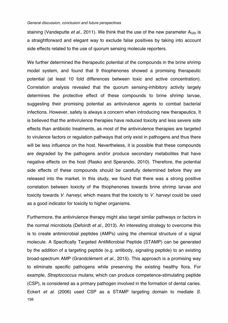
General discussion, conclusion and future perspectives
156
staining (Vandeputte et al., 2011). We think that the use of the new parameter AQSI is
a straightforward and elegant way to exclude false positives by taking into account
side effects related to the use of quorum sensing molecule reporters.
We further determined the therapeutic potential of the compounds in the brine shrimp
model system, and found that 9 thiophenones showed a promising therapeutic
potential (at least 10 fold differences between toxic and active concentration).
Correlation analysis revealed that the quorum sensing-inhibitory activity largely
determines the protective effect of these compounds to brine shrimp larvae,
suggesting their promising potential as antivirulence agents to combat bacterial
infections. However, safety is always a concern when introducing new therapeutics. It
is believed that the antivirulence therapies have reduced toxicity and less severe side
effects than antibiotic treatments, as most of the antivirulence therapies are targeted
to virulence factors or regulation pathways that only exist in pathogens and thus there
will be less influence on the host. Nevertheless, it is possible that these compounds
are degraded by the pathogens and/or produce secondary metabolites that have
negative effects on the host (Rasko and Sperandio, 2010). Therefore, the potential
side effects of these compounds should be carefully determined before they are
released into the market. In this study, we found that there was a strong positive
correlation between toxicity of the thiophenones towards brine shrimp larvae and
toxicity towards V. harveyi, which means that the toxicity to V. harveyi could be used
as a good indicator for toxicity to higher organisms.
Furthermore, the antivirulence therapy might also target similar pathways or factors in
the normal microbiota (Defoirdt et al., 2013). An interesting strategy to overcome this
is to create antimicrobial peptides (AMPs) using the chemical structure of a signal
molecule. A Specifically Targeted AntiMicrobial Peptide (STAMP) can be generated
by the addition of a targeting peptide (e.g. antibody, signaling peptide) to an existing
broad-spectrum AMP (Grandclément et al., 2015). This approach is a promising way
to eliminate specific pathogens while preserving the existing healthy flora. For
example, Streptococcus mutans, which can produce competence-stimulating peptide
(CSP), is considered as a primary pathogen involved in the formation of dental caries.
Eckert et al. (2006) used CSP as a STAMP targeting domain to mediate S.
Chapter VII
157
mutans-specific delivery of an antimicrobial peptide domain, and this class of STAMP
is capable of selectively eliminating S. mutans from multispecies biofilms without
affecting closly related noncariogenic oral bacteria.
For a long time, it was generally assumed that pathogens are unlikely to develop
resistance to quorum sensing disruption as it engenders only a milder evolutionary
pressure. Recently, Defoirdt et al., (2010) challenged this assumption and argued that
there is also a risk of resistance development using quorum sensing inhibitors. The
authors provided an overview of data indicating that there can be variation in the
expression of quorum sensing core genes among different strains of a certain species.
These core genes are involved in the production of signal molecule synthases and
receptors, as well as in quorum sensing signal transduction. Since the variability of
core genes can be caused by horizontal gene transfer, if this variation is associated
with a difference in fitness under treatment of quorum sensing inhibitors, natural
selection would automatically result (Defoirdt et al., 2010). Furthermore, the same
report also suggested that it is critically important to correctly evaluate the effect of
quorum sensing disruption on the fitness of bacteria, i.e. growth tests should be
performed under conditions that are as similar as possible to the clinical situation
instead of in nutrient-rich synthetic growth media (Martinez et al., 2007).
The first demonstration of resistance evolvement in cells to quorum sensing disruption
was by Maeda et al. (2012) using both realistic lab constructs and clinical strains. It
was shown that cells could develop resistance to quorum sensing disruption
compounds through different mechanisms, and that these mutations also occur in a
clinical setting, which indicates that cells can definitely evolve resistance even in the
absence of previous exposure to them (Maeda et al. 2012). Additional clinical
evidence of the ability of resistance development in strains was provided by
García-Contreras et al. (2013), demonstrating that Mexican clinical isolates from urine,
blood and catheter tip specimens from children showed resistance to brominated
furanone (C-30) and to 5-fluorouracil. More recently, quorum sensing was found to
enhance the stress tolerance of Pseudomonas aeruginosa to oxidative stress,
osmotic, thermal and heavy metals, and the link between quorum sensing and stress
was shown to have important physiological and ecological consequences, including
General discussion, conclusion and future perspectives
158
allowing the development of quorum sensing disruption resistance in cells and preventing social cheating (García-Contreras et al., 2015).
Further investigations are also required to determine the possibility of resistance to more quorum sensing disruption compounds, such as signal-degrading enzymes. Future investigations on the therapeutic development of antivirulence strategies should be proceed with more care and caution to avoid the undesired fate currently associated with antibiotic development.
7.4 INDOLE SIGNALING IN V. harveyi
Increasing evidences suggest that the bacterial metabolite indole can also act as a signal molecule in some bacteria. It has even been considered to be an interkingdom signal in commensal bacteria (Bansal et al., 2010). The production of indole is observed in over 85 bacterial species including both Gram-negative and Gram-positive bacteria ( Lee and Lee, 2010). Even many bacterial species that cannot produce indole will respond to the presence of extracellular indole, and exhibit several changes in behavior (Figure 7.3). In addition, several indole derivatives have also been reported to affect bacterial behaviors, and indole signaling has been employed for the design of small molecules that have the potential to control bacterial virulence and lead to the potential discovery of new therapeutics (Melander et al., 2014).
Figure 7.3. Overview of bacterial processes controlled by indole
Chapter VII
159
In this study, we determined the impact of indole on the virulence of V. harveyi in
Chapter 5. We demonstrated that addition of indole (50 µM or more) significantly decreased the virulence of V. harveyi towards both gnotobiotic brine shrimp larvae and conventionally reared giant river prawn larvae. This concentration is similar to the concentration produced by wild type V. harveyi when grown in Luria-Bertani broth, hence, doubling the concentration of extracellular indole results in decreased virulence. Since indole has been found to exert a beneficial effect on higher animals (Bansal et al., 2010), we pretreated the pathogen with indole in order to exclude a direct effect of indole on the host. Our findings are in accordance with previous report which has revealed that indole can protect sea bass (Dicentrarchus labrax) larvae from V. anguillarum (Li et al., 2014). Therefore, indole decreasing virulence seems to be conserved among distantly related vibrios that are pathogenic to aquatic organisms. Subsequently, we indicated that indole could decrease production of several major virulence factors in V. harveyi, including biofilm formation, exopolysaccharide production and swimming motility. These factors have been reported to play a key role in bacterial adherence and in colonization to epithelial cells that are importance for infection (Bansal et al., 2007).
Although indole production has been found in many species, the complex pathways by which indole exerts its effects are far from understood, even in the most intensively studied species, E. coli. It has been revealed that the E. coli LuxR-homologue SdiA is involved in indole signaling, however, there is no proof of the direct binding of indole and SdiA, and the interactions between indole and SdiA still remain to be investigated further ( Lee et al., 2007). In another report, Hirakawa et al. (2005) demonstrated that indole-induced drug resistance in E. coli was mediated by the two-component systems BaeSR and CpxAR. In vibrio species, indole was hypothesised to act through the RNA polymerase regulator DksA in V. cholerae (Beyhan et al., 2009). The V.
harveyi ATCC BAA-1116 genome does not contain a homologue of sdiA, but it does contain a dksA homologue that shows 82% identity at nucleotide level with V.
cholerae dksA. However, we were not able to obtain a dksA deletion mutant in several attempts for further investigation (collaboration with Gary Vora, US Naval Research Institute, Washington DC), and this might indicate that this gene is essential in V.
harveyi.
General discussion, conclusion and future perspectives
160
Further, in this study, we found a link between indole sensing and other regulatory
mechanisms in V. harveyi. According to the results, we hypothesized that indole could
induce a stress response by inducing expression of the alternative sigma factor RpoS,
which is known to be related to stress. Further we found that RpoS down-regulated
indole production, which might be a feedback mechanism to maintain homeostasis.
These results are consistent with what has been observed in another important
aquaculture pathogen, V. anguillarum (Li et al., 2014). In addition to this, indole was
found to interfere with the three-channel quorum sensing system in V. harveyi by
interacting with signal transduction. This could also in part explain the decreased
virulence towards different hosts since quorum sensing is required for full virulence of
V. harveyi in these model systems (Defoirdt and Sorgeloos, 2012).
Finally, we demonstrated that the auxin hormones indole-3-acetic acid and
indole-3-acetamide, which are indole analogues produced by plants including
micro-algae, showed similar effects as indole on the virulence of V. harveyi. To our
knowledge, this is the first report about the effect of auxins produced by micro-algae
on aquatic bacteria. Furthermore, it has been reported that there is a lower incidence
of disease in the green water aquaculture system, which means animals are cultured
in water containing high levels of micro-algae (Natrah et al., 2014). According to our
findings in this study, this might be explained by the presence of (relatively) high
levels of indole analogues in the digestive tract of animals that have ingested high
levels of micro-algae. It also could be an interesting novel strategy to control bacterial
disease in aquaculture by selecting micro-algae that are capable of producing high
levels of auxins.
7.5 SENSING OF CATECHOLAMINE STRESS HORMONES PRODUCED BY THE HOST INCREASES THE VIRULENCE OF V. harveyi
Host stress has long been known to influence the outcome of many bacterial
infections. During a stress reaction, stress hormones such as glucocorticoids and
catecholamines, are released by many higher organisms (Verbrugghe et al., 2012). In
addition to the effects of these stress hormones on the host immune system, research
over the past decades has revealed that catecholamines can also alter the growth and
Chapter VII
161
virulence of several pathogens and consequently influence the pathogen-host interactions. This new perspective leads to the development of a new research field termed microbial endocrinology, which is regarded as a means of understanding the dialogue between animals and their microflora (Sharaff and Freestone, 2011).
In Chapter 6, we evaluated the impact of two kinds of catecholamines, norepinephrine and dopamine, on the growth and virulence of V. harveyi. Results indicated that both catecholamines could significantly increase the growth of V.
harveyi in serum-supplemented medium, which might be attributed to the capacity of catecholamines to supply the pathogen of iron. The increased availability of iron that is supplied through the intervention of catecholamines probably plays a key role in the effects of stress on the outcome of an infection (Verbrugghe et al., 2012). In addition to increase iron uptake in a direct way, we also found that both norepinephrine and dopamine could significantly induce siderophore production, which has been reported to be able to internalize the iron removed by catecholamines (Freestone et al., 2003).
Further, we found that both norepinephrine and dopamine can enhance the production of some virulence factors in V. harveyi that are essential for colonization and adhesion to the host surfaces, such as swimming motility, exopolysaccharide production and biofilm formation. As part of the ‘fight or flight’ response, stress in a host will increase the systemic catecholamine levels (Freestone et al., 2008). Our results in this study suggested that the increasing concentration of catecholamines might be indicator informing the pathogen that the host is less fit and it is time to relocate to a suitable one.
In animals, catecholamines have been found to exert their effects by binding to specific adrenergic and dopaminergic receptors, and the binding can be blocked by antagonists specific to the catecholamine receptors (Freestone et al., 1999). In the present study, the results demonstrated that ɑ- but not β-adrenergic receptor antagonists could block responses to norepinephrine, but did not show any effects on dopamine responsiveness. On the contrary, dopaminergic receptor antagonists neutralized induction caused by dopamine, but did not neutralize induction by norepinephrine. This suggests that bacterial response system for catecholamine
General discussion, conclusion and future perspectives
162
recognition shows a degree of specificity similar to that demonstrated for catecholamine receptors in animals.
In terms of a bacterial catecholamine receptor, there are so far only few reports examining the existence of such receptors. It has also been revealed that these receptors show large difference depending on the type of pathogen, the host or the type of stress hormone (Sharaff and Freestone, 2011). For instance, Escherichia coli
O157:H7 and Salmonella enterica have been reported to sense norepinephrine and epinephrine as quorum sensing signals through histidine sensor kinases (QseC and QseE), while norepinephrine was found to enhance the growth and virulence of Pseudomonas aeruginosa through the las quorum sensing pathway (Hughes et al., 2009; Pullinger et al., 2010; Hegde et al., 2009). In this study, a QseC antagonist, LED209 was found to be able to neutralize the effects of both norepinephrine and dopamine, indicating that a similar response system for catecholamines might exist in V. harveyi. This is also substantiated by the fact that the V. harveyi ATCC BAA-1116 genome contains a QseC homolog. Furthermore, catecholamines have been found to regulate virulence gene expression and enhance the potential to cause disease in all these species mentioned above. Therefore, these receptors could be considered as regulators of multiple virulence factors, making them a possible target for antivirulence therapy to combat bacterial infections.
The fact that stress hormones can influence the outcome of an infection has also been confirmed by in vivo test. However, such studies are scarce, and contradictory results have been reported (Cray et al., 1998; Kudahl et al., 2007; Dutta et al., 2009; Wesley et al., 2009). In order to obtain a better understanding of the interactions between the host immune system and pathogen, it is of utmost importance that (highly controlled) animal models are created and more in vivo tests are conducted to investigate the effects of stress hormones on bacterial infections in different hosts. In this study, we investigated the impact of norepinephrine and dopamine and the respective receptor antagonists on the virulence of V. harveyi in our highly controlled brine shrimp larvae system. Significantly higher brine shrimp mortality was observed when exposing V.
harveyi to norepinephrine or dopamine prior to the challenge, and the induced virulence could be neutralized by specific antagonists. However, pretreatment of V.
Chapter VII
163
harveyi with the antagonists had no effect on virulence in the absence of
catecholamines. Furthermore, previous work in our lab has also confirmed the
increased virulence of V. harveyi by catecholamines in giant freshwater prawn larvae
(Pande et al., 2014).
7.6 CONCLUSIONS
! This study revealed an important mechanism by which quorum sensing controls
the virulence of V. harveyi by demonstrating (i) that quorum sensing positively
regulates motility by affecting flagellar biosynthesis, (ii) that LuxR is involved in
the regulation of motility and (iii) that flagellar motility significantly affects
virulence of V. harveyi in our gnotobiotic brine shrimp model.
! In this study we determined the quorum sensing-inhibiting activities of 20 new
synthetic thiophenones towards the quorum sensing model bacterium V. harveyi,
and 6 thiophenones were identified to be able to inhibit quorum sensing at
submicromolecular levels. There was a strong positive correlation between the
specific quorum sensing-disrupting activity of the thiophenones and the
protection of brine shrimp larvae against pathogenic V. harveyi, and 6 quorum
sensing-disrupting thiophenones were considered to be highly promising.
! We proposed a new parameter, AQSI, to determine specific quorum sensing
inhibitory activity based on experiments with a quorum sensing reporter strain.
This parameter allowed us to exclude 5 false positives out of the 17 thiophenone
compounds that were able to inhibit bioluminescence in V. harveyi. We think that
the use of this new parameter is a straightforward and elegant way to exclude
false positives by taking into account side effects related to the use of quorum
sensing molecule reporters.
! This study demonstrated for the first time that indole sensing could reduce the
virulence of V. harveyi towards gnotobiotic brine shrimp larvae and conventionally
reared giant river prawn larvae. Indole decreased the production of several
virulence factors in V. harveyi, including biofilm formation, exopolysaccharide
production and swimming motility.
! The alternative sigma factor RpoS was found to be involved in the production of
indole in V. harveyi, and indole could interfere with the three-channel quorum
General discussion, conclusion and future perspectives
164
sensing system of V. harveyi.
! Auxin hormones that are produced by micro-algae showed similar effects as
indole, suggesting that the selection of micro-algae that can produce high levels
of auxins could be an interesting novel strategy to control bacterial disease in
aquaculture.
! The catecholamines norepinephrine and dopamine increased the virulence of V.
harveyi by increasing swimming motility, biofilm formation, exopolysaccharide
production and growth in environments with low iron availability. These effects
could be neutralized by antagonists for eukaryotic catecholamine receptors and
the bacterial catecholamine receptor antagonist LED209.
! Different effects of the adrenergic and dopaminergic receptors antagonists
indicate the presence of specific sensing systems for different catecholamines in
V. harveyi.
7.7 FUTURE PERSPECTIVES
The present study demonstrated that the flagellar motility significantly affects
virulence of V. harveyi in our gnotobiotic brine shrimp model. Additionally, flagellar
motility has also been considered as an important virulence factor in numerous
pathogens. Can flagellar motility serve as a therapeutic or vaccine target? To our
knowledge, little practical work has been done to evaluate motility as a therapeutic
target. Tsutsui et al., (2000) reported that a proton pump inhibitor (PPI), which could
markedly inhibit the motility of Helicobacter pylori, has already been widely used for
treatment of ulcers in humans. In another report, flagellin proved to be a specific and
efficient antigen to inhibit infection of V. cholerae towards rabbits (Yancey et al., 1979).
In addition, flagellar preparations of Pseudomonas aeruginosa were used for the
development of a vaccine formula for cystic fibrosis patients, which has been proven
to be effective and safe both in animal models and human volunteers (Holder and
Naglich, 1986; Doring and Dorner, 1997). However, flagella and motility have not
been intensively studied as therapeutic target in aquaculture so far. Due to the
apparent role of bacterial flagellin as an immunomodulatory agent, it might represent
one promising ingredient in composite subunit vaccines (Josenhans and Suerbaum,
2002).
Chapter VII
165
To date, most research with respect to exploring novel antivirulence strategies to
combat bacterial infections in aquaculture have focused on the pathogen-pathogen
signaling. Quorum sensing disruption has been considered to be an effective strategy
to control diseases caused by various pathogens. Application of compounds with
quorum sensing-inhibitory activity are one of the promising tools for quorum sensing
disruption. However, several questions remain to be answered, e.g. whether it is
possible to control the pathogen without affecting the normal microbiota. Additionally,
although most of the antivirulence agents have been tested both in vitro and in vivo,
large-scale trials are still required to prove their safety in real aquaculture
environments before bringing them into practical applications. Furthermore,
increasing evidence suggests that pathogens will probably develop resistance to
antivirulence agent as well. Therefore, it is important to further develop different
strategies that could be used synergistically to maximize the chance of successfully
protecting the animals.
The development of alternative antivirulence therapies will require continued
investigations to obtain a better understanding of the bacterial virulence strategies,
signaling pathways and potential ways to exploit them. In addition to quorum sensing,
more pathogenicity mechanisms in bacterial pathogens have been unraveled to be
potential targets for antivirulence therapy. For example, host-pathogen signaling
mediated by catecholamine stress hormones. In this study, we demonstrated that
catecholamines norepinephrine and dopamine could significantly enhance the
virulence of V. harveyi, and specific sensing systems for different catecholamines
seem to be present in V. harveyi. In the future, there is a need to identify bacterial
adrenergic receptors and improve our understanding on why there are multiple
sensors responding to one or more signals. Perhaps there is a specialized need for
certain receptors in specific niches.
Finally, it would also be interesting to search new interkingdom communication
mechanisms, in order to provide not only new biological insights into pathogenicity but
also novel strategy for antivirulence research. For example, V. anguillarum was found
to be affected by sensing of several host factors (e.g. mucin, bile salts and cholesterol)
and subsequently showed a decreased virulence towards see bass larvae.

General discussion, conclusion and future perspectives
166

Appendix A
REFERENCES

References
167
REFERENCES
A Abraham TJ. (2006). Virulence of Vibrio harveyi possessing a transferable
chloramphenicol resistance determinant to larvae of Indian white shrimp Fenneropenaeus indicus (Decapoda). Indian Journal of Marine Sciences 35:275–278.
Abuaita BH, Withey JH. (2009). Bicarbonate Induces Vibrio cholerae virulence gene expression by enhancing ToxT activity. Infection and Immunity 77:4111–4120.
Aguirre-Guzmán G, Vázquez-Juárez R, Ascencio F. (2001). Differences in the Susceptibility of American White Shrimp Larval Substages (Litopenaeus vannamei) to Four Vibrio Species. Journal of Invertebrate Pathology 78:215–219.
Akinbowale OL, Peng H, Barton MD. (2006). Antimicrobial resistance in bacteria isolated from aquaculture sources in Australia. Journal of Applied Microbiology 100:1103–1113.
Alcaide E, Gil Sanz C, Sanjuan E, Esteve D, Amaro C, Silveira L. (2001). Vibrio harveyi causes disease in seahorse, Hippocampus sp. Journal of Fish Diseases 24:311–313.
Alderman DJ, Hastings TS. (1998). Antibiotic use in aquaculture: development of antibiotic resistance – potential for consumer health risks*. International Journal of Food Science & Technology 33:139–155.
Amaro C, Aznar R, Alcaide E, Lemos ML. (1990). Iron-binding compounds and related outer membrane proteins in Vibrio cholerae non-O1 strains from aquatic environments. Applied and Environmental Microbiology 56:2410–2416.
Andrus CR, Walter M, Crosa JH, Payne SM. (1983). Synthesis of siderophores by pathogenic Vibrio species. Curr Microbiol 9:209–214.
Anetzberger C, Pirch T, Jung K. (2009). Heterogeneity in quorum sensing-regulated bioluminescence of Vibrio harveyi. Molecular Microbiology 73:267–277.
Anyanful A, Dolan Livengood JM, Lewis T, Sheth S, DeZalia MN, Sherman MA, et al. (2005). Paralysis and killing of Caenorhabditis elegans by enteropathogenic Escherichia coli requires the bacterial tryptophanase gene. Molecular
References
168
Microbiology 57:988–1007.
Ashen JB, Cohen JD, Goff LJ. (1999). GC‐SIM‐MS detection and quantification of free indole‐3‐acetic acid in bacterial galls on the marine alga Prionitis Lanceolata (Rhodophyta). Journal of Phycology 35: 493–500.
Ashley PJ. (2007). Fish welfare: Current issues in aquaculture. Applied Animal Behaviour Science 104:199–235.
Austin B, Zhang X. (2006). Vibrio harveyi: a significant pathogen of marine vertebrates and invertebrates. Lett Appl Microbiol 43:119–124.
Austin B and Austin DDA. (1999). Bacterial fish pathogens: diseases of farmed and wild fish. 3rd edn. Berlin: Springer.
Avendaño-Herrera R, Núñez S, Barja JL, Toranzo AE. (2008). Evolution of drug resistance and minimum inhibitory concentration to enrofloxacin in Tenacibaculum maritimum strains isolated in fish farms. Aquacult Int 16:1–11.
B Baca-DeLancey RR, South MM, Ding X, Rather PN. (1999). Escherichia coli genes
regulated by cell-to-cell signaling. Proceedings of the National Academy of Sciences 96:4610–4614.
Bansal T, Englert D, Lee J, Hegde M, Wood TK, Jayaraman A. (2007). Differential effects of epinephrine, norepinephrine, and indole on Escherichia coli O157:H7 chemotaxis, colonization, and gene expression. Infection and Immunity 75:4597–4607.
Bansal T, Alaniz RC, Wood TK, Jayaraman A. (2010). The bacterial signal indole increases epithelial-cell tight-junction resistance and attenuates indicators of inflammation. Proc Natl Acad Sci USA 107:228–233.
Baron C (2010). Antivirulence drugs to target bacterial secretion systems. Current Opinion in Microbiology 13, 100-105.
Baruah K, Cam DTV, Dierckens K, Wille M, Defoirdt T, Sorgeloos P, Bossier P. (2009). In vivo effects of single or combined N-acyl homoserine lactone quorum sensing signals on the performance of Macrobrachium rosenbergii larvae. Aquaculture 288, 233–238.
References
169
Bassler BL, Wright M, Silverman MR (1994). Multiple signalling systems controlling expression of luminescence in Vibrio harveyi: sequence and function of genes encoding a second sensory pathway. Mol. Microbiol. 13: 273–286.
Bassler BL, Greenberg EP, Stevens AM. (1997). Cross-species induction of luminescence in the quorum-sensing bacterium Vibrio harveyi. Journal of Bacteriology 179:4043–4045.
Bassler CMWABL. (2007). QUORUM SENSING: Cell-to-Cell Communication in Bacteria. 1–31.
Basu S, Sun H, Brian L, Quatrano RL, Muday GK. (2002). Early Embryo Development in Fucus distichusIs Auxin Sensitive. PLANT PHYSIOLOGY 130: 292–302.
Bearson BL, and Bearson SM (2008). The role of the QseC quorum sensing sensor kinase in colonization and norepinephrine-enhanced motility of Salmonella enteric serovar Typhimurium. Microb. Pathog. 44, 271-278.
Becket P, Gillan D, Lanterbecq D, Jangoux M, Rasolofonirina, R, Rakotovao J, and Eeckhaut I (2004) The skin ulceration disease in cultivated juveniles of Holothuria scabra (Holothuroidea, Echinodermata). Aquaculture 242, 13–30.
Bergmeier C, Siekmeier R, Gross W. (2004). Distribution spectrum of paraoxonase activity in HDL fractions. Clinical Chemistry 50: 2309–2315.
Beyhan S, Saini SG, Yildiz FH, Bartlett DH. (2009). Indole Acts as an Extracellular Cue Regulating Gene Expression in Vibrio cholerae. Journal of Bacteriology 191:3504–3516.
Belay T, Aviles H, Vance M, Fountain K, Sonnenfeld G. (2003). Catecholamines and in vitro growth of pathogenic bacteria: enhancement of growth varies greatly among bacterial species. Life Sciences 73:1527–1535.
Benneche T, Herstad G, Rosenberg M, Assev S, Scheie AA. (2011). Facile synthesis of 5-(alkylidene)thiophen-2(5H)-ones. A new class of antimicrobial agents. RSC Adv 1:323–332.
Benneche T, Chamgordani EJ, Scheie AA. (2012). Reaction of (Z)-5-(Bromomethylene)thiophen-2(5H)-one with Some Nucleophiles in Search for New Biofilm Inhibitors. Synthetic Communications 43:431–437.
Benneche T, Chamgordani EJ, Herstad G, Tannœs BSM, Scheie AA. 5-Alkylidenethiophen-2(5H)-ones as biofilm inhibitors. Submitted.
References
170
Beyhan S, Saini SG, Yildiz FH, Bartlett DH. (2009). Indole Acts as an Extracellular Cue Regulating Gene Expression in Vibrio cholerae. Journal of Bacteriology 191:3504–3516.
Billecke S, Draganov D, Counsell R, Stetson P, Watson C, Hsu C, et al. (2000). Human serum paraoxonase (PON1) isozymes Q and R hydrolyze lactones and cyclic carbonate esters. Drug Metab Dispos 28: 1335–1342.
Blair DF. (1995). How Bacteria Sense and Swim. Annu Rev Microbiol 49: 489–520.
Bondad-Reantaso MG, Subasinghe RP, Arthur JR, Ogawa K, Chinabut S, Adlard R, et al. (2005). Disease and health management in Asian aquaculture. Veterinary Parasitology 132:249–272.
Bobadilla Fazzini RA, Skindersoe ME, Bielecki P, Puchałka J, Givskov M, Martins dos Santos VAP. (2013). Protoanemonin: a natural quorum sensing inhibitor that selectively activates iron starvation response. Environmental Microbiology 15: 111–120.
Borchardt SA, Allain EJ, Michels JJ, Stearns GW, Kelly RF, McCoy WF. (2001). Reaction of acylated homoserine lactone bacterial signaling molecules with oxidized halogen antimicrobials. Applied and Environmental Microbiology 67:3174–3179.
Brackman G, Defoirdt T, Miyamoto C, Bossier P, Van Calenbergh S, Nelis H, et al. (2008). Cinnamaldehyde and cinnamaldehyde derivatives reduce virulence in Vibrio spp. by decreasing the DNA-binding activity of the quorum sensing response regulator LuxR. BMC Microbiol 8:149.
Brackman G, Quntar Al AAA, Enk CD, Karalic I, Nelis HJ, Van Calenbergh S, et al. (2013). Synthesis and evaluation of thiazolidinedione and dioxazaborocane analogues as inhibitors of AI-2 quorum sensing in Vibrio harveyi. Bioorganic & Medicinal Chemistry 21: 660–667.
Bramhachari PV, Dubey SK. (2006). Isolation and characterization of exopolysaccharide produced by Vibrio harveyi strain VB23. Lett Appl Microbiol 43:571–577.
Broughton EI, Walker DG. (2009). Prevalence of Antibiotic-Resistant Salmonella in Fish in Guangdong, China. Foodborne Pathogens and Disease 6:519–521.
Burridge L, Weis JS, Cabello F, Pizarro J, Bostick K. (2010). Chemical use in salmon aquaculture: A review of current practices and possible environmental effects.
References
171
Aquaculture 306:7–23.
Butler A, Sandy M. (2009). Mechanistic considerations of halogenating enzymes. Nature 460:848–854.
Burton C L, Chhabra S R, Swift S, Baldwin T J, Withers H, Hill S J and Williams P. (2002). The growth response of Escherichia coli to neurotransmitters and related catecholamine drugs requires a functional enterobactin biosynthesis and uptake system. Infect. Immun. 70, 5913-5923.
C Cao JG, Meighen EA. (1989). Purification and structural identification of an
autoinducer for the luminescence system of Vibrio harveyi. J Biol Chem 264:21670–21676.
Cabello FC. (2006). Heavy use of prophylactic antibiotics in aquaculture: a growing problem for human and animal health and for the environment. Environmental Microbiology 8:1137–1144.
Cabello FC, Godfrey HP, Tomova A, Ivanova L, Dölz H, Millanao A, et al. (2013). Antimicrobial use in aquaculture re-examined: its relevance to antimicrobial resistance and to animal and human health. Environmental Microbiology 15:1917–1942.
Carlier A, Uroz S, Smadja B, Fray R, Latour X, Dessaux Y, et al. (2003). The Ti Plasmid of Agrobacterium tumefaciens Harbors an attM-Paralogous Gene, aiiB, Also Encoding N-Acyl Homoserine Lactonase Activity. Applied and Environmental Microbiology 69:4989–4993.
Cavalli RO, Lavens P, Sorgeloos P. (2001). Reproductive performance of Macrobrachium rosenbergi female in captivity. Journal of the World Aquaculture Society 32 (1), 60–67.
Chant EL, Summers DK. (2007). Indole signalling contributes to the stable maintenance of Escherichia coli multicopy plasmids. Molecular Microbiology 63:35–43.
Chen X, Schauder S, Potier N, Van Dorsselaer A, Pelczer I, Bassler BL, et al. (2002). Structural identification of a bacterial quorum-sensing signal containing boron. Nature 415:545–549.
References
172
Chen Y, Dai J, Morris JG, Johnson JA. (2010). Genetic analysis of the capsule polysaccharide (K antigen) and exopolysaccharide genes in pandemic Vibrio parahaemolyticus O3:K6. BMC Microbiol 10:274.
Chen YN, Fan HF, Hsieh SL and Kuo CM (2003). Physiological involvement of DA in ovarian development of the freshwater giant prawn, Macrobrachium rosenbergii. Aquaculture 228, 383-395.
Choi H, Mascuch SJ, Villa FA, Byrum T, Teasdale ME, Smith JE, et al. (2012). Honaucins A-C, potent inhibitors of inflammation and bacterial quorum sensing: synthetic derivatives and structure-activity relationships. Chemistry & Biology 19: 589–598.
Christensen QH, Grove TL, Booker SJ, Greenberg EP. (2013). A high-throughput screen for quorum-sensing inhibitors that target acyl-homoserine lactone synthases. Proceedings of the National Academy of Sciences, 110(34), 13815-13820.
Chu W, Zere TR, Weber MM, Wood TK, Whiteley M, Hidalgo-Romano B, et al. (2012). Indole production promotes Escherichia coli mixed-culture growth with Pseudomonas aeruginosa by inhibiting quorum signaling. Applied and Environmental Microbiology 78:411–419.
Clarke MB, Hughes DT, Zhu C, Boedeker EC, Sperandio V. (2006). The QseC sensor kinase: a bacterial adrenergic receptor. Proceedings of the National Academy of Sciences 103:10420–10425.
Clatworthy AE, Pierson E, Hung DT. (2007). Targeting virulence: a new paradigm for antimicrobial therapy. Nature chemical biology 3:541–548.
Clemons KV, Gadberry JL. (1982). Increased indole detection for Pasteurella multocida. J Clin Microbiol 15:731–732.
Cogan TA, Thomas AO, Rees LEN, Taylor AH, Jepson MA, Williams PH, et al. (2007). Norepinephrine increases the pathogenic potential of Campylobacter jejuni. 56:1060–1065.
Costa LG, Cole TB, Vitalone A, Furlong CE. (2005). Measurement of paraoxonase (PON1) status as a potential biomarker of susceptibility to organophosphate toxicity. Clinica Chimica Acta 352: 37–47.
Costerton JW, Irvin RT, Cheng KJ, Sutherland IW. (1981). The Role of Bacterial Surface Structures in Pathogenesis. Critical Reviews in Microbiology 8:303–338.
References
173
Coulanges V, Andre P, Ziegler O, Buchheit L, Vidon DJ. (1997). Utilization of iron-catecholamine complexes involving ferric reductase activity in Listeria monocytogenes. Infection and Immunity 65:2778–2785.
Cray WC, Casey TA, Bosworth BT, Rasmussen MA. (1998). Effect of dietary stress on fecal shedding of Escherichia coli O157:H7 in calves. Applied and Environmental Microbiology 64:1975–1979.
D Dalsgaard I, Høi L, Siebeling RJ, Dalsgaard A. (1999). Indole-positive Vibrio vulnificus
isolated from disease outbreaks on a Danish eel farm. Dis Aquat Organ 35:187–194.
Darshanee Ruwandeepika HA, Sanjeewa Prasad Jayaweera T, Paban Bhowmick P, Karunasagar I, Bossier P, Defoirdt T. (2012). Pathogenesis, virulence factors and virulence regulation of vibrios belonging to the Harveyi clade. Rev Aquacult 4:59–74.
Das A, Saha D, Pal J. (2009). Antimicrobial resistance and in vitro gene transfer in bacteria isolated from the ulcers of EUS-affected fish in India. Lett Appl Microbiol 49:497–502.
De Keersmaecker SCJ, Sonck K, Vanderleyden J. (2006). Let LuxS speak up in AI-2 signaling. Trends in Microbiology 14:114–119.
De Kievit TR, Iglewski BH. (2000). Bacterial quorum sensing in pathogenic relationships. Infection and Immunity 68:4839–4849.
De Schryver P, Defoirdt T, Sorgeloos P. (2014). Early Mortality Syndrome Outbreaks: A Microbial Management Issue in Shrimp Farming? Rall, GF (ed). PLoS Pathog 10:e1003919.
Decho AW. (1990). Microbial Exopolymer Secretions in Ocean Environments: Their Role(S) in Food Webs and Marine Processes. Oceanogr Mar Biol Annu Rev 28:73–153.
Defoirdt T, Bossier P, Sorgeloos P, Verstraete W. (2005). The impact of mutations in the quorum sensing systems of Aeromonas hydrophila, Vibrio anguillarum and Vibrio harveyi on their virulence towards gnotobiotically cultured Artemia franciscana. Environmental Microbiology 7:1239–1247.
References
174
Defoirdt T, Crab R, Wood TK, Sorgeloos P, Verstraete W, Bossier P. (2006). Quorum sensing-disrupting brominated furanones protect the gnotobiotic brine shrimp Artemia franciscana from pathogenic Vibrio harveyi, Vibrio campbellii, and Vibrio parahaemolyticus isolates. Applied and Environmental Microbiology 72:6419–6423.
Defoirdt T, Boon N, Sorgeloos P, Verstraete W, Bossier P. (2007a). Quorum sensing and quorum quenching in Vibrio harveyi: lessons learned from in vivo work. ISME J 2:19–26.
Defoirdt T, Miyamoto CM, Wood TK, Meighen EA, Sorgeloos P, Verstraete W, et al. (2007b). The natural furanone (5Z)-4-bromo-5-(bromomethylene)-3-butyl-2(5H)-furanone disrupts quorum sensing-regulated gene expression in Vibrio harveyi by decreasing the DNA-binding activity of the transcriptional regulator protein luxR. Environmental Microbiology 9:2486–2495.
Defoirdt T, Boon N, Sorgeloos P, Verstraete W, Bossier P. (2008). Quorum sensing and quorum quenching in Vibrio harveyi: lessons learned from in vivo work. ISME J 2:19–26.
Defoirdt T, Darshanee Ruwandeepika HA, Karunasagar I, Boon N, Bossier P. (2009). Quorum sensing negatively regulates chitinase in Vibrio harveyi. Environmental Microbiology Reports 2:44–49.
Defoirdt T, Darshanee Ruwandeepika HA, Karunasagar I, Boon N and Bossier P. (2010a). Quorum sensing negatively regulates chitinase in Vibrio harveyi. Environ. Microbiol. Rep 2: 44–49.
Defoirdt T, Boon N, Bossier P. (2010b). Can bacteria evolve resistance to quorum sensing disruption? PLoS Pathog 6:e1000989.
Defoirdt T, Thanh LD, Van Delsen B, De Schryver P, Sorgeloos P, Boon N, et al. (2011a). N-acylhomoserine lactone-degrading Bacillus strains isolated from aquaculture animals. Aquaculture 311:258–260.
Defoirdt T, Sorgeloos P, Bossier P. (2011b). Alternatives to antibiotics for the control of bacterial disease in aquaculture. Current Opinion in Microbiology 14:251–258.
Defoirdt T, Benneche T, Brackman G, Coenye T, Sorgeloos P, Scheie AA. (2012a). A quorum sensing-disrupting brominated thiophenone with a promising therapeutic potential to treat luminescent vibriosis. PLoS ONE 7:e41788.
Defoirdt T, Sorgeloos P. (2012b). Monitoring of Vibrio harveyi quorum sensing activity
References
175
in real time during infection of brine shrimp larvae. ISME J 6:2314–2319.
Defoirdt T, Brackman G, Coenye T. (2013a). Quorum sensing inhibitors: how strong is the evidence? Trends in Microbiology 21:619–624.
Defoirdt T. (2013b). Virulence mechanisms of bacterial aquaculture pathogens and antivirulence therapy for aquaculture. Rev Aquacult 6:100–114.
Defoirdt T. (2013c). Antivirulence therapy for animal production: filling an arsenal with novel weapons for sustainable disease control. PLoS Pathog. 9, e1003603.
Defoirdt T, Pande GSJ, Baruah K, Bossier P. (2013d). The apparent quorum-sensing inhibitory activity of pyrogallol is a side effect of peroxide production. Antimicrobial Agents and Chemotherapy 57:2870–2873.
Defoirdt T. (2014). Virulence mechanisms of bacterial aquaculture pathogens and antivirulence therapy for aquaculture. Rev Aquacult 6:100–114.
DeMoss RD, Moser K. (1969). Tryptophanase in diverse bacterial species. Journal of Bacteriology 98:167–171.
Dhabhar FS, and McEwen BS. (1997). Acute stress enhances while chronic stress suppresses cell-mediated immunity in vivo: a potential role for leukocyte trafficking. Brain Behav. Immun. 11, 286–306.
Dhabhar FS. (2009). A hassle a day may keep the pathogens away: The fight-or-flight stress response and the augmentation of immune function. Integr Comp Biol 49:215–236.
Di Martino P, Fursy R, Bret L, Sundararaju B, Phillips RS. (2011). Indole can act as an extracellular signal to regulate biofilm formation of Escherichia coli and other indole-producing bacteria. Canadian Journal of Microbiology 49:443–449.
Diggle SP, Gardner A, West SA, Griffin AS. (2007). Evolutionary theory of bacterial quorum sensing: when is a signal not a signal? Philosophical Transactions of the Royal Society B: Biological Sciences 362:1241–1249.
Diggles BK, Moss GA, Carson J, Anderson CD. (2000). Luminous Vibriosis in Rock Lobster Jasus Verreauxi (Decapoda: Palinuridae) Phyllosoma Larvae Associated with Infection by Vibrio harveyi. Nature 43:127–137.
Domka J, Lee J, Wood TK. (2006). YliH (BssR) and YceP (BssS) Regulate Escherichia coli K-12 Biofilm Formation by Influencing Cell Signaling. Applied and Environmental Microbiology 72:2449–2459.
References
176
Dong Y H, Gusti A R, Zhang Q, Xu J L, Zhang L H. (2002). Identification of quorum-quenching N-acyl homoserine lactonases from Bacillus species. Applied and environmental microbiology, 68(4), 1754-1759.
Dong Y-H, Wang L-H, Zhang L-H. (2007). Quorum-quenching microbial infections: mechanisms and implications. Philosophical Transactions of the Royal Society B: Biological Sciences 362:1201–1211.
Donlan RM, Costerton JW. (2002). Biofilms: survival mechanisms of clinically relevant microorganisms. Clin Microbiol Rev 15:167–193.
Doring G, Dorner F. (1997). A multicenter vaccine trial using the Pseudomonas aeruginosa flagella vaccine IMMUNO in patients with cystic fibrosis. Behring Inst Mitt 338–344.
Dorsch M, Lane D, Stackebrandt E. (1992). Towards a Phylogeny of the Genus Vibrio Based on 16S rRNA Sequences. International Journal of Systematic and Evolutionary Microbiology 42:58–63.
Doukyu N, Aono R. (1997). Biodegradation of indole at high concentration by persolvent fermentation with Pseudomonas sp. ST-200. Extremophiles 1:100–105.
Draganov DI, Teiber JF, Speelman A, Osawa Y, Sunahara R, La Du BN. (2005). Human paraoxonases (PON1, PON2, and PON3) are lactonases with overlapping and distinct substrate specificities. J Lipid Res 46: 1239–1247.
Duarte CM, Holmer M, Olsen Y, Soto D, Marbà N, Guiu J, et al. (2009). Will the Oceans Help Feed Humanity? BioScience 59:967–976.
Dung TT, Haesebrouck F, Tuan NA, Sorgeloos P, Baele M, Decostere A. (2008). Antimicrobial Susceptibility Pattern of Edwardsiella ictaluri Isolates from Natural Outbreaks of Bacillary Necrosis of Pangasianodon hypophthalmus in Vietnam. Microbial drug resistance .14(4):311–316.
Dutta V, Huff GR, Huff WE, Johnson MG, Nannapaneni R, Sayler RJ. (2009). The Effects of Stress on Respiratory Disease and Transient Colonization of Turkeys with Listeria monocytogenes Scott A.
E Eckerson HW, Wyte CM, La Du BN. (1983). The human serum
References
177
paraoxonase/arylesterase polymorphism. American Journal of Human Genetics 35: 1126–1138.
Eckert R, He J, Yarbrough DK, Qi F, Anderson MH, Shi W. (2006). Targeted killing of Streptococcus mutans by a pheromone-guided ‘smart’ antimicrobial peptide. Antimicrobial Agents and Chemotherapy 50: 3651–3657.
Ensley BD, Ratzkin BJ, Osslund TD, Simon MJ, Wackett LP, Gibson DT. (1983). Expression of naphthalene oxidation genes in Escherichia coli results in the biosynthesis of indigo. Science 222:167–169.
F
FAO (2014) The State of World Fisheries and Aquaculture 2014. FAO, Rome, Italy.
Fernández Alarcón C, Miranda CD, Singer RS, López Y, Rojas R, Bello H, et al. (2010). Detection of the floR Gene in a Diversity of Florfenicol Resistant Gram-Negative Bacilli from Freshwater Salmon Farms in Chile. Zoonoses and Public Health 57:181–188.
Finch RG, Pritchard DI, Bycroft BW, Williams P, Stewart GS. (1998). Quorum sensing: a novel target for anti-infective therapy. Journal of Antimicrobial Chemotherapy 42:569–571.
Fishman A, Tao Y, Rui L, Wood TK. (2005). Controlling the regiospecific oxidation of aromatics via active site engineering of toluene para-monooxygenase of Ralstonia pickettii PKO1. J Biol Chem 280:506–514.
Freeman JA and Bassler BL. (1999). A genetic analysis of the function of LuxO, a two-component response regulator involved in quorum sensing in Vibrio harveyi. Mol. Microbiol. 31: 665–677.
Freestone PP, Haigh RD, Williams PH, Lyte M. (1999). Stimulation of bacterial growth by heat-stable, norepinephrine-induced autoinducers. FEMS Microbiology Letters 172:53–60.
Freestone PP, Haigh RD, Williams PH, and Lyte M. (2003). Involvement of enterobactin in norepinephrine-mediated iron supply from transferrin to enterohaemorrhagic Escherichia coli. FEMS Microbiol. Lett. 222, 39-43.
Freestone PP, Haigh RD, Lyte M. (2007). Blockade of catecholamine-induced growth by adrenergic and dopaminergic receptor antagonists in Escherichia coli
References
178
O157:H7, Salmonella enterica and Yersinia enterocolitica. BMC Microbiol 7:8.
Freestone PPE, Sandrini SM, Haigh RD, Lyte M. (2008). Microbial endocrinology: how stress influences susceptibility to infection. Trends in Microbiology 16:55–64.
Fuqua C, Greenberg EP. (2002). Signalling: Listening in on bacteria: acyl-homoserine lactone signalling. Nat Rev Mol Cell Biol 3:685–695.
G
Garbe TR, Kobayashi M, Yukawa H. (2000). Indole-inducible proteins in bacteria suggest membrane and oxidant toxicity. Arch Microbiol 173:78–82.
García-Contreras R, Nunez-Lopez L, Jasso-Chávez R, Kwan BW, Belmont JA, Rangel-Vega A, & Wood TK. (2015). Quorum sensing enhancement of the stress response promotes resistance to quorum quenching and prevents social cheating. The ISME journal. 9, 115-125.
Gerth K, Metzger R, Reichenbach H. (1993). Induction of myxospores in Stigmatella aurantiaca (myxobacteria): inducers and inhibitors of myxospore formation, and mutants with a changed sporulation behaviour. Microbiology 139:865–871.
Gode-Potratz CJ and McCarter LL. (2011). Quorum sensing and silencing in Vibrio parahaemolyticus. Journal of bacteriology, 193(16): 4224-4237.
Gotoh K, Kodama T, Hiyoshi H, Izutsu K, Park K-S, Dryselius R, et al. (2010). Bile Acid-Induced Virulence Gene Expression of Vibrio parahaemolyticus Reveals a Novel Therapeutic Potential for Bile Acid Sequestrants Ratner, AJ (ed). PLoS ONE 5:e13365.
Grandclément C, Tannières M, Moréra S, Dessaux Y, Faure DD, Cámara M. (2015). Quorum quenching: role in nature and applied developments Cámara, M (ed). FEMS Microbiology Reviews fuv038.
H
Haenen O, Fouz B, Amaro C, Isern MM. (2014). Vibriosis in aquaculture. 16th EAFP Conference, Tampere, Finland, 4th September 2013. Bull Eur Ass Fish Pathol,
References
179
34:138–148.
Hammer BK, Bassler BL. (2003). Quorum sensing controls biofilm formation in Vibrio cholerae. Molecular Microbiology 50:101–104.
Han L-J (2006). The auxin concentration in sixteen Chinese marine algae. Chin J Ocean Limnol 24: 329–332.
Han Y, Yang C-L, Yang Q, Qi Z, Liu W, Xu Z-H, et al. (2011). Mutation of tryptophanase gene tnaA in Edwardsiella tarda reduces lipopolysaccharide production, antibiotic resistance and virulence. Environmental Microbiology Reports 3:603–612.
Harris LJ, Owens L. (1999). Production of exotoxins by two luminous Vibrio harveyi strains known to be primary pathogens of Penaeus monodon larvae. Diseases of aquatic organisms 38:11–22.
Hegde M, Wood TK, Jayaraman A. (2009). The neuroendocrine hormone norepinephrine increases Pseudomonas aeruginosa PA14 virulence through the las quorum-sensing pathway. Applied Microbiology and Biotechnology 84:763–776.
Hendrie MS, Hodgkiss W, Shewan JM. (1970). The identification, taxonomy and classification of luminous bacteria. Journal of General Microbiology, 64(2), 151-169.
Henke JM, Bassler BL. (2004a). Quorum sensing regulates type III secretion in Vibrio harveyi and Vibrio parahaemolyticus. Journal of Bacteriology 186:3794–3805.
Henke JM and Bassler BL. (2004b). Three parallel quorum sensing systems regulate gene expression in V. harveyi. J. Bacteriol. 186: 6902–6914.
Hidalgo-Romano B, Gollihar J, Brown SA, Whiteley M, Valenzuela E Jr, Kaplan HB, et al. (2014). Indole inhibition of N-acylated homoserine lactone-mediated quorum signalling is widespread in Gram-negative bacteria. Microbiology 160:2464–2473.
Higgins DA, Pomianek ME, Kraml CM, Taylor RK, Semmelhack MF, Bassler BL. (2007). The major Vibrio cholerae autoinducer and its role in virulence factor production. Nature 450:883–886.
Hirakawa H, Inazumi Y, Masaki T, Hirata T, Yamaguchi A. (2005). Indole induces the expression of multidrug exporter genes in Escherichia coli. Molecular Microbiology 55:1113–1126.
References
180
Hirakawa H, Kodama T, Takumi-Kobayashi A, Honda T, Yamaguchi A. (2009). Secreted indole serves as a signal for expression of type III secretion system translocators in enterohaemorrhagic Escherichia coli O157�:�H7. Microbiology 155:541–550.
Hiroaki Naka JHC. (2011). Genetic Determinants of Virulence in the Marine Fish Pathogen Vibrio anguillarum. Fish Pathol 46:1–10.
Hoagland KD, Rosowski JR, Gretz MR, Roemer SC. (1993). DIATOM EXTRACELLULAR POLYMERIC SUBSTANCES: FUNCTION, FINE STRUCTURE, CHEMISTRY, AND PHYSIOLOGY. Journal of Phycology 29:537–566.
Holder IA, Naglich JG. (1986). Experimental Studies of the Pathogenesis of Infections due to Pseudomonas aeruginosa: Immunization Using Divalent Flagella Preparations. Journal of Trauma and Acute Care Surgery 26:118.
Holmström K, Gräslund S, Wahlström A, Poungshompoo S, Bengtsson BE, Kautsky N. (2003). Antibiotic use in shrimp farming and implications for environmental impacts and human health. International Journal of Food Science & Technology 38:255–266.
Hsiao A, Liu Z, Joelsson A, Zhu J. (2006). Vibrio cholerae virulence regulator-coordinated evasion of host immunity. Proceedings of the National Academy of Sciences 103:14542–14547.
Hsieh SL, Chen SM, Yang YH and Kuo CM. (2006). Involvement of norepinephrine in the hyperglycemic response of the freshwater giant prawn, Macrobrachium rosenbergii, under cold shock. Comparat. Biochem. Physiol. Part A 143, 254-263.
Hsieh Y-C, Liang S-M, Tsai W-L, Chen Y-H, Liu T-Y, Liang C-M. (2003). Study of capsular polysaccharide from Vibrio parahaemolyticus. Infection and Immunity 71:3329–3336.
Huang JJ, Han J-I, Zhang L-H, Leadbetter JR. (2003). Utilization of Acyl-Homoserine Lactone Quorum Signals for Growth by a Soil Pseudomonad and Pseudomonas aeruginosa PAO1. Applied and Environmental Microbiology 69:5941–5949.
Huang YL, Ki JS, Case RJ. (2008). Diversity and acyl-homoserine lactone production among subtidal biofilm-forming bacteria. Aquatic microbial ecology 52:185–193.
Hughes DT, Sperandio V. (2008). Inter-kingdom signalling: communication between bacteria and their hosts. Nat Rev Micro 6:111–120.
References
181
Hughes DT, Clarke MB, Yamamoto K, Rasko DA, and Sperandio V. (2009). The QseC adrenergic signaling cascade in enterohemorrhagic E. coli (EHEC). PLoS Pathog. 5, e1000553.
Hung DT, Shakhnovich EA, Pierson E, Mekalanos JJ. (2005). Small-molecule inhibitor of Vibrio cholerae virulence and intestinal colonization. Science 310:670–674.
I
Imae Y, Atsumi T. (1989). Na+-driven bacterial flagellar motors. J Bioenerg Biomembr 21: 705–716.
Ishida Y, Ahmed AM, Mahfouz NB, Kimura T, EL-Khodery SA, Moawad AA, et al. (2010). Molecular Analysis of Antimicrobial Resistance in Gram-Negative Bacteria Isolated from Fish Farms in Egypt. Journal of Veterinary Medical Science 72:727–734.
Ishimaru K, Muroga K. (1997). Taxonomical Re-examination of Two Pathogenic Vibrio Species Isolated from Milkfish and Swimming Crab. Fish Pathol 32:59–64.
J Jakobsen TH, van Gennip M, Phipps RK, Shanmugham MS, Christensen LD, Alhede
M, et al. (2012a). Ajoene, a sulfur-rich molecule from garlic, inhibits genes controlled by quorum sensing. Antimicrobial Agents and Chemotherapy 56: 2314–2325.
Jakobsen TH, Bragason SK, Phipps RK, Christensen LD, van Gennip M, Alhede M, et al. (2012b). Food as a source for QS inhibitors: Iberin from Horseradish Revealed as a Quorum Sensing Inhibitor of Pseudomonas aeruginosa. Applied and Environmental Microbiology 78: AEM.05992–11–2421.
Janssens JCA, De Keersmaecker SCJ, De Vos DE, Vanderleyden J. (2008a). Small Molecules for Interference with Cell-Cell-Communication Systems in Gram-Negative Bacteria. CMC 15:2144–2156.
Janssens JCA, Steenackers H, Robijns S, Gellens E, Levin J, Zhao H, et al. (2008b). Brominated furanones inhibit biofilm formation by Salmonella enterica serovar Typhimurium. Applied and Environmental Microbiology 74:6639–6648.
Jaques S, Kim YK and McCarter LL. (1999). Mutations conferring resistance to
References
182
phenamil and amiloride, inhibitors of sodium-driven motility of Vibrio parahaemolyticus. Proceedings of the National Academy of Sciences, 96(10): 5740-5745.
Jayaraman A, Wood TK. (2008). Bacterial Quorum Sensing: Signals, Circuits, and Implications for Biofilms and Disease. Annu Rev Biomed Eng. 10:145–167.
Johnson FH, Shunk IV. (1936). An Interesting New Species of Luminous Bacteria. Journal of Bacteriology 31:585–593.
Jones S, Yu B, Bainton NJ, Birdsall M, Bycroft BW, Chhabra SR, et al. (1993). The lux autoinducer regulates the production of exoenzyme virulence determinants in Erwinia carotovora and Pseudomonas aeruginosa. The EMBO Journal 12:2477–2482.
Josenhans C and Suerbaum S. (2002). The role of motility as a virulence factor in bacteria. Int. J. Med. Microbiol., 291: 605–614.
K
Kalia VC, Rani A, Lal S, Cheema S, Raut CP. (2007). Combing databases reveals potential antibiotic producers. wwwexpertopincom/edc 2:211–224.
Kalia VC. (2013). Quorum sensing inhibitors: An overview. Biotechnology Advances 31:224–245.
Kamath AV, Vaidyanathan CS. (1990). New pathway for the biodegradation of indole in Aspergillus niger. Applied and Environmental Microbiology 56:275–280.
Karavolos MH, Spencer H, Bulmer DM, Thompson A, Winzer K, Williams P, et al. (2008). Adrenaline modulates the global transcriptional profile of Salmonella revealing a role in the antimicrobial peptide and oxidative stress resistance responses. BMC Genomics 9:458.
Karavolos MH, Bulmer DM, Spencer H, Rampioni G, Schmalen I, Baker S, et al. (2011). Salmonella Typhi sense host neuroendocrine stress hormones and release the toxin haemolysin E. EMBO reports 12:252–258.
Karavolos MH, Winzer K, Williams P, Khan CMA. (2013). Pathogen espionage: multiple bacterial adrenergic sensors eavesdrop on host communication systems. Molecular Microbiology 87:455–465.
References
183
Karunasagar I, Pai R, Malathi GR, Karunasagar I. (1994). Mass mortality of Penaeus monodon larvae due to antibiotic-resistant Vibrio harveyi infection. Aquaculture 128:203–209.
Kawagishi I, Maekawa Y, Atsumi T, Homma M and Imae Y (1995). Isolation of the polar and lateral flagellum-defective mutants in Vibrio alginolyticus and identification of their flagellar driving energy sources. Journal of bacteriology, 177(17), 5158-5160.
Kawagishi I, Nakada M, Nishioka N and Homma M. (1997). Cloning of a vibrio alginolyticus rpoN gene That Is Required for Polar Flagellar Formation. J Bacteriol. 179, 6851-6854.
Kendall MM, Rasko DA, and Sperandio V. (2007). Global effects of the cell-to-cell signaling molecules autoinducer-2, autoinducer-3, and epinephrine in luxS mutant of enterohemorragic Escherichia coli. Infect. Immun. 75, 4875-4884.
Keshavan ND, Chowdhary PK, Haines DC, González JE. (2005). L-Canavanine made by Medicago sativa interferes with quorum sensing in Sinorhizobium meliloti. Journal of Bacteriology 187: 8427–8436.
Khajanchi BK, Kozlova EV, Sha J, Popov VL, Chopra AK. (2011). The two-component QseBC signalling system regulates in vitro and in vivo virulence of Aeromonas hydrophila. Microbiology 158:259–271.
Khemayan K, Pasharawipas T, Puiprom O, Sriurairatana S, Suthienkul O, Flegel TW. (2006). Unstable lysogeny and pseudolysogeny in Vibrio harveyi siphovirus-like phage 1. Applied and Environmental Microbiology 72:1355–1363.
Kim J, Park W. (2013). Indole inhibits bacterial quorum sensing signal transmission by interfering with quorum sensing regulator folding. Microbiology 159:2616–2625.
Kinney KS, Austin CE, Morton DS, Sonnenfeld G. (1999). Catecholamine enhancement of Aeromonas hydrophila growth. Microbial Pathogenesis 26:85–91.
Klose KE and Mekalanos JJ. (1998). Differential Regulation of Multiple Flagellins in Vibrio cholerae. Journal of bacteriology 180, 303-316.
Kraxberger-Beatty T, McGarey DJ, Grier HJ, Lim DV. (1990). Vibrio harveyi, an opportunistic pathogen of common snook, Centropomus undecimalis (Bloch), held in captivity. Journal of Fish Diseases 13:557–560.
References
184
Kudahl AB, Østergaard S, Sørensen JT, Nielsen SS. (2007). A stochastic model simulating paratuberculosis in a dairy herd. Preventive Veterinary Medicine 78:97–117.
L
LaSarre B, Federle MJ. (2013). Exploiting quorum sensing to confuse bacterial pathogens. Microbiol Mol Biol Rev 77: 73–111.
Lee J, Bansal T, Jayaraman A, Bentley WE, Wood TK. (2007a). Enterohemorrhagic Escherichia coli biofilms are inhibited by 7-hydroxyindole and stimulated by isatin. Applied and Environmental Microbiology 73:4100–4109.
Lee J, Jayaraman A, Wood TK. (2007b). Indole is an inter-species biofilm signal mediated by SdiA. BMC Microbiol 7:42.
Lee J, Attila C, Cirillo SLG, Cirillo JD, Wood TK. (2009a). Indole and 7-hydroxyindole diminish Pseudomonas aeruginosa virulence. Microbial Biotechnology 2:75–90.
Lee J, Maeda T, Hong SH, Wood TK. (2009b). Reconfiguring the quorum-sensing regulator SdiA of Escherichia coli to control biofilm formation via indole and N-acylhomoserine lactones. Applied and Environmental Microbiology 75:1703–1716.
Lee J-H, Lee J. (2010). Indole as an intercellular signal in microbial communities. FEMS Microbiology Reviews 34:426–444.
Lee K, Liu PC, Chuang WH. (2002). Pathogenesis of Gastroenteritis Caused by Vibrio carchariae in Cultured Marine Fish. Mar Biotechnol 4:267–277.
Lee K-K, Chen Y-L, Liu P-C. (1999). Hemostasis of Tiger Prawn Penaeus monodon Affected by Vibrio harveyi, Extracellular Products, and a Toxic Cysteine Protease. Blood Cells, Molecules, and Diseases 25:180–192.
Lelong C, Aguiluz K, Luche S, Kuhn L, Garin J, Rabilloud T, et al. (2007). The Crl-RpoS regulon of Escherichia coli. Mol Cell Proteomics 6:648–659.
Lenz DH, Mok KC, Lilley BN, Kulkarni RV, Wingreen NS, Bassler BL. (2004). The small RNA chaperone Hfq and multiple small RNAs control quorum sensing in Vibrio harveyi and Vibrio cholerae. Cell 118:69–82.
Li H-L, Liu D-P, Liang C-C. (2003). Paraoxonase gene polymorphisms, oxidative
References
185
stress, and diseases. J Mol Med 81: 766–779.
Li X, Defoirdt T, Yang Q, Laureau S, Bossier P, Dierckens K. (2014a). Host-induced increase in larval sea bass mortality in a gnotobiotic challenge test with Vibrio anguillarum. Dis Aquat Org 108:211–216.
Li X, Yang Q, Dierckens K, Milton DL, Defoirdt T. (2014b). RpoS and Indole Signaling Control the Virulence of Vibrio anguillarum towards Gnotobiotic Sea Bass (Dicentrarchus labrax) Larvae Manganelli, R (ed). PLoS ONE 9:e111801.
Lilley BN, Bassler BL. (2000). Regulation of quorum sensing in Vibrio harveyi by LuxO and Sigma-54. Molecular Microbiology 36:940–954.
Lin B, Wang Z, Malanoski AP, O'Grady EA, Wimpee CF, Vuddhakul V, et al. (2010). Comparative genomic analyses identify the Vibrio harveyi genome sequenced strains BAA-1116 and HY01 as Vibrio campbellii. Environmental Microbiology Reports 2:81–89.
Lin YH, Xu JL, Hu J, Wang L-H, Ong SL, Leadbetter JR, et al. (2003). Acyl-homoserine lactone acylase from Ralstonia strain XJ12B represents a novel and potent class of quorum-quenching enzymes. Molecular Microbiology 47:849–860.
Liu P-C, Lee K-K, Yii K-C, Kou G-H, Chen S-N. (1996). News & Notes: Isolation of Vibrio harveyi from Diseased Kuruma Prawns Penaeus japonicus. Curr Microbiol 33:129–132.
Liu PC, Lee K, Chen SN. (1997). Susceptibility of different isolates of Vibrio harveyi to antibiotics. Microbios 91:175–180.
Liu PC, Chuang WH, Lee K. (2003). Infectious gastroenteritis caused by Vibrio harveyi (V. carchariae) in cultured red drum, Sciaenops ocellatus. Journal of Applied Ichthyology 19:59–61.
Liu PC, Lee K, Chen SN. (1996). Pathogenicity of different isolates of Vibrio harveyi in tiger prawn, Penaeus monodon. Lett Appl Microbiol 22:413–416.
Liu PC, Lee K, Tu CC, Chen SN. (1997). Purification and characterization of a cysteine protease produced by pathogenic luminous Vibrio harveyi. Curr Microbiol 35:32–39.
Liu X, McCarron RC, Nordin JH. (1996). A cysteine protease that processes insect vitellin. Purification and partial characterization of the enzyme and the
References
186
proenzyme. J Biol Chem 271:33344–33351.
Livak KJ, Schmittgen TD. (2001). Analysis of Relative Gene Expression Data Using Real-Time Quantitative PCR and the 2�ΔΔCT Method. Methods 25:402–408.
Lönn-Stensrud J, Benneche T, Scheie AA. Mendez-Vilas A, editor. (2011) Furanones and thiophenones in control of Staphylococcus epidermidis biofilm infections. Science and Technology against Microbial Pathogens. 155–159.
Lundberg U. (2005). Stress hormones in health and illness: The roles of work and gender. Psychoneuroendocrinology 30:1017–1021.
Lupp C and Ruby EG. (2005). Vibrio fischeri uses two quorum-sensing systems for the regulation of early and late colonization factors. J, Bacteriol. 187(11):3620-9.
Lyon GJ, Muir TW. (2003). Chemical Signaling among Bacteria and Its Inhibition. Chemistry & Biology 10:1007–1021.
Lyte M, Ernst S. (1992). Catecholamine induced growth of gram negative bacteria. Life Sciences 50:203–212.
Lyte M, Freestone PP, Neal CP, Olson BA, Haigh RD, Bayston R, and Williams PH. (2003). Stimulation of Staphylococcus epidermidis growth and biofilm formation by catecholamine inotropes. Lancet 361, 130-135.
Lyte M. (2004). Microbial endocrinology and infectious disease in the 21st century. Trends in Microbiology 12:14–20.
Lyte M, Vulchanova L, Brown DR. (2011). Stress at the intestinal surface: catecholamines and mucosa-bacteria interactions. Cell Tissue Res 343:23–32.
M
Maddox MB, Manzi JJ, (1976). The effects of algal supplements on static system culture on Macrobrachium rosenbergii (de Man) larvae. Proceedings of the World Mariculture Society 7, 677–698.
Maeda T, García-Contreras R, Pu M, Sheng L, Garcia LR, Tomás M, & Wood TK. (2012). Quorum quenching quandary: resistance to antivirulence compounds. The ISME journal, 6(3), 493-501.
Mah T-FC, O'Toole GA. (2001). Mechanisms of biofilm resistance to antimicrobial
References
187
agents. Trends in Microbiology 9:34–39.
Manefield M, de Nys R, Naresh K, Roger R, Givskov M, Peter S, et al. (1999). Evidence that halogenated furanones from Delisea pulchra inhibit acylated homoserine lactone (AHL)-mediated gene expression by displacing the AHL signal from its receptor protein. Microbiology 145:283–291.
Manefield M, Rasmussen TB, Henzter M, Andersen JB, Steinberg P, Kjelleberg S, et al. (2002). Halogenated furanones inhibit quorum sensing through accelerated LuxR turnover. Microbiology 148:1119–1127.
Marques A, François J-M, Dhont J, Bossier P, and Sorgeloos P. (2004). Influence of yeast quality on performance of gnotobiotically grown Artemia. J Exp. Mar. Biol. Ecol. 310, 247-264.
Marques A, Dinh T, Ioakeimidis C, Huys G, Swings J, Verstraete W, et al. (2005). Effects of bacteria on Artemia franciscana cultured in different gnotobiotic environments. Applied and Environmental Microbiology 71:4307–4317.
Martin GG, Rubin N, Swanson E. (2004). Vibrio parahaemolyticus and V. harveyi cause detachment of the epithelium from the midgut trunk of the penaeid shrimp, Sicyonia ingentis. Dis Aquat Org 60:21–29.
Martínez, J. L., Baquero, F., & Andersson, D. I. (2007). Predicting antibiotic resistance. Nature Reviews Microbiology, 5(12), 958-965.
Matson JS, Withey JH, DiRita VJ. (2007). Regulatory networks controlling Vibrio cholerae virulence gene expression. Infection and Immunity 75:5542–5549.
McCarter LL. (2001). Polar Flagellar Motility of the Vibrionaceae. Microbiology and Molecular Biology Reviews 65:445–462.
McCarter LL. (2004). Dual Flagellar Systems Enable Motility under Different Circumstances. J. Mol. Microbiol. Biotechnol. 7:18–29.
McGee K, Hörstedt P and Milton DL. (1996). Identification and characterization of additional flagellin genes from Vibrio anguillarum. Journal of bacteriology, 178(17): 5188-5198.
McIntosh D, Cunningham M, Ji B, Fekete FA, Parry EM, Clark SE, et al. (2008). Transferable, multiple antibiotic and mercury resistance in Atlantic Canadian isolates of Aeromonas salmonicida subsp. salmonicida is associated with carriage of an IncA/C plasmid similar to the Salmonella enterica plasmid pSN254.
References
188
J Antimicrob Chemother 61:1221–1228.
Mekalanos JJ. (1992). Environmental signals controlling expression of virulence determinants in bacteria. Journal of Bacteriology 174:1–7.
Melander RJ, Minvielle MJ, Melander C. (2014). Controlling bacterial behavior with indole-containing natural products and derivatives. Tetrahedron 70:6363–6372.
Merrell DS, Butler SM, Qadri F, Dolganov NA, Alam A, Cohen MB. et al. (2002). Host-induced epidemic spread of the cholera bacterium. Nature, 417(6889), 642-645.
Meyer FP. (1991). Aquaculture disease and health management. Journal of animal science 69:4201–4208.
Miller MB, Bassler BL. (2001). Quorum Sensing in Bacteria. Annual Reviews in Microbiology, 55(1), 165-199.
Mok KC, Wingreen NS, Bassler BL. (2003). Vibrio harveyi quorum sensing: a coincidence detector for two autoinducers controls gene expression. The EMBO Journal 22:870–881.
Montero AB, Austin B. (1999). Characterization of extracellular products from an isolate of Vibrio harveyi recovered from diseased post-larval Penaeus vannamei (Bonne). Journal of Fish Diseases 22:377–386.
Moriarty D. (1999). Disease control in shrimp aquaculture with probiotic bacteria. Microbial bioassays: New frontiers Proceedings of the Eighth International Symposium on Micriobial Ecology Atlantic Canada Society for Micriobial Ecology, Halifax, Canada.
Mueller RS, McDougald D, Cusumano D, Sodhi N, Kjelleberg S, Azam F, et al. (2007). Vibrio cholerae strains possess multiple strategies for abiotic and biotic surface colonization. Journal of Bacteriology 189:5348–5360.
Mueller RS, Beyhan S, Saini SG, Yildiz FH, Bartlett DH. (2009). Indole Acts as an Extracellular Cue Regulating Gene Expression in Vibrio cholerae. Journal of Bacteriology 191:3504–3516.
Muir RE, Gober JW. (2001). Regulation of late flagellar gene transcription and cell division by flagellum assembly in Caulobacter crescentus. Molecular Microbiology 41: 117–130.
References
189
Munro J, Oakey J, Bromage E, Owens L. (2003). Experimental bacteriophage-mediated virulence in strains of Vibrio harveyi. Diseases of aquatic organisms 54:187–194.
N
Nakano M, Takahashi A, Sakai Y, Kawano M, Harada N, Mawatari K, et al. (2007a). Catecholamine-induced stimulation of growth in Vibrio species. Lett Appl Microbiol 44:649–653.
Nakano M, Takahashi A, Sakai Y, Nakaya Y. (2007b). Modulation of pathogenicity with norepinephrine related to the type III secretion system of Vibrio parahaemolyticus. J Infect Dis 195:1353–1360.
Namba K, Vonderviszt F. (1997). Molecular architecture of bacterial flagellum. Q Rev Biophys 30: 1–65.
Natrah FMI, Ruwandeepika HA, Pawar S, Karunasagar I, Sorgeloos P, Bossier P and Defoirdt T. (2011a). Regulation of virulence factors by quorum sensing in Vibrio harveyi. Vet. Microbiol. 154:124-129.
Natrah FMI, Defoirdt T, Sorgeloos P, & Bossier P. (2011b). Disruption of bacterial cell-to-cell communication by marine organisms and its relevance to aquaculture. Marine Biotechnology, 13(2), 109-126.
Natrah FMI, Alam MI, Pawar S, Harzevili AS, Nevejan N, Boon N, et al. (2012). The impact of quorum sensing on the virulence of Aeromonas hydrophila and Aeromonas salmonicida towards burbot (Lota lota L.) larvae. Veterinary Microbiology 159:77–82.
Natrah FMI, Bossier P, Sorgeloos P, Yusoff FM, Defoirdt T. (2014). Significance of microalgal–bacterial interactions for aquaculture. Rev Aquacult 6:48–61.
Nealson KH, Platt T, Hastings JW. (1970). Cellular Control of the Synthesis and Activity of the Bacterial Luminescent System. Journal of Bacteriology 104:313–322.
New MB. (2003). Farming freshwater prawns: a manual for the culture of the giant river prawn, Macrobrachium rosenbergii. FAO Fisheries Technical Paper No. 428, pp. 145–146.
Newton W A, Snell E E. (1965). Formation and Interrelationships of Tryptophanase
References
190
and Tryptophan Synthetases in Escherichia Coli. Journal of Bacteriology 89:355–364.
Ng WL, Bassler BL. (2009). Bacterial Quorum-Sensing Network Architectures. Annu Rev Genet 43:197–222.
Ng WL, Wei Y, Perez LJ, Cong J, Long T, Koch M, et al. (2010). Probing bacterial transmembrane histidine kinase receptor-ligand interactions with natural and synthetic molecules. Proceedings of the National Academy of Sciences 107:5575–5580.
Nhan DT, Cam DTV, Wille M, Defoirdt T, Bossier P, Sorgeloos P. (2010). Quorum quenching bacteria protect Macrobrachium rosenbergii larvae from Vibrio harveyi infection. Journal of Applied Microbiology 109:1007–1016.
Ni N, Choudhary G, Li M, Wang B. (2008). Pyrogallol and its analogs can antagonize bacterial quorum sensing in Vibrio harveyi. Bioorganic & Medicinal Chemistry Letters 18: 1567–1572.
Nicolas J-L, Basuyaux O, Mazurie J, Thebault A. (2002). Vibrio carchariae, a pathogen of the abalone Haliotis tuberculata. Diseases of aquatic organisms 50:35–43.
Nikaido E, Yamaguchi A, Nishino K. (2008). AcrAB multidrug efflux pump regulation in Salmonella enterica serovar Typhimurium by RamA in response to environmental signals. J Biol Chem 283:24245–24253.
Nikaido E, Giraud E, Baucheron S, Yamasaki S. (2012). Effects of indole on drug resistance and virulence of Salmonella enterica serovar Typhimurium revealed by genome-wide analyses. Gut Pathog, 4(1), 5-5.
Nishimori E, Hasegawa O, Numata T, Wakabayashi H. (1998). Vibrio carchariae Causes Mass Mortalities in Japanese Abalone, Sulculus diversicolor supratexta. Fish Pathol 33:495–502.
Nishino K, Honda T, Yamaguchi A. (2005). Genome-wide analyses of Escherichia coli gene expression responsive to the BaeSR two-component regulatory system. Journal of Bacteriology 187:1763–1772.
Norizan SNM, Yin W-F, Chan K-G. (2013). Caffeine as a potential quorum sensing inhibitor. Sensors 13: 5117–5129.
References
191
O
Oakey HJ, Owens L. (2000). A new bacteriophage, VHML, isolated from a toxin-producing strain of Vibrio harveyi in tropical Australia. Journal of Applied Microbiology 89:702–709.
O'Toole R, Milton DL and Wolf-Watz H. (1996). Chemotactic motility is required for invasion of the host by the fish pathogen Vibrio anguillarum. Molecular microbiology, 19(3), 625-637.
Ottaviani E, Franceschi C. (1996). The neuroimmunology of stress from invertebrates to man. Progress in Neurobiology 48:421–440.
Owens L, Austin DA, Austin B. (1996). Effect of strain origin on siderophore production in Vibrio harveyi isolates. Diseases of aquatic organisms 27:157–160.
Ozer EA, Pezzulo A, Shih DM, Chun C, Furlong C, Lusis AJ, et al. (2005). Human and murine paraoxonase 1 are host modulators of Pseudomonas aeruginosa quorum-sensing. FEMS Microbiology Letters 253: 29–37.
P
Packiavathy IASV, Sasikumar P, Pandian SK, Veera Ravi A. (2013). Prevention of quorum-sensing-mediated biofilm development and virulence factors production in Vibrio spp. by curcumin. Applied Microbiology and Biotechnology 97: 10177–10187.
Pan L, Liu H and Zhao Q. (2014). Effect of salinity on the biosynthesis of amines in Litopenaeus vannamei and the expression of gill related ion transport genes. J. Ocean Univ. China 13, 453-459.
Pande GSJ, Natrah FMI, Sorgeloos P, Bossier P, Defoirdt T. (2013a). The Vibrio campbellii quorum sensing signals have a different impact on virulence of the bacterium towards different crustacean hosts. Veterinary Microbiology 167:540–545.
Pande GSJ, Scheie AA, Benneche T, Wille M, Sorgeloos P, Bossier P, et al. (2013b). Quorum sensing-disrupting compounds protect larvae of the giant freshwater prawn Macrobrachium rosenbergii from Vibrio harveyi infection. Aquaculture 406-407:121–124.
References
192
Pande GSJ, Suong NT, Bossier P, Defoirdt T. (2014). The catecholamine stress hormones norepinephrine and dopamine increase the virulence of pathogenic Vibrio anguillarum and Vibrio campbellii. FEMS Microbiology Ecology 90:761–769.
Park S-Y, Kang H-O, Jang H-S, Lee J-K, Koo B-T, Yum D-Y. (2005). Identification of extracellular N-acylhomoserine lactone acylase from a Streptomyces sp. and its application to quorum quenching. Applied and Environmental Microbiology 71:2632–2641.
Park S-Y, Lee SJ, Oh T-K, Oh J-W, Koo B-T, Yum D-Y, et al. (2003). AhlD, an N-acylhomoserine lactonase in Arthrobacter sp., and predicted homologues in other bacteria. Microbiology 149:1541–1550.
Parsek MR, Val DL, Hanzelka BL, Cronan JE, Greenberg EP. (1999). Acyl homoserine-lactone quorum-sensing signal generation. Proceedings of the National Academy of Sciences 96:4360–4365.
Pass DA, Dybdahl R, Mannion MM. (1987). Investigations into the causes of mortality of the pearl oyster, Pinctada maxima (Jamson), in Western Australia. Aquaculture 65:149–169.
Peddie S, Wardle R. (2005). Peddie: Crustaceans: The impact and control of vibriosisin shrimp culture worldwide. Aquaculture Health International.
Petrova OE, Sauer K. (2012). Sticky situations: key components that control bacterial surface attachment. Journal of Bacteriology 194:2413–2425.
Piedrahita RH. (2003). Reducing the potential environmental impact of tank aquaculture effluents through intensification and recirculation. Aquaculture 226:35–44.
Platt TG, Fuqua C. (2010). What's in a name? The semantics of quorum sensing. Trends in Microbiology 18:383–387.
Prasad S, Morris PC, Hansen R, Meaden PG, Austin B. (2005). A novel bacteriocin-like substance (BLIS) from a pathogenic strain of Vibrio harveyi. Microbiology 151:3051–3058.
Pullinger GD, Carnell SC, Sharaff FF, van Diemen PM, Dziva F, Morgan E, et al. (2010). Norepinephrine augments Salmonella enterica-induced enteritis in a manner associated with increased net replication but independent of the putative adrenergic sensor kinases QseC and QseE. Infection and Immunity
References
193
78:372–380.
Pybus V, Loutit MW, Lamont IL, Tagg JR. (1994). Growth inhibition of the salmon pathogen Vibrio ordalii by a siderophore produced by Vibrio anguillarum strain VL4355. Journal of Fish Diseases 17:311–324.
Q
Qin QW, Wu TH, Jia TL, Hegde A, Zhang RQ. (2006). Development and characterization of a new tropical marine fish cell line from grouper, Epinephelus coioides susceptible to iridovirus and nodavirus. Journal of Virological Methods 131:58–64.
R
Raffa RG, Raivio TL. (2002). A third envelope stress signal transduction pathway in Escherichia coli. Molecular Microbiology 45:1599–1611.
Rasch M, Buch C, Austin B, Slierendrecht WJ, Ekmann KS, Larsen JL, et al. (2004). An Inhibitor of Bacterial Quorum Sensing Reduces Mortalities Caused by Vibriosis in Rainbow Trout (Oncorhynchus mykiss, Walbaum). Systematic and Applied Microbiology 27:350–359.
Rasko DA, Moreira CG, Li DR, Reading NC, Ritchie JM, Waldor MK, et al. (2008). Targeting QseC Signaling and Virulence for Antibiotic Development. Science 321:1078–1080.
Rasko DA, Sperandio V. (2010). Anti-virulence strategies to combat bacteria-mediated disease. Nature Reviews Drug Discovery 9:117–128.
Rasmussen TB, Bjarnsholt T, Skindersoe ME, Hentzer M, Kristoffersen P, Köte M, et al. (2005a). Screening for quorum-sensing inhibitors (QSI) by use of a novel genetic system, the QSI selector. Journal of Bacteriology 187:1799–1814.
Rasmussen TB, Skindersoe ME, Bjarnsholt T, Phipps RK, Christensen KB, Jensen PO, et al. (2005b). Identity and effects of quorum-sensing inhibitors produced by Penicillium species. Microbiology (Reading, Engl) 151: 1325–1340.
Rasmussen TB, Givskov M. (2006). Quorum sensing inhibitors: a bargain of effects. Microbiology 152:895–904.
References
194
Rasmussen L, White EL, Pathak A, Ayala JC, Wang H, Wu JH. et al. (2011). A high-throughput screening assay for inhibitors of bacterial motility identifies a novel inhibitor of the Na+-driven flagellar motor and virulence gene expression in Vibrio cholerae. Antimicrobial agents and chemotherapy, 55(9): 4134-4143.
Rattanama P, Srinitiwarawong K, Thompson JR, Pomwised R, Supamattaya K, Vuddhakul V. (2009). Shrimp pathogenicity, hemolysis, and the presence of hemolysin and TTSS genes in Vibrio harveyi isolated from Thailand. Diseases of aquatic organisms 86:113–122.
Reading NC, Torres AG, Kendall MM, Hughes DT, Yamamoto K, Sperandio V. (2007). A novel two-component signaling system that activates transcription of an enterohemorrhagic Escherichia coli effector involved in remodeling of host actin. Journal of Bacteriology 189:2468–2476.
Reiche EMV, Nunes SOV, Morimoto HK. (2004). Stress, depression, the immune system, and cancer. The Lancet Oncology 5:617–625.
Romero J, Feijoo CG, Navarrete P. (2014). Antibiotics in Aquaculture – Use, Abuse and Alternatives Carvalho, E (ed). Health and Environment in Aquaculture 1–41.
Ruangpan L, Danyadol Y, Direkbusarakom S, Siurairatana S, Flegel TW. (1999). Lethal toxicity of Vibrio harveyi to cultivated Penaeus monodon induced by a bacteriophage. Diseases of aquatic organisms 35:195–201.
Ruangsri J, Wannades M, Wanlem S, Songnui A, Arunrat S, Tanmark N, et al. (2004). Pathogenesis and virulence of Vibrio harveyi from southern part of Thailand in black tiger shrimp, Penaeus monodon Fabricius. Songklanakarin Journal of Science and Technology (Thailand).
Rui H, Liu Q, Ma Y, Wang Q and Zhang Y. (2008). Roles of LuxR in regulating extracellular alkaline serine protease A, extracellular polysaccharide and mobility of Vibrio alginolyticus. FEMS Microbiol. Lett. 285(2):155-162.
Russo VEA, Brody S, Cove D, Ottolenghi S. (1992). Development: The Molecular Genetic Approach. Softcover reprint of the original 1st ed. 1992 Edition.
Rutherford ST, van Kessel JC, Shao Y, Bassler BL. (2011). AphA and LuxR/HapR reciprocally control quorum sensing in vibrios. Genes & Development 25:397–408.
Ruwandeepika HAD, Defoirdt T, Bhowmick PP, Karunasagar I, Bossier P. (2010). Expression of virulence genes in luminescent and nonluminescent isogenic
References
195
vibrios and virulence towards gnotobiotic brine shrimp (Artemia franciscana). Journal of Applied Microbiology 110:399–406.
Ruwandeepika HAD, Bhowmick PP, Karunasagar I, Bossier P, Defoirdt T. (2011a). Quorum sensing regulation of virulence gene expression in Vibrio harveyi in vitro and in vivo during infection of gnotobiotic brine shrimp larvae. Environmental Microbiology Reports 3:597–602.
Ruwandeepika HAD, Defoirdt T, Bhowmick PP, Karunasagar I, Karunasagar I, Bossier P. (2011b). In vitro and in vivo expression of virulence genes in Vibrio isolates belonging to the Harveyi clade in relation to their virulence towards gnotobiotic brine shrimp (Artemia franciscana). Environmental Microbiology 13:506–517.
Ruwandeepika D, Arachchige H, Jayaweera T, Paban Bhowmick P, Karunasagar I, Bossier P and Defoirdt T. (2012). Pathogenesis, virulence factors and virulence regulation of vibrios belonging to the Harveyi clade. Reviews in Aquaculture, 4: 59–74.
S
Sae-Oui D, Tunsutapanich A, Ruangpan L. (1987). Vibrio harveyi a causative agent of white shrimp Nauplii, Penaeus merguiensis. Thai Fisheries Gazette (Thailand).
Saeed MO. (1995). Association of Vibrio harveyi with mortalities in cultured marine fish in Kuwait. Aquaculture 136:21–29.
Sandrini SM, Shergill R, Woodward J, Muralikuttan R, Haigh RD, Lyte M, et al. (2010). Elucidation of the mechanism by which catecholamine stress hormones liberate iron from the innate immune defense proteins transferrin and lactoferrin. Journal of Bacteriology 192:587–594.
Sandy M, Butler A. (2009). Microbial Iron Acquisition: Marine and Terrestrial Siderophores. Chem Rev 109:4580–4595.
Schmittgen TD, and Livak KJ. (2008). Analyzing real-time PCR data by the comparative CT method. Nature Protocols 3, 1101-1108.
Schwyn B, and Neilands JB. (1987). Universal CAS assay for the detection and determination of siderophores. Anal. Biochem. 160, 47–60.
Shao Y, and Bassler BL. (2012). Quorum-sensing non-coding small RNAs use unique
References
196
pairing regions to differentially control mRNA targets. Molecular microbiology, 83(3), 599-611.
Sharaff F, Freestone P. (2011). Microbial Endocrinology. Open Life Sciences 6:685–694.
Sharif DI, Gallon J, Smith CJ, Dudley E. (2008). Quorum sensing in Cyanobacteria: N-octanoyl-homoserine lactone release and response, by the epilithic colonial cyanobacterium Gloeothece PCC6909. ISME J 2:1171–1182.
Shin SH, Lim Y, Lee SE, Yang NW, and Rhee JH. (2001). CAS agar diffusion assay for the measurement of siderophores in biological fluids. J. Microbiol. Meth. 44, 89-95.
Skaar EP. (2010). The Battle for Iron between Bacterial Pathogens and Their Vertebrate Hosts Madhani, HD (ed). PLoS Pathog 6:e1000949.
Skindersoe ME, Ettinger-Epstein P, Rasmussen TB, Bjarnsholt T, de Nys R, Givskov M. (2008). Quorum Sensing Antagonism from Marine Organisms. Mar Biotechnol 10:56–63.
Smith P, Christofilogiannis P. (2007). Application of Normalised Resistance Interpretation to the detection of multiple low-level resistance in strains of Vibrio anguillarum obtained from Greek fish farms. Aquaculture 272:223–230.
Smith P. (2008). Antimicrobial resistance in aquaculture. Rev Sci Tech 27:243–264.
Soffientino B, Gwaltney T, Nelson DR, Specker JL, Mauel M, Gómez-Chiarri M. (1999). Infectious necrotizing enteritis and mortality caused by Vibrio carchariae in summer flounder Paralichthys dentatus during intensive culture. Diseases of aquatic organisms 38:201–210.
Soto-Rodriguez SA, Roque A, Lizarraga-Partida M, Guerra-Flores A, and Gomez-Gil B. (2003). Virulence of luminous vibrios to Artemia franciscana nauplii. Dis Aquat Org 53:231–240.
Spaepen S, Vanderleyden J. (2011). Auxin and Plant-Microbe Interactions. Cold Spring Harb Perspect Biol 3:a001438–a001438.
Stamm I, Lottspeich F, Plaga W. (2005). The pyruvate kinase of Stigmatella aurantiaca is an indole binding protein and essential for development. Molecular Microbiology 56:1386–1395.
References
197
Steenackers HP, Levin J, Janssens JC, Weerdt AD, Balzarini J, Vanderleyden J, et al. (2010). Structure–activity relationship of brominated 3-alkyl-5-methylene-2(5H)-furanones and alkylmaleic anhydrides as inhibitors of Salmonella biofilm formation and quorum sensing regulated bioluminescence in Vibrio harveyi. Bioorganic & Medicinal Chemistry 18:5224–5233.
Stepanović S, Vuković D, Hola V, Bonaventura GD, Djukić S, Ćirković I, et al. (2007). Quantification of biofilm in microtiter plates: overview of testing conditions and practical recommendations for assessment of biofilm production by staphylococci. APMIS 115:891–899.
Stirk WA, Arthur GD, Lourens AF, Novák O, Strnad M, van Staden J. (2004). Changes in cytokinin and auxin concentrations in seaweed concentrates when stored at an elevated temperature. Journal of Applied Phycology 16: 31–39.
Stirk WA, Ördög V, van Staden J, Jäger K. (2002). Cytokinin- and auxin-like activity in Cyanophyta and microalgae. Journal of Applied Phycology 14: 215–221.
Stirk WA, Ördög V, Novák O, Rol� k J, Strnad M, B lint P, et al. (2013). Auxin and cytokinin relationships in 24 microalgal strains1 Bassi, R (ed). Journal of Phycology 49:459–467.
Stull TL, Hyun L, Sharetzsky C, Wooten J, John P McCauley J, Smith AB III. (1995). Production and Oxidation of Indole by Haemophilus influenzae. J Biol Chem 270:5–8.
Sun B, Zhang X-H, Tang X, Wang S, Zhong Y, Chen J, et al. (2007). A single residue change in Vibrio harveyi hemolysin results in the loss of phospholipase and hemolytic activities and pathogenicity for turbot (Scophthalmus maximus). Journal of Bacteriology 189:2575–2579.
Surette MG, Miller MB, Bassler BL. (1999). Quorum sensing in Escherichia coli, Salmonella typhimurium and Vibrio harveyi: a new family of genes responsible for autoinducer production. Proc. Natl. Acad. Sci. USA 96: 1639–1644.
Swift S, Lynch MJ, Fish L, Kirke DF, Tomás JM, Stewart GS, et al. (1999). Quorum sensing-dependent regulation and blockade of exoprotease production in Aeromonas hydrophila. Infection and Immunity 67:5192–5199.
Syrpas M, Ruysbergh E, Blommaert L, et al. (2014) Haloperoxidase mediated quorum quenching by Nitzschia cf pellucida: Study of the metabolization of N-acyl homoserine lactones by a benthic diatom. Marine drugs, 12(1): 352-367.
References
198
Szpilewska H, Czy � A, Wgrzyn G. (2003). Experimental Evidence for the Physiological Role of Bacterial Luciferase in the Protection of Cells Against Oxidative Stress. Curr Microbiol 47:379–382.
T
Tao Y, Fishman A, Bentley WE, Wood TK. (2004). Altering toluene 4-monooxygenase by active-site engineering for the synthesis of 3-methoxycatechol, methoxyhydroquinone, and methylhydroquinone. Journal of Bacteriology 186:4705–4713.
Tarakhovskaya ER, Maslov YI, Shishova MF. (2007). Phytohormones in algae. Russian Journal of Plant Physiology, 54(2), 163–170.
Teasdale ME, Liu J, Wallace J, Akhlaghi F, Rowley DC. (2009). Secondary metabolites produced by the marine bacterium Halobacillus salinus that inhibit quorum sensing-controlled phenotypes in gram-negative bacteria. Applied and Environmental Microbiology 75:567–572.
Tendencia EA, la Peña de LD. (2001). Antibiotic resistance of bacteria from shrimp ponds. Aquaculture 195:193–204.
Tendencia EA. (2004). The first report of Vibrio harveyi infection in the sea horse Hippocampus kuda Bleekers 1852 in the Philippines. Aquacult. Res. 35, 1292–1294.
Teo JW, Suwanto A, Poh CL. (2000). Novel beta-lactamase genes from two environmental isolates of Vibrio harveyi. Antimicrobial Agents and Chemotherapy 44:1309–1314.
Teo JWP, Zhang L-H, Poh CL. (2003a). Cloning and characterization of a metalloprotease from Vibrio harveyi strain AP6. Gene 303:147–156.
Teo JWP, Zhang L-H, Poh CL. (2003b). Cloning and characterization of a novel lipase from Vibrio harveyi strain AP6. Gene 312:181–188.
Teplitski M. (2004). Chlamydomonas reinhardtii Secretes Compounds That Mimic Bacterial Signals and Interfere with Quorum Sensing Regulation in Bacteria. PLANT PHYSIOLOGY 134:137–146.
Teplitski M, Mathesius U, Rumbaugh KP. (2011). Perception and degradation of N-acyl homoserine lactone quorum sensing signals by mammalian and plant

References
199
cells. Chem Rev 111:100–116.
Tian Y, Wang Q, Liu Q, Ma Y, Cao X, Guan L and Zhang Y. (2008). Involvement of LuxS in the regulation of motility and flagella biogenesis in Vibrio alginolyticus. Biosci. Biotechnol. Biochem. 72(4): 1063-71.
Tinh NTN, Yen VHN, Dierckens K, Sorgeloos P, Bossier P. (2008). An acyl homoserine lactone-degrading microbial community improves the survival of first-feeding turbot larvae (Scophthalmus maximus L.). Aquaculture 285:56–62.
Toe A, Areechon N, and Srisapoome P. (2012). Molecular characterization and immunological response analysis of a novel transferrin-like, pacifastin heavy chain protein in giant freshwater prawn, Macrobrachium rosenbergii (De Man, 1879). Fish Shellfish Immunol. 33, 801-12.
Tsutsui N, Taneike I, Ohara T, Goshi S, Kojio S, Iwakura N, et al. (2000). A novel action of the proton pump inhibitor rabeprazole and its thioether derivative against the motility of Helicobacter pylori. Antimicrobial Agents and Chemotherapy 44:3069–3073.
Tu KC and Bassler BL. (2007). Multiple small RNAs act additively to integrate sensory information and control quorum sensing in Vibrio harveyi. Genes. Dev. 21: 221–233.
U
Uroz S, D'Angelo-Picard C, Carlier A, Elasri M, Sicot C, Petit A, et al. (2003). Novel bacteria degrading N-acylhomoserine lactones and their use as quenchers of quorum-sensing-regulated functions of plant-pathogenic bacteria. Microbiology 149:1981–1989.
V
van Kessel JC, Ulrich LE, Zhulin IB and Bassler BL. (2013a). Analysis of Activator and Repressor Functions Reveals the Requirements for Transcriptional Control by LuxR, the Master Regulator of Quorum Sensing in Vibrio harveyi. mBio, 4(4), e00378-13.
van Kessel JC, Rutherford ST, Shao Y, Utria AF. (2013b). Individual and Combined Roles of the Master Regulators AphA and LuxR in Control of the Vibrio harveyi Quorum-Sensing Regulon. Journal of Bacteriology 195:436–443.
References
200
Vandeputte OM, Kiendrebeogo M, Rasamiravaka T, Stévigny C, Duez P, Rajaonson S, et al. (2011). The flavanone naringenin reduces the production of quorum sensing-controlled virulence factors in Pseudomonas aeruginosa PAO1. Microbiology 157:2120–2132.
Vattem DA, Mihalik K, Crixell SH, McLean RJC. (2007). Dietary phytochemicals as quorum sensing inhibitors. Fitoterapia 78:302–310.
Vega NM, Allison KR, Khalil AS, Collins JJ. (2012). Signaling-mediated bacterial persister formation. Nature chemical biology 8:431–433.
Verbrugghe E, Boyen F, Gaastra W, Bekhuis L, Leyman B, Van Parys A, et al. (2012). The complex interplay between stress and bacterial infections in animals. Veterinary Microbiology 155:115–127.
Verschuere L, Rombaut G, Huys G, Dhont J, Sorgeloos P, Verstraete W. (1999). Microbial Control of the Culture of Artemia Juveniles through Preemptive Colonization by Selected Bacterial Strains. Applied and Environmental Microbiology 65:2527–2533.
Vikram A, Jesudhasan PR, Jayaprakasha GK, Pillai BS, Patil BS. (2010). Grapefruit bioactive limonoids modulate E. coli O157:H7 TTSS and biofilm. International Journal of Food Microbiology 140:109–116.
W
Walsh C. (2003). Antibiotics: Actions, Origins, Resistance. Natural Product Reports, 22(2), 304-305.
Wang D, Ding X, Rather PN. (2001). Indole Can Act as an Extracellular Signal in Escherichia coli. Journal of Bacteriology 183:4210–4216.
Wang LH, Weng LX, Dong YH, & Zhang LH. (2004). Specificity and enzyme kinetics of the quorum-quenching N-acyl homoserine lactone lactonase (AHL-lactonase). Journal of Biological Chemistry, 279(14), 13645-13651.
Wang X, Wang Q, Yang M, Xiao J, Liu Q, Wu H, et al. (2011). QseBC controls flagellar motility, fimbrial hemagglutination and intracellular virulence in fish pathogen Edwardsiella tarda. Fish and Shellfish Immunology 30:944–953.
Wang Z, O'Shaughnessy TJ, Soto CM, Rahbar AM, Robertson KL, Lebedev N, et al. (2012). Function and regulation of Vibrio harveyi proteorhodopsin: acquired
References
201
phototrophy in a classical organoheterotroph. PLoS ONE 7:e38749.
Wesley IV, Rostagno M, Hurd HS, Trampel DW. (2009). Prevalence of Campylobacter jejuni and Campylobacter coli in Market-Weight Turkeys On-Farm and at Slaughter. Journal of food protection 72:43–48.
Whitehead NA, Barnard AML, Slater H, Simpson NJL, George P.C. Salmond. (2001). Quorum-sensing in Gram-negative bacteria. FEMS Microbiology Reviews 25:365–404.
WHO. 2014. Antimicrobial resistance: global report on surveillance. http://www.who.int/drugresistance/documents/surveillancereport/en/ "World population projected to reach 9.6 billion by 2050 – UN report". UN News Centre. June 14, 2013. Retrieved June 16, 2013.
Williams PH, Rabsch W, Methner U, Voigt W, Tschäpe H, and Reissbrodt R. (2006). Catecholate receptor proteins in Salmonella enterica: role in virulence and implications for vaccine development. Vaccine, 24, 3840-3844.
Winzer K, and Williams P. (2001). Quorum sensing and the regulation of gene expression in pathogenic bacteria. Int. J. Med. Microbiol. 291, 131-143.
"World population projected to reach 9.6 billion by 2050 – UN report". UN News Centre. June 14, 2013. Retrieved June 16, 2013.
Wu H, Song Z, Givskov M, Doring G, Worlitzsch D, Mathee K, et al. (2001). Pseudomonas aeruginosa mutations in lasI and rhlI quorum sensing systems result in milder chronic lung infection. Microbiology 147:1105–1113.
X
Xavier KB, Bassler BL. (2005). Interference with AI-2-mediated bacterial cell-cell communication. Nature 437:750–753.
Y
Yang F, Wang L-H, Wang J, Dong Y-H, Hu JY, Zhang L-H. (2005). Quorum quenching enzyme activity is widely conserved in the sera of mammalian species. FEBS Letters 579: 3713–3717.
Yang Q, Defoirdt T. (2014). Quorum sensing positively regulates flagellar motility in
References
202
pathogenic Vibrio harveyi. Environmental Microbiology 17(4): 960-968.
Yancey RJ, Willis DL, Berry LJ. (1979). Flagella-induced immunity against experimental cholera in adult rabbits. Infection and Immunity 25:220–228.
Yates EA, Philipp B, Buckley C, Atkinson S, Chhabra SR, Sockett RE, et al. (2002). N-acylhomoserine lactones undergo lactonolysis in a pH-, temperature-, and acyl chain length-dependent manner during growth of Yersinia pseudotuberculosis and Pseudomonas aeruginosa. Infection and Immunity 70:5635–5646.
Yildiz FH, Dolganov NA, Schoolnik GK. (2001). VpsR, a Member of the Response Regulators of the Two-Component Regulatory Systems, Is Required for Expression of vps Biosynthesis Genes and EPSETr-Associated Phenotypes in Vibrio cholerae O1 El Tor. Journal of Bacteriology 183:1716–1726.
Yonekura K, Maki-Yonekura S, Namba K. (2002). Growth mechanism of the bacterial flagellar filament. Research in Microbiology 153: 191–197.
Z
Zane H K, Naka H, Rosconi F, et al. (2014). Biosynthesis of amphi-enterobactin siderophores by Vibrio harveyi BAA-1116: identification of a bifunctional nonribosomal peptide synthetase condensation domain. Journal of the American Chemical Society 136(15): 5615-5618.
Zasloff M. (2012). Antimicrobial peptides of multicellular organisms. Nature 415, 389-395.
Zhang G, Zhang F, Ding G, Li J, Guo X, Zhu J, et al. (2012). Acyl homoserine lactone-based quorum sensing in a methanogenic archaeon. ISME J 6:1336–1344.
Zhang W-W, Sun K, Cheng S, Sun L. (2008). Characterization of DegQVh, a serine protease and a protective immunogen from a pathogenic Vibrio harveyi strain. Applied and Environmental Microbiology 74:6254–6262.
Zhang X, Austin B. (2000). Pathogenicity of Vibrio harveyi to salmonids. Journal of Fish Diseases 23:93–102.
Zhang X, Meaden PG, Austin B. (2001). Duplication of hemolysin genes in a virulent isolate of Vibrio harveyi. Applied and Environmental Microbiology 67:3161–3167.

References
203
Zhu J, Miller MB, Vance RE, Dziejman M, Bassler BL and Mekalanos JJ. (2002). Quorum-sensing regulators control virulence gene expression in Vibrio cholerae. Proc. Natl. Acad. Sci. U.S.A. 99(5): 3129-34.
Zorrilla I, Arijo S, Chabrillon M, Diaz P, Martinez Manzanares E, Balebona MC, et al. (2003). Vibrio species isolated from diseased farmed sole, Solea senegalensis (Kaup), and evaluation of the potential virulence role of their extracellular products. Journal of Fish Diseases 26:103–108.

References
204

Appendix B
SUMMARY/SAMENVATTING

Summary/Samenvatting
205
SUMMARY
Vibrio harveyi is amongst the most significant pathogens in the larviculture and aquaculture industry. It is able to infect a wide range of marine vertebrates and invertebrates, causing significant losses to the aquaculture industry worldwide. The pathogenicity mechanism of V. harveyi is not yet completely understood. The inhibition of the production of virulence factors that are required to cause disease, i.e. antivirulence therapy, has been proposed as a novel strategy to control bacterial infections. The production of virulence factors in V. harveyi is under strict regulatory control, and one of the regulatory mechanisms is quorum sensing, bacterial cell-to-cell communication. Disruption of quorum sensing is the most intensively studied strategy to inhibit virulence factor production. In this work, we evaluated the impact of pathogen-pathogen signaling and sensing of host factors on the virulence of V.
harveyi in a model system with gnotobiotic brine shrimp larvae.
First, a literature research was done on the current knowledge on V. harveyi, including the virulence, pathogenesis, and the regulatory mechanisms of virulence factors (Chapter 2). This chapter also discusses antivirulence therapy as a strategy for the future treatment of bacterial infections.
In Chapter 3, we verified the effect of quorum sensing on swimming motility in V.
harveyi and on the expression of selected genes involved in flagellar motility. We further investigated the importance of flagellar motility for the virulence of V. harveyi by applying a motility inhibitor. Our results suggested an important mechanism by which quorum sensing controls the virulence of V. harveyi by demonstrating (i) that quorum sensing positively regulates motility by affecting flagellar biosynthesis, (ii) that LuxR is involved in the regulation of motility and (iii) that flagellar motility significantly affects virulence of V. harveyi in our gnotobiotic brine shrimp model.
In Chapter 4, the quorum sensing-disrupting activity, protective effect and toxicity of 20 novel thiophenone compounds were determined. Six thiophenones were identified to be able to inhibit quorum sensing at submicromolecular levels. There was a strong positive correlation between the specific quorum sensing-disrupting activity of the
Sammary/Samenvatting
206
thiophenones and the protection of brine shrimp larvae against pathogenic V. harveyi,
and 6 quorum sensing-disrupting thiophenones were considered to be highly
promising. We also found that there was a strong positive correlation between toxicity
of the thiophenones towards brine shrimp larvae and toxicity towards V. harveyi,
which means that the toxicity to V. harveyi could be used as a good indicator for
toxicity to higher organisms. Furthermore, in this chapter, we proposed a new
parameter, AQSI, to determine specific quorum sensing inhibitory activity based on
experiments with a quorum sensing reporter strain. This parameter allowed us to
exclude 5 false positives out of the 17 thiophenone compounds that were able to
inhibit bioluminescence in V. harveyi. We think that the use of this new parameter is a
straightforward and elegant way to exclude false positives by taking into account side
effects related to the use of quorum sensing molecule reporters.
In Chapter 5, we aimed at determining the impact of indole sensing on the virulence
of V. harveyi, and at investigating whether indole analogues produced by micro-algae
induce a similar response as indole. The results demonstrated for the first time that
indole sensing could reduce the virulence of V. harveyi towards gnotobiotic brine
shrimp larvae and conventionally reared giant river prawn larvae. Indole also
decreased the production of several virulence factors in V. harveyi, including biofilm
formation, exopolysaccharide production and swimming motility. The alternative
sigma factor RpoS was found to be involved in the production of indole in V. harveyi,
and indole could interfere with the three-channel quorum sensing system of V. harveyi.
Further, we also demonstrated that auxin hormones that are produced by micro-algae
showed similar effects as indole, suggesting that the selection of micro-algea that can
produce high levels of auxins could be an interesting novel strategy to control bacterial
disease in aquaculture.
In Chapter 6, we investigated the impact of the catecholamines norepinephrine and
dopamine (neurotransmitters produced by higher organisms) on the growth of V.
harveyi in serum-based medium, on the expression of various virulence-related
characteristics and on virulence towards gnotobiotic brine larvae. According to the
results, the catecholamines norepinephrine and dopamine increased the virulence of
V. harveyi by increasing swimming motility, biofilm formation, exopolysaccharide
Summary/Samenvatting
207
production and growth in environments with low iron availability. These effects could
be neutralized by antagonists for eukaryotic catecholamine receptors and the
bacterial catecholamine receptor antagonist LED209. Moreover, different effects of
the adrenergic and dopaminergic receptors antagonists indicate the presence of
specific sensing systems for different catecholamines in V. harveyi.
Finally, in Chapter 7, the most important results obtained in this thesis are highlighted
and discussed. Suggestions for further research are proposed, including perspectives
on exploring novel antivirulence strategies.
In conclusion, the work presented in this thesis indicates the complexity of the
virulence mechanisms in V. harveyi and proposes a novel mechanism by which indole
and host factors regulate the virulence of V. harveyi. Additionally, thiophenones are
found to be promising antivirulence agent for infections caused by V. harveyi, and we
suggest that the use of new parameter AQSI is a straightforward and elegant way to
exclude false positives in screening quorum sensing inhibitors by taking into account
side effects related to the use of quorum sensing molecule reporters.

Sammary/Samenvatting
208

Summary/Samenvatting
209
SAMENVATTING
Vibrio harveyi is één van de belangrijkste ziekteverwekkers in de larvicultuur en aquacultuur industrie. Deze bacterie kan een brede waaier van mariene vertebraten en invertebraten infecteren, en dit leidt wereldwijd tot ernstige verliezen in de aquacultuurindustrie. Het mechanisme waarmee V. harveyi zijn gastheren infecteert is nog niet volledig gekend. Men heeft antivirulentie therapie, het blokkeren van de productie van virulentiefactoren die nodig zijn om ziekte te veroorzaken, voorgesteld als een nieuwe strategie om bacteriële infecties te bestrijden. De productie van virulentiefactoren in V. harveyi is zeer sterk gereguleerd, en één van de regulatiemechanismen is quorum sensing, bacteriële cel tot celcommunicatie. Het verstoren van quorum sensing is de meest intensief bestudeerde strategie om de productie van virulentiefactoren te blokkeren. In dit werk hebben we de invloed van pathogeen-pathogeen communicatie en het reageren op gastheerfactoren op de virulentie van V. harveyi bestudeerd in een modelsysteem met gnotobiotische pekelkreeftlarven.
Vooreerst werd een literatuurstudie uitgevoerd i.v.m. de huidige kennis over V.
harveyi, met inbegrip van virulentie, pathogenese en regulatiemechanismen die de productie van virulentiefactoren regelen (Chapter 2). Bovendien wordt in dit hoofdstuk antivirulentie therapie besproken als een strategie voor ziektebestrijding in de toekomst.
Vervolgens (Chapter 3) onderzochten we de invloed van quorum sensing op de zwembeweging van V. harveyi en op de expressive van een aantal geselecteerde genen die betrokken zijn bij deze beweging. Vervolgens onderzochten we het belang van de zwembeweging voor de virulentie van V. harveyi door gebruik te maken van een specifieke inhibitor. Onze resultaten toonden aan dat (i) quorum sensing de zwembeweging reguleert door de aanmaak van flagellen te regulatie, (ii) dat de quorum sensing regulator LuxR betrokken is bij deze regualtie en (iii) dat de zwembeweging een belangrijke invloed heeft op de virulentie van V. harveyi in ons gnotobiotisch model.
Sammary/Samenvatting
210
In het vierde hoofdstuk (Chapter 4) bepaalden we de quorum sensing verstorende
capaciteit van 20 nieuwe synthetische thiofenonen. Zes thiofenonen waren in staat
om quorum sensing te blokkeren aan submicromolaire concentraties. Er was een
sterke positieve correlatie tussen de specifieke quorum sensing verstorende activiteit
van de thiofenonen en de bescherming die ze boden tegen V. harveyi in het
gnotobiotisch model met pekelkreeftjes. Er was eveneens een sterke positieve
correlatie tussen de toxiciteit van de componenten t.o.v. steriele pekelkreeftjes en
toxiciteit t.o.v. V. harveyi, wat betekent dat toxiciteit t.o.v. V. harveyi een goede
indicator is voor toxiciteit t.o.v. hogere organismen. Bovendien stelden we in dit
hoofdstuk een nieuwe parameter, AQSI, voor om specifieke quorum sensing
verstorende activiteit te bepalen gebaseerd op experimenten met een quorum sensing
reporter stam. Het bepalen van deze parameter liet ons toe om 5 vals positieven uit te
sluiten. Het gebruik van deze parameter is een eenvoudige en elegante manier om
vals positieven uit te sluiten door rekening te houden met neveneffecten gerelateerd
aan het gebruik van reporterstammen.
In het vijfde hoofdstuk (Chapter 5) onderzochten we de invloed van indool op de
virulentie van V. harveyi en onderzochten we of indoolanalogen geproduceerd door
micro-algen een gelijkaardige respons veroorzaakten. De resultaten toonden aan dat
indool de virulentie van V. harveyi verlaagt, zowel t.o.v. gnotobiotische pekelkreeftjes
als t.o.v. conventioneel gekweekte reuzen zoetwatergarnalen. Indool verlaagde
eveneens de productie van verschillende virulentiefactoren zoals biofilm vorming,
eopolysaccharide productie en zwembeweging. De alternatieve sigma factor RpoS
bleek betrokken te zijn bij de productie van indool in V. harveyi, en indool interfereerde
met het driewegs quorum sensing system van V. harveyi. Tenslotte toonden we aan
dat auxine plantenhormonen die o.a. geproduceerd worden door micro-algen
gelijkaardige effecten teweeg brachten. Dit wijst erop dat de selectie van micro-algen
die in staat zijn om hoge concentratie auxines te produceren een interessante nieuwe
strategie zou kunnen zijn om ziekten te bestrijden in de aquacultuur.
In het zesde hoofdstuk (Chapter 6) onderzochten we de invloed van de
catecholamines noradrenaline en dopamine (neurotransmitters geproduceerd door
hogere organismen) op de groei van V. harveyi in medium met serum, op de productie
Summary/Samenvatting
211
van verschillende virulentiefactoren en op virulentie t.o.v. gnotobiotische
pekelkreeftjes. Beide catecholamines verhoogden de virulentie van V. harveyi door de
zwembeweging, biofilmvorming, exopolysaccharide vorming en groei in een
omgeving met lage beschikbaarheid van ijzer te bevorderen. Deze effecten konden
geneutraliseerd worden door antagonisten van eukaryote catecholamine receptoren
en door een antagonist van de bacteriële catecholamine receptor QseC (LED209).
Bovendien suggereren de verschillende activiteiten van de verschillende antagonisten
dat er een bepaalde mate van specificiteit bestaat wat betreft de detectie van de
verschillende catecholamines.
In het laatste hoofdstuk (Chapter 7) worden de belangrijkste resultaten van dit
onderzoek benadrukt en besproken. Daarenboven worden suggesties naar voor
gebracht voor verder onderzoek, zoals het verkennen van nieuwe strategieën voor
antivirulentie therapie.

Sammary/Samenvatting
212

Appendix C
Acknowledgements

Acknowledgement
211
ACKNOWLEDGEMENTS
The dissertation would not have been possible without the guidance and help of several individuals that have contributed directly or indirectly to bring the dissertation to a successful end.
First and foremost, I would like to express my greatest gratitude to my promoter, Prof. Dr. ir. Peter Bossier, for his continuous guidance and support throughout my doctoral studies in Ghent University. I deeply appreciate all his contributions of time and ideas to my PhD. Also, my sincere appreciations for his constant understanding and patience especially during the very stressful moments of my PhD life in a foreign country.
My deepest appreciations to my other promoter, Dr. ir. Tom Defoirdt. Millions of thanks for his impressive kindness and patience, his invaluable advices in inspiring ideas, his constant dedication in coaching me, and his endless support and encouragement in making my PhD journey such an incredible experience. It is true that without his precious and constructive comments none of my work would success. Without him I would not be able to complete my study in time. The joy and enthusiasm he has for his research was contagious and motivational for me, even during tough times in the PhD pursuit. His keen and vigorous academic observation enlightens me not only in this thesis but also in my future life. May all your future endeavors will always be filled with successes. Thank your very much for being such a wonderful mentor.
I express my deep gratitude to Prof. Dr. Patrick Sorgeloos, who offer me the opportunity to pursue my PhD at the Laboratory of Aquaculture & Artemia Reference Center (ARC).
I am especially thankful to the members of examination and reading committee, Prof. Dr. Patrick Sorgeloos (Ghent University, Belgium), Prof. Dr. ir. Tom Van de Wiele (Ghent University, Belgium), Prof. Dr. Tom Coenye (Ghent University, Belgium), Prof. Dr. ir. Sven Mangelinckx (Ghent University, Belgium), and Prof. Dr. Ana Maria Otero

Acknowledgement
212
Casal (University of Santiago de Compostela). Thanks a lot for their critical reviews
and extremely valuable suggestions to improve this thesis.
Special thanks to Caroline and Alex, our dearest secretaries, for their constant help
and patience to all kinds of issues during my living in Gent. I also want to extend my
acknowledgement to all the ARC fellows that have always been a support in all areas.
Big thanks to Kristof, Marc, Brigitte, Tom B, Anita, Christ, Jorg, Peter DS, Kartik,
Aaron, Mieke, Jean, Mathieu, Margriet, Gilbert, Nancy and Geert; thanks for your
assistance in carrying out the experiments, for your friendship and for everything...
A special gratitude to my dearest fellow colleagues who shared all the up and down
moments during my PhD: Parisa, Hung, Hong, Dung, Bing, Lenny, Pande, Joseph,
Gladys, Lenny, Toi, Thai, Sofie, Magsodi, Spyros, and other sandwich PhD
colleagues. Special thanks also goes to Michael, Xuan, Eamy, Pande, thanks for
always being there for me whenever I need a friend to talk to.
I pay my deepest gratitude to my closest friends in Belgium, Diya and Leilei, for giving
me lot of love one can live in it. Without your care and sharing of my stressful feelings,
I would never be able to achieve my target. At the same time, my heartfelt thanks to
Zhen, Hao, Xiaohuan, Meng, Haolin, Penglu and others for making my life in Gent
colourful. Although we are a very small community, my moments spending with you
will be cherished forever. All the best!
Special thanks also go to Chinese Scholarship Council and Ghent University for giving
me the financial assistance to pursue my PhD study in Belgium.
I could not find words to express my gratitude to my dear mother and father, who
made me into this person that I am through endless encouragement and support. As
your single daughter, I know how hard for you to be apart with me. You deserve the
credit for all my success, and I am so proud to be your daughter. Finally, great
appreciation to my future boyfriend, who never showed up during these years, allows
me to focus on my PhD study and finish it on time.

Appendix D
CURRICULUM VITAE

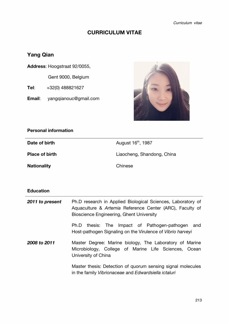
Curriculum vitae
213
CURRICULUM VITAE
Yang Qian
Address: Hoogstraat 92/0055,
Gent 9000, Belgium
Tel: +32(0) 488821627
Email: [email protected]
Personal information
Date of birth August 16th, 1987
Place of birth Liaocheng, Shandong, China
Nationality Chinese
Education
2011 to present Ph.D research in Applied Biological Sciences, Laboratory of Aquaculture & Artemia Reference Center (ARC), Faculty of Bioscience Engineering, Ghent University
Ph.D thesis: The Impact of Pathogen-pathogen and Host-pathogen Signaling on the Virulence of Vibrio harveyi
2008 to 2011 Master Degree: Marine biology, The Laboratory of Marine Microbiology, College of Marine Life Sciences, Ocean University of China
Master thesis: Detection of quorum sensing signal molecules in the family Vibrionaceae and Edwardsiella ictaluri
Curriculum vitae
214
Publications
Qian Yang, Nguyen D Q Anh, Peter Bossier, Tom Defoirdt: Norepinephrine and
dopamine increase motility, biofilm formation, and virulence of Vibrio harveyi. Frontiers in Microbiology 11/2014; 5:584.
Qian Yang, Tom Defoirdt: Quorum sensing positively regulates flagellar motility in
pathogenic Vibrio harveyi. Environmental Microbiology 02/2014;
Qian Yang, Yin Han, Nguyen Thi Ngoc Tinh, Nguyen Thi Hien, Peter Bossier: Detection of Quorum Sensing Signal Molecules in Edwardsiella ictaluri Ei-151. Indian Journal of Microbiology 12/2012; 52(4).
Qian Yang, Y Han, X-H Zhang: Detection of quorum sensing signal molecules in the
family Vibrionaceae. Journal of Applied Microbiology 03/2011; 110(6):1438-48.
Xuan Li, Qian Yang, Kristof Dierckens, Debra L Milton, Tom Defoirdt: RpoS and
Indole Signaling Control the Virulence of Vibrio anguillarum towards Gnotobiotic Sea
Bass (Dicentrarchus labrax) Larvae. PLoS ONE 10/2014; 9(10):e111801.
Xuan Li, Tom Defoirdt, Qian Yang, Stanislas Laureau, Peter Bossier, Kristof Dierckens: Host-induced increase in larval sea bass mortality in a gnotobiotic
challenge test with Vibrio anguillarum.. Diseases of Aquatic Organisms 04/2014; 108(3):211-6.
Yongxia Li, Shulin Yan, Qian Yang, Zizhong Qi, Xiao-Hua Zhang, Yu-Bin Fu: Algoriphagus faecimaris sp. Nov., isolated from coastal sediment. International Journal of Systematic and Evolutionary Microbiology 12/2011; 61(Pt 12):2856-60.
Yin Han, Chun-Li Yang, Qian Yang, Zizhong Qi, Wenzheng Liu, Zhi-Hong Xu, Wei-Ming Zhu, Peter Bossier, Xiao-Hua Zhang: Mutation of tryptophanase gene tnaA
in Edwardsiella tarda reduces lipopolysaccharide production, antibiotic resistance
2004 to 2008 Bachelor Degree: Bioscience, College of Life Sciences, Liaocheng University
Curriculum vitae
215
and virulence. Environmental Microbiology Reports 10/2011; 3(5):603-12.
Xuan Li, Yin Han, Qian Yang, Xiao-Hua Zhang: Detection of quorum sensing signal
molecules and mutation of luxS gene in Vibrio ichthyoenteri. Research in Microbiology 10/2009; 161(1):51-7.
Participation to international conference and training course
Larvi 2013 conference, Ghent, Belgium (2-5/09/2013) The impact of quorum sensing on motility in Vibrio harveyi (Poster presentation)
AQUA EUROPE 2014 conference, San Sebastián, Spain (14-17/10/2014) Quorum sensing in Vibrio harveyi positively regulates flagellar motility, an essential virulence factor (Oral presentation)
International Symposium on Quorum Sensing Inhibition, Santiago, Spain (3-5/06/2015) Novel quorum sensing-disrupting thiophenones with a promising potential to treat vibriosis in aquaculture. (Oral presentation)
MSc Students Supervised
Name: NGUYEN DUC QUYNH ANH Degree MSc in Aquaculture, Faculty of Bio-engineering, Ghent
University, Belgium
Title The impact of host stress hormones on luminescent vibriosis in brine shrimp (Artemia franciscana)


