Embed Size (px)
Citation preview
Review
Consequences of changes in BDNF levels on serotonin neurotransmission,5-HT transporter expression and function: Studies in adult mice hippocampus
Thierry Deltheil a,b, Bruno P. Guiard a,b, Jean-Philippe Guilloux a,b, Lorelei Nicolas a,b,Claudine Deloménie b, Christelle Repérant a,b, Erwan Le Maitre c,
Isabelle Leroux-Nicollet c, Saloua Benmansour d,François Coudoré a,b, Denis J. David a,b,
Alain M. Gardier a,b,!
a Univ. Paris Sud, EA 3544, Fac. Pharmacie, Chatenay-Malabry cedex F-92296, Franceb IFR141, Faculté de Pharmacie, Univ. Paris-Sud, 5 Rue J.-B. Clément, 92296 Chatenay-Malabry cedex, France
c FRE 2735 CNRS, IFRMP 23, UFR de Médecine et de Pharmacie, 22 Boulevard Gambetta, 76183 Rouen cedex1, Franced Department of Pharmacology, University of Texas Health Science Center at San Antonio, Texas 78229, USA
Available online 3 October 2007
Abstract
In vivo intracerebral microdialysis is an important neurochemical technique that has been applied extensively in genetic and pharmacological studiesaimed at investigating the relationship between neurotransmitters. Among themain interests ofmicrodialysis application is the infusion of drugs throughthemicrodialysis probe (reverse dialysis) in awake, freelymoving animals. As an example of the relevance of intracerebralmicrodialysis, this reviewwillfocus on our recent neurochemical results showing the impact of Brain-DerivedNeurotrophic Factor (BDNF) on serotonergic neurotransmission in basaland stimulated conditions. Indeed, although the elevation of 5-HToutflow induced by chronic administration of selective serotonin reuptake inhibitors(SSRIs) causes an increase in BDNF protein levels and expression (mRNA) in the hippocampus of rodents, the reciprocal interaction has not beendemonstrated yet. Thus, the neurochemical sight of this question will be addressed here by examining the consequences of either a constitutive decreaseor increase in brain BDNF protein levels on hippocampal extracellular levels of 5-HT in conscious mice.© 2007 Elsevier Inc. All rights reserved.
Keywords: Antidepressant drugs; Serotonin; BDNF; Neurogenesis; Genetically modified animals; Conventional microdialysis; Zero net flux method of quantitativemicrodialysis
Contents
1. Introduction . . . . . . . . . . . . . . . . . . . . . . . . . . . . . . . . . . . . . . . . . . . . . . . . . . . . . . . . . . . . . . 1752. Microdialysis: principles and methodology . . . . . . . . . . . . . . . . . . . . . . . . . . . . . . . . . . . . . . . . . . . . . . 175
2.1. Mice . . . . . . . . . . . . . . . . . . . . . . . . . . . . . . . . . . . . . . . . . . . . . . . . . . . . . . . . . . . . . . 1752.2. Conventional intracerebral microdialysis . . . . . . . . . . . . . . . . . . . . . . . . . . . . . . . . . . . . . . . . . . . 1762.3. Zero net flux method of quantitative intracerebral microdialysis . . . . . . . . . . . . . . . . . . . . . . . . . . . . . . . 1762.4. Statistical analysis . . . . . . . . . . . . . . . . . . . . . . . . . . . . . . . . . . . . . . . . . . . . . . . . . . . . . . . 177
3. Strategy 1: effects of decreasing BDNF levels on 5-HT neurotransmission in the hippocampus. Comparison ofBDNF+/+versus adult BDNF+/!mice . . . . . . . . . . . . . . . . . . . . . . . . . . . . . . . . . . . . . . . . . . . . . . . . . 1783.1. Basal levels with the zero net flux method of quantitative microdialysis . . . . . . . . . . . . . . . . . . . . . . . . . . . 1783.2. 5-HT transporter activity: [3H]5-HT uptake in hippocampal synaptosomes from BDNF+/+versus BDNF+/!mice . . . . . . . . 1783.3. Basal extracellular 5-HT levels with conventional intracerebral microdialysis in mice . . . . . . . . . . . . . . . . . . . . 178
Available online at www.sciencedirect.com
Pharmacology, Biochemistry and Behavior 90 (2008) 174–183www.elsevier.com/locate/pharmbiochembeh
! Corresponding author. Univ. Paris Sud, EA 3544, Fac. Pharmacie, Chatenay-Malabry cedex F-92296, France. Tel.: +33 1 46 83 54 16; fax: +33 1 46 83 53 55.E-mail address: [email protected] (A.M. Gardier).
0091-3057/$ - see front matter © 2007 Elsevier Inc. All rights reserved.doi:10.1016/j.pbb.2007.09.018
3.4. Paroxetine-induced changes in hippocampal 5-HT levels in mice . . . . . . . . . . . . . . . . . . . . . . . . . . . . . . 1783.5. [3H]citalopram binding site densities to hippocampal slices in mutant BDNF+/!mice . . . . . . . . . . . . . . . . . . . . 1783.6. 5-HT transporter expression: SERT mRNA levels in the brain stem of BDNF+/+versus BDNF+/!mice . . . . . . . . . . 179
4. Strategy 2: effects of increasing BDNF levels on dialysate 5-HT in the hippocampus of adult wild-type BDNF+/+mice . . . . . . . 1794.1. Effects of local intra-hippocampal BDNF injection . . . . . . . . . . . . . . . . . . . . . . . . . . . . . . . . . . . . . . 1794.2. Effects of local intra-hippocampal BDNF injection on paroxetine-induced changes in hippocampalextracellular 5-HT levels . . . . . . . . . . . . . . . . . . . . . . . . . . . . . . . . . . . . . . . . . . . . . . . . . . . . . . . 179
5. Final remarks . . . . . . . . . . . . . . . . . . . . . . . . . . . . . . . . . . . . . . . . . . . . . . . . . . . . . . . . . . . . . 1796. Conclusion . . . . . . . . . . . . . . . . . . . . . . . . . . . . . . . . . . . . . . . . . . . . . . . . . . . . . . . . . . . . . . 181Acknowledgements . . . . . . . . . . . . . . . . . . . . . . . . . . . . . . . . . . . . . . . . . . . . . . . . . . . . . . . . . . . . 182References . . . . . . . . . . . . . . . . . . . . . . . . . . . . . . . . . . . . . . . . . . . . . . . . . . . . . . . . . . . . . . . . . 182
1. Introduction
Most of the antidepressants such as Selective SerotoninReuptake Inhibitors (SSRI) act as indirect agonists of monoaminereceptors. While SSRI drugs produce relatively rapid blockade ofserotonin (5-HT) transporters (SERT) in vitro, the onset of clinicalbenefits usually takes several (4–6) weeks to occur. This gap intiming between SSRI near-immediate effect on neurotransmittersystems and the slow symptomatic recovery is a paradox that hasnot been completely solved yet. At presynpatic level, SSRI-inducedblockade of SERTresults in a rapid suppression of the firing activityof 5-HT neurons in the brainstem (Blier, 2001). Consequently,despite the 5-HT reuptake inhibition also taking place at nerveterminals, there is a decrease in 5-HT release via activation of 5-HT1A (somatodendritic) or 5-HT1B (nerve terminal) autoreceptors(Rutter et al., 1995). Thus, depending on the brain area, only a smallincrease or no change at all in the synaptic availability of 5-HToccurs (Romero et al., 1996;Malagie et al., 1996). As the treatmentis prolonged, a robust and time-dependent downregulation of the 5-HT transporter SERT is observed (Pineyro et al., 1994;Benmansour et al., 2002), while 5-HT1A autoreceptors graduallydesensitize leading to a progressive recovery to normal of the firingrate of 5-HT neurons as well as to an increased 5-HTneurotransmission in synpases (Blier et al., 1986; Chaput et al.,1986; El Mansari et al., 2005). At post-synaptic levels, a growthfactor, the Brain-Derived Neurotrophic Factor (BDNF), requiresactivation of the high-affinity protein kinase receptor family TrkB(Tropomyosine-related kinase B) to exert its biological effects. Theproperties of BDNF are different according to the brain regionstudied: for example, it regulates synaptic plasticity in the adultvisual cortex (Tsanov and Manahan-Vaughan, 2007). In addition,BDNF regulates lipid biosynthesis (Suzuki et al., 2007). In the adulthippocampus, BDNF might be involved in this delay of onset ofSSRI. Indeed, chronic, but not acute, SSRI treatment by increasing5-HT neurotransmission causes an increase in BDNF protein levelsand expression (mRNA)most notably in the dentate gyrus granularcell layer of the hippocampus in adult rats (Nibuya et al., 1995,1996) and mice (Santarelli et al., 2003). Thus, a positive regulationof 5-HTon the expression of the gene coding for BDNFmay occurin adult hippocampus. These effects could be related to increases inneurogenesis, i.e., ability of progenitors cells to differentiate intoneurons or glia cells (Malberg et al., 2000). This cascade of eventsmay contribute to the therapeutic effects of antidepressant drugs.However, the actual knowledge regarding the relationship betweenBDNF and serotonin (5-HT) in the hippocampus is limited. For
example, is there any reciprocal effect of BDNF on 5-HTneurotransmission? To answer this question, we have developeda dual experimental strategy by inducing either a decrease or anincrease in BDNF protein levels.
First, we studied the SSRI response in heterozygousBDNF+/!mice, in which brain BDNF protein levels are decreased byhalf (Korte et al., 1995). These constitutive BDNF+/!micedevelop enhanced inter-male aggressiveness and hyperphagiaaccompanied by significant weight gain in early adulthood; thesebehavioral abnormalities are known to correlate with 5-HT dys-function (Lyons et al., 1999).
Second, we increased BDNF protein levels by its localinfusion into adult hippocampus by reverse microdialysis in wild-type mice. Indeed, it was found that BDNF increases activity ofbrainmonoaminergic systems in rats (Siuciak et al., 1996). BDNFinfusion into the forebrain results in an elevation of 5-HTneuronalfiber density and also protects serotoninergic neurons fromneurotoxic damage in rats (Mamounas et al., 1995). In anotherstudy, intra-hippocampal BDNF injection induces an antidepres-sant-like effect in rats that was dose-dependent (a dose as low as0.25 !g of BDNF induced it), was observed 3 days and lasted upto 10 days after its bilateral injection (Shirayama et al., 2002).
In the present study, both in vivo conventional and quantitativeintracerebral microdialysis studies have been performed in thesetwo animal models, and wemeasured extracellular levels of 5-HTin the adult hippocampus of awake, freely moving mice.
2. Microdialysis: principles and methodology
2.1. Mice
Male wild-type BDNF+/+and heterozygous mutant BDNF+/!mice (3 to 4 months of age and 25–30 g in body weight) were bredon a mixed S129/Sv!C57BL/6 genetic background (Korte et al.,1995) and raised at the animal facility of the university of Paris XI(Chatenay-Malabry, France). Heterozygous adult mice with onefunctional BDNF allele (BDNF+/!) exhibited reduced BDNFprotein levels in the hippocampus (data not shown). All animalswere genotyped by polymerase chain reaction (Guiard et al., 2007).
For local BDNF injection, adult male 129S6/SvEvTac wild-type mice (Taconic Farms, Ry, Denmark) were used. All micewere 7–8 weeks old, weighed 23–25 g, and were housed ingroups of 6 mice per cage under standard conditions (12:12 hlight–dark cycle, 22±1 °C ambient temperature, 60% relativehumidity, food and water ad libitum).
175T. Deltheil et al. / Pharmacology, Biochemistry and Behavior 90 (2008) 174–183
2.2. Conventional intracerebral microdialysis
Concentric dialysis probes (0.30 mm outer diameter) wereconstructed of cuprophane and set up as described previously(Guiard et al., 2004; Malagie et al., 2001). Probes were implantedinto the ventral hippocampus (active length of 1.6 mm) inanesthetized mice (chloral hydrate, 400 mg/kg, intraperitoneally,i.p.) according to the mouse brain atlas of Paxinos and Franklin(2001). The stereotaxic coordinates from Bregma (in mm) were:A=!3.4, L=3.4, V=4.0. The next day, after recovery fromsurgery, probes were continuously perfused with artificialcerebrospinal fluid (aCSF) in awake animals at a flow rate of1.5 !l/min. Dialysate samples were collected every 15 min forthe measurement of 5-HT by using high-performance liquidchromatography coupled to an amperometric detector (1049A,Hewlett-Packard, Les Ulis, France). The limit of sensitivity for5-HTwas"0.5 fmol/sample (signal-to-noise ratio 2). After 1 hof stabilization necessary to reach uniform concentrations of5-HT in dialysates, four samples were collected to measurebasal extracellular 5-HT values. Drugs were then injected
intraperitoneally (paroxetine 4 and/or 8 mg/kg) at t=0 andsubsequent dialysate fractions were collected. BDNF wasdissolved in artificial cerebrospinal fluid (aCSF) (compositionin mM: NaCl 147, KCl 3.5, CaCl2 1.26, MgCl2 1.2, NaH2PO4
1.0, NaHCO3 25.0, pH 7.4±0.2) and administered locally intothe ventral hippocampus (vHi) via a silica catheter glued to themicrodialysis probe [(0.2 !L/min for 2 min by using a Picoplusmicroinjector (Harvard Apparatus, Les Ulis, France)], at the doseof 20 and 100 ng. The tyrosine kinase inhibitor, K252a (10 !M)was dissolved in aCSF containing 0.1% of dimethyl sulfoxide(DMSO), respectively, and was locally perfused by ‘reversemicrodialysis’ at a flow rate of 1.5 !L/min. For each experiment,control group received the appropriate vehicle.
2.3. Zero net flux method of quantitative intracerebralmicrodialysis
Four samples were collected to determine basal hippocampal5-HT levels before local perfusion of increasing concentrationsof 5-HT (0, 5, 10 and 20 nM). The dialysate 5-HTconcentrations
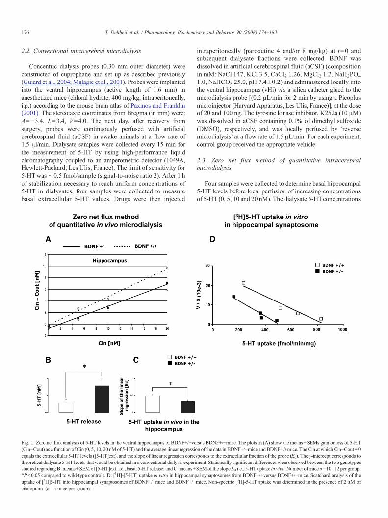
Fig. 1. Zero net flux analysis of 5-HT levels in the ventral hippocampus of BDNF+/+versus BDNF+/!mice. The plots in (A) show the means±SEMs gain or loss of 5-HT(Cin–Cout) as a function of Cin (0, 5, 10, 20 nMof 5-HT) and the average linear regression of the data in BDNF+/!mice andBDNF+/+mice. TheCin atwhich Cin–Cout=0equals the extracellular 5-HT levels ([5-HT]ext), and the slope of linear regression corresponds to the extracellular fraction of the probe (Ed). The y-intercept corresponds totheoretical dialysate 5-HT levels that would be obtained in a conventional dialysis experiment. Statistically significant differences were observed between the two genotypesstudied regardingB:means±SEMof [5-HT]ext, i.e., basal 5-HT release; andC:means±SEMof the slopeEd i.e., 5-HTuptake in vivo. Number of mice n=10–12 per group.!Pb0.05 compared to wild-type controls. D: [3H]-[5-HT] uptake in vitro in hippocampal synaptosomes from BDNF+/+versus BDNF+/!mice. Scatchard analysis of theuptake of [3H]5-HT into hippocampal synaptosomes of BDNF+/+mice and BDNF+/!mice. Non-specific [3H]-5-HT uptake was determined in the presence of 2 !M ofcitalopram. (n=5 mice per group).
176 T. Deltheil et al. / Pharmacology, Biochemistry and Behavior 90 (2008) 174–183
(Cout) obtained during perfusion of the various concentrationsof 5-HT (Cin) were used to construct a linear regression curve foreach animal (Guiard et al., 2007). The net change in 5-HT (Cin–Cout) was plotted on the y-axis against Cin on the x-axis.Extracellular 5-HT levels ([5-HT]ext) and the extraction fractionof the probe (Ed) were determined as described by Parsonset al. (1991). The concentration of 5-HT in the extracellularspace [5-HT]ext is estimated from the concentration at whichCin–Cout=0 and corresponds to a point at which there is no netdiffusion of 5-HT across the dialysis membrane. The extractionfraction (Ed) is the slope of the linear regression curve and hasbeen shown to provide an estimate of changes in transporter-mediated 5-HT uptake (Gardier et al., 2003; Parsons et al., 1991).
2.4. Statistical analysis
All data are reported as means±SEMs. Following linearregression of the data for each animal in the zero net fluxmicrodialysis experiments, unpaired two-tailed Student's t-testswere used to assess the effects of genotype on extracellular levelsof 5-HT in the hippocampus and Ed. For conventionalmicrodialysis experiments (paroxetine i.p.; BDNF injection),statistical analyses were performed on areas under the curve
(AUC) values for the amount of 5-HT outflow collected duringthe 0–120 min post-treatment period. To compare different AUCvalues in each group of mice, a one-way ANOVAwith treatmentfactor followed by Fischer protected least significance difference(PLSD) post-hoc test was conducted. In addition, basal 5-HTlevels in the ventral hippocampus across groups of mice involvedin conventional microdialysis studies have been compared byusing a Student's t-test.
For the 5-HT uptake experiments performed in hippocampalsynaptosomes, results were analyzed by non-linear regressionand the uptake capacity (Vmax) and Km of [3H]-5-HT werecalculated. Then, two-way ANOVA was performed with 5-HTconcentration as a within-subject variable and genotype as abetween-subject variable.
For [3H]-citalopram autoradiography study, the opticaldensity of selected brain region was measured and convertedinto fmol/mg tissue using the standard curve. Non-specificbinding was subtracted from total binding to evaluate specificbinding in each brain region of each animal. Measurements weremade on three sections from each brain region, and the valueswere averaged for each animal. The values for each region foreach animal were then analyzed by Student's t-test fordifferences between genotypes.
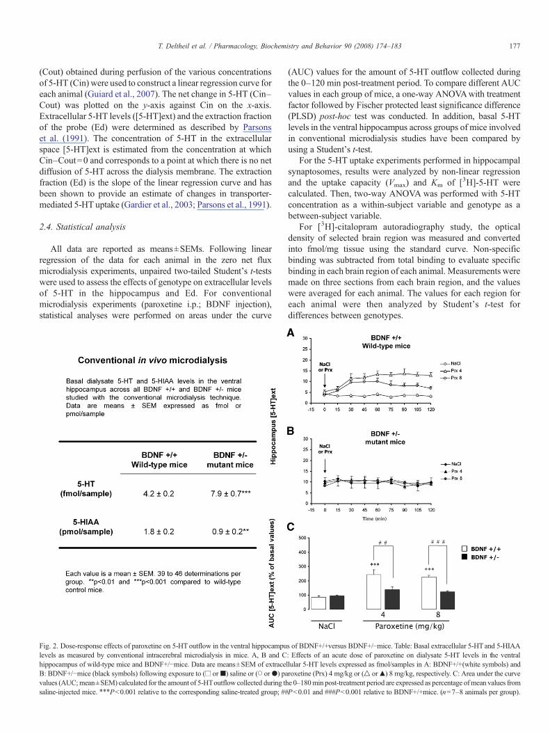
Fig. 2. Dose-response effects of paroxetine on 5-HT outflow in the ventral hippocampus of BDNF+/+versus BDNF+/!mice. Table: Basal extracellular 5-HT and 5-HIAAlevels as measured by conventional intracerebral microdialysis in mice. A, B and C: Effects of an acute dose of paroxetine on dialysate 5-HT levels in the ventralhippocampus of wild-type mice and BDNF+/!mice. Data are means±SEM of extracellular 5-HT levels expressed as fmol/samples in A: BDNF+/+(white symbols) andB: BDNF+/!mice (black symbols) following exposure to (! or") saline or (# or$) paroxetine (Prx) 4 mg/kg or (% or&) 8 mg/kg, respectively. C: Area under the curvevalues (AUC;mean±SEM) calculated for the amount of 5-HToutflow collected during the 0–180min post-treatment period are expressed as percentage ofmean values fromsaline-injected mice. !!!Pb0.001 relative to the corresponding saline-treated group; ##Pb0.01 and ###Pb0.001 relative to BDNF+/+mice. (n=7–8 animals per group).
177T. Deltheil et al. / Pharmacology, Biochemistry and Behavior 90 (2008) 174–183
For all data, significant level was set at P# 0.05. All analyseswere conducted using a Statview 5.0 (JMP Software, Cary, NC).
3. Strategy 1: effects of decreasing BDNF levels on 5-HTneurotransmission in the hippocampus. Comparison ofBDNF+/+versus adult BDNF+/!mice
3.1. Basal levels with the zero net flux method of quantitativemicrodialysis
The zero net flux method of quantitative microdialysis wasused to evaluate basal extracellular 5-HT levels in the ventralhippocampus of BDNF+/!and BDNF+/+mice. In a recentreport, we demonstrated that the extracellular 5-HT levelscorrected for in vivo recovery were significantly higher inBDNF+/!mice compared to wild-type mice (Fig. 1A) (Guiardet al., 2007). Thus, constitutive deletion of a single copy of theBDNF gene is associated with an increase in basal 5-HT levelsin the ventral hippocampus. This effect may reflect eitherincrease in hippocampal 5-HT release and/or decrease in 5-HTuptake in vivo. Previous studies have shown that manipulationsthat decrease neurotransmitter uptake also decrease the recoveryof neurotransmitter from the tissue as reflected in the extractionfraction, Ed (Parsons et al., 1991). In agreement with anelevated basal extracellular 5-HT concentrations, BDNF+/!mice exhibited a significantly lower Ed compared to wild-typemice Fig. 1B and C).
3.2. 5-HT transporter activity: [3H]5-HTuptake in hippocampalsynaptosomes from BDNF+/+versus BDNF+/!mice
In vitro [3H]-5-HT uptake by synaptosomes prepared fromthe hippocampus was decreased in BDNF+/!mice compared toBDNF+/+mice (Fig. 1D). Constitutive reductions in BDNFaffected Vmax (528±32 vs. 942±59 pmol/mg protein per min inBDNF+/!mice and BDNF+/+mice, respectively, P=0.007),but was without significant effect on Km values for [3H]-5-HTuptake (35±3 vs. 59±11 nM in BDNF+/!mice and BDNF+/+mice, respectively, PN0.05; Guiard et al., 2007).
3.3. Basal extracellular 5-HT levels with conventionalintracerebral microdialysis in mice
Conventional microdialysis data confirmed that constitutivedecreases in BDNF expression also produce an elevation in basal
dialysate 5-HT concentrations (Table in Fig. 2). In addition,dialysate levels of its major metabolite, 5-hydroxyindoleaceticacid (5-HIAA) were significantly reduced in the ventral hippo-campus in BDNF+/!mice compared to BDNF+/+mice (P=0.01;Guiard et al., 2007).
3.4. Paroxetine-induced changes in hippocampal 5-HT levelsin mice
In the ventral hippocampus of BDNF+/+mice extracellular5-HT levels were increased by paroxetine administered at thedose of 4 mg/kg compared to the corresponding group of wild-type mice treated with vehicle (Fig. 2A and C). In BDNF+/!mice, extracellular 5-HT levels were not affected by paroxetineneither at 4 mg/kg nor at 8 mg/kg compared to thecorresponding group of mutant mice treated with vehicle(Fig. 2B and C). Interestingly, the neurochemical effects ofparoxetine did not differ between BDNF+/!and BDNF+/+micein the frontal cortex and the dorsal raphe nucleus, both regionsexpressing SERT protein (Guiard et al., 2007).
3.5. [3H]citalopram binding site densities to hippocampalslices in mutant BDNF+/!mice
To further explore the underlying causes of the decreases inextraction fraction in hippocampal 5-HT reported above forBDNF+/!mice, we performed autoradiography in the hippocam-pus of theses mutants. Examination of [3H]-citalopram bindingsite densities revealed a significant reduction in the number of[3H]-citalopram binding sites in the ventral hippocampus ofBDNF+/!mutants compared to BDNF+/+mice. In particular, asignificant decrease in [3H]-citalopram binding sites wasmeasured in the CA3 (Pb0.01, Table 1), but not in the dentategyrus and CA1 (PN0.05, Table 1) sub-regions of the hippocam-pus in BDNF+/!mutants compared to BDNF+/+mice. As well,no differences in the density of the labelingwere noted in the otherbrain regions such as frontal cortex, striatum and raphe nucleiwith respect to genotype (Guiard et al., 2007).
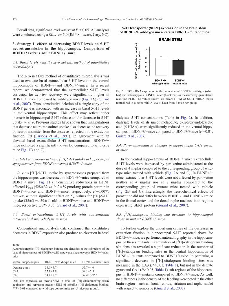
Fig. 3. SERT mRNA expression in the brain stem of BDNF+/+wild-type (whitebar) and heterozygous BDNF+/!mice (black bar) as measured by quantitativereal-time PCR. The values shown are means±SEM of SERT mRNA levelsnormalized to "-actin mRNA levels. Data from 7 mice per group.
Table 1Autoradiographic [3H]-citalopram binding site densities in the subregions of theventral hippocampus of BDNF+/+wild-type versus heterozygous BDNF+/!adultmice
Ventral hippocampus BDNF+/+wild-type mice BDNF+/-mutant mice
Dentate gyrus 34.6±3.7 35.7±4.6CA1 37.1±1.8 34.1±2.5CA3 74.4±2.7 59.4±3.7!!
Data are expressed as mean±SEM in fmol of [3H]-citalopram/mg tissueequivalent and represent means±SEM of specific [3H]-citalopram binding.!!Pb0.01 compared to wild-type control mice (n=5 mice per group).
178 T. Deltheil et al. / Pharmacology, Biochemistry and Behavior 90 (2008) 174–183
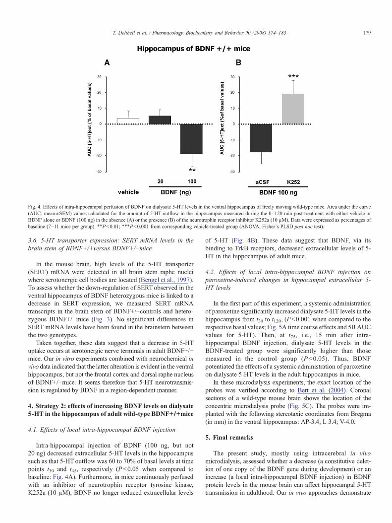
3.6. 5-HT transporter expression: SERT mRNA levels in thebrain stem of BDNF+/+versus BDNF+/!mice
In the mouse brain, high levels of the 5-HT transporter(SERT) mRNA were detected in all brain stem raphe nucleiwhere serotonergic cell bodies are located (Bengel et al., 1997).To assess whether the down-regulation of SERTobserved in theventral hippocampus of BDNF heterozygous mice is linked to adecrease in SERT expression, we measured SERT mRNAtranscripts in the brain stem of BDNF+/+controls and hetero-zygous BDNF+/!mice (Fig. 3). No significant differences inSERT mRNA levels have been found in the brainstem betweenthe two genotypes.
Taken together, these data suggest that a decrease in 5-HTuptake occurs at serotonergic nerve terminals in adult BDNF+/!mice. Our in vitro experiments combined with neurochemical invivo data indicated that the latter alteration is evident in the ventralhippocampus, but not the frontal cortex and dorsal raphe nucleusof BDNF+/!mice. It seems therefore that 5-HT neurotransmis-sion is regulated by BDNF in a region-dependent manner.
4. Strategy 2: effects of increasing BDNF levels on dialysate5-HT in the hippocampus of adult wild-type BDNF+/+mice
4.1. Effects of local intra-hippocampal BDNF injection
Intra-hippocampal injection of BDNF (100 ng, but not20 ng) decreased extracellular 5-HT levels in the hippocampussuch as that 5-HT outflow was 60 to 70% of basal levels at timepoints t30 and t45, respectively (Pb0.05 when compared tobaseline: Fig. 4A). Furthermore, in mice continuously perfusedwith an inhibitor of neurotrophin receptor tyrosine kinase,K252a (10 !M), BDNF no longer reduced extracellular levels
of 5-HT (Fig. 4B). These data suggest that BDNF, via itsbinding to TrkB receptors, decreased extracellular levels of 5-HT in the hippocampus of adult mice.
4.2. Effects of local intra-hippocampal BDNF injection onparoxetine-induced changes in hippocampal extracellular 5-HT levels
In the first part of this experiment, a systemic administrationof paroxetine significantly increased dialysate 5-HT levels in thehippocampus from t30 to t120, (Pb0.001 when compared to therespective basal values; Fig. 5A time course effects and 5BAUCvalues for 5-HT). Then, at t75, i.e., 15 min after intra-hippocampal BDNF injection, dialysate 5-HT levels in theBDNF-treated group were significantly higher than thosemeasured in the control group (Pb0.05). Thus, BDNFpotentiated the effects of a systemic administration of paroxetineon dialysate 5-HT levels in the adult hippocampus in mice.
In these microdialysis experiments, the exact location of theprobes was verified according to Bert et al. (2004). Coronalsections of a wild-type mouse brain shows the location of theconcentric microdialysis probe (Fig. 5C). The probes were im-planted with the following stereotaxic coordinates from Bregma(in mm) in the ventral hippocampus: AP-3.4; L 3.4; V-4.0.
5. Final remarks
The present study, mostly using intracerebral in vivomicrodialysis, assessed whether a decrease (a constitutive delet-ion of one copy of the BDNF gene during development) or anincrease (a local intra-hippocampal BDNF injection) in BDNFprotein levels in the mouse brain can affect hippocampal 5-HTtransmission in adulthood. Our in vivo approaches demonstrate
Fig. 4. Effects of intra-hippocampal perfusion of BDNF on dialysate 5-HT levels in the ventral hippocampus of freely moving wild-type mice. Area under the curve(AUC; mean±SEM) values calculated for the amount of 5-HT outflow in the hippocampus measured during the 0–120 min post-treatment with either vehicle orBDNF alone or BDNF (100 ng) in the absence (A) or the presence (B) of the neurotrophin receptor inhibitor K252a (10 µM). Data were expressed as percentages ofbaseline (7–11 mice per group). !!Pb0.01; !!!Pb0.001 from corresponding vehicle-treated group (ANOVA, Fisher's PLSD post hoc test).
179T. Deltheil et al. / Pharmacology, Biochemistry and Behavior 90 (2008) 174–183

that both strategies led to changes in basal extracellular 5-HTlevels in the hippocampus. In both cases, decreasing or increasingBDNF protein levels altered 5-HT uptake, i.e., the 5-HTtransporter SERT function in the mouse adult hippocampus.
When BDNF gene expression was reduced (in heterozygousBDNF+/!mice), basal extracellular 5-HT levels increased atserotonergic nerve terminals in the hippocampus. Indeed, asexpected, a decrease in hippocampal SERT activity measured inthesemutants results in an increased basal extracellular 5-HT level,thus in an increase in 5-HT neurotransmission. In addition, anacute systemic administration of a SSRI, paroxetine, becameinactive in the ventral hippocampus and 5-HT uptake in vitro (insynaptosomes) aswell as in vivo (zero net flux) was blunted. Thesechanges were not detected at serotonergic nerve terminal regionssuch as the frontal cortex and striatum of adult heterozygousBDNF+/!mice (Szapacs e al., 2004). These latter results are notsurprising since the striatum is not a brain region involved inadult neurogenesis and changes in BDNF levels followingantidepressant drug treatment. The fact that the constitutivedecrease in brainBDNF levels alters the effects of paroxetine in theventral hippocampus, but neither in the frontal cortex nor in thedorsal raphe nucleus, strongly supports the region-specificalteration of the serotonin transporter SERT in mice.
We extended this observation by applying intracerebral in vivomicrodialysis in the vicinity of cell bodies of 5-HTneurons locatedin the dorsal raphe nucleus (Guiard et al., 2007). A reduction inSERT function rather than in SERT densities occurred in the adulthippocampus (Guiard et al., 2007). Changes in SERT mRNAexpression in the brain stem of these mutant mice cannot accountfor these changes. Furthermore, they were not associated with afunctional desensitization of 5-HT1A autoreceptors in the raphenuclei since the capacity of a 5-HT1A receptor agonist to decreaseeither raphe 5-HT neuronal activity or the body temperature wasunchanged in BDNF+/!mice compared to their wild-typelittermates (Guiard et al., 2007). These results suggest thatBDNF is necessary for an appropriate uptake of 5-HT to occur atserotonergic nerve terminals in the hippocampus of adult mice.The effect of BDNF in the presence of paroxetine could beattributed to an increase in hippocampal 5-HT release.
As expected, when BDNF protein levels were increased (bylocal intra-hippocampal BDNF injection in wild-type mice), basalextracellular 5-HT levels decreased at serotonergic nerve terminalsin the hippocampus. These effects are selective and depend on theactivation of TrkB receptors since theywere blocked byK252a, aninhibitor of neurotrophin receptor tyrosine kinase.
The hypothesis that decreases in Ca2+-dependent release of5-HT or increases in 5-HT uptake may be responsible for thisdecrease must be further investigated. Preliminary data obtainedin rats (Benmansour et al., Soc. For Neurosci. Atlanta, USA,2006) suggest that BDNF has neither effect on the affinity of aSSRI for SERT binding sites nor on SERT density in CA3region of adult hippocampus. In addition, the effects of an acutesystemic administration of paroxetine on dialysate 5-HT waspotentiated by BDNF injection in the hippocampus. Thus, inthis case, SERT the main target of this antidepressant drug isselectively inhibited by this SSRI. BDNF further increased theamount of 5-HT in hippocampal synapses.
Fig. 5. Effects of intra-hippocampal perfusion of BDNF on paroxetine-inducedincreases in extracellular 5-HT levels in the ventral hippocampus of freely movingwild-type mice. Basal dialysates 5-HT levels in the vHi of mice treated did notsignificantly differ between these groups of mice [(in fmol/20 !L) (mean±SEM)4.15±0.67 (n=7); 3.66±0.5 (n=9) for protein/vehicle and paroxetine/BDNFrespectively] (F (1,14)=0.348, PN0.05). A: Time course: Data are means±SEMof dialysate 5-HTexpressed as percentages of basal values. Mice received (arrow)either paroxetine (4 mg/kg; i.p.)/vehicle (!) or paroxetine (4 mg/kg; i.p.)/BDNF(100 ng) ("). B: Area under the curve (AUC; mean±SEM) values calculated forthe amount of 5-HT outflow in the vHi measured after the perfusion of eithervehicle or BDNF 60–120 min (B) post treatment period and expressed aspercentages of baseline (7–11 mice per group). !Pb0.05; from correspondingvehicle-treated group (Two-way ANOVA, Fisher's PLSD post hoc test). i.p.intraperitoneal. @Pb0.05 significantly different from paroxetine/vehicle treatedgroup at the corresponding time (Two-way ANOVA, Fisher's PLSD post hoc test)§§§Pb0.001 from t0 to t120 from baseline value for each group (ANOVA forrepeated measures, Fisher's PLSDpost hoc test). C: Coronal section of a wild-typemouse brain showing the location of the microdialysis probe according to Paxinosand Franklin (2001). The probes were implanted in the ventral hippocampus. Thelength of the black bar corresponds to 1 mm. The arrow indicates the tip of themicrodialysis membrane.
180 T. Deltheil et al. / Pharmacology, Biochemistry and Behavior 90 (2008) 174–183
Recent reports suggest that neurogenesis is associated withchronic, but not acute, administration of all various types ofantidepressant drugs in both the subventricular zone and adulthippocampus. At the present time, increases in various phasesof neurogenesis (cell proliferation, migration, differentiation,survival of newly formed neurons and synaptogenesis) wereobserved in adult hippocampus following chronic treatmentwith antidepressant drugs such as tranylcypromine, reboxetine,(Malberg et al., 2000); fluoxetine (Malberg et al., 2000; Manevet al., 2001; Santarelli et al., 2003); tricyclic antidepressant drugs,e.g., imipramine, desipramine (Santarelli et al., 2003; Chen et al.,2006a; Holick et al., 2007); the CRF(1) receptor antagonistSSR125543A and the V(1b) receptor antagonist SSR149415(Alonso et al., 2004); agomelatine (Banasr et al., 2006); MCHR1receptor antagonist, SNAP 94847 (David et al., 2007).
Similarly, intra-hippocampal BDNF infusion for two weeksincreased neurogenesis of granule cells in the dentate gyrus ofthe hippocampus in adult rats (Scharfman et al., 2005).
There is a great deal of interest in neurotrophin therapy toprevent neurodegenerative diseases as well as to treat mooddisorders. However, we need first to investigate growth factors'effects in various animal models of anxiety-depression. Indeed,all the above described studies using SSRIs have been performedin normal animals. In mice subjected to the chronic mild stress(CMS) procedure, a model of depression with predictive validity(Alonso et al., 2004), repeated administration of fluoxetine(10 mg/kg/day i.p. for 28 days) significantly reversed the reduc-tion of cell proliferation produced by CMS. This result suggeststhat clinically effective antidepressant drugs affect plasticitychanges in the hippocampal formation. Furthermore, the largesize of neurotrophin and the blood–brain barrier represent majorhurdles in the use of peptide therapeutics. Intracerebral micro-dialysis is a key technique allowing to overcome some of thesedifficulties.
Taken together, the microdialysis technique gave symmet-rical results between the two strategies. BDNF can modulate theamount of 5-HT in synapses either by decreasing (inheterozygous BDNF+/!mice) or increasing (local BDNFinjection) the activity of SERT or 5-HT release or both.
According to Altar et al. (1997), neurotrophins can exertdifferent roles. First, their long distance retrograde signallingand participation to the development of the peripheral nervoussystem involves their retrograde transport from terminals to thecell bodies of neurons. A local action of BDNF in the adultcentral nervous system (as in the present study after its localinjection within the hippocampus of adult mice), involves theanterograde transport, for example, from neuron cell bodies totheir terminals, then is released to bind to its post-synapticreceptor on target cells (Altar et al., 1997). Endogenous BDNFis produced by neurons in the peripheral and central nervoussystems. BDNF protein and mRNA are distributed throughoutthe brain, thus suggesting that both retrograde and anterogradetransports are probably widespread. We can thus infer that, inour experimental conditions, exogenous BDNF either was takenup by presynaptic non-serotonergic neurons and was released insynapses, or acted directly on post-synaptic serotonergic nerveterminals located in the hippocampus, through its binding to
TrkB receptors. The post-synaptic localization of the full-length, active form of these receptors was already demonstratedin adult rat cerebral cortex and hippocampus (Wu et al., 1996).
BDNF is known to enhance synaptic neurotransmission inhippocampal neurons through TrkB receptor activation at excita-tory glutamatergic synapses and phosphorylation of post-synapticionotropic receptors such as N-methyl-D-asparate (NMDA) re-ceptors (Suen et al., 1997). BDNF thus modulates hippocampallong-term potentiation (LTP), a cellular and molecular model ofplasticity associated with learning and memory. However, intra-hippocampal injection of BDNF decreased rather than increasedclearance rate of 5-HT. How can BDNF decrease hippocampal 5-HT release here? The 5-HT outflow measured with the micro-dialysis technique is a balance between 5-HT uptake and itsrelease. Taken together, the data obtained with BDNF and co-administration ofBDNFwith paroxetine suggest that the effects ofBDNF on 5-HT uptake predominates over those on 5-HT release,thus leading to a decrease in dialysate 5-HT levels following asingle administration of BDNF.
According to monoaminergic hypothesis of depression anincrease in 5-HT levels lead to antidepressant-like activity inrodents. Since BDNF modulates 5-HT outflow would BDNFhave an antidepressant like activity in rodents? Previous studiesfound that infusion of BDNF into the brain produced antidepres-sant-like activity in various animal models of depression (Siuciaket al., 1997). The dose we used has more physiological relevancethan a higher dose since the total amount of BDNF in the hip-pocampus corresponds to a weight of about 150 ng/g wet weighttissue (Szapacs et al., 2004). In another study, intra-hippocampalBDNF injection in rats induced an antidepressant-like effect thatwas dose-dependent (a dose as low as 0.25 !g of BDNF inducedit), was observed 3 days and lasted up to 10 days after its bilateralinjection (Shirayama et al., 2002). Whether or not behaviouraland neurochemical responses could be associated with TrkBreceptor expression in particular hippocampal subfields, needs tobe investigated. In this latter study, the diffusion of BDNF fromthe site of injection was limited ($0.5 mm) and peak levels ofBDNF immunolabeling were observed 2 h after injection to rats.Multiple infusions gave an effect similar to that of a singleinjection (Shirayama et al., 2002). In the present study, maximaleffects of BDNFon dialysate 5-HTwas found in the hippocampus45min after injection tomice. Since BDNF potentiates the effectsof paroxetine on dialysate 5-HT, we are currently investigatingwhether a co-administration of BDNF and paroxetine lead toantidepressant-like activity in mice.
6. Conclusion
The present data may help better understand the physiopa-thology of depression and the mechanism of antidepressantefficacy as they link abnormalities of two distinct neurotrans-mitter systems (i.e. reduced BDNF expression and reduced 5-HT re-uptake). Thus, both SERT and BDNF may be implicatedin the mechanism of action of antidepressant drugs. SERT genepresents a polymorphism in its promoter region. Mice carryingone or two methionine allele corresponding to the humanmethionine BDNF gene polymorphism display anxiety like
181T. Deltheil et al. / Pharmacology, Biochemistry and Behavior 90 (2008) 174–183
behaviour (Chen et al., 2006b). Genetically modified animalshelp identifying interactions between SERT and BDNF, brainregions and neuronal pathways involved in these interactions:this research area might be fruitful to improve antidepressanttreatment and/or to discover new therapeutic targets. Today, it isquite difficult to consider BDNF as a treatment of depressivedisorders because of its peptidic structure, BDNF is rapidlydegraded by endopeptidases. Small non-peptidic TrkB receptoragonists that cross the blood brain barrier have to be designed.
Acknowledgements
During the performance of this work, B.P. G., J-P. G., T.D. andL.N. were recipients of a fellowship from the “Ministère del'Enseignement Supérieur et de la Recherche”. In addition, B.P. G.was a recipient of a fellowship from “La Fondation pour laRechercheMédicale”.We are grateful to GlaxoSmithKline for thegenerous gifts of paroxetine.
References
Alonso R, Griebel G, Pavone G, Stemmelin J, Le Fur G, Soubrie P. Blockade ofCRF(1) or V(1b) receptors reverses stress-induced suppression of neurogen-esis in a mouse model of depression. Mol Psychiatry 2004;9:278–86.
Altar CA, Cai N, Bliven T, Juhasz M, Conner JM, Acheson AL, et al.Anterograde transport of brain-derived neurotrophic factor and its role in thebrain. Nature 1997;389:856–60.
Banasr M, Soumier A, Hery M, Mocaer E, Daszuta A. Agomelatine, a newantidepressant, induces regional changes in hippocampal neurogenesis. BiolPsychiatry 2006;59:1087–96.
Bengel D, Johren O, Andrews AM, Heils A, Mossner R, Sanvitto GL, et al.Cellular localization and expression of the serotonin transporter in mousebrain. Brain Res 1997;778:338–45.
Benmansour S, Owens WA, Cecchi M, Morilak DA, Frazer A. Serotoninclearance in vivo is altered to a greater extent by antidepressant-induceddownregulation of the serotonin transporter than by acute blockade of thistransporter. J Neurosci 2002;22:6766–72.
Benmansour S, Piotrowski JP, Frazer A. BDNF inhibits the effect of an SSRI onserotonin clearanceSoc For Neurosci. Atlanta; 2006. poster 289.4.
Bert L, Favale D, Jego G, Greve P, Guilloux JP, Guiard BP, et al. Rapid andprecise method to locate microdialysis probe implantation in the rodentbrain. J Neurosci Methods 2004;140:53–7.
Blier P. Pharmacology of rapid-onset antidepressant treatment strategies. J ClinPsychiatry 2001;15:12–7.
Blier P, de Montigny C, Azzaro AJ. Modification of serotonergic andnoradrenergic neurotransmissions by repeated administration of monoamineoxidase inhibitors: electrophysiological studies in the rat central nervoussystem. J Pharmacol Exp Ther 1986;237:987–94.
Chaput Y, Blier P, de Montigny C. In vivo electrophysiological evidence for theregulatory role of autoreceptors on serotoninergic terminals. J Neurosci1986;6:2796–801.
Chen H, Pandey GN, Dwivedi Y. Hippocampal cell proliferation regulation byrepeated stress and antidepressants. NeuroReport. 2006a;17:863–7.
Chen ZY, Jing D, Bath KG, Ieraci A, Khan T, Siao CJ, et al. Genetic variantBDNF (Val66Met) polymorphism alters anxiety-related behavior. Science2006b;314:140–3.
David DJ, KlemenhagenKC, Holick KA, SaxeMD,Mendez I, Santarelli L, et al.Efficacy of the MCHR1 antagonist N-[3-(1-{[4-(3,4-difluorophenoxy)phenyl]methyl}(4-piperidyl))-4-methylphenyl]-2-methylpropanamide(SNAP 94847) in mouse models of anxiety and depression following acuteand chronic administration is independent of hippocampal neurogenesis.J Pharmacol Exp Ther 2007;321:237–48.
El Mansari M, Sánchez C, Chouvet G, Renaud B, Haddjeri N. Effects of acuteand long-term administration of escitalopram and citalopram on serotonin
neurotransmission: an in vivo electrophysiological study in rat brain.Neuropsychopharmacology 2005;30:1269–77.
Gardier AM, David DJ, Jego G, Przybylski C, Jacquot C, Durier S, et al. Effectsof chronic paroxetine treatment on dialysate serotonin in 5-HT1B receptorknockout mice. J Neurochem 2003;86:13–24.
Guiard BP, David DJP, Deltheil T, Chenu F, Le Maître E, Renoir T, et al. Brain-derived neurotrophic factor-deficient mice exhibit a hippocampal hyperser-otonergic phenotype. Int J NeuropsychoPharmacology 2007;11:1–14.
Holick KA, Lee DC, Hen R, Dulawa SC. Behavioral effects of chronicfluoxetine in BALB/cJ mice do not require adult hippocampal neurogenesisor the serotonin 1A receptor. Neuropsychopharmacology 2007 [11 Apr],[Electronic publication ahead of print].
Korte M, Carroll P, Wolf E, Brem G, Thoenen H, Bonhoeffer T. Hippocampallong-term potentiation is impaired in mice lacking brain-derived neuro-trophic factor. Proc Natl Acad Sci U S A 1995;92:8856–60.
Lyons WE, Mamounas LA, Ricaurte GA, Coppola V, Reid SW, Bora SH, et al.Brain-derived neurotrophic factor-deficient mice develop aggressivenessand hyperphagia in conjunction with brain serotonergic abnormalities. ProcNatl Acad Sci U S A 1999;96:15239–44.
Malagie I, Trillat AC, Douvier E, Dessalles MC, Jacquot C, Gardier AM.Regional differences in the effect of the combined treatment of WAY 100635and fluoxetine: an in vivo microdialysis study. Naunyn–Schmiedeberg'sArch Pharmacol 1996;354:785–90.
Malagie I, Trillat AC, Bourin M, Jacquot C, Hen R, Gardier AM. 5-HT1Bautoreceptors limit the effects of selective serotonin re-uptake inhibitors inmouse hippocampus and frontal cortex. J Neurochem 2001;76:865–71.
Malberg JE, Eisch AJ, Nestler EJ, Duman RS. Chronic antidepressant treatmentincreases neurogenesis in adult rat hippocampus. JNeurosci 2000;20:9104–10.
Mamounas LA, Blue ME, Siuciak JA, Altar CA. Brain-derived neurotrophicfactor promotes the survival and sprouting of serotonergic axons in rat brain.J Neurosci 1995;15:7929–39.
Manev H, Uz T, Smalheiser NR, Manev R. Antidepressants alter cellproliferation in the adult brain in vivo and in neural cultures in vitro. EurJ Pharmacol 2001;411:67–70.
Nibuya M, Morinobu S, Duman RS. Regulation of BDNF and Trkb mRNA inrat brain by chronic electroconvulsive seizure and antidepressant drugtreatments. J Neurosci 1995;15:7539–47.
Nibuya M, Nestler EJ, Duman RS. Chronic antidepressant administrationincreases the expression of cAMP response element binding protein (CREB)in rat hippocampus. J Neurosci 1996;16:2365–72.
Parsons LH, Smith AD, Justice Jr JB. The in vivo microdialysis recovery ofdopamine is altered independently of basal level by 6-hydroxydopaminelesions to the nucleus accumbens. J Neurosci Methods 1991;40:139–47.
Paxinos G, Franklin KBJ. The mouse brain in stereotaxic coordinates. 2ndEdition. Academic Press; 2001.
Pineyro G, Blier P, Dennis T, de Montigny C. Desensitization of the neuronal 5-HT carrier following its long-term blockade. J Neurosci 1994;14: 3036–47.
Romero L, Hervas I, Artigas F. The 5-HT1A antagonist WAY-100635selectively potentiates the presynaptic effects of serotonergic antidepressantsin rat brain. Neurosci Lett 1996;219:123–6.
Rutter JJ, Gundlah C, Auerbach SB. Systemic uptake inhibition decreasesserotonin release via somatodendritic autoreceptor activation. Synapse1995;20:225–33.
Santarelli L, Saxe M, Gross C, Surget A, Battaglia F, Dulawa S, et al.Requirement of hippocampal neurogenesis for the behavioral effects ofantidepressants. Science 2003;301:805–9.
Scharfman H, Goodman J, Macleod A, Phani S, Antonelli C, Croll S. Increasedneurogenesis and the ectopic granule cells after intrahippocampal BDNFinfusion in adult rats. Exp Neurol 2005;192:348–56.
Shirayama Y, Chen AC, Nakagawa S, Russell DS, Duman RS. Brain-derivedneurotrophic factor produces antidepressant effects in behavioral models ofdepression. J Neurosci 2002;22(8):3251–61.
Siuciak JA, Boylan C, Fritsche M, Altar CA, Lindsay RM. BDNF increasesmonoaminergic activity in rat brain following intracerebroventricular orintraparenchymal administration. Brain Res 1996;710:11–20.
Siuciak JA, Lewis DR, Wiegand SJ, Lindsay RM. Antidepressant-like effect ofbrain-derived neurotrophic factor (BDNF). Pharmacol Biochem Behav1997;56:131–7.
182 T. Deltheil et al. / Pharmacology, Biochemistry and Behavior 90 (2008) 174–183

Suen PC, Wu K, Levine ES, Mount HT, Xu JL, Lin SY, et al. Brain-derivedneurotrophic factor rapidly enhances phosphorylation of the postsynaptic N-methyl-D-aspartate receptor subunit 1. Proc Natl Acad Sci U S A1997;94:8191–5.
Suzuki S, Kiyosue K, Hazama S, Ogura A, Kashihara M, Hara T, et al. Brain-derived neurotrophic factor regulates cholesterol metabolism for synapsedevelopment. J Neurosci 2007;27:6417–27.
Szapacs ME, Mathews TA, Tessarollo L, Ernest Lyons W, Mamounas LA,Andrews AM. Exploring the relationship between serotonin and brain-derived
neurotrophic factor: analysis of BDNF protein and extraneuronal 5-HT in micewith reduced serotonin transporter or BDNF expression. J Neurosci Methods2004;140:81–92.
Tsanov M, Manahan-Vaughan D. Intrinsic, light-independent and visualactivity-dependent mechanisms cooperate in the shaping of the fieldresponse in rat visual cortex. J Neurosci 2007;27:8422–9.
Wu K, Xu JL, Suen PC, Levine E, Huang YY, Mount HT, et al. Functional TrkBneurotrophin receptors are intrinsic components of the adult brainpostsynaptic density. Brain Res Mol Brain Res 1996;43:286–90.
183T. Deltheil et al. / Pharmacology, Biochemistry and Behavior 90 (2008) 174–183


![Glutamatergic neurotransmission and synaptic plasticity: molecular, clinical, and phylogenetic aspects [Portuguese]](https://img.pdfslide.net/doc/110x75/634c6757738f1906320e170a/glutamatergic-neurotransmission-and-synaptic-plasticity-molecular-clinical-and.jpg)


![Monocyte chemotactic protein-1 provokes mast cell aggregation and [3H]5HT release](https://img.pdfslide.net/doc/110x75/634811a4031992cdcf01d95c/monocyte-chemotactic-protein-1-provokes-mast-cell-aggregation-and-3h5ht-release.jpg)


