Embed Size (px)
Citation preview
European Journal of Pharmaceutics and Biopharmaceutics 75 (2010) 179–185
Contents lists available at ScienceDirect
European Journal of Pharmaceutics and Biopharmaceutics
journal homepage: www.elsevier .com/locate /e jpb
Research paper
Controlled release of metronidazole benzoate from poly e-caprolactone electrospunnanofibers for periodontal diseases
Maedeh Zamani a, Mohammad Morshed a,*, Jaleh Varshosaz b, Marziyeh Jannesari a
a Department of Textile Engineering, Isfahan University of Technology, Isfahan, Iranb Department of Pharmaceutics, School of Pharmacy and Pharmaceutical Sciences, Isfahan University of Medical Sciences, Isfahan, Iran
a r t i c l e i n f o
Article history:Received 26 August 2009Accepted in revised form 2 February 2010Available online 6 February 2010
Keywords:NanofibersElectrospinningDrug deliveryPeriodontal diseasesMetronidazole benzoatePoly e-caprolactone
0939-6411/$ - see front matter � 2010 Elsevier B.V. Adoi:10.1016/j.ejpb.2010.02.002
* Corresponding author. Address: Department ofUniversity of Technology, Isfahan, 84156-83111, Iran+98 311 3912444.
E-mail addresses: [email protected] (M. ZamaMorshed), [email protected] (J. Varshosaz)(M. Jannesari).
a b s t r a c t
Poly e-caprolactone (PCL) nanofibers containing metronidazole benzoate (MET) were successfully elec-trospun and evaluated for periodontal diseases. Solutions of 10.5% w/v PCL and 5–15% w/w MET inmixtures of dichloromethane (DCM)/N,N-dimethylformamide (DMF) with ratios of 90:10, 80:20 and70:30 v/v were prepared, and the nanofibers were produced by electrospinning technique. Scanning elec-tron microscopy (SEM) was used to investigate the morphology and average diameter of the electrospunnanofibers. DSC results indicated a molecular dispersion of MET in the PCL nanofibers and showed adecrease in crystallinity of PCL nanofibers by adding MET. Results showed that an increase in theDCM:DMF ratio led to a decrease in the solution conductivity and an increase in the solution viscosityas well as in the nanofibers diameter. Also increasing metronidazole benzoate concentration caused anincrease in the solution conductivity and a decrease in the solution viscosity as well as in the nanofibersdiameter. In vitro drug release studies in phosphate buffer solution (pH 7.4) showed that the drug releaserate was affected by the solvents ratio and the drug concentration. Moreover, the burst release was low,and sustained drug release was prolonged to at least 19 days.
� 2010 Elsevier B.V. All rights reserved.
1. Introduction
Periodontal disease is a general term for a number of patholog-ical conditions characterized by inflammation and degeneration ofthe gums (gingival), supporting bone (alveolar bone), periodontalligament and cementum [1]. The extension of inflammation frommarginal gingival into the supporting periodontal tissues marksthe transition from gingivitis to periodontitis [2]. One of the mostimportant clinical features of periodontitis is periodontal pocket[3]. The epithelium of the gingiva migrates along the tooth surfaceforming ‘periodontal pocket’ that provides an ideal environmentfor the growth and proliferation of microorganisms [1]. The aimof current periodontal therapy is to remove the bacterial depositsfrom the tooth surface and to shift the pathogenic bacteria toone compatible with periodontal health by mechanical cleaningand systemic or local application of antimicrobial agents [4].Although systemic administration of antibiotics is useful, high oraldoses are necessary to achieve effective concentrations in the gin-gival fluid. However, long-term use may lead to the development
ll rights reserved.
Textile Engineering, Isfahan. Tel.: +98 311 3915023; fax:
ni), [email protected] (M., [email protected]
of resistant bacterial strains. These drawbacks have led researchersworldwide to focus on localized delivery of antibiotics directly atthe diseased site [5]. The effectiveness of local delivery but notcontrolled release is that it reaches the base of periodontal pocketand is maintained for an adequate length of time for the antibioticeffect to occur. But there is not any mechanism to retain therapeu-tic levels for a prolonged period of time. Controlled delivery sys-tems are designed to release drug slowly for more prolongeddrug availability and sustained drug action [3]. These systemscan be divided into two main categories: nondegradable devicesand degradable devices. The latter offer the advantage of notrequiring the patient to revisit for the removal of the device. There-fore, high patient compliance will be afforded. It persuadedresearchers to focus on developing different types of degradabledelivery systems such as films, fibers, microcapsules.
The first literature about using monolithic fibers as a drug deliv-ery system for periodontal diseases was by Goodson et al. who stud-ied on ethylene vinyl acetate fibers incorporated tetracyclinehydrochloride (TCL), which exhibited in vitro drug release up to9 days [6]. Afterwards, other researchers attempted to develop con-trolled release devices using various polymers and antibiotics andevaluated in vitro or in vivo for the treatment of periodontal diseases.Among antibiotics, MET is a front line chemotherapeutic agent fortreating infections by anaerobic bacteria associated with periodon-tal diseases due to the low minimum inhibitory concentration (MIC)it requires [4]. The chemical structure of MET is shown in Fig. 1. Poly
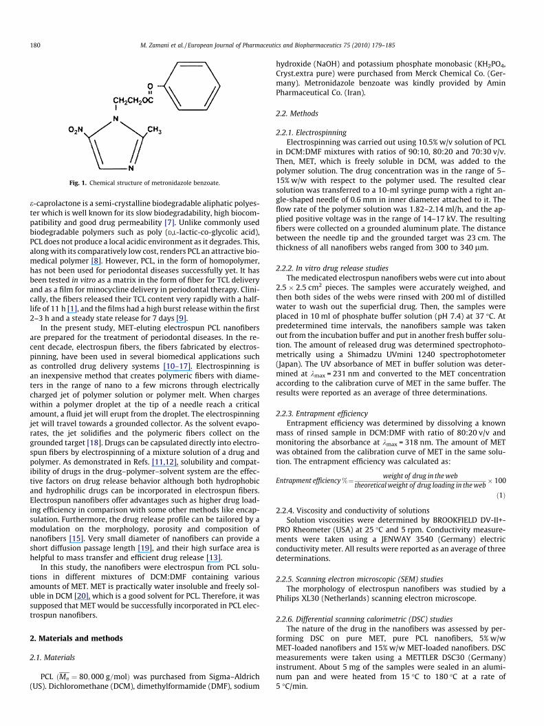
Fig. 1. Chemical structure of metronidazole benzoate.
180 M. Zamani et al. / European Journal of Pharmaceutics and Biopharmaceutics 75 (2010) 179–185
e-caprolactone is a semi-crystalline biodegradable aliphatic polyes-ter which is well known for its slow biodegradability, high biocom-patibility and good drug permeability [7]. Unlike commonly usedbiodegradable polymers such as poly (D,L-lactic-co-glycolic acid),PCL does not produce a local acidic environment as it degrades. This,along with its comparatively low cost, renders PCL an attractive bio-medical polymer [8]. However, PCL, in the form of homopolymer,has not been used for periodontal diseases successfully yet. It hasbeen tested in vitro as a matrix in the form of fiber for TCL deliveryand as a film for minocycline delivery in periodontal therapy. Clini-cally, the fibers released their TCL content very rapidly with a half-life of 11 h [1], and the films had a high burst release within the first2–3 h and a steady state release for 7 days [9].
In the present study, MET-eluting electrospun PCL nanofibersare prepared for the treatment of periodontal diseases. In the re-cent decade, electrospun fibers, the fibers fabricated by electros-pinning, have been used in several biomedical applications suchas controlled drug delivery systems [10–17]. Electrospinning isan inexpensive method that creates polymeric fibers with diame-ters in the range of nano to a few microns through electricallycharged jet of polymer solution or polymer melt. When chargeswithin a polymer droplet at the tip of a needle reach a criticalamount, a fluid jet will erupt from the droplet. The electrospinningjet will travel towards a grounded collector. As the solvent evapo-rates, the jet solidifies and the polymeric fibers collect on thegrounded target [18]. Drugs can be capsulated directly into electro-spun fibers by electrospinning of a mixture solution of a drug andpolymer. As demonstrated in Refs. [11,12], solubility and compat-ibility of drugs in the drug–polymer–solvent system are the effec-tive factors on drug release behavior although both hydrophobicand hydrophilic drugs can be incorporated in electrospun fibers.Electrospun nanofibers offer advantages such as higher drug load-ing efficiency in comparison with some other methods like encap-sulation. Furthermore, the drug release profile can be tailored by amodulation on the morphology, porosity and composition ofnanofibers [15]. Very small diameter of nanofibers can provide ashort diffusion passage length [19], and their high surface area ishelpful to mass transfer and efficient drug release [13].
In this study, the nanofibers were electrospun from PCL solu-tions in different mixtures of DCM:DMF containing variousamounts of MET. MET is practically water insoluble and freely sol-uble in DCM [20], which is a good solvent for PCL. Therefore, it wassupposed that MET would be successfully incorporated in PCL elec-trospun nanofibers.
2. Materials and methods
2.1. Materials
PCL ðMn ¼ 80;000 g=molÞ was purchased from Sigma–Aldrich(US). Dichloromethane (DCM), dimethylformamide (DMF), sodium
hydroxide (NaOH) and potassium phosphate monobasic (KH2PO4,Cryst.extra pure) were purchased from Merck Chemical Co. (Ger-many). Metronidazole benzoate was kindly provided by AminPharmaceutical Co. (Iran).
2.2. Methods
2.2.1. ElectrospinningElectrospinning was carried out using 10.5% w/v solution of PCL
in DCM:DMF mixtures with ratios of 90:10, 80:20 and 70:30 v/v.Then, MET, which is freely soluble in DCM, was added to thepolymer solution. The drug concentration was in the range of 5–15% w/w with respect to the polymer used. The resulted clearsolution was transferred to a 10-ml syringe pump with a right an-gle-shaped needle of 0.6 mm in inner diameter attached to it. Theflow rate of the polymer solution was 1.82–2.14 ml/h, and the ap-plied positive voltage was in the range of 14–17 kV. The resultingfibers were collected on a grounded aluminum plate. The distancebetween the needle tip and the grounded target was 23 cm. Thethickness of all nanofibers webs ranged from 300 to 340 lm.
2.2.2. In vitro drug release studiesThe medicated electrospun nanofibers webs were cut into about
2.5 � 2.5 cm2 pieces. The samples were accurately weighed, andthen both sides of the webs were rinsed with 200 ml of distilledwater to wash out the superficial drug. Then, the samples wereplaced in 10 ml of phosphate buffer solution (pH 7.4) at 37 �C. Atpredetermined time intervals, the nanofibers sample was takenout from the incubation buffer and put in another fresh buffer solu-tion. The amount of released drug was determined spectrophoto-metrically using a Shimadzu UVmini 1240 spectrophotometer(Japan). The UV absorbance of MET in buffer solution was deter-mined at kmax = 231 nm and converted to the MET concentrationaccording to the calibration curve of MET in the same buffer. Theresults were reported as an average of three determinations.
2.2.3. Entrapment efficiencyEntrapment efficiency was determined by dissolving a known
mass of rinsed sample in DCM:DMF with ratio of 80:20 v/v andmonitoring the absorbance at kmax = 318 nm. The amount of METwas obtained from the calibration curve of MET in the same solu-tion. The entrapment efficiency was calculated as:
Entrapment efficiency %¼ weight of drug in the webtheoretical weight of drug loading in the web
� 100
ð1Þ
2.2.4. Viscosity and conductivity of solutionsSolution viscosities were determined by BROOKFIELD DV-II+-
PRO Rheometer (USA) at 25 �C and 5 rpm. Conductivity measure-ments were taken using a JENWAY 3540 (Germany) electricconductivity meter. All results were reported as an average of threedeterminations.
2.2.5. Scanning electron microscopic (SEM) studiesThe morphology of electrospun nanofibers was studied by a
Philips XL30 (Netherlands) scanning electron microscope.
2.2.6. Differential scanning calorimetric (DSC) studiesThe nature of the drug in the nanofibers was assessed by per-
forming DSC on pure MET, pure PCL nanofibers, 5% w/wMET-loaded nanofibers and 15% w/w MET-loaded nanofibers. DSCmeasurements were taken using a METTLER DSC30 (Germany)instrument. About 5 mg of the samples were sealed in an alumi-num pan and were heated from 15 �C to 180 �C at a rate of5 �C/min.
M. Zamani et al. / European Journal of Pharmaceutics and Biopharmaceutics 75 (2010) 179–185 181
2.2.7. Statistical analysisStatistical analysis was carried out using the SPSS 13 statistical
package. Analysis of variance was followed by the least significantdifference (LSD). Post hoc test was used for comparison of differentdata. P < 0.05 was considered as the significant level.
The drug release mechanism was investigated using Peppasequation:
Mt
M1¼ Ktn ð2Þ
where Mt/M1 is the drug fraction released at time t, K is a constantdepending on the structural and geometric characteristic of the sys-tem, n is the diffusional coefficient related to the release mechanism[21].
Fig. 2. SEM photographs of electrospun PCL nanofibers containing 5% w/w MET as afunction of DCM:DMF ratio (v/v): (a) 70:30, (b) 80:20, and (c) 90:10.
3. Results and discussion
DCM is a good solvent for both PCL and MET. But it is not a suit-able solvent for electrospinning of PCL because of its mediumdielectric constant (9.1 at 25 �C) and poor electric conductivity[22]. While DMF is a nonsolvent for PCL, but it has a high dielectricconstant (36.7 at 25 �C) [22,23]. DMF was used to enhance elec-trospinning process in some other investigations [22,23]. There-fore, a mixture of DCM:DMF was used as a solvent in this study.
3.1. Physical characteristics of the electrospun nanofibers
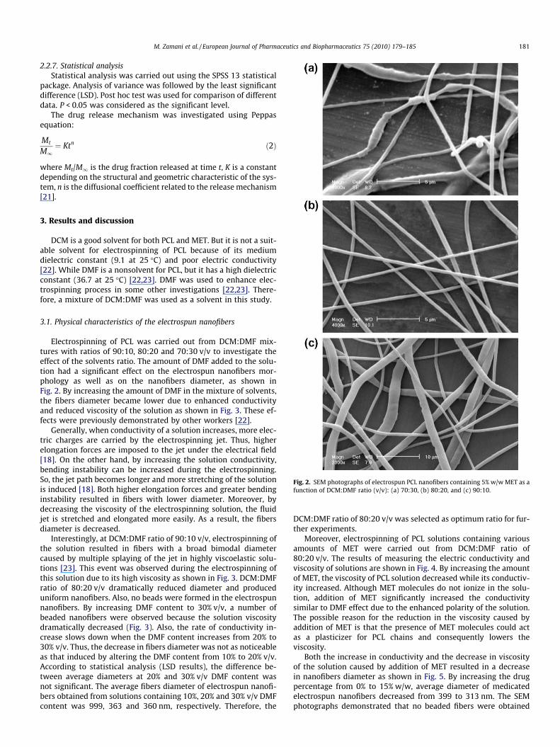
Electrospinning of PCL was carried out from DCM:DMF mix-tures with ratios of 90:10, 80:20 and 70:30 v/v to investigate theeffect of the solvents ratio. The amount of DMF added to the solu-tion had a significant effect on the electrospun nanofibers mor-phology as well as on the nanofibers diameter, as shown inFig. 2. By increasing the amount of DMF in the mixture of solvents,the fibers diameter became lower due to enhanced conductivityand reduced viscosity of the solution as shown in Fig. 3. These ef-fects were previously demonstrated by other workers [22].
Generally, when conductivity of a solution increases, more elec-tric charges are carried by the electrospinning jet. Thus, higherelongation forces are imposed to the jet under the electrical field[18]. On the other hand, by increasing the solution conductivity,bending instability can be increased during the electrospinning.So, the jet path becomes longer and more stretching of the solutionis induced [18]. Both higher elongation forces and greater bendinginstability resulted in fibers with lower diameter. Moreover, bydecreasing the viscosity of the electrospinning solution, the fluidjet is stretched and elongated more easily. As a result, the fibersdiameter is decreased.
Interestingly, at DCM:DMF ratio of 90:10 v/v, electrospinning ofthe solution resulted in fibers with a broad bimodal diametercaused by multiple splaying of the jet in highly viscoelastic solu-tions [23]. This event was observed during the electrospinning ofthis solution due to its high viscosity as shown in Fig. 3. DCM:DMFratio of 80:20 v/v dramatically reduced diameter and produceduniform nanofibers. Also, no beads were formed in the electrospunnanofibers. By increasing DMF content to 30% v/v, a number ofbeaded nanofibers were observed because the solution viscositydramatically decreased (Fig. 3). Also, the rate of conductivity in-crease slows down when the DMF content increases from 20% to30% v/v. Thus, the decrease in fibers diameter was not as noticeableas that induced by altering the DMF content from 10% to 20% v/v.According to statistical analysis (LSD results), the difference be-tween average diameters at 20% and 30% v/v DMF content wasnot significant. The average fibers diameter of electrospun nanofi-bers obtained from solutions containing 10%, 20% and 30% v/v DMFcontent was 999, 363 and 360 nm, respectively. Therefore, the
DCM:DMF ratio of 80:20 v/v was selected as optimum ratio for fur-ther experiments.
Moreover, electrospinning of PCL solutions containing variousamounts of MET were carried out from DCM:DMF ratio of80:20 v/v. The results of measuring the electric conductivity andviscosity of solutions are shown in Fig. 4. By increasing the amountof MET, the viscosity of PCL solution decreased while its conductiv-ity increased. Although MET molecules do not ionize in the solu-tion, addition of MET significantly increased the conductivitysimilar to DMF effect due to the enhanced polarity of the solution.The possible reason for the reduction in the viscosity caused byaddition of MET is that the presence of MET molecules could actas a plasticizer for PCL chains and consequently lowers theviscosity.
Both the increase in conductivity and the decrease in viscosityof the solution caused by addition of MET resulted in a decreasein nanofibers diameter as shown in Fig. 5. By increasing the drugpercentage from 0% to 15% w/w, average diameter of medicatedelectrospun nanofibers decreased from 399 to 313 nm. The SEMphotographs demonstrated that no beaded fibers were obtained
Fig. 3. Effects of DCM:DMF ratio on the solution properties: (a) solution viscosityand (b) solution conductivity.
Fig. 4. Effects of MET concentration on the solution properties: (a) solutionviscosity and (b) solution conductivity.
182 M. Zamani et al. / European Journal of Pharmaceutics and Biopharmaceutics 75 (2010) 179–185
by electrospinning of these solutions containing various amountsof MET.
All electrospun nanofiber mats produced in this study weredesirably smooth and flexible and yet retained the generalmechanical characteristics of PCL such as elasticity as was studiedphysically by touch. This flexibility, in addition to hydrophobicity,
provides easy handling ability during implantation and a comfort-able texture for use.
3.2. Differential scanning calorimetric study
DSC studies were performed to understand the physical state ofthe drug in the electrospun nanofibers. The thermogram of crystal-line MET showed an endothermic sharp peak at 103 �C and an en-thalpy (DH) of 790 J/g due to melting temperature of MET (Fig. 6a).Also, an endothermic melting peak at 57 �C was observed for purePCL nanofibers (DH = 1979.2 J/g), (Fig. 6b). The melting endothermpeak of MET was detected neither in the 5% w/w MET-loaded(Fig. 6c) nor in 15% w/w MET-loaded (Fig. 6d) nanofibers. The ab-sence of detectable crystalline domain, even at high concentrationof MET, indicates that drug was molecularly dispersed in amor-phous state in polymeric matrix. On the other hand, by addingMET to the solutions, the melting enthalpy of PCL nanofibers be-came lower and appeared at almost the same temperature of57.1 �C. By increasing the MET content of nanofibers, the meltingenthalpy of PCL nanofibers decreased from DH of 1979.2 J/g toDH of 1222 J/g and DH of 888.3 J/g for 5% and 15% w/w MET con-tent, respectively. These results show that increasing the amountof MET in the semi-crystalline PCL nanofibers caused a reductionin the crystallinity.
3.3. In vitro drug release
Calculated values of entrapment efficiency of different formula-tions after rinsing are presented in Table 1.
The results display a systematic dependence of the entrapmentefficiency to the amount of drug incorporated, while the solventscomposition did not have a significant effect on it. It is believedthat reduced entrapment efficiency observed by increasing theamount of drug was mainly due to further exposure of the drugto the water. It means that at higher concentration of MET, a moreportion of MET was located on the web surface. Therefore, it candissolve in the rinsing water and is washed out easily.
Figs. 7 and 8 show the release profiles of MET from various elec-trospun PCL nanofibers. As illustrated in these figures, drug releasecontinued for a period of at least 19 days. Indeed, the drug releasefrom PCL nanofibers is ideally prolonged in comparison with vari-ous controlled drug release systems that have previously beenstudied for periodontal diseases [1,2,4,9,24–27]. Furthermore,none of the electrospun mats showed high burst release implyingthe perfect inclusion of the drug inside the fibers.
Since MET is highly soluble in DCM which is the main portion ofsolvents mixture, the solution remains homogenous during theelectrospinning process without separation of drug crystals. Onthe other hand, both MET and PCL have hydrophobic properties.Thus, the affinity and compatibility between the drug and polymeris vital for perfect encapsulation of the drug inside the electrospunnanofibers during the rapid stretching and quick solvent evapora-tion of electrospinning process [11]. In this research, successfulencapsulation of drug inside the nanofibers was the main causeof ideally prolonged drug release as well as low burst release. Shortperiod of drug release and initial burst caused by incompatibility ofdrug–polymer–solvent system were reported in some investiga-tions [10–13,15,16].
Fig. 7 shows the effect of solvents ratio on the release profiles ofMET from electrospun PCL nanofibers. It can be seen that drug re-lease profiles were not significantly affected by decreasing DMFcontent from 30% to 20% v/v. It means that morphological changesof nanofibers caused by altering DCM:DMF ratio from 70:30 to80:20 v/v did not have a significant effect on release profiles andthese formulations released MET at approximately the same rates.However, for 10% v/v DMF content, nanofibers showed a different
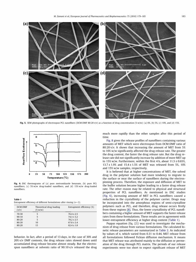
Fig. 5. SEM photographs of electrospun PCL nanofibers (DCM:DMF 80:20 v/v) as a function of drug concentration (% w/w): (a) 0%, (b) 5%, (c) 10%, and (d) 15%.
Fig. 6. DSC thermograms of (a) pure metronidazole benzoate, (b) pure PCLnanofibers, (c) 5% w/w drug-loaded nanofibers, and (d) 15% w/w drug-loadednanofibers.
Table 1Entrapment efficiency of different formulations after rinsing (n = 3).
DCM:DMF(v/v)
Theoretical drug loading(% w/w)
Entrapment efficiency (%)
70:30 5 76.4 ± 2.590:10 5 74.3 ± 3.280:20 5 78.2 ± 2.580:20 10 67.8 ± 2.480:20 15 62.4 ± 1.8
M. Zamani et al. / European Journal of Pharmaceutics and Biopharmaceutics 75 (2010) 179–185 183
behavior. In fact, after a period of 13 days, in the case of 30% and20% v/v DMF contents, the drug release rates slowed down untilaccumulated drug release became almost steady. But the electro-spun nanofibers at solvents ratio of 90:10 v/v released the drug
much more rapidly than the other samples after this period oftime.
Fig. 8 gives the release profiles of nanofibers containing variousamounts of MET which were electrospun from DCM:DMF ratio of80:20 v/v. It shows that increasing the amount of MET from 5%to 10% w/w significantly affected the drug release rate. The greaterthe drug content, the faster the drug release rate. But the drug re-lease rate did not significantly increase by addition of more MET upto 15% w/w. Furthermore, within the first 4 h, about 11.5 ± 0.65%,13.7 ± 1.9% and 15.4 ± 1.5% of MET was released from 5%, 10%and 15% w/w samples, respectively.
It is believed that at higher concentrations of MET, the solveddrug in the polymer solution had more tendency to migrate tothe surface or near the surface of nanofibers during the electros-pinning process. Therefore, the exposure and diffusion of MET tothe buffer solution became higher leading to a faster drug releaserate. The other reason may be related to physical and structuralproperties of PCL nanofibers. As mentioned in DSC studies(Fig. 6), increasing amount of MET in PCL nanofibers caused areduction in the crystallinity of the polymer carrier. Drugs maybe incorporated into the amorphous regions of semi-crystallinepolymers such as PCL, and therefore, drug release occurs firstlyfrom these regions [8]. Thus, the lower crystallinity of PCL nanofi-bers containing a higher amount of MET supports the faster releaserates from these formulations. These results are in agreement withlower entrapment efficiency at higher drug content (Table 1).
Peppas equation (Eq. (2)) was used to investigate the mecha-nism of drug release from various formulations. The calculated ki-netic release parameters are summarized in Table 2. As indicatedby values of n, which varied from 0.31 to 0.44, MET release fromall formulations followed Fickian diffusion mechanism. It meansthat MET release was attributed mainly to the diffusion or perme-ation of the drug through PCL matrix. The periods of our releaseexperiments were too short to expect significant release of MET
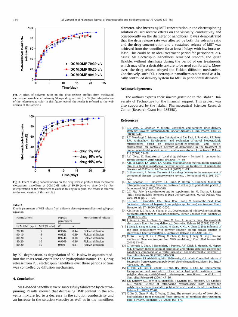
Fig. 7. Effect of solvents ratio on the drug release profiles from medicatedelectrospun nanofibers containing 5% w/w drug vs. time (n = 3). (For interpretationof the references to color in this figure legend, the reader is referred to the webversion of this article.)
Fig. 8. Effect of drug concentrations on the drug release profiles from medicatedelectrospun nanofibers at DCM:DMF ratio of 80:20 (v/v) vs. time (n = 3). (Forinterpretation of the references to color in this figure legend, the reader is referredto the web version of this article.)
Table 2Kinetic parameters of MET release from different electrospun nanofibers using Peppasequation.
Formulations Peppasparameters
Mechanism of release
DCM:DMF (v/v) MET (% w/w) R2 n
70:30 5 0.9604 0.44 Fickian diffusion90:10 5 0.9823 0.39 Fickian diffusion80:20 5 0.9748 0.38 Fickian diffusion80:20 10 0.9609 0.36 Fickian diffusion80:20 15 0.989 0.31 Fickian diffusion
184 M. Zamani et al. / European Journal of Pharmaceutics and Biopharmaceutics 75 (2010) 179–185
by PCL degradation, as degradation of PCL is slow in aqueous med-ium due to its semi-crystalline and hydrophobic nature. Thus, drugrelease from PCL electrospun nanofibers over these periods of timewas controlled by diffusion mechanism.
4. Conclusion
MET-loaded nanofibers were successfully fabricated by electros-pinning. Results showed that decreasing DMF content in the sol-vents mixture led to a decrease in the solution conductivity andan increase in the solution viscosity as well as in the nanofibers
diameter. Also increasing MET concentration in the electrospinningsolution caused reverse effects on the viscosity, conductivity andconsequently on the diameter of nanofibers. It was demonstratedthat the drug release rate was affected by both the solvents ratioand the drug concentration and a sustained release of MET wasachieved from the nanofibers for at least 19 days with low burst re-lease. This could be an ideal treatment period for periodontal dis-eases. All electrospun nanofibers remained smooth and quiteflexible, without shrinkage during the period of our treatments,which may offer a desirable texture to be used comfortably. More-over, the drug release obeyed the Fickian diffusion mechanism.Conclusively, such PCL electrospun nanofibers can be used as a lo-cally controlled delivery system for MET in periodontal diseases.
Acknowledgements
The authors express their sincere gratitude to the Isfahan Uni-versity of Technology for the financial support. This project wasalso supported by the Isfahan Pharmaceutical Sciences ResearchCenter (Research Grant No: 285358).
References
[1] S.P. Vyas, V. Sihorkar, V. Mishra, Controlled and targeted drug deliverystrategies towards intraperiodontal pocket diseases, J. Clin. Pharm. Ther. 25(2000) 1–42.
[2] R.C. Mundargi, S. Srirangarajan, S.A. Agnihotri, S.A. Patil, S. Ravindra, S.B. Setty,T.M. Aminabhavi, Development and evaluation of novel biodegradablemicrospheres based on poly(D,L-lactide-co-glycolide) and poly(e-caprolactone) for controlled delivery of doxycycline in the treatment ofhuman periodontal pocket: in vitro and in vivo studies, J. Controlled Release119 (2007) 59–68.
[3] P.V. Divya, K. Nandakumar, Local drug delivery – Periocol in periodontics,Trends Biomater. Artif. Organs 19 (2006) 74–80.
[4] A.H. El-Kamel, L.Y. Ashri, I.A. Alsarra, Micromatricial metronidazole benzoatefilm as a local mucoadhesive delivery system for treatment of periodontaldiseases, AAPS Pharm. Sci. Technol. 8 (2007) E1–E11.
[5] G. Greenstein, A. Polson, The role of local drug delivery in the management ofperiodontal diseases: a comprehensive review, J. Periodontol. 69 (1998) 507–520.
[6] J.M. Goodson, D. Holborow, R.L. Dunn, P. Hogen, S. Dunham, Monolithictetracycline-containing fibers for controlled delivery to periodontal pocket, J.Periodontol. 54 (1983) 575–579.
[7] C.G. Pitt, Poly (e-caprolactone) and its copolymers, in: M. Chasin, R. Langer(Eds.), Biodegradable Polymers as Drug Delivery Systems, Marcel Dekker, NewYork, 1990, pp. 71–120.
[8] E.L. Van, L. Grondahl, K.N. Chua, K.W. Leong, V. Nurcombe, S.M. Cool,Controlled release of heparin from poly(e-caprolactone) electrospun fibers,Biomaterials 27 (2006) 2042–2050.
[9] K.D. Kyun, K.S. Yun, J.S. Young, et al., Development of minocycline containingpolycaprolactone film as local drug delivery, Taeban Chikkwa Visa Hyophoe 28(1990) 279–290.
[10] J. Zeng, X. Xu, X. Chen, Q. Liang, X. Bian, L. Yang, X. Jing, Biodegradableelectrospun fibers for drug delivery, J. Controlled Release 92 (2003) 227–231.
[11] J. Zeng, L. Yang, Q. Liang, X. Zhang, H. Guan, X. XU, X. Chen, X. Jing, Influence ofthe drug compatibility with polymer solution on the release kinetics ofelectrospun fiber formulation, J. Controlled Release 105 (2005) 43–51.
[12] X. Xu, L. Yang, X. Xu, X. Wang, X. Chen, Q. Liang, J. Zeng, X. Ling, Ultrafinemedicated fibers electrospun from W/O emulsions, J. Controlled Release 108(2005) 33–42.
[13] G. Verreck, I. Chun, J. Rosenblatt, J. Peeters, A.V. Dijck, J. Mensch, M. Noppe,M.E. Brewster, Incorporation of drugs in an amorphous state into electrospunnanofibers composed of a water-insoluble, nonbiodegradable polymer, J.Controlled Release 92 (2003) 349–360.
[14] E.R. Kenawy, F.I. Abdel-Hay, M.H. El-Newehy, G.E. Wnek, Controlled release ofketoprofen from electrospun poly (vinyl alcohol) nanofibers, Mater. Sci. Eng. A459 (2007) 90–396.
[15] K. Kim, Y.K. Luu, C. Chang, D. Fang, B.S. Hsiao, B. Chu, M. Hadjiargyrou,Incorporation and controlled release of a hydrophilic antibiotic usingpoly(lactide-co-glycolide)-based electrospun nanofibrous scaffolds, J.Controlled Release 98 (2004) 47–56.
[16] E.R. Kenawy, G.L. Bowlin, K. Mansfield, J. Layman, D.G. Simpson, E.H. Sanders,G.E. Wnek, Release of tetracycline hydrochloride from electrospunpoly(ethylene-co-vinylacetate), poly(lactic acid), and a blend, J. ControlledRelease 81 (2002) 57–64.
[17] X. Xu, X. Chen, P. Ma, X. Wang, X. Jing, The release behavior of doxorubicinhydrochloride from medicated fibers prepared by emulsion-electrospinning,Euro. J. Pharm. Biopharm. 70 (2008) 165–170.
M. Zamani et al. / European Journal of Pharmaceutics and Biopharmaceutics 75 (2010) 179–185 185
[18] S. Ramakrishna, K. Fujihara, W.E. Teo, T.C. Lim, Z. Ma, An Introduction toElectrospinning and Nanofibers, World Scientific, Singapore, 2005.
[19] Z.M. Huang, C.L. He, A. Yang, Y. Zhang, X. Han, J. Yin, Q. Wu, Encapsulatingdrugs in biodegradable ultrafine fibers through co-axial electrospinning, J.Biomed. Mater. Res. Part A 77A1 (2006) 169–179.
[20] S. Sweetman, Martindale: The Complete Drug Reference, 34th ed.,Pharmaceutical Press, London, 2005.
[21] P. Costa, J. Manuel, Modeling and comparison of dissolution profiles, Eur. J.Pharm. Sci. 13 (2001) 123–133.
[22] K.H. Lee, H.Y. Kim, M.S. Khil, Y.M. Ra, D.R. Lee, Characterization of nano-structured poly(e-caprolactone) nonwoven mats via electrospinning, Polymer44 (2003) 1287–1294.
[23] C.M. Hsu, S. Shivkumar, N,N-dimethylformamide additions to the solution forthe electrospinning of poly(e-caprolactone) nanofibers, Macromol. Mater. Eng.289 (2004) 334–340.
[24] M. Addy, L. Rawle, R. Handley, et al., The development and in-vitro evaluationof acrylic strips and dialysis tubing for local drug delivery, J. Periodontol. 53(1982) 693–699.
[25] A.E. Collins, P.B. Desay, D.J. MacCarthy, D.B. Shanely, Evaluation ofcontrolled release compact containing tetracycline hydrochloride bonded totooth for the treatment of periodontal diseases, Int. J. Pharm. 51 (1989) 103–114.
[26] L. Perioli, V. Ambrogi, D. Rubini, S. Giovagnoli, M. Ricci, P. Blasi, C. Rossi,Novel mucoadhesive buccal formulation containing metronidazole for thetreatment of periodontal diseases, J. Controlled Release 95 (2004)521–533.
[27] K.S. Abdellaoui, A. Monti, J. Barr, J. Heller, R. Gurny, Optimization of a novelbiodegradable device based on auto-catalyzed poly (ortho esters) forcontrolled delivery of tetracycline to periodontal pocket, Biomaterials 22(2001) 1659–1666.


![Methyl 5-bromo-2-[methyl(methylsulfonyl)amino]benzoate](https://img.pdfslide.net/doc/110x75/632436d203238a9ff60acbf8/methyl-5-bromo-2-methylmethylsulfonylaminobenzoate.jpg)


