Embed Size (px)
Citation preview

Molecular Biology of the CellVol. 19, 994-1006, March 2008
Coordinated Vascular Endothelial Growth FactorExpression and Signaling During Skeletal MyogenicDifferentiationBrad A. Bryan,* Tony E. Walshe,* Dianne C. Mitchell,† Josh S. Havumaki,‡Magali Saint-Geniez,* Arindel S. Maharaj,* Angel E. Maldonado,* andPatricia A. D’Amore*
*Schepens Eye Research Institute, Boston, MA 02114; †Acceleron Pharma, Cambridge, MA 02139; ‡Universityof Massachusetts, Amherst, MA 01003
Submitted September 4, 2007; Revised November 28, 2007; Accepted December 11, 2007Monitoring Editor: Richard Hynes
Angiogenesis is largely controlled by hypoxia-driven transcriptional up-regulation and secretion of vascular endothelialgrowth factor (VEGF) and its binding to the endothelial cell tyrosine receptor kinases, VEGFR1 and VEGFR2. Recentexpression analysis suggests that VEGF is expressed in a cell-specific manner in normoxic adult tissue; however, thetranscriptional regulation and role of VEGF in these tissues remains fundamentally unknown. In this report wedemonstrate that VEGF is coordinately up-regulated during terminal skeletal muscle differentiation. We reveal that thisregulation is mediated in part by MyoD homo- and hetero-dimeric transcriptional mechanisms. Serial deletions of theVEGF promoter elucidated a region containing three tandem CANNTG consensus MyoD sites serving as essential sitesof direct interaction for MyoD-mediated up-regulation of VEGF transcription. VEGF-null embryonic stem (ES) cellsexhibited reduced myogenic differentiation compared with wild-type ES cells, suggesting that VEGF may serve a role inskeletal muscle differentiation. We demonstrate that VEGFR1 and VEGFR2 are expressed at low levels in myogenicprecursor cells and are robustly activated upon VEGF stimulation and that their expression is coordinately regulatedduring skeletal muscle differentiation. VEGF stimulation of differentiating C2C12 cells promoted myotube hypertrophyand increased myogenic differentiation, whereas addition of sFlt1, a VEGF inhibitor, resulted in myotube hypotrophy andinhibited myogenic differentiation. We further provide evidence indicating VEGF-mediated myogenic marker expression,mitogenic activity, migration, and prosurvival functions may contribute to increased myogenesis. These data suggest anovel mechanism whereby VEGF is coordinately regulated as part of the myogenic differentiation program and serves anautocrine function regulating skeletal myogenesis.
INTRODUCTION
Angiogenesis often coincides with increased vascular per-meability, allowing extravasation of plasma proteins that laydown a provisional scaffold for migrating endothelial cells(ECs). Subsequent degradation of the extracellular matrix bymatrix-metalloproteases relieves pericyte-EC contacts andliberates extracellular matrix-sequestered growth factors.ECs then proliferate and migrate to their final destination toassemble as lumen-bearing cords. These processes arelargely controlled by transcriptional up-regulation and sub-sequent secretion of three splice variant isoforms of vascularendothelial growth factor (VEGF) and their binding to theendothelial cell tyrosine receptor kinases: VEGFR1 andVEGFR2, leading to activation of a number of downstreamsignaling cascades (Ferrara et al., 2003).
The function and regulation of VEGF has primarily beenstudied in pathological angiogenesis, such as in tumors ofcritical mass where hypoxia-driven HIF1� activation and
subsequent binding to hypoxia-induced regulatory elementsin the VEGF promoter modulates VEGF mRNA expression(Liu et al., 1995; Forsythe et al., 1996; Mazure et al., 1996).Although HIF1� is known to play a role in the regulation ofVEGF gene expression in tissues of the embryo and adult,deletion of the hypoxia-responsive element in the VEGFpromoter surprisingly leads to only a subtle defect—adult-onset progressive motor neuron degeneration—with othertissues largely unaffected (Oosthuyse et al., 2001). This ob-servation suggests that the importance of hypoxia-drivenHIF1� regulation of VEGF may be historically overesti-mated. Moreover, a recent study by Maharaj et al. (2006) hasshown that VEGF is expressed at appreciable levels in acell-specific manner in nondiseased normoxic adult tissue.This finding, together with the absence of generalized vas-cular defects in HIF1� null mice, suggests that VEGF geneexpression in normal adult tissues is controlled by yet to beidentified novel mechanisms.
In addition to its induction of new blood vessel growth,VEGF serves an important role in the maintenance anddevelopment of endothelial fenestrations (Esser et al., 1998;Lammert et al., 2003; Yokomori et al., 2003) and in en-dothelial survival in vitro (Darland et al., 2003) and in vivo(Kamba et al., 2006). A growing number of reports suggestthat VEGF has autocrine effects on nonvascular cells such asneuronal cells (Ogunshola et al., 2002; Jin et al., 2006; Nishi-
This article was published online ahead of print in MBC in Press(http://www.molbiolcell.org/cgi/doi/10.1091/mbc.E07–09–0856)on December 19, 2007.
Address correspondence to: Patricia A. D’Amore ([email protected]).
994 © 2008 by The American Society for Cell Biology

jima et al., 2007), muscle (Germani et al., 2003), and bone(Fons et al., 2004; Byun et al., 2007). These findings challengethe dogma that VEGF serves solely as a paracrine growthfactor specifically targeting ECs (D’Amore, 2007) and mayhelp to explain why VEGF is expressed in normoxic adulttissues, where there is not active ongoing angiogenesis.
In this report, we utilize skeletal muscle as a model systemto examine the tissue-specific regulation and function ofVEGF under physiological conditions. Although a plethoraof data implicates exercise-induced hypoxia as the key sig-nal modulating VEGF expression in the skeletal muscle(Gustafsson and Kraus, 2001; Olfert et al., 2001; Hudlicka etal., 2002), the level of VEGF expression remains high in theskeletal muscle of sedentary mice (Maharaj et al., 2006),suggesting a regulatory mechanism not involving hypoxia.Our data, using multipotent mesenchymal cell lines that arecapable of terminally differentiating into skeletal myocytes,demonstrate that skeletal differentiation is associated withincreased VEGF expression, suggesting that VEGF is coor-dinately regulated during myogenesis. We further demon-strate that the myogenic transcription factor, MyoD, and itsheterodimeric binding proteins, E12 and E47, up-regulatethe expression of endogenous VEGF through direct interac-tion with the VEGF promoter. Moreover, VEGF-null embry-onic stem (ES) cells exhibit a significant reduction in skeletalmyogenesis compared with wild-type ES cells, suggestingthat VEGF is necessary for skeletal myogenesis. Consistentwith these observations, we report that VEGFR1 andVEGFR2 are expressed at low levels in C2C12 cells and arerobustly activated upon VEGF stimulation, indicating thatVEGF can potentially signal in an autocrine manner. Indeed,VEGF stimulation of C2C12 cells results in increased termi-nal skeletal muscle differentiation, as evidenced by in-creased myotube formation, myogenic marker expression,and myotube hypertrophy. Conversely, treatment with thesoluble VEGF inhibitor sFlt1 effectively blocks myogenesisand reduces myotube size. These data suggest a novel mech-anism by which VEGF expression is coordinately regulatedduring skeletal myogenesis and demonstrate an autocrinefunction whereby VEGF serves as an essential regulator inthis process.
MATERIALS AND METHODS
LacZ StainingAdult C57BL/6 mice expressing the �-galactosidase (lacZ) reporter genecDNA with a nuclear localization signal and an internal ribosome entry siteinserted into the 3� untranslated region (3�UTR) of the VEGF gene were usedin these studies (Miquerol et al., 1999). This gene yields a bicistronic mRNAthat produces both functional VEGF and a reporter �-galactosidase (�-gal)protein. VEGF expression was visualized in adult skeletal muscle and incryosections of VEGF-LacZ mouse embryos. Embryos were fixed overnight at4°C in 4% paraformaldehyde in phosphate-buffered saline (PBS). For cryosec-tions, embryos were embedded in OCT compound (Sakura Finetechnical,Torrance, CA). Whole-mounted adult skeletal muscle or cryosections werestained for LacZ using the in situ �-galactosidase staining kit, according to themanufacturer’s protocol (Stratagene, La Jolla, CA).
Semiquantitative RT-PCRSkeletal muscle from the hindlimbs of 2-mo-old C57BL/6 mice and lysatesfrom C2C12 and 10T1/2 cultures were collected under RNase-free conditions.Total RNA was extracted using Trizol reagent (Invitrogen, Carlsbad, CA),according to the manufacturer’s protocol. Residual DNA was removed bytreatment with 1 U DNase I (Ambion, Austin, TX) at 37°C for 20 min. Onemicrogram of RNA was reverse-transcribed in the presence of 500 ng of oligodT12–18 with Superscript II reverse transcriptase (Invitrogen) in a 20-�lreaction at 42°C for 50 min and digested with 2 U of RNase H. One microliterof cDNA was used as a template in a 25-�l amplification mixture containing200 mM dNTPs, 1 U Taq DNA polymerase (Roche Diagnostics, Indianapolis,IN), and 0.2 �M of the appropriate primer pairs capable of amplifying allthree isoforms of VEGF (forward: 5� CCT CCG AAA CCA TGA ACT TTC
TGC TC 3�; reverse: 5� CAG CCT GGC TCA CCG CCT TGG CTT 3�). Productswere amplified for 35 cycles and separated by agarose gel electrophoresis,stained with ethidium bromide, and visualized by UV light. Quantitation ofband intensity was performed using Image J software (http://rsb.info.nih.gov/ij/; NIH).
Real-Time PCRmRNA was purified as described above using the Trizol method. One micro-gram of RNA was reverse- transcribed as described above, except 500 ng ofrandom hexamers were used. One twentieth of the total cDNA (50 ng ofequivalent RNA) was used in each amplification reaction. VEGF isoformswere quantified using the Prism 9700 Sequence Detection System (AppliedBiosystems, Foster City, CA) according to the manufacturer’s instructions.Reactions were performed in 25 �l with 0.3 �M primers specific for VEGFisoforms (Zhang et al., 2002) and SYBR Green master mix (ABI, Columbia,MD). PCR cycles consisted of an initial denaturation step at 95°C for 10 min,followed by 40 cycles at 95°C for 15 s and at 60°C for 60 s. To confirmamplification specificity, PCR products from each primer pair were subjectedto a melting curve analysis. A standard curve was constructed for each PCRreaction and was derived from the serial dilution (10�3 to 10�9 ng DNA perreaction) of a plasmid coding for each isoform—VEGF188, VEGF164, andVEGF120—and amplified using the SYBR Green system. The level of isoformexpression in each sample was calculated relative to the standard curve. Eachsample was run in triplicate, and each experiment included three nontemplatecontrol wells. Results were expressed as the mean � SD.
Cell Culture and TreatmentC2C12 (generous gift from Dr. M. T. Chin at Harvard Medical School) and10T1/2 cells (purchased from ATCC, Manassas, VA; CCL-226) were main-tained at subconfluent levels in growth medium (GM) consisting of DMEM(GIBCO, Rockville, MD) supplemented with 10% fetal bovine serum (FBS;Hyclone, Logan, UT). 10T1/2 cells, which were used for differentiation ex-periments, were stably transfected with a MyoD expression plasmid (gener-ous gift from Dr. Mingyao Liu, Texas A&M Health Science Center). To inducedifferentiation, cells were grown to 100% confluence and GM was replacedwith differentiation medium (DM) consisting of DMEM supplemented with2% horse serum (Invitrogen) and 80 U/ml penicillin/streptomycin C. DMwas replaced every 24 h.
Bovine retinal endothelial cells (BRECs) were maintained in EBM sup-plemented with 10% horse serum, 80 U/ml penicillin/streptomycin C, and12 �g/ml bovine brain extract. The cells were plated on plastic coated with50 �g/ml fibronectin.
Wild-type and VEGF-null mouse ES cells (generous gift from Andras Nagy,Mount Sinai Hospital, Toronto, Canada) were cultured on gelatin-coateddishes in high-glucose DMEM (GIBCO BRL) with 15% fetal bovine serum (LotFRB25667, Hyclone), sodium pyruvate (GIBCO, stock solution diluted 1:100),nonessential amino acids (GIBCO, stock solution diluted1:100), �-mercapto-ethanol (GIBCO, final concentration 30 �M), 190 �g/ml l-glutamine, 60 U/mlpenicillin G, 60 �g/ml streptomycin (glutamine pen–strep mix, Irvine Scien-tific, Santa Ana, CA), supplemented with media (1:300 dilution) conditionedby Chinese hamster ovary cells overexpressing LIF (provided by GeneticsInstitute, Cambridge, MA) as a source of LIF to maintain the ES cells in anundifferentiated state. ES cells were cultured in a humidified tissue cultureincubator at 10% CO2 and 37°C and passaged every 2 to 3 d. ES cells weredifferentiated into cystic embryoid bodies (CEB) as previously described (Nget al., 2004). Briefly, trypsinized ES cells were suspended in the same culturemedium as described above, but without LIF. A total of 60 aliquots (30 �l) ofES cell suspension containing 2.5 �103 cells were plated as individual dropsonto 100-mm2 bacteriological dishes (Valmark, Brampton, Canada). Theplates were then inverted and the cells were incubated in hanging drops; thiswas defined as day 0 of differentiation. The CEB were cultivated via hangingdrop for 40–45 h, and then the dishes were turned right side up and 10 ml ofES culture media without LIF was added so that the CEB were then insuspension. Every 3 d, half of the culture media was removed and replacedwith fresh media. For attached cultures, day 4 or day 5 CEB were transferredto gelatin-coated glass coverslips, onto which the CEB attached, flattened, andspread.
Recombinant human VEGF165 (obtained from National Cancer Institute,www.cancer.gov) was added to the cultures at a final concentration of 25ng/ml. Recombinant mouse VEGF-R1/Fc chimera (sFlt1; R&D Systems, Min-neapolis, MN; 471-F1, Lot BSL1107011) was added to the cultures at a finalconcentration of 100 ng/ml. Recombinant adenovirus (AV) vectors containingeither a null cassette or sFlt1 were obtained from Qbiogene (Montreal,Canada). Undifferentiated C2C12 cells were incubated with AV vectors atmultiplicities of infection (MOIs) of 103 viral genomes per cell.
Western BlottingTissues were collected in lysis buffer (10 mM Tris-HCl, pH 7.4, 5 mM EDTA,50 mM NaCl, 1% Triton X-100, 50 mM NaF, 1 mM phenylmethylsulfonylfluoride [PMSF], 2 mM Na3VO4, and 20 mg/ml aprotinin). Proteins werequantified using the Dc protein assay kit (Bio-Rad, Richmond, CA). Cellextracts were analyzed by SDS-PAGE and probed with anti-MyoD (sc-304,
VEGF: Role and Regulation in Skeletal Muscle
Vol. 19, March 2008 995

Santa Cruz Biotechnology, Santa Cruz, CA), anti-VEGFR1 (sc-316, Santa CruzBiotechnology), anti-VEGFR2 (a generous gift from A. Kazlauskas, SchepensEye Research Institute), anti-myosin heavy chain (MHC; MF20, Iowa Hybrid-oma Bank), anti-myogenin (F5D, Iowa Hybridoma Bank), PY20 (05-321, Up-state Biotechnology, Lake Placid, NY), and anti-tubulin (CP06-100UG, Onco-gene Research Products, Boston, MA). Binding was detected with theappropriate HRP-conjugated secondary antibody (mouse: NA931V, rabbit:NA934V, Amersham Biosciences, Piscataway, NJ) and ECL-Plus WesternBlotting Detection System (RPN2132, GE Healthcare, Waukesha, WI).
ImmunofluorescenceCells were fixed in ice-cold methanol for 10 min. Reactions were blocked for60 min with PBS containing 0.2% bovine serum albumin, followed by 60 minof incubation with anti-MHC antibody (1:20 dilution; Iowa Hybridoma Bank).Fluorescein-conjugated goat anti-mouse antibody was added for 30 min (1:1000; 81-6511, Molecular Probes, Eugene, OR). Nuclear staining was observedafter 10 min of 4�,6-diamidino-2-phenylindole (DAPI) treatment (1:100 dilu-tion; Molecular Probes). Fluorescence images were captured on a CCD cameramounted on an inverted research microscope using Spot imaging software(Diagnostic Instruments, Sterling Heights, MI).
Luciferase Reporter AssaysVEGF-promoter luciferase constructs have been previously described(Loureiro et al., 2005). C2C12 and 10T1/2 cells were cotransfected at 60–80%confluence using Lipofectamine 2000 with equal amounts of VEGF fireflyluciferase (Fluc)-reporter plasmid, Renilla control luciferase (Rluc; Promega,Madison, WI), and the indicated plasmid to a total of 1.5 �g DNA per well ofa 24-well plate. Fresh medium was added 6 h after transfection and leftundisturbed until cell lysates were collected after 48-h incubation. Luciferaseexpression was detected using the Dual Luciferase Reporter Assay System(Promega) following the manufacturer’s instructions with a Turner Lumi-nometer (Turner Designs, Sunnyvale, CA). VEGF promoter activity, as de-tected by Fluc activity, was normalized against Rluc readings and this ratiowas divided by the ratio of readings from promoterless luciferase to deter-mine fold-change. Assays were performed three times in triplicate withsimilar results; one representative experiment is shown. Statistical analysis ofthe data were performed to determine significance using analysis of variance.
Chromatin Immunoprecipitation AssayChromatin immunoprecipitation assay (ChIP) was performed following theprotocol outlined by the ChIP assay kit (Upstate Biotechnology). Briefly,C2C12 cells were fixed with 1% formaldehyde, scraped into conical tubes,pelleted, and lysed in SDS lysis buffer containing 1 mM phenylmethylsulfonylfluoride, 1 �g/ml aprotinin, and 1 �g/ml pepstatin A. DNA was sheared tofragments of 200–500 base pairs by eight 10-s sonications. The chromatin wasprecleared with salmon sperm DNA/protein A-agarose slurry (Upstate Bio-technology) for 1 h at 4°C with gentle agitation. The agarose beads werepelleted, and the precleared supernatant was incubated with antibodies toIgG or MyoD overnight at 4°C. The region between �256 to �122 basesupstream of the VEGF 5�UTR was PCR amplified from the immunoprecipi-tated chromatin using the appropriate primers (Forward: 5�-CACTCTCCT-GTCTCCCCTGA-3�; Reverse: 5�-CACTACCGCGAAATGGAAAG-3�). AfterPCR, the PCR product was resolved on a 2.5% agarose gel and stained withethidium bromide. Samples were visualized under UV light.
Invasion/Migration AssayC2C12 cells were seeded on six-well plates and grown to 100% confluence inDMEM � 10% FBS (serum starvation was not used in this experiment becauselow serum forced the cells to differentiate and inhibited migration almostcompletely) and wounded with a sterile pipette tip to remove cells by twoperpendicular linear scratches. After washing, the cells were cultured withDMEM � 10% FBS under the following conditions: mock treatment, 25 ng/mlVEGF, or 100 ng/ml sFlt1. The progress of migration was photographedimmediately after injury and at 12 h after wounding, near the crossing point,with an inverted microscope equipped with a digital camera (SPOT; Diag-nostic Imaging, Sterling Heights, MI).
Proliferation AssayProliferation assays were performed according to the method of Lyons (2000).Briefly, C2C12 cells were resuspended at 106 cells/ml in PBS, and 5-(and-6)-carboxyfluorescein diacetate succinimidyl ester (CFDASE) was added to afinal concentration of 5 �M and incubated at 37°C for 10 min. The cells werethen washed two times with DMEM media supplemented with 10% FBS,plated in six-well plates, and allowed to proliferate for 48 h in DMEM � 10%FBS in the presence of control or 100 ng/ml sFlt1. Cells were collected bytrypsinization and washed two times in PBS, and fluorescence was read usinga Becton Dickinson Facsort flow cytometer (Mountain View, CA).
Apoptosis AssayApoptosis was measured using the annexin V binding method (Koopman etal., 1994). Briefly, C2C12 cells at day 2 of differentiation conditions weretreated with control or 100 ng/ml sFlt1, and cells were washed once with PBS,trypsinized, and resuspended briefly in serum containing medium to quenchthe trypsin. Cells were washed once in PBS, and 1 � 105 cells were added toannexin V binding reagent for 15 min in the dark. Propidium iodide wasincluded in the reaction to allow the identification of cells in late apoptosis.Fluorescence was read using a Becton Dickinson Facsort flow cytometer.
RESULTS
Expression of VEGF in the Developing and Adult SkeletalMuscleTo investigate VEGF expression in the mouse embryo andadult skeletal muscle, representative sections from VEGF-lacZ E18.5 mouse limbs and adult skeletal muscle wholemounts were stained with X-gal. Robust LacZ staining wasobserved in both the embryonic and adult time points (Fig-ure 1A). Although a high level of VEGF expression is notsurprising in embryonic samples, with the exception of ex-ercise-induced hypoxia, little published data exists regard-ing the physiological expression and control of VEGF in theadult skeletal muscle. Our data are consistent with a reportpublished by Maharaj et al., (2006), indicating strong expres-sion of VEGF in adult mouse skeletal muscle.
The VEGF gene encodes for at least three biochemicallydistinct protein isoforms generated through alternativesplicing: VEGF120, VEGF164, and VEGF188 (Tischer et al.,1991; Shima et al., 1996). These different gene products ex-hibit tissue-specific expression during embryogenesis (Ng etal., 2001) and in the adult (Bacic et al., 1995; Ng et al., 2001;Maharaj et al., 2006). To examine VEGF isoform levels inadult skeletal muscle, we collected skeletal muscle from thehindlimb of 2-mo-old mice and performed semiquantitativeRT-PCR using primers that specifically amplify each VEGFisoform. Previous observations indicate a good correlationbetween VEGF mRNA and protein levels and give strongreason to believe that the mRNA levels are an accuratereflection of protein levels (Shima et al., 1995; Cheng et al.,1997). As seen in Figure 1B, VEGF164 and VEGF188 were theonly detectable isoforms expressed in adult mouse skeletalmuscle tissue. VEGF164 and VEGF188 are known to bind toheparan sulfate within the extracellular matrix (Park et al.,1993; Ballaun et al., 1995), suggesting an autocrine or para-crine action as the primary means of VEGF signaling inskeletal muscle. We confirmed these results using real timePCR using primers that specifically amplify each VEGF iso-form. Indeed, our results confirmed that VEGF164 andVEGF188 are the predominant isoforms in adult skeletalmuscle, with no detectable levels of VEGF120 (Figure 1C).
VEGF Is Up-Regulated during Myogenic DifferentiationWe next sought to elucidate the molecular mechanism con-trolling VEGF expression in skeletal muscle. We utilized invitro cell culture models that closely recapitulate the forma-tion and maintenance of skeletal muscle. Two cell lines serveas excellent model systems to examine the molecular mech-anisms controlling skeletal muscle differentiation: the mul-tipotent mesenchymal progenitor cell line C2C12 (Bains etal., 1984) and the fibroblast cell line 10T1/2 (Davis et al.,1987). To determine if VEGF is endogenously expressed inthese cell lines, we performed semiquantitative RT-PCR todetect the VEGF isoform expression. As demonstrated inFigure 2A, VEGF120, VEGF164, and VEGF188 were all ex-pressed in both cell lines, indicating that these lines likelyserve as good model systems to examine the mechanismscontrolling VEGF gene expression. It is worth noting that
B. A. Bryan et al.
Molecular Biology of the Cell996

VEGF was expressed at higher levels in C2C12 cells com-pared with 10T1/2 cells.
On reaching confluence and serum withdrawl, C2C12cells undergo differentiation from proliferative myoblasts toterminally differentiated myotubes, fusing together to formmultinucleated cells that express markers of differentiatedskeletal muscle (Figure 2B and C). 10T1/2 cells, when exog-enously overexpressing the myogenic transcription factorMyoD, have also been demonstrated to undergo terminaldifferentiation similar to C2C12 cells (Pinney et al., 1988). Tobetter understand the expression of VEGF during skeletalmuscle differentiation, we induced C2C12 and 10T1/2 cellsto undergo terminal differentiation and performed semi-quantitative RT-PCR on samples taken in the proliferativestate and throughout the process of differentiation. Within
1 d of differentiation conditions, the levels of all VEGFisoforms where increased in both 10T1/2 (Figure 2D and E)and C2C12 (Figure 2F and G) cells; levels returned to base-line levels after 5 d of differentiation. These data suggest thatVEGF expression is coordinately controlled with the processof myogenic differentiation.
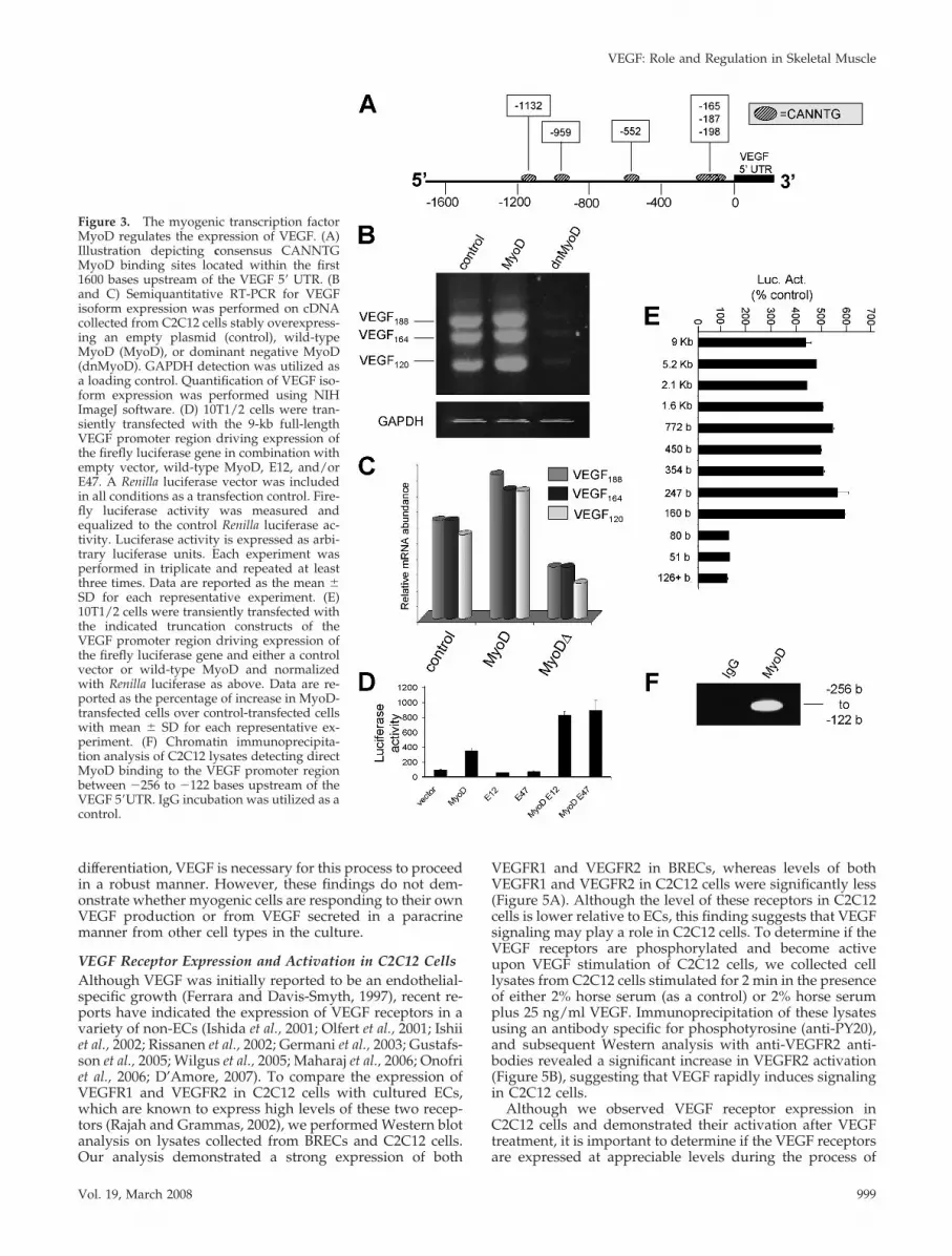
The Myogenic Transcription Factor MyoD Controls VEGFTranscriptional ExpressionGiven the unique expression patterns of the VEGF isoformsduring skeletal myogenesis, we sought to determine themolecular mechanisms controlling this process. Using TESSanalysis (http://www.cbil.upenn.edu/cgi-bin/tess/tess) ofthe human VEGF promoter, we identified six putativeCANNTG sites within the promoter region, 1600 bases up-stream of the coding region (Figure 3A). MyoD homodimersor heterodimers of MyoD plus E12 or E47 serve as transcrip-tion factor complexes that bind to CANNTG consensus sitesin the promoter regions of genes, performing major func-tions in specification and differentiation of skeletal muscleprecursor cells (Murre et al., 1989).
These predictions led us to test whether MyoD regulatesVEGF expression during the muscle differentiation program.In C2C12 cells we stably overexpressed a control vector, awild-type MyoD, or a dominant negative MyoD (dnMyoD)in which MyoD is fused to the lysosomal protease cathepsinB, thus proteolytically digesting any MyoD multimersand/or detour the multimers from their usual subcellulardestination to the lysosome (Li et al., 1996). We then usedsemiquantitative RT-PCR to examine the endogenous VEGFexpression levels in each condition. Overexpression of wild-type MyoD led to a significant increase in VEGF isoformlevels over the control condition, whereas dnMyoD expres-sion resulted in a marked reduction in VEGF isoform ex-pression compared with the control (Figure 3B and C). Thesedata suggest that MyoD is necessary to maintain a baselinelevel of VEGF transcriptional expression in C2C12. This isconsistent with our findings that C2C12 cells, which ex-pressed endogenous MyoD, synthesize higher levels ofVEGF than 10T1/2 cells, which lack endogenous MyoD(Figure 2A).
To confirm our finding that MyoD modulates VEGF ex-pression in skeletal muscle precursor cells, we utilized lu-ciferase reporter assays. C2C12 cells were transfected withthe 9-kb (full length) VEGF promoter luciferase reporterplasmid and combinations of 1) control vector, 2) wild-typeMyoD, 3) E12, or 4) E47, and luciferase assays were per-formed on the cell lysates. As observed in Figure 3D, coex-pression of the VEGF luciferase plasmid with MyoD led toan approximately fourfold increase in VEGF-reporter ex-pression over the control. Transfection with either E12 orE47 alone resulted in no increase in VEGF-reporter expres-sion; however, cotransfection of either MyoD and E12 orMyoD and E47 led to eightfold increase in luciferase activityover the control. These data suggest that MyoD and theE-box proteins act synergistically to up-regulate VEGF-re-porter expression.
We next set out to determine the specific transcriptionalbinding sites that mediate the MyoD-dependent increase ofVEGF expression. C2C12 cells were transfected with trunca-tion mutants of the VEGF promoter luciferase reporter plas-mid and either a control plasmid or wild-type MyoD. StrongMyoD-driven VEGF-reporter expression was observed infull-length promoter constructs and truncations down to�160 bases upstream of the transcriptional start site (Figure3E). However, truncations less than this region led to com-plete loss of MyoD-mediated transcriptional expression of
Figure 1. VEGF is expressed in embryonic and adult skeletal mus-cle. (A) Cryosections of embryonic mouse limbs (E18.5) and wholemounts of adult mouse skeletal muscle were collected from VEGF-LacZ mice and stained for LacZ using in situ �-galactosidase. Scalebar (E18.5) 50 �m; scale bar (adult) 100 �m. Support of mRNA wascollected from the hindlimb skeletal muscle of 2-mo-old C57BL/6mice, and semiquantitative RT-PCR detection of VEGF188, VEGF164,and VEGF120 was performed. As a reaction control, a PCR reactioncomposed of mixed VEGF plasmids each encoding single VEGFisoforms was utilized. (C) mRNA was collected from the hindlimbskeletal muscle of 2-mo-old C57BL/6 mice and real-time RT-PCRdetection of VEGF188, VEGF164, and VEGF120 was performed. Therelative amount of each VEGF isoform is represented as a percent-age of the total VEGF expression (VEGF188 � VEGF164 � VEGF120 �100%). Error bars, SDs.
VEGF: Role and Regulation in Skeletal Muscle
Vol. 19, March 2008 997

the reporter gene, suggesting that a region between �247bases and �160 bases upstream of the transcription start siteis essential for MyoD activity on this promoter. Indeed,within this region lies three closely spaced CANNTG sites at�165, �187, and �198 bases upstream of the VEGF 5� UTR.
MyoD Physically Associates with the VEGF-PromoterTo demonstrate that MyoD directly interacts and forms acomplex with the MyoD sites in this region, we performedChIP using sheared DNA isolated from C2C12 cells. Immu-noprecipitation of chromatin-bound DNA using an antibodyagainst endogenous MyoD was followed by PCR usingprimers that amplified the �256- to �122-base pair region ofthe VEGF promoter spanning the length of the three essen-tial consensus MyoD sites. As demonstrated in Figure 3F,the MyoD-specific antibody is capable of immunoprecipitat-ing the VEGF promoter fragment containing the three MyoDsites, whereas control immunoprecipitation using anti-IgGfailed to produce a PCR product.
VEGF-Null Embryonic Stem Cells Exhibit DecreasedSkeletal MyogenesisThe essential role of VEGF during vasculogenesis and an-giogenesis is well established (Ferrara, 1999); however, verylittle is known regarding nonvascular roles for VEGF. Todetermine if VEGF influences skeletal myogenic differenti-ation, we utilized a CEB model in which ECs are cultivatedto form aggregates, which efficiently differentiate into anumber of cell lineages, including skeletal muscle. This sys-
tem closely recapitulates the early steps of muscle develop-ment in vivo (Rohwedel et al., 1994) and serves as an excel-lent in vitro system to study this process. ES cells isolatedfrom wild-type or VEGF-null mice were differentiated intoCEB and then transferred to gelatin-coated glass coverslips,where they attached, flattened, spread, and differentiated.Over a period of 11 d, we performed immunofluorescentstaining specific for the skeletal muscle specific marker,MHC, and calculated the percentage of each field that waspositive for MHC. Myosin heavy chain was first detected atday 7 in both the wild-type and VEGF-null ES cultures insparse, isolated patches throughout the CEB. At this timepoint staining in wild-type cultures represented �3.5% ofthe total area of the representative fields, whereas MHCstaining in VEGF-null cells represented �0.3% of the totalarea of the representative fields (Figure 4A and B). At days9 and 11 of differentiation, VEGF-null ES cultures consis-tently demonstrated a reduction in MHC expression com-pared with CEB derived from wild-type ES cells. Becausethe MHC antibody (Iowa Hybridoma Bank, MF20) we usedin these experiments is known to react with any striatedmuscle actin, including cardiac tissue, we performed West-ern blots on wild-type and VEGF-null ES cell lysates at day9 of differentiation to detect the expression of the skeletalmuscle specific protein, myogenin. As expected, a significantreduction in myogenin protein expression is observed inVEGF-null ES cell lysates compared with wild-type ES celllysates (Figure 4C). These data demonstrate that while theloss of VEGF does not appear to affect the onset of myogenic
Figure 2. VEGF expression is regulated during skele-tal myogenic differentiation. (A) Semiquantitative RT-PCR for VEGF isoform and MyoD expression was per-formed on cell lysates of C2C12 and 10T1/2 cells.Steady-state mRNA levels were detected and GAPDHexpression was utilized to indicate equal loading. (B)Phase-contrast images of C2C12 cells were taken in theproliferative phase (GM) and after 3 d of differentiation(DM). (Arrows indicate the presence of myotubes after3-d differentiation, 40� magnification) (C) Western blotanalysis of the muscle-lineage–specific marker MyoDwas performed on lysates collected from proliferating(GM) and 3-d differentiated (DM) C2C12 cells. Tubulinexpression was utilized to indicate equal loading. (D–G)RNA was collected from C2C12 and 10T1/2 cells in theproliferative phase of growth (GM) and over 4 d ofdifferentiation (DM1-4). Semiquantitative RT-PCR forVEGF isoform expression was performed for each timepoint for C2C12 (D) and 10T1/2 (F). Analysis ofGAPDH expression was utilized as a loading control.Quantification of VEGF isoform expression duringC2C12 (E) and 10T1/2 (G) differentiation was per-formed using NIH ImageJ software.
B. A. Bryan et al.
Molecular Biology of the Cell998
differentiation, VEGF is necessary for this process to proceedin a robust manner. However, these findings do not dem-onstrate whether myogenic cells are responding to their ownVEGF production or from VEGF secreted in a paracrinemanner from other cell types in the culture.
VEGF Receptor Expression and Activation in C2C12 CellsAlthough VEGF was initially reported to be an endothelial-specific growth (Ferrara and Davis-Smyth, 1997), recent re-ports have indicated the expression of VEGF receptors in avariety of non-ECs (Ishida et al., 2001; Olfert et al., 2001; Ishiiet al., 2002; Rissanen et al., 2002; Germani et al., 2003; Gustafs-son et al., 2005; Wilgus et al., 2005; Maharaj et al., 2006; Onofriet al., 2006; D’Amore, 2007). To compare the expression ofVEGFR1 and VEGFR2 in C2C12 cells with cultured ECs,which are known to express high levels of these two recep-tors (Rajah and Grammas, 2002), we performed Western blotanalysis on lysates collected from BRECs and C2C12 cells.Our analysis demonstrated a strong expression of both
VEGFR1 and VEGFR2 in BRECs, whereas levels of bothVEGFR1 and VEGFR2 in C2C12 cells were significantly less(Figure 5A). Although the level of these receptors in C2C12cells is lower relative to ECs, this finding suggests that VEGFsignaling may play a role in C2C12 cells. To determine if theVEGF receptors are phosphorylated and become activeupon VEGF stimulation of C2C12 cells, we collected celllysates from C2C12 cells stimulated for 2 min in the presenceof either 2% horse serum (as a control) or 2% horse serumplus 25 ng/ml VEGF. Immunoprecipitation of these lysatesusing an antibody specific for phosphotyrosine (anti-PY20),and subsequent Western analysis with anti-VEGFR2 anti-bodies revealed a significant increase in VEGFR2 activation(Figure 5B), suggesting that VEGF rapidly induces signalingin C2C12 cells.
Although we observed VEGF receptor expression inC2C12 cells and demonstrated their activation after VEGFtreatment, it is important to determine if the VEGF receptorsare expressed at appreciable levels during the process of
Figure 3. The myogenic transcription factorMyoD regulates the expression of VEGF. (A)Illustration depicting consensus CANNTGMyoD binding sites located within the first1600 bases upstream of the VEGF 5� UTR. (Band C) Semiquantitative RT-PCR for VEGFisoform expression was performed on cDNAcollected from C2C12 cells stably overexpress-ing an empty plasmid (control), wild-typeMyoD (MyoD), or dominant negative MyoD(dnMyoD). GAPDH detection was utilized asa loading control. Quantification of VEGF iso-form expression was performed using NIHImageJ software. (D) 10T1/2 cells were tran-siently transfected with the 9-kb full-lengthVEGF promoter region driving expression ofthe firefly luciferase gene in combination withempty vector, wild-type MyoD, E12, and/orE47. A Renilla luciferase vector was includedin all conditions as a transfection control. Fire-fly luciferase activity was measured andequalized to the control Renilla luciferase ac-tivity. Luciferase activity is expressed as arbi-trary luciferase units. Each experiment wasperformed in triplicate and repeated at leastthree times. Data are reported as the mean �SD for each representative experiment. (E)10T1/2 cells were transiently transfected withthe indicated truncation constructs of theVEGF promoter region driving expression ofthe firefly luciferase gene and either a controlvector or wild-type MyoD and normalizedwith Renilla luciferase as above. Data are re-ported as the percentage of increase in MyoD-transfected cells over control-transfected cellswith mean � SD for each representative ex-periment. (F) Chromatin immunoprecipita-tion analysis of C2C12 lysates detecting directMyoD binding to the VEGF promoter regionbetween �256 to �122 bases upstream of theVEGF 5�UTR. IgG incubation was utilized as acontrol.
VEGF: Role and Regulation in Skeletal Muscle
Vol. 19, March 2008 999

skeletal myogenesis. To address this, we collected cell ly-sates from C2C12 cultures in their proliferative phase andthroughout myogenic differentiation. Western blot analysisrevealed an inverse relationship between the expression ofVEGF receptors and differentiation (Figure 5C); althoughVEGFR1 expression was highest in the proliferative stateand gradually decreased during myogenic differentiation,VEGFR2 expression was relatively low in the proliferativestate and significantly increased during differentiation.These data suggest that the induction of VEGF expressionduring skeletal myogenesis via MyoD-dependent up-regu-lation may induce an autocrine signaling pathway that per-forms a necessary role in myogenic differentiation.
VEGF Stimulation of C2C12 Cells Enhances MyogenicDifferentiationGiven that C2C12 cells provide a well-documented model inwhich to examine the effects of VEGF on myogenesis, weutilized phase-contrast microscopy to quantify myotube for-mation during C2C12 myogenic differentiation in responseto VEGF stimulation or inhibition. As observed in Figure 6Aand C, the addition of 25 ng/ml VEGF to differentiatingC2C12 cells resulted in an �30% increase in the number ofmyotubes compared with untreated control cells. Moreover,inhibition of autocrine VEGF signaling via the expression ofsFlt1 led to an �70% reduction in myotube numbers relativeto AV-control (Figure 6A and C). We confirmed these resultsusing fluorescent detection of the skeletal muscle–specificdifferentiation marker, MHC. Similar to our findings mea-suring myotube numbers, addition of VEGF led to an in-crease in MHC-positive cells compared with cells that re-ceived control treatment (Figure 6B), whereas inhibition ofautocrine VEGF signaling using sFlt1 resulted in decreasedMHC-positive cells relative to controls (Figure 6B). Addi-tionally, VEGF stimulation of differentiating C2C12 cellsresulted in a significant increase in myotube hypertrophy,although sFlt1 inhibition of VEGF signaling led to a decrease
Figure 4. VEGF regulates skeletal myogenesis during embryonicstem cell differentiation. (A) ES cells were differentiated using thehanging drop method as described in Materials and Methods. At 7, 9,and 11 d of differentiation, cells were fixed and MHC was detectedby immunofluorescence (green) and DAPI (blue) was performed(40� magnification). (B) Quantification of MHC positive area withinthe field was performed using NIH ImageJ software. Data arereported as the mean � SD for each representative experiment. (C)Lysates from ES cells were collected at day 9 of differentiation andWestern-blotted with anti-myogenin and anti-tubulin antibodies.
Figure 5. VEGF receptor expression and activation during skeletalmyogenesis. (A) Lysates from BREC and C2C12 cells were Westernblotted for VEGFR1 and VEGFR2. Tubulin levels were utilized as aloading control. (B) Lysates from C2C12 cells grown in 2% horseserum with (VEGF) or without (control) the 25 ng/ml VEGF (2 min)were immunoprecipitated with anti-VEGFR2 antibodies and West-ern blotted with anti-PY20 and anti-VEGF-R2 antibodies were per-formed to detect VEGFR2 phosphorylation. (C) Lysates from C2C12cells in proliferative conditions (GM) and days 1 through 4 ofmyogenic differentiation (DM1-4) were subjected to Western anal-ysis to detect levels of VEGFR1 and VEGFR2. Detection of MHClevels demonstrated myogenic differentiation and tubulin proteinlevels were used as a loading control.
B. A. Bryan et al.
Molecular Biology of the Cell1000

in myotube size compared with controls (Figure 6D). Theseresults strongly suggest that VEGF signals to C2C12 cellsthrough an autocrine mechanism.
VEGF Regulates C2C12 Cell Fate DecisionsTo determine the mechanism by which VEGF promotesmyogenic differentiation, we examined its effect on MHCexpression in C2C12 cells. At day 3 of myogenic differenti-ation, levels of MHC were up-regulated in VEGF-treatedcells compared with controls (Figure 7A), revealing thatVEGF is capable of promoting skeletal myogenic differenti-ation by up-regulating key proteins involved in musclefunction. Moreover, VEGF neutralization resulted in a sig-nificant down-regulation of MHC expression comparedwith controls, suggesting that VEGF is essential for completemyogenic differentiation.
Myoblast migration is central to both developmental andregenerative myogenesis (Schultz and McCormick, 1994;Christ and Brand-Saberi, 2002), in which differentiation, mi-gration, and cell-to-cell contacts are coupled (O’Connor etal., 2007). Given that VEGF has been shown to induce cellmigration in a number of cell types (Gruber et al., 1995;Ratajska et al., 1995; Yoshida et al., 1996; Grosskreutz et al.,1999; Castellon et al., 2002), it seemed likely that it wouldperform a similar function during myoblast migration. Totest this possibility, we performed invasion/migration as-
says on C2C12 cells treated with 25 ng/ml VEGF, 100 ng/mlrecombinant sFlt1, or with untreated cells as a control. Con-fluent monolayers of cells were physically wounded with ascratch and allowed to migrate in order to “heal” for 12 h,eliminating the possibility of proliferation accounting for thewound closure. As demonstrated in Figure 7B, at 12 h afterthe initial scratch VEGF-treated cells had completely closedthe wound, whereas sFlt1-treated cells exhibited a signifi-cant reduction in invasion/migration compared with thecontrol. These data suggest that VEGF stimulates C2C12 cellmigration and that endogenous VEGF plays an essential rolein C2C12 invasion/migration.
In addition to its action on the endothelium, VEGF hasbeen heavily implicated as a survival factor for nonvascularcells (Zachary, 2001; Darland et al., 2003; Nishijima et al.,2007). To determine if the promyogenic effect of VEGF sig-naling might occur through its anti-apoptotic signaling, dif-ferentiated C2C12 cells were treated with 100 ng/ml recom-binant sFlt1 for 48 h and examined for apoptosis viaAnnexin V:propidium iodide staining and subsequent flowcytometric analysis. As demonstrated in Figure 7C, sFlt1-treated cells exhibited an �50% increase in the number ofapoptotic cells compared with control cells, suggesting thatVEGF signaling is essential for cell survival during C2C12differentiation, an effect that may contribute to the promyo-genic effects of VEGF.
Figure 6. VEGF regulates C2C12 skeletalmyogenic differentiation. (A and B) C2C12cells were differentiated for 3 d in the follow-ing conditions: control, VEGF, AV-control, orAV-sFlt1. Phase-contrast images (40� magni-fication; A) and immunofluorescent detectionof MHC (MHC; green) and DAPI (blue; 100�magnification; B) were collected at day 3 ofdifferentiation. (C) Quantification of myotubenumber per field was performed using NIHImageJ software. Data are reported as themean � SD for each representative experi-ment. (D) Quantification of relative MHC-pos-itive cell size using NIH Image J software (reddashed line indicates median size of MHCpositive control or AV-control cells).
VEGF: Role and Regulation in Skeletal Muscle
Vol. 19, March 2008 1001

Muscles are formed by fusion of individual postmitoticmyoblasts to form multinucleated myotubes; however, dur-ing skeletal muscle regeneration, which is associated withroutine maintenance, hypertrophy, and repair, satellite stemcells are activated from their quiescent state, proliferate, andfinally differentiate (Adams, 2006; Zammit et al., 2006). Be-cause VEGF is a mitogen for ECs (Leung et al., 1989), wetested whether it might also regulate myoblast proliferation.Using CFDASE cell labeling as a measure of cell prolifera-tion, we observed that sFlt1-treated cells retained signifi-cantly more CFDASE than control cells, indicating thatVEGF neutralization reduced the division rate of C2C12 cells(Figure 7D). We found no difference in CFDASE retentionbetween control and VEGF-treated cells (data not shown);however, this was not surprising given that the cells weregrown in 10% FBS and were likely replicating at maximumcapacity. Given the tendency of C2C12 cells to undergodifferentiation and thereby enter G0 at lower serum concen-trations, we did not test the mitogenic effects of VEGF at lowserum concentrations.
DISCUSSION
The formation of vascular systems is essential for normalgrowth, differentiation, and organ function where the vas-cular bed is specialized to meet the needs of the organ itsupplies. To achieve the high degree of structural organiza-tion necessary to serve the specific tissue requirements,blood vessel formation is tightly regulated by the specificexpression and secretion of factors such as VEGF. VEGF hasbeen shown to be expressed at varying levels in a cell-specific manner in virtually all vascularized adult tissues(Maharaj et al., 2006) and is up-regulated in a number of celltypes in response to hypoxic conditions (Liu et al., 1995;Forsythe et al., 1996; Mazure et al., 1996). The data presentedin this report demonstrate a novel regulatory mechanism bywhich VEGF is up-regulated during skeletal muscle differ-entiation by the myogenic regulatory factor, MyoD. More-over, although VEGF is well established as a modulator ofblood vessel formation (Ferrara et al., 2003), the expression ofVEGF receptors by many cell types in addition to EC pointsto nonvascular roles for VEGF in the adult (Maharaj et al.,2006; D’Amore, 2007). Indeed, our data suggest that VEGFmediates skeletal myogenesis and that VEGF expressed bydifferentiating myogenic precursors functions in an auto-crine manner.
Through differential mRNA splicing, the single murineVEGF gene gives rise to at least three isoforms including,VEGF120, VEGF164, and VEGF188 (Ferrara et al., 1992; Shimaet al., 1996). VEGF120 lacks a heparan sulfate binding site andis therefore freely diffusible, whereas VEGF188, with twoheparan sulfate binding sites, associates with the cell surfaceand extracellular matrix, and VEGF164 has intermediateproperties (Park et al., 1993; Ferrara and Davis-Smyth, 1997).The various VEGF isoforms play distinct roles in vasculardevelopment, and their patterns of expression vary duringorgan development and in the adult (Ng et al., 2001; Robin-son and Stringer, 2001; Skryabin et al., 2004). Our data dem-onstrating VEGF188 and VEGF164 expression in skeletal mus-cle is consistent with a number of other studies in human,rat, and mouse (Ng et al., 2001; Ishii et al., 2002; Jensen et al.,2004; Maharaj et al., 2006). Given that skeletal muscle issubjected to a considerable amount of daily stresses includ-ing exercise-induced hypoxia (Hoppeler, 1999) and struc-tural damage (Russell et al., 1992), necessitating angiogene-sis, VEGF expression in this tissue is not surprising. Inaddition, we demonstrate that multipotent cell lines (C2C12and 10T1/2), which under the appropriate conditions aremolecularly similar to myogenic satellite stem cells, expressall three VEGF isoforms. These data suggest that myogenicprecursors, in addition to expressing the sequestered formsof VEGF (VEGF188 and VEGF164), may express diffusibleVEGF120, which could function to attract distant vessels tosites of myoblast differentiation. Although multiple reportsdemonstrate variable tissue-specific isoform expression pat-terns for VEGF, the mechanism controlling this process re-mains unknown.
HIF1 serves as a master regulator for the expression of anumber of genes involved in the hypoxic response, includ-ing glucose transporters and glycolytic enzymes as well asVEGF (Damert et al., 1997; Minchenko et al., 2002; Mobasheriet al., 2005), and hypoxic regulation of VEGF expression viaHIF-1 is the best characterized mechanism of VEGF control.Interestingly, deletion of the hypoxia-response element inthe VEGF promoter in mice results in a viable mouse withonly minor phenotypes (Oosthuyse et al., 2001). In contrast,VEGF�/� and �/� mice are embryonic lethal (Ferrara etal., 1996). Thus, the dispensable nature of HIF-1 suggests
Figure 7. VEGF serves multiple roles in C2C12 cells. (A) Lysateswere collected from C2C12 cells grown at 3 d of differentiation andtreated with 25 ng/ml VEGF, 100 ng/ml sFlt1, or left untreated andsubsequently subjected to Western analysis to detect steady-statelevels of MHC. Tubulin was utilized as a loading control. (B) C2C12cells were grown to 100% confluence, treated as in A and woundedwith a sterile pipette tip to remove cells. Photographs were taken(40� magnification) at t � 0 and t � 12 h (control, VEGF, and sFlt1)after the injury. (C) Fluorescence-activated cell sorter analysis forannexin V:propidium iodide staining of C2C12 cells at day 2 ofdifferentiation and treated 100 ng/ml sFlt1. (D) Flow cytometricanalysis of CFDASE stability in proliferating C2C12 cells treatedwith 25 ng/ml VEGF or 100 ng/ml sFlt1 (red dashed line indicatesmedian fluorescence of control cells).
B. A. Bryan et al.
Molecular Biology of the Cell1002

that HIF-1 is much less important in the regulation of VEGFthan previously suspected. Furthermore, there are a smallbut growing number of reports documenting mechanismsthat modulate VEGF expression in normoxic conditions. Forinstance, myogenic Akt signaling controls both muscle fiberhypertrophy and VEGF synthesis, independent of HIF-1activity, illustrating a mechanism through which blood ves-sel recruitment can be coupled to normal tissue growth(Takahashi et al., 2002). Moreover, analysis of VEGF mRNAlevels across an array of adult tissues demonstrates a widevariability in the expression pattern for VEGF (Ng et al.,2001), suggesting complex transcriptional modulation of thisgene. TESS promoter analysis (http://www.cbil.upenn.edu/cgi-bin/tess/tess) of the human VEGF promoter reveals awide array of cell-type specific putative transcriptional reg-ulatory sites for transcription factors, such as c-Myb (in-volved in proliferation and differentiation of hematopoeticprogenitor cells), myogenin (involved in differentiation ofskeletal muscle), T-cell factor (TCF; involved in T-cell spe-cific differentiation), and peroxisome proliferator-activatedreceptor (PPAR; transcription factor specific for liver, kid-ney, heart, and brown adipose tissue), to name only a few.
Our observation of a number of putative CANNTGMyoD-binding sites within 9 kb upstream of the VEGFtranscriptional start site motivated us to investigate if skel-etal muscle-specific regulation of VEGF exists independentof hypoxic regulation. In skeletal muscle, MyoD functions inan instructive chromatin context and directly regulates genesexpressed throughout the myogenic program (McKinsey et al.,2001; Pownall et al., 2002). For example, expression ofMyoD is sufficient to convert fibroblast and adipoblastcells into skeletal muscle cells (Tapscott et al., 1988). In-deed, our analysis revealed that VEGF is transcriptionallyup-regulated during skeletal muscle differentiation viathe myogenic transcription factor MyoD and its bindingpartners, E12 and E47. Many studies have demonstratedVEGF up-regulation in skeletal muscle regeneration,transplantation, and exercise models (Rissanen et al., 2002;Smythe et al., 2002; Arsic et al., 2004; Prior et al., 2004; Yanet al., 2005; Wagatsuma, 2007; Xia et al., 2006), and ourreport expands on that knowledge by providing a specificmechanism by which VEGF is up-regulated during normalskeletal muscle differentiation. Indeed, in light of the manyputative tissue-specific transcription factor binding sites lo-cated in the VEGF promoter, we speculate that the differen-tiation-dependent expression of VEGF that we have docu-mented in skeletal muscle may be a paradigm for VEGFregulation in adult tissues.
The most studied aspects of VEGF in skeletal muscle in-volves its role in inducing angiogenesis during muscle forma-tion and after bouts of exercise (Wagner, 2001; Haas, 2002;Bloor, 2005). Two contradicting studies have previously at-tempted to examine the effects of VEGF on skeletal myogen-esis. One study suggests that VEGF strongly affects myocytemigration and survival but reports no effect on myogenicmarker expression (Germani et al., 2003). The interpretation ofthese results remains complicated because the authors reportvery early expression of MHC, in contrast to a large number ofreports that demonstrate that MHC is only detectable by West-ern blotting in C2C12 cells at 2 to 3 d of differentiation (Miller,1990; Andres and Walsh, 1996). Another report demonstratedincreased proliferation and decreased apoptosis in C2C12 cellsin response to exogenous VEGF, as well as an increase inmyotube number, size, and multinucleation (Arsic et al., 2004);however, this group did not examine a possible autocrine rolefor VEGF.
We have comprehensively examined the role and regula-tion of VEGF during skeletal myogenesis and demonstratethat VEGF stimulation increases myotube number, myo-genic marker expression, and myotube size, whereas inhibi-tion of VEGF signaling decreases these processes. These dataindicate that VEGF may be essential for proper myogenicdifferentiation. Given the ubiquitous distribution of VEGF,we speculate that VEGF may perform similar autocrine rolesfor a number of developmental processes. For instance,VEGF is overexpressed in chondrogenic and osteogenic dif-ferentiation cascades, and inhibition of VEGF activity resultsin reduced bone differentiation, suggesting that this factorplays an important role during cartilage and bone formation(Mayer et al., 2005; Bluteau et al., 2007). Moreover, differen-tiating kidney podocytes strongly up-regulate VEGF andVEGFR2, and treatment with VEGF reduces podocyte apo-ptosis �40%, suggesting VEGF promotes survival in thesecells through VEGFR2 (Guan et al., 2006). Genetic overex-pression of VEGF in adult rats results in an approximatelytwofold increase in hippocampal neurogenesis associatedwith improved cognition, whereas inhibition of VEGF ex-pression by RNA interference completely blocks the envi-ronmental induction of neurogenesis, suggesting that VEGFserves as a mediator of neurogenesis and cognition (Duringand Cao, 2006). It seems clear that future studies will eluci-date more instances where VEGF is utilized for the functionof nonvascular cell types.
The coordinated regulation of the VEGF receptors duringskeletal myogenesis possibly reflects the underlying biologyof the system. Our data suggest that proliferating, undiffer-entiated C2C12 cells predominantly express VEGFR1; how-ever, differentiating C2C12 cells down-regulate VEGFR1 anddramatically increase the expression of VEGF2. Because thedifferentiated C2C12 cells have completely exited the cellcycle, VEGFR2 signaling no longer leads to cell proliferationbut instead appears to contribute to myogenic differentia-tion. A similar induction of VEGFR2 expression has beenobserved in differentiation of podocytes (Guan et al., 2006).The expression of VEGFR2 has been shown in other systemsto be induced by VEGF (Enholm et al., 2001), and given ourdata indicate that VEGF is up-regulated during myogenicdifferentiation, this may account for the observed increasedVEGFR2 expression. Our findings that VEGF can mediatemyogenic differentiation and survival are consistent with anautocrine role for VEGFR2 signaling in differentiating anddifferentiated skeletal muscle. At the same time, the VEGFproduced by the differentiating myocytes is necessary forthe vascularization of the developing skeletal muscle.
Although these results are important in elucidating themolecular mechanisms controlling tissue-specific vasculardevelopment and maintenance, they also present broadermedical implications for anti-angiogenic treatment for dis-eases such as cancer and macular degeneration. Patientsundergoing long-term anti-VEGF therapy may experienceunexpected side effects due to interference with normal ves-sel regrowth/stability in skeletal muscle tissue and/or pro-longed muscle regeneration time after injury. For example,although developmental differentiation of skeletal muscledoes not occur in the adult, day-to-day wear and tear ofadult skeletal muscle accounts for �1–2% replacement ofmuscle fibers per week (Schmalbruch and Lewis, 2000)—processes that involve satellite stem cell activation andsubsequent myogenic differentiation and that could be sus-ceptible to anti-VEGF therapies. Consistent with this specu-lation, patients treated with Avastin report asthenia, which,though nonspecific, may result from reduced muscle regen-eration. Because of the low rate of skeletal muscle turnover/
VEGF: Role and Regulation in Skeletal Muscle
Vol. 19, March 2008 1003

repair, abnormalities might be expected to require ex-tended treatment to develop. In addition, because patientsreceiving Avastin often exhibit a spectrum of medicalissues, subtle changes in muscle function might be diffi-cult to detect. A beneficial effect of VEGF overexpressionhas been reported for skeletal muscle regenerationthrough both proangiogenic effects and myogenic regen-erative effects (Arsic et al., 2004; Messina et al., 2007).These observations suggest that anti-VEGF drugs couldinhibit muscle regeneration through dual mechanisms—blocking angiogenesis and blocking satellite stem cell ac-tivation and subsequent myogenesis.
ACKNOWLEDGMENTS
We thank Dr. M. Horwitz from the University of Washington, Seattle, WA, forthe use of the dominant negative MyoD plasmid construct and Dr. M. Liufrom the Texas A&M University Health Science Center, Houston, TX, for theuse of the wild-type MyoD plasmid construct. This study was supported bythe National Institutes of Health Grants EY05318, EY015435, and CA45548.P.A.D. is a Research to Prevent Blindness Senior Scientific Investigator.
REFERENCES
Adams, G. R. (2006). Satellite cell proliferation and skeletal muscle hypertro-phy. Appl. Physiol. Nutr. Metab. 31, 782–790.
Andres, V., and Walsh, K. (1996). Myogenin expression, cell cycle withdrawal,and phenotypic differentiation are temporally separable events that precedecell fusion upon myogenesis. J. Cell Biol. 132, 657–666.
Arsic, N., Zacchigna, S., Zentilin, L., Ramirez-Correa, G., Pattarini, L., Salvi,A., Sinagra, G., and Giacca, M. (2004). Vascular endothelial growth factorstimulates skeletal muscle regeneration in vivo. Mol. Ther. 10, 844–854.
Bacic, M., Edwards, N. A., and Merrill, M. J. (1995). Differential expression ofvascular endothelial growth factor (vascular permeability factor) forms in rattissues. Growth Factors 12, 11–15.
Bains, W., Ponte, P., Blau, H., and Kedes, L. (1984). Cardiac actin is the majoractin gene product in skeletal muscle cell differentiation in vitro. Mol. Cell.Biol. 4, 1449–1453.
Ballaun, C., Weninger, W., Uthman, A., Weich, H., and Tschachler, E. (1995).Human keratinocytes express the three major splice forms of vascular endo-thelial growth factor. J. Invest. Dermatol. 104, 7–10.
Bloor, C. M. (2005). Angiogenesis during exercise and training. Angiogenesis8, 263–271.
Bluteau, G., Julien, M., Magne, D., Mallein-Gerin, F., Weiss, P., Daculsi, G.,and Guicheux, J. (2007). VEGF and VEGF receptors are differentially ex-pressed in chondrocytes. Bone 40, 568–576.
Byun, J. H., Park, B. W., Kim, J. R., and Lee, J. H. (2007). Expression of vascularendothelial growth factor and its receptors after mandibular distraction os-teogenesis. Int. J. Oral Maxillofac. Surg. 36, 338–344.
Castellon, R., Hamdi, H. K., Sacerio, I., Aoki, A. M., Kenney, M. C., andLjubimov, A. V. (2002). Effects of angiogenic growth factor combinations onretinal endothelial cells. Exp. Eye Res. 74, 523–535.
Cheng, S. Y., Nagane, M., Huang, H. S., and Cavenee, W. K. (1997). Intrace-rebral tumor-associated hemorrhage caused by overexpression of the vascularendothelial growth factor isoforms VEGF121 and VEGF165 but not VEGF189.Proc. Natl. Acad. Sci. USA 94, 12081–12087.
Christ, B., and Brand-Saberi, B. (2002). Limb muscle development. Int. J. Dev.Biol. 46, 905–914.
D’Amore, P. A. (2007). Vascular endothelial cell growth factor-a: not just forendothelial cells anymore. Am J. Pathol. 171, 14–18.
Damert, A., Machein, M., Breier, G., Fujita, M. Q., Hanahan, D., Risau, W., andPlate, K. H. (1997). Up-regulation of vascular endothelial growth factor ex-pression in a rat glioma is conferred by two distinct hypoxia-driven mecha-nisms. Cancer Res. 57, 3860–3864.
Darland, D. C., Massingham, L. J., Smith, S. R., Piek, E., Saint-Geniez, M., andD’Amore, P. A. (2003). Pericyte production of cell-associated VEGF is differ-entiation-dependent and is associated with endothelial survival. Dev. Biol.264, 275–288.
Davis, R. L., Weintraub, H., and Lassar, A. B. (1987). Expression of a singletransfected cDNA converts fibroblasts to myoblasts. Cell 51, 987–1000.
During, M. J., and Cao, L. (2006). VEGF, a mediator of the effect of experienceon hippocampal neurogenesis. Curr. Alzheimer Res. 3, 29–33.
Enholm, B., Karpanen, T., Jeltsch, M., Kubo, H., Stenback, F., Prevo, R.,Jackson, D. G., Yla-Herttuala, S., and Alitalo, K. (2001). Adenoviral expressionof vascular endothelial growth factor-C induces lymphangiogenesis in theskin. Circ. Res. 88, 623–629.
Esser, S., Wolburg, K., Wolburg, H., Breier, G., Kurzchalia, T., and Risau, W.(1998). Vascular endothelial growth factor induces endothelial fenestrations invitro. J. Cell Biol. 140, 947–959.
Ferrara, N., Houck, K., Jakeman, L., and Leung, D. W. (1992). Molecular andbiological properties of the vascular endothelial growth factor family ofproteins. Endocr. Rev. 13, 18–32.
Ferrara, N., Carver-Moore, K., Chen, H., Dowd, M., Lu, L., O’Shea, K. S.,Powell-Braxton, L., Hillan, K. J., and Moore, M. W. (1996). Heterozygousembryonic lethality induced by targeted inactivation of the VEGF gene.Nature 380, 439–442.
Ferrara, N., and Davis-Smyth, T. (1997). The biology of vascular endothelialgrowth factor. Endocr. Rev. 18, 4–25.
Ferrara, N. (1999). Molecular and biological properties of vascular endothelialgrowth factor. J. Mol. Med. 77, 527–543.
Ferrara, N., Gerber, H. P., and LeCouter, J. (2003). The biology of VEGF andits receptors. Nat. Med. 9, 669–676.
Fons, P., Herault, J. P., Delesque, N., Tuyaret, J., Bono, F., and Herbert, J. M.(2004). VEGF-R2 and neuropilin-1 are involved in VEGF-A-induced differen-tiation of human bone marrow progenitor cells. J. Cell. Physiol. 200, 351–359.
Forsythe, J. A., Jiang, B. H., Iyer, N. V., Agani, F., Leung, S. W., Koos, R. D.,and Semenza, G. L. (1996). Activation of vascular endothelial growth factorgene transcription by hypoxia-inducible factor 1. Mol. Cell. Biol. 16, 4604–4613.
Germani, A., Di Carlo, A., Mangoni, A., Straino, S., Giacinti, C., Turrini, P.,Biglioli, P., and Capogrossi, M. C. (2003). Vascular endothelial growth factormodulates skeletal myoblast function. Am J. Pathol. 163, 1417–1428.
Grosskreutz, C. L., Anand-Apte, B., Duplaa, C., Quinn, T. P., Terman, B. I.,Zetter, B., and D’Amore, P. A. (1999). Vascular endothelial growth factor-induced migration of vascular smooth muscle cells in vitro. Microvasc Res. 58,128–136.
Gruber, B. L., Marchese, M. J., and Kew, R. (1995). Angiogenic factors stim-ulate mast-cell migration. Blood 86, 2488–2493.
Guan, F., Villegas, G., Teichman, J., Mundel, P., and Tufro, A. (2006). Auto-crine VEGF-A system in podocytes regulates podocin and its interaction withCD2AP. Am J. Physiol. Renal Physiol. 291, F422–F428.
Gustafsson, T., and Kraus, W. E. (2001). Exercise-induced angiogenesis-re-lated growth and transcription factors in skeletal muscle, and their modifica-tion in muscle pathology. Front. Biosci. 6, D75–D89.
Gustafsson, T., Ameln, H., Fischer, H., Sundberg, C. J., Timmons, J. A., andJansson, E. (2005). VEGF-A splice variants and related receptor expression inhuman skeletal muscle following submaximal exercise. J. Appl. Physiol. 98,2137–2146.
Haas, T. L. (2002). Molecular control of capillary growth in skeletal muscle.Can. J. Appl. Physiol. 27, 491–515.
Hoppeler, H. (1999). Vascular growth in hypoxic skeletal muscle. Adv. Exp.Med. Biol. 474, 277–286.
Hudlicka, O., Milkiewicz, M., Cotter, M. A., and Brown, M. D. (2002). Hyp-oxia and expression of VEGF-A protein in relation to capillary growth inelectrically stimulated rat and rabbit skeletal muscles. Exp. Physiol. 87, 373–381.
Ishida, A., Murray, J., Saito, Y., Kanthou, C., Benzakour, O., Shibuya, M., andWijelath, E. S. (2001). Expression of vascular endothelial growth factor recep-tors in smooth muscle cells. J. Cell. Physiol. 188, 359–368.
Ishii, H., Oota, I., Arakawa, T., and Takuma, T. (2002). Differential geneexpression of vascular endothelial growth factor isoforms and their receptorsin the development of the rat masseter muscle. Arch. Oral Biol. 47, 505–510.
Jensen, L., Pilegaard, H., Neufer, P. D., and Hellsten, Y. (2004). Effect of acuteexercise and exercise training on VEGF splice variants in human skeletalmuscle. Am J. Physiol. Regul. Integr. Comp. Physiol. 287, R397–R402.
Jin, K., Mao, X. O., and Greenberg, D. A. (2006). Vascular endothelial growthfactor stimulates neurite outgrowth from cerebral cortical neurons via Rhokinase signaling. J. Neurobiol. 66, 236–242.
Kamba, T. et al. (2006). VEGF-dependent plasticity of fenestrated capillaries inthe normal adult microvasculature. Am J. Physiol. Heart Circ. Physiol. 290,H560–H576.
B. A. Bryan et al.
Molecular Biology of the Cell1004

Koopman, G., Reutelingsperger, C. P., Kuijten, G. A., Keehnen, R. M., Pals,S. T., and van Oers, M. H. (1994). Annexin V for flow cytometric detection ofphosphatidylserine expression on B cells undergoing apoptosis. Blood 84,1415–1420.
Lammert, E., Gu, G., McLaughlin, M., Brown, D., Brekken, R., Murtaugh,L. C., Gerber, H. P., Ferrara, N., and Melton, D. A. (2003). Role of VEGF-A invascularization of pancreatic islets. Curr. Biol. 13, 1070–1074.
Leung, D. W., Cachianes, G., Kuang, W. J., Goeddel, D. V., and Ferrara, N.(1989). Vascular endothelial growth factor is a secreted angiogenic mitogen.Science 246, 1306–1309.
Li, F. Q., Coonrod, A., and Horwitz, M. (1996). Preferential MyoD homodimerformation demonstrated by a general method of dominant negative mutationemploying fusion with a lysosomal protease. J. Cell Biol. 135, 1043–1057.
Liu, Y., Cox, S. R., Morita, T., and Kourembanas, S. (1995). Hypoxia regulatesvascular endothelial growth factor gene expression in endothelial cells. Iden-tification of a 5� enhancer. Circ. Res. 77, 638–643.
Loureiro, R. M., Maharaj, A. S., Dankort, D., Muller, W. J., and D’Amore, P. A.(2005). ErbB2 overexpression in mammary cells upregulates VEGF throughthe core promoter. Biochem. Biophys. Res. Commun. 326, 455–465.
Lyons, A. B. (2000). Analysing cell division in vivo and in vitro using flowcytometric measurement of CFSE dye dilution. J. Immunol. Methods 243,147–154.
Maharaj, A. S., Saint-Geniez, M., Maldonado, A. E., and D’Amore, P. A.(2006). Vascular endothelial growth factor localization in the adult. Am J.Pathol. 168, 639–648.
Mayer, H., Bertram, H., Lindenmaier, W., Korff, T., Weber, H., and Weich, H.(2005). Vascular endothelial growth factor (VEGF-A) expression in humanmesenchymal stem cells: autocrine and paracrine role on osteoblastic andendothelial differentiation. J. Cell. Biochem. 95, 827–839.
Mazure, N. M., Chen, E. Y., Yeh, P., Laderoute, K. R., and Giaccia, A. J. (1996).Oncogenic transformation and hypoxia synergistically act to modulate vas-cular endothelial growth factor expression. Cancer Res. 56, 3436–3440.
McKinsey, T. A., Zhang, C. L., and Olson, E. N. (2001). Control of muscledevelopment by dueling HATs and HDACs. Curr. Opin. Genet Dev. 11,497–504.
Messina, S., Mazzeo, A., Bitto, A., Aguennouz, M., Migliorato, A., DePasquale, M. G., Minutoli, L., Altavilla, D., Zentilin, L., Giacca, M., et al.(2007). VEGF overexpression via adeno-associated virus gene transfer pro-motes skeletal muscle regeneration and enhances muscle function in mdxmice. FASEB J. 21, 3737–3746.
Miller, J. B. (1990). Myogenic programs of mouse muscle cell lines: expressionof myosin heavy chain isoforms, MyoD1, and myogenin. J. Cell Biol. 111,1149–1159.
Minchenko, A., Leshchinsky, I., Opentanova, I., Sang, N., Srinivas, V.,Armstead, V., and Caro, J. (2002). Hypoxia-inducible factor-1-mediatedexpression of the 6-phosphofructo-2-kinase/fructose-2,6-bisphosphatase-3(PFKFB3) gene. Its possible role in the Warburg effect. J. Biol. Chem. 277,6183–6187.
Miquerol, L., Gertsenstein, M., Harpal, K., Rossant, J., and Nagy, A. (1999).Multiple developmental roles of VEGF suggested by a LacZ-tagged allele.Dev. Biol. 212, 307–322.
Mobasheri, A., Richardson, S., Mobasheri, R., Shakibaei, M., and Hoyland,J. A. (2005). Hypoxia inducible factor-1 and facilitative glucose transportersGLUT1 and GLUT 3, putative molecular components of the oxygen andglucose sensing apparatus in articular chondrocytes. Histol. Histopathol. 20,1327–1338.
Murre, C. et al. (1989). Interactions between heterologous helix-loop-helixproteins generate complexes that bind specifically to a common DNA se-quence. Cell 58, 537–544.
Ng, Y. S., Rohan, R., Sunday, M. E., Demello, D. E., and D’Amore, P. A. (2001).Differential expression of VEGF isoforms in mouse during development andin the adult. Dev. Dyn. 220, 112–121.
Ng, Y. S., Ramsauer, M., Loureiro, R. M., and D’Amore, P. A. (2004). Identi-fication of genes involved in VEGF-mediated vascular morphogenesis usingembryonic stem cell-derived cystic embryoid bodies. Lab. Invest. 84, 1209–1218.
Nishijima, K. et al. (2007). Vascular endothelial growth factor-A is a survivalfactor for retinal neurons and a critical neuroprotectant during the adaptiveresponse to ischemic injury. Am. J. Pathol. 171, 53–67.
O’Connor, R. S., Mills, S. T., Jones, K. A., Ho, S. N., and Pavlath, G. K. (2007).A combinatorial role for NFAT5 in both myoblast migration and differentia-tion during skeletal muscle myogenesis. J. Cell Sci. 120, 149–159.
Ogunshola, O. O., Antic, A., Donoghue, M. J., Fan, S. Y., Kim, H., Stewart,W. B., Madri, J. A., and Ment, L. R. (2002). Paracrine and autocrine functionsof neuronal vascular endothelial growth factor (VEGF) in the central nervoussystem. J. Biol. Chem. 277, 11410–11415.
Olfert, I. M., Breen, E. C., Mathieu-Costello, O., and Wagner, P. D. (2001).Skeletal muscle capillarity and angiogenic mRNA levels after exercise train-ing in normoxia and chronic hypoxia. J. Appl. Physiol. 91, 1176–1184.
Onofri, C., Theodoropoulou, M., Losa, M., Uhl, E., Lange, M., Arzt, E., Stalla,G. K., and Renner, U. (2006). Localization of vascular endothelial growthfactor (VEGF) receptors in normal and adenomatous pituitaries: detection ofa non-endothelial function of VEGF in pituitary tumours. J. Endocrinol. 191,249–261.
Oosthuyse, B. et al. (2001). Deletion of the hypoxia-response element in thevascular endothelial growth factor promoter causes motor neuron degenera-tion. Nat. Genet. 28, 131–138.
Park, J. E., Keller, G. A., and Ferrara, N. (1993). The vascular endothelialgrowth factor (VEGF) isoforms: differential deposition into the subepithelialextracellular matrix and bioactivity of extracellular matrix-bound VEGF. Mol.Biol. Cell 4, 1317–1326.
Pinney, D. F., Pearson-White, S. H., Konieczny, S. F., Latham, K. E., andEmerson, C. P., Jr. (1988). Myogenic lineage determination and differentiation:evidence for a regulatory gene pathway. Cell 53, 781–793.
Pownall, M. E., Gustafsson, M. K., and Emerson, C. P., Jr. (2002). Myogenicregulatory factors and the specification of muscle progenitors in vertebrateembryos. Annu. Rev. Cell Dev. Biol. 18, 747–783.
Prior, B. M., Yang, H. T., and Terjung, R. L. (2004). What makes vessels growwith exercise training? J. Appl. Physiol. 97, 1119–1128.
Rajah, T. T., and Grammas, P. (2002). VEGF and VEGF receptor levels inretinal and brain-derived endothelial cells. Biochem. Biophys. Res. Commun.293, 710–713.
Ratajska, A., Torry, R. J., Kitten, G. T., Kolker, S. J., and Tomanek, R. J. (1995).Modulation of cell migration and vessel formation by vascular endothelialgrowth factor and basic fibroblast growth factor in cultured embryonic heart.Dev. Dyn. 203, 399–407.
Rissanen, T. T. et al. (2002). Expression of vascular endothelial growth factorand vascular endothelial growth factor receptor-2 (KDR/Flk-1) in ischemicskeletal muscle and its regeneration. Am J. Pathol. 160, 1393–1403.
Robinson, C. J., and Stringer, S. E. (2001). The splice variants of vascularendothelial growth factor (VEGF) and their receptors. J. Cell Sci. 114, 853–865.
Rohwedel, J., Maltsev, V., Bober, E., Arnold, H. H., Hescheler, J., and Wobus,A. M. (1994). Muscle cell differentiation of embryonic stem cells reflectsmyogenesis in vivo: developmentally regulated expression of myogenic de-termination genes and functional expression of ionic currents. Dev. Biol. 164,87–101.
Russell, B., Dix, D. J., Haller, D. L., and Jacobs-El, J. (1992). Repair of injuredskeletal muscle: a molecular approach. Med. Sci. Sports Exerc. 24, 189–196.
Schmalbruch, H., and Lewis, D. M. (2000). Dynamics of nuclei of muscle fibersand connective tissue cells in normal and denervated rat muscles. MuscleNerve 23, 617–626.
Schultz, E., and McCormick, K. M. (1994). Skeletal muscle satellite cells. Rev.Physiol. Biochem. Pharmacol. 123, 213–257.
Shima, D. T., Deutsch, U., and D’Amore, P. A. (1995). Hypoxic induction ofvascular endothelial growth factor (VEGF) in human epithelial cells is medi-ated by increases in mRNA stability. FEBS Lett. 370, 203–208.
Shima, D. T., Kuroki, M., Deutsch, U., Ng, Y. S., Adamis, A. P., and D’Amore,P. A. (1996). The mouse gene for vascular endothelial growth factor. Genomicstructure, definition of the transcriptional unit, and characterization of tran-scriptional and post-transcriptional regulatory sequences. J. Biol. Chem. 271,3877–3883.
Skryabin, K. G. et al. (2004). Differential expression of the isoforms of humanvascular endothelial growth factor and new approaches to therapeutic angio-genesis. Dokl. Biol. Sci. 397, 298–300.
Smythe, G. M., Lai, M. C., Grounds, M. D., and Rakoczy, P. E. (2002).Adeno-associated virus-mediated vascular endothelial growth factor genetherapy in skeletal muscle before transplantation promotes revascularizationof regenerating muscle. Tissue Eng. 8, 879–891.
Takahashi, A., Kureishi, Y., Yang, J., Luo, Z., Guo, K., Mukhopadhyay, D.,Ivashchenko, Y., Branellec, D., and Walsh, K. (2002). Myogenic Akt signalingregulates blood vessel recruitment during myofiber growth. Mol. Cell. Biol.22, 4803–4814.
Tapscott, S. J., Davis, R. L., Thayer, M. J., Cheng, P. F., Weintraub, H., andLassar, A. B. (1988). MyoD1, a nuclear phosphoprotein requiring a Mychomology region to convert fibroblasts to myoblasts. Science 242, 405–411.
VEGF: Role and Regulation in Skeletal Muscle
Vol. 19, March 2008 1005

Tischer, E., Mitchell, R., Hartman, T., Silva, M., Gospodarowicz, D., Fiddes,J. C., and Abraham, J. A. (1991). The human gene for vascular endothelialgrowth factor. Multiple protein forms are encoded through alternative exonsplicing. J. Biol. Chem. 266, 11947–11954.
Wagatsuma, A. (2007). Endogenous expression of angiogenesis-related factorsin response to muscle injury. Mol. Cell Biochem. 298, 151–159.
Wagner, P. D. (2001). Skeletal muscle angiogenesis. A possible role for hyp-oxia. Adv. Exp. Med. Biol. 502, 21–38.
Wilgus, T. A., Matthies, A. M., Radek, K. A., Dovi, J. V., Burns, A. L., Shankar,R., and DiPietro, L. A. (2005). Novel function for vascular endothelial growthfactor receptor-1 on epidermal keratinocytes. Am J. Pathol. 167, 1257–1266.
Xia, J. H., Xie, A. N., Zhang, K. L., Xu, L., and Zheng, X. Y. (2006). The vascularendothelial growth factor expression and vascular regeneration in infarctedmyocardium by skeletal muscle satellite cells. Chin. Med. J. (Engl.) 119,117–121.
Yan, H. et al. (2005). Superior neovascularization and muscle regeneration inischemic skeletal muscles following VEGF gene transfer by rAAV1pseudotyped vectors. Biochem. Biophys. Res. Commun. 336, 287–298.
Yokomori, H., Oda, M., Yoshimura, K., Nagai, T., Ogi, M., Nomura, M., andIshii, H. (2003). Vascular endothelial growth factor increases fenestral perme-ability in hepatic sinusoidal endothelial cells. Liver Int. 23, 467–475.
Yoshida, A., Anand-Apte, B., and Zetter, B. R. (1996). Differential endothelialmigration and proliferation to basic fibroblast growth factor and vascularendothelial growth factor. Growth Factors 13, 57–64.
Zachary, I. (2001). Signaling mechanisms mediating vascular protective ac-tions of vascular endothelial growth factor. Am J. Physiol. Cell Physiol. 280,C1375–C1386.
Zammit, P. S., Partridge, T. A., and Yablonka-Reuveni, Z. (2006). The skeletalmuscle satellite cell: the stem cell that came in from the cold. J. Histochem.Cytochem. 54, 1177–1191.
Zhang, L., Conejo-Garcia, J. R., Yang, N., Huang, W., Mohamed-Hadley, A.,Yao, W., Benencia, F., and Coukos, G. (2002). Different effects of glucosestarvation on expression and stability of VEGF mRNA isoforms in murineovarian cancer cells. Biochem. Biophys. Res. Commun. 292, 860–868.
B. A. Bryan et al.
Molecular Biology of the Cell1006


