Embed Size (px)
Citation preview
Corneal transparency: genesis, maintenance and dysfunction
Yureeda Qazi, MBBS*,Department of Ophthalmology, John Moran Eye Center, University of Utah, Salt Lake City-84132, Utah. [email protected]
Gilbert Wong, MD*,Department of Ophthalmology, John Moran Eye Center, University of Utah, Salt Lake City-84132, Utah. [email protected]
Bryan Monson, MD,Department of Ophthalmology, John Moran Eye Center, University of Utah, Salt Lake City-84132, Utah. [email protected]
Jack Stringham, andUniversity of Utah School of Medicine, University of Utah, Salt Lake City- 84132, [email protected]
Balamurali K. Ambati, MD, PhDDepartment of Ophthalmology, John Moran Eye Center, University of Utah, Salt Lake City-84132, Utah. [email protected]
AbstractOptimal vision is contingent upon transparency of the cornea. Corneal neovascularization, traumaand, surgical procedures such as photorefractive keratectomy and graft rejection after penetratingkeratoplasty can lead to corneal opacification. In this article we identify the underlying basis ofcorneal transparency and factors that compromise the integrity of the cornea. With evidence fromwork on animal models and clinical studies, we explore the molecular mechanisms of both cornealavascularity and its dysfunction. We also seek to review therapeutic regimens that can safelysalvage and restore corneal transparency
IntroductionSight has been a primary factor of advantage in natural selection and evolution therebyconferring great importance to the maintenance of corneal transparency [63]. The cornea'sprimary physiologic functions are transmission of incident light, refraction and provision ofprotection to intraocular structures from trauma and pathogens [20]. More than 60% of thetotal refractive power of the eye is attributed to the cornea making it vital to focusing lightonto the retina for visual processing [115].
Correspondence to: Balamurali K. Ambati.*These authors contributed equally to the preparation of the manuscript.Declaration of conflict of interest: None of the authors have any conflict of interest, financial or otherwise, in the publication of thismanuscript.Publisher's Disclaimer: This is a PDF file of an unedited manuscript that has been accepted for publication. As a service to ourcustomers we are providing this early version of the manuscript. The manuscript will undergo copyediting, typesetting, and review ofthe resulting proof before it is published in its final citable form. Please note that during the production process errors may bediscovered which could affect the content, and all legal disclaimers that apply to the journal pertain.
NIH Public AccessAuthor ManuscriptBrain Res Bull. Author manuscript; available in PMC 2011 April 14.
Published in final edited form as:Brain Res Bull. 2010 February 15; 81(2-3): 198–210. doi:10.1016/j.brainresbull.2009.05.019.
NIH
-PA Author Manuscript
NIH
-PA Author Manuscript
NIH
-PA Author Manuscript
For optimal vision, the cornea must efficiently transmit incident light by maintaining itstransparency. The cornea is thus a highly complex sensory organ made largely ofextracellular matrix components, developed and differentiated in a manner conferringoptical transparency. In our review we assimilate pertinent structural features and molecularmechanisms by which the cornea is kept transparent and correlate them with pathologicaland surgical conditions under which corneal transparency is compromised. In light of pastand current research endeavors, we have reviewed effective therapies and highlightedavenues for intriguing and promising research in the field of corneal angiogenesis.
Development of the cornea and relevant animal models in angiogenesisresearch
The development of the human eye is an orderly process of complex events most of whichoccur simultaneously. Some of these tissues induce the development of other ocular tissuesand affect tissue formation through interactions among each other. If these critical processesare disturbed at any point from fertilization of the ovum until early postnatal period, severecongenital anomalies may result [58].
The eye is derived from three embryonic layers:
- Surface ectoderm and its derivative, the neural crest
- Neural ectoderm
- Mesoderm
Development of the eye can be divided into three main stages: embryogenesis,organogenesis and differentiation.
Embryogenesis is the first period during which the three primary germinal layers areestablished, the ectoderm, mesoderm and endoderm. The outer layer of cells forms theectoderm; a longitudinal ectodermal thickening over the dorsal surface of the embryo knownas the primitive streak forms over the embryo. The ectoderm anterior to the primitive streak,also known as neuroectoderm, forms a large portion of the eye and brain. It is the cells of theneuroectoderm that divide to form the neural tube. As folding of the neural tube occurs, aridge of cells known as the neural crest cells form at the edges of the converging ends of thetube [17,94,140]. These cells migrate dorsolaterally to form portions of the eye and orbit.
The corneal epithelium and endothelium are first apparent at fifth gestational week, at the12mm stage. At week 7 the anterior segment is formed by the invasion of the neural crestcells under the surface ectoderm, forming the corneal epithelium and occurs in three stagesresponsible for the formation of the corneal endothelium, corneal stroma and stroma of theiris respectively. The corneal endothelium forms as a two-cell cuboidal layer. These cellsbegin to secrete a basement membrane by week 8, which later becomes Descemet'smembrane [134,193].
It is by the 8th gestational week that differentiation of tissues begins to form a fullyfunctional eye. The stroma under the corneal epithelium thickens slowly and condenses toform Bowman's membrane at 4 months of gestation, at the 100mm stage [159]. TheBowman's layer is seen as an extension of filaments from the basal lamina of the epithelium.
The primitive cornea is formed once the surface ectoderm detaches from the lens vesicle anddifferentiates into a two-layered epithelium which rests on a basal lamina [167,171,181].These cells develop junctional complexes by gestational week 6. At week 7, neural crestcells begin to move towards the lens vesicle in three stages as aforementioned. By the third
Qazi et al. Page 2
Brain Res Bull. Author manuscript; available in PMC 2011 April 14.
NIH
-PA Author Manuscript
NIH
-PA Author Manuscript
NIH
-PA Author Manuscript
month of gestation, fibroblasts and collagen fibrils appear; fibroblasts begin to secreteglycosaminoglycans which form the ground substance of the cornea. Keratan sulfateproduction is also seen at this time. At month 4, tight junctions begin to form between theapices of the corneal endothelial cells. As development proceeds, the cornea enlarges withproduction of aqueous humor and concomitant dehydration of the stroma leading to theformation of a transparent ocular tissue- the avascular cornea [196].
Through exhaustive mouse studies of genes, transcription factors and growth factors, todaywe have a greater understanding of the molecular mediators of corneal development. Usingthe technique of identifying genes of interest by correlating ocular phenotype withchromosomal rearrangement anomalies, a number of genes including but not limited toPAX6, PITX2, FOXC1, MAF, TMEM114, SOX2, OTX2 and BMP4 have been implicated inocular development [126]. In the developing embryo, a transcription factor of interest in thedeveloping cornea is Kruppel-like transcription factor (Klf4) which is highly expressed inthe cornea [141]. Klf4 is responsible for maintaining corneal epithelial integrity and properstromal hydration. In Klf4 conditional null (Klf4CN) mice, corneas lose transparency anddevelop corneal edema and epithelial fragility. These findings are explained by aconcomitant decrease in the levels of keratin-12 and aquaporin-5 in Klf4CN mice [179].
Genes that promote and maintain avascularity of the cornea and thus fundamental totransparency are of particular interest to investigators of corneal neovascularization. Twogenes of particular interest are PAX6 and Destrin.
Pax6 belongs to the Pax family of developmental genes which have a conserved paired-boxmotif. The paired-box motif was originally identified in Drosophila melanogaster and isknown to play a key regulatory role in the development of the eye. Mutations in Pax6 areinevitably associated with ocular malformations [78]. Heterozygotes develop aniridiawhereas homozygotes have anophthalmia, loss of nares and abnormalities in the brain [164].A clinical syndrome known as Peters anomaly which has anterior segment dysgenesis isassociated with mutations in Pax6 [78,138]. Peters anomaly can be identified by thepresence of an avascular corneal opacity which is usually associated with but not limited tomicrocornea, microphthalmos, aniridia, cataractous lens, iris coloboma and myopia.
Aniridia and spontaneous corneal neovascularization in humans is associated with mutationsin Pax6 similar to mouse models of Pax6 heterozygotes (Pax6+/-) which have aniridia,small eyes, atrophic corneal epithelium, goblet cells in the cornea and in some casesspontaneous vascularization of the cornea can also be found [151,152]. These mice providean excellent platform for the study of ocular surface disorders, aniridia-related keratopathyand corneal neovascularization.
Destrin, also known as actin-depolymerizing factor, is critical in the normal development ofthe cornea. Autosomal recessive mutations in destrin lead to corneal epithelial hyperplasiasuggesting an interrelation between actin cytoskeleton remodeling and corneal epithelial cellgrowth [86]. Corneal disease-1 (corn1) and corn1 (2J), spontaneous mutation mouse modelsof destrin, have an irregular, thickened and vascularized cornea. The corneal thickening seenin corn1/corn1 mice has been linked to aberrant levels of lumican expression in cornealepithelial cells [187]. Soon after birth corn1 mice exhibit signs of developmental anomalies;corneal epithelial hyperplasia and corneal neovascularization are seen by post-natal day 20[174]. These mice also spontaneously grow lymphatic endothelium-specific hyaluronanreceptor (LYVE-1) LYVE-1+++/CD31+ lymphatic vessels in the cornea [48]. Hence, thecorn1 mouse model is of remarkable value in the study of ocular surface diseases, corneallymph- and hemangiogenesis.
Qazi et al. Page 3
Brain Res Bull. Author manuscript; available in PMC 2011 April 14.
NIH
-PA Author Manuscript
NIH
-PA Author Manuscript
NIH
-PA Author Manuscript
Corneal architecture and its role in corneal transparencyThe transmission and refraction of light through the cornea depends primarily on a highlyspecialized ultrastructure. The human cornea is approximately 540 μm thick [150] and iscomprised mainly of the stroma, which lies between Bowman's layer anteriorly andDescemet's membrane posteriorly. The corneal epithelium forms the most superficial layerof the cornea while the endothelium lies deepest and is in contact with the aqueous humor ofthe anterior chamber. The epithelium and endothelium play important roles in maintainingcorneal transparency by serving as a mechanical barrier to fluid diffusion and by creating agradient that allows osmotic transport of water out of the stroma [59].
The stroma is comprised of keratocytes lying between lamellae of collagen bundles.Collagen type I and type V are the predominant forms in mammalian corneas [150],although collagen type IV is the main constituent of Descemet's membrane [19,96,137,180].These collagen fibrils have a diameter of approximately 10-20 nm, while the wavelength oflight is 500 nm [85]. Each collagen fibril lies at a fixed distance from the other (20 nm) andfibril density within the lamellae increases in the centre of the stroma relative to theperiphery [26,131].
The extracellular matrix which lies between the collagen bundles and keratocytes iscomposed predominantly of water, proteoglycans, glycoproteins and inorganic salts.Proteoglycans consist of a core protein with a variable number of glycosaminoglycan sidechains with sulphated groups. By interacting with collagen fibrils, proteoglycans determineboth the architecture of the corneal matrix and its water-retaining properties [27]. As proofof this principle, Lum-/- mice that lack the main keratan sulfate proteoglycan, lumican,develop corneal haze and abnormally thick collagen fibers in the posterior corneal stroma[33]. It is proposed that in the Lum-/- mouse, corneal clouding occurs due to abnormal fibrilassembly, lateral fusion of fibrils due to an absent lumican protein core, and alteredinterfibrillar spacing because of the lack of lumican-bound keratan sulfate [87].
The refractive index changes from 1.38 to 1.373 across the stromal anterior-posteriordimension and yet the total amount of light scattered is estimated at less than 1% [89]. Twocomplementary theories have been preferred to explain this phenomenon. According to the“lattice theory”, the regular arrangement of collagen fibrils (which have a smaller diameterthan the wavelength of light) causes destructive interference of any scattered waves exceptfor those in the direction of the incident beam [124]. Goldman and Bendek modified thistheory, proposing that the relatively miniscule distance between collagen fibrils relative tothe wavelength of light minimized the possibility of scatter regardless of fibril arrangement[73]. Evidence from the Lum-/-mouse experiments suggests that rather than the specificarrangement of collagen fibrils, it is the interfibrillar spacing and proteoglycan content thatplays an important role in ensuring corneal transparency.
A further structural adaptation to minimize light scatter is thought to be the expression ofcorneal crystallins in keratocytes [91]. Keratocytes lie between stromal lamellae at a densityof 23,000 cells/mm3 [125]. In vivo confocal microscopy reveals that while light scattering is(as expected) maximal at the anterior superficial epithelial and posterior endothelialsurfaces, light scatter within the stroma is restricted to keratocyte nuclei [90]. Initially,keratocytes from rabbits were found to express water-soluble proteins that are identical tolens crystallins, for example, aldehyde dehydrogenase 1A1, which is homologous to neu-crystallin [92]. Other corneal crystallins, like aldehyde dehydrogenase 3A1, have beenidentified in most mammalian species and are thought to both absorb UV light directly andprotect from UV-induced enzyme damage [62]. The role of corneal crystallins in reducinglight scatter has been demonstrated by the use of reflectance confocal microscopy [91]. In
Qazi et al. Page 4
Brain Res Bull. Author manuscript; available in PMC 2011 April 14.
NIH
-PA Author Manuscript
NIH
-PA Author Manuscript
NIH
-PA Author Manuscript
this study, corneal keratocytes were plated on collagen coated polyacrylamide gels (toreduce light scatter) and grown in serum-free conditions to maintain keratocytedifferentiation. Upon the addition of TGF-beta1 the keratocytes differentiated intomyofibroblasts. This was associated with a 50% (p<0.05) increase in light scatter and 45%decrease in ALDH1A1 expression compared to controls. Reduced expression of cornealcrystallins also occurs following transcorneal freeze injury in rabbits [92]and reducedALDH3A1 expression (with accompanying keratocyte differentiation into fibroblasts) wasalso found in patients with corneal haze following failed corneal transplant [145]. These datasuggest a major role for corneal crystallin expression in the maintenance of stromaltransparency.
The corneal stroma is carefully structured to minimize light scatter with importantcontributions from the extracellular matrix with its proteoglycans, carefully spaced collagenfibrils and crystallin-expressing keratocytes. Dysfunction in any of these components cancause a loss of transparency and crucial loss of function.
Corneal Haze in Response to InjuryCorneal wound healing is a complex process, involving cellular changes and signalingmolecules from cells of every layer of the cornea with contributions by the lacrimal glandand infiltrating inflammatory cells. Except for minor epithelial abrasions, the injured normaladult cornea rarely returns to its original transparency and tensile strength.
As with injuries in other parts of the human body, the process of wound healing beginsimmediately after the injury. The corneal epithelium heals by a sequence of three processes:migration, mitosis, and differentiation [108]. If the injury breaches the acellular Bowman'slayer, the corneal stroma becomes involved; this process of healing begins with apoptosis ofthe keratocytes immediately deep to the wound, followed by the activation, transformation,and migration of keratocytes that are further away from the wound[191]. The population ofactivated keratocytes becomes fibroblast-like and secretes extracellular matrix components.Later, some of these fibroblasts differentiate into myofibroblasts, which have contractileproperties [93].
If the endothelium is not damaged by the injury, it mainly responds by secretion of signalingmolecules (as with the other layers of the cornea). However, if the endothelial structure iscompromised, the defect is repaired by migration and enlargement of the surroundinghealthy endothelial cells. In humans, mitotic regeneration of endothelial cells is very limited[166]. In addition to the involvement of each layer of the cornea in response to injury, thereis also participation of outside inflammatory cells. For example, circulating monocytesmigrating into the wound can also transform into fibroblasts [29].
All layers of the cornea must heal to maintain corneal transparency, but the layer that mostcrucial to corneal haze in response to injury is the corneal stroma. The following sectionswill explore components of the corneal stromal keratocyte response to injury and its relationto corneal haze.
The development of corneal haze in response to injury is dynamic and can change over thecourse of years after the initial event [39,61,67,130]. In the period immediately after theinjury, some of the corneal haze is due to corneal edema. Traditionally, it has been thoughtthat the corneal haze that develops after this initial phase was from irregularly orientedcollagen fibers and accumulation of macromolecules such as proteins, glycosaminoglycans,and lipids [128]. However, studies in the last decade have suggested that the intrinsictransparency of the corneal fibroblast also plays a role. One theory implicates theinvolvement of intracellular keratocyte crystallin proteins, which are analogous to the water-
Qazi et al. Page 5
Brain Res Bull. Author manuscript; available in PMC 2011 April 14.
NIH
-PA Author Manuscript
NIH
-PA Author Manuscript
NIH
-PA Author Manuscript
soluble crystallin proteins of the lens. The keratocyte crystallin proteins are found inabundant amounts in quiescent keratocytes. In several mouse, rabbit and human studies,when the quiescent keratocyte transforms into an activated fibroblast, the expression of theseproteins is markedly reduced, and this is associated with increased reflectivity of thekeratocyte [92,99,145,176], and thus increased corneal opacity. In the human, cornealcrystallin proteins that appear to be relevant to corneal haze include aldehyde dehydrogenase(ALDH) and transketolase (TKT).
Several authors who have been proponents of the keratocyte crystallin theory have alsoproposed other theories for increased keratocyte reflectivity. A proteomic study published in2004 found that activated corneal fibroblasts had increased expression of proteins protectingagainst oxidative stress and protein misfolding. The authors concluded that proteinmisfolding from oxidative stress could be a contributor to keratocyte reflectivity [99].Another protein expression study published in 2005 proposed that the increased expressionof intracytoplasmic actin increased keratocyte light-scattering. Lastly, a confocalmicroscopy study in 2004 suggested that changes in the cellular organelles could increasekeratocyte reflectivity [128]. The protein expression profile eventually regresses back to aphenotype similar to the original quiescent keratocytes from which they were derived, yetthey do not exhibit the exact same phenotype. This partly explains the changes that result inthe nearly normal clarity of the corneal injury site.
The transformation of the corneal fibroblast into the myofibroblasts occurs during the laterstages of wound healing, and is characterized by the development of intracytoplasmic α-smooth muscle actin. Contraction of the myofibroblasts results in the irregular contour of thehealing scar [93], which may contribute to corneal haze. A recent study showed thatextracellular matrix metalloproteinase inducer/CD147 (EMMPRIN) specifically promotedα-smooth muscle actin expression by corneal fibroblasts and contraction of myofibroblasts[84].
As mentioned above, it has long been established that a major component in corneal haze inwound healing is a change in the composition and configuration of the extracellular matrix.In the normal uninjured cornea, the extracellular matrix of the stroma is primarily composedof proteoglycans and Type I collagen. The two main types of proteoglycans found in thenormal uninjured stroma are keratin sulfate proteoglycan (KSPG), which predominates inthe adult cornea, and dermatan sulfate proteoglycan (DSPG), which predominates in thefetus. It is thought that the proteoglycans regulate the diameter and spacing of the collagenfibrils [108]. When the corneal stroma is injured, KSPG and DSPG from areas adjacent tothe wound enter to fill in the wound [29]. Subsequently, there is production of newproteoglycans; however, KSPG synthesis is significantly downregulated while DSPGsynthesis is greatly upregulated. Specifically, there is upregulation in production biglycan, aform of DSPG that is seen to accumulate in corneas with chronic pathological conditions[13,68].
After the corneal wound site is cleared of debris, the production of collagen by thekeratocyte begins. The predominant form is collagen type I, but collagens type III, IV, V,and VI are synthesized as well [76,108]. Electron microscopy of injured rabbit corneasshows that the collagen matrix is initially a mix of parallel and randomly oriented arrays thateventually remodels into a lamellar pattern. However, the final lamellar pattern that wasobserved two years after injury consisted of shorter and narrower lamellae [43]. The newproteoglycans and collagen laid down in the corneal scar are not of the same transparencyand strength as the original stroma.
Qazi et al. Page 6
Brain Res Bull. Author manuscript; available in PMC 2011 April 14.
NIH
-PA Author Manuscript
NIH
-PA Author Manuscript
NIH
-PA Author Manuscript
Matrix metalloproteinases (MMPs) foster scar remodeling from a hazy, disorganized arrayinto clearer lamellae. MMPs are proteins which cleave extracellular proteins. In cornealwound healing, five MMPs play central roles in corneal wound healing: fibroblastcollagenase (MMP-1), gelatinase A (MMP-2), stromelysin (MMP-3), neutrophil collagenase(MMP-8), and gelatinase B (MMP-9). All of these are expressed early in the corneal woundhealing process, implying that they contribute to the initial tissue destruction after an injury.However, MMPs-1, 2 and 3 are specifically expressed by the corneal fibroblasts and havesustained expression many months after the initial injury. These three degrade collagen, andone of them degrades proteoglycans (MMP-3), so they likely play a role in the selectivecatabolism of the scar matrix. Then, new collagen and proteoglycans can be synthesized inan arrangement that is closer to the original lamellar pattern prior to the corneal injury[65,108].
More complex than the actual corneal wound healing processes described above are themolecular signals that modulate the actions of the stromal keratocyte. There are a myriad ofsignals which act in autocrine, paracrine and exocrine pathways. Among the many proteins,the key signal molecule in the process of corneal wound healing is transforming growthfactor beta (TGF-β). Among the three isoforms of TGF-β, the TGF-β1 isoform is the mainone involved in corneal wound healing. One study showed that TGF-β1 postponed re-epithelialization, stimulated migration keratocytes to the vicinity of the wound of the stroma,promoted keratocyte proliferation and stimulated myofibroblast transformation. On the otherhand, TGF-β3 and IL-10 seemed to oppose the action of TGF-β1 [32]. TGF-β has beenfound to interact with a number of other signaling molecules. Fibroblast growth factor-2(FGF-2) inhibits expression of TGF-β1 while connective tissue growth factor (CTGF) andepidermal growth factor (EGF) work synergistically with TGF-β1 [71,80], but the exactroles are unclear.
The spatial orientation of these factors frames the understanding of these signalingpathways. A number of studies have shown that corneal epithelial-stromal interactionsgreatly influence the transparency of a healed corneal wound. This is extremely importantfrom the perspective of refractive surgery, in which different methods vary by the layer ofthe cornea manipulated. There are two possible theories which are not mutually exclusive.The first theory is that the epithelial cells secrete specific growth factors or cytokinesdepending on the degree of damage. The second theory is that the epithelial substratum,including the basement membrane and acellular Bowman's layer, may have a barrierfunction for some of the molecular signals. Therefore, only if the Bowman's layer isbreached will a significant stromal response occur [112,203]. Many future studies will needto be done to elucidate a complete picture of the sequence and spatial orientation ofsignaling pathways involved in corneal wound healing.
Corneal Haze in the Setting of Photorefractive Keratectomy (PRK)Despite the increase in successful postoperative outcomes in PRK since its introduction inthe late 1980s, corneal haze remains a major complication of the procedure when comparedto other methods of corneal refractive surgery. Like the processes leading to corneal haze inresponse to penetrating injuries, the pathophysiology of PRK-associated corneal haze is notfully understood. Risk factors for post-PRK corneal haze are many; they include deeperablation, small ablation diameters with abrupt transition edges, pregnancy, oralcontraceptives, exposure to UV radiation, viral keratitis, systemic viral illness, trauma,ocular surface disorders, increased corneal temperature during ablation, a rough stromal bed,and dark-colored irides [108]. All of these factors can theoretically alter the pro-inflammatory/wound repair milieu of the cornea. The increased keratocyte response, in turn,leads to distortion of the photoablated stromal surface and increased reflectivity from the
Qazi et al. Page 7
Brain Res Bull. Author manuscript; available in PMC 2011 April 14.
NIH
-PA Author Manuscript
NIH
-PA Author Manuscript
NIH
-PA Author Manuscript
activated/transformed keratocytes and augmented fibrosis [129]. This is the reason thatsteroids, cooled BSS, antioxidants, chemotherapeutic agents such as mitomycin C (MMC),and TGF-β inhibitor have been shown to decrease or prevent the development of PRK haze[105,108,129,175,185]. In rabbits, post-PRK corneal haze is linked with increasedmyofibroblasts generation due to heightened TGF-β signaling to the stroma[139].
However, it is likely that the basal epithelial-stromal interaction is most crucial factor in thepathophysiology of post-PRK corneal haze; this relationship explains the almost nonexistentrisk of corneal haze after laser in-situ keratomileusis (LASIK). Methods to facilitate re-epithelialization or placement of a basement membrane over the corneal stroma have beenproven to decrease corneal haze. Using alcohol rather than mechanical epithelial removal,using urokinase-type plasminogen activator (to presumably enhance epithelial migration),and creating a smooth post-ablation surface result in reduced corneal haze[44,108,168,169,170]. The use of amniotic membrane and amniotic membrane-derivedointments also show positive results in reducing corneal haze. It is hypothesized that thetherapeutic effect of the amniotic membrane lies in its thick basement membrane as well asits anti-inflammatory factors [40,42,104,144]. Finding reliable methods to eliminate post-PRK corneal haze may help PRK regain popularity as a corneal refractive procedure as wellas aid the understanding of corneal wound healing.
Corneal Avascularity and AngiogenesisCorneal avascularity is an essential element of corneal transparency [5,35]. The disruptionof this state of avascularity has a significant impact; four percent of the U.S. population havedecreased vision from neovascularization [35]. Corneal neovascularization (KNV) is underthe control of local pro- and anti-angiogenic factors [10,34,97,120]. The natural balance ofthese factors maintains corneal avascularity. KNV is induced by the upregulation ofangiogenic cytokines due to inflammation or hypoxic conditions [10,34,96,120]. Theremaining portion of this section will discuss the process of angiogenesis as well as specificpro- and antiangiogenic factors.
The overall process of angiogenesis involves the degradation of the extracellular matrix andthe vascular basement membrane by matrix metalloproteinases (MMP) allowing endothelialcells to invade and form vessels [100,162,177]. Under inflammatory conditions, the invasionof endothelial cells into the cornea is largely stimulated by the actions of macrophageswhich enhance inflammation through the recruitment of additional macrophages while alsoproducing pro-angiogenic factors [4,161,183]. Chemokines recruit macrophages to areas ofinflammation by binding to specific receptors; one of these receptors is CCR5 [4]. Amongthese pro-angiogenic factors released by the macrophages are macrophage migratoryinhibitory factor and basic fibroblast growth factor (bFGF). Macrophage migratoryinhibitory factor promotes interactions between endothelial cells increasing the angiogeniceffects of leukocytes, while bFGF stimulates the proliferation and migration of endothelialcells [183].
The most significant role of macrophages in KNV is their secretion of vascular endothelialgrowth factor (VEGF) [4]. It is a potent pro-angiogenic factor that is required for KNV[3,4,6]. VEGF binds to its receptors flt (fms-like tyrosine kinase, also known as VEGFR-1)and KDR (VEGFR-2) which are expressed on the vascular endothelium. Flt is atransmembrane receptor consisting of seven immunoglobin domains with an intracellulartyrosine kinase [184]. Upon VEGF binding, receptor transduction promotes endothelial cellproliferation and migration [4]. The major source of VEGF in the cornea originates frominvading macrophages [4,96]. VEGF further amplifies inflammatory CNV by recruitingmore macrophages [135].
Qazi et al. Page 8
Brain Res Bull. Author manuscript; available in PMC 2011 April 14.
NIH
-PA Author Manuscript
NIH
-PA Author Manuscript
NIH
-PA Author Manuscript
While VEGF and bFGF promote endothelial cell proliferation and migration, the VEGF-Aisoform is needed to initiate vascular formation [69]. The formation of vasculature is thenstabilized and remodeled by the binding of angiopoietin-1 to Tie-2 receptors on endothelialcells [69,178]. In subtle contrast to angiopoietin-1, angiopoietin-2 promotes destabilizationof vasculature which can bud into new vessels in the presence of VEGF [120,189].
In addition to inflammation-induced angiogenesis, KNV is also induced under hypoxicconditions in attempt to supply oxygen to the cornea. Under hypoxic conditions, pro-angiogenic factors are upregulated as anti-angiogenic factors are down regulated. Forexample, pigment epithelium-derived factor effectively inhibits KNV under normoxicconditions by hindering endothelial cell migration toward angiogenic inducers [54]. Underhypoxic conditions, pigment epithelium-derived factor is down-regulated, creating apermissive environment for KNV [54]. In addition to crippling the eye's defense againstKNV, hypoxic conditions upregulate VEGF, thus stimulating angiogenesis [16,47,88,116].This process stimulates KNV in about half a million Americans who wear soft contactlenses [116].
The eye has developed multiple methods to protect the cornea from vascularization. The Fasligand (FasL) provides a barrier against both inflammatory cells and new blood vessels[177]. FasL is highly expressed on the cornea and it induces apoptosis to invadinginflammatory cells and endothelial cells that are Fas positive [75,177]. Additionally,thrombospondin-1 (TSP-1) reduces angiogenesis in response to corneal injury [135]. TSP-1activates CD36, a transmembrane glycoprotein found on macrophages and endothelial cells[53,135]. Activated CD36 on a macrophage reduces VEGF secretion, thus inhibitingendothelial cell proliferation, migration, and the recruitment of additional macrophages[135]. Furthermore, activated CD36 on endothelial cells targets them for apoptosis [135].
The most crucial element discovered to date in maintaining corneal avascularity is solubleVEGF receptor-1 (sFlt-1). If one of the redundant defense mechanisms against CNVmentioned before is impaired, avascularity is still maintained. However, sFlt-1 is required toprevent neovascularization [3,5,35,64]. SFlt-1 is formed by the splicing of Flt mRNA into ashorter fragment [83]. It inhibits the angiogenic effects of VEGF by acting as a decoyreceptor while also inactivating the membrane bound VEGF receptor, Flt [6]. The presenceof IFN-γ, a cytokine, further enhances sFlt-1 expression while also sequestering VEGF IFN-γ [107].
LymphangiogenesisA clear human cornea lacks both blood and lymphatic vasculature. Corneallymphangiogenesis is induced with corneal neovascularization and it correlates with thedegree of corneal neovascularization, though it has been found that the lymphatics of thecornea regress sooner and more fully than blood vasculature [49,50].
Lymphangiogenesis is the creation of new lymphatics by lymphangioblasts or the sproutingof lymphatics from the preexisting limbal lymphatics [106,142]. In the cornea, lymphaticshave been observed to arise within the cornea which then connect to the limbal lymphaticvessels and also by growing from the limbal lymphatics [123]. Proof of lymphangiogenesisin the cornea has been hindered due to the invisibility of lymphatics [47]. This has recentlychanged with the discovery of specific lymphatic markers such as vascular endothelialgrowth factor receptor 3 (VEGFR3), LYVE-1, podoplanin, prox1, and D2-40 (an antibodyagainst an endothelial marker) [47,50,155].
The formation of lymphatics begins with the homeobox gene prox1 [69]. Endothelial cellsexpressing this gene bud from veins and differentiate as lymphocyte endothelial cells
Qazi et al. Page 9
Brain Res Bull. Author manuscript; available in PMC 2011 April 14.
NIH
-PA Author Manuscript
NIH
-PA Author Manuscript
NIH
-PA Author Manuscript
(LECs) which leads to the formation of the lymphatic system [69,190]. Without thesebudding endothelial cells expressing the homeobox gene prox1, the lymphatic system failsto develop [190]. Prox1 also upregulates the gene encoding the tyrosine kinase receptorVEGFR-3 which regulates the growth and maintenance of lymphatic vessels [47,98,121].
VEGFR-3 is expressed on LECs and is activated by its ligands VEGF-C and VEGF-D[47,50,98,121,188]. This activation leads to the proliferation, migration, and survival ofLECs [121]. VEGF-C is essential for sprouting the first lymphatic vessels from embryonicveins [46,98]. Pro-inflammatory cytokines upregulate the release of VEGF-C and are notaffected by hypoxia as is VEGF-A [160].
Pro-inflammatory conditions recruit macrophages which further enhanceslymphangiogenesis in two ways. First, macrophages release VEGF-C and VEGF-D whichinduces local sprouting of preexisting LECs [47,102,123]. Second, CD11b positivemacrophages transdifferentiate into LECs which initially form aggregates that are laterintegrated into sprouting lymphatic vessels [102,123]. Without macrophages,lymphangiogenesis does not occur [123].
Fibroblast growth factor-2 (FGF-2) stimulates both lymphangiogenesis and angiogenesis,though it has been shown that lymphatic vessels are considerably more sensitive to FGF-2than blood vessels [37,110]. FGF-2 has proved to stimulate lymphangiogenesis in the mousecornea via two routes: first, by upregulating VEGF-C expression and second, by binding toits receptors promoting migration and proliferation of LECs [37,110,173].
It has also been found that VEGF-A induces a lymphangiogenic response and thatneuropilin-2 is important in lymphatic development [136,202]. Despite the many pro-lymphangiogenic factors there are relatively few known anti-lymphangiogenic factors. Itwas recently discovered that neostatin-7, the C-terminal 28 kDa endostatin-spanningproteolytic fragment of collagen XVIII, has anti-lymphangiogenic properties [28,36,106].Neostatin-7 reduced bFGF-induced corneal lymphangiogenesis, binds to VEGFR-3 in vitro,and corneal lymphangiogenesis and VEGF-C expression is enhanced in collagen XVIIIknockout mice [106]. Over expression of endostatin, another proteolytic fragment ofcollagen XVII, has also shown ability to diminish lymphangiogenesis in carcinogen inducedskin tumors in mice [28]. It has been proposed by Takashi Kojima et al. that endostatin-containing fragments may be among the factors that prevent new lymphatic vessel formationafter wound healing [106].
Anti-angiogenic therapiesAn increased understanding of the anti-angiogenic regulators and the pro-angiogenic factorsthat maintain the avascularity of the homeostatic cornea has lead to a number of anti-angiogenic pharmaceutical strategies. These strategies have targeted both the generalinflammatory cascade as well as the inhibition of selective pro-angiogenic factors.
SteroidsSteroids, have remained the mainstay therapy of corneal neovascularization in the clinic.Specifically, topical and subconjunctival administration of triamcinolone has been shown toinhibit corneal neovascularization[133,158]. However steroids sometimes do notsuccessfully inhibit neovascularization, and carry with them serious side effects includinginherent risk of infection, glaucoma, and cataract formation[9]. Corticosteroids are thereforeinadequate, and alternative therapeutic options are required.
Qazi et al. Page 10
Brain Res Bull. Author manuscript; available in PMC 2011 April 14.
NIH
-PA Author Manuscript
NIH
-PA Author Manuscript
NIH
-PA Author Manuscript
VEGF inhibitorsVascular Endothelial Growth Factor (VEGF) plays a key role in angiogenesis in the humancornea. Comprised of 5 isoforms, VEGF promotes several steps within normal vasculargrowth including the induction of angiogenesis, endothelial cell proliferation, enhancedinflammatory response, proteolytic activities and increased vascular permeability[21,23,35].
Several cellular components within the human cornea have been found to excrete VEGFwhen under duress or inflammation including corneal endothelial and epithelial cells,fibroblasts, macrophages, and limbal vessel vascular endothelial cells[3,70,146]. VEGFantagonists disrupt these pathways, thus preventing and regressing cornealneovascularization.
Bevacizumab (Avastin) is a full-length, recombinant monoclonal antibody that binds allVEGF isoforms. It effectively inhibits the VEGF–receptor interaction, hence inhibitingvascular permeability and capillary formation[165]. A related compound, Ranibizumab(Lucentis), is a high affinity recombinant monoclonal antibody derived from the same parentmurine antibody as bevacizumab. It is much smaller than the parent molecule andneutralizes all isoforms of VEGF-A[5].
Bevacizumab and ranibizumab have been widely used in the management of diabeticretinopathy and age-related maculopathy (AMD)[7,165]. Initial reports in treating cornealneovascularization have shown bevacizumab to be safe and effective in both the animalmodel[22,197,198], as well as multiple human clinical reports[42]. Of specific clinicalimportance is the role VEGF inhibitors play in the prophylactic treatment of penetratingkeratoplasty which will be discussed in a later section.
Studies have investigated various drug delivery approaches of bevacizumab inneovascularization treatment. Two series consisting of patients with cornealneovascularization that effectively responded bevacizumab eye drops have beendescribed[22,56]. However, difficulty that older patients may experience with the use of eyedrops may make single injection a superior drug delivery method[31]. Subconjunctivaladministration of bevacizumab has been shown to inhibit corneal neovascularization inexperimental rabbit models[143]. In human patients, short term results of subconjunctivalinjection of bevacizumab has been shown to be well tolerated, and shows regression ofcorneal neovascularization[12,200].
A recent study suggests that bevacizumab not only inhibits hemangiogenesis but alsolymphangiogenesis[22] by the inhibition of the VEGF-A isotype as discussed previously.This isotype plays a particularly important role in corneal lymphangiogenesis. This givesrise to the hope that bevacizumab may be of particular usefulness in preventing immunerejections after penetrating keratoplasty as lymphangiogenesis is a component of graftrejection (as will subsequently be discussed), and in preventing tumor metastasis vialymphatic vessels[47,111].
Furthermore, bevacizumab may play a complementary role in preventing cornealneovascularization in herpetic stromal keratitis[31] as endothelium and stroma infected withherpes contain elevated levels of VEGF. In recurrent pterygium, the efficacy ofbevacizumab is debatable. Short-term results of an initial study demonstrated thatsubconjunctival bevacizumab did not significantly induce regression of corneal vessels inrecurrent pterygium[12]. However, latest evidence indicates successful use of bevacizumabin the management of recurrent pterygium[192].
Qazi et al. Page 11
Brain Res Bull. Author manuscript; available in PMC 2011 April 14.
NIH
-PA Author Manuscript
NIH
-PA Author Manuscript
NIH
-PA Author Manuscript
Matrix metalloproteinase-2Matrix metalloproteinase-2 (MMP-2) has also recently been implicated as a regulator ofangiogenesis in inflammation-related corneal neovascularization, and are the target of newtreatment modalities[113,162]. Propolis extracts containing artepillin C and caffeic acidphenyl ester (CAPE)[182], has been shown to inhibit cell proliferation, migration andcapillary tube formation in the human cornea. Propolis reduced the number of newly formedvessels, and inhibiting expression of MMPs as well as VEGF production from variouscells[103]. Doxycycline, an inhibitor of MMP-2, has also been found to effectively suppresscorneal neovascularization while in combination with Triamcinolone acetonide[5].
Agents under investigationIn addition to anti-VEGF therapy and MMP investigations, studies are ongoing for otherpromising treatment modalities. A number of various drugs and chemical compounds havebeen proposed including heparin[117], cyclosporine A[52], nonsteroidal anti-inflammatorydrugs[57], methotrexate[95], FK506[18], and thalidomide[2,101]. Pigment epithelium-derived factor (PEDF) has recently been identified as an angiogenic inhibitor that typicallyaids in maintaining the quiescent vasculature in disease free cornea, and thus may be oftherapeutic value in pathological neovascularization[54].
Combination treatmentsArgon laser coagulation provides a potential treatment in closing pathologic blood vessels inthe cornea[74]. Combination of bevacizumab injections and argon laser coagulation prior topenetrating keratoplasty may be a promising tool to enhance transplant survival[72]. Arecent study demonstrated the inhibition of corneal neovascularization with topicallyadministered combinations of triamcinolone acetonide with low molecular weightheparin[9]. Triamcinolone acetonide and doxycycline combinations also demonstratedefficient suppression of corneal neovascularization; yet these drugs were ineffective atsimilar concentrations used alone[9].
Photodynamic therapy (PDT) is a widely practiced and efficacious mode of treatment forvascular disorders ranging from tumors to vascular diseases of the eye[109]. PDT involvesadministration of a photosensitizer which is activated by localized application of light of asuitable wavelength, targeting the tissue of interest. Upon excitation, the photosensitizerreleases highly reactive oxygen species that are destructive to blood vessels and neoplasticcells; these changes are irreversible. The changes induced in the microvasculature involvedamage to the endothelial cells and basement membrane. This breach of architecturalintegrity generates a focal thrombogenic response, sealing off the lumen[109]. PDT has beensafely and successfully used to regress corneal neovascularization in mice, rats, rabbits andhumans, but multiple sessions may be required[30,60,66,81,149,199].
While VEGF and MMPs as targets comprise the mainstay of current drug therapy andresearch, an increasing understanding of other factors involved in the complex angiogenicpathway may provide the basis for future drug strategies. FHL2 gene expression in the playsan important role in developing vasculature following corneal injury[41] as does nitric oxidesynthase. Cyclooxygenase-2 (COX-2) plays an important role in the progression ofangiogenesis and is the known rate-limiting enzyme in angiogenic prostaglandin synthesis,including PGE2, which induces VEGF[118,122]. Significant positive correlation betweenVEGF and COX-2 has been seen in both head and neck squamous cell carcinoma as well asnon–small cell lung carcinoma[122].
Qazi et al. Page 12
Brain Res Bull. Author manuscript; available in PMC 2011 April 14.
NIH
-PA Author Manuscript
NIH
-PA Author Manuscript
NIH
-PA Author Manuscript
Protection of Corneal Transplants from RejectionWhen transparency of a native cornea cannot be maintained at a functional level for thepatient, corneal transplantation is often the next intervention. Once transplanted, the majorcause of corneal graft failure is allograft rejection. Despite this fact, corneal transplantationhas a very high success rate. Over 90% of low-risk corneal transplants retain clarity yearsafter transplantation using only local immunosuppression [108]. The success of penetratingkeratoplasty (PKP) is determined by factors which confer the same immune privilege foundin the pristine cornea. These factors include limiting access to the graft by the host immunesystem, suppressing antigen expression and migration of antigen-presenting cells, andinhibiting or modulating the host immune response. While most research to date hasrevealed that the protection against allograft rejection is a combination of avoiding higher-risk conditions and using agents to suppress host response, there is growing evidence thatthere are active agents in the host and the allograft that prevent allograft rejection.
Blocking access to the donor cornea from the host immune system is the first line of defenseagainst corneal allograft rejection, of which corneal avascularity is an essential component.Increased blood vessel vascularity of the host bed as well as neovascularization of the donorgraft increases the risk for corneal rejection, presumably by increasing delivery of hostantigen-presenting cells and immune cells once the host immune system is activated. Innormal low-risk grafts, it is common practice to avoid exposing suture knots and ends whichmay stimulate neovascularization, to remove sutures associated with stromalneovascularization, and to treat neovascularization aggressively using topical steroids.Furthermore, larger grafts and decentered grafts increase the risk for rejection as suchprocedures place the donor graft closer to host limbal vessels [108].
Because vascular endothelial growth factor (VEGF) plays a significant role in cornealavascularity, many have experimented with VEGF inhibition to prevent or treat cornealtransplant rejection. The results are mixed. A few studies have shown favorable results fromthe use of anti-VEGF therapy post-transplant for animals and humans with high-riskvascularity in the recipient corneal bed [11,72]. A recent case report showed successfulregression of graft neovascularization with the use of subconjunctival bevacizumab (an anti-VEGF monoclonal antibody) along with aggressive subconjunctival and topical steroidtherapy [79]. On the other hand, a recent case series showed limited success ofsubconjunctival bevacizumab monotherapy for vascularized rejecting corneal grafts, inwhich all 3 grafts eventually failed [8].
Despite the longstanding notion that hemangiogenesis is intimately linked with cornealallograft rejection, lymphangiogenesis also plays an important role in PKP graft rejection.This could be a major reason for host immune activation against a corneal allograft despitethe lack of hemangiogenesis into the graft, as the new lymphatics would allow antigenpresenting cells and antigenic material to access the host immune system. Several studiesshowed that drainage of antigen-presenting cells into specific lymph nodes induced cornealallograft rejection [148,195]. Later studies have shown histological evidence of lymphaticvessels within a majority of corneal allografts in mice and humans [47,155].
Lymphangiogenesis, like hemangiogenesis, has also been linked with VEGF signaling.Specifically, lymphangiogenesis has been associated with VEGF isoforms A and C (VEGF-A, VEGF-C) as well as VEGF receptor-3 (VEGF-R3) [127]. Inhibition of VEGF resulted indecreased hemangiogenesis and lymphangiogenesis and increased corneal graft survival inseveral studies [24,45,82]. Inhibition of cell-surface integrin receptor α1β1 via antibodyblockade and knock-out mice also inhibits hemangio- and lymphangiogenesis and improves
Qazi et al. Page 13
Brain Res Bull. Author manuscript; available in PMC 2011 April 14.
NIH
-PA Author Manuscript
NIH
-PA Author Manuscript
NIH
-PA Author Manuscript
graft survival, presumably by suppressing the ability of endothelial cells to migrate throughthe extracellular matrix for angiogenesis [38].
While increased hemangiogenesis and lymphangiogenesis lead to host immune activationagainst a corneal allograft, the converse seems to be true as well. Steroids traditionally havebeen used to bring about the regression of allograft neovascularization. More recently,immunosuppressants such as rapamycin (Sirolimus) have been shown to prevent rejectionand neovascularization of allografts [172].
The fact that the development of blood and lymph vessels does not occur in all cornealtransplants indicates that there may be anti-angiogenic factors native to the transplantedcornea. Several molecules show promise of playing this role. FasL is a molecule thatmodulates cell apoptosis through binding to its cell surface receptor, Fas. Interestingly, thereare two forms of FasL, membrane-bound FasL (mFasL) and soluble FasL (sFasL), which iscleaved from mFasL by matrix metalloproteinases. Each form appears to antagonize theother; mFasL promotes apoptosis while sFasL opposes mFasL action [163]. Several studieshave looked at FasL in the cornea. One study in 1997 concluded that the presence of FasLdecreases corneal rejection. Another study showed that Fas-FasL interaction is involved incorneal neovascularization, although the authors could not find a single unifying theory toexplain their results [177]. Yet another study found that higher sFasL levels were correlatedwith corneal allograft rejection [157]. The authors noted that sFasL is produced by theendothelial cells. The conflicting results among these studies and even within one of thesestudies may be due to the opposing actions of mFasL and sFasL. Future studies are neededto distinguish mFasL from sFasL action as well whether their actions are directly linked tocorneal transplant rejection through neovascularization.
The soluble VEGF receptors, sVEGFR-1 (sFlt-1) and sVEGFR-2 (sFlk-1), inhibitneovascularization [5,114,201]. The interactions among VEGF, Flt-1, Flk-1, sFlt-1, andsFlk-1 and their production within cells of the cornea have been demonstrated on normaland vascularized corneas; future studies on transplanted corneas are needed.
Donor and host antigen-presenting cells play an important part in activating the host immunesystem. It has recently been confirmed that the central cornea does in fact contain antigenpresenting cells (APCs). Interestingly, they do not express major histocompatibility (MHC)antigens until they are stimulated as in corneal transplantation [77,119]. Previously, therewas no demonstrated benefit of HLA matching in corneal allograft rejection[1]. However,recent case series and mono-center trials indicate growing evidence for the protective effectof HLA-I, HLA-A and HLA-DR typing on corneal allograft survival[14,15,25,132,156].Several studies have also noted increased graft survival with pre-transplant treatment ofdonor corneas with UV light or hyperbaric oxygen [153,154,186]. The suppression of hostAPC action is also crucial in preventing host immune cell activation. Inhibition of variouscytokines, such as interleukin (IL-1) and tumor necrosis factor α (TNF-α) inhibits host APCmigration into the donor graft, increasing graft survival [51,55,194].
The effector arm of the host immune reaction in allograft rejection involves T-lymphocytes.Immunosuppressants like Cyclosporine A and FK-506 traditionally used to prevent andcontrol corneal allograft rejection target T-cell activation and proliferation. Some of thenewer experimental treatments for corneal allograft rejection have been focused onmonoclonal antibodies to block the cell-to-cell interactions needed to activate T-cells. Inaddition, several experiments have examined modulating the T-cell phenotype away fromthe Th1 pathway, which is thought to be more responsible for corneal graft rejection than theTh2 pathway [108].
Qazi et al. Page 14
Brain Res Bull. Author manuscript; available in PMC 2011 April 14.
NIH
-PA Author Manuscript
NIH
-PA Author Manuscript
NIH
-PA Author Manuscript
As noted above, several studies have shown that exogenous modulation of cytokine levelscan protect the corneal allograft. Fewer studies have examined cytokine profiles in the hostthat promote corneal graft survival. Lower levels of TNF-α and TNF-β, and higher levels ofTGF-β2 occur in human and animal eyes with graft acceptance [147]. A deeperunderstanding of cytokine profiles that inhibit graft rejection will likely usher in newtreatments for corneal graft rejection.
ConclusionThe cornea uniquely melds the roles of refractive power and structural integrity into anoptically transparent tissue. This review has sought to outline the key processes andmolecules involved in the genesis, maintenance, and disruption of corneal transparency. Thecornea's development is guided by a complex concert of gene activation at key times inembroyogenesis (especially Pax6 and destrin), and its transparency is maintained during lifeby precise collagen lamellar architecture, homeostatic fluid regulation by the epithelium &endothelium, and an ensemble of anti-hemangiogenic and anti-lymphangiogenic factors ledby soluble VEGFR-1. These elements are closely linked to corneal immune privilege whichenables the best long-term survival rates of any solid tissue transplant. Immune cellinflammation after corneal injury or surgery can induce haze, scarring, and transplantrejection. Much future research will be needed in the areas of minimizing haze formationduring wound healing, the mechanisms inhibiting lymphangiogenesis, and preventing &reversing corneal transplant rejection.
References1. The collaborative corneal transplantation studies (CCTS). Effectiveness of histocompatibility
matching in high-risk corneal transplantation. The Collaborative Corneal Transplantation StudiesResearch Group. Arch Ophthalmol. 1992; 110:1392–403. [PubMed: 1417537]
2. Abbas A, Khan B, Feroze AH, Hyman GF. Thalidomide prevents donor corneal graftneovascularization in an alkali burn model of corneal angiogenesis. J Pak Med Assoc. 2002;52:476–82. [PubMed: 12553678]
3. Amano S, Rohan R, Kuroki M, Tolentino M, Adamis AP. Requirement for vascular endothelialgrowth factor in wound- and inflammation-related corneal neovascularization. Invest OphthalmolVis Sci. 1998; 39:18–22. [PubMed: 9430540]
4. Ambati BK, Anand A, Joussen AM, Kuziel WA, Adamis AP, Ambati J. Sustained inhibition ofcorneal neovascularization by genetic ablation of CCR5. Invest Ophthalmol Vis Sci. 2003; 44:590–3. [PubMed: 12556387]
5. Ambati BK, Nozaki M, Singh N, Takeda A, Jani PD, Suthar T, Albuquerque RJ, Richter E, SakuraiE, Newcomb MT, Kleinman ME, Caldwell RB, Lin Q, Ogura Y, Orecchia A, Samuelson DA,Agnew DW, Leger J, Green WR, Mahasreshti PJ, Curiel DT, Kwan D, Marsh H, Ikeda S, LeiperLJ, Collinson JM, Bogdanovich S, Khurana TS, Shibuya M, Baldwin ME, Ferrara N, Gerber HP,De Falco S, Witta J, Baffi JZ, Raisler BJ, Ambati J. Corneal avascularity is due to soluble VEGFreceptor-1. Nature. 2006; 443:993–7. [PubMed: 17051153]
6. Ambati BK, Patterson E, Jani P, Jenkins C, Higgins E, Singh N, Suthar T, Vira N, Smith K,Caldwell R. Soluble vascular endothelial growth factor receptor-1 contributes to the cornealantiangiogenic barrier. Br J Ophthalmol. 2007; 91:505–8. [PubMed: 17151056]
7. Avery RL, Pieramici DJ, Rabena MD, Castellarin AA, Nasir MA, Giust MJ. Intravitrealbevacizumab (Avastin) for neovascular age-related macular degeneration. Ophthalmology. 2006;113:363–372 e5. [PubMed: 16458968]
8. Awadein A. Subconjunctival bevacizumab for vascularized rejected corneal grafts. J CataractRefract Surg. 2007; 33:1991–3. [PubMed: 17964413]
9. Aydin E, Kivilcim M, Peyman GA, Esfahani MR, Kazi AA, Sanders DR. Inhibition of experimentalangiogenesis of cornea by various doses of doxycycline and combination of triamcinolone acetonide
Qazi et al. Page 15
Brain Res Bull. Author manuscript; available in PMC 2011 April 14.
NIH
-PA Author Manuscript
NIH
-PA Author Manuscript
NIH
-PA Author Manuscript
with low-molecular-weight heparin and doxycycline. Cornea. 2008; 27:446–53. [PubMed:18434849]
10. Azar DT. Corneal angiogenic privilege: angiogenic and antiangiogenic factors in cornealavascularity, vasculogenesis, and wound healing (an American Ophthalmological Society thesis).Trans Am Ophthalmol Soc. 2006; 104:264–302. [PubMed: 17471348]
11. Bachmann BO, Bock F, Wiegand SJ, Maruyama K, Dana MR, Kruse FE, Luetjen-Drecoll E,Cursiefen C. Promotion of graft survival by vascular endothelial growth factor a neutralizationafter high-risk corneal transplantation. Arch Ophthalmol. 2008; 126:71–7. [PubMed: 18195221]
12. Bahar I, Kaiserman I, McAllum P, Rootman D, Slomovic A. Subconjunctival bevacizumabinjection for corneal neovascularization. Cornea. 2008; 27:142–7. [PubMed: 18216566]
13. Barry-Lane PA, Wilson SE, Cavanagh HD, Petroll WM, Jester JV. Characterization of SV40-transfected cell strains from rabbit keratocytes. Cornea. 1997; 16:72–8. [PubMed: 8985637]
14. Bartels MC, Doxiadis II, Colen TP, Beekhuis WH. Long-term outcome in high-risk cornealtransplantation and the influence of HLA-A and HLA-B matching. Cornea. 2003; 22:552–6.[PubMed: 12883350]
15. Bartels MC, Otten HG, van Gelderen BE, Van der Lelij A. Influence of HLA-A, HLA-B, andHLA-DR matching on rejection of random corneal grafts using corneal tissue for retrospectiveDNA HLA typing. Br J Ophthalmol. 2001; 85:1341–6. [PubMed: 11673303]
16. Battegay EJ. Angiogenesis: mechanistic insights, neovascular diseases, and therapeutic prospects. JMol Med. 1995; 73:333–46. [PubMed: 8520966]
17. Beauchamp GR, Knepper PA. Role of the neural crest in anterior segment development anddisease. J Pediatr Ophthalmol Strabismus. 1984; 21:209–14. [PubMed: 6502411]
18. Benelli U, Lepri A, Del Tacca M, Nardi M. FK-506 delays corneal graft rejection in a model ofcorneal xenotransplantation. J Ocul Pharmacol Ther. 1996; 12:425–31. [PubMed: 8951679]
19. BenEzra D, Foidart JM. Collagens and non collagenous proteins in the human eye. I. Cornealstroma in vivo and keratocyte production in vitro. Curr Eye Res. 1981; 1:101–10. [PubMed:7028399]
20. Bennett, ES.; Weissman, BA. Clinical Contact Lens Practice. Lippincott Williams and Wilkins;2004.
21. Bock F, Konig Y, Dietrich T, Zimmermann P, Baier M, Cursiefen C. Inhibition of angiogenesis inthe anterior chamber of the eye. Ophthalmologe. 2007; 104:336–44. [PubMed: 17372736]
22. Bock F, Konig Y, Kruse F, Baier M, Cursiefen C. Bevacizumab (Avastin) eye drops inhibit cornealneovascularization. Graefes Arch Clin Exp Ophthalmol. 2008; 246:281–4. [PubMed: 17934753]
23. Bock F, Onderka J, Dietrich T, Bachmann B, Kruse FE, Paschke M, Zahn G, Cursiefen C.Bevacizumab as a potent inhibitor of inflammatory corneal angiogenesis and lymphangiogenesis.Invest Ophthalmol Vis Sci. 2007; 48:2545–52. [PubMed: 17525183]
24. Bock F, Onderka J, Dietrich T, Bachmann B, Pytowski B, Cursiefen C. Blockade of VEGFR3-signalling specifically inhibits lymphangiogenesis in inflammatory corneal neovascularisation.Graefes Arch Clin Exp Ophthalmol. 2008; 246:115–9. [PubMed: 17909835]
25. Bohringer D, Spierings E, Enczmann J, Bohringer S, Sundmacher R, Goulmy E, Reinhard T.Matching of the minor histocompatibility antigen HLA-A1/H-Y may improve prognosis in cornealtransplantation. Transplantation. 2006; 82:1037–41. [PubMed: 17060851]
26. Boote C, Dennis S, Meek K. Spatial mapping of collagen fibril organisation in primate cornea-anX-ray diffraction investigation. J Struct Biol. 2004; 146:359–67. [PubMed: 15099577]
27. Borcherding MS, Blacik LJ, Sittig RA, Bizzell JW, Breen M, Weinstein HG. Proteoglycans andcollagen fibre organization in human corneoscleral tissue. Exp Eye Res. 1975; 21:59–70.[PubMed: 124659]
28. Brideau G, Makinen MJ, Elamaa H, Tu H, Nilsson G, Alitalo K, Pihlajaniemi T, Heljasvaara R.Endostatin overexpression inhibits lymphangiogenesis and lymph node metastasis in mice. CancerRes. 2007; 67:11528–35. [PubMed: 18089781]
29. Brightbill, FS., editor. Corneal Surgery: Theory, Technique & Tissue. Mosby; New York: 1999.30. Brooks BJ, Ambati BK, Marcus DM, Ratanasit A. Photodynamic therapy for corneal
neovascularisation and lipid degeneration. Br J Ophthalmol. 2004; 88:840. [PubMed: 15148229]
Qazi et al. Page 16
Brain Res Bull. Author manuscript; available in PMC 2011 April 14.
NIH
-PA Author Manuscript
NIH
-PA Author Manuscript
NIH
-PA Author Manuscript
31. Carrasco MA. Subconjunctival bevacizumab for corneal neovascularization in herpetic stromalkeratitis. Cornea. 2008; 27:743–5. [PubMed: 18580272]
32. Carrington LM, Albon J, Anderson I, Kamma C, Boulton M. Differential regulation of key stagesin early corneal wound healing by TGF-beta isoforms and their inhibitors. Invest Ophthalmol VisSci. 2006; 47:1886–94. [PubMed: 16638995]
33. Chakravarti S, Petroll WM, Hassell JR, Jester JV, Lass JH, Paul J, Birk DE. Corneal opacity inlumican-null mice: defects in collagen fibril structure and packing in the posterior stroma. InvestOphthalmol Vis Sci. 2000; 41:3365–73. [PubMed: 11006226]
34. Chan CK, Pham LN, Zhou J, Spee C, Ryan SJ, Hinton DR. Differential expression of pro- andantiangiogenic factors in mouse strain-dependent hypoxia-induced retinal neovascularization. LabInvest. 2005; 85:721–33. [PubMed: 15856049]
35. Chang JH, Gabison EE, Kato T, Azar DT. Corneal neovascularization. Curr Opin Ophthalmol.2001; 12:242–9. [PubMed: 11507336]
36. Chang JH, Javier JA, Chang GY, Oliveira HB, Azar DT. Functional characterization of neostatins,the MMP-derived, enzymatic cleavage products of type XVIII collagen. FEBS Lett. 2005;579:3601–6. [PubMed: 15978592]
37. Chang LK, Garcia-Cardena G, Farnebo F, Fannon M, Chen EJ, Butterfield C, Moses MA,Mulligan RC, Folkman J, Kaipainen A. Dose-dependent response of FGF-2 forlymphangiogenesis. Proc Natl Acad Sci U S A. 2004; 101:11658–63. [PubMed: 15289610]
38. Chen L, Huq S, Gardner H, de Fougerolles AR, Barabino S, Dana MR. Very late antigen 1blockade markedly promotes survival of corneal allografts. Arch Ophthalmol. 2007; 125:783–8.[PubMed: 17562989]
39. Chew SJ, Beuerman RW, Kaufman HE. Real-time confocal microscopy of keratocyte activity inwound healing after cryoablation in rabbit corneas. Scanning. 1994; 16:269–74. [PubMed:7994487]
40. Choi YS, Kim JY, Wee WR, Lee JH. Effect of the application of human amniotic membrane onrabbit corneal wound healing after excimer laser photorefractive keratectomy. Cornea. 1998;17:389–95. [PubMed: 9676911]
41. Chu PH, Yeh LK, Lin HC, Jung SM, Ma DH, Wang IJ, Wu HH, Shiu TF, Chen J. Deletion of theFHL2 gene attenuating neovascularization after corneal injury. Invest Ophthalmol Vis Sci. 2008;49:5314–8. [PubMed: 18708619]
42. Chung SA, Kim EK, Ryu IH, Kim JK, Lee HK. Effectiveness of cultured human keratinocyteonlays on epithelial healing and clinical outcome after photorefractive keratectomy. J RefractSurg. 2008; 24:826–32. [PubMed: 18856238]
43. Cintron C, Szamier RB, Hassinger LC, Kublin CL. Scanning electron microscopy of rabbit cornealscars. Invest Ophthalmol Vis Sci. 1982; 23:50–63. [PubMed: 7085221]
44. Csutak A, Silver DM, Tozser J, Hassan Z, Berta A. Urokinase-type plasminogen activator toprevent haze after photorefractive keratectomy, and pregnancy as a risk factor for haze in rabbits.Invest Ophthalmol Vis Sci. 2004; 45:1329–33. [PubMed: 15111585]
45. Cursiefen C, Cao J, Chen L, Liu Y, Maruyama K, Jackson D, Kruse FE, Wiegand SJ, Dana MR,Streilein JW. Inhibition of hemangiogenesis and lymphangiogenesis after normal-risk cornealtransplantation by neutralizing VEGF promotes graft survival. Invest Ophthalmol Vis Sci. 2004;45:2666–73. [PubMed: 15277490]
46. Cursiefen C, Chen L, Borges LP, Jackson D, Cao J, Radziejewski C, D'Amore PA, Dana MR,Wiegand SJ, Streilein JW. VEGF-A stimulates lymphangiogenesis and hemangiogenesis ininflammatory neovascularization via macrophage recruitment. J Clin Invest. 2004; 113:1040–50.[PubMed: 15057311]
47. Cursiefen C, Chen L, Dana MR, Streilein JW. Corneal lymphangiogenesis: evidence, mechanisms,and implications for corneal transplant immunology. Cornea. 2003; 22:273–81. [PubMed:12658100]
48. Cursiefen C, Ikeda S, Nishina PM, Smith RS, Ikeda A, Jackson D, Mo JS, Chen L, Dana MR,Pytowski B, Kruse FE, Streilein JW. Spontaneous corneal hem- and lymphangiogenesis in micewith destrin-mutation depend on VEGFR3 signaling. Am J Pathol. 2005; 166:1367–77. [PubMed:15855638]
Qazi et al. Page 17
Brain Res Bull. Author manuscript; available in PMC 2011 April 14.
NIH
-PA Author Manuscript
NIH
-PA Author Manuscript
NIH
-PA Author Manuscript
49. Cursiefen C, Maruyama K, Jackson DG, Streilein JW, Kruse FE. Time course of angiogenesis andlymphangiogenesis after brief corneal inflammation. Cornea. 2006; 25:443–7. [PubMed:16670483]
50. Cursiefen C, Schlotzer-Schrehardt U, Kuchle M, Sorokin L, Breiteneder-Geleff S, Alitalo K,Jackson D. Lymphatic vessels in vascularized human corneas: immunohistochemical investigationusing LYVE-1 and podoplanin. Invest Ophthalmol Vis Sci. 2002; 43:2127–35. [PubMed:12091407]
51. Dana MR, Dai R, Zhu S, Yamada J, Streilein JW. Interleukin-1 receptor antagonist suppressesLangerhans cell activity and promotes ocular immune privilege. Invest Ophthalmol Vis Sci. 1998;39:70–7. [PubMed: 9430547]
52. Dana MR, Streilein JW. Loss and restoration of immune privilege in eyes with cornealneovascularization. Invest Ophthalmol Vis Sci. 1996; 37:2485–94. [PubMed: 8933765]
53. Dawson DW, Pearce SF, Zhong R, Silverstein RL, Frazier WA, Bouck NP. CD36 mediates the Invitro inhibitory effects of thrombospondin-1 on endothelial cells. J Cell Biol. 1997; 138:707–17.[PubMed: 9245797]
54. Dawson DW, Volpert OV, Gillis P, Crawford SE, Xu H, Benedict W, Bouck NP. Pigmentepithelium-derived factor: a potent inhibitor of angiogenesis. Science. 1999; 285:245–8. [PubMed:10398599]
55. Dekaris I, Zhu SN, Dana MR. TNF-alpha regulates corneal Langerhans cell migration. J Immunol.1999; 162:4235–9. [PubMed: 10201952]
56. DeStafeno JJ, Kim T. Topical bevacizumab therapy for corneal neovascularization. ArchOphthalmol. 2007; 125:834–6. [PubMed: 17562998]
57. Deutsch TA, Hughes WF. Suppressive effects of indomethacin on thermally inducedneovascularization of rabbit corneas. Am J Ophthalmol. 1979; 87:536–40. [PubMed: 443319]
58. Duke-Elder, S.; Cook, C. Normal and Abnormal Development, Part 1, Embryology. CV Mosby;London: 1963.
59. Edelhauser HF. The balance between corneal transparency and edema: the Proctor Lecture. InvestOphthalmol Vis Sci. 2006; 47:1754–67. [PubMed: 16638979]
60. Epstein RJ, Hendricks RL, Harris DM. Photodynamic therapy for corneal neovascularization.Cornea. 1991; 10:424–32. [PubMed: 1834434]
61. Essepian JP, Rajpal RK, Azar DT, New K, Antonacci R, Shields W, Stark WJ. The use of confocalmicroscopy in evaluating corneal wound healing after excimer laser keratectomy. Scanning. 1994;16:300–4. [PubMed: 7994491]
62. Estey T, Cantore M, Weston PA, Carpenter JF, Petrash JM, Vasiliou V. Mechanisms involved inthe protection of UV-induced protein inactivation by the corneal crystallin ALDH3A1. J BiolChem. 2007; 282:4382–92. [PubMed: 17158879]
63. Fernald RD. Evolution of eyes. Curr Opin Neurobiol. 2000; 10:444–50. [PubMed: 10981612]64. Fineman MS. Ocular paintball injuries. Curr Opin Ophthalmol. 2001; 12:186–90. [PubMed:
11389344]65. Fini ME, Girard MT, Matsubara M, Bartlett JD. Unique regulation of the matrix metalloproteinase,
gelatinase B. Invest Ophthalmol Vis Sci. 1995; 36:622–33. [PubMed: 7890493]66. Fossarello M, Peiretti E, Zucca I, Serra A. Photodynamic therapy of corneal neovascularization
with verteporfin. Cornea. 2003; 22:485–8. [PubMed: 12827058]67. Frueh BE, Cadez R, Bohnke M. In vivo confocal microscopy after photorefractive keratectomy in
humans. A prospective, long-term study. Arch Ophthalmol. 1998; 116:1425–31. [PubMed:9823340]
68. Funderburgh JL, Funderburgh ML, Mann MM, Corpuz L, Roth MR. Proteoglycan expressionduring transforming growth factor beta -induced keratocyte-myofibroblast transdifferentiation. JBiol Chem. 2001; 276:44173–8. [PubMed: 11555658]
69. Gale NW, Thurston G, Hackett SF, Renard R, Wang Q, McClain J, Martin C, Witte C, Witte MH,Jackson D, Suri C, Campochiaro PA, Wiegand SJ, Yancopoulos GD. Angiopoietin-2 is requiredfor postnatal angiogenesis and lymphatic patterning, and only the latter role is rescued byAngiopoietin-1. Dev Cell. 2002; 3:411–23. [PubMed: 12361603]
Qazi et al. Page 18
Brain Res Bull. Author manuscript; available in PMC 2011 April 14.
NIH
-PA Author Manuscript
NIH
-PA Author Manuscript
NIH
-PA Author Manuscript
70. Gan L, Fagerholm P, Palmblad J. Vascular endothelial growth factor (VEGF) and its receptorVEGFR-2 in the regulation of corneal neovascularization and wound healing. Acta OphthalmolScand. 2004; 82:557–63. [PubMed: 15453853]
71. Garrett Q, Khaw PT, Blalock TD, Schultz GS, Grotendorst GR, Daniels JT. Involvement of CTGFin TGF-beta1-stimulation of myofibroblast differentiation and collagen matrix contraction in thepresence of mechanical stress. Invest Ophthalmol Vis Sci. 2004; 45:1109–16. [PubMed:15037576]
72. Gerten G. Bevacizumab (avastin) and argon laser to treat neovascularization in corneal transplantsurgery. Cornea. 2008; 27:1195–9. [PubMed: 19034142]
73. Goldman JN, Benedek GB. The relationship between morphology and transparency in thenonswelling corneal stroma of the shark. Invest Ophthalmol. 1967; 6:574–600. [PubMed:6073962]
74. Gordon YJ, Mann RK, Mah TS, Gorin MB. Fluorescein-potentiated argon laser therapy improvessymptoms and appearance of corneal neovascularization. Cornea. 2002; 21:770–3. [PubMed:12410033]
75. Griffith TS, Brunner T, Fletcher SM, Green DR, Ferguson TA. Fas ligand-induced apoptosis as amechanism of immune privilege. Science. 1995; 270:1189–92. [PubMed: 7502042]
76. Guerriero E, Chen J, Sado Y, Mohan RR, Wilson SE, Funderburgh JL, Sundarraj N. Loss ofalpha3(IV) collagen expression associated with corneal keratocyte activation. Invest OphthalmolVis Sci. 2007; 48:627–35. [PubMed: 17251459]
77. Hamrah P, Liu Y, Zhang Q, Dana MR. The corneal stroma is endowed with a significant numberof resident dendritic cells. Invest Ophthalmol Vis Sci. 2003; 44:581–9. [PubMed: 12556386]
78. Hanson I, Van Heyningen V. Pax6: more than meets the eye. Trends Genet. 1995; 11:268–72.[PubMed: 7482776]
79. Harooni H, Reddy V, Root T, Ambati B. Bevacizumab for graft rejection. Ophthalmology. 2007;114:1950. [PubMed: 17908597]
80. He J, Bazan HE. Epidermal growth factor synergism with TGF-beta1 via PI-3 kinase activity incorneal keratocyte differentiation. Invest Ophthalmol Vis Sci. 2008; 49:2936–45. [PubMed:18579759]
81. Holzer MP, Solomon KD, Vroman DT, Sandoval HP, Margaron P, Kasper TJ, Crosson CE.Photodynamic therapy with verteporfin in a rabbit model of corneal neovascularization. InvestOphthalmol Vis Sci. 2003; 44:2954–8. [PubMed: 12824237]
82. Hos D, Bock F, Dietrich T, Onderka J, Kruse FE, Thierauch KH, Cursiefen C. Inflammatorycorneal (lymph)angiogenesis is blocked by VEGFR-tyrosine kinase inhibitor ZK 261991, resultingin improved graft survival after corneal transplantation. Invest Ophthalmol Vis Sci. 2008;49:1836–42. [PubMed: 18436817]
83. Houck KA, Leung DW, Rowland AM, Winer J, Ferrara N. Dual regulation of vascular endothelialgrowth factor bioavailability by genetic and proteolytic mechanisms. J Biol Chem. 1992;267:26031–7. [PubMed: 1464614]
84. Huet E, Vallee B, Szul D, Verrecchia F, Mourah S, Jester JV, Hoang-Xuan T, Menashi S, GabisonEE. Extracellular matrix metalloproteinase inducer/CD147 promotes myofibroblast differentiationby inducing alpha-smooth muscle actin expression and collagen gel contraction: implications intissue remodeling. FASEB J. 2008; 22:1144–54. [PubMed: 17965264]
85. Ihanamaki T, Pelliniemi LJ, Vuorio E. Collagens and collagen-related matrix components in thehuman and mouse eye. Prog Retin Eye Res. 2004; 23:403–34. [PubMed: 15219875]
86. Ikeda S, Cunningham LA, Boggess D, Hawes N, Hobson CD, Sundberg JP, Naggert JK, Smith RS,Nishina PM. Aberrant actin cytoskeleton leads to accelerated proliferation of corneal epithelialcells in mice deficient for destrin (actin depolymerizing factor). Hum Mol Genet. 2003; 12:1029–37. [PubMed: 12700171]
87. Iozzo RV. The biology of the small leucine-rich proteoglycans. Functional network of interactiveproteins. J Biol Chem. 1999; 274:18843–6. [PubMed: 10383378]
88. Jackson JR, Seed MP, Kircher CH, Willoughby DA, Winkler JD. The codependence ofangiogenesis and chronic inflammation. FASEB J. 1997; 11:457–65. [PubMed: 9194526]
Qazi et al. Page 19
Brain Res Bull. Author manuscript; available in PMC 2011 April 14.
NIH
-PA Author Manuscript
NIH
-PA Author Manuscript
NIH
-PA Author Manuscript
89. Jalbert I, Stapleton F. The corneal stroma during contact lens wear. Cont Lens Anterior Eye. 2005;28:3–12. [PubMed: 16318829]
90. Jester JV. Corneal crystallins and the development of cellular transparency. Semin Cell Dev Biol.2008; 19:82–93. [PubMed: 17997336]
91. Jester JV, Budge A, Fisher S, Huang J. Corneal keratocytes: phenotypic and species differences inabundant protein expression and in vitro light-scattering. Invest Ophthalmol Vis Sci. 2005;46:2369–78. [PubMed: 15980224]
92. Jester JV, Moller-Pedersen T, Huang J, Sax CM, Kays WT, Cavangh HD, Petroll WM, PiatigorskyJ. The cellular basis of corneal transparency: evidence for ‘corneal crystallins’. J Cell Sci. 1999;112(Pt 5):613–22. [PubMed: 9973596]
93. Jester JV, Petroll WM, Cavanagh HD. Corneal stromal wound healing in refractive surgery: therole of myofibroblasts. Prog Retin Eye Res. 1999; 18:311–56. [PubMed: 10192516]
94. Johnston MC. A radioautographic study of the migration and fate of cranial neural crest cells in thechick embryo. Anat Rec. 1966; 156:143–55. [PubMed: 5969670]
95. Joussen AM, Kruse FE, Volcker HE, Kirchhof B. Topical application of methotrexate forinhibition of corneal angiogenesis. Graefes Arch Clin Exp Ophthalmol. 1999; 237:920–7.[PubMed: 10541903]
96. Kabosova A, Azar DT, Bannikov GA, Campbell KP, Durbeej M, Ghohestani RF, Jones JC,Kenney MC, Koch M, Ninomiya Y, Patton BL, Paulsson M, Sado Y, Sage EH, Sasaki T, SorokinLM, Steiner-Champliaud MF, Sun TT, Sundarraj N, Timpl R, Virtanen I, Ljubimov AV.Compositional differences between infant and adult human corneal basement membranes. InvestOphthalmol Vis Sci. 2007; 48:4989–99. [PubMed: 17962449]
97. Kaminska GM, Niederkorn JY. Spontaneous corneal neovascularization in nude mice. Localimbalance between angiogenic and anti-angiogenic factors. Invest Ophthalmol Vis Sci. 1993;34:222–30. [PubMed: 8425828]
98. Karkkainen MJ, Haiko P, Sainio K, Partanen J, Taipale J, Petrova TV, Jeltsch M, Jackson DG,Talikka M, Rauvala H, Betsholtz C, Alitalo K. Vascular endothelial growth factor C is required forsprouting of the first lymphatic vessels from embryonic veins. Nat Immunol. 2004; 5:74–80.[PubMed: 14634646]
99. Karring H, Thogersen IB, Klintworth GK, Enghild JJ, Moller-Pedersen T. Proteomic analysis ofthe soluble fraction from human corneal fibroblasts with reference to ocular transparency. MolCell Proteomics. 2004; 3:660–74. [PubMed: 15054125]
100. Kato T, Kure T, Chang JH, Gabison EE, Itoh T, Itohara S, Azar DT. Diminished cornealangiogenesis in gelatinase A-deficient mice. FEBS Lett. 2001; 508:187–90. [PubMed: 11718713]
101. Kenyon BM, Browne F, D'Amato RJ. Effects of thalidomide and related metabolites in a mousecorneal model of neovascularization. Exp Eye Res. 1997; 64:971–8. [PubMed: 9301478]
102. Kerjaschki D. The crucial role of macrophages in lymphangiogenesis. J Clin Invest. 2005;115:2316–9. [PubMed: 16138185]
103. Keshavarz M, Mostafaie A, Mansouri K, Shakiba Y, Motlagh HR. Inhibition of cornealneovascularization with propolis extract. Arch Med Res. 2009; 40:59–61. [PubMed: 19064129]
104. Kim TH, Lee DY, Rho JH, Rho SH, Yoo KW, Ahn HB, Yoo YH, Park WC. Application ofnewly developed amniotic membrane ointment for photorefractive keratectomy in rabbits.Ophthalmic Res. 2006; 38:58–61. [PubMed: 16357491]
105. Kitazawa Y, Tokoro T, Ito S, Ishii Y. The efficacy of cooling on excimer laser photorefractivekeratectomy in the rabbit eye. Surv Ophthalmol. 1997; 42 1:S82–8. [PubMed: 9603293]
106. Kojima T, Azar DT, Chang JH. Neostatin-7 regulates bFGF-induced corneal lymphangiogenesis.FEBS Lett. 2008; 582:2515–20. [PubMed: 18570894]
107. Kommineni VK, Nagineni CN, William A, Detrick B, Hooks JJ. IFN-gamma acts as anti-angiogenic cytokine in the human cornea by regulating the expression of VEGF-A and sVEGF-R1. Biochem Biophys Res Commun. 2008; 374:479–84. [PubMed: 18639520]
108. Krachmer, JH.; Mannis, MJ.; Holland, EJ., editors. Cornea. Elsevier Mosby; New York: 2005.109. Krammer B. Vascular effects of photodynamic therapy. Anticancer Res. 2001; 21:4271–7.
[PubMed: 11908681]
Qazi et al. Page 20
Brain Res Bull. Author manuscript; available in PMC 2011 April 14.
NIH
-PA Author Manuscript
NIH
-PA Author Manuscript
NIH
-PA Author Manuscript
110. Kubo H, Cao R, Brakenhielm E, Makinen T, Cao Y, Alitalo K. Blockade of vascular endothelialgrowth factor receptor-3 signaling inhibits fibroblast growth factor-2-induced lymphangiogenesisin mouse cornea. Proc Natl Acad Sci U S A. 2002; 99:8868–73. [PubMed: 12070340]
111. Kuchle M, Cursiefen C, Nguyen NX, Langenbucher A, Seitz B, Wenkel H, Martus P, NaumannGO. Risk factors for corneal allograft rejection: intermediate results of a prospective normal-riskkeratoplasty study. Graefes Arch Clin Exp Ophthalmol. 2002; 240:580–4. [PubMed: 12136292]
112. Kuo IC. Corneal wound healing. Curr Opin Ophthalmol. 2004; 15:311–5. [PubMed: 15232470]113. Kvanta A, Sarman S, Fagerholm P, Seregard S, Steen B. Expression of matrix
metalloproteinase-2 (MMP-2) and vascular endothelial growth factor (VEGF) in inflammation-associated corneal neovascularization. Exp Eye Res. 2000; 70:419–28. [PubMed: 10865990]
114. Lai CM, Brankov M, Zaknich T, Lai YK, Shen WY, Constable IJ, Kovesdi I, Rakoczy PE.Inhibition of angiogenesis by adenovirus-mediated sFlt-1 expression in a rat model of cornealneovascularization. Hum Gene Ther. 2001; 12:1299–310. [PubMed: 11440623]
115. Land MF, Fernald RD. The evolution of eyes. Annu Rev Neurosci. 1992; 15:1–29. [PubMed:1575438]
116. Lee P, Wang CC, Adamis AP. Ocular neovascularization: an epidemiologic review. SurvOphthalmol. 1998; 43:245–69. [PubMed: 9862312]
117. Lepri A, Benelli U, Bernardini N, Bianchi F, Lupetti M, Danesi R, Del Tacca M, Nardi M. Effectof low molecular weight heparan sulphate on angiogenesis in the rat cornea after chemicalcauterization. J Ocul Pharmacol. 1994; 10:273–80. [PubMed: 8207330]
118. Lim M, Goldstein MH, Tuli S, Schultz GS. Growth factor, cytokine and protease interactionsduring corneal wound healing. Ocul Surf. 2003; 1:53–65. [PubMed: 17075633]
119. Liu Y, Hamrah P, Zhang Q, Taylor AW, Dana MR. Draining lymph nodes of corneal transplanthosts exhibit evidence for donor major histocompatibility complex (MHC) class II-positivedendritic cells derived from MHC class II-negative grafts. J Exp Med. 2002; 195:259–68.[PubMed: 11805152]
120. Maisonpierre PC, Suri C, Jones PF, Bartunkova S, Wiegand SJ, Radziejewski C, Compton D,McClain J, Aldrich TH, Papadopoulos N, Daly TJ, Davis S, Sato TN, Yancopoulos GD.Angiopoietin-2, a natural antagonist for Tie2 that disrupts in vivo angiogenesis. Science. 1997;277:55–60. [PubMed: 9204896]
121. Makinen T, Veikkola T, Mustjoki S, Karpanen T, Catimel B, Nice EC, Wise L, Mercer A,Kowalski H, Kerjaschki D, Stacker SA, Achen MG, Alitalo K. Isolated lymphatic endothelialcells transduce growth, survival and migratory signals via the VEGF-C/D receptor VEGFR-3.EMBO J. 2001; 20:4762–73. [PubMed: 11532940]
122. Marrogi AJ, Travis WD, Welsh JA, Khan MA, Rahim H, Tazelaar H, Pairolero P, Trastek V, JettJ, Caporaso NE, Liotta LA, Harris CC. Nitric oxide synthase, cyclooxygenase 2, and vascularendothelial growth factor in the angiogenesis of non-small cell lung carcinoma. Clin Cancer Res.2000; 6:4739–44. [PubMed: 11156228]
123. Maruyama K, Ii M, Cursiefen C, Jackson DG, Keino H, Tomita M, Van Rooijen N, Takenaka H,D'Amore PA, Stein-Streilein J, Losordo DW, Streilein JW. Inflammation-inducedlymphangiogenesis in the cornea arises from CD11b-positive macrophages. J Clin Invest. 2005;115:2363–72. [PubMed: 16138190]
124. Maurice DM. The structure and transparency of the cornea. J Physiol. 1957; 136:263–86.[PubMed: 13429485]
125. McLaren JW, Nau CB, Kitzmann AS, Bourne WM. Keratocyte density: comparison of twoconfocal microscopes. Eye Contact Lens. 2005; 31:28–33. [PubMed: 15665669]
126. Mihelec M, St Heaps L, Flaherty M, Billson F, Rudduck C, Tam PP, Grigg JR, Peters GB,Jamieson RV. Chromosomal rearrangements and novel genes in disorders of eye development,cataract and glaucoma. Twin Res Hum Genet. 2008; 11:412–21. [PubMed: 18637741]
127. Mimura T, Amano S, Usui T, Kaji Y, Oshika T, Ishii Y. Expression of vascular endothelialgrowth factor C and vascular endothelial growth factor receptor 3 in corneal lymphangiogenesis.Exp Eye Res. 2001; 72:71–8. [PubMed: 11133184]
128. Moller-Pedersen T. Keratocyte reflectivity and corneal haze. Exp Eye Res. 2004; 78:553–60.[PubMed: 15106935]
Qazi et al. Page 21
Brain Res Bull. Author manuscript; available in PMC 2011 April 14.
NIH
-PA Author Manuscript
NIH
-PA Author Manuscript
NIH
-PA Author Manuscript
129. Moller-Pedersen T, Cavanagh HD, Petroll WM, Jester JV. Neutralizing antibody to TGFbetamodulates stromal fibrosis but not regression of photoablative effect following PRK. Curr EyeRes. 1998; 17:736–47. [PubMed: 9678420]
130. Moller-Pedersen T, Li HF, Petroll WM, Cavanagh HD, Jester JV. Confocal microscopiccharacterization of wound repair after photorefractive keratectomy. Invest Ophthalmol Vis Sci.1998; 39:487–501. [PubMed: 9501858]
131. Muller LJ, Pels E, Schurmans LR, Vrensen GF. A new three-dimensional model of theorganization of proteoglycans and collagen fibrils in the human corneal stroma. Exp Eye Res.2004; 78:493–501. [PubMed: 15106928]
132. Munkhbat B, Hagihara M, Shimazaki J, Kanai N, Morita N, Gansuvd B, Kato S, Tsubota K, TsujiK. The impact of HLA-A matching in corneal transplantation. Tokai J Exp Clin Med. 1999;24:63–71. [PubMed: 10641994]
133. Murata M, Shimizu S, Horiuchi S, Taira M. Inhibitory effect of triamcinolone acetonide oncorneal neovascularization. Graefes Arch Clin Exp Ophthalmol. 2006; 244:205–9. [PubMed:16044325]
134. Murphy C, Alvarado J, Juster R. Prenatal and postnatal growth of the human Descemet'smembrane. Invest Ophthalmol Vis Sci. 1984; 25:1402–15. [PubMed: 6511224]
135. Mwaikambo BR, Sennlaub F, Ong H, Chemtob S, Hardy P. Activation of CD36 inhibits andinduces regression of inflammatory corneal neovascularization. Invest Ophthalmol Vis Sci. 2006;47:4356–64. [PubMed: 17003426]
136. Nagy JA, Vasile E, Feng D, Sundberg C, Brown LF, Detmar MJ, Lawitts JA, Benjamin L, Tan X,Manseau EJ, Dvorak AM, Dvorak HF. Vascular permeability factor/vascular endothelial growthfactor induces lymphangiogenesis as well as angiogenesis. J Exp Med. 2002; 196:1497–506.[PubMed: 12461084]
137. Nakayasu K, Tanaka M, Konomi H, Hayashi T. Distribution of types I, II, III, IV and V collagenin normal and keratoconus corneas. Ophthalmic Res. 1986; 18:1–10. [PubMed: 3513078]
138. Nanjo Y, Kawasaki S, Mori K, Sotozono C, Inatomi T, Kinoshita S. A novel mutation in thealternative splice region of the PAX6 gene in a patient with Peters' anomaly. Br J Ophthalmol.2004; 88:720–1. [PubMed: 15090434]
139. Netto MV, Mohan RR, Sinha S, Sharma A, Dupps W, Wilson SE. Stromal haze, myofibroblasts,and surface irregularity after PRK. Exp Eye Res. 2006; 82:788–97. [PubMed: 16303127]
140. Noden, DM. Periocular mesenchyme, neural crest and mesodermal interactions. In: Jakobiec,FA., editor. Ocular anatomy, embryology and teratology. Harper and Row; Philadelphia: 1982. p.97-119.
141. Norman B, Davis J, Piatigorsky J. Postnatal gene expression in the normal mouse cornea bySAGE. Invest Ophthalmol Vis Sci. 2004; 45:429–40. [PubMed: 14744882]
142. Paavonen K, Puolakkainen P, Jussila L, Jahkola T, Alitalo K. Vascular endothelial growth factorreceptor-3 in lymphangiogenesis in wound healing. Am J Pathol. 2000; 156:1499–504. [PubMed:10793061]
143. Papathanassiou M, Theodossiadis PG, Liarakos VS, Rouvas A, Giamarellos-Bourboulis EJ,Vergados IA. Inhibition of corneal neovascularization by subconjunctival bevacizumab in ananimal model. Am J Ophthalmol. 2008; 145:424–431. [PubMed: 18207123]
144. Park WC, Tseng SC. Modulation of acute inflammation and keratocyte death by suturing, blood,and amniotic membrane in PRK. Invest Ophthalmol Vis Sci. 2000; 41:2906–14. [PubMed:10967044]
145. Pei Y, Reins RY, McDermott AM. Aldehyde dehydrogenase (ALDH) 3A1 expression by thehuman keratocyte and its repair phenotypes. Exp Eye Res. 2006; 83:1063–73. [PubMed:16822507]
146. Philipp W, Speicher L, Humpel C. Expression of vascular endothelial growth factor and itsreceptors in inflamed and vascularized human corneas. Invest Ophthalmol Vis Sci. 2000;41:2514–22. [PubMed: 10937562]
147. Pleyer U, Milani JK, Ruckert D, Rieck P, Mondino BJ. Determinations of serum tumor necrosisfactor alpha in corneal allografts. Ocul Immunol Inflamm. 1997; 5:149–55. [PubMed: 9326759]
Qazi et al. Page 22
Brain Res Bull. Author manuscript; available in PMC 2011 April 14.
NIH
-PA Author Manuscript
NIH
-PA Author Manuscript
NIH
-PA Author Manuscript
148. Plskova J, Duncan L, Holan V, Filipec M, Kraal G, Forrester JV. The immune response to cornealallograft requires a site-specific draining lymph node. Transplantation. 2002; 73:210–5.[PubMed: 11821732]
149. Primbs GB, Casey R, Wamser K, Snyder WJ, Crean DH. Photodynamic therapy for cornealneovascularization. Ophthalmic Surg Lasers. 1998; 29:832–8. [PubMed: 9793949]
150. Quantock AJ, Young RD. Development of the corneal stroma, and the collagen-proteoglycanassociations that help define its structure and function. Dev Dyn. 2008; 237:2607–21. [PubMed:18521942]
151. Ramaesh T, Collinson JM, Ramaesh K, Kaufman MH, West JD, Dhillon B. Cornealabnormalities in Pax6+/- small eye mice mimic human aniridia-related keratopathy. InvestOphthalmol Vis Sci. 2003; 44:1871–8. [PubMed: 12714618]
152. Ramaesh T, Ramaesh K, Martin Collinson J, Chanas SA, Dhillon B, West JD. Developmental andcellular factors underlying corneal epithelial dysgenesis in the Pax6+/-mouse model of aniridia.Exp Eye Res. 2005; 81:224–35. [PubMed: 16080917]
153. Ray-Keil L, Chandler JW. Rejection of murine heterotopic corneal transplants. Transplantation.1985; 39:473–7. [PubMed: 3887689]
154. Ray-Keil L, Chandler JW. Reduction in the incidence of rejection of heterotopic murine cornealtransplants by pretreatment with ultraviolet radiation. Transplantation. 1986; 42:403–6.[PubMed: 3532451]
155. Regina M, Zimmerman R, Malik G, Gausas R. Lymphangiogenesis concurrent withhaemangiogenesis in the human cornea. Clin Experiment Ophthalmol. 2007; 35:541–4.[PubMed: 17760636]
156. Reinhard T, Bohringer D, Enczmann J, Kogler G, Mayweg S, Wernet P, Sundmacher R.Improvement of graft prognosis in penetrating normal-risk keratoplasty by HLA class I and IImatching. Eye. 2004; 18:269–77. [PubMed: 15004576]
157. Reinhard T, Bonig H, Mayweg S, Bohringer D, Gobel U, Sundmacher R. Soluble Fas ligand andtransforming growth factor beta2 in the aqueous humor of patients with endothelial immunereactions after penetrating keratoplasty. Arch Ophthalmol. 2002; 120:1630–5. [PubMed:12470135]
158. Riazi-Esfahani M, Peyman GA, Aydin E, Kazi AA, Kivilcim M, Sanders DR. Prevention ofcorneal neovascularization: evaluation of various commercially available compounds in anexperimental rat model. Cornea. 2006; 25:801–5. [PubMed: 17068457]
159. Riordan-Eva P, Whitcher JP. Vaughn and Asbury's General Ophthalmology, Lange MedicalBooks/McGraw Hill. 2004:25–27.
160. Ristimaki A, Narko K, Enholm B, Joukov V, Alitalo K. Proinflammatory cytokines regulateexpression of the lymphatic endothelial mitogen vascular endothelial growth factor-C. J BiolChem. 1998; 273:8413–8. [PubMed: 9525952]
161. Sakurai E, Anand A, Ambati BK, van Rooijen N, Ambati J. Macrophage depletion inhibitsexperimental choroidal neovascularization. Invest Ophthalmol Vis Sci. 2003; 44:3578–85.[PubMed: 12882810]
162. Samolov B, Steen B, Seregard S, van der Ploeg I, Montan P, Kvanta A. Delayed inflammation-associated corneal neovascularization in MMP-2-deficient mice. Exp Eye Res. 2005; 80:159–66.[PubMed: 15670794]
163. Sano Y, Sotozono C. Role of Fas ligand in ocular tissue. Cornea. 2002; 21:S30–2. [PubMed:11995807]
164. Schedl A, Ross A, Lee M, Engelkamp D, Rashbass P, van Heyningen V, Hastie ND. Influence ofPAX6 gene dosage on development: overexpression causes severe eye abnormalities. Cell. 1996;86:71–82. [PubMed: 8689689]
165. Schmidt-Erfurth UM, Richard G, Augustin A, Aylward WG, Bandello F, Corcostegui B, Cunha-Vaz J, Gaudric A, Leys A, Schlingemann RO. Guidance for the treatment of neovascular age-related macular degeneration. Acta Ophthalmol Scand. 2007; 85:486–94. [PubMed: 17655610]
166. Schultz G, Cipolla L, Whitehouse A, Eiferman R, Woost P, Jumblatt M. Growth factors andcorneal endothelial cells: III. Stimulation of adult human corneal endothelial cell mitosis in vitroby defined mitogenic agents. Cornea. 1992; 11:20–7. [PubMed: 1559343]
Qazi et al. Page 23
Brain Res Bull. Author manuscript; available in PMC 2011 April 14.
NIH
-PA Author Manuscript
NIH
-PA Author Manuscript
NIH
-PA Author Manuscript
167. Sellheyer K, Spitznas M. Surface differentiation of the human corneal epithelium during prenataldevelopment. Graefes Arch Clin Exp Ophthalmol. 1988; 226:482–8. [PubMed: 3192100]
168. Serrao S, Lombardo M. One-year results of photorefractive keratectomy with and without surfacesmoothing using the technolas 217C laser. J Refract Surg. 2004; 20:444–9. [PubMed: 15523955]
169. Serrao S, Lombardo M. Corneal epithelial healing after photorefractive keratectomy: analyticalstudy. J Cataract Refract Surg. 2005; 31:930–7. [PubMed: 15975458]
170. Serrao S, Lombardo M, Mondini F. Photorefractive keratectomy with and without smoothing: abilateral study. J Refract Surg. 2003; 19:58–64. [PubMed: 12553608]
171. Sevel D, Isaacs R. A re-evaluation of corneal development. Trans Am Ophthalmol Soc. 1988;86:178–207. [PubMed: 2979018]
172. Shi W, Gao H, Xie L, Wang S. Sustained intraocular rapamycin delivery effectively preventshigh-risk corneal allograft rejection and neovascularization in rabbits. Invest Ophthalmol Vis Sci.2006; 47:3339–44. [PubMed: 16877400]
173. Shin JW, Min M, Larrieu-Lahargue F, Canron X, Kunstfeld R, Nguyen L, Henderson JE, BikfalviA, Detmar M, Hong YK. Prox1 promotes lineage-specific expression of fibroblast growth factor(FGF) receptor-3 in lymphatic endothelium: a role for FGF signaling in lymphangiogenesis. MolBiol Cell. 2006; 17:576–84. [PubMed: 16291864]
174. Smith RS, Hawes NL, Kuhlmann SD, Heckenlively JR, Chang B, Roderick TH, Sundberg JP.Corn1: a mouse model for corneal surface disease and neovascularization. Invest Ophthalmol VisSci. 1996; 37:397–404. [PubMed: 8603845]
175. Stojanovic A, Ringvold A, Nitter T. Ascorbate prophylaxis for corneal haze after photorefractivekeratectomy. J Refract Surg. 2003; 19:338–43. [PubMed: 12777030]
176. Stramer BM, Fini ME. Uncoupling keratocyte loss of corneal crystallin from markers of fibroticrepair. Invest Ophthalmol Vis Sci. 2004; 45:4010–5. [PubMed: 15505050]
177. Stuart PM, Pan F, Plambeck S, Ferguson TA. FasL-Fas interactions regulate neovascularization inthe cornea. Invest Ophthalmol Vis Sci. 2003; 44:93–8. [PubMed: 12506060]
178. Suri C, Jones PF, Patan S, Bartunkova S, Maisonpierre PC, Davis S, Sato TN, Yancopoulos GD.Requisite role of angiopoietin-1, a ligand for the TIE2 receptor, during embryonic angiogenesis.Cell. 1996; 87:1171–80. [PubMed: 8980224]
179. Swamynathan SK, Katz JP, Kaestner KH, Ashery-Padan R, Crawford MA, Piatigorsky J.Conditional deletion of the mouse Klf4 gene results in corneal epithelial fragility, stromal edema,and loss of conjunctival goblet cells. Mol Cell Biol. 2007; 27:182–94. [PubMed: 17060454]
180. Sundarraj N, Willson J. Monoclonal antibody to human basement membrane collagen type IV.Immunology. 1982; 47:133–40. [PubMed: 6811420]
181. Tisdale AS, Spurr-Michaud SJ, Rodrigues M, Hackett J, Krachmer J, Gipson IK. Development ofthe anchoring structures of the epithelium in rabbit and human fetal corneas. Invest OphthalmolVis Sci. 1988; 29:727–36. [PubMed: 3366564]
182. Totan Y, Aydin E, Cekic O, Cihan Dagloglu M, Borazan M, Daglioglu K, Gultek A. Effect ofcaffeic acid phenethyl ester on corneal neovascularization in rats. Curr Eye Res. 2001; 23:291–7.[PubMed: 11852431]
183. Usui T, Yamagami S, Kishimoto S, Seiich Y, Nakayama T, Amano S. Role of macrophagemigration inhibitory factor in corneal neovascularization. Invest Ophthalmol Vis Sci. 2007;48:3545–50. [PubMed: 17652722]
184. Veikkola T, Karkkainen M, Claesson-Welsh L, Alitalo K. Regulation of angiogenesis viavascular endothelial growth factor receptors. Cancer Res. 2000; 60:203–12. [PubMed: 10667560]
185. Vetrugno M, Maino A, Cardia G, Quaranta GM, Cardia L. A randomised, double masked, clinicaltrial of high dose vitamin A and vitamin E supplementation after photorefractive keratectomy. BrJ Ophthalmol. 2001; 85:537–9. [PubMed: 11316710]
186. Wang F. UVA/riboflavin-induced apoptosis in mouse cornea. Ophthalmologica. 2008; 222:369–72. [PubMed: 18698146]
187. Wang I, Kao CW, Liu C, Saika S, Nishina PM, Sundberg JP, Smith RS, Kao WW.Characterization of Corn1 mice: Alteration of epithelial and stromal cell gene expression. MolVis. 2001; 7:20–6. [PubMed: 11182022]
Qazi et al. Page 24
Brain Res Bull. Author manuscript; available in PMC 2011 April 14.
NIH
-PA Author Manuscript
NIH
-PA Author Manuscript
NIH
-PA Author Manuscript
188. Watari K, Nakao S, Fotovati A, Basaki Y, Hosoi F, Bereczky B, Higuchi R, Miyamoto T,Kuwano M, Ono M. Role of macrophages in inflammatory lymphangiogenesis: Enhancedproduction of vascular endothelial growth factor C and D through NF-kappaB activation.Biochem Biophys Res Commun. 2008; 377:826–31. [PubMed: 18951870]
189. White RR, Shan S, Rusconi CP, Shetty G, Dewhirst MW, Kontos CD, Sullenger BA. Inhibitionof rat corneal angiogenesis by a nuclease-resistant RNA aptamer specific for angiopoietin-2. ProcNatl Acad Sci U S A. 2003; 100:5028–33. [PubMed: 12692304]
190. Wigle JT, Harvey N, Detmar M, Lagutina I, Grosveld G, Gunn MD, Jackson DG, Oliver G. Anessential role for Prox1 in the induction of the lymphatic endothelial cell phenotype. EMBO J.2002; 21:1505–13. [PubMed: 11927535]
191. Wilson SE. Role of apoptosis in wound healing in the cornea. Cornea. 2000; 19:S7–12. [PubMed:10832715]
192. Wu PC, Kuo HK, Tai MH, Shin SJ. Topical bevacizumab eyedrops for limbal-conjunctivalneovascularization in impending recurrent pterygium. Cornea. 2009; 28:103–4. [PubMed:19092418]
193. Wulle KG. Electron microscopy of the fetal development of the corneal endothelium andDescemet's membrane of the human eye. Invest Ophthalmol. 1972; 11:897–904. [PubMed:4634956]
194. Yamada J, Dana MR, Zhu SN, Alard P, Streilein JW. Interleukin 1 receptor antagonist suppressesallosensitization in corneal transplantation. Arch Ophthalmol. 1998; 116:1351–7. [PubMed:9790635]
195. Yamagami S, Dana MR, Tsuru T. Draining lymph nodes play an essential role in alloimmunitygenerated in response to high-risk corneal transplantation. Cornea. 2002; 21:405–9. [PubMed:11973391]
196. Yanoff, M.; Duker, JS. Ophthalmology. Mosby; St. Louis, MO: 2004.197. Yoeruek E, Spitzer MS, Tatar O, Aisenbrey S, Bartz-Schmidt KU, Szurman P. Safety profile of
bevacizumab on cultured human corneal cells. Cornea. 2007; 26:977–82. [PubMed: 17721300]198. Yoeruek E, Ziemssen F, Henke-Fahle S, Tatar O, Tura A, Grisanti S, Bartz-Schmidt KU,
Szurman P. Safety, penetration and efficacy of topically applied bevacizumab: evaluation ofeyedrops in corneal neovascularization after chemical burn. Acta Ophthalmol. 2008; 86:322–8.[PubMed: 17995975]
199. Yoon KC, You IC, Kang IS, Im SK, Ahn JK, Park YG, Ahn KY. Photodynamic therapy withverteporfin for corneal neovascularization. Am J Ophthalmol. 2007; 144:390–395. [PubMed:17631268]
200. You IC, Kang IS, Lee SH, Yoon KC. Therapeutic effect of subconjunctival injection ofbevacizumab in the treatment of corneal neovascularization. Acta Ophthalmol. 2008
201. Yu H, Wu J, Li H, Wang Z, Chen X, Tian Y, Yi M, Ji X, Ma J, Huang Q. Inhibition of cornealneovascularization by recombinant adenovirus-mediated sFlk-1 expression. Biochem BiophysRes Commun. 2007; 361:946–52. [PubMed: 17692288]
202. Yuan L, Moyon D, Pardanaud L, Breant C, Karkkainen MJ, Alitalo K, Eichmann A. Abnormallymphatic vessel development in neuropilin 2 mutant mice. Development. 2002; 129:4797–806.[PubMed: 12361971]
203. Zieske JD, Guimaraes SR, Hutcheon AE. Kinetics of keratocyte proliferation in response toepithelial debridement. Exp Eye Res. 2001; 72:33–9. [PubMed: 11133180]
Qazi et al. Page 25
Brain Res Bull. Author manuscript; available in PMC 2011 April 14.
NIH
-PA Author Manuscript
NIH
-PA Author Manuscript
NIH
-PA Author Manuscript
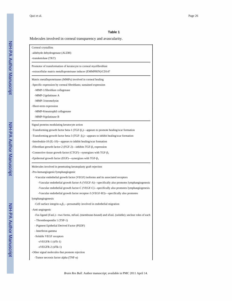
NIH
-PA Author Manuscript
NIH
-PA Author Manuscript
NIH
-PA Author Manuscript
Qazi et al. Page 26
Table 1
Molecules involved in corneal transparency and avascularity.
Corneal crystallins
-aldehyde dehydrogenase (ALDH)
-transketolase (TKT)
Promotor of transformation of keratocyte to corneal myofibroblast
-extracellular matrix metalloproteinase inducer (EMMPRIN)/CD147
Matrix metalloproteinases (MMPs) involved in corneal healing
-Specific expression by corneal fibroblasts; sustained expression
-MMP-1/fibroblast collagenase
-MMP-2/gelatinase A
-MMP-3/stromelysin
-Short-term expression
-MMP-8/neutrophil collagenase
-MMP-9/gelatinase B
Signal proteins modulating keratocyte action
-Transforming growth factor beta-1 (TGF-β1)—appears to promote healing/scar formation
-Transforming growth factor beta-3 (TGF- β3)—appears to inhibit healing/scar formation
-Interleukin-10 (IL-10)—appears to inhibit healing/scar formation
-Fibroblast growth factor-2 (FGF-2)—inhibits TGF-β1 expression
-Connective tissue growth factor (CTGF)—synergizes with TGF-β1
-Epidermal growth factor (EGF)—synergizes with TGF-β1
Molecules involved in penetrating keratoplasty graft rejection
-Pro-hemangiogenic/lymphangiogenic
-Vascular endothelial growth factor (VEGF) isoforms and its associated receptors
-Vascular endothelial growth factor-A (VEGF-A)—specifically also promotes lymphangiogenesis
-Vascular endothelial growth factor-C (VEGF-C)—specifically also promotes lymphangiogenesis
-Vascular endothelial growth factor receptor-3 (VEGF-R3)—specifically also promotes
lymphangiogenesis
-Cell surface integrin α1β1—presumably involved in endothelial migration
-Anti-angiogenic
-Fas ligand (FasL)—two forms, mFasL (membrane-bound) and sFasL (soluble); unclear roles of each
- Thrombospondin 1 (TSP-1)
- Pigment Epithelial Derived Factor (PEDF)
- Interferon gamma
-Soluble VEGF receptors
-sVEGFR-1 (sFlt-1)
-sVEGFR-2 (sFlk-1)
-Other signal molecules that promote rejection
-Tumor necrosis factor alpha (TNF-α)
Brain Res Bull. Author manuscript; available in PMC 2011 April 14.

NIH
-PA Author Manuscript
NIH
-PA Author Manuscript
NIH
-PA Author Manuscript
Qazi et al. Page 27
-Tumor necrosis factor beta (TNF-β)
-Interleukin-1 (IL-1)
-Other signal molecules that inhibit rejection
-Transforming growth factor beta-2 (TGF- β2)
Brain Res Bull. Author manuscript; available in PMC 2011 April 14.
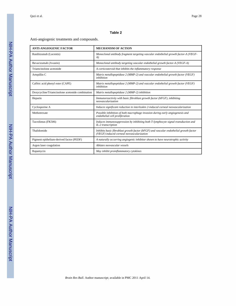
NIH
-PA Author Manuscript
NIH
-PA Author Manuscript
NIH
-PA Author Manuscript
Qazi et al. Page 28
Table 2
Anti-angiogenic treatments and compounds.
ANTI-ANGIOGENIC FACTOR MECHANISM OF ACTION
Ranibizumab (Lucentis) Monoclonal antibody fragment targeting vascular endothelial growth factor-A (VEGF-A)
Bevacizumab (Avastin) Monoclonal antibody targeting vascular endothelial growth factor-A (VEGF-A)
Triamcinolone acetonide A corticosteroid that inhibits the inflammatory response
Artepillin C Matrix metallopeptidase 2 (MMP-2) and vascular endothelial growth factor (VEGF)inhibition
Caffeic acid phenyl ester (CAPE) Matrix metallopeptidase 2 (MMP-2) and vascular endothelial growth factor (VEGF)inhibition
Doxycycline/Triamcinolone acetonide combination Matrix metallopeptidase 2 (MMP-2) inhibition
Heparin Immunoreactivity with basic fibroblast growth factor (bFGF), inhibitingneovascularization
Cyclosporine A Induces significant reduction in interleukin 2-induced corneal neovascularization
Methotrexate Possible inhibition of both macrophage invasion during early angiogenesis andendothelial cell proliferation
Tacrolimus (FK506) Induces immunosuppression by inhibiting both T-lymphocyte signal transduction andIL-2 transcription
Thalidomide Inhibits basic fibroblast growth factor (bFGF) and vascular endothelial growth factor(VEGF) induced corneal neovascularization
Pigment epithelium-derived factor (PEDF) A naturally occurring angiogenic inhibitor shown to have neurotrophic activity
Argon laser coagulation Ablates neovascular vessels
Rapamycin May inhibit proinflammatory cytokines
Brain Res Bull. Author manuscript; available in PMC 2011 April 14.


