Embed Size (px)
Citation preview

0014-2980/02/0606-1593$17.50+.50/0© WILEY-VCH Verlag GmbH, D-69451 Weinheim, 2002
The fate of duplicated major histocompatibilitycomplex class Ia genes in a dodecaploidamphibian, Xenopus ruwenzoriensis
Benedicte Sammut, Anne Marcuz and Louis Du Pasquier
Basel Institute for Immunology, Basel, Switzerland
The dodecaploid anuran amphibian Xenopus ruwenzoriensis represents the only polyploidspecies of Xenopus in which the full silencing of the extra copies of the major histocompati-bility complex (MHC) has not occurred. Xenopus ruwenzoriensis is a recent polyploid thathas evolved within one of the two tetraploid groups of Xenopus through allopolyploidization.Family studies of its MHC haplotype suggested a polysomic inheritance of the MHC class Iand II genes. Four class Ia bands can be detected per individual in Southern blot analysisand, similarly, four different cDNA sequences are expressed per individual. The Xenopusclass Ia sequences we analyzed belong to only one of the old class I lineages and show ahomogenization of their § 3 domain sequences. This homogenization occurred after specia-tion within the Xenopus ruwenzoriensis species, either due to gene conversion or inter-alleles/loci recombination. A re-evaluation of the polymorphism of class Ia in Xenopus, bylooking at the rate of non-synonymous versus synonymous substitutions, suggests thatXenopus MHC class Ia genes are not under strong overdominant selection. This is a rare sit-uation among vertebrates. The observed polymorphism is most likely due to the interlocusgenetic exchanges related to the peculiar mode of speciation of the genus.
Key words: Evolution / Polymorphism / Duplication / Homogenization / Immune system
Received 7/12/01Revised 5/3/02Accepted 8/3/02
[I 22683]
Abbreviations: Ia: Classical class I Ib: Non-classical classI 3’UTR: 3’ untranslated region > : Alpha domain ARS:Antigen recognition site dN: Frequency of non-synonymoussubstitutions dS: Frequency of synonymous substitutionsgDNA: Genomic DNA Chr: Chromosomes XNC: Xenopusnon-classical class I
1 Introduction
The MHC region that plays a central role in self/non-selfdiscrimination is one of the best-characterized geneticregions in the mammalian genome [1, 2]. This regioncontains genes encoding the class I and class II mole-cules presenting peptides to T cells and a number ofother genes belonging to the so-called class III regionthat are not related evolutionarily to genes of the immunesystem properly. Some genes of the class III regionencode proteins essential for the function of the MHCclass I molecules, others encode products that have littleor no functional interaction with class I or class II [1–3].
MHC class I and class II genes have been identified in allvertebrate classes except in Agnatha, which seem tolack an adaptive immune system [4–7]. The fact that nei-
ther class I nor class II genes have been found in jawlessfish or invertebrates suggests that the MHC “suddenly”emerged in a jawed vertebrate ancestor. Kasahara [8]proposed that the MHC region arose as a result of twoancient genome-wide duplications that took place 400million years ago in a common ancestor of jawed verte-brates. The signature of the chromosomal duplicationwould be the existence of four paralogous MHC clustersin all vertebrates. So far they have been partially demon-strated in mammals [7, 9, 10]. In the paralogous MHCregions identified so far in mammals, some genes werepreserved, while others were either silenced or translo-cated. Even if each paralogous cluster can be identified,it is very difficult nowadays to understand the fate ofsome of the duplicated genes because these eventstook place such a long time ago.
Xenopus, an anuran amphibian whose various specieswere generated by interspecies hybridization throughallopolyploidization [11, 12] offers a unique model tostudy the gene duplication mechanism postulated by themodel of Kasahara. Tetra-, octo- and dodecaploid spe-cies of Xenopus were generated at various times over aperiod ranging from 80 to 10 millions years ago. Bystudying the MHC in these species, we could perhapscatch the silencing events in the act.
Eur. J. Immunol. 2002. 32: 1593–1604 MHC class Ia in dodecaploid Xenopus 1593

Fig. 1. Class Ia hybridizing bands in a population of X. ruwenzoriensis (XR1–6) where XR1 is one of the grandfathers of the stud-ied family and XR2–6 are sibs. The filter was hybridized at 65°C overnight with the Xeru UAA1.1 § 2 domain probe and washedunder high stringency conditions [(a) 0.2× SSC, 65°C, 15 min] or the Xeru UAA1.1 § 3 domain probe and washed under low strin-gency conditions [(b) 2× SSC, 65°C, 15 min].
From functional studies and preliminary Southern blotdata, we already know that among all species of Xeno-pus studied so far, there is a trend toward diploidizationof the MHC ([13], and Salter-Cid and colleagues, submit-ted for publication). Consequently, the number of MHCgenes is reduced compared to the number expected inrespect of the ploidy level. Only the most recent dode-caploid X. ruwenzoriensis [with 2n=108 chromosomes(Chr)] showed some evidence for polysomic inheritanceof MHC genes in functional studies [5, 13]. However, noprecise sequence analysis of the alleles, of the numberof loci, of the polymorphism or of the number of ex-pressed genes has been carried out so far. These are thefeatures dealt with in this study.
2 Results
2.1 Each individual of X. ruwenzoriensisexpresses a maximum of four class Ia chaingenes
The species X. ruwenzoriensis, with 108 chromosomes,is likely to be derived from interspecies hybridization andallopolyploidization within the laevis group [11, 12]. Thesimplest laevis group species have 36 chromosomesand have diploidized their MHC [13]. Only one locus ofthe class Ia chain is detected both by Southern andcDNA analysis [14, 15]. With 108 chromosomes (=3×36),
X. ruwenzoriensis could in principle harbor three timesthe number of genes present in X. laevis, i.e. in a hetero-zygous situation and if no silencing has taken place, asmany as six alleles could be detected.
Southern blot performed with homologous X. ruwenzo-riensis probes confirm the earlier estimation establishedwith laevis probes [14, 15]: no more than four class Iahybridizing bands are detected in any X. ruwenzoriensis(Fig. 1a). Under low-stringency conditions ruwenzorien-sis § 3 domain probe detects a large number of the Xeno-pus bands, which corresponds to class Ia and class Ib§ 3 domains (Fig. 1b). This allows a rough estimation of
the number of X. ruwenzoriensis class Ib XNC (roughly20). It corresponds to the number of laevis XNC genesdetected using similar methods [15], meaning that theclass Ib number did not increase as much as expected inrespect of the level of ploidy. Thus, the global number ofX. ruwenzoriensis MHC class I genes suggests thatsilencing has occurred, affecting duplicated MHC classIa and Ib.
The cDNA from four X. ruwenzoriensis sibs XR2, 4, 5 and6 revealed two to four different sequences per individual,never more, when eight would have been expected ifboth parents had been heterozygous (Fig. 1 and Table 1).As a first approximation, this agrees with the number ofbands detected in Southern blot. However, the pre-diction of allele distribution made from the Southern
1594 B. Sammut et al. Eur. J. Immunol. 2002. 32: 1593–1604

Fig. 2. Protein alignments of the four X. ruwenzoriensis class Ia alleles compared to the X. laevis UAA1f. Sequences were alignedwith the Clustal X program method. The presumed g strand (S) and § helical (H) structure is derived from crystal structures ofhuman and mouse class I molecules [39]. The * represents polymorphic residues in mammals; ˚ are evolutionarily conserved[25] and ˆindicates the residues described as ARS [24]. The symbol “–” indicates the gaps introduced in the alignment. “b” and“8” refer to the positions proposed to interact with g 2-microglobulin and CD8, respectively [15]. Bold residues in the cytoplasmictail can be phosphorylated in mammals [40]. Underlined residues indicate the set of primers.
Table 1. Distribution of the four class Ia alleles found amongthe X. ruwenzoriensis different sibsa)
Allele names XR2 XR4 XR5 XR6
Xeru UAA1.1 + + + +
Xeru UAA1.2 + +
Xeru UAA1.3 + + +
Xeru UAA1.4 + + + +
Number hybridizingbands in Southernblot
4–5 2–3 2–3 4–5
a) The profile of class Ia § 2 hybridization obtained inSouthern blot (Fig. 1) is also mentioned.
blot does not correspond to the distribution of allelesobtained from the sequences (Table 1). For instance,individuals 2 and 6 yield the same four alleles by
sequence but have different RFLP, suggesting that iden-tical coding sequences can be found on different RFLP,depending on the constitutive haplotypes.
Since the ruwenzoriensis species is due to an allopoly-ploidization [16], its paralogous class Ia sequences cor-respond to as many ancient alleles as those that comefrom the parental species. We therefore designed themas Xeru UAA1.1 to Xeru UAA1.4 for Xenopus ruwenzo-riensis Classis-Una, family-A, chain-A locus-1 alleles 1to 4 in accordance with the proposal by Mayer et al.[17, 18].
The four sequences correspond to the class Ia alleliclineage A, i.e. one of the two major class Ia lineagesdescribed in X. laevis [19, 20]. The identification of theX. ruwenzoriensis class Ia A lineage was possible thanksto the comparison of their cytoplasmic, transmembraneand 3’UTR sequences (Fig. 2). Indeed, by subjectingX. ruwenzoriensis XR6 peripheral blood (PBL) cDNA to
Eur. J. Immunol. 2002. 32: 1593–1604 MHC class Ia in dodecaploid Xenopus 1595
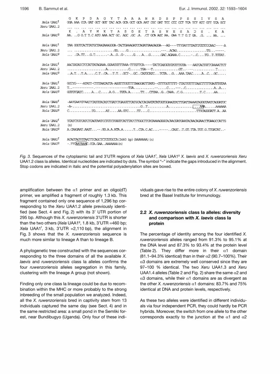
Fig. 3. Sequences of the cytoplasmic tail and 3’UTR regions of Xela UAA1f, Xela UAA1g X. laevis and X. ruwenzoriensis XeruUAA1.2 class Ia alleles. Identical nucleotides are indicated by dots. The symbol “–” indicate the gaps introduced in the alignment.Stop codons are indicated in italic and the potential polyadenylation sites are boxed.
amplification between the § 1 primer and an oligo(dT)primer, we amplified a fragment of roughly 1.3 kb. Thisfragment contained only one sequence of 1,296 bp cor-responding to the Xeru UAA1.2 allele previously identi-fied (see Sect. 4 and Fig. 2) with its 3’ UTR portion of295 bp. Although this X. ruwenzoriensis 3’UTR is shorterthan the two others (Xela UAA1g, 1.8 kb, 3’UTR =460 bp;Xela UAA1f, 3 kb, 3’UTR =2,110 bp), the alignment inFig. 3 shows that the X. ruwenzoriensis sequence ismuch more similar to lineage A than to lineage B.
A phylogenetic tree constructed with the sequences cor-responding to the three domains of all the available X.laevis and ruwenzoriensis class Ia alleles confirms thefour ruwenzoriensis alleles segregation in this family,clustering with the lineage A group (not shown).
Finding only one class Ia lineage could be due to recom-bination within the MHC or more probably to the stronginbreeding of the small population we analyzed. Indeed,all the X. ruwenzoriensis bred in captivity stem from 13individuals captured the same day (see Sect. 4) and inthe same restricted area: a small pond in the Semliki for-est, near Bundibugyo (Uganda). Only four of these indi-
viduals gave rise to the entire colony of X. ruwenzoriensisbred at the Basel Institute for Immunology.
2.2 X. ruwenzoriensis class Ia alleles: diversityand comparison with X. laevis class Iaprotein
The percentage of identity among the four identified X.ruwenzoriensis alleles ranged from 91.3% to 95.1% atthe DNA level and 87.3% to 93.4% at the protein level(Table 2). They differ more in their § 1 domain(81.1–94.3% identical) than in their § 2 (90.7–100%). Their§ 3 domains are extremely well conserved since they are
97–100 % identical. The two Xeru UAA1.3 and XeruUAA1.4 alleles (Table 2 and Fig. 2) share the same § 2 and§ 3 domains, while their § 1 domains are as divergent as
the other X. ruwenzoriensis § 1 domains: 83.7% and 75%identical at DNA and protein levels, respectively.
As these two alleles were identified in different individu-als via four independent PCR, they could hardly be PCRhybrids. Moreover, the switch from one allele to the othercorresponds exactly to the junction at the § 1 and § 2
1596 B. Sammut et al. Eur. J. Immunol. 2002. 32: 1593–1604

Table 2. Similarities of MHC class Ia alleles from Xenopus ruwenzoriensisa)
§ 1 § 2 § 3 § 1-cyto
XeruUAA
1.1 1.2 1.3 1.4 1.1 1.2 1.3 1.4 1.1 1.2 1.3 1.4 1.1 1.2 1.3 1.4
1.1 81.8 81.1 86.4 90.7 91.8 91.8 97 97 97 91.4 91.3 92.5
1.2 78.4 94.3 83.7 86 92.1 90.7 96.7 97 97 88.9 95.1 92.1
1.3 77.3 89.8 83.7 84.9 91.4 100 96.7 97.8 100 87.3 93.4 95.5
1.4 79.5 78.4 75 84.9 91.4 100 96.7 97.8 100 87.3 89.8 92.8
a) Percentages of DNA and amino acid (shown in bold italics) identity of the X. ruwenzoriensis sequences shown in Fig. 2. Thepercentages are given for § 1, § 2, § 3 domains and for the coding region between the § 1 domain and the cytoplasmic tail.
domain separation, which strongly suggests an exon-shuffling event.
The property of sharing one or even more domainsbetween alleles has already been reported anddescribed in HLA class I where one allele shares itsexons 1 and 2 with one allele and its exons 3 to 8 withanother. Homologous recombination involvement wassuggested as an explanation for this characteristic [21].However, without complete knowledge of the sequencesflanking these alleles, Holmes and Parham [22] could noteliminate that an allelic conversion involving a relativelylarge segment of DNA was the mechanism. As we do nothave the Xeru UAA1.3 and Xeru UAA1.4 intronicsequences, we cannot favor one or the other of thesetwo explanations.
The transmembrane (TM) and cytoplasmic regions (cyto)of the four paralogues are nearly totally (TM) or totally(cyto) conserved. The highest polymorphism is seen inthe peptide binding region (PBR) constituted by the § 1and § 2 domain of the § chain [23, 24]. As in all verte-brates, the variability is mostly encountered betweenpositions 60–80 of the § 1 domain, and to a minor degreein the § 2 domain, among residues located at the surfaceof the two § helices of the groove [25]. The variability isalso seen between positions 110 and 130 of the § 2domain, which correspond to residues forming g sheetsof the groove. As already observed [25], residues thatinteract with antigenic peptide termini through hydrogenbonding are conserved (Fig. 2).
The frequency of non-synonymous substitutions, dN(amino acid altering), in the antigen recognition site (ARS)of X. ruwenzoriensis is slightly higher (but inferior to 1)than in the remaining codons (Table 3) as described forclass I molecules under positive selection [26]. Unlike inall the species studied so far (Table 4) and despite agreat diversity within the PBR, dN is smaller than the fre-
quency of the synonymous substitutions, dS (no aminoacid altering), in the ARS. This situation also encounteredin X. laevis class Ia [19] is due rather to a high dS than toa reduction of dN. Indeed, one can assume than, even inthe ARS, diversification is limited and some structurallyimportant amino acids are conserved. Thus, beside thepositive Darwinian selection known to favor an aminoacid change [26], another type of pressure allowing ahigh dS (i.e. without altering the amino acid) must beexerted in the ARS of Xenopus.
The calculation of the ratio dN/dS (Table 4) shows amoderate trend towards overdominant selection for theX. ruwenzoriensis class Ia ARS sequences. Indeed, allother vertebrates from teleost fish to mammals show amuch stronger bias towards the ARS dN/dS ratio. This isvalid not only for the highly polyploid X. ruwenzoriensisbut also for the pseudo-tetraploid species of X. laevis.
Fig. 2 aligns the X. ruwenzoriensis class Ia allele se-quences with the first Xenopus class I gene described byShum et al. [14]. Besides the expected diversity in the § 1and § 2 domains, Fig. 2 shows that there is one aminoacid less in every X. ruwenzoriensis § 3 domain than inthe X. laevis. Compared to the X. laevis sequences, themissing X. ruwenzoriensis amino acid is located at theend of the first loop between strand A and B of the Ig C1domain, a region not known to affect any function.
2.3 All the X. ruwenzoriensis class Ia sequencesare specific for this 108-chromosomespecies
The generation of X. ruwenzoriensis species involvesa series of interspecific hybridization followed by ge-nome duplication during gametogenesis [11, 12]. SeveralXenopus species are thought to be putative ancestors ofthe X. ruwenzoriensis either for morphological (X. amieti
Eur. J. Immunol. 2002. 32: 1593–1604 MHC class Ia in dodecaploid Xenopus 1597

Table 3. Mean number of nucleotide substitutions per 100 synonymous sites (dS) and per 100 non-synonymous sites (dN) ± SDof the Xenopus class Ia alleles shown in Fig. 2 and in human sequencesa)
ARS Remaining codons in § 1 and§ 2 domain
Exon4 ( § 3 domain)
(n = 57 codons) (n = 124,125)b) (n = 90, 92 codons)c)
dS dN dS dN dS dN
X. ruwenz (4 or 3)d) 30.9± 9 17.6±4 14.9±3 5.6± 1 10.01± 3.7 1.26± 0.69
X. laevis + X. laevis/gilli 39.2±10.1 19.5±5.2 28.1±5.7 9.7± 2.4 2.22± 1.35 2.00± 0.8
(7 or 8)e)
X. laevis and ruwen. (11) 60.0± 1.7 53.6±8.0 52.0±8.2 39.2± 5.4 124.5 ±76 ±63 74.98±44.33
X. laevis XNC cl Ib (41) 106.8 35.4±3.3 82.2±8.6 31.0±22 18.7± 8.8 8.8 ± 1.1
Human, overall means A,B, C intralocus (19)
4.7± 2.6 14.1±2.4 5.1±2.1 2.4± 0.8 5.8 ± 2.0 1.1 ± 0.6
a) For the human sequences, the values are taken from [26]. dS, dN and ± SD were computed with the help of the programSNAP (Synonymous/Non-synonymous Program: http://hiv-web.lanl.gov/SNAP/WEBSNAP/SNAP.htmlby) adapted from [41]and incorporating a statistic developed in [42]. The class Ia sequences for Xenopus dN and dS calculations are from [19] andXenopus laevis class Ib data from [15]. The numbers of sequences used in the analysis are given in parentheses.
b) n=124 for Xenopus, and 125 for the human.c) n=90 for the Xenopus, and 92 for the human.d) Xeru UAA1.3 and Xeru UAA1.4 sharing the § 3 domain, the number of alleles used for the ARS and remaining codons dN, dS
calculations is four, while the one used for the § 3 domain calculations is only three.e) As one X. laevis/gilli lg3 MHC class Ia alleles is truncated in its 5’ region, the number of X. laevis/gilli sequences used in the
dN, dS calculation is three for the ARS and remaining codons and four for the § 3 domain. Thus, the total number of comparedalleles for X. laevis-X. laevis/gilli is seven for the ARS, remaining codons and eight for the § 3 domain.
72 Chr, X. fraseri 36 Chr) or geographical (X. vestitus72 Chr, X. wittei 72 Chr) reasons [16, 27, 28]. The parentsable to generate a 108-chromosome species have to beone 72- and one 36-chromosome individual. Therefore, ifall X. ruwenzoriensis alleles showed a deletion, one couldanticipate that it would also be present in one or two ofthe parental species. Otherwise there would be evidenceof homogenization within the X. ruwenzoriensis or itsclosest ancestral species.
By PCR on genomic DNA (gDNA), we amplified the § 3domains of several Xenopus species and aligned thededuced amino acid sequences (Fig. 4). The amino aciddeletion could not be detected in any of the sequencesof tetraploid or octoploid species of the laevis group.This deletion seems to act as a tag for the X. ruwenzo-riensis species and was therefore most likely acquiredafter the generation of the ruwenzoriensis species withinthe laevis group. It may have appeared on one allele andspread via a homogenization due to gene interchange(conversion or recombination) as already describedwithin other species [5, 29]. It is therefore not surprisingto observe it in the X. ruwenzoriensis sequences. How-ever, in the case of X. ruwenzoriensis the deletion, being
a tag for all sequences in a genome, ultimately comesfrom different species of the laevis group without a dele-tion. It represents one of the best signatures ofhomogenization.
In addition, the X. ruwenzoriensis class Ia § 3 domainsequences are more conserved in X. ruwenzoriensisalleles than in any other alleles (Fig. 4). Among the Xeno-pus § 3 domain sequences, the variability is greater inthe 36- and 72-chromosome Xenopus groups (11 and12 variable amino acid positions) than in the 108-chromosome X. ruwenzoriensis (2 variable amino acidpositions).
Based on Fig. 4 numbering and in reference to the XelaUAA1f sequence, the variable positions encountered inevery Xenopus allele has been grouped according to theploidy level of the species (Fig. 5). At the DNA level, thetotal number of substitutions is roughly three times lessin X. ruwenzoriensis than in the other species. Fig. 5 alsounderlines that no special similarity between the X.ruwenzoriensis and the fraseri or wittei Xenopus (thoughtto be close to their ancestor) can be detected. X. ruwen-zoriensis § 3 domain sequences have clearly been
1598 B. Sammut et al. Eur. J. Immunol. 2002. 32: 1593–1604

Table 4. The non-synonymous and synonymous ratio dN/dSfrom several species class I PBRa)
Ratio dN/dS ARS(n = 56, 57)b)
Ratio dN/dSremaining
(n = 124, 125)c)
Salmo salar (20) 2.1 ND
X. ruwenzoriensis (4) 0.56 0.37
X. laevis (7) 0.49 0.34
Xenopus (11) 0.89 0.75
X. laevis Ib (41) 0.33 0.37
Axolotl (2) 1.96 1.15
Chicken (5) 1.6 1.13
Mouse
Interlocus (24) 1.52 0.81
Intralocus (12) 1.6 0.71
All comparisons (36) 1.55 0.78
Human
Interlocus (47) 3.05 0.44
Intralocus (19) 3 0.47
All comparisons (66) 3.05 0.44
a) This ratio is based on Table 3 for Xenopus class Ia andclass Ib [15] § 1 and § 2 domains. We calculated the ratiofor the axolotl from the Amme 3 (AMU138) and Amme 4(AMU137) class I sequences, for the chicken B15(L28958), B-F19 (Z54360), B-F12 (Z54359), B13(AFO13494) and B2 (AFO13492) alleles. The mammaliandN and dS values were obtained from [26] and thesalmon data from [32]. Since dN/dS data for variousvertebrates have been obtained following Bjorman’sdefinition of ARS [24], we calculated the Xenopus valuesaccording to the same principles and did not introduce inthis table the actual values of trouts (see discussion and[33]). The number of sequences compared is given inparentheses. n=number of codons compared.
b) n=56 for Xenopus and axolotl, and 57 for chicken, humanand mouse.
c) n=123 for chicken, 124 for mouse and 125 for human andaxolotl.
homogenized and this homogenization affects X. ruwen-zoriensis § 3 domain sequences more than the se-quences of any other species. All these features are inagreement with the notion that X. ruwenzoriensissequences were generated recently [13].
3 Discussion
In this study, we clearly establish that each X. ruwenzo-riensis individual can express up to four different MHCclass Ia gene sequences. At this point, we do not knowwhether the transcripts detected are all translated butthe messages look functional. This number of four classIa is the same as the one estimated by domain specifichybridizations on Southern blot. It is smaller than thenumber expected if all the MHC class I genes had beenconserved throughout speciation events. X. ruwenzo-riensis has three times more chromosomes than thepseudo-tetraploid X. laevis. Since X. laevis (because ofdiploidization) contains and expresses only class Ialocus (i.e. two alleles at the heterozygous situation), X.ruwenzoriensis could at least contain and express (3×2,i.e. 6) alleles in a full heterozygous situation. Finding onlya maximum of four genes and four transcripts demon-strates that two genes were either silenced or deleted.The absence of any extra signal on Southern blot sug-gests that these two genes were in fact deleted. Thiselimination might be directly related to the function of theMHC class Ia molecule if, as already suggested, pos-sessing too many class Ia molecules can be detrimental[30, 31].
Zoologists agree that X. ruwenzoriensis is a relativelyyoung species [13]. Therefore, finding more than twoalleles expressed per individual means that we areobserving the reduction in the number of class Ia genesin progress. It is conceivable that in an undefined num-ber of years (millions?), X. ruwenzoriensis, like all othertetraploid or octoploid species, will have kept only onelocus of class Ia.
Because of our dN/dS data, one can speculate whetherXenopus class Ia gene polymorphism is under positiveselection or not. The analysis of the X. ruwenzoriensispolymorphism prompted some comparisons with otherspecies of Xenopus and other vertebrates. This broughtup an interesting difference between Xenopus se-quences and all other vertebrate sequences.
The pattern of nucleotide substitutions in the ARS andthe remaining codons does not indicate a strong selec-tion of the polymorphism. The dN/dS ratio of ARS, sup-posed to be higher in species when polymorphism isselected, is rather low in genus Xenopus. This is not afeature shared by all cold-blooded vertebrates (Table 4).In the salmonid, two types of results have been pub-lished. In the Atlantic salmon [32], and in related trouts oftwo species [33], the dN/dS ratio comparisons yieldresults similar to those of mammals with evidence foroverdominant selection. However, the outbreed troutresults are similar to those obtained in Xenopus. Thus,
Eur. J. Immunol. 2002. 32: 1593–1604 MHC class Ia in dodecaploid Xenopus 1599

Fig. 4. Alignment of MHC class I § 3 domain amino acid sequences deduced from cDNA clones in polyploid species of Xenopus.X. laevis sequences are from Flajnik et al. [19]. X. fraseri, muelleri, vestitus, amieti and wittei sequences are shorter since theyhave been amplified on gDNA by PCR between the underlined primers (see Sect. 4). Bold characters indicate the speciesthought to have given rise to X. ruwenzoriensis (X. vestitus, X. wittei, X. fraseri and X. amieti). Dots indicate identities to XelaUAA1f; dash (underlined by an arrow) indicates the gap introduced in the alignment (Clustal X method); “S” indicates g strand and8 and b refers to CD8 and g 2-microglobulin contact residues, respectively.
we do not think that the low dN/dS ratio reflects a majordifference in antigenic load between warm- and cold-blooded vertebrates.
This lack of positive selection also seems to be specificto class I sequences only since the dN/dS ratio in X.ruwenzoriensis and laevis class II g -chain sequences arecomparable to the ratio found in the other vertebrates(Sammut, in preparation). Four non-mutually exclusivehypothesis are commonly proposed to explain the gen-eration and the maintenance of polymorphism within theMHC [26]: (1) high mutation rate, (2) genetic exchangebetween loci and alleles, (3) balancing selection (withheterozygous advantage), and (4) some form of fre-quency dependent selection. The effect of overdominantselection is present in Xenopus but seems weak. It couldbe masked by the effect of polymorphism on the modeof speciation in Xenopus implying a lot of genetic mate-rial exchange. We do not have global data reporting ahigher rate of mutation in Xenopus but the mode of spe-
ciation of the polyploid Xenopus certainly favors inter-locus genetic exchanges. As proof of this possibility,X. ruwenzoriensis still shows some multivalents in mei-osis [16].
The relatively low dN/dS ratio is seen in all Xenopus spe-cies from tetraploid to dodecaploid, i.e. always in poly-ploid forms. Unfortunately, we do not have data on thesimpler X. tropicalis that could serve as a reference. Atthis point, we do not know whether this unique featurereveals a different selective pressure on the MHC class Iin Xenopus or whether it is the result of the mode of thepolymorphism generation. Since other cold-bloodedvertebrate and bird class I sequences show the samedN/dS feature as seen in the mammalian sequences,we favor the second hypothesis.
The homogenization of the § 3 sequences in X. ruwenzo-riensis, the exon shuffling, and the minimal differencesamong the X. ruwenzoriensis alleles compared to the
1600 B. Sammut et al. Eur. J. Immunol. 2002. 32: 1593–1604

Fig. 5. Nucleotide sequences of amino acid sequences shown in Fig. 4, showing substitutions encountered in the § 3 domains ofthe different polyploid species. Xela UAA1f amino acid sequence is indicated as a master and sequences are grouped accordingto the level of polyploidy. In every group, when a non-synonymous substitution, compared to Xela UAA1f, is encountered (red),the corresponding amino acid change is indicated in red under the group of sequences, while the position of synonymous substi-tutions are indicated by blue stars. Dots correspond to the identities and dashes indicate the gaps introduced in the alignment.As in Fig. 4, “S” indicates g strand and 8 and b refer to CD8 and g 2-microglobulin contact residues, respectively.
Eur. J. Immunol. 2002. 32: 1593–1604 MHC class Ia in dodecaploid Xenopus 1601

other Xenopus sequences are all indications of the pos-sibility of intergene exchanges. The mechanisms are stillunknown but conversion or interlocus recombinationcould be involved.
4 Materials and methods
4.1 Animals
X. laevis with the MHC haplotype f [34], X. fraseri (2n=36 Chr), X. vestitus, X. amieti (2n=72 Chr) and X. ruwenzo-riensis (2n=108 Chr) were bred occasionally and maintainedat the Basel Institute for Immunology. They came from differ-ent families and in each case were raised from a small num-ber of couples collected in Uganda, Rwanda and Cameroonin 1975 [13, 27]. Extra gDNA samples from X. vestitus, X.muelleri (2n=36 Chr) and X. wittei (2n=72 Chr) were providedby Dr. R. C. Tinsley at the University of Bristol. Different X.ruwenzoriensis from the same family (the grandfather, XR1,and five sibs, XR2 to 6), one X. fraseri, two X. muelleri, two X.amieti, four X. vestitus from three different families and twoX. wittei from different families were analyzed. By compari-son with X. tropicalis (20 Chr), species with 36 chromo-somes are defined as tetraploid (4n), those with 72 chromo-somes as octoploid (8n) and those with 108 chromosomesas dodecaploid (12n).
4.2 PBL isolation, RNA extraction, cDNA and PCR
Peripheral blood cells were obtained from circulating bloodby pipetting and PBL were separated and collected after aFicoll gradient separation as described [35].
Total cellular RNA (less than 1 ? g) isolated from the PBL fromXR2, 4, 5 and 6 using Gibco TRIzol kit was reverse tran-scribed using the (dT)17 adapter-primer (5’-GACTCGAG-TCGACATCGAT(17)-3’) as described previously [36].
The resulting cDNA was subjected to one-sided PCRaccording to [37]. Briefly, the amplification was achievedusing a single gene-specific primer and a reverse adapter-primer. This primer ( § 1s: 5’-GGCAGTCACTCCCCTGCG-3’)was designed on the 5’ end of the Xela UAA1f § 1 domain, aregion usually conserved within a species [14]. The PCR pro-file was 94°C for 5 min followed by 30 cycles of 94°C for1 min 30 s, 55°C for 30 s, 72°C for 1 min 30 s with a finalextension of 10 min.
Another PCR was achieved using the § 1s primer in combi-nation with a specific reverse primer (5’-AGCTGAAACAAT-AGATGAAAG-3’), based on the Xela UAA1f cytoplasmic tailsequence to amplify a fragment of X. ruwenzoriensis class Igene cDNA (1 kb).
The PCR profile for this amplification was 94°C for 5 min,followed by 30 cycles of 94°C for 1 min, 60°C for 30 s, 72°Cfor 45 min with a final extension time of 10 min.
In both amplifications, 1 ? l of the 25- ? l PBL cDNA wasadded to 5 ? l 1× PCR buffer (GenAmp, Hoffmann-La Roche,Basel, Switzerland), 50 ? M dNTPs, 10 pmol of each primer( § 1s and adapter or § 1s and specific reverse primer) and 1 UAmpliTaq DNA polymerase (Hoffmann-La Roche). PCRproducts were cloned into the TA cloning vector accordingto the manufacturer’s instructions (TA cloning kit, Invitrogen,San Diego, CA).
Random PCR clones, 10–30 for each of the four X. ruwenzo-riensis clonings and 10–20 for the other Xenopus clonings,were sequenced by dideoxy chain termination chemistryusing universal primers (MWG Biotec, Ebersberg, Germany)in conjunction with the thermo sequenase kit (Amersham,Arlington Heights, IL). Sequences were obtained via an auto-mated sequencer (Licor 4000L) and submitted to a Blast Xsearch to identify class I sequences. Alignments, phyloge-nies and bootstrapping were conducted using the Clustal Xsoftware package [38].
Some clones were diluted, re-amplified via PCR (30 cycles:94°C for 30 s, 55°C for 30 s, 72°C for 30 s followed by a finalextension of 10 min) with the § 1s primer in combination withspecific X. ruwenzoriensis reverse primers. These reverseprimers were based on the class I sequences obtainedthrough the first two PCR to amplify cDNA segments spe-cific to X. ruwenzoriensis § 2 or § 3 domains. These domainfragments were purified using either Qiaquick PCR purifica-tion or QiaEX II gel extraction kits (Quiagen, Hoffmann-LaRoche), randomly primed (Life Technologies, Gaithersburg,MD) with [32P]dCTP (Amersham, Arlington Heights, IL) andthen used as homologous probes to hybridize Southernblots.
4.3 gDNA and PCR amplifications on gDNA
Blood cells from XR1 to XR6 were incubated in lysis buffer(0.1 M EDTA, 0.05 M Tris-HCl, 0.1% SDS) at 65°C for45 min, supplemented with proteinase K (100 ? g/ml lysisbuffer) and incubated at 37°C for 8 h. DNA was recoveredfrom the lysate after two 5 M NaCl extractions. This gDNAwas then used in PCR amplifications and in Southern blotanalysis. Two primers (forward: 5’-CACCCTCATGTAAGAA-TTTC-3’, reverse: 5’-GTGCTCCACATGACAGGC-3’) basedon the Xela UAA1f/X. ruwenzoriensis class Ia paralogoussequences comparison, were designed in the 5’ and 3’regions of the § 3 domain. With these primers, we amplifiedclass I § 3 domains on gDNA obtained from various Xeno-pus. Each PCR was carried out in a total volume of 50 ? lcontaining 250 ? g gDNA, 50 ? M of each dNTPs, 10 pmol ofeach primer, 5 ? l of 1× PCR buffer and 1 U AmpliTaq. ThePCR conditions were 94°C for 1 min, 55°C for 30 s, 72°C for
1602 B. Sammut et al. Eur. J. Immunol. 2002. 32: 1593–1604

30 s followed by a final extension of 10 min. For every ampli-fication and cloning, 10–30 random PCR clones weresequenced as mentioned above.
4.4 Southern blots
The gDNA (10 ? g) was digested for 8 h with restrictionenzymes, electrophoresed, capillary transferred to nylon(Zetaprobe GT, Bio-Rad Labs) and then hybridized withradiolabeled probes (see figures) in 1.6× SSC, 1% SDS,10% dextran sulfate, 0.5% pyrophosphate, 0.5% nonfatpowdered milk and 500 ? g/ml sonicated salmon sperm DNAfor 8 h at 65°C. The membrane was washed under low orhigh stringency conditions: either 2× SSC, 0.5% SDS at65°C for 15 min, or 0.2× SSC, 0.5% SDS at 65°C for 15 min.
Acknowledgements: We thank Dr. Martin Flajnik for hissuggestions and comments, Allison Dwileski and Lucy Trip-pmacher for their help with the manuscript and figures andErwin Schilliger for photography. The Basel Institute forImmunology was founded and supported by F. Hoffmann-LaRoche Ltd., Basel, Switzerland
References
1 Klein, J., Natural history of the major histocompatibility complex,New York 1986.
2 Marguilies, D. H., The major histocompatibility complex. In Paul,W. E. (Ed.) Fundamental immunology, 4th edn. Lippincott-Raven,Philadelphia 1999, pp 263–285.
3 Campbell, R. D. and Trowsdale, J., Map of the human MHC.Immunol. Today 1993. 14: 349–352.
4 Du Pasquier, L. and Flajnik, M. F., Origin and evolution of thevertebrate immune system. In Paul, W. E. (Ed.) Fundamentalimmunology, 4th edn. Lippincott-Raven, Philadelphia 1999, pp605–650.
5 Flajnik, M. F., Ohta, Y., Namikawa-Yamada, C. and Nonaka,M., Insight into the primordial MHC from studies in ectothermicvertebrates. Immunol. Rev. 1999. 167: 59–67.
6 Kasahara, M., Flajnik, M. F., Ishibashi, T. and Natori, T., Evolu-tion of the major histocompatibility complex: a current overview.Transplant. Immunol. 1995. 3: 1–20.
7 Kasahara, M., The chromosomal duplication model of the majorhistocompatibility complex. Immunol. Rev. 1999. 167: 17–32.
8 Kasahara, M., Genome dynamics of the major histocompatibilitycomplex: insights from genome paralogy. Immunogenetics 1999.50: 134–145.
9 Kasahara, M., What do the paralogous regions in the genometell us about the origin of the adaptive immune system? Immunol.Rev. 1998. 166: 159–175.
10 Abi Rached, L., McDermott, M. F. and Pontarotti, P., The MHCbig bang. Immunol. Rev. 1999. 167: 33–44.
11 Kobel, H. R. and Du Pasquier, L., Genetics of polyploid Xeno-pus. Trends Genet. 1986. 12: 310–315.
12 Kobel, H. R., Loumont, C. and Tinsley, R. C., The extant spe-cies. In Tinsley, R. C. and Kobel, H. R. (Eds.) The biology ofXenopus. Clarendon Press, Oxford 1996, pp 9–33.
13 Du Pasquier, L., Miggiano, V. C., Kobel, H. R. and Fischberg,M., The genetic control of histocompatibility reactions in naturaland laboratory-made polyploid individuals of the clawed toadXenopus. Immunogenetics 1977. 5: 129–141.
14 Shum, B. P., Avila, D., Du Pasquier, L., Kasahara, M. and Flaj-nik, M. F., Isolation of a classical MHC class I cDNA from anamphibian. Evidence for only one class I locus in the XenopusMHC. J. Immunol. 1993. 151: 5376–5386.
15 Flajnik, M. F., Kasahara, M., Shum, B. P., Salter-Cid, L., Taylor,E. and Du Pasquier, L., A novel type of class I gene organizationin vertebrates: a large family of non-MHC-linked class I genes isexpressed at the RNA level in the amphibian Xenopus. EMBO J.1993. 12: 4385–4396.
16 Tymowska, J., Polyploidy and cytogenetic variation in frogs ofthe genus Xenopus. In Green, D. M. and Sessions, S. K. (Eds.)Amphibian cytogenetics and evolution. Academic Press, SanDiego 1991, pp 259–297.
17 Klein, J., Bontrop, R. E., Dawkins, R. L., Erlich, H. A., Gyllens-ten, U. B., Heise, E. R., Jones, P. P., Parham, P., Wakeland, E.K. and Watkins, D. I., Nomenclature for the major histocompati-bility complexes of different species: a proposal. Immunogenet-ics 1990. 31: 217–219.
18 Mayer, W. E., Williams, N. S., O’Huigin, C., Vincek, V., Zaleska-Rutczynska, Z. and Klein, J., Class I major histocompatibilitycomplex genes of the red-necked Wallaby, Macropus rufogri-seus. Mol. Phylogenet. Evol. 1993. 2: 23–30.
19 Flajnik, M. F., Ohta, Y., Greenberg, A. S., Salter-Cid, L., Carri-zosa, A., Du Pasquier, L. and Kasahara, M., Two ancient alleliclineages at the single classical class I locus in the Xenopus MHC.J. Immunol. 1999. 163: 3826–3833.
20 Nonaka, M., Yamada-Namikawa, C., Flajnik, M. F. and DuPasquier, L., Trans-species polymorphism of the major histo-compatibility complex-encoded proteasome subunit LMP7 in anamphibian genus, Xenopus. Immunogenetics 2000. 51: 186–192.
21 Parham, P., Lawlor, D. A., Lomen, C. E. and Ennis, P. D., Diver-sity and diversification of HLA-A,B,C alleles. J. Immunol. 1989.142: 3937–3950.
22 Holmes, N. and Parham, P., Exon shuffling in vivo can generatenovel HLA class I molecules. EMBO J. 1985. 4: 2849–2854.
23 Bjorkman, P. J., Saper, M. A., Samraoui, B., Bennett, W. S.,Strominger, J. L. and Wiley, D. C., Structure of the human classI histocompatibility antigen, HLA-A2. Nature 1987. 329: 506–512.
24 Bjorkman, P. J., Saper, M. A., Samraoui, B., Bennett, W. S.,Strominger, J. L. and Wiley, D. C., The foreign antigen bindingsite and T cell recognition regions of class I histocompatibilityantigens. Nature 1987. 329: 512–518.
25 Kaufman, J., Salomonsen, J. and Flajnik, M., Evolutionary con-servation of MHC class I and class II molecules – different yet thesame. Semin. Immunol. 1994. 6: 411–424.
26 Hughes, A. L. and Nei, M., Pattern of nucleotide substitution atmajor histocompatibility complex class I loci reveals overdomi-nant selection. Nature 1988. 335: 167–170.
27 Tinsley, R. C., Loumont, C. and Kobel, H. R., Geographical dis-tribution and ecology. In Tinsley, R. C. and Kobel, H. R. (Eds.)The Biology of Xenopus. Clarendon Press, Oxford 1996, pp35–41.
28 Kobel, H. R., Barandun, B. and Thiebaud, C. H., MitochondrialRDNA phylogeny in Xenopus. Herpatological J. 1998. 8: 13–17.
Eur. J. Immunol. 2002. 32: 1593–1604 MHC class Ia in dodecaploid Xenopus 1603

29 Hughes, A. L. and Yeager, M., Natural selection at major histo-compatibility complex loci of vertebrates. Annu. Rev. Genet.1998. 32: 415–435.
30 Lawlor, D. A., Zemmour, J., Ennis, P. D. and Parham, P., Evolu-tion of class I MHC genes and proteins: from natural selection tothymic selection. Annu. Rev. Immunol. 1990. 8: 23–63.
31 Bevan, M. J., High determinant density may explain the phenom-enon of alloreactivity. Immunol. Today 1984. 5: 128–130.
32 Grimholt, U., Olsaker, I., de Vries Lindstrom, C. and Lie, O., Astudy of variability in the MHC class II beta 1 and class I alpha 2domain exons of Atlantic salmon, Salmo salar L. Anim. Genet.1994. 25: 147–153.
33 Shum, B. P., Guethlein, L., Flodin, L. R., Adkison, M. A., Hed-rick, R. P., Nehring, R. B., Stet, R. J., Secombes, C. and Par-ham, P., Modes of salmonid MHC class I and II evolution differfrom the primate paradigm. J. Immunol 2001. 166: 3297–3308.
34 Du Pasquier, L., Chardonnens, X. and Miggiano, V. C., A majorhistocompatibility complex in the toad Xenopus laevis (Daudin).Immunogentics 1975. 1: 482–494.
35 Du Pasquier, L., Flajnik, M. F., Hsu, E. and Guiet, C., Methodsused to study the immune system of Xenopus (Amphibian,Anura). In Lefkovits, I. and Pernis, B. (Eds.) Immunologicalmethods. Academic Press, Orlando 1985, pp 425–465.
36 Sammut, B., Laurens, V. and Tournefier, A., Isolation of MHCclass I cDNAs from the axolotl Ambystoma mexicanum. Immuno-genetics 1997. 45: 285–294.
37 Frohman, M. A., Rapid production of full-length CdNAs from raretranscripts: amplification using a single gene-specific oligonucle-otide primer. Proc. Natl. Acad. Sci. USA 1988. 85: 8998–9002.
38 Thompson, J. D., Gibson, T. J., Plewniak, F., Jeanmougin, F.and Higgins, D. G., The CLUSTAL–X windows interface: flexiblestrategies for multiple sequence alignment aided by quality anal-ysis tools. Nucleic Acids Res. 1997. 25: 4876–4882.
39 Saper, M. A., Bjorkman, P. J. and Wiley, D. C., Refined structureof the human histocompatibility antigen HLA-A2 at 2.6 A resolu-tion. J. Mol. Biol. 1991. 219: 277–319.
40 Guild, B. C. and Strominger, J. L., HLA-A2 antigen phosphoryla-tion in vitro by cyclic AMP-dependent protein kinase. Sites ofphosphorylation and segmentation in class i major histocom-patibility complex gene structure. J. Biol. Chem. 1984. 259:13504–13510.
41 Nei, M., Stephens, J. C. and Saitou, N., Methods for computingthe standard errors of branching points in an evolutionary treeand their application to molecular data from humans and apes.Mol. Biol. Evol. 1985. 2: 66–85.
42 Ota, T. and Nei, M., Variance and covariances of the numbers ofsynonymous and nonsynonymous substitutions per site. Mol.Biol. Evol. 1994. 11: 613–619.
Correspondence: Benedicte Sammut, 5 rue du Rhin,F-68730 Blotzheim, Francee-mail address: sammutbe — hotmail.com
1604 B. Sammut et al. Eur. J. Immunol. 2002. 32: 1593–1604


