Embed Size (px)
Citation preview

JOURNAL OF VIROLOGY, Apr. 2002, p. 3365–3373 Vol. 76, No. 70022-538X/02/$04.00�0 DOI: 10.1128/JVI.76.7.3365–3373.2002Copyright © 2002, American Society for Microbiology. All Rights Reserved.
Coxsackievirus B3 Replication Is Reduced by Inhibition of theExtracellular Signal-Regulated Kinase (ERK) Signaling Pathway
Honglin Luo, Bobby Yanagawa, Jingchun Zhang, Zongshu Luo, Mary Zhang, Mitra Esfandiarei,Christopher Carthy, Janet E. Wilson, Decheng Yang, and Bruce M. McManus*
Department of Pathology and Laboratory Medicine, McDonald Research Laboratories/The iCAPTUR4E Center, St. Paul’s Hospital/Providence Health Care-University of British Columbia, Vancouver, British Columbia, Canada
Received 27 August 2001/Accepted 26 December 2001
Coxsackievirus B3 (CVB3) is the most common human pathogen for viral myocarditis. We have previouslyshown that the signaling protein p21ras GTPase-activating protein (RasGAP) is cleaved and that mitogen-activated protein kinases (MAPKs) ERK1/2 are activated in the late phase of CVB3 infection. However, the roleof intracellular signaling pathways in CVB3-mediated myocarditis and the relative advantages of such path-ways to host or virus remain largely unclear. In this study we extended our prior studies by examining theinteraction between CVB3 replication and intracellular signaling pathways in HeLa cells. We observed thatCVB3 infection induced a biphasic activation of ERK1/2, early transient activation versus late sustainedactivation, which were regulated by different mechanisms. Infection by UV-irradiated, inactivated virus capableof receptor binding and endocytosis triggered early ERK1/2 activation, but was insufficient to trigger lateERK1/2 activation. By using a general caspase inhibitor (zVAD.fmk) we further demonstrated that late ERK1/2activation was not a result of CVB3-mediated caspase cleavage. Treatment of cells with U0126, a selectiveinhibitor of MAPK kinase (MEK), significantly inhibited CVB3 progeny release and decreased virus proteinproduction. Furthermore, inhibition of ERK1/2 activation circumvented CVB3-induced apoptosis and viralprotease-mediated RasGAP cleavage. Taken together, these data suggest that ERK1/2 activation is importantfor CVB3 replication and contributes to virus-mediated changes in host cells. Our findings demonstratecoxsackievirus takeover of a particular host signaling mechanism and uncover a prospective approach tostymie virus spread and preserve myocardial integrity.
Coxsackievirus B3 (CVB3), a member of the Picornaviridaefamily, is the most common human pathogen that has beenassociated with the pathogenesis of myocarditis and idiopathicdilated cardiomyopathy (DCM) (5, 42). Although viral myo-carditis was originally considered predominantly an immunesystem-mediated disease of the heart (39), recently early directvirus-mediated injury occurring prior to infiltrating immuneresponses has been shown to have important implications inthe progression of CVB3 myocarditis. In cultured cells, CVB3infection is capable of inducing a direct cytopathic effect (CPE)and cell apoptosis (10, 61). Immunocompromised mice dem-onstrate an early and extended coagulative necrosis and con-traction band necrosis following CVB3 infection (11, 43).Prominent cytopathic alterations colocalized to cells with viralreplication by in situ hybridization of both positive- and neg-ative-strand viral RNA reinforce the importance of direct vi-rus-induced damage (29). Previous studies by our laboratory(56, 64) and others (33, 44) have suggested that early host generesponses to viral infection play a key role in determining theseverity of myocarditis and disease progression to DCM. How-ever the early determining factors, in particular, the interplayof virus-host signaling pathways, remain to be determined.
CVB3 has a short life cycle, which typically culminates inrapid cell death and release of progeny virus. Subsequent tovirus attachment to a target cell receptor, viral RNA is released
into the cell and acts as a template for the translation of thevirus polyprotein and replication of the virus genome. Viralreceptors include the coxsackievirus and adenovirus receptor(6, 22, 38, 59) and the decay-accelerating factor (DAF) core-ceptor (37, 50). Viral proteins are initially synthesized as alarge polyprotein, which is subsequently cleaved into individualstructural and nonstructural proteins by virus-encoded pro-teases 2A, 3C, and 3CD. In addition to degrading the viralpolyprotein, viral proteases can cleave multiple host proteins(4, 15). CVB3 protein 3D, an RNA-dependent RNA polymer-ase, is essential for transcription of the negative-strand viralRNA intermediate, which then serves as a template for syn-thesis of multiple progeny genomes.
Many viruses are known to manipulate host signaling ma-chinery to regulate virus replication and host gene responses.Such pathways include the mitogen-activated protein kinases(MAPKs), which respond to diverse extracellular stimuli andwhich transduce signals from the cell membrane to the nucleus(7, 30). MAPKs constitute a superfamily of highly relatedserine/threonine kinases. At least seven members of theMAPK family have been identified in mammals: extracellularsignal-regulated kinases 1 and 2 (ERK1/2) (7, 30), c-Jun NH2-terminal kinase (JNK)/stress-activated protein kinase (7, 30),p38 MAPK (7, 30), big MAPK 1 (BMK1) (32), ERK6 (31), andERK7 (1). Each MAPK pathway generally consists of threekinase modules composed of a MAPK, a MAPK kinase(MAPKK), and a MAPKK kinase. These kinase modules aredifferentially activated by a variety of cellular stimuli and con-tribute to distinct cellular function. The ERK1/2 module in-cludes Raf, MEK1/2, and ERK1/2, which regulate a wide range
* Corresponding author. Mailing address: Cardiovascular ResearchLaboratory, University of British Columbia-St. Paul’s Hospital, 1081Burrard St., Vancouver, British Columbia, Canada V6Z 1Y6. Phone:(604) 806-8586. Fax: (604) 806-8351. E-mail: [email protected].
3365
of cellular functions including cell proliferation, transforma-tion, differentiation, and, notably, cell survival and death (14,35). Small GTP-binding protein Ras has been shown to acti-vate the Raf/MEK/ERK cascade by binding Raf and anchoringit at the cell membrane, where it is phosphorylated and acti-vated by other kinases (34). Recently, the ERK pathway hasbeen implicated in the regulation of viral gene expression andreplication for human cytomegalovirus (26), simian virus 40(53), human immunodeficiency virus type 1 (HIV-1) (23, 66),and influenza virus (46). However, to date, the host signalingmechanisms involved in CVB3-mediated myocarditis are stilllargely unclear.
In a previous study, we observed that CVB3 infection re-sulted in the cleavage of RasGAP and activation of ERK1/2late in viral infection (21). Here, we extend such findings andreport that CVB3 infection leads to a biphasic activation ofERK1/2, each activation triggered by very different mecha-nisms. We further show that inhibition of this pathway resultsin decreased viral protein synthesis and viral progeny releaseand augmented cell survival.
MATERIALS AND METHODS
Cell culture, virus, and materials. HeLa cells (American Type Culture Col-lection) were grown and maintained in Dulbecco’s modified Eagle’s media(DMEM) supplemented with 10% heat-inactivated fetal calf serum. CVB3 (Kan-dolf strain) was propagated in HeLa cells and stored at �80°C. Virus titer wasroutinely determined prior to infection by a plaque assay of HeLa cell mono-layers as described below. UV-irradiated virus was prepared as described previ-ously (5).
All other supplies were purchased from Sigma Chemical Co. unless otherwisespecified. Polyclonal phosphorylated ERK1/2 and ERK1/2 antibodies were pur-chased from New England Biolabs. The polyclonal CVB3 VP1 antibody wasobtained from Accurate Chemicals. The polyclonal anti-caspase 3 antibody wasobtained from Santa Cruz Biotechnology, and the monoclonal RasGAP antibodywas obtained from Transduction Laboratories. U0126, the MEK inhibitor, waspurchased from Promega. General caspase inhibitor benzyloxycarbonyl-Val-Ala-Asp-fluoromethylketone (zVAD.fmk) was obtained from Bachem.
Virus infection. HeLa cells were grown in complete medium, and upon reach-ing 70 to 80% confluence cells were serum starved by incubation in serum-freeDMEM for 24 h. For viral infection, growth-arrested HeLa cells were infected ata multiplicity of infection (MOI) of 10 with CVB3 or sham treated with phos-phate-buffered saline (PBS) for 1 h. Cells were washed with PBS and cultured inserum-free DMEM. For inhibitor experiments, HeLa cells were incubated withMEK inhibitor U0126 or caspase inhibitor zVAD.fmk for 30 min. Cells werethen infected for 1 h, washed with PBS, and placed in serum-free media con-taining fresh inhibitor unless otherwise specified.
Western blot analysis. Cell lysates were prepared as described previously (63).Equal amounts of protein were subjected to sodium dodecyl sulfate-polyacryl-amide gel electrophoresis and then transferred to nitrocellulose membranes.Membranes were blocked for 1 h with nonfat dry milk solution (5% in Tris-buffered saline) containing 0.1% Tween 20. Blots were then incubated for 1 hwith the primary antibody followed by incubation for 1 h with the secondaryantibody (horseradish peroxidase conjugated). Immunoreactive bands were vi-sualized by chemiluminescence (ECL; Amersham).
Cell viability assay. A modified 3,4-(5-dimethylthiazol-2-yl)-5-(3-carboxy-methoxy phenyl)-2-(4-sulfophenyl)-2H-tetrazolium salt (MTS) assay (Promega),which measures mitochondrial function, was used to determine cell viability (13).HeLa cells were grown in 96-well plates and serum starved for 24 h. FollowingCVB3 infection, culture medium was replaced with serum-free DMEM. Twenty-four hours postinfection, cells were incubated for 4 h in MTS solution, andabsorbance was measured with an enzyme-linked immunosorbent assay platereader (490 nm). MTS assays were performed in triplicate.
Plaque assay. CVB3 titer in cell supernatant was determined on monolayers ofHeLa cells by an agar overlay plaque assay in triplicate as previously described(3). Briefly, samples were serially diluted 10-fold and overlaid on 90 to 95%confluent monolayers of HeLa cells in six-well plates and incubated for 1 h.Medium was removed, and 2 ml of complete DMEM containing 0.75% agar wasoverlaid in each well. Cells were incubated at 37°C for 72 h, fixed with Carnoy’s
fixative (75% ethanol–25% acetic acid) for 30 min, and stained with 1% crystalviolet. Plaques were counted, and viral concentration was calculated as PFU permilliliter.
Morphological analysis. HeLa cells are examined following CVB3 infectionfor cellular morphological changes by phase-contrast microscopy.
RESULTS
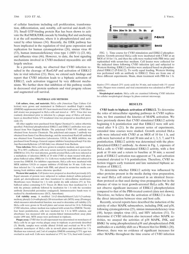
CVB3 leads to biphasic activation of ERK1/2. To determinethe roles of intracellular signaling pathways in CVB3 replica-tion, we first examined the kinetics of MAPK activation. Wehave previously shown that CVB3 stimulated ERK1/2 activitybeginning 6 h postinfection and that ERK1/2 were still acti-vated after 9 h (21). In the present series of experiments,extended time courses were studied. Growth arrested HeLacells were infected with CVB3 at an MOI of 10 for 1 h, andcells were harvested at 10 and 30 min and at 1, 3, 5, 7, and 9 hpostinfection. ERK1/2 activity was determined with the phos-phorylated-ERK1/2 antibody. As shown in Fig. 1, exposure ofHeLa cells to CVB3 stimulated ERK1/2 activity, with a firstpeak at 10 min and a return to baseline at 30 min; a secondpeak of ERK1/2 activation was apparent at 7 h, and activationremained elevated to 9 h postinfection. Therefore, CVB3 in-fection triggers early transient and late sustained biphasic ac-tivation of ERK1/2.
To determine whether ERK1/2 activity was influenced byother proteins present in the media during virus preparation,we used HeLa cell extract processed in an identical freeze-thaw protocol as that used during virus propagation but in theabsence of virus to treat growth-arrested HeLa cells. We didnot observe significant increases of ERK1/2 phosphorylationcompared to that of the PBS-treated control (data not shown).Therefore, we believe that the activation of ERK1/2 is due tothe direct interaction between virus and host cells.
Recently, several reports have described the induction of theactivity of other MAPK submembers, including JNK and p38,following cytomegalovirus (25), simian immunodeficiency virus(48), herpes simplex virus (41), and HIV infection (51). Todetermine if CVB3 infection also increased other MAPK ac-tivities, we assayed the activities of p38 MAPK, JNK, andBMK1 by using phosphorylated-p38 and phosphorylated-JNKantibodies or a mobility shift on a Western blot for BMK1 (28).However, there was no evidence of significant increases forother MAPKs throughout the time course of CVB3 infection
FIG. 1. Time course for CVB3 stimulation and ERK1/2 phosphor-ylation. Growth-arrested HeLa cells were incubated with CVB3 at anMOI of 10 for 1 h, and then the cells were washed with PBS twice andreplenished with serum-free medium. Cell lysates were collected forthe indicated times following CVB3 infection (pi) and subjected toWestern blotting. ERK1/2 activities were analyzed based on phosphor-ylated ERK1/2 (P-Erk1/2). To verify equal loading, Western blottingwas performed with an antibody to ERK1/2. Data are from one ofthree different experiments. Sham, sham treatment with PBS for 1 h.
3366 LUO ET AL. J. VIROL.

(data not shown), which suggests that activation of ERK was arather specific event.
UV-irradiated CVB3 stimulates early-phase phosphoryla-tion of ERK1/2. To further elucidate the mechanism of CVB3-mediated ERK1/2 activation, we used UV-irradiated virus.Such inactivated virus fails to express viral proteins due tothymidine dimers, which prevent transcription of viral genes,but retains the capability for receptor binding and endocytosisinto host cells (5). Since specific ligand-receptor interactionscan result in rapid and transient activation of ERK1/2 (35), thepresence of ERK activation immediately following infectionsuggests that this is a direct receptor-mediated event. As Fig. 2shows, the first (early) ERK1/2 phosphorylation event remains,whereas the second (late) event completely disappears, follow-ing UV-irradiated virus infection, suggesting that CVB3-recep-tor interaction is responsible for early ERK1/2 activation whileviral protein production appears necessary for late-phaseERK1/2 activation following CVB3 infection.
Inhibition of ERK1/2 activation results in significant reduc-tion in viral progeny production and viral protein synthesis.To determine the potential role of ERK activation in viralreplication, we used a selective inhibitor of the ERK pathway,U0126, which inhibits MEK, immediately upstream of ERK.Treatment of cells with U0126 (20 �M) resulted in a completeinhibition of both CVB3-induced early- and late-phase ERKphosphorylation (Fig. 3A).
First, we investigated the effect of the MEK inhibitor on theexpression of viral proteins. HeLa cells were incubated withdifferent concentrations of U0126 for 30 min prior to infection.U0126 was also present during infection and in subsequentincubation periods. Nine hours postinfection cell lysates werecollected and Western blotting was performed using a CVB3polyclonal antibody that recognizes viral structural proteinVP1. Densitometric analysis of Western blotting showed thatthe intracellular expression of the VP1 protein was reduced byU0126 in a dose-dependent manner (Fig. 3B). Next, we wantedto examine whether U0126 affected viral titers. Twenty-fourhours postinfection supernatants were collected and viral titerswere determined by plaque assay of monolayer HeLa cells. AsFig. 3C shows, the presence of U0126 reduced viral progenyrelease in a dose-dependent manner. Compared with controls,
5, 10, and 20 �M U0126 reduced viral progeny by 46, 61, and85%, respectively. It should be noted that, at all concentrationsof U0126 used in this study, there was no evidence of celldeath, as detected by the MTS assay (data not shown).
FIG. 2. UV-irradiated CVB3 stimulates early-phase phosphoryla-tion of ERK1/2. HeLa cells were infected with either wild-type virus orUV-irradiated virus, and 10 min and 9 h after infection (pi) cell lysateswere harvested and Western blotting was performed to determineERK activation. To verify equal loading, Western blotting was per-formed with an anti-ERK1/2 antibody. Data are from one of twodifferent experiments. Sham is as defined for Fig. 1.
FIG. 3. MEK inhibitor reduces viral progeny release and viral pro-tein synthesis. (A) Inhibition of ERK activation by MEK inhibitorU0126 was determined by Western blotting with an anti-phosphory-lated ERK antibody. HeLa cells were preincubated with U0126 (20�M) for 30 min and then were infected with CVB3 (MOI � 10). Onehour later, cells were washed twice with PBS and replenished withserum-free medium containing fresh U0126. To verify equal loading,Western blotting was performed with an anti-ERK1/2 antibody. Thedata are representative of two different experiments. Sham, pi, andP-Erk1/2 are as defined for Fig. 1. (B) HeLa cells were treated withU0126 exactly as for panel A. Cellular lysates were collected fromCVB3-infected HeLa cells 9 h postinfection, and Western blot analysisusing a CVB3 polyclonal antibody that recognizes viral structure pro-tein VP1 was performed. Results (means � standard errors [SE]; n �3) were quantitated by densitometric analysis using National Institutesof Health Image, version 1.61, and normalized to control levels (sham-infected cells without U0126) arbitrarily set to 1.0. (C) HeLa cells weretreated with different concentrations of U0126 exactly as for panel A.Medium was collected from CVB3-infected HeLa cells 24 h afterinfection, and virus titers were determined by plaque assays on HeLacell monolayers. Values are means � SE from three independentexperiments, in each of which titrations were carried out in triplicate.
VOL. 76, 2002 ERK1/2 REGULATES CVB3 REPLICATION 3367

Exposure to U0126 following the first peak of ERK1/2 ac-tivation (3 h postinfection) attenuated viral progeny produc-tion, but to a lesser extent than administration prior to infec-tion (data not shown), suggesting that both phases of ERKactivation contribute to viral replication. Taken together, theseresults indicate that ERK plays an important role in CVB3replication.
Inhibition of ERK1/2 activation prevents CVB3-mediatedCPE and caspase activation. Apoptosis may play a critical rolein viral myocarditis (12, 19, 20). We have previously demon-strated two separate but related phenomena, CPE and apo-ptosis, following CVB3 infection of HeLa cells (10). We there-fore decided to see whether ERK1/2 activation is required forCVB3-induced CPE and apoptosis. Cell viability was deter-mined by the MTS assay, which measures mitochondrial func-tion. As shown in Fig. 4A, inhibition of ERK1/2 by U0126resulted in a dose-dependent reduction of CVB3-mediated celldeath 24 h postinfection, with 20 �M U0126 producing anapproximate 60% increase of viable cells. Morphology of cellstreated with U0126 is shown in Fig. 4B. CVB3-infected cellsdisplayed typical features of apoptosis by 24 h postinfection.Consistent with the cell viability change, pretreatment withU0126 markedly suppressed the morphological changes in-duced by CVB3 infection. We also examined the effect ofU0126 on CVB3-mediated caspase activation. As shown in Fig.4C, U0126 significantly inhibited CVB3-induced caspase 3cleavage in a dose-dependent manner. Therefore, we believethat ERK1/2 activation is required for CVB3-induced apopto-sis, although it is unclear whether the apoptosis and CPE aredirectly mediated by the ERK signaling pathway or whetherthey are indirectly associated with ERK-regulated viral repli-cation or viral infectivity.
Caspases are the main executioners of apoptosis through anumber of cleavage events. It has been reported that many hostproteins are cleaved during the course of apoptosis (57). Wethus determined whether ERK1/2 activity was a result of cas-pase activation by using general caspase inhibitor zVAD.fmk.As shown in Fig. 5, caspase 3 activation could be demonstratedat 9 h postinfection by the presence of the 17-kDa cleavageproduct. ERK1/2 activation was also increased at 9 h postin-fection. Preincubation of cells with 100 �M zVAD.fmk mostlyblocked CVB3-induced caspase cleavage but had no effecton ERK activation. This result shows that the observedsecond peak of ERK1/2 phosphorylation occurred beforeinitiation of caspase activity and suggests that the caspasepathway and subsequent apoptotic processes do not influ-ence MAPK.
MEK inhibitor blocks CVB3-mediated RasGAP cleavage.To gain further insight into the mechanisms and regulation ofintracellular signaling pathways in CVB3-mediated host cell
expression, we next considered the contribution of the MEKinhibitor to CVB3-induced RasGAP cleavage. HeLa cells weretreated with different concentrations of U0126. Nine hourspostinfection cell lysates were collected and subjected to West-ern blotting for RasGAP, a protein which negatively regulatesthe activation of Ras by hydrolysis of GTP to GDP. The re-sults, depicted in Fig. 6, show that MEK inhibition significantlyblocked CVB3-mediated RasGAP cleavage. This result sug-gests that ERK activation may be achieved through a positive-feedback mechanism. As such, ERK activation enhances viralreplication, resulting in a cleavage of host signaling proteinRasGAP, further promoting Ras activity and subsequent acti-vation of the ERK cascade (Fig. 7).
DISCUSSION
In the present study, we have shown that virus infectioninduces biphasic activation of MAPK ERK1/2 in target cells.Immediately following CVB3 or UV-irradiated CVB3 infec-tion, ERK is transiently phosphorylated. Toward the finalstages of virus replication, sustained activation of ERK wasobserved, but this event was absent in infection by UV-irradi-ated virus. We further show that specific inhibition of ERKleads to attenuation of the virus life cycle, as determined bylower virus protein and progeny production, decreased viruscleavage of host proteins, and attenuation of host cell death.This study has provided new insights into our understanding ofthe interplay of virus and host signaling induced by CVB3infection and demonstrates that ERK1/2 signaling is requiredfor CVB3 pathogenesis.
The significance of host activation of MAPK in other modelsof viral infection has been reported. In the HIV-1 model,MAPK activation is beneficial to viral replication and inhibi-tion of these phosphorylation events seems to inhibit viralreplication (23). Similarly, the following viruses also interactwith ERK: adenovirus type 7 (2), Borna disease virus (45),influenza A virus (46), and hepatitis C virus (17). Such wide-spread ERK involvement suggests either involvement in aglobal virus strategy to enhance its own replicative machineryor a universal host protective response to infection.
We have found that CVB3 infection induces a biphasic ac-tivation of ERK1/2 at 10 min and 7 h postinfection. The mech-anisms for ERK1/2 activation following viral infection may beinduced by direct virus-receptor binding, such as for HIV ac-tivation of ERK (47), or by exposure to a viral protein such asthe hepatitis C virus core protein (16) or HIV Tat protein (49).The first peak occurs immediately following infection, whichstrongly suggests that signaling is initiated directly from a re-ceptor-coreceptor complex. We further showed that UV-irra-diated and inactivated CVB3 does indeed activate early but not
FIG. 4. MEK inhibitor blocks CVB3-induced CPE and apoptosis. (A) HeLa cells were treated with U0126 as described for Fig. 3A. Cellviability was determined at 24 h postinfection (pi) by the MTS assay, which measures mitochondrial function, at 24 h after infection. Values aremeans � standard errors (SE; n � 6). The level of MTS in sham-infected cells in the absence of U0126 was defined as 100% survival. Similar resultswere obtained in three independent experiments. (B) Representative phase-contrast microscopy of HeLa cells treated with medium containing orlacking U0126 24 h postinfection. (C) HeLa cells were pretreated with vehicle or various concentrations of U0126 for 30 min, followed by infectionwith CVB3 for 1 h, 9 h after infection. Western blotting was performed to examine the cleavage of caspase 3. Results were quantitated bydensitometric analysis using National Institutes of Health Image, version 1.61, and normalized to the control level (sham-infected cells withoutU0126), which was arbitrarily set to 1.0. Values are means � SE (n � 3).
3368 LUO ET AL. J. VIROL.

VOL. 76, 2002 ERK1/2 REGULATES CVB3 REPLICATION 3369

late ERK signaling. The DAF coreceptor is anchored to theextracellular surface by glycosylphosphatidylinositol (GPI),which is localized to cholesterol-rich invaginations of theplasma membrane, or caveolae, which are proposed sites forincreased outside-to-inside signal transduction and transcytosis(40). This virus receptor has been found to associate with tyro-sine kinases p56lck and p59fyn, among others, and with down-stream ERK1/2 as an essential part of T-cell development (52).Recent evidence has shown that p56lck is required for CVB3infection of T-cell lines and that ERK may be a downstreamtarget of such signaling pathways (33). Since DAF expression isnot always necessary for viral entry, this evidence raises thequestion of whether CVB3 binding to coxsackievirus and ad-enovirus receptor can trigger MAPK activation or whether theintracellular presence, with or without replication, of viral RNA isrequired (38). We therefore speculate that such a DAF-GPIsignaling complex is responsible for early ERK activation inCVB3-infected HeLa cells, but further studies are necessary toconfirm the nature of intersection between receptor complex,tyrosine kinases, and ERK signaling.
A plausible explanation for these early events in the inter-
action between CVB3 and host cells is that ERK signaling isindeed a host protective mechanism to which CVB3 hasadapted. Activation of ERK1/2 is a well-documented cell-pro-tective and cell-beneficial response in the heart to a wide va-riety of damaging agents such as pressure or volume overloadand ischemia-reperfusion injury (9, 67). It is conceivable thatcardiac myocytes may also invoke the ERK response to CVB3as a defense mechanism. The virus, in turn, may have adaptedto such preexisting signaling pathways to benefit its own rep-lication. A relatively short CVB3 life cycle and lack of RNApolymerase proofreading function may allow for rapid adjust-ments to changing characteristics of the host cell system, whichwould beget an overall high mutation rate.
We also show that ERK is stimulated 7 h postinfection,which is consistent with peak virus replication and caspase 3activation (10). We show that UV-irradiated virus, incapable ofreplication, does not trigger late ERK activation, which sug-gests that the observed high level of late-phase ERK activationis dependent on viral gene expression. Several viral gene prod-ucts have been shown to intersect the ERK1/2 signaling path-way, including the hepatitis C virus core protein (16) and theHIV Tat protein (49). Although no comparable data for apicornavirus protein have yet been identified, previous work inour laboratory has shown that RasGAP is cleaved and ERK1/2is activated during virus infection (21). Such findings raise thepossibility that a viral protease is responsible for ERK1/2 ac-tivation via cleavage of an upstream effector molecule. Thesefindings indicate that CVB3 may have developed multiplemechanisms to ensure activation of the MAPK ERK1/2 andthat timely activation may have important consequences dur-ing the course of an infection.
Activation of the ERK pathway results in phosphorylation ofnumerous ERK target proteins, which mediate multiple cellu-lar functions. In the HIV-1 model, ERK1/2 activation aug-ments viral infectivity and replication, which may occur bydirect phosphorylation of viral protein Vif (65). It is not knownat present how CVB3 viral replication is regulated by the ERKsignaling pathway. Perhaps this process involves direct phos-phorylation of intracellular components which are required for
FIG. 5. ERK1/2 activation was not due to caspase activation. HeLacells were preincubated with zVAD.fmk (100 �M) for 30 min and theninfected with CVB3 for 1 h. Cell lysates were collected 9 h postinfec-tion (pi). ERK1/2 activation and caspase 3 cleavage were determinedby Western blotting using a phosphorylated ERK1/2 (P-Erk1/2) anti-body and a caspase 3 antibody. The data are representative of twodifferent experiments.
FIG. 6. MEK inhibitor inhibits CVB3-mediated RasGAP cleavage.HeLa cells were pretreated for 30 min with various concentrations ofU0126 and then were infected by CVB3 for 1 h. At 9 h after additionof CVB3, HeLa cells were harvested and Western blot analysis wasperformed using an antibody that recognizes RasGAP. Results werequantitated by densitometric analysis using National Institutes ofHealth Image, version 1.61, and normalized to the control level (sham-infected cells without U0126), which was arbitrarily set to 1.0. Valuesare means � standard errors (n � 3).
FIG. 7. A proposed model of the mechanism of ERK activationduring CVB3 infection. CVB3 binds with its receptor and initiatesearly transient ERK phosphorylation. Virus replication mediatesRasGAP cleavage, which triggers late-phase ERK phosphorylation.Subsequently, there is a positive feedback to augment ERK activationand viral replication.
3370 LUO ET AL. J. VIROL.

viral replication. Alternatively, ERK1/2 activity may be neces-sary to activate CVB3 viral proteins, for example, RNA-de-pendent RNA polymerase 3D, which is essential for the initi-ation of viral RNA replication.
Recently MEK inhibitors have been reported to prevent theactivation of both ERK1/2 and BMK1. MEK inhibitors, includ-ing U0126 and PD98059, are specific for ERK1/2 at low doses,but high doses block mitogen-induced activation of bothERK1/2 and BMK1 (27). The dose of U0126 used here issufficiently specific for CVB3-medicated ERK1/2 activation;BMK1 was not activated throughout the course of CVB3 in-fection.
CVB3 infection, as well as expression of viral capsid proteinsand proteases, induces direct CPE and cell apoptosis (10, 18,19). Inhibition of such caspase activation blocks the loss ofviability and prevents progeny virus release. Since caspase in-hibition does not affect ERK activation, but ERK inhibitionprotects against CVB3-induced CPE and apoptosis, we con-clude that ERK activation occurs upstream of caspase activa-tion. The mechanism by which inhibition of ERK preventsCVB3-induced apoptosis is likely an indirect result of a de-crease in virus replication and/or infectivity, analogous to theERK contribution to influenza virus infection (46). In additionto the effects of virus replication on host cell apoptotic path-ways, we offer the possibility of a more direct role for ERK inthe CVB3-induced cell death signaling pathway in HeLa cells.Although many studies have supported the general view thatactivation of the ERK pathway delivers a survival signal thatcounteracts proapoptotic effects associated with p38 and JNKactivation (62), there are some studies that relate ERK and theapoptosis cascade. For example, it has been reported that ERKactivation is required for cisplatin-induced apoptosis of HeLacells and functions upstream of caspase activation to initiatethe apoptotic signal (60). Overproduction of proto-oncogenesdownstream of ERK, such as c-myc, may trigger apoptosis atthe level of the mitochondria (54). Joe et al. (24) have alsoshown that dominant inhibitory Ras delays Sindbis virus-in-duced apoptosis in neuronal cells. The mechanism of indirectinduction of apoptosis by CVB3 replication and the possiblecontribution by direct host signaling events remain an impor-tant area for further investigation.
Ras is a 21-kDa, GTP-binding protein that plays a criticalrole in signal transduction pathways mediating many importantcellular functions. In the GTP-bound state, Ras interacts withand transmits signals to downstream effector molecules, suchas those in the ERK signaling pathway. Hydrolysis of GTP toGDP switches Ras to the inactive state. RasGAP negativelyregulates the GTP-bound state by stimulating the intrinsic RasGTPase by hydrolysis of GTP to GDP (8, 58). We have pre-viously shown that RasGAP is cleaved after CVB3 infection,potentially triggering the Ras pathway and subsequent phos-phorylation of ERK1/2 (21). In this study we report that aMEK inhibitor blocks viral replication and CVB3-inducedRasGAP cleavage. This result suggests a possible mechanismby which viral protein cleavage of RasGAP and late-phaseERK activation prompt a positive-feedback loop to further in-crease viral replication, with subsequent cleavage of RasGAP.
Increasing our understanding of virus-induced death signal-ing in viral myocarditis has become particularly important inlight of direct virus-mediated mechanistic connections between
myocarditis and DCM. These findings are qualified by thepresence of the CVB3 genome in patients with DCM (36) andmore-recent findings that suggest that CVB3 may persist insuch tissues disguised in a stable double-stranded form (55).The contribution of virus-myocyte interactions, as opposed toimmune infiltration, has profound consequences for the degreeof myocardial injury. Therefore, greater understanding of keysignaling pathways that are beneficial to the virus may lead tonovel therapies to stymie the progression from myocarditis toan end-stage disease which impacts a significant and growingpopulation.
ACKNOWLEDGMENTS
This work was funded by grants from the Heart and Stroke Foun-dation of British Columbia and Yukon (B.M.M.) and the MedicalResearch Council of Canada (B.M.M.) and by doctoral traineeshipsfrom the Canadian Institutes of Health Research and Heart andStroke Foundation of Canada (joint; B.Y.), the Heart and StrokeFoundation of British Columbia and Yukon (M.E.), and the MichaelSmith Foundation for Health Research (B.Y. and M.E.).
We thank Reinhard Kandolf (University of Tubingen, Tubingen,Germany) for providing the CVB3.
REFERENCES
1. Abe, M. K., W. L. Kuo, M. B. Hershenson, and M. R. Rosner. 1999. Extra-cellular signal-regulated kinase 7 (ERK7), a novel ERK with a C-terminaldomain that regulates its activity, its cellular localization, and cell growth.Mol. Cell. Biol. 19:1301–1312.
2. Alcorn, M. J., J. L. Booth, K. M. Coggeshall, and J. P. Metcalf. 2001.Adenovirus type 7 induces interleukin-8 production via activation of extra-cellular regulated kinase 1/2. J. Virol. 75:6450–6459.
3. Anderson, D. R., J. E. Wilson, C. M. Carthy, D. Yang, R. Kandolf, and B. M.McManus. 1996. Direct interactions of coxsackievirus B3 with immune cellsin the splenic compartment of mice susceptible or resistant to myocarditis.J. Virol. 70:4632–4645.
4. Badorff, C., G. H. Lee, B. J. Lamphear, M. E. Martone, K. P. Campbell, R. E.Rhoads, and K. U. Knowlton. 1999. Enteroviral protease 2A cleaves dystro-phin: evidence of cytoskeletal disruption in an acquired cardiomyopathy.Nat. Med. 5:320–326.
5. Beck, M. A., N. M. Chapman, B. M. McManus, J. C. Mullican, and S. Tracy.1990. Secondary enterovirus infection in the murine model of myocarditis.Pathologic and immunologic aspects. Am. J. Pathol. 136:669–681.
6. Bergelson, J. M., J. A. Cunningham, G. Droguett, E. A. Kurt-Jones, A.Krithivas, J. S. Hong, M. S. Horwitz, R. L. Crowell, and R. W. Finberg. 1997.Isolation of a common receptor for coxsackie B viruses and adenoviruses 2and 5. Science 275:1320–1323.
7. Blenis, J. 1993. Signal transduction via the MAP kinases: proceed at yourown RSK. Proc. Natl. Acad. Sci. USA 90:5889–5892.
8. Bollag, G., and F. McCormick. 1991. Differential regulation of rasGAP andneurofibromatosis gene product activities. Nature 351:576–579.
9. Bueno, O. F., L. J. De Windt, K. M. Tymitz, S. A. Witt, T. R. Kimball, R.Klevitsky, T. E. Hewett, S. P. Jones, D. J. Lefer, C. F. Peng, R. N. Kitsis, andJ. D. Molkentin. 2000. The MEK1-ERK1/2 signaling pathway promotescompensated cardiac hypertrophy in transgenic mice. EMBO J. 19:6341–6350.
10. Carthy, C. M., D. J. Granville, K. A. Watson, D. R. Anderson, J. E. Wilson,D. Yang, D. W. Hunt, and B. M. McManus. 1998. Caspase activation andspecific cleavage of substrates after coxsackievirus B3-induced cytopathiceffect in HeLa cells. J. Virol. 72:7669–7675.
11. Chow, L. H., K. W. Beisel, and B. M. McManus. 1992. Enteroviral infectionof mice with severe combined immunodeficiency. Evidence for direct viralpathogenesis of myocardial injury. Lab. Investig. 66:24–31.
12. Colston, J. T., B. Chandrasekar, and G. L. Freeman. 1998. Expression ofapoptosis-related proteins in experimental coxsackievirus myocarditis. Car-diovasc. Res. 38:158–168.
13. Cory, A. H., T. C. Owen, J. A. Barltrop, and J. G. Cory. 1991. Use of anaqueous soluble tetrazolium/formazan assay for cell growth assays in culture.Cancer Commun. 3:207–212.
14. Davis, R. J. 1993. The mitogen-activated protein kinase signal transductionpathway. J. Biol. Chem. 268:14553–14556.
15. Ehrenfeld, E. 1982. Poliovirus-induced inhibition of host-cell protein synthe-sis. Cell 28:435–436.
VOL. 76, 2002 ERK1/2 REGULATES CVB3 REPLICATION 3371

16. Fukuda, K., K. Tsuchihara, M. Hijikata, S. Nishiguchi, T. Kuroki, and K.Shimotohno. 2001. Hepatitis C virus core protein enhances the activation ofthe transcription factor, Elk1, in response to mitogenic stimuli. Hepatology33:159–165.
17. Giambartolomei, S., F. Covone, M. Levrero, and C. Balsano. 2001. Sustainedactivation of the Raf/MEK/Erk pathway in response to EGF in stable celllines expressing the hepatitis C virus (HCV) core protein. Oncogene 20:2606–2610.
18. Goldstaub, D., A. Gradi, Z. Bercovitch, Z. Grosmann, Y. Nophar, S. Luria,N. Sonenberg, and C. Kahana. 2000. Poliovirus 2A protease induces apo-ptotic cell death. Mol. Cell. Biol. 20:1271–1277.
19. Henke, A., H. Launhardt, K. Klement, A. Stelzner, R. Zell, and T. Munder.2000. Apoptosis in coxsackievirus B3-caused diseases: interaction betweenthe capsid protein VP2 and the proapoptotic protein siva. J. Virol. 74:4284–4290.
20. Herzum, M., V. Ruppert, B. Kuytz, H. Jomaa, I. Nakamura, and B. Maisch.1994. Coxsackievirus B3 infection leads to cell death of cardiac myocytes. J.Mol. Cell. Cardiol. 26:907–913.
21. Huber, M., K. A. Watson, H. C. Selinka, C. M. Carthy, K. Klingel, B. M.McManus, and R. Kandolf. 1999. Cleavage of RasGAP and phosphorylationof mitogen-activated protein kinase in the course of coxsackievirus B3 rep-lication. J. Virol. 73:3587–3594.
22. Ito, M., M. Kodama, M. Masuko, M. Yamaura, K. Fuse, Y. Uesugi, S.Hirono, Y. Okura, K. Kato, Y. Hotta, T. Honda, R. Kuwano, and Y. Aizawa.2000. Expression of coxsackievirus and adenovirus receptor in hearts of ratswith experimental autoimmune myocarditis. Circ. Res. 86:275–280.
23. Jacque, J. M., A. Mann, H. Enslen, N. Sharova, B. Brichacek, R. J. Davis,and M. Stevenson. 1998. Modulation of HIV-1 infectivity by MAPK, avirion-associated kinase. EMBO J. 17:2607–2618.
24. Joe, A. K., G. Ferrari, H. H. Jiang, X. H. Liang, and B. Levine. 1996.Dominant inhibitory Ras delays Sindbis virus-induced apoptosis in neuronalcells. J. Virol. 70:7744–7751.
25. Johnson, R. A., S. M. Huong, and E. S. Huang. 2000. Activation of themitogen-activated protein kinase p38 by human cytomegalovirus infectionthrough two distinct pathways: a novel mechanism for activation of p38.J. Virol. 74:1158–1167.
26. Johnson, R. A., X. L. Ma, A. D. Yurochko, and E. S. Huang. 2001. The roleof MKK1/2 kinase activity in human cytomegalovirus infection. J. Gen. Virol.82:493–497.
27. Kamakura, S., T. Moriguchi, and E. Nishida. 1999. Activation of the proteinkinase ERK5/BMK1 by receptor tyrosine kinases. Identification and char-acterization of a signaling pathway to the nucleus. J. Biol. Chem. 274:26563–26571.
28. Kato, Y., R. I. Tapping, S. Huang, M. H. Watson, R. J. Ulevitch, and J. D.Lee. 1998. Bmk1/Erk5 is required for cell proliferation induced by epidermalgrowth factor. Nature 395:713–716.
29. Klingel, K., P. Rieger, G. Mall, H. C. Selinka, M. Huber, and R. Kandolf.1998. Visualization of enteroviral replication in myocardial tissue by ultra-structural in situ hybridization: identification of target cells and cytopathiceffects. Lab. Investig. 78:1227–1237.
30. Lange-Carter, C. A., C. M. Pleiman, A. M. Gardner, K. J. Blumer, and G. L.Johnson. 1993. A divergence in the MAP kinase regulatory network definedby MEK kinase and Raf. Science 260:315–319.
31. Lechner, C., M. A. Zahalka, J. F. Giot, N. P. Moller, and A. Ullrich. 1996.ERK6, a mitogen-activated protein kinase involved in C2C12 myoblast dif-ferentiation. Proc. Natl. Acad. Sci. USA 93:4355–4359.
32. Lee, J. D., R. J. Ulevitch, and J. Han. 1995. Primary structure of BMK1: anew mammalian map kinase. Biochem. Biophys. Res. Commun. 213:715–724.
33. Liu, P., K. Aitken, Y. Y. Kong, M. A. Opavsky, T. Martino, F. Dawood, W. H.Wen, I. Kozieradzki, K. Bachmaier, D. Straus, T. W. Mak, and J. M. Pen-ninger. 2000. The tyrosine kinase p56lck is essential in coxsackievirus B3-mediated heart disease. Nat. Med. 6:429–434.
34. Marais, R., Y. Light, H. F. Paterson, and C. J. Marshall. 1995. Ras recruitsRaf-1 to the plasma membrane for activation by tyrosine phosphorylation.EMBO J. 14:3136–3145.
35. Marshall, C. J. 1995. Specificity of receptor tyrosine kinase signaling: tran-sient versus sustained extracellular signal-regulated kinase activation. Cell80:179–185.
36. Martino, T. A., P. Liu, and M. J. Sole. 1994. Viral infection and the patho-genesis of dilated cardiomyopathy. Circ. Res. 74:182–188.
37. Martino, T. A., M. Petric, M. Brown, K. Aitken, C. J. Gauntt, C. D. Rich-ardson, L. H. Chow, and P. P. Liu. 1998. Cardiovirulent coxsackieviruses andthe decay-accelerating factor (CD55) receptor. Virology 244:302–314.
38. Martino, T. A., M. Petric, H. Weingartl, J. M. Bergelson, M. A. Opavsky,C. D. Richardson, J. F. Modlin, R. W. Finberg, K. C. Kain, N. Willis, C. J.Gauntt, and P. P. Liu. 2000. The coxsackie-adenovirus receptor (CAR) isused by reference strains and clinical isolates representing all six serotypes ofcoxsackievirus group B and by swine vesicular disease virus. Virology 271:99–108.
39. Mason, J. W., J. B. O’Connell, A. Herskowitz, N. R. Rose, B. M. McManus,
M. E. Billingham, T. E. Moon, et al. 1995. A clinical trial of immunosup-pressive therapy for myocarditis. N. Engl. J. Med. 333:269–275.
40. Mayor, S., K. G. Rothberg, and F. R. Maxfield. 1994. Sequestration ofGPI-anchored proteins in caveolae triggered by cross-linking. Science 264:1948–1951.
41. McLean, T. I., and S. L. Bachenheimer. 1999. Activation of cJUN N-terminalkinase by herpes simplex virus type 1 enhances viral replication. J. Virol.73:8415–8426.
42. McManus, B. M., L. H. Chow, S. J. Radio, S. M. Tracy, M. A. Beck, N. M.Chapman, K. Klingel, and R. Kandolf. 1991. Progress and challenges in thepathological diagnosis of myocarditis. Eur. Heart J. 12:18–21.
43. McManus, B. M., L. H. Chow, J. E. Wilson, D. R. Anderson, J. M. Gulizia,C. J. Gauntt, K. E. Klingel, K. W. Beisel, and R. Kandolf. 1993. Directmyocardial injury by enterovirus: a central role in the evolution of murinemyocarditis. Clin. Immunol. Immunopathol. 68:159–169.
44. Peng, T., T. Sadusky, Y. Li, G. R. Coulton, H. Zhang, and L. C. Archard.2001. Altered expression of Bag-1 in coxsackievirus B3 infected mouse heart.Cardiovasc. Res. 50:46–55.
45. Planz, O., S. Pleschka, and S. Ludwig. 2001. MEK-specific inhibitor U0126blocks spread of Borna disease virus in cultured cells. J. Virol. 75:4871–4877.
46. Pleschka, S., T. Wolff, C. Ehrhardt, G. Hobom, O. Planz, U. R. Rapp, and S.Ludwig. 2001. Influenza virus propagation is impaired by inhibition of theRaf/MEK/ERK signalling cascade. Nat. Cell Biol. 3:301–305.
47. Popik, W., J. E. Hesselgesser, and P. M. Pitha. 1998. Binding of humanimmunodeficiency virus type 1 to CD4 and CXCR4 receptors differentiallyregulates expression of inflammatory genes and activates the MEK/ERKsignaling pathway. J. Virol. 72:6406–6413.
48. Popik, W., and P. M. Pitha. 1998. Early activation of mitogen-activatedprotein kinase kinase, extracellular signal-regulated kinase, p38 mitogen-activated protein kinase, and c-Jun N-terminal kinase in response to bindingof simian immunodeficiency virus to Jurkat T cells expressing CCR5 recep-tor. Virology 252:210–217.
49. Rusnati, M., C. Urbinati, B. Musulin, D. Ribatti, A. Albini, D. Noonan, C.Marchisone, J. Waltenberger, and M. Presta. 2001. Activation of endothelialcell mitogen activated protein kinase ERK(1/2) by extracellular HIV-1 Tatprotein. Endothelium 8:65–74.
50. Shafren, D. R., R. C. Bates, M. V. Agrez, R. L. Herd, G. F. Burns, and R. D.Barry. 1995. Coxsackieviruses B1, B3, and B5 use decay accelerating factoras a receptor for cell attachment. J. Virol. 69:3873–3877.
51. Shapiro, L., K. A. Heidenreich, M. K. Meintzer, and C. A. Dinarello. 1998.Role of p38 mitogen-activated protein kinase in HIV type 1 production invitro. Proc. Natl. Acad. Sci. USA 95:7422–7426.
52. Shenoy-Scaria, A. M., J. Kwong, T. Fujita, M. W. Olszowy, A. S. Shaw, andD. M. Lublin. 1992. Signal transduction through decay-accelerating factor.Interaction of glycosyl-phosphatidylinositol anchor and protein tyrosine ki-nases p56lck and p59fyn 1. J. Immunol. 149:3535–3541.
53. Sontag, E., S. Fedorov, C. Kamibayashi, D. Robbins, M. Cobb, and M.Mumby. 1993. The interaction of SV40 small tumor antigen with proteinphosphatase 2A stimulates the map kinase pathway and induces cell prolif-eration. Cell 75:887–897.
54. Soucie, E. L., M. G. Annis, J. Sedivy, J. Filmus, B. Leber, D. W. Andrews, andL. Z. Penn. 2001. Myc potentiates apoptosis by stimulating Bax activity at themitochondria. Mol. Cell. Biol. 21:4725–4736.
55. Tam, P. E., and R. P. Messner. 1999. Molecular mechanisms of coxsackie-virus persistence in chronic inflammatory myopathy: viral RNA persiststhrough formation of a double-stranded complex without associated genomicmutations or evolution. J. Virol. 73:10113–10121.
56. Taylor, L. A., C. M. Carthy, D. Yang, K. Saad, D. Wong, G. Schreiner, L. W.Stanton, and B. M. McManus. 2000. Host gene regulation during coxsack-ievirus B3 infection in mice: assessment by microarrays. Circ. Res. 87:328–334.
57. Thornberry, N. A., and Y. Lazebnik. 1998. Caspases: enemies within. Science281:1312–1316.
58. Trahey, M., G. Wong, R. Halenbeck, B. Rubinfeld, G. A. Martin, M. Ladner,C. M. Long, W. J. Crosier, K. Watt, K. Koths. 1988. Molecular cloning of twotypes of GAP complementary DNA from human placenta. Science 242:1697–1700.
59. Wang, X., and J. M. Bergelson. 1999. Coxsackievirus and adenovirus recep-tor cytoplasmic and transmembrane domains are not essential for coxsack-ievirus and adenovirus infection. J. Virol. 73:2559–2562.
60. Wang, X., J. L. Martindale, and N. J. Holbrook. 2000. Requirement forERK activation in cisplatin-induced apoptosis. J. Biol. Chem. 275:39435–39443.
61. Wessely, R., A. Henke, R. Zell, R. Kandolf, and K. U. Knowlton. 1998.Low-level expression of a mutant coxsackieviral cDNA induces a myocyto-pathic effect in culture: an approach to the study of enteroviral persistence incardiac myocytes. Circulation 98:450–457.
62. Xia, Z., M. Dickens, J. Raingeaud, R. J. Davis, and M. E. Greenberg. 1995.Opposing effects of ERK and JNK-p38 MAP kinases on apoptosis. Science270:1326–1331.
63. Yan, C., H. Luo, J. D. Lee, J. Abe, and B. C. Berk. 2001. Molecular cloning
3372 LUO ET AL. J. VIROL.

of mouse ERK5/BMK1 splice variants and characterization of ERK5 func-tional domains. J. Biol. Chem. 276:10870–10878.
64. Yang, D., J. Yu, Z. Luo, C. M. Carthy, J. E. Wilson, Z. Liu, and B. M. McManus.1999. Viral myocarditis: identification of five differentially expressed genes incoxsackievirus B3-infected mouse heart. Circ. Res. 84:704–712.
65. Yang, X., and D. Gabuzda. 1998. Mitogen-activated protein kinase phos-phorylates and regulates the HIV-1 Vif protein. J. Biol. Chem. 273:29879–29887.
66. Yang, X., and D. Gabuzda. 1999. Regulation of human immunodeficiencyvirus type 1 infectivity by the ERK mitogen-activated protein kinase signalingpathway. J. Virol. 73:3460–3466.
67. Yue, T. L., C. Wang, J. L. Gu, X. L. Ma, S. Kumar, J. C. Lee, G. Z.Feuerstein, H. Thomas, B. Maleeff, and E. H. Ohlstein. 2000. Inhibition ofextracellular signal-regulated kinase enhances ischemia/reoxygenation-in-duced apoptosis in cultured cardiac myocytes and exaggerates reperfusioninjury in isolated perfused heart. Circ. Res. 86:692–699.
VOL. 76, 2002 ERK1/2 REGULATES CVB3 REPLICATION 3373


