Embed Size (px)
Citation preview

Critical roles of macrophages in the formation of intracranialaneurysm
Yasuhisa Kanematsu, MD PhD1,4,5, Miyuki Kanematsu, MD1,4,6, Chie Kurihara, BS1,4,Yoshiteru Tada, MD, PhD1,4,5, Tsung-Ling Tsou, BS1,4, Nico van Rooijen, MD, Michael T.Lawton, MD7, William L. Young, MD1,2,3,4, Elena I. Liang, BS1,4, Yoshitsugu Nuki, MDPhD1,4, and Tomoki Hashimoto, MD1,4
1Department of Anesthesia and Perioperative Care, University of California, San Francisco,California, U.S.A. 2Department of Neurology University of California, San Francisco, California,U.S.A. 3Department of Neurosurgery, University of California, San Francisco, California, U.S.A.4Center for Cerebrovascular Research, University of California, San Francisco, California, U.S.A.5Department of Neurosurgery, Institute of Health Biosciences, University of Tokushima,Tokushima, Japan 6Department of Thoracic, Endocrine Surgery and Oncology, Institute of HealthBiosciences, University of Tokushima, Tokushima, Japan 7Department of Molecular Cell Biology,Vrije Universiteit, Amsterdam, The Netherlands
AbstractBackground and Purpose—Abnormal vascular remodeling triggered by hemodynamicstresses and inflammation is believed to be a key process in the pathophysiology of intracranialaneurysms. Numerous studies have shown infiltration of inflammatory cells, especiallymacrophages, into intracranial aneurysmal walls in humans. Using a mouse model of intracranialaneurysms, we tested whether macrophages play critical roles in the formation of intracranialaneurysms.
Methods—Intracranial aneurysms were induced in adult male mice using a combination of asingle injection of elastase into the cerebrospinal fluid and angiotensin-II-induced hypertension.Aneurysm formation was assessed three weeks later. Roles of macrophages were assessedutilizing clodronate liposome-induced macrophage depletion. In addition, the incidence ofaneurysms was assessed in mice lacking monocyte chemotactic protein-1 (MCP-1, CCL2), andmice lacking matrix metalloproteinase-12 (MMP-12, macrophage elastase).
Results—Intracranial aneurysms in this model showed leukocyte infiltration into the aneurysmalwall, the majority of leukocytes being macrophages. Mice with macrophage depletion had asignificantly reduced incidence of aneurysms compared to control mice (1/10 vs. 6/10; P < 0.05).Similarly, there was a reduced incidence of aneurysms in mice lacking MCP-1, compared toincidence of aneurysms in wild-type mice (2/10 vs. 14/20, P < 0.05). There was no difference inthe incidence of aneurysms between mice lacking MMP-12 and wild-type mice.
Conclusions—These data suggest critical roles of macrophages and proper macrophagefunctions in the formation of intracranial aneurysms in this model.
Correspondence to: Tomoki Hashimoto, MD, Department of Anesthesia and Perioperative Care, University of California, SanFrancisco, 1001 Potrero Avenue, No. 3C-38, San Francisco, CA 94110, USA. Phone: (415) 206-8958. Fax: (415) 206-8170;[email protected] of interest / DisclosuresNone
NIH Public AccessAuthor ManuscriptStroke. Author manuscript; available in PMC 2012 January 1.
Published in final edited form as:Stroke. 2011 January ; 42(1): 173–178. doi:10.1161/STROKEAHA.110.590976.
NIH
-PA Author Manuscript
NIH
-PA Author Manuscript
NIH
-PA Author Manuscript

KeywordsIntracranial aneurysm; stroke; inflammation; animal model; macrophage
IntroductionPotential roles of inflammation in the pathophysiology of intracranial aneurysms—bothruptured and unruptured— have been suggested by observational and genetic studies.1-6
Macrophage infiltration has been well-documented in both ruptured and unrupturedintracranial aneurysms in humans.2, 3, 7 A higher degree of inflammation in aneurysmsseems to be associated with aneurysmal wall destruction and rupture.3, 7
We have recently showed that macrophages and macrophage-derived cytokines are criticalfor hemodynamically-induced outward vascular remodeling.8, 9 Vascular remodelingcoupled with inflammation is considered as a key part in the pathophysiology of intracranialaneurysms.4, 10 Sustained vascular remodeling may lead to aneurysmal growth and rupture.1, 11 By mediating inflammation and hemodynamically-induced vascular remodeling,macrophages may play critical roles in the development, growth, and rupture of intracranialaneurysms.
In this study, we examined whether macrophages are critical for the formation ofintracranial aneurysms using a mouse model of intracranial aneurysms that replicates keyfeatures of human intracranial aneurysms. First, we assessed the effects of macrophagedepletion by clodronate liposome on the formation of aneurysms. Second, aneurysmformation was assessed in mice lacking monocyte chemotactic protein-1 (MCP-1, CCL2).MCP-1 is a chemotactic factor that is critical for proper macrophage functions. MCP-1knockout mice have reduced macrophage/monocyte counts and impaired macrophagefunctions. Therefore, they have been used as a genetic equivalent of mice withpharmacological depletion of macrophages and monocytes in various physiological andpathological settings.12, 13
Materials and MethodsExperiments were conducted in accordance with the guidelines approved by the Universityof California, San Francisco, Institutional Animal Care and Use Committee.
We used the elastase-induced intracranial aneurysms in 8 to 9 week old hypertensive miceas previously described.11 In this model, two well-known clinical factors associated withhuman intracranial aneurysms—hypertension and the disruption of elastic lamina— werecombined to induce intracranial aneurysm formation in mice. We performed a singlestereotaxic injection of elastase into the cerebrospinal fluid at the right basal cistern. Avolume of 2.5 μL of elastase solution (17 milli-units) was injected at a rate of 0.2 μL/min(Ultramicropump, World Precision Instruments). Hypertension was induced by a continuoussubcutaneous infusion of angiotensin-II at 1000 ng/kg/min for three weeks via an implantedosmotic pump (Alzet pump, Durect).11, 14
Systolic blood pressure was measured in mice before treatment, one week after elastaseinjection, and two weeks after elastase injection using the tail cuff method. After threeweeks, we sacrificed the mice and perfused the animals with bromophenol blue dye. Twoblinded observers assessed the formation of intracranial aneurysms by examining of Circleof Willis and its major branches under a dissecting microscope (10×). Intracranialaneurysms were operationally defined as a localized outward bulging of the vascular wall in
Kanematsu et al. Page 2
Stroke. Author manuscript; available in PMC 2012 January 1.
NIH
-PA Author Manuscript
NIH
-PA Author Manuscript
NIH
-PA Author Manuscript

the Circle of Willis or in its major primary branches, as previously described.10, 11 Afterinspecting the Circle of Willis, the whole brain samples were frozen in OCT forimmunohistochemical staining.
Macrophage depletionMacrophage depletion was achieved by an intravenous injection of liposome-encapsulateddichloromethylene diphosphonate (clodronate liposome).15 We used 8 to 9-week-old maleC57BL/6J mice (n = 10 in each group). Clodronate was a gift from Roche DiagnosticsGmbH (Mannheim, Germany). Animals received clodronate liposome intravenously twodays before elastase and angiotensin-II treatment. This regimen was reported to cause areduction of macrophages to less than 10% of the baseline count.9, 16 Animals in the controlgroup received the same volume of phosphate-buffered saline-containing liposome (PBSliposome). We assessed the efficiency of macrophage depletion by examining macrophagesin the spleen using immunohistochemistry, as previously described.9, 16
Incidence of aneurysms in MCP-1 knockout mice and MMP-12 knockout miceIn addition, the incidence of aneurysms was assessed in MCP-1 knockout mice andMMP-12 knockout mice (n = 10). Wild-type mice with the same background (both C57BL/6J) were used as control mice (n = 20).
Immunohistochemical analysisDetails of immunohistochemical analysis were described in Online Data Supplement.
Statistical analysisAll results were expressed as mean ± SD. Differences between multiple groups wereanalyzed by one-way ANOVA, followed by the Tukey-Kramer post hoc test. Fisher's exacttest was used to analyze the incidence of aneurysms. Statistical significance was taken at P <0.05.
ResultsPresence of macrophages in experimental intracranial aneurysm
Figure 1 shows representative intracranial aneurysms in hypertensive mice that received asingle injection of elastase into the cerebrospinal fluid. Large saccular aneurysm formationwas found along the Circle of Willis or its major branches, which is consistent with ourprevious study (A-D).11 Most aneurysms were larger than 250 μm in diameter,approximately 2 to 5 times larger than their parent arteries, as we previously reported (A-D).Some mice had multiple aneurysms (D).
A cerebral artery from the sham operation group revealed an endothelial cell lining with athin layer of smooth muscle cells, as previously described (Figure 2A).11 In contrast,intracranial aneurysms had a partially thickened vascular wall with inflammatory-like cellinfiltration (Figure 2B). Endothelial cell layers seemed to be generally intact, but smoothmuscle cell layers had thickened in the area with intense inflammatory cell infiltration.
Pan-leukocyte staining using anti-CD45 antibody and macrophage staining using anti-CD68antibody in the cerebral artery from the sham operation group showed a lack ofinflammatory cells and macrophage infiltration (Figure 2C and 2E). In intracranialaneurysms, numerous leukocytes (CD45 positive cells) were detected in the adventitia andmedia of the aneurysmal wall (Figure 2D), especially in the thickened part of aneurysmalwall, which is generally consistent with observations in human intracranial aneurysms.2, 3, 7
Kanematsu et al. Page 3
Stroke. Author manuscript; available in PMC 2012 January 1.
NIH
-PA Author Manuscript
NIH
-PA Author Manuscript
NIH
-PA Author Manuscript

Macrophage staining showed macrophage infiltration into the aneurysmal wall with adistribution similar to that of leukocytes (Figure 2F-G). Double staining with anti-CD68 andanti-CD45 revealed that a majority of leukocytes in intracranial aneurysms in this modelwere macrophages (Figure 2H).
Effects of macrophage depletion on intracranial aneurysm formationTen mice underwent macrophage depletion treatment with clodronate liposome two daysbefore the induction of intracranial aneurysms, and another ten mice received PBS liposome.All twenty mice received a single stereotaxic injection of elastase into the cerebrospinalfluid to disrupt the elastic lamina, and a continuous infusion of angiotensin-II to inducehypertension.
When we examined the mice three weeks after aneurysm induction, mice that receivedmacrophage depletion treatment using clodronate liposome had a reduced incidence ofintracranial aneurysms compared to mice that received PBS liposome (mice with intactmacrophages) (1/10 vs. 6/10; 10% vs. 60%, P < 0.05) (Figure 3A), indicating a critical roleof macrophages in the formation of intracranial aneurysms in this model.
While there were abundant macrophages in the intracranial aneurysm from a PBS liposome-treated mouse (Figure 3D, 3E), middle cerebral arteries from mice in either the shamoperation group (Figure 3B, 3C) or the macrophage depletion group (F, G) did not showmacrophage infiltration into the vascular wall.
Quantification of macrophages (n = 5 in each group) showed that the number ofmacrophages was higher in the mice treated with elastase, angiotensin-II, and PBS liposometreated mice compared to mice in the sham group or macrophage depletion group (0.5 ± 0.3vs. 11.1 ± 3.3, P < 0.05; 0.5 ± 0.3 vs. 1.0 ± 0.3, P < 0.05) (Figure 3H).
Immunohistochemical staining for CD68 positive cells (monocyte / macrophage) in thespleen (n = 5 at each time point) was used to assess the efficiency and time-course ofclodronate liposome treatment as previously described (Figure 4A-B).9, 16 Treatment withclodronate liposome decreased the CD68 positive area in the spleen by 88% from thebaseline (28.8 ± 4.1 vs. 3.2 ± 1.0%, P < 0.05), showing an effective macrophage / monocytereduction by the clodronate treatment. At two weeks, CD68 positive area returned to thebaseline (28.8 ± 4.1 vs. 23.1 ± 2.6%).
Angiotensin-II treatment caused hypertension in both groups. After one week and twoweeks, systolic blood pressure was higher than the baseline. Macrophage depletiontreatment did not affect systolic blood pressure (Figure 4C).
Reduced incidence of intracranial aneurysm in MCP-1 knockout miceSince MCP-1 knockout mice have reduced monocyte/macrophage counts and impairedmacrophage functions,17 we used MCP-1 knockout mice to further test the critical role ofmacrophages in the formation of intracranial aneurysms. Twenty wild-type mice and tenMCP-1 knockout mice underwent intracranial aneurysm induction.
MCP-1 knockout mice had a lower incidence of aneurysms compared to wild-type mice(2/10 vs. 14/20; 20 vs. 70%, P < 0.05) (Figure 5A). Immunohistochemical staining ofcerebral arteries for macrophages showed a lack of macrophage infiltration in MCP-1knockout mice (Figure 5B). Quantification of macrophages in the middle cerebral artery (n= 5 in each group) showed reduced macrophage infiltration to the middle cerebral artery inMCP-1 knockout mice compared to macrophage infiltration in wild-type mice (2.8 ± 1.4 vs.10.9 ± 0.8, P < 0.05, Figure 5E).
Kanematsu et al. Page 4
Stroke. Author manuscript; available in PMC 2012 January 1.
NIH
-PA Author Manuscript
NIH
-PA Author Manuscript
NIH
-PA Author Manuscript

Macrophages produce matrix metalloproteinases (MMP) that are critical for vascularremodeling.8, 9 Previously, we have shown that high activity of MMPs in intracranialaneurysms of this model.11 MMP inhibitor, doxycycline, significantly reduced the incidenceof intracranial aneurysms.11 MMP-9 knockout mice, but not MMP-2 knockout mice, have areduced incidence of intracranial aneurysms.11 While MMP-9 is the main gelatinaseproduced by macrophages, MMP-12 represents the major elastase from macrophages.MMP-12 could be the proteinase responsible for facilitating structural changes of elasticlamina, resulting in physiological and pathological vascular remodeling.18 Therefore, weinvestigated the roles of MMP-12 in the formation of intracranial aneurysms. However,there was no difference in the incidence of intracranial aneurysms between MMP-12knockout mice and wild-type mice (50% vs. 70%). Moreover, immunohistochemicalstaining for macrophages (CD68 positive cells) showed a similar number of macrophagesthat accumulated in MMP-12 knockout mice compared to the number of macrophages inwild-type mice (9.4 ± 2.3 vs. 10.9 ± 0.8, Figure 5E).
Continuous infusion of angiotensin-II increased systolic blood pressure from the baselineafter one week and two weeks in MCP-1 knockout mice and MMP-12 knockout mice. Therewas no significant difference between wild-type mice and MCP-1 or MMP-12 knockoutmice at one week and two weeks (Figure 5F).
DiscussionIn this study, we have shown the critical roles of macrophages in the formation ofintracranial aneurysms in mice. We used a recently developed intracranial aneurysm modelin which intracranial aneurysms were induced by a combination of single stereotaxicinjection of elastase into the cerebrospinal fluid and pharmacologically-inducedhypertension in mice. Intracranial aneurysms in this mouse model closely resemblehistological changes that are observed in human intracranial aneurysms.11 Using this model,we first showed infiltration of inflammatory cells, mostly macrophages, into the aneurysmalwall. Second, mice with pharmacological depletion of macrophages had a significantlyreduced incidence of intracranial aneurysms compared to mice with intact macrophages. Inaddition, MCP-1 knockout mice, mice with reduced monocyte/macrophage counts andimpaired macrophage function, had a significantly reduced incidence of intracranialaneurysms compared to wild-type mice. These findings strongly indicate that macrophagesplay critical roles in the formation of intracranial aneurysms in this model, especially duringthe early stages of aneurysmal formation and growth.
Intracranial aneurysms are commonly found in locations where abnormal hemodynamicstresses are exerted on the vascular wall.19 Abnormal hemodynamic stresses trigger aninflammatory process by activating endothelial cells and monocytes/macrophages. Thesecells secrete proteinases, including MMPs and elastases.1 MMPs can destabilize the vascularwall directly by facilitating vascular remodeling by digestion of the vascular matrix, andindirectly by activating and releasing of other proteinases and angiogenic factors.20 We havepreviously shown critical roles of macrophages and MMPs in adaptive vascular remodelingof large arteries.9 Intracranial aneurysms may represent a result of maladaptive vascularremodeling in which inflammatory cells maintain active and abnormal remodeling processesthat lead to aneurysm growth and rupture.1
Similar to our study, Aoki et al. used MCP-1 knockout mice in a different mouse intracranialaneurysm model in which intracranial aneurysms were induced by a combination of fourmanipulations over five months: treatment with beta aminopropio-nitrile (irreversible lysyloxidase inhibitor), unilateral carotid artery ligation, bilateral posterior renal artery ligation,and high-salt drinking water.21 In their study, aneurysmal changes, defined as disruption of
Kanematsu et al. Page 5
Stroke. Author manuscript; available in PMC 2012 January 1.
NIH
-PA Author Manuscript
NIH
-PA Author Manuscript
NIH
-PA Author Manuscript

elastic lamina with or without the formation of aneurysms, were less frequent in MCP-1knockout mice compared to wild-type mice, which is generally consistent with our data.21 Intheir study, there was a weak trend for MCP-1 knockout mice to have a reduced incidence ofaneurysm compared to wild-type mice (10% vs. 20%), while our study showed a statisticallysignificant reduction of the incidence of aneurysms in MCP-1 knockout mice compared towild-type mice (20% vs. 70%). Such difference between these two studies might be due to adifference in severity of the phenotype between these two models. Aneurysms induced by asingle injection of elastase into the cerebrospinal fluid in hypertensive mice tend to be largerand macroscopically apparent.11 In contrast, the mouse aneurysm model used by Aoki et al.yielded smaller aneurysms with more subtle histological changes.21, 22
Previously, potential roles of MMPs were shown in the formation of intracranial aneurysms.6, 11, 22 We have shown that a broad-spectrum inhibitor of MMPs can suppress theformation of intracranial aneurysms. Although MMP-2 was not critical for the formation ofintracranial aneurysms, mice lacking MMP-9 had a reduced incidence of aneurysms.11
Macrophage-derived MMP-9 may be playing critical roles in the formation of intracranialaneurysms.22 In this study, we assessed roles of another key MMP that is produced bymacrophages—MMP-12, a macrophage elastase. However, a lack of MMP-12 did not affectthe incidence of aneurysms. Unlike MMP-9, MMP-12 may not play a significant role in theformation of intracranial aneurysm. Since exogenous elastase was used to induce aneurysmsin our model, it may be the case that the early processes that require endogenous elastasessuch as MMP-12 may have been bypassed in this model. Alternatively, roles ofmacrophages and MMPs may play different roles between different stages—early and latestages— of aneurysm formation and growth.
Clodronate liposome, the treatment we used to deplete macrophages, may have unknownside effects. However, in our experiments, the animals that received clodronate liposome didnot show any apparent signs of adverse effects. In our previous study, we have shown thatclodronate liposome treatment did not have effects on other leukocyte subpopulation,platelets, or red blood cells.9 Our methods to deplete macrophages did not completelydeplete the target cell population. This may have resulted in an incomplete suppression ofaneurysm formation. Alternatively, other cell types may have been compensated due to arelative lack of macrophages.
In summary, data from this study strongly indicated critical roles of macrophages in theformation of intracranial aneurysms in mice. Macrophages and macrophage-derivedcytokines may be maintaining abnormal and active aneurysmal wall remodeling that lead toaneurysmal growth and rupture. Pharmacological therapy that modifies inflammationmediated by macrophages may be studied for the prevention of progression, growth, andrupture of intracranial aneurysms.
Supplementary MaterialRefer to Web version on PubMed Central for supplementary material.
AcknowledgmentsNone
Funding
This study was funded by NIH R01NS055876 (TH), NIH R01NS027713 (WLY) and NIH P01NS04415 (TH,WLY)
Kanematsu et al. Page 6
Stroke. Author manuscript; available in PMC 2012 January 1.
NIH
-PA Author Manuscript
NIH
-PA Author Manuscript
NIH
-PA Author Manuscript
References1. Hashimoto T, Meng H, Young WL. Intracranial aneurysms: Links between inflammation,
hemodynamics and vascular remodeling. Neurol Res 2006;28:372–380. [PubMed: 16759441]2. Chyatte D, Bruno G, Desai S, Todor DR. Inflammation and intracranial aneurysms. Neurosurgery
1999;45:1137–1146. [PubMed: 10549930]3. Kataoka K, Taneda M, Asai T, Kinoshita A, Ito M, Kuroda R. Structural fragility and inflammatory
response of ruptured cerebral aneurysms. A comparative study between ruptured and unrupturedcerebral aneurysms. Stroke 1999;30:1396–1401. [PubMed: 10390313]
4. Shi C, Awad IA, Jafari N, Lin S, Du P, Hage ZA, Shenkar R, Getch CC, Bredel M, Batjer HH,Bendok BR. Genomics of human intracranial aneurysm wall. Stroke 2009;40:1252–1261. [PubMed:19228845]
5. Inoue K, Mineharu Y, Inoue S, Yamada S, Matsuda F, Nozaki K, Takenaka K, Hashimoto N,Koizumi A. Search on chromosome 17 centromere reveals tnfrsf13b as a susceptibility gene forintracranial aneurysm: A preliminary study. Circulation 2006;113:2002–2010. [PubMed: 16618819]
6. Kim SC, Singh M, Huang J, Prestigiacomo CJ, Winfree CJ, Solomon RA, Connolly ES Jr. Matrixmetalloproteinase-9 in cerebral aneurysms. Neurosurgery 1997;41:642–666. [PubMed: 9310982]
7. Frosen J, Piippo A, Paetau A, Kangasniemi M, Niemela M, Hernesniemi J, Jaaskelainen J.Remodeling of saccular cerebral artery aneurysm wall is associated with rupture: Histologicalanalysis of 24 unruptured and 42 ruptured cases. Stroke 2004;35:2287–2293. [PubMed: 15322297]
8. Ota R, Kurihara C, Tsou TL, Young WL, Yeghiazarians Y, Chang M, Mobashery S, Sakamoto A,Hashimoto T. Roles of matrix metalloproteinases in flow-induced outward vascular remodeling. JCereb Blood Flow Metab 2009;29:1547–1558. [PubMed: 19513084]
9. Nuki Y, Matsumoto MM, Tsang E, Young WL, van Rooijen N, Kurihara C, Hashimoto T. Roles ofmacrophages in flow-induced outward vascular remodeling. J Cereb Blood Flow Metab2009;29:495–503. [PubMed: 19002198]
10. Hashimoto N, Handa H, Hazama F. Experimentally induced cerebral aneurysms in rats. SurgNeurol 1978;10:3–8. [PubMed: 684603]
11. Nuki Y, Tsou TL, Kurihara C, Kanematsu M, Kanematsu Y, Hashimoto T. Elastase-inducedintracranial aneurysms in hypertensive mice. Hypertension 2009;54:1337–1344. [PubMed:19884566]
12. Nakao S, Kuwano T, Tsutsumi-Miyahara C, Ueda S, Kimura YN, Hamano S, Sonoda KH, Saijo Y,Nukiwa T, Strieter RM, Ishibashi T, Kuwano M, Ono M. Infiltration of cox-2-expressingmacrophages is a prerequisite for il-1 beta-induced neovascularization and tumor growth. J ClinInvest 2005;115:2979–2991. [PubMed: 16239969]
13. Stamatovic SM, Shakui P, Keep RF, Moore BB, Kunkel SL, Van Rooijen N, Andjelkovic AV.Monocyte chemoattractant protein-1 regulation of blood-brain barrier permeability. J Cereb BloodFlow Metab 2005;25:593–606. [PubMed: 15689955]
14. Kanematsu Y, Kanematsu M, Kurihara C, Tsou TL, Nuki Y, Liang EI, Makino H, Hashimoto T.Pharmacologically induced thoracic and abdominal aortic aneurysms in mice. Hypertension2010;55:1267–1274. [PubMed: 20212272]
15. Van Rooijen N, Sanders A. Liposome mediated depletion of macrophages: Mechanism of action,preparation of liposomes and applications. J Immunol Methods 1994;174:83–93. [PubMed:8083541]
16. Danenberg HD, Fishbein I, Gao J, Monkkonen J, Reich R, Gati I, Moerman E, Golomb G.Macrophage depletion by clodronate-containing liposomes reduces neointimal formation afterballoon injury in rats and rabbits. Circulation 2002;106:599–605. [PubMed: 12147543]
17. Lu B, Rutledge BJ, Gu L, Fiorillo J, Lukacs NW, Kunkel SL, North R, Gerard C, Rollins BJ.Abnormalities in monocyte recruitment and cytokine expression in monocyte chemoattractantprotein 1-deficient mice. J Exp Med 1998;187:601–608. [PubMed: 9463410]
18. Brown PM, Zelt DT, Sobolev B. The risk of rupture in untreated aneurysms: The impact of size,gender, and expansion rate. J Vasc Surg 2003;37:280–284. [PubMed: 12563196]
19. Schievink WI. Intracranial aneurysms. N Engl J Med 1997;336:28–40. [PubMed: 8970938]
Kanematsu et al. Page 7
Stroke. Author manuscript; available in PMC 2012 January 1.
NIH
-PA Author Manuscript
NIH
-PA Author Manuscript
NIH
-PA Author Manuscript
20. Tronc F, Mallat Z, Lehoux S, Wassef M, Esposito B, Tedgui A. Role of matrix metalloproteinasesin blood flow-induced arterial enlargement: Interaction with no. Arterioscler Thromb Vasc Biol2000;20:E120–126. [PubMed: 11116076]
21. Aoki T, Kataoka H, Ishibashi R, Nozaki K, Egashira K, Hashimoto N. Impact of monocytechemoattractant protein-1 deficiency on cerebral aneurysm formation. Stroke 2009;40:942–951.[PubMed: 19164781]
22. Aoki T, Kataoka H, Morimoto M, Nozaki K, Hashimoto N. Macrophage-derived matrixmetalloproteinase-2 and -9 promote the progression of cerebral aneurysms in rats. Stroke2007;38:162–169. [PubMed: 17122420]
Kanematsu et al. Page 8
Stroke. Author manuscript; available in PMC 2012 January 1.
NIH
-PA Author Manuscript
NIH
-PA Author Manuscript
NIH
-PA Author Manuscript

Figure 1. Representative intracranial aneurysms in hypertensive mice that received a singleinjection of elastase into the cerebrospinal fluidArrows indicate aneurysms. Large aneurysm formation was found along the Circle of Willisor its major branches (A-D). Dissection of aneurysms revealed saccular shape of theaneurysms (A, B). Some of the mice had multiple aneurysms (D). Bar = 1 mm, ACA:anterior cerebral artery, MCA: middle cerebral artery, PCA: posterior cerebral artery, ICA:internal carotid artery.
Kanematsu et al. Page 9
Stroke. Author manuscript; available in PMC 2012 January 1.
NIH
-PA Author Manuscript
NIH
-PA Author Manuscript
NIH
-PA Author Manuscript
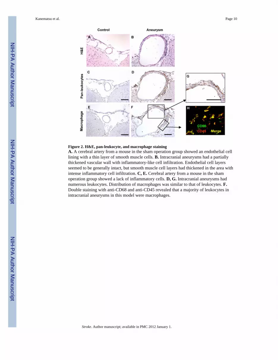
Figure 2. H&E, pan-leukocyte, and macrophage stainingA. A cerebral artery from a mouse in the sham operation group showed an endothelial celllining with a thin layer of smooth muscle cells. B. Intracranial aneurysms had a partiallythickened vascular wall with inflammatory-like cell infiltration. Endothelial cell layersseemed to be generally intact, but smooth muscle cell layers had thickened in the area withintense inflammatory cell infiltration. C, E. Cerebral artery from a mouse in the shamoperation group showed a lack of inflammatory cells. D, G. Intracranial aneurysms hadnumerous leukocytes. Distribution of macrophages was similar to that of leukocytes. F.Double staining with anti-CD68 and anti-CD45 revealed that a majority of leukocytes inintracranial aneurysms in this model were macrophages.
Kanematsu et al. Page 10
Stroke. Author manuscript; available in PMC 2012 January 1.
NIH
-PA Author Manuscript
NIH
-PA Author Manuscript
NIH
-PA Author Manuscript

Figure 3. Macrophage depletion and intracranial aneurysm formationA. Mice with macrophage depletion treatment using clodronate liposome had a reducedincidence of intracranial aneurysms compared to mice that received PBS liposome (P <0.05). B-G. While there were an abundant number of macrophages in the intracranialaneurysms from PBS liposome treated mice, middle cerebral arteries from mice in either thesham operation group or the macrophage depletion group did not show macrophageinfiltration into the vascular wall. H. The number of macrophages was higher in mice treatedwith elastase, angiotensin-II, and PBS liposome compared to mice in the sham group ormacrophage depletion group (n = 5 in each group, P < 0.05).
Kanematsu et al. Page 11
Stroke. Author manuscript; available in PMC 2012 January 1.
NIH
-PA Author Manuscript
NIH
-PA Author Manuscript
NIH
-PA Author Manuscript

Figure 4.A. Macrophage staining of the spleen from mice that received clodronate liposome. Brownarea indicates macrophage positive area. B. Clodronate liposome treatment decreased CD68positive area in the spleen by 88% from the baseline (P < 0.05), showing an effectivemacrophage / monocyte reduction by the clodronate treatment. At two weeks, CD68 positivearea returned to the baseline. C. Successful induction of hypertension by angiotensin-II.
Kanematsu et al. Page 12
Stroke. Author manuscript; available in PMC 2012 January 1.
NIH
-PA Author Manuscript
NIH
-PA Author Manuscript
NIH
-PA Author Manuscript

Figure 5.A. MCP-1 knockout mice had a lower incidence of aneurysms compared to wild-type mice(P < 0.05). There was no difference in the incidence of intracranial aneurysms betweenMMP-12 knockout mice and wild-type mice. B-D. While macrophage infiltration wasobserved in the cerebral arteries of wild-type mice (B) and MMP-12 knockout mice (D),cerebral arteries in MCP-1 knockout mice showed a lack of macrophage infiltration (C). F.Successful induction of hypertension by angiotensin-II.
Kanematsu et al. Page 13
Stroke. Author manuscript; available in PMC 2012 January 1.
NIH
-PA Author Manuscript
NIH
-PA Author Manuscript
NIH
-PA Author Manuscript


![[Dissecting aortic aneurysm simulating an acute myocardial infarct]](https://img.pdfslide.net/doc/110x75/63558f23922cbb7c550ca86c/dissecting-aortic-aneurysm-simulating-an-acute-myocardial-infarct.jpg)


