Embed Size (px)
Citation preview

Crystal structure of the DNA-bound VapBC2antitoxin/toxin pair from Rickettsia felisMarıa J. Mate1,2, Renaud Vincentelli1,2, Nicolas Foos1,2, Didier Raoult3,
Christian Cambillau1,2 and Miguel Ortiz-Lombardıa1,2,*
1Aix-Marseille Universite, Architecture et Fonction des Macromolecules Biologiques, Campus de Luminy, Case932, 13288 Marseille Cedex 09, France, 2CNRS, Architecture et Fonction des Macromolecules Biologiques,UMR6098, Campus de Luminy, Case 932, 13288 Marseille Cedex 09, France and 3Unite des Rickettsies, CNRSUMR IRD 6236, IFR 48, Faculte de Medecine, Marseille, France
Received July 30, 2011; Revised November 11, 2011; Accepted November 13, 2011
ABSTRACT
Besides their commonly attributed role in the main-tenance of low-copy number plasmids, toxin/anti-toxin (TA) loci, also called ‘addiction modules’,have been found in chromosomes and associatedto a number of biological functions such as: reduc-tion of protein synthesis, gene regulation andretardation of cell growth under nutritional stress.The recent discovery of TA loci in obligatory intracel-lular species of the Rickettsia genus has promptednew research to establish whether they work asstress response elements or as addiction systemsthat might be toxic for the host cell. VapBC2 is aTA locus from R. felis, a pathogen responsible forflea-borne spotted fever in humans. The VapC2toxin is a PIN-domain protein, whereas the antitoxin,VapB2, belongs to the family of swapped-hairpinb-barrel DNA-binding proteins. We have used a com-bination of biophysical and structural methods tocharacterize this new toxin/antitoxin pair. Ourresults show how VapB2 can block the VapC2toxin. They provide a first structural description ofthe interaction between a swapped-hairpin b-barrelprotein and DNA. Finally, these results suggest howthe VapC2/VapB2 molar ratio can control theself-regulation of the TA locus transcription.
INTRODUCTION
Toxin–antitoxin (TA) complexes were first discovered asfactors that protect low-copy number plasmids in bacteriafrom segregation loss (1). One protein in the pair is toxicto the cell and stable, whereas its corresponding antitoxinis unstable and requires continuous transcription to re-versibly inhibit the toxin by engaging in a non-covalentcomplex. If the plasmid is not correctly segregated during
cell division, one of the daughter cells will not carry theplasmid but will still have a number of copies of the TAcomplex. Eventually, the labile antitoxin will be degradedby proteases while the stable toxin will persist and killthe plasmid-free cell. In this way, the presence of TAloci prevents the proliferation of plasmid-free cells ingrowing bacterial cultures, and for that reason TAsystems have been described as ‘addiction modules’.More recently, TA loci have been associated to other bio-logical functions such as attenuation of protein synthesisand retardation of cell growth under conditions of nutri-tional stress (2–4). In this regard, TA would play a role instress physiology rather than in bacterial programmed celldeath.The most common activity of TA toxins seems to be
that of an mRNA-specific endonuclease. Such activityhas been already confirmed for RelE and MazF (5).VapC toxins from enteric bacteria have recently shownto specifically cleave initiator tRNAfMet (4). Nevertheless,two other activities have been reported: CcdB and ParEare inhibitors of DNA gyrase and the recentlycharacterized HipA toxin is a protein kinase (6). In turn,the antitoxins consist of two domains, a C0-terminalone involved in protein–protein interactions and anN0-terminal domain responsible for DNA-binding (7,8).They belong to one of several different classes of DNAbinding proteins. The binding to the toxin and to theDNA stabilizes the antitoxin, which otherwise exhibits alargely disordered structure highly susceptible to proteoly-sis. However, the main function of the TA binding toDNA is related to transcriptional control; TA operonsare all self-regulated at the transcription level by the anti-toxins, which bind to the TA operon promoters. In thisrespect, the toxin components can be considered ascorepressors of their own transcription because TAcomplexes bind to DNA more strongly than antitoxinsalone.TA pairs have been classified into families based on the
toxin structural features and mode of action (9). One of
*To whom correspondence should be addressed. Tel: +33 491825593; Fax: +33 491266720; Email: [email protected]
Published online 2 December 2011 Nucleic Acids Research, 2012, Vol. 40, No. 7 3245–3258doi:10.1093/nar/gkr1167
� The Author(s) 2011. Published by Oxford University Press.This is an Open Access article distributed under the terms of the Creative Commons Attribution Non-Commercial License (http://creativecommons.org/licenses/by-nc/3.0), which permits unrestricted non-commercial use, distribution, and reproduction in any medium, provided the original work is properly cited.

these, the VapBC (virulence associated protein) family,is defined by the presence of a PIN-domain protein asthe toxic component (VapC). The PIN domain(PFAM: PF01850) was first annotated on the basis ofsequence similarity to the N0-terminal domain of thetype IV pili protein, pilT, from Myxococcus xanthus(PIN, PilT N0-terminus). In prokaryotes the majority ofPIN-domain proteins are the toxic components ofchromosomally encoded TAs. PIN-domain proteinsexhibit structural homology to the T4 RNase H nucleasedomain (10). Actually, the PIN-domain of the yeast Dis3protein and the VapC toxin from enteric bacteria bearendoribonuclease activity (4,11), supporting the notionof a ribonuclease function for other PIN-domain toxins.Although sequence similarity is low within thePIN-domain, multiple-sequence alignments have shownthat active site residues are highly conserved.Other TA systems extensively studied include
Escherichia coli MazEF and archaeal RelBE. The MazFtoxin cleaves mRNA specifically at ACA sequences. Thecrystal structure of the MazEF complex shows aheterohexameric organization with a MazE dimer sand-wiched between two MazF dimers (12). The interactionbetween MazE and MazF is mediated by the flexibleC0-terminus of the antitoxin that wraps around the toxinhomodimer. The RelE toxin is a global inhibitor of trans-lation that is activated by nutritional stress to cleavemRNA positioned at the ribosomal A-site (13,14).RelB inhibits the action of RelE by wrapping aroundthe toxin, thus preventing it from binding to the ribosomalA-site (15).It is worth to note that a particular type of toxins can
form TA systems with antitoxins from different proteinsuper-families (16). Thus, the RelE/ParE toxins havebeen found associated with antitoxin transcriptionalfactors belonging to the MetJ/Arc, the PhD/YefM orthe cHTH super-families. Likewise, sequence analyseshave identified several PIN-domain toxins immediatelyadjacent to genes encoding homologues of transcriptionalregulators belonging to at least four different classes,namely HTH, RHH, Phd/YefM and AbrB. These obser-vations might indicate that TA loci have evolved throughgene shuffling or partner switching (9,16).Most of the structures of TA complexes reported so
far comprise only the toxin and the C0-terminal domainof the antitoxin that wraps around it. Thus, very littlestructural information has been obtained concerning theN0-terminal DNA binding domain of antitoxins. The caseof MazEF is an exception (12). The structure of theMazEF heterohexamer has shown that the antitoxinMazE homodimer contains an N0-terminal, intertwinedb-barrel. This structure classifies MazE as a member ofthe large super-family of DNA binding proteins repre-sented by the N0-terminal domain of the transcriptionalfactor AbrB (12, 17). Proteins from this family consist offour b-strands arranged in two anti-parallel hairpins thatare interleaved in the dimer, justifying the name given totheir fold: swapped-hairpin barrel.Two crystal structures of a promoter-bound TA
complex have been reported, namely those of FitABfrom Neisseria gonorrhoeae, bound to a 36-bp DNA
fragment from the fitAB promoter (7) and of E. coliHipAB, bound to a 21-bp operator DNA (18). The anti-toxin FitA contains an N0-terminal domain with a DNAbinding ribbon–helix–helix (RHH) motif, and aC0-terminal extended domain including an a-helix thatinteracts with the PIN-domain of the FitB toxin. FourFitA and four FitB molecules form a tetramer ofheterodimers that explains why the binding to the toxinenhances the antitoxin affinity for the DNA. This struc-ture also explains why the presence of the toxin and DNApartners stabilize the inherently flexible structure of theantitoxin. In turn, HipAB bind to DNA as a dimer ofheterodimers, which also results in an enhanced DNArecognition with respect to the binding of the antitoxinalone (19).
Although well known in plasmids and in the chromo-somes of free-living bacteria and archaea, the presence ofTA loci was only recently confirmed in obligatory intra-cellular species. Thus, the complete genome sequence ofRickettsia felis, a flea-borne pathogen responsible forspotted fever in humans (20,21), has revealed thepresence of at least 13 TA loci (22). The Rickettsia TAloci are presumed to work as addiction systems, wherethe elimination of TA-containing Rickettsia from hostcells could lead to the release of the toxin in the cyto-plasm of host, which would result in cell death (23).One of the R. felis TA loci includes VapC2, a proteinthat exhibits in vitro RNase activity and that can inhibitthe growth of transformed E. coli and S. cerevisiaecultures (23).
We present here evidence, from multi-angle light scat-tering and refractometry data, for the existence of twotypes of R. felis VapBC2 complexes and reveal the struc-ture of one of them, the octameric VapBC2 complexbound to a 27-bp dsDNA fragment from its ownpromoter. As in the case of FitAB, four toxins and fourantitoxins form a tetramer of heterodimers that bind tothe dsDNA. The toxin VapC2 contains a PIN-domainthat forms homodimers homologous to those of FitB.However, the DNA-binding domain of the VapB2 anti-toxin does not fold as the RHH domain of FitA. Instead,VapB2 forms homodimers that display a swapped-hairpinb-barrel fold, similar to MazE, thus illustrating the richinterplay between toxin and antitoxin super-families.Hence, this is the first structure of a TA pair composedof a toxin belonging to the VapC family and an antitoxinbelonging to the AbrB/MazE superfamily. It is also thefirst experimental structure showing the complex of aswapped-hairpin b-barrel protein with a DNA fragment.We discuss how these data help to understand the inter-play between the toxin inhibition and transcription repres-sion functions of the antitoxin protein.
MATERIALS AND METHODS
Protein production
For expression in E. coli, R. felis vapB2 and vapC2 werecloned in operon using the Gateway (Life Technologies,Carlsbad, CA, USA) compatible destination vectorpETG20A, where VapB2 was fused to thioredoxin
3246 Nucleic Acids Research, 2012, Vol. 40, No. 7

(TRX) at its N0-terminus to improve protein expressionand solubility (24). TRX was His-tagged and followed bythe tobacco virus (TEV) protease cleavage sequence,which respectively allowed affinity purification andrelease of the target protein.
The VapBC2 complex was expressed in BL21 Rosetta(DE3) pLysS cells (Novagen). Bacteria were grown at37�C in auto-inducing media (25) for 4 h andtransferred to 17�C when OD600nm reached �2 (24). Theselenomethionine-labelled protein was also producedusing autoinducing media (25). Cultures were harvestedby centrifugation at 4000g during 10min and frozen at�80�C. The cell pellet was resuspended in a buffer con-taining 300mM NaCl, 50mM Tris–HCl pH 8.0, 0.25mg/ml lysozyme, 10mM imidazole, 20 mg/ml DNaseI, 20mMMgSO4 and a cocktail of protease inhibitors (CompleteEDTA-free, Roche). Cells were disrupted by sonicationand the lysate clarified by centrifugation at 20 000g for30min at 4�C.
The TA complex was loaded on a Ni2+-affinity chroma-tography, HisTrap column (GE Healthcare) and eluted ina buffer containing 300mM NaCl, 50mM Tris–HCl pH8.0 and 250mM imidazole. After this first purification stepthe His-tagged thioredoxin fusion was cleaved by theaddition of TEV protease. The TA complex was thenrecovered in the flow-through of a second Ni2+-affinitychromatography.
Oligonucleotides for crystallization were annealed byslow cooling in a buffer containing 20mM Tris–HCl(pH 8.0) and 150mM NaCl. The sequence of theforward oligonucleotide was 50-TTAATATATACTAATTAATATATACTA-30. The VapBC2 complex was mixedwith the duplex DNA in a 4:1 molar ratio and purified bygel filtration in a preparative Superdex 75 HR16/60column, using a buffer containing 25mM Tris–HCl pH8.0 and 150mM NaCl.
The purity of the sample was verified by SDS–poly-acrylamide gel electrophoresis on NuPAGE� Bis-Trisgels (Invitrogen).
Multiangle light scattering
The molecular weight was calculated using analyticalsize-exclusion chromatography (SEC) on a high-performance liquid chromatography (HPLC) system(Waters) coupled with online multiangle laser light scat-tering, ultra-violet light absorbance and refractive indexdetectors (MALLS/UV/RI) (Wyatt Technology, SantaBarbara, CA, USA). SEC was performed on a Shodex�
KW-804 column with 25mM HEPES pH 7.5, 150mMNaCl as the eluent. The molecular weight was calculatedusing ASTRA V software (Wyatt Technology) with a re-fractive index increment (dn/dc) of 0.185ml/g for proteinsamples. In the ‘protein conjugate’ analysis of VapBC2–DNA complexes we used extinction coefficients, at280 nm, of 0.95ml/(mg� cm) and 14.31ml/(mg� cm)for VapBC2 and for the DNA, respectively. In thisanalysis, the refractive index increment of DNA was setto 0.166ml/g (26).
Surface plasmon resonance
A Biacore� 1000 surface plasmon resonance system wasused to study the direct binding of the VapBC2 complex toits putative cognate DNAs, as predicted from sequenceanalysis of the TA locus. Two different 55-mer DNA frag-ments, with sequences:
vapBC2_A: 50-ATATTTAGAATAGTTGTTTGCTTTAATAATAAAGTTAATATATACTAATT-30
vapBC2_B: 50-AGTTGTTTGCTTTAATAATAAAGTTAATATATACTAATTAATATATACTA-3 0
were individually immobilized (332 and 268 resonanceunits (RU), respectively) on research grade streptavidin(SA) sensor chips. Purified VapBC2 in Tris buffer[50mM Tris–HCl (pH 8.0), 150mM NaCl, and 0.005%(v/v) Tween-20] was then injected at concentrationsranging from 0.25 mM to 9 mM over the immobilizedDNAs at 25�C at a flow rate of 20 ml/min. Dissociationconstants (KD) were derived by non-linear curve fitting ofthe standard Langmuir binding isotherm using the signalat the equilibrium response level with the BIAevaluationsoftware (version 4.1).
Complex crystallization
The VapBC2 complex bound to the 27-bp dsDNA wasconcentrated to 6mg/ml. Crystals were obtained by thevapour diffusion method using a nanodrop-dispensingrobot (Honeybee 961; Cartesian) in 96-well Greiner crys-tallization plates. The drops contained 300 nl of thecomplex and 100 nl of the crystallization agent, asolution of 12% PEG4000, 4% PEG400, and 100mMMES pH 6.5. Subsequent optimizations were manuallyperformed in 24-well plates. Crystals were very fragile,therefore cryoprotection was achieved by graduallyincreasing the concentration of PEG400 to 12%. Thecrystals were then flash-cooled in liquid nitrogen.Crystals belonged to the space group P212121 with unitcell parameters a=80.8 A, b=92.3 A, whereas c wasfound to vary from crystal to crystal in the range of143–164 A. A Matthew’s coefficient of 2.62, correspondingto a solvent content of 58.2%, was found to be compatiblewith the presence in the asymmetric unit of four TAhomodimers and one molecule of double stranded DNA.
Crystal structure determination and refinement
Data were collected at the European SynchrotronRadiation Facility, ESRF (Grenoble, France). A single-wavelength anomalous diffraction (SAD) data set was col-lected at the Selenium peak wavelength (l=0.9785 A) atbeamline ID14eh4. Besides, two native data sets, from twodifferent crystals, diffracting to 2.5 and 2.85 A resolutionwere collected at beamlines ID14eh1 and ID23eh2,respectively. All data sets were reduced using XDS (27)and SCALA (28) (Table 1). The structure of the complexwas solved by the SIRAS (Single Isomorphous Replace-ment with Anomalous Signal) method. Substructuredetermination, phase calculation, and density modifica-tion were carried out with autoSHARP (29) andSOLOMON (30). Phases provided by SOLOMON were
Nucleic Acids Research, 2012, Vol. 40, No. 7 3247

introduced in BUCANEER to build an initial model ofthe protein components of the asymmetric unit. The restof the model was manually built using COOT (31). Themodel was refined against the native data set withautoBUSTER (32) using TLS (translation/libration/screw) parameters, with one TLS group per chain. Thefinal Rfree is 22% and Rcryst is 17% (Table 1). Model val-idation was done with Molprobity (33). PISA (34) wasused for the analysis of interfaces. Electrostatic calcula-tions were carried out with the program APBS (35), andthe electrostatic potential distribution was representedusing Chimera (36), which was also used to preparefigures.
Structure deposition
Coordinates and structure factors for the DNA-boundVapBC2 complex have been deposited in the PDB underID 3zvk.
RESULTS
The R. felis VapBC2 complex binds to its promoter DNA
TA complexes are known to repress the transcription oftheir coding genes by binding to their promoter DNA. Asequence analysis was carried out to identify possiblebinding sites for R. felis VapBC2. We aimed at findingsequence palindromes or repeats close to the TA
promoter, as found in other TA operator sites (37). Thisanalysis resulted in two possible binding sequences fromthe VapBC2 locus promoter that we analysed by surfaceplasmon resonance. One of them, vapBC2_B, displaysin vitro good affinity with a KD of 0.11±0.004mM(Figure 1a), whereas the other sequence, vapBC2_A,shows a �40-fold higher KD of 4.15±0.76 mM. A closerinspection of the vapBC2_B sequence revealed that the 27most downstream bases include a nearly perfect palin-drome, a hallmark of TA operator sequences (37). There-fore, we decided to pursue the rest of this study with thisshorter version of the VapBC2 operator sequence.
Rickettsiae felis VapBC2 forms octamers when bound toits cognate DNA and hexamers in its absence
To gain insight into the stoichiometry of the complexesmade by VapB2, VapC2 and their cognate DNA, westudied their oligomeric states by size exclusion chroma-tography in conjunction with multiangle light scattering(SEC/MALS). This technique allows the calculation,from MALS data, of an average molecular weight formacromolecules within each peak eluting from the SECcolumn. We studied first purified VapBC2 samples in theabsence of DNA. Our analyses show that VapBC2 formsan 82.4 kDa complex (Figure 1b). Given the molecularmasses of the VapC2 toxin (15.36 kDa) and of the VapB2antitoxin (9.01 kDa), a number of TA ensembles are
Table 1. X-ray data and refinement statistics
SeMet data set Native low resol Native high resolID14eh4 ID23eh2 ID14eh1
Data collectionSpace group P212121 P212121 P212121Cell dimensions (A,�) 81.5 92.9 163.4 82.0 92.8 164.1 80.8 92.3 150.5Resolution (A)a 49.0–3.5 (3.7–3.5) 49.2–2.9 (3.1–2.9) 47.3–2.5 (2.6–2.5)No. observationsMeasured 244 369 115 005 191 495Unique 16 251 28 411 39 577
Completeness (%)a 100 (98.8) 99.9 (99.9) 99.7 (99.7)Rmeas (%)a 10.9 (51.2) 9.6 (55.5) 6.6 (56.3)Multiplicitya 15.0 (15.4) 4.0 (4.1) 4.9 (4.8)hhIi/s(hIi)ia 23.2 (8.6) 10.9 (2.9) 17.4 (3.2)Phasing powerAnomalousb 0.935 (4.6 A)Isomorphousb,c 0.565 (6.4 A)
Refinement and validationRcryst/Rfree (%)d 17.23/21.72No. atoms: protein/DNA/water 6431/1063/360R.m.s.d.e
Bonds/angles (A,�) 0.010/1.28B factor (A2) main/side chain 2.72/6.25
Ramachandran plotf (# residues, %)Favoured regions 764, 97.7Allowed regions 781, 99.9Outliers 1, 0.1
aValues in parentheses are for the highest resolution shell.bValue in parenthesis: resolution for which phasing power drops below 1.0.cIsomorphous phasing power for acentric reflections using native low resolution (ID23eh2) data set.dRfree is Rcryst calculated for a test set of randomly selected 5% of the data.eRoot mean standard deviation.fRamachandran plot statistics as reported by Molprobity (33).
3248 Nucleic Acids Research, 2012, Vol. 40, No. 7

compatible with the measured mass within a 5% error.Among them, complexes with 3:4 (total mass 82.12 kDa)and 4:2 (79.46 kDa) stoichiometry are the likeliest ones,given the relative amounts of the proteins as observed onSDS acrylamide gels performed with the same samples.Because both molecules belong to protein families knownto form homodimers, we consider the 4:2 complex as themost probable one. We also note that this stoichiometrycoincides with that of the MazEF complex, whichincludes an antitoxin of the same family than VapB2.
Then, based on the previously known complex of theFitAB toxin with DNA, samples incubated at a 4:4:1(VapB2:VapC2:DNA) molar ratio were also studied bySEC/MALS. In this case, the analysis of the molecularmass was carried out using the ‘‘protein conjugate’’model, as implemented in the ASTRA software. In thismethod, the combined use of light scattering, ultravioletand refractive index detectors enables the assessment ofthe molar mass, size, and relative polymer fractions of acopolymer such as a protein-DNA complex. With thisapproach, a total mass of 111 kDa breaks downinto 93.41 and 17.58 kDA for the protein and DNAcomponents, respectively (Figure 1c). These molarmasses are consistent with those expected for a 4:4:1(VapB2:VapC2:DNA) complex, 97.48 and 17.17 kDa,respectively.
Overall structure of the R. felis VapBC2 complex boundto its promoter DNA
We determined the structure of the VapBC2 TA com-plex bound to a 27-bp double stranded DNA molecule(Figure 2) by single isomorphous replacement with anom-alous signal (SIRAS), using selenomethionine-substitutedproteins. The final model, refined to 2.5 A, contains fourmolecules of the toxin VapC2 (monomers A, B, C and Dencompassing residues 1–134 for the first one and 1–133for the other three), four molecules of the antitoxin VapB2(monomers E, F, G and H comprising residues 2–58, 1–78,2–77 and 1–57, respectively), 26 base pairs out of the 27present in the double stranded DNA fragment used forcrystallization, 360 molecules of water and 4 moleculesof MES, the buffer used in the crystallization condition.In a second native crystal, diffracting to 2.85 A, all the
27-bp of the DNA fragments are visible. Whereas thecrystal packing contacts are slightly different (seebelow), the conformation of the proteins and of theDNA are largely the same than in the structure at 2.5 A.The quaternary structure of the complex can be con-
veniently described as a tetramer of VapBC2 heterodimersbound to the DNA fragment (Figure 2a). In this regard,this complex resembles that of FitAB bound to itspromoter. However, the VapBC2 complex is dramaticallymore compact than that of FitAB. The heterodimers
Figure 1. Analysis of the complexes formed by the VapBC2 system in the absence and presence of their cognate DNA. (a) SPR sensorgramsobtained by passing purified VapBC2 at the labelled concentrations (assuming an octamer) on the vapBC2_B DNA, annealed to its complement andbound to a streptavidin sensor chip. The inset shows the steady-state analysis of the binding. (b) About 300 mg of purified VapBC2 were subjected tosize-exclusion chromatography coupled to MALS/RI/UV detectors as described in the ‘Materials and Methods’ section. The molar mass (black line),derived from refractive index measurements, and the absorption at 280 nm (gray line) were plotted as a function of the elution time. (c) Same as in(b) with 400 mg of purified VapBC2 incubated with the dsDNA used for crystallization at a 4:1 molar ratio. For clarity, the graphs of derived molarmass are only shown around the main absorption peak areas in (b) and (c). Molar mass values at the absorption peaks are indicated, with thoseresulting from protein-conjugate analysis in parentheses.
Nucleic Acids Research, 2012, Vol. 40, No. 7 3249

are arranged circularly via alternative toxin–toxin andantitoxin–antitoxin dimerization interfaces. The wholeVapBC2 complex has a pseudo-dyad axis, perpendicularto the DNA double helix (Figure 2). This pseudo-dyadaxis intersects the DNA at the A13/T15 base pair. Theinteraction of the VapBC2 complex with the DNA ismediated by the two antitoxin homodimers. The inter-action with DNA is described in detail below.
Structure of the VapC2 toxin
VapC2 consists of 134 amino acids forming a compactprotein with an a/b/a fold. The topology of VapC2includes 10 elements of secondary structure (Figure 3a),namely b1 (residues 2 to 5), a1 (residues 7–16), a2(residues 19–29), b2 (residues 33–37), a3 (residues 38–50), a4 (residues 54–65), b3 (residues 69–71), a5(residues 76–90), a6 (residues 99–109), b4 (residues 112–115), b5 (residues 128–131). A central, twisted, parallelb-sheet made of five strands (ordered 32145) is surroundedby six a-helices, with four a-helices on one side of the sheetand two on the other side. As predicted from sequencealignments VapC2 folds as a PIN-domain. This assign-ment was further confirmed by a search for similar struc-tures using the DALI server (38) that gave severalmatches, all of them PIN-domain containing proteins.The closest structural homologues are FitBfrom Neisseria gonorrhoeae (7) (PDB: 2H1C, chainA, Z-score=17.1), VapC2 (PDB ID: 3H87, chain A,Z=15.1) and VapC-5 (39) (PDB ID: 3DBO, chain B,Z=15.0) from Mycobaterium tuberculosis and twoarchaeal proteins (PDB: 1Y82, Z=15.7; 1V96,Z=15.3). PIN-domain proteins share two nearly invari-ant aspartic acid residues and a conserved glutamicamino-acid (40), which in VapC2 correspond to residues
Asp6, Asp99 and Glu43, respectively. During the cloninga spontaneous mutation of Asp6 to glycine resulted inVapC2, perhaps inhibiting its toxic effect and allowingthe overexpression of the protein.
Remarkably, VapC2 residues 116–123 (between strandsb4 and b5) form a coil where other PIN-domain proteinsbear a short helix, commonly described as helix a6. Thishelix includes a less conserved acidic residue that resides inthe vicinity of the three well-conserved acidic amino acidscharacteristic of the domain. In VapC2, Glu120 can beregarded as the fourth acidic residue. In fact, theelectron density for residues 117–124 is weak and someof them, or their side chains, could not be modelled.Indeed, this part of the model slightly differs among thefour toxins. In monomers C and D this region is exposedto the solvent, and thereby less well defined than inmonomers A and B.
VapC2 forms nearly symmetric homodimers that bury asolvent accessible surface area (SASA) of 1083±16A2
per monomer, representing about 14% of the totalsurface of the toxin (Figure 3b). The main contribu-tions to the dimer interface arise from helices a3 and a5,which cross-interact symmetrically, and from the loopbetween strand b3 and helix a5. As in FitA, Phe73(Phe78 in FitA) plays a crucial role in dimerization:its backbone carbonyl and amine atoms establishhydrogen bonds to the corresponding atoms in theother monomer. Furthermore, the backbone carbonyl ofPhe73 also forms a hydrogen bond to Ser38 in theother monomer. In the same loop, Asn72 from onetoxin hydrogen binds the backbone amine of Ala75 fromthe neighbouring toxin. Finally, Tyr46 residues make astacking interaction with the aliphatic chains of Arg85.Besides these contacts, interactions between
(a) (b)
(c)
VapB2dimer
VapC2dimer
VapBC2dimer
VapBC2dimer
Figure 2. Three orthogonal views of the VapBC2-DNA complex. (a) VapB2 antitoxins are shown in different tones of blue and VapC2 toxins inyellow and orange tones. A toxin and antitoxin dimers are labelled using the colours of their components. Two toxin/antitoxin heterodimers are alsomarked; in one of them (bottom left) the antitoxin C0-terminus is ordered whereas in the other one (top right) it is not visible. (b) The DNA issignificantly bent. (c) The VapBC2 complex interacts with the concave face of the DNA. In the three views, the pseudo-dyad axis, calculated usingthe two toxin homodimers as reference, is shown as a red rod with black caps.
3250 Nucleic Acids Research, 2012, Vol. 40, No. 7

monomers are variable and not symmetrical. In thisregard, we note that Glu43 in monomer A is involved inhomodimerization contacts via a hydrogen bond to Arg85from monomer B.
Structure of the VapB2 antitoxin
The antitoxin VapB2 consists of 78 amino acids. Thesecondary structure of each monomer comprises twobeta-hairpins (Figure 3c): b1 (residues 6–9)/b2 (residues12–15) and b3 (residues 28–33)/b4 (residues 36–41) con-nected by a loop including a short 310 helix (residues19–21) and followed by helix a1 (residues 46–55).Residues 59–78, which form an extended coil, are onlyvisible in monomers F and G (Figure 3c). The inter-action with the DNA is mediated by the b-hairpins,whereas the extended coil mediates the inhibitory inter-action with VapC2 toxin homodimers (Figure 4b, seebelow).
Hence, the VapB2 fold differs from that of FitA (7), thecognate antitoxin of the VapC2 homologue FitB. A struc-ture similarity search in the DALI server (38) using theN0-terminal domain of VapB2 (up to residue Pro41)yielded two main matches of medium-low statistical sig-nificance: the antitoxin MazE (12, 41) (PDB: 1MVF, chainE, Z-score=4.4 and PDB: 1UB4, chain C, Z-score=3.3)and the DNA binding domain of AbrB (42, 43) (PDB:2RO4, chain B, Z-score=3.1 and PDB: 1Z0R, chain A,Z-score=3.0), a B. subtilis transcription-state regulator.This result is consistent with sequence similarity searchesusing BLAST against the UniProt database, which gaveSpoVT/AbrB-like proteins (PFAM: PF04014) as the
closest hits to VapB2. The sequence identity for thefirst 41 residues of VapB2 is 22% with MazE and 15%with AbrB.It is therefore adequate to classify VapB2 in the super-
family of AbrB-like transcription factors, also calledswapped-hairpin b-barrel transcription factors. Proteinsof this superfamily fold as homodimeric b-barrels viatwo pairs of interleaved beta-hairpins (17). The only sig-nificant difference with other members of this family withknown structures is that, in VapB2, the two b-hairpins areconnected by a loop including a three-residues long 310helix instead of a (5–7)-residues long a-helix. Notably,the first residue in the 310 helix, Lys19, and the first oneafter that helix, Arg22, interacts with DNA phosphatesfrom the minor groove (see below). Structural superpos-ition of the DNA-binding domains of VapB2, MazE(PDB: 1UB4) and AbrB (PDB: 2RO4) shows that thefirst b-hairpin of VapB2 is more similar to that of MazEthan to that of AbrB, which is shorter. The 310 helixpresent in VapB2 superposes to the first turn of theshort helix present in MazE and AbrB. The rest of thelinker is an extended coil that roughly superposes tothose of MazE and AbrB (Supplementary Figure S1).The topology of the swapped-hairpin b-barrel in
the VapB2 dimers is b1, b2, b20, b10, b3, b4, b40, b30
(Figure 3c). Dimerization buries a SASA of 1589±15A2 per VapB2 monomer. This extensive dimerizationinterface is stabilized by a central hydrophobic core andby a network of hydrogen bonds, most of them betweenmain-chain atoms. The interface also includes two saltbridges, Lys5-Glu27 and Asn9-Arg16.
(a)
(c)
(b)
β1α1
α2
β2
β3β4 β5
α3
α4
α5α6
β1β2
310
β3β4Γ
α1
E120
D99
G6
E43Y46
R85
S38D72
F73
Figure 3. Structures of the VapC2 toxin and the VapB2 antitoxin in the VapBC2–DNA complex. (a) VapC2 toxin monomer A is shown as a ribbondiagram with labelled secondary structure elements. Blue and red circles mark the position of the N0- and C0-termini, respectively. (b) View of thetoxin homodimer, formed by monomers A and B, along is 2-fold axis (black dot). The four acidic residues of their putative active sites are shown assticks coloured by element, with carbons in pink. They are labelled in monomer B. Residues involved in dimerization are depicted as sticks withcoloured by element, with carbons in yellow. They are labelled in monomer A. Hydrogen bonds are represented as dashed black lines. The inset givesa close-up view of the central part of the homodimer. (c) The antitoxin homodimer formed by monomers F and G viewed across the 2-fold axis of itsb-barrel core. Secondary structure elements are labelled on monomer G. Note the asymmetry of the C0-termini with respect to the homodimer 2-foldaxis. Colour codes are as in Figure 2. For simplicity, one-letter codes are used for aminoacid labels.
Nucleic Acids Research, 2012, Vol. 40, No. 7 3251

Two antitoxins suffice to block the four putative activesites of the toxins
VapB2 antitoxins interact with VapC2 toxins through twocontiguous sites. In the first site (Figure 4a), the VapB2b3/b4 hairpin, the a1 helix and the loop that connectsthese two elements contact helices a2 and a4 from theVapC2 toxin. All four TA heterodimers engage in thistype of interaction, which buries a total area of1739±256A2 (941±125A2 from VapB2 and 798±132A2 from VapC2). These contacts are essentially driven byvan der Waals forces resulting from the interaction of theantitoxin a1 helix with a pocket defined by the toxin a2and a4 helices, as well as by the C0-terminus of its a1 helix.Trp46 from the VapB2 antitoxin playing a dominant role,as it fills a pocket lined by residues Leu25, Glu26, Ile34,Phe64, Arg67 and Leu68 from VapC2.A striking feature of the VapBC2 complex is that a
single antitoxin is able to block the access to the twoputative active sites of each toxin homodimer. This 1:2inhibition was also found in the MazEF complex (12).The second VapC2/VapB2 interaction site (Figure 4b)involves a single antitoxin (monomers F and G) and thetwo toxins in a VapC2 dimer (toxin dimers AB and CD,respectively). It is noteworthy that antitoxin monomers Fand G form one of the two antitoxin homodimers.
Therefore, their interaction with the toxin homodimerseffectively breaks the dyad symmetry of the wholeVapBC2 complex. Residues 59–78 from the antitoxinmediate this type of interaction, which, contrary to theprevious one, is stabilized by a dense network of polarcontacts involving both main-chain and side-chainatoms. Especially noteworthy are the electrostaticcontacts established by VapB2 Arg66 with residuesGlu43 and Asp99 from the proximal VapC2 monomer.Contacts between the inhibitory antitoxins and theproximal toxin monomers are conserved. Conversely,contacts with the distal VapC2 monomers vary from onetoxin homodimer to the other. Thus, Tyr76 from VapB2monomer F establishes a hydrogen bond with Glu43 fromthe distal VapC2 monomer A. In turn, obstruction of theactive site of the distal VapC2 monomer D is mainlyassured by Arg74 from VapB2 monomer G, whichcontacts Glu43 and Asp99 from the distal toxin. Finally,in all cases VapB2 residues Ile61, Phe62, Tyr76 and Phe77engage in van der Waals contacts that anchor theC0-terminus of the antitoxin to the surface of the toxins.
DNA conformation
DNA duplexes from adjacent asymmetric units arealigned end-to-end to form a supercoiled helix through
(a)
(c)
(b)
W46P43
R66
Y76R74
I61F62
F77
Q72
K73
E78K53
R85
R74
Q72D99
N97
K73
K75
Y76
Figure 4. Antitoxin/toxin interactions. (a) Site 1: interaction between the antitoxin monomer G and its proximal toxin (monomer C). Residues withthe strongest contributions are labelled in yellow and white for the antitoxin and the toxin, respectively. Hydrogen bonds are represented as dashedblack lines. This type of interaction surface is present at all toxin/antitoxin interfaces. (b) Site 2: inhibitory interaction between the antitoxinmonomer F and the toxin homodimer made by monomers A and B. The molecular surface of the toxin dimer is coloured by its electrostaticpotential, at 150mM ionic strength, ranging from �51mV (red) to 51mV (blue) at 298K. Residues from the antitoxin are labelled in yellow.Hydrogen bonds are represented as black lines. (c) Site 2: a detailed view of the interaction of antitoxin monomer G with its distal toxin (monomerD, dark blue) in an orientation equivalent to that of panel b. Residues with the strongest contributions are labelled in black and blue (except forGlu43 in white) for the antitoxin and the toxin, respectively. Hydrogen bonds are represented as dashed black lines. Note the different positions andinteractions of residues Arg74 and Tyr76 from the antitoxin. Colour codes are as in Figure 2. For simplicity, one-letter codes are used for aminoacidlabels.
3252 Nucleic Acids Research, 2012, Vol. 40, No. 7

the crystal lattice. The supercoil results from anoverall bend of 36.8�, as calculated by CURVES (44)(Figure 2b). Bending is maximal in the central part ofthe DNA and as a result, the molecule offers a concavesurface to the interaction with the VapBC2 complex(Figures 2b and 5b).
The most remarkable feature of structure of this DNAis that the minor groove in the AT-rich central region(bases 13–16, forward chain) is severely compressed,shrinking to less than half the width of canonicalB-DNA: 2.6 A for base-pairs A13/T15 and A14/T14,compared to the standard 6 A. In contrast, the minorgroove width increases to 8.5–8.7 A in the region directlyinteracting with VapB2 (bases 3–11 and 18–26, forwardchain). The minor groove is again compressed at the DNA
ends, presumably due to crystal packing forces. The oscil-lations in the minor groove width correlate with changesin the width of the major groove. However, all along theDNA molecule, the major groove is wider than in a ca-nonical B-DNA (11.5 A). Thus, in the centre of thefragment the major groove width is �12.4 A, whereas inthe VapB2-interacting area it increases to �15.5 A(Supplementary Figure S2).
Interaction of the antitoxins VapB2 with DNA
The sequence of the vapBC2 promoter used in this studyincludes an imperfect palindrome spanning from baseT2 to base T27 (forward sequence), with half-sequenceTA(A/G)TATATA(C/T)TAA. Within this palindrome,two regions are directly recognized by the antitoxin homo-dimers: base-pairs A3/T25 to T12/A16 by the EH VapB2dimer and base-pairs A17/T11 to G26/A2 by the FGVapB2 dimer (Figure 5a). Since each of these regionsis recognized by an antitoxin homodimer, it is notsurprising to note that they are also internally nearly pal-indromic (Figure 2), being A(A/G)TAT theirhalf-sequence.Globally, the VapB2 homodimers interact with the con-
cave face of the curved DNA: when looking at thecomplex along overall longitudinal axis of the DNA, thedyad axes of the two antitoxin homodimers make an angleof �110� (Figure 5b). In the VapB2 homodimers the firstb-hairpins from each antitoxin (strands b1 and b2) formtogether a saddle-shaped surface that deeply penetratesthe DNA major grove (Figure 5a), providing a stronghydrophobic base to the interaction. A total 1151±7A2 area is buried by the interaction of each antitoxinhomodimer with a half site, resulting in a solvationenergy gain of �14 kcalmol�1, as estimated by PISA(34). Two residues, Asn9 and Gln11, make specifichydrogen bonds with the DNA bases. The Asn9residues, which are at the C0-terminus of the b1 strand,contact the central base-pairs while the Gln11 residues, inthe b1-b2 loop, contact the penultimate base-pairs of thepseudo-palindromic interacting regions. The Asn9residues specifically recognize the O4 positions of the thy-midines belonging to the central base-pairs. The positionof Asn9 is stabilized by the Arg16 residue from the sameantitoxin, which in turn contacts the DNA backbone, thuscontributing to the opening of the major groove. The roleof Gln11 is particularly interesting since it adjusts to theadenine/guanine variability within the palindrome recog-nized by each antitoxin dimer. Indeed, the side-chainamide group of a glutamine residue interacts equallywell with the N6 of an adenine or with the O6 of aguanine. Besides these contacts, the main-chain aminefrom each Gln11 recognizes the N7 position, common toadenines and guanines. Finally, residues Lys19, from the310 helix between strands b2 and b3, and Arg22, in theloop that follows that helix, further contribute to stabilizethe antitoxin–DNA interaction through electrostaticcontacts with the DNA phosphate backbone. A full mapof the VapB2/DNA contacts is available in Suppl.Figure 3.
K19K19R22R22
R16R16
N9N9
S12S12S12
Q11Q11Q11N9
R16
R22K19
G3T6T21
T8A24
(a)
FGdimer
EHdimer
(c)
K19
R22Q11Q11
N9N9
N9
R22R22Q11K19K19
(b)
Figure 5. Antitoxin/DNA interaction. (a) Detail of the tight interactionof the antitoxin homodimer made by monomers F and G with theDNA. Antitoxin residues are labelled in black for monomer F and inwhite with a black contour for monomer G. DNA bases labels are initalics. Hydrogen bonds are represented as dashed black lines. (b)Overall view of the disposition of the antitoxin homodimers (only theb-barrels are shown) with respect to the DNA. The 2-fold axes of theantitoxin homodimers are shown as black-capped red rods. Colourcodes are as in Figure 2. For simplicity, one-letter codes are used foraminoacid labels. (c) Sequence conservation in the swapped-hairpinb-barrel domain at the DNA interface. The sequence of VapB2 wasaligned with the Pfam representative set of sequences for the PF04014family using Muscle (58). Conservation was mapped onto the molecularsurface of the VapB2 homodimer FG (residues 1–45) using the identityhistogram model implemented in Chimera (36). Protein residue labelsare as in panel a. A colour key bar shows the colour correspondencefor lower (cyan) to higher (maroon) conservation.
Nucleic Acids Research, 2012, Vol. 40, No. 7 3253

Sequence conservation in the swapped-hairpinb-barrel DNA binding domain is consistent with the dif-ferent roles of the b2–b3 and the b1–b2 loops. Thus,the DNA backbone binding residues in the b2–b3 loopare the most conserved at the interface with DNA.Conversely, residues in the b1–b2 loop region, whichmake direct base-specific residues and so drive the rec-ognition of a particular sequence, are less conserved(Figure 5c).
DISCUSSION
A (VapC2)4(VapB2)2 heterohexamer likely constitutes thetoxin-inhibited complex whereas a (VapC2)4(VapB2)4heterooctamer binds to the locus operator DNA
PIN domains resemble structurally to the T4 RNaseH(10). In keeping with this observation, the PIN domainof the eukaryotic exosomal protein Rrp44 displaysendoribonucleolytic activity (11) and VapC toxins fromenteric bacteria specifically cleave the initiator tRNAfMet
(4). The three conserved acidic residues of the PIN domainare clustered at the surface of the protein and superposewell to part of the active site of the T4 RNaseH, namelyRNaseH residues Asp19, Asp71 and Asp132. In VapC2these residues correspond to amino acids Asp6, Asp99 andGlu43, respectively (Figure 3b). In the structure describedin this work Asp6 is mutated to glycine. An Asp!Alasubstitution in the equivalent residue (Asp7) of anenteric VapC toxin resulted in an inactive enzyme in vivo(4). We infer that the VapC2 protein in our construct isalso inactive.A fourth acidic residue is present in PIN domains. Its
position at the sequence level can vary from a protein toanother, as it lies in a region structurally more variablethan the former three residues. Glu120, which sits in aflexible loop that in other PIN domains folds as a shorthelix, is the likely candidate for this fourth acidic residuein VapC2. Actually, the side chain of Glu120 could onlybe fully modelled in monomer A, where it is hydrogenbound to residues Thr115 and Asn8 from the samemonomer. The larger active site of T4 RNaseH coordinatestwo divalent metal cations whereas, in principle, the activesite of PIN domains could bind one. Nevertheless, in linewith other known structures of PIN domain proteins, thatof VapC2 in the VapBC2–DNA complex does not includeany metal cation. The Asp6Gly point mutation present inour VapC2 construct is a likely explanation for thisabsence. Notwithstanding, it is also possible that the en-zymatic mechanism of VapC2 does not imply metal cofac-tors, that they may have been displaced by the interactionwith the antitoxin or that the metals are only transientlybound to the enzyme during the reaction.Globally, the putative active site of VapC2 resides on a
surface pocket near the dimerization interface of the toxinhomodimer; therefore, both active sites are close to eachother. In fact, they are connected by a groove that spansthe full length of the toxin homodimer. In the structure ofVapBC2 this groove is filled by one antitoxin C0-terminus.However, it is clear that a putative RNA substrate wouldalso fit. Consistent with this hypothesis, the groove
presents an overall positive charge, except for the activesite pockets. Since the rest of the dimer bears a negativeelectrostatic potential, the groove could aptly attract anegatively charged substrate such as an RNA fragment.Based on the fact that the VapB2 anti-toxin blocks theaccess to the putative active and substrate-binding sitesof VapC2, we propose that this mode of interaction cor-responds to the toxin-inhibited complex.
In the other structurally characterized TA complexesinvolving PIN-domain toxins, namely N. gonorrhoeaeFitAB and M. tuberculosis VapBC-5, each toxin activesite is blocked in a topologically similar way, that is, byocclusion of the active and substrate-binding sites(Supplementary Figure S4), further supporting our hy-pothesis that the VapB2/VapC2 interaction that we seecorresponds to the toxin-inhibited complex. In theFitAB structure, each FitB toxin monomer is blocked byone FitA antitoxin monomer (7). Yet, in the case ofR. felis VapBC2, a single VapB2 antitoxin monomersuffices to occlude the two active sites of a VapC2 toxinhomodimer. This situation is reminiscent of the MazEFcomplex, where a single monomer of MazE, a swapped-hairpin b-barrel antitoxin similar to VapB2, interactswith both monomers of a MazF toxin homodimer (12).We suggest that this interaction scheme, observed incomplexes with two different types of toxins, might be acharacteristic trait of swapped-hairpin b-barrel DNA-binding antitoxins. The molecular characterization ofother TA pairs involving swapped-hairpin b-barrel anti-toxins will be needed to confirm this hypothesis.
Notwithstanding, the significance of this finding can bebetter understood in view of our SEC/MALS results,which shows that VapbC2:VapB2 stoichiometry changesfrom 4:2 to 4:4 upon binding to DNA. In the absence oftheir cognate DNA, MazE and MazF form a hetero-hexamer comprising two MazF toxin homodimers and aMazE antitoxin homodimer (12). A 4:2 stoichiometry hadalso been inferred for the ccdAB toxin–antitoxin system(45). However, in the presence of their target DNA ccdABwould form oligomeric species with a 1:1 molar ratio(45,46). Hence, it seems conceivable that, at leastfor toxin–antitoxin systems involving swapped-hairpinb-barrel DNA-binding antitoxins, the complexesrequired for toxin inhibition and for transcription self-repression differ. Thus, our data transposes to R. felisVapBC2 and, at the same time rationalizes, the modelfirst proposed for the CcdAB system (46) (Figure 5). Inthese operons the antitoxin genes are coded upstream thetoxin ones, therefore antitoxins will be likely produced athigher levels than their cognate toxins. However, the anti-toxins’ high susceptibility to proteases will compensateand eventually revert this advantage. While the levels oftoxin and antitoxin are high enough and close to a 1:1molar ratio, they will form complexes able to represstheir own transcription. At some point, the levels of anti-toxin may decrease so that such complexes are no longerfavoured but still there are enough antitoxin molecules toinhibit the more stable toxins. Then, if the coding DNA isstill present, transcription can resume before antitoxinlevels decay so much that toxins can poison the hostcell. Hence, the fact that the toxin-inhibitory and the
3254 Nucleic Acids Research, 2012, Vol. 40, No. 7
transcriptional repressor complexes bear a different stoi-chiometry adds a further level of control to the TA system.
DNA sequence and toxin/antitoxin interfaces drive therecognition of the locus operator by the VapBC2 complex
The recognition of specific DNA sequences by proteinsdepends on two types of readout mechanisms that com-plement each other to assure specificity and affinity. Thefirst recognized mechanism is the so-called ‘direct’ or‘base’ readout that involves the formation of hydrogenbonds with specific bases, primarily in the major groove.A second mechanism was subsequently identified anddubbed ‘indirect’ or ‘shape’ readout (47,48). Shapereadout depends on the propensity of a given DNAsequence to assume a conformation that facilitates itsbinding to a particular protein. The nucleotides involvedin shape readout do not need to be in contact with theprotein. Indeed, they are often in linker sequences con-necting two half-sites that bind to different proteinsubunits. Such is the case of the A-tract in the coreregion of the sequence recognized by the cancer-associatedhuman papillomavirus E2 protein (49). We find a similarsituation in the AT-rich promoter region recognized bythe VapBC2 complex, which includes a central linkerregion that separates the two half-sites contacted by theantitoxin homodimers. In AT-rich sequences, ApT andApA (TpT) base pair steps usually display negative rollangles that lead to a compression of the minor groove(50). We do observe negative roll angle values betweenbases 12 and 16 in the central region (SupplementaryFigure S2), which result in a severely compressed minorgroove in this region. Conversely, TpA steps act as ‘hingesteps’ that tend to compress the major groove and providea higher deformability that can affect the entropy ofcomplex formation (51). In the DNA bound to VapBC2,two TpA steps, T11pA12 and T15pA16, separate thecentral, minor-groove-compressed region from the
binding half-sites, which are themselves rich in TpAsteps and therefore more deformable. Compression ofthe DNA minor groove is widely found in protein-DNArecognition, where it can serve other purposes than mod-ifying the DNA shape. Thus, narrow minor grooveslocally enhance the negative electrostatic potential ofDNA and, in that way, they attract positively chargedamino acids, preferably arginines, into the minor groove(52,53). In the VapBC2–DNA complex, the amino groupsof two lysines, residues Lys19 from the VapB2 moleculesF and H, do contact the phosphate backbone from thecentral minor groove of the target DNA.We conclude that the central compression of the DNA
minor groove, combined with deformable bindinghalf-sites, enables the antitoxin to insert into adjacentmajor grooves in the concave face of the bent DNA.This is reminiscent of the binding mechanism by whichthe Fis protein selects DNA targets based on the intrinsicwidth of the minor groove, using the separation betweenhelix–turn–helix (HTH) motifs as a ruler (54). However,the structure that we have solved shows that the VapBC2complex actively participates in this adaptation process:the VapBC2 complex is not perfectly 2-fold symmetrical,showing a 175� rotation, instead of 180�, between the twotoxin homodimers. This slight asymmetry reflects the needof this complex to adapt to the relative positions of thehalf-sites and strongly supports the notion that a combin-ation of DNA and protein properties is needed toallow their mutual recognition and eventual interaction.To better understand the source of this asymmetry, wemust consider the fact that a VapBC2 hexamericcomplex exists in solution in the absence of DNA.At least for the CcdAB, Phd/doc and ParDE systems, ahigh toxin:antitoxin ratio would lift DNA binding or tran-scriptional repression (46,55–57). Although we have notdetermined this behaviour in the case of VapBC2, it isplausible that VapBC2 hexamers cannot bind DNA, or
antitoxindegradation
antitoxindegradation
toxicactivity
VapB2 VapC2 VapB2 VapC2
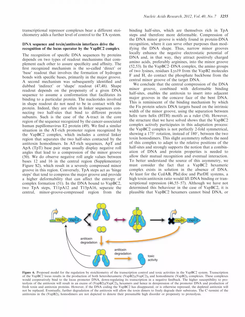
Figure 6. Proposed model for the regulation by stoichiometry of the transcription control and toxic activities in the VapBC2 system. Transcriptionof the VapBC2 locus results in the production of both heterohexameric (VapB2)2(VapC2)4 and homodimeric (VapB2)2 complexes. These complexeswould cooperatively bind to the locus promoter DNA, down-regulating its transcription in a negative feedback. The higher susceptibility to pro-teolysis of the antitoxin will result in an excess of (VapB2)2(VapC2)4 hexamers and hence in derepression of the promoter DNA and production offresh toxin and antitoxin proteins. However, if the DNA coding the VapBC2 has disappeared, or is otherwise repressed, the depleted antitoxin willnot be replaced. Eventually, further degradation of the antitoxin will allow the toxin dimers to freely degrade their substrates. The C0-termini of theantitoxins in the (VapB2)2 homodimers are not depicted to denote their presumable high disorder or propensity to proteolysis.
Nucleic Acids Research, 2012, Vol. 40, No. 7 3255
bind to it with lower affinity than VapB2 antitoxinhomodimers. One could then hypothesize that while thetoxin:antitoxin ratio is kept low, VapBC2 heterohexamersand VapB2 homodimers would coexist. In this scenario,VapB2 homodimers would bind to the recognitionhalf-sites of their cognate DNA, from where they couldrecruit VapBC2 hexamers to form a fully active repressioncomplex. The antitoxin hinge region (residues Pro41 toAsn44) is likely to play a major role in the adaptation ofthe VapBC2 full complex to its cognate DNA. Finally,when the levels of antitoxin dwindle, VapBC2 hexamerswill prevail, bind with lower affinity, or not at all to theDNA and repression will be lifted, so that the toxin:anti-toxin ratio can be re-equilibrated (Figure 6).
CONCLUSION
The study of the VapBC2 system has provided newinsights into the mechanisms that underlie toxin inhibitionby antitoxins of the swapped-hairpin b-barrel family.Furthermore, our combination of biophysical and struc-tural methods has shed light onto how the differentcomplexes formed by VapB2 and VapC2, but possiblyalso by other toxin/antitoxin pairs, integrate the controlof the toxin activity with their participation in the regula-tion of their own transcription.This work represents as well the first experimental struc-
ture of a protein from the swapped-hairpin b-barrel familybound to DNA. The structure essentially confirmsprevious models based on the unbound structures ofMazE (41) and AbrB (42), while providing a firstdetailed view of the contacts involved in DNA binding.Indeed, the structure underlines the importance of a largeinteraction surface that results in a marked widening ofthe DNA major groove, which combined with the relativepositioning of the VapB2 antitoxin homodimers in thecontext of the complex, possibly enhances the bendingof the DNA. Thus, the mutual adjustments between anintrinsically bendable DNA and the VapB2/VapC2 inter-faces, both within the VapBC2 heterohexamer andbetween this complex and the DNA-bound VapB2homodimer, may explain the increased affinity of theternary toxin–antitoxin–DNA complex.
ACCESSION NUMBER
3zvk.
SUPPLEMENTARY DATA
Supplementary Data are available at NAR Online:Supplementary Figures S1–S4 and SupplementaryReference [59].
ACKNOWLEDGEMENTS
The authors thank Gregory Gimenez for suggestingpossible sequences for the VapBC2 locus promoter,Stephanie Blangy and Catarina Rodrigues for their assist-ance with SEC/MALS experiments, Mariella Tegoni for
help with the SPR assays and Gilles Audoly for helpfuldiscussions. They also thank the staff of the ID14eh1,ID14eh4 and ID23eh2 beamlines at the EuropeanSynchrotron Radiation Facility (ESRF; Grenoble,France) for their help and support.
FUNDING
The Ville de Marseille provided a financial installation aid(to M.O.-L.). M.J.M. was supported by the FondationInfectiopole Sud. Funding for open access charge:Internal Resources.
Conflict of interest statement. None declared.
REFERENCES
1. Gerdes,K., Rasmussen,P.B. and Molin,S. (1986) Unique type ofplasmid maintenance function: postsegregational killing ofplasmid-free cells. Proc. Natl Acad. Sci. USA, 83, 3116–3120.
2. Pedersen,K., Christensen,S.K. and Gerdes,K. (2002) Rapidinduction and reversal of a bacteriostatic condition by controlledexpression of toxins and antitoxins. Mol. Microbiol., 45, 501–510.
3. Gerdes,K. (2000) Toxin-antitoxin modules may regulate synthesisof macromolecules during nutritional stress. J. Bacteriol., 182,561–572.
4. Winther,K.S. and Gerdes,K. (2011) Enteric virulence associatedprotein VapC inhibits translation by cleavage of initiator tRNA.Proc. Natl Acad. Sci. USA, 108, 7403–7407.
5. Yamaguchi,Y. and Inouye,M. (2009) mRNA interferases,sequence-specific endoribonucleases from the toxin-antitoxinsystems. Prog. Mol. Biol. Transl. Sci., 85, 467–500.
6. Correia,F.F., D’Onofrio,A., Rejtar,T., Li,L., Karger,B.L.,Makarova,K., Koonin,E.V. and Lewis,K. (2006) Kinase activityof overexpressed HipA is required for growth arrest andmultidrug tolerance in Escherichia coli. J. Bacteriol., 188,8360–8367.
7. Mattison,K., Wilbur,J.S., So,M. and Brennan,R.G. (2006)Structure of FitAB from Neisseria gonorrhoeae bound to DNAreveals a tetramer of toxin-antitoxin heterodimers containing pindomains and ribbon-helix-helix motifs. J. Biol. Chem., 281,37942–37951.
8. Wilbur,J.S., Chivers,P.T., Mattison,K., Potter,L., Brennan,R.G.and So,M. (2005) Neisseria gonorrhoeae FitA interacts with FitBto bind DNA through its ribbon-helix-helix motif. Biochemistry,44, 12515–12524.
9. Gerdes,K., Christensen,S.K. and Løbner-Olesen,A. (2005)Prokaryotic toxin-antitoxin stress response loci. Nat. Rev.Microbiol., 3, 371–382.
10. Arcus,V.L., Backbro,K., Roos,A., Daniel,E.L. and Baker,E.N.(2004) Distant structural homology leads to the functionalcharacterization of an archaeal PIN domain as an exonuclease.J. Biol. Chem., 279, 16471–16478.
11. Lebreton,A., Tomecki,R., Dziembowski,A. and Seraphin,B. (2008)Endonucleolytic RNA cleavage by a eukaryotic exosome. Nature,456, 993–996.
12. Kamada,K., Hanaoka,F. and Burley,S.K. (2003) Crystal structureof the MazE/MazF complex: molecular bases of antidote-toxinrecognition. Mol. Cell, 11, 875–884.
13. Christensen,S.K. and Gerdes,K. (2003) RelE toxins from bacteriaand Archaea cleave mRNAs on translating ribosomes, which arerescued by tmRNA. Mol. Microbiol., 48, 1389–1400.
14. Pedersen,K., Zavialov,A.V., Pavlov,M.Y., Elf,J., Gerdes,K. andEhrenberg,M. (2003) The bacterial toxin RelE displayscodon-specific cleavage of mRNAs in the ribosomal A site. Cell,112, 131–140.
15. Takagi,H., Kakuta,Y., Okada,T., Yao,M., Tanaka,I. andKimura,M. (2005) Crystal structure of archaeal toxin-antitoxinRelE-RelB complex with implications for toxin activity andantitoxin effects. Nat. Struct. Mol. Biol., 12, 327–331.
3256 Nucleic Acids Research, 2012, Vol. 40, No. 7
16. Anantharaman,V. and Aravind,L. (2003) New connections in theprokaryotic toxin-antitoxin network: relationship with theeukaryotic nonsense-mediated RNA decay system. Genome Biol.,4, R81.
17. Coles,M., Djuranovic,S., Soding,J., Frickey,T., Koretke,K.,Truffault,V., Martin,J. and Lupas,A.N. (2005) AbrB-liketranscription factors assume a swapped hairpin fold that isevolutionarily related to double-psi beta barrels. Structure, 13,919–928.
18. Schumacher,M.A., Piro,K.M., Xu,W., Hansen,S., Lewis,K. andBrennan,R.G. (2009) Molecular mechanisms of HipA-mediatedmultidrug tolerance and its neutralization by HipB. Science, 323,396–401.
19. Black,D.S., Kelly,A.J., Mardis,M.J. and Moyed,H.S. (1991)Structure and organization of hip, an operon that affectslethality due to inhibition of peptidoglycan or DNA synthesis.J. Bacteriol., 173, 5732–5739.
20. La Scola,B., Meconi,S., Fenollar,F., Rolain,J.-M., Roux,V. andRaoult,D. (2002) Emended description of Rickettsia felis (Bouyeret al. 2001), a temperature-dependent cultured bacterium.Int. J. Syst. Evol. Microbiol., 52, 2035–2041.
21. Raoult,D., La Scola,B., Enea,M., Fournier,P.E., Roux,V.,Fenollar,F., Galvao,M.A. and de Lamballerie,X. (2001)A flea-associated Rickettsia pathogenic for humans.Emerg. Infect. Dis., 7, 73–81.
22. Ogata,H., Renesto,P., Audic,S., Robert,C., Blanc,G.,Fournier,P.-E., Parinello,H., Claverie,J.-M. and Raoult,D. (2005)The genome sequence of Rickettsia felis identifies the firstputative conjugative plasmid in an obligate intracellular parasite.PLoS Biol., 3, e248.
23. Audoly,G., Vincentelli,R., Edouard,S., Georgiades,K.,Mediannikov,O., Gimenez,G., Socolovschi,C., Mege,J.-L.,Cambillau,C. and Raoult,D. (2011) Toxic effect ofrickettsial toxin VapC on its eukaryotic host. PLoS ONE, 6,e26528.
24. Vincentelli,R., Cimino,A., Geerlof,A., Kubo,A., Satou,Y. andCambillau,C. (2011) High-throughput proteinexpression screening and purification in Escherichia coli. Methods,55, 65–72.
25. Studier,F.W. (2005) Protein production by auto-inductionin high density shaking cultures. Protein Expr. Purif., 41,207–234.
26. Krasna,A.I., Dawson,J.R. and Harpst,J.A. (1970)Characterization of acid-denatured DNA by low-angle lightscattering. Biopolymers, 9, 1017–1028.
27. Kabsch,W. (2010) XDS. Acta Crystallogr. D Biol. Crystallogr.,66, 125–132.
28. Evans,P. (2006) Scaling and assessment of data quality. ActaCrystallogr. D Biol. Crystallogr., 62, 72–82.
29. Vonrhein,C., Blanc,E., Roversi,P. and Bricogne,G. (2007)Automated structure solution with autoSHARP. MethodsMol. Biol., 364, 215–230.
30. Abrahams,J.P. and Leslie,A.G. (1996) Methods used in thestructure determination of bovine mitochondrial F1 ATPase.Acta Crystallogr. D Biol. Crystallogr., 52, 30–42.
31. Emsley,P., Lohkamp,B., Scott,W.G. and Cowtan,K. (2010)Features and development of Coot. Acta Crystallogr. D Biol.Crystallogr., 66, 486–501.
32. Blanc,E., Roversi,P., Vonrhein,C., Flensburg,C., Lea,S.M. andBricogne,G. (2004) Refinement of severely incomplete structureswith maximum likelihood in BUSTER-TNT. Acta Crystallogr.D Biol. Crystallogr., 60, 2210–2221.
33. Davis,I.W., Leaver-Fay,A., Chen,V.B., Block,J.N., Kapral,G.J.,Wang,X., Murray,L.W., Arendall,W.B. III, Snoeyink,J.,Richardson,J.S. et al. (2007) MolProbity: all-atom contactsand structure validation for proteins and nucleic acids.Nucleic Acids Res., 35, W375–W383.
34. Krissinel,E. and Henrick,K. (2007) Inference of macromolecularassemblies from crystalline state. J. Mol. Biol., 372, 774–797.
35. Baker,N.A., Sept,D., Joseph,S., Holst,M.J. and McCammon,J.A.(2001) Electrostatics of nanosystems: application tomicrotubules and the ribosome. Proc. Natl Acad. Sci. USA, 98,10037–10041.
36. Pettersen,E.F., Goddard,T.D., Huang,C.C., Couch,G.S.,Greenblatt,D.M., Meng,E.C. and Ferrin,T.E. (2004) UCSFChimera—a visualization system for exploratory research andanalysis. J. Comput. Chem., 25, 1605–1612.
37. Bailey,S.E.S. and Hayes,F. (2009) Influence of operator sitegeometry on transcriptional control by the YefM-YoeBtoxin-antitoxin complex. J. Bacteriol., 191, 762–772.
38. Holm,L. and Rosenstrom,P. (2010) Dali server: conservationmapping in 3D. Nucleic Acids Res., 38, W545–W549.
39. Miallau,L., Faller,M., Chiang,J., Arbing,M., Guo,F., Cascio,D.and Eisenberg,D. (2009) Structure and proposed activity of amember of the VapBC family of toxin-antitoxin systems.VapBC-5 from Mycobacterium tuberculosis. J. Biol. Chem., 284,276–283.
40. Makarova,K.S., Aravind,L., Galperin,M.Y., Grishin,N.V.,Tatusov,R.L., Wolf,Y.I. and Koonin,E.V. (1999) Comparativegenomics of the Archaea (Euryarchaeota): evolution ofconserved protein families, the stable core, and the variable shell.Genome Res., 9, 608–628.
41. Loris,R., Marianovsky,I., Lah,J., Laeremans,T., Engelberg-Kulka,H., Glaser,G., Muyldermans,S. and Wyns,L. (2003) Crystalstructure of the intrinsically flexible addiction antidote MazE.J. Biol. Chem., 278, 28252–28257.
42. Sullivan,D.M., Bobay,B.G., Kojetin,D.J., Thompson,R.J.,Rance,M., Strauch,M.A. and Cavanagh,J. (2008) Insights into thenature of DNA binding of AbrB-like transcription factors.Structure, 16, 1702–1713.
43. Bobay,B.G., Andreeva,A., Mueller,G.A., Cavanagh,J. andMurzin,A.G. (2005) Revised structure of the AbrB N-terminaldomain unifies a diverse superfamily of putative DNA-bindingproteins. FEBS Lett., 579, 5669–5674.
44. Lavery,R., Moakher,M., Maddocks,J.H., Petkeviciute,D. andZakrzewska,K. (2009) Conformational analysis of nucleic acidsrevisited: Curves+. Nucleic Acids Res., 37, 5917–5929.
45. Dao-Thi,M.-H., Charlier,D., Loris,R., Maes,D., Messens,J.,Wyns,L. and Backmann,J. (2002) Intricate interactionswithin the ccd plasmid addiction system. J. Biol. Chem., 277,3733–3742.
46. Afif,H., Allali,N., Couturier,M. and Van Melderen,L. (2001) Theratio between CcdA and CcdB modulates the transcriptionalrepression of the ccd poison-antidote system. Mol. Microbiol., 41,73–82.
47. Otwinowski,Z., Schevitz,R.W., Zhang,R.G., Lawson,C.L.,Joachimiak,A., Marmorstein,R.Q., Luisi,B.F. and Sigler,P.B.(1988) Crystal structure of trp repressor/operator complex atatomic resolution. Nature, 335, 321–329.
48. Rohs,R., Jin,X., West,S.M., Joshi,R., Honig,B. and Mann,R.S.(2010) Origins of specificity in protein-DNA recognition.Annu. Rev. Biochem., 79, 233–269.
49. Hizver,J., Rozenberg,H., Frolow,F., Rabinovich,D. andShakked,Z. (2001) DNA bending by an adenine—thymine tractand its role in gene regulation. Proc. Natl Acad. Sci. USA, 98,8490–8495.
50. Haran,T.E. and Mohanty,U. (2009) The unique structureof A-tracts and intrinsic DNA bending. Q. Rev. Biophys., 42,41–81.
51. Olson,W.K., Gorin,A.A., Lu,X.J., Hock,L.M. and Zhurkin,V.B.(1998) DNA sequence-dependent deformability deduced fromprotein-DNA crystal complexes. Proc. Natl Acad. Sci. USA, 95,11163–11168.
52. Rohs,R., West,S.M., Sosinsky,A., Liu,P., Mann,R.S. andHonig,B. (2009) The role of DNA shape in protein-DNArecognition. Nature, 461, 1248–1253.
53. Joshi,R., Passner,J.M., Rohs,R., Jain,R., Sosinsky,A.,Crickmore,M.A., Jacob,V., Aggarwal,A.K., Honig,B. andMann,R.S. (2007) Functional specificity of a Hox proteinmediated by the recognition of minor groove structure. Cell, 131,530–543.
54. Stella,S., Cascio,D. and Johnson,R.C. (2010) The shape of theDNA minor groove directs binding by the DNA-bending proteinFis. Genes Dev., 24, 814–826.
55. Johnson,E.P., Strom,A.R. and Helinski,D.R. (1996) Plasmid RK2toxin protein ParE: purification and interaction with the ParDantitoxin protein. J. Bacteriol., 178, 1420–1429.
Nucleic Acids Research, 2012, Vol. 40, No. 7 3257

56. Magnuson,R. and Yarmolinsky,M.B. (1998) Corepression of theP1 addiction operon by Phd and Doc. J Bacteriol, 180,6342–6351.
57. Garcia-Pino,A., Balasubramanian,S., Wyns,L., Gazit,E., DeGreve,H., Magnuson,R.D., Charlier,D., van Nuland,N.A.J. andLoris,R. (2010) Allostery and intrinsic disorder mediatetranscription regulation by conditional cooperativity. Cell, 142,101–111.
58. Edgar,R.C. (2004) MUSCLE: multiple sequence alignment withhigh accuracy and high throughput. Nucleic Acids Res., 32,1792–1797.
59. Luscombe,N.M., Laskowski,R.A. and Thornton,J.M. (1997)NUCPLOT: a program to generate schematic diagrams ofprotein-nucleic acid interactions. Nucleic Acids Res., 25,4940–4945.
3258 Nucleic Acids Research, 2012, Vol. 40, No. 7


