Embed Size (px)
Citation preview

CTGF inhibits cell motility and COX-2 expression in oral cancer cells
Jing-Yuan Chuang1, Wan-Yu Yang
1, Chih-Ho Lai2,3, Chia-Der Lin
4, Ming-Hsui Tsai
4
and Chih-Hsin Tang5,3
*
1Department of Medical Laboratory Science and Biotechnology, China Medical
University, Taichung Taiwan
2Department of Microbiology, School of Medicine, China Medical University and
Hospital, Taichung, Taiwan
3Graduate Institute of Basic Medical Science, China Medical University and Hospital,
Taichung, Taiwan
4Department of Otolaryngology, China Medical University Hospital, Taichung, Taiwan
5Department of Pharmacology, School of Medicine, China Medical University and
Hospital, Taichung, Taiwan
Address correspondence to:
Chih-Hsin, Tang
Department of Pharmacology, School of Medicine, China Medical University.
No. 91, Hsueh-Shih Road, Taichung, Taiwan.
Tel: 886-4-22052121-7726; Fax: 886-4-22053764
E-mail: [email protected]

2
ABSTRACT
Oral squamous cell carcinoma (SCC) has a striking tendency to migrate and
metastasize. Cyclooxygenase (COX)-2, the inducible isoform of prostaglandin
synthase, has been implicated in tumor metastasis. Connective tissue growth factor
(CTGF), a secreted protein that binds to integrins, modulates the invasive behavior of
certain human cancer cells. However, the effect of CTGF on migration activity and
COX-2 expression in human oral cells is mostly unknown. Here we found that CTGF
reduced the migration and expression of COX-2 in human oral cancer cells. v
monoclonal antibody (mAb), phosphatidylinositol 3-kinase inhibitor (PI3K;
Ly294002 and wortmannin) and Akt inhibitor reversed the CTGF-inhibited the
migration and COX-2 down-regulation of oral cancer cells. CTGF stimulation
decreased the phosphorylation of focal adhesion kinase (FAK), PI3K and Akt. In
addition, c-Jun siRNA also antagonized the CTGF-inhibited migration and COX-2
expression. Moreover, CTGF decreased the binding of c-Jun to the AP-1 element on
the COX-2 promoter. Taken together, our results indicated that CTGF inhibits the
migration of oral cancer cells by decreasing COX-2 expression through the v
integrin receptor, FAK, PI3K, Akt, c-Jun and AP-1 signal transduction pathway.
Running title: CTGF inhibits the migration of oral cancer
Key words: CTGF; Migration; Oral cancer; FAK; Integrin

3
INTRODUCTION
Oral squamous cell carcinoma (SCC) represents 1–2% of all human
malignancies [Banoczy, 1997; Neville and Day, 2002]. It is characterized by a high
degree of local invasiveness and a high rate of metastasis to cervical lymph nodes.
The migration of oral SCC into maxillary and mandibular bones is a common clinical
problem [Lyons and Jones, 2007]. Because oral cancer is a type of highly malignant
tumor with a potent capacity to invade locally and metastasize distantly [Greenberg et
al., 2003; Thomas and Speight, 2001], an approach that decreases its ability to invade
and metastasize may facilitate the development of effective adjuvant therapy.
Cyclooxygenases (COXs) are the rate-limiting enzymes that catalyze
the
conversion of arachidonic acid to prostaglandins (PGs). Two COX isoforms with
distinct tissue distributions and physiological functions have been identified [Smith et
al., 2000; Warner and Mitchell, 2004]. COX-1 is constitutively expressed in many
tissues and plays important roles in the control of homeostasis [Morita, 2002].
Conversely, COX-2 is an inducible enzyme and is activated by extracellular stimuli
such as growth factors and pro-inflammatory cytokines [Turini and DuBois, 2002].
Over-expression of COX-2 is frequently found in many types of cancer, including
colon, lung, breast, pancreas, head, and neck cancers [Hida et al., 1998; Hwang et al.,
1998; Sano et al., 1995] and is usually associated with poor prognosis and short
survival. COX-2 also plays an important role in oral cancer cell migration, and
COX-2 inhibitors or siRNA had also been reported to rescues the migration of oral
cancer cells [Yang et al.]. Therefore, COX-2 may play a critical role in tumorigenesis,
and its disruption may prevent metastasis.
Connective tissue growth factor (CTGF, also known as CCN2) belongs to the
CCN family [Bork, 1993]. This family consists of six members, CTGF, NOVH,
CYR61, WISP1, WISP2 and WISP3 [Perbal, 2004] that all possess an N-terminal
signal peptide identifying them as secreted proteins. CCN proteins probably carry out
their biological activity through binding and activating of the cell
surface integrins
[Perbal, 2004]. Focal adhesion kinase (FAK), a potential candidate signaling molecule,
4
has been shown to be capable of regulating integrin-mediated signaling [Crouch et al.,
1996; Hadden and Henke, 2000]. However, the downstream signaling pathways that
mediate integrin-FAK signaling are diverse, and the factors determining which
pathway is used remain obscure. It has been reported that the CCN proteins involved
the stimulation of cellular proliferation, migration, adhesion, extracellular matrix
formation, and also the regulation of angiogenesis and tumorigenesis [Lau and Lam,
1999]. Overexpression of CTGF, WISP1, and CYR61 in tumor cells have been linked
to tumor size and lymph node metastasis [Xie et al., 2001], suggesting that these CCN
proteins are involved in the progression of human cancers.
Previous studies have shown that CTGF modulates cell migration and invasion
in cancer cells [Chang et al., 2004; Chen et al., 2007; Tan et al., 2009]. COX-2 has
been reported that modulates the cell migration and invasion of oral cancer cells
[Yang et al.]. However, the effect of CTGF on COX-2 expression and migration
activity in human oral cancer cells is mostly unknown. We hypothesized that CTGF
might be capable of regulating oral cancer cell migration and COX-2 expression. Here
we found that CTGF reduced the migration and the expression of COX-2 in human
oral cancer cells. In addition, v integrin, FAK, phosphatidylinositol 3-kinase
(PI3K), Akt and AP-1 signaling pathways may be involved in the decrease of COX-2
expression and cell migration by CTGF.
MATERIALS and METHODS
Materials:
Anti-mouse and anti-rabbit IgG-conjugated horseradish peroxidase, rabbit
polyclonal antibodies specific for -actin, p85, Akt, c-Jun, -actin and the small
interfering RNAs (siRNAs) against COX-2, c-Jun and control (for experiments using
targeted siRNA transfection; each consists of a scrambled sequence that will not lead
to the specific degradation of any known cellular mRNA) were purchased from Santa

5
Cruz Biotechnology (Santa Cruz, CA). Rabbit polyclonal antibodies specific for
p-FAK, p-p85 and p-Akt were purchased from Cell Signaling and Neuroscience
(Danvers, MA). Rabbit polyclonal antibodies (neutralizing antibodies) specific for
v3(MAB1976Z), v5(MAB1961Z) and 51(MAB1969) integrin were
purchased from Chemicon (Temecula, CA). Rabbit polyclonal antibody specific for
COX-2 was purchased from Cayman Chemical (Ann Arbor, MI). The recombinant
human CTGF was purchased from PeproTech (Rocky Hill, NJ, USA). NS398,
Ly294002, wortmannin, Akt inhibitor and curcumin were purchased from Calbiochem
(San Diego, CA). Celebrex was purchased from Pharmacia Co. (Piscataway, NJ). The
phosphorylation site mutant of FAK(Y397F) was a gift from Dr. J. A. Girault (Institut
du Fer a` Moulin, Moulin, France). The p85(p85; deletion of 35 amino acids from
residues 479-513 of p85) and Akt (Akt K179A) dominant-negative mutants were gifts
from Dr. W. M. Fu (National Taiwan University, Taipei, Taiwan). All other chemicals
were obtained from Sigma-Aldrich (St. Louis, MO).
Cell Culture
The human oral cancer cell line SCC4, SAS and Cal-27 was obtained from the
American Type Culture Collection (Rockville, MD). The cells were maintained in
DMEM supplemented with 20 mM HEPES and 10% heat-inactivated FCS, 2 mM
glutamine, penicillin (100 U/ml), and streptomycin (100 g/ml) at 37 °C with 5%
CO2.
Migration Assay
The migration assay was performed using Transwell (Costar, NY; pore size,
8-μm) in 24-well dishes. Before the migration assay, cells were pretreated for 30 min
with different concentrations of inhibitors, including the Ly294002, wortmannin or
vehicle control (0.1% DMSO). Approximately 1×10⁴ cells in 100 μl of serum-free
medium were placed in the upper chamber, and 300 l of the same medium
containing CTGF was placed in the lower chamber. The plates were incubated for 24

6
h at 37°C in 5% CO2, and then cells were fixed in methanol for 15 min and stained
with 0.05% crystal violet in PBS for 15 min. Cells on the upper side of the filters were
removed with cotton-tipped swabs, and the filters were washed with PBS. Cells on the
underside of the filters were examined and counted under a microscope. Each clone
was plated in triplicate in each experiment, and each experiment was repeated at least
three times. The number of migrating cells in each experiment was adjusted with a
cell viability assay to correct for proliferation effects of CTGF (corrected migrating
cell number = counted migrating cell number/percent of viable cells) [Tang et al.,
2008].
Quantitative Real-Time PCR
Total RNA was extracted from oral cancer cells using a TRIzol kit (MDBio Inc.,
Taipei, Taiwan). The reverse transcription reaction was performed using 2 g of total
RNA that was reverse transcribed into cDNA using oligo(dT) primer. The quantitative
real time PCR (qPCR) analysis was carried out using Taqman® one-step PCR Master
Mix (Applied Biosystems, CA). 2 l of total cDNA mixtures were added per 25-μl
reaction with sequence-specific primers and Taqman® probes. Sequences for all
target gene primers and probes were purchased commercially (-actin was used as
internal control) (Applied Biosystems, CA). qPCR assays were carried out in triplicate
(one independent RNA sample for each treatment) on an StepOnePlus sequence
detection system. The cycling conditions were 10-min polymerase activation at 95 °C
followed by 40 cycles at 95 °C for 15 s and 60 °C for 60 s. The threshold was set
above the non-template control background and within the linear phase of target gene
amplification to calculate the cycle number at which the transcript was detected
(denoted CT).
Western Blot Analysis
Cellular lysates were prepared as described [Tang et al., 2008]. Proteins were
resolved on SDS-PAGE and transferred to Immobilon polyvinyldifluoride (PVDF)

7
membranes. The blots were blocked with 4% BSA for 1 hr at room temperature and
then probed with rabbit anti-human antibodies against FAK, p-FAK, p85 or p-p85
(1:1000) for 1 hr at room temperature. After three washes, the blots were subsequently
incubated with a donkey anti-rabbit peroxidase-conjugated secondary antibody
(1:1000) for 1 hr at room temperature. The blots were visualized with enhanced
chemiluminescence and Kodak X-OMAT LS film (Eastman Kodak, Rochester, NY).
Chromatin Immunoprecipitation Assay
Chromatin immunoprecipitation analysis was performed as described [Huang
and Chen, 2005]. DNA immunoprecipitated with anti-c-Jun was purified and
extracted with phenol-chloroform. The purified DNA pellet was subjected to PCR,
and PCR products were resolved with 1.5% agarose gel electrophoresis and visualized
with UV light. The primers 5'-TAAGGGGAGAGGAGGGAAAAAT-3' and
5'-ACAATTGTCGCTAACCGAG-3' were utilized to amplify across the COX-2
promoter region (–119 to –14) [Chang et al., 2005].
Statistics
The values given are means ± S.E.M. Statistical analysis between two samples
was performed using the Student’s t test. Statistical comparisons involving more than
two groups were performed using one-way analysis of variance (ANOVA) with
Bonferroni’s post hoc test. In all cases, p < 0.05 was considered to be significant.
RESULTS
CTGF reduced migration and COX-2 expression in oral cancer cells
CTGF has been reported regulates migration and invasion of human cancer cells
[Chang et al., 2004; Chen et al., 2007; Tan et al., 2009]. The CTGF for oral cancer cell
migration was examined using the Transwell assay. Treatment of SCC4 cells with

8
CTGF (5-50 ng/ml) reduced cell migration (Fig. 1A). In addition, CTGF also
dose-dependently reduced other human oral cancer cell migration (SAS and Cal-27
cells) (Fig. 1B). However, treatment of these oral cancer cells with CTGF did not
affect cell viability by DAPI staining and MTT assay (data not shown). Previous study
has shown that COX-2 mediated cell motility in human oral cancer cells [Yang et al.].
We therefore, hypothesized that COX-2 may be involved in CTGF-mediated oral
cancer migration. Treatment of cells with CTGF reduced mRNA and protein
expression of COX-2 (Fig. 1C&D). To confirm COX-2 mediated CTGF-reduced cell
migration, the COX-2 specific inhibitors (Celebrex and NS-398) were used. Celebrex
and NS-398 reversed CTGF-mediated cell migration (Fig. 1E). When the SCC4 cells
were transfected with COX-2 or control siRNA for 24 hr, the Western blot analysis
showed that the expression of protein levels of COX-2 was suppressed by transfection
with COX-2 siRNA (Fig. 1F; upper panel). Transfection of cells with COX-2 siRNA
reversed the CTGF- reduced cell migration (Fig. 1F; lower panel). We also examined
human oral cancer tissues for expression of CTGF and COX-2 using Western blot
analysis. We found that oral cancer tissues expressed CTGF and COX-2 (Fig. 1G).
These data suggest that CTGF-reduced cancer migration may occur via
down-regulation of the COX-2.
CTGF reduced oral cancer cell migration through v integrin
Previous study has shown CTGF affects cell migration through integrin receptor
signaling [Chen et al., 2007; Tan et al., 2009]. We therefore hypothesized that integrin
receptor signaling pathway may be involved in CTGF-mediated oral cancer cell
migration. Pretreatment of cells for 30 min with anti-vbut not anti-v3 and
monoclonal antibody (mAb) markedly reversed the CTGF-reduced cancer
migration (Fig. 2A). In addition, treatment of cells with vbut not v3 and
mAb also reversed the CTGF-reduced COX-2 expression (Fig. 2B). These results
indicate that CTGF reduced cell migration in human oral cancer cells via v
integrin receptor.

9
The FAK, PI3K and Akt signaling pathways are involved in the CTGF-reduced
migration of oral cancer cells
FAK has been shown to be capable of regulating integrin-mediated signaling
[Mitra and Schlaepfer, 2006; Schlaepfer and Hunter, 1998]. Phosphorylation of
tyrosine 397 of FAK has been used as a marker of FAK activity. As shown in Fig. 2C,
FAK phosphorylation decreased in a time-dependent manner in response to CTGF
stimulation. Transfection of cells with FAK(Y397F) mutant reversed the
CTGF-reduced cell migration and COX-2 expression (Fig. 2D&E). Phosphorylation
of tyrosine 397 of FAK may provide a binding site for the Src homology 2 domain of
the p85 subunit of PI3K [Chan et al., 1999]. We then examined whether CTGF
stimulation also reduces the association of FAK with PI3K. Treatment of oral cancer
cells with CTGF led to a significant decrease of phosphorylation of p85 subunit of
PI3K (Fig. 3A). To explore whether PI3K is involved in CTGF-reduced cell migration,
PI3K inhibitor Ly294002 and wortmannin were used. As shown in Fig. 3B,C,E,
pretreatment of SCC4 cells with Ly294002 and wortmannin reversed CTGF-inhibited
migration activity and COX-2 expression of oral cancer cells. We then directly
measured the Akt phosphorylation in response to CTGF stimulation. Figure 3A shows
that CTGF decreased Akt phosphorylation (serine 473) in a time-dependent manner.
Furthermore, Akt inhibitor also antagonized CTGF-inhibited cell migration and
COX-2 expression (Fig. 3B,D,E). Transfection of cells with p85 and Akt mutant also
antagonized the potentiating effect of CTGF (Fig. 3F&G). Taken together, these
results indicate that the v5 integrin/FAK/PI3K and Akt pathway is involved in
CTGF-inhibited migration activity and COX-2 expression of human oral cancer cells.
Involvement of AP-1 in CTGF-inhibited cell migration and COX-2 expression
The promoter region of human COX-2 contains AP-1, NF-κB,
CCAAT/enhancer-binding protein and SP binding sites [van de Stolpe and van der
Saag, 1996]. AP-1 plays a critical role in COX-2 expression [Lu et al.]. To examine

10
the role of the AP-1 binding site in CTGF-mediated migration and COX-2 expression,
an AP-1 inhibitor (curcumin) was used. Pretreatment of cells with curcumin reversed
CTGF-reduced cell migration and COX-2 expression (Fig. 4A-C). It has been
reported that the AP-1 binding site between –67 and –61 was important for the
activation of the COX-2 gene [Iniguez et al., 2000]. AP-1 activation was further
evaluated by analyzing the chromatin immunoprecipitation assay. In vivo dis-binding
of c-Jun to the AP-1 element of the COX-2 promoter occurred after CTGF stimulation
(Fig. 5A). Dis-binding of c-Jun to the AP-1 element by CTGF was reversed by
Ly294002 and Akt inhibitor (Fig. 5A). Transfection of cells with c-Jun siRNA
suppressed the expression of c-Jun (Fig. 5B). CTGF-inhibited cell migration and
COX-2 expression was also reversed by c-Jun siRNA but not by control siRNA (Fig.
5C&D). Taken together, these data suggest that activation of the FAK, PI3K, Akt,
c-Jun, and AP-1 pathways are required for the CTGF-inhibited cell migration and
COX-2 expression in human oral cancer cells.
DISCUSSION
The elucidation of the molecular biology of cancer cells in recent years has
identified various molecular pathways that are altered in different cancers. This
information is currently being exploited to develop potential therapies that target
molecules in these pathways. To achieve metastasis, cancer cells must evade multiple
barriers and overcome certain rules. Several discrete steps are discernible in the
biological cascade leading to metastasis: loss of cellular adhesion, increased motility
and invasiveness, entry and survival into the circulation, entrance into new tissue, and
eventual colonization of a distant site [Gupta and Massague, 2006]. The mechanism
of metastasis is a complicated and multistage process, however our study showed that
CTGF inhibits cell migration and the expression of COX-2 in human oral cancer cells.
Here, we provide evidence that COX-2 acts as crucial transducers of cell signaling,

11
regulating cell migration and CTGF acts as a critical mediator of the metastasis
activity of cancer cells in the tumor microenvironment.
COX-2 is a pleiotropic enzyme that mediates many physiological functions such
as inhibition of cell apoptosis, augmentation of angiogenesis, and increased cell
motility. These COX-2–mediated functions are regulated in part by various proteins
such as B-cell lymphoma [Sun et al., 2002], myeloid cell leukemia-1, VEGF-A [Ma et
al., 2002] and metalloproteinases [Dohadwala et al., 2002]. It has been reported that
the expression of COX-2 is associated with a metastatic phenotype of oral cancer cells
[Yang et al.]. In this study, we found that CTGFinhibited COX-2 expression in
human oral cancer cells. On the other hand, COX-2 inhibitors antagonized
CTGF-reduced cell motility. In addition, the inhibition of CTGF-inhibited COX-2
protein expression with siRNA significantly reversed CTGF-inhibited migration.
However, COX-2 inhibitors and siRNA did not affect the basal migration activity of
oral cancer cells (Supplemental data Fig. 1S). Therefore, COX-2 may be the
CTGF-responsive mediator, and lead to decrease cancer migration and metastasis.
FAK, a potential candidate signaling molecule, has been shown to be capable of
regulating integrin-mediated signaling [Miranti and Brugge, 2002]. We demonstrate
that CTGF decreased phosphorylation of tyrosine 397 of FAK. Furthermore, the
FAK(Y397F) mutant antagonized the CTGF-inhibited migration activity and COX-2
expression, suggesting that FAK inhibition is an obligatory event in CTGF-reduced
migration in these cells. FAK contains tyrosine residues in motifs for binding to SH2
domain. Phosphorylated tyrosine 397 of FAK has been shown to serve as a binding
site for the SH2 domain of the p85 subunit of PI3K [Chen et al., 1996].
Phosphorylation of the p85 subunit is required for activation of the p110 catalytic
subunit of PI3K. Pretreatment of oral cancer cells with PI3K inhibitors Ly294002 and
wortmannin antagonized the decrease of chemomigration by CTGF. The cytoplasmic
serine kinase Akt was found to be reduced by CTGF stimulation in oral cancer cells.
In addition, treatment of cells with Akt inhibitor also reversed CTGF-inhibited cell
migration and COX-2 expression. This was further confirmed by the result that the

12
dominant-negative mutant of p85 and Akt reversed the migration and COX-2
expression by CTGF stimulation. Take together, our results provide evidence that
CTGF reduces migration and COX-2 expression in oral cancer cells through
FAK/PI3K/Akt signaling pathway.
It has been reported that chemokines induced COX-2 secretion through AP-1
dependent pathway [Chien et al., 2009; Lu et al.]. The AP-1 sequence binds to
members of the Jun and Fos families of transcription factors. These nuclear proteins
interact with the AP-1 site as Jun homodimers or Jun-Fos heterodimers formed by
protein dimerization through their leucine zipper motifs. The results of this study
show that CTGF reduced cell migration and COX-2 expression was reversed by c-Jun
siRNA. Furthermore, CTGF decreased the binding of c-Jun to the AP-1 element on
COX-2 promoter, as shown by ChIP assay. The dis-binding of c-Jun to the AP-1
element was also reversed by Ly294002 and Akt inhibitor. These results indicate that
CTGF might act through the v integrin, FAK, PI3K, Akt, c-Jun and AP-1
pathway to reduce cell migration and COX-2 expression in human oral cancer cells.
In addition, Ly294002, wortmannin, Akt inhibitor and curcumin or FAK mutant also
reversed CTGF-inhibited cell migration in other oral cancer cells (SAS and Cal-27
cells) (Supplemental data Fig. 2S). Therefore, the same signaling pathways are
involved in all oral cancer cells.
In conclusion, we present here a novel mechanism of CTGF-reduced migration
of human oral cancer cells by down-regulation of COX-2. CTGF inhibits COX-2
expression by binding to the v integrin receptor and reduction of FAK, PI3K, Akt
which inhibits binding of c-Jun to AP-1 site, resulting in the reduction tumor
migration.

13
Acknowledgments
This work was supported by grants from the National Science Council of Taiwan
(NSC99-2320-B-039-003-MY3); Taiwan Department of Health, China Medical
University Hospital Cancer Research Center of Excellence (DOH100-TD-C-111-005);
China Medical University (CMU-98-S-43). We thank Dr. J. A. Girault for providing
the FAK(Y397F) mutant; Dr. W. M. Fu for providing p85 and Akt mutants.
14
FIGURE LEGENDS
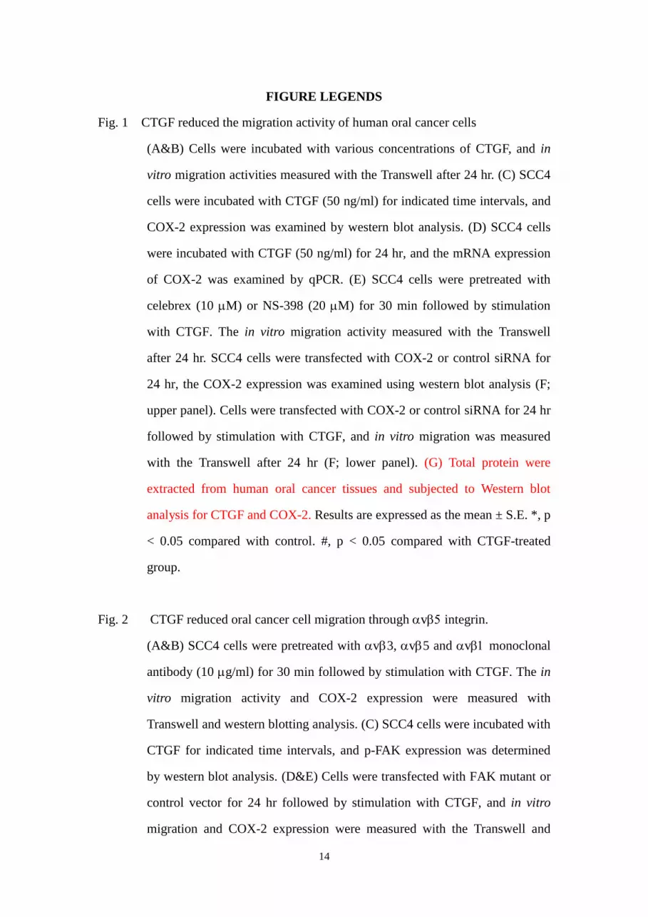
Fig. 1 CTGF reduced the migration activity of human oral cancer cells
(A&B) Cells were incubated with various concentrations of CTGF, and in
vitro migration activities measured with the Transwell after 24 hr. (C) SCC4
cells were incubated with CTGF (50 ng/ml) for indicated time intervals, and
COX-2 expression was examined by western blot analysis. (D) SCC4 cells
were incubated with CTGF (50 ng/ml) for 24 hr, and the mRNA expression
of COX-2 was examined by qPCR. (E) SCC4 cells were pretreated with
celebrex (10 M) or NS-398 (20 M) for 30 min followed by stimulation
with CTGF. The in vitro migration activity measured with the Transwell
after 24 hr. SCC4 cells were transfected with COX-2 or control siRNA for
24 hr, the COX-2 expression was examined using western blot analysis (F;
upper panel). Cells were transfected with COX-2 or control siRNA for 24 hr
followed by stimulation with CTGF, and in vitro migration was measured
with the Transwell after 24 hr (F; lower panel). (G) Total protein were
extracted from human oral cancer tissues and subjected to Western blot
analysis for CTGF and COX-2. Results are expressed as the mean ± S.E. *, p
< 0.05 compared with control. #, p < 0.05 compared with CTGF-treated
group.
Fig. 2 CTGF reduced oral cancer cell migration through v integrin.
(A&B) SCC4 cells were pretreated with v3, v5 and v monoclonal
antibody (10 g/ml) for 30 min followed by stimulation with CTGF. The in
vitro migration activity and COX-2 expression were measured with
Transwell and western blotting analysis. (C) SCC4 cells were incubated with
CTGF for indicated time intervals, and p-FAK expression was determined
by western blot analysis. (D&E) Cells were transfected with FAK mutant or
control vector for 24 hr followed by stimulation with CTGF, and in vitro
migration and COX-2 expression were measured with the Transwell and

15
western blotting analysis. Results are expressed as the mean ± S.E. *, p <
0.05 compared with control. #, p < 0.05 compared with CTGF-treated group.
Fig. 3 PI3K/Akt pathway is involved in CTGF-mediated migration in human oral
cancer cells.
(A) SCC4 cells were incubated with CTGF for indicated time intervals, and
p-PI3K and p-Akt expression was determined by western blot analysis. (B-E)
SCC4 cells were pretreated with Ly294002 (10 M), wortmannin (10 M)
or Akt inhibitor (10 M) for 30 min followed by stimulation with CTGF.
The in vitro migration activity and COX-2 expression were measured with
Transwell, western blotting analysis and qPCR. (F&G) Cells were
transfected with p85 and Akt mutant or control vector for 24 hr followed by
stimulation with CTGF, and in vitro migration and COX-2 expression were
measured with the Transwell and western blotting analysis. Results are
expressed as the mean ± S.E. *, p < 0.05 compared with control. #, p < 0.05
compared with CTGF-treated group.
Fig. 4 AP-1 is involved in CTGF-mediated migration in human oral cancer cells.
(A-C) SCC4 cells were pretreated with curcumin (10 M) for 30 min
followed by stimulation with CTGF. The in vitro migration activity and
COX-2 expression were measured with Transwell, western blotting analysis
and qPCR. Results are expressed as the mean ± S.E. *, p < 0.05 compared
with control. #, p < 0.05 compared with CTGF-treated group.
Fig. 5 FAK/PI3K/Akt pathway is mediated CTGF-reduced AP-1 activation.
(A) Cells were pretreated with Ly294002, and Akt inhibitor then stimulated
with CTGF for 120 min, and the chromatin immunoprecipitation assay was
then performed. Chromatin was immunoprecipitated with anti-c-Jun. One
percentage of the precipitated chromatin was assayed to verify equal loading

16
(input). (B) SCC4 cells were transfected with c-Jun or control siRNA for 24
hr, the c-Jun expression was examined using western blot analysis. (C&D)
Cells were transfected with c-Jun or control siRNA for 24 hr followed by
stimulation with CTGF, and in vitro migration and COX-2 expression were
measured with the Transwell and western blotting analysis. Results are
expressed as the mean ± S.E. *, p < 0.05 compared with control. #, p < 0.05
compared with CTGF-treated group.

17
REFERENCES
Banoczy J. 1997. Oral cancer and precancerous lesions. Fogorv Sz 90 Spec No:27.
Bork P. 1993. The modular architecture of a new family of growth regulators related
to connective tissue growth factor. FEBS Lett 327:125-30.
Chan TO, Rittenhouse SE, Tsichlis PN. 1999. AKT/PKB and other D3
phosphoinositide-regulated kinases: kinase activation by phosphoinositide-dependent
phosphorylation. Annu Rev Biochem 68:965-1014.
Chang CC, Shih JY, Jeng YM, Su JL, Lin BZ, Chen ST, Chau YP, Yang PC, Kuo ML.
2004. Connective tissue growth factor and its role in lung adenocarcinoma invasion
and metastasis. J Natl Cancer Inst 96:364-75.
Chang YJ, Wu MS, Lin JT, Chen CC. 2005. Helicobacter pylori-Induced invasion and
angiogenesis of gastric cells is mediated by cyclooxygenase-2 induction through
TLR2/TLR9 and promoter regulation. J Immunol 175:8242-52.
Chen HC, Appeddu PA, Isoda H, Guan JL. 1996. Phosphorylation of tyrosine 397 in
focal adhesion kinase is required for binding phosphatidylinositol 3-kinase. J Biol
Chem 271:26329-34.
Chen PS, Wang MY, Wu SN, Su JL, Hong CC, Chuang SE, Chen MW, Hua KT, Wu
YL, Cha ST, Babu MS, Chen CN, Lee PH, Chang KJ, Kuo ML. 2007. CTGF
enhances the motility of breast cancer cells via an
integrin-alphavbeta3-ERK1/2-dependent S100A4-upregulated pathway. J Cell Sci
120:2053-65.
Chien MH, Ku CC, Johansson G, Chen MW, Hsiao M, Su JL, Inoue H, Hua KT, Wei
LH, Kuo ML. 2009. Vascular endothelial growth factor-C (VEGF-C) promotes
angiogenesis by induction of COX-2 in leukemic cells via the VEGF-R3/JNK/AP-1
pathway. Carcinogenesis 30:2005-13.
Crouch DH, Fincham VJ, Frame MC. 1996. Targeted proteolysis of the focal
adhesion kinase pp125 FAK during c-MYC-induced apoptosis is suppressed by
integrin signalling. Oncogene 12:2689-96.
Dohadwala M, Batra RK, Luo J, Lin Y, Krysan K, Pold M, Sharma S, Dubinett SM.
2002. Autocrine/paracrine prostaglandin E2 production by non-small cell lung cancer
cells regulates matrix metalloproteinase-2 and CD44 in cyclooxygenase-2-dependent
invasion. J Biol Chem 277:50828-33.
Greenberg JS, El Naggar AK, Mo V, Roberts D, Myers JN. 2003. Disparity in
pathologic and clinical lymph node staging in oral tongue carcinoma. Implication for
therapeutic decision making. Cancer 98:508-15.
Gupta GP, Massague J. 2006. Cancer metastasis: building a framework. Cell
127:679-95.
18
Hadden HL, Henke CA. 2000. Induction of lung fibroblast apoptosis by soluble
fibronectin peptides. Am J Respir Crit Care Med 162:1553-60.
Hida T, Yatabe Y, Achiwa H, Muramatsu H, Kozaki K, Nakamura S, Ogawa M,
Mitsudomi T, Sugiura T, Takahashi T. 1998. Increased expression of cyclooxygenase
2 occurs frequently in human lung cancers, specifically in adenocarcinomas. Cancer
Res 58:3761-4.
Huang WC, Chen CC. 2005. Akt phosphorylation of p300 at Ser-1834 is essential for
its histone acetyltransferase and transcriptional activity. Mol Cell Biol 25:6592-602.
Hwang D, Scollard D, Byrne J, Levine E. 1998. Expression of cyclooxygenase-1 and
cyclooxygenase-2 in human breast cancer. J Natl Cancer Inst 90:455-60.
Iniguez MA, Martinez-Martinez S, Punzon C, Redondo JM, Fresno M. 2000. An
essential role of the nuclear factor of activated T cells in the regulation of the
expression of the cyclooxygenase-2 gene in human T lymphocytes. J Biol Chem
275:23627-35.
Lau LF, Lam SC. 1999. The CCN family of angiogenic regulators: the integrin
connection. Exp Cell Res 248:44-57.
Lu DY, Leung YM, Huang SM, Wong KL. Bradykinin-induced cell migration and
COX-2 production mediated by the bradykinin B1 receptor in glioma cells. J Cell
Biochem 110:141-50.
Lyons AJ, Jones J. 2007. Cell adhesion molecules, the extracellular matrix and oral
squamous carcinoma. Int J Oral Maxillofac Surg 36:671-9.
Ma L, del Soldato P, Wallace JL. 2002. Divergent effects of new cyclooxygenase
inhibitors on gastric ulcer healing: Shifting the angiogenic balance. Proc Natl Acad
Sci U S A 99:13243-7.
Miranti CK, Brugge JS. 2002. Sensing the environment: a historical perspective on
integrin signal transduction. Nat Cell Biol 4:E83-90.
Mitra SK, Schlaepfer DD. 2006. Integrin-regulated FAK-Src signaling in normal and
cancer cells. Curr Opin Cell Biol 18:516-23.
Morita I. 2002. Distinct functions of COX-1 and COX-2. Prostaglandins Other Lipid
Mediat 68-69:165-75.
Neville BW, Day TA. 2002. Oral cancer and precancerous lesions. CA Cancer J Clin
52:195-215.
Perbal B. 2004. CCN proteins: multifunctional signalling regulators. Lancet 363:62-4.
Sano H, Kawahito Y, Wilder RL, Hashiramoto A, Mukai S, Asai K, Kimura S, Kato
H, Kondo M, Hla T. 1995. Expression of cyclooxygenase-1 and -2 in human
colorectal cancer. Cancer Res 55:3785-9.
Schlaepfer DD, Hunter T. 1998. Integrin signalling and tyrosine phosphorylation: just
the FAKs? Trends Cell Biol 8:151-7.

19
Smith WL, DeWitt DL, Garavito RM. 2000. Cyclooxygenases: structural, cellular,
and molecular biology. Annu Rev Biochem 69:145-82.
Sun Y, Tang XM, Half E, Kuo MT, Sinicrope FA. 2002. Cyclooxygenase-2
overexpression reduces apoptotic susceptibility by inhibiting the cytochrome
c-dependent apoptotic pathway in human colon cancer cells. Cancer Res 62:6323-8.
Tan TW, Lai CH, Huang CY, Yang WH, Chen HT, Hsu HC, Fong YC, Tang CH.
2009. CTGF enhances migration and MMP-13 up-regulation via alphavbeta3 integrin,
FAK, ERK, and NF-kappaB-dependent pathway in human chondrosarcoma cells. J
Cell Biochem 107:345-56.
Tang CH, Tan TW, Fu WM, Yang RS. 2008. Involvement of matrix
metalloproteinase-9 in stromal cell-derived factor-1/CXCR4 pathway of lung cancer
metastasis. Carcinogenesis 29:35-43.
Thomas GJ, Speight PM. 2001. Cell adhesion molecules and oral cancer. Crit Rev
Oral Biol Med 12:479-98.
Turini ME, DuBois RN. 2002. Cyclooxygenase-2: a therapeutic target. Annu Rev
Med 53:35-57.
van de Stolpe A, van der Saag PT. 1996. Intercellular adhesion molecule-1. J Mol
Med 74:13-33.
Warner TD, Mitchell JA. 2004. Cyclooxygenases: new forms, new inhibitors, and
lessons from the clinic. FASEB J 18:790-804.
Xie D, Nakachi K, Wang H, Elashoff R, Koeffler HP. 2001. Elevated levels of
connective tissue growth factor, WISP-1, and CYR61 in primary breast cancers
associated with more advanced features. Cancer Res 61:8917-23.
Yang SF, Chen MK, Hsieh YS, Chung TT, Hsieh YH, Lin CW, Su JL, Tsai MH,
Tang CH. PGE2/EP1 signaling pathway enhances ICAM-1 expression and cell
motility in oral cancer cells. J Biol Chem.


![[Gary W. Cox] Making Votes Count](https://img.pdfslide.net/doc/110x75/6361c4fb40b666b8ec0e1200/gary-w-cox-making-votes-count.jpg)


