Embed Size (px)
Citation preview

Myocardial Connective Tissue Growth Factor (CCN2/CTGF) Attenuates Left Ventricular Remodeling afterMyocardial InfarctionJørgen Gravning1,2, Stein Ørn3, Ole Jørgen Kaasbøll1,2, Vladimir N. Martinov1,2, Cord Manhenke3,
Kenneth Dickstein3,4, Thor Edvardsen1,2,5, Havard Attramadal1,2,5*, Mohammed Shakil Ahmed1,2
1 Institute for Surgical Research, Oslo University Hospital, Rikshospitalet, Oslo, Norway, 2 Center for Heart Failure Research, University of Oslo, Oslo, Norway, 3 Division of
Cardiology, Stavanger University Hospital, Stavanger, Norway, 4 Institute of Internal Medicine, University of Bergen, Bergen, Norway, 5 Department of Cardiology, Oslo
University Hospital, Rikshospitalet, Oslo, Norway
Abstract
Aims: Myocardial CCN2/CTGF is induced in heart failure of various etiologies. However, its role in the pathophysiology of leftventricular (LV) remodeling after myocardial infarction (MI) remains unresolved. The current study explores the role of CTGFin infarct healing and LV remodeling in an animal model and in patients admitted for acute ST-elevation MI.
Methods and Results: Transgenic mice with cardiac-restricted overexpression of CTGF (Tg-CTGF) and non-transgeniclittermate controls (NLC) were subjected to permanent ligation of the left anterior descending coronary artery. Despitesimilar infarct size (area of infarction relative to area at risk) 24 hours after ligation of the coronary artery in Tg-CTGF and NLCmice, Tg-CTGF mice disclosed smaller area of scar tissue, smaller increase of cardiac hypertrophy, and less LV dilatation anddeterioration of LV function 4 weeks after MI. Tg-CTGF mice also revealed substantially reduced mortality after MI. Remote/peri-infarct tissue of Tg-CTGF mice contained reduced numbers of leucocytes, macrophages, and cells undergoingapoptosis as compared with NLC mice. In a cohort of patients with acute ST-elevation MI (n = 42) admitted to hospital forpercutaneous coronary intervention (PCI) serum-CTGF levels (s-CTGF) were monitored and related to infarct size and LVfunction assessed by cardiac MRI. Increase in s-CTGF levels after MI was associated with reduced infarct size and improvedLV ejection fraction one year after MI, as well as attenuated levels of CRP and GDF-15.
Conclusion: Increased myocardial CTGF activities after MI are associated with attenuation of LV remodeling and improvedLV function mediated by attenuation of inflammatory responses and inhibition of apoptosis.
Citation: Gravning J, Ørn S, Kaasbøll OJ, Martinov VN, Manhenke C, et al. (2012) Myocardial Connective Tissue Growth Factor (CCN2/CTGF) Attenuates LeftVentricular Remodeling after Myocardial Infarction. PLoS ONE 7(12): e52120. doi:10.1371/journal.pone.0052120
Editor: Fadi N Salloum, Virginia Commonwealth University Medical Center, United States of America
Received May 3, 2012; Accepted November 9, 2012; Published December 20, 2012
Copyright: � 2012 Gravning et al. This is an open-access article distributed under the terms of the Creative Commons Attribution License, which permitsunrestricted use, distribution, and reproduction in any medium, provided the original author and source are credited.
Funding: This study was supported by grants from the South-Eastern Norway Regional Health Authority (grant numbers 2009086 and 2011006), The NorwegianCouncil on Cardiovascular Disease (grant number 6484), and The Norwegian Research Council. The funders had no role in study design, data collection andanalysis, decision to publish, or preparation of the manuscript.
Competing Interests: The authors Jørgen Gravning, Vladimir N. Martinov, M. Shakil Ahmed, and Havard Attramadal are inventors in a patent application relatedto the potential application of CCN2/CTGF as a cardioprotectant (PCT/GB2009/002218;WO/2010/032007). This does not alter the authors’ adherence to all thePLOS ONE policies on sharing data and materials.
* E-mail: [email protected]
Introduction
Myocardial infarction (MI) commonly triggers left ventricular
(LV) remodeling, progressive deterioration of cardiac function,
and ultimately, the clinical syndrome of heart failure (HF) [1].
However, current understanding of the pathophysiologic mecha-
nisms of LV remodeling is still fragmentary. Thus, unraveling of
autocrine/paracrine factors and cytokines involved in infarct
healing and LV remodeling after MI may provide novel targets for
pharmacologic therapeutics.
Myocardial connective tissue growth factor (CCN2/CTGF), a
secreted matricellular protein of the CCN family, is upregulated in
HF of both ischemic and non-ischemic etiologies, in experimental
models as well as in humans [2,3]. Plasma CTGF levels have also
previously been reported to be elevated in HF patients and
correlated to NYHA-class [4]. Although increased tissue expres-
sion of CTGF in disease is often associated with fibrosis [5], it is
not yet known to what extent CTGF causes fibrosis. Thus, despite
the unresolved role of elevated myocardial CTGF activities in
heart failure, myocardial CTGF expression may reflect both
fibrosis and functional derangement of the heart. A recent study
reported preserved cardiac function in transgenic mice with
cardiac-restricted overexpression of CTGF following chronic
infusion of angiotensin II [6]. In a recent report from our
laboratory, enhanced resistance towards ischemia/reperfusion
injury was observed both in transgenic mice with cardiac-restricted
overexpression of CTGF (Tg-CTGF) and in Langendorff-perfused
hearts pre-treated with recombinant human CTGF (rh-CTGF)
before ischemia, due to increased activity of the Akt/GSK-3bsignaling pathway [7]. However, to what extent the cardioprotec-
tive effects of CTGF are maintained in chronic ischemia, infarct
healing and LV remodeling remain to be investigated. Cardiac-
PLOS ONE | www.plosone.org 1 December 2012 | Volume 7 | Issue 12 | e52120

restricted expression of CTGF resulted in minimal increase of
myocardial fibrosis per se, and indicated that, at the least, additional
co-factors are required for CTGF to stimulate relevant fibrosis [7].
Yet, to what extent MI provides basis for enhanced profibrotic
actions of CTGF is unknown. No reports have yet explored the
relationship between CTGF and LV remodeling following MI.
In this study we therefore investigated the role of CTGF in post-
MI remodeling of the left ventricle both in an animal model of
myocardial infarction and in a cohort of patients admitted for first-
time, acute ST-elevation MI (STEMI) treated by primary PCI,
and assessed with cardiac MRI and analysis of s-CTGF levels at
several time points after the acute event. The current study
unravels novel salutary actions of CTGF in infarct healing and LV
remodeling after MI.
Materials and Methods
All animal studies were performed in accordance with the NIH
Guidelines for the Use of Laboratory Animals and were approved
by the institutional and national boards for laboratory animal
research. The patient study was approved by the Regional Ethics
Committee of Western Norway and all patients provided written
informed consent prior to inclusion.
Experimental MI and Heart Failure in MiceThe transgenic mice with cardiac-restricted overexpression of
CTGF (Tg-CTGF mice) employed in this study have been
described and characterized previously [7]. Briefly, the Tg-CTGF
mice were generated by pronuclear injection of a DNA cassette
containing the ORF of rat CTGF cDNA under control of the
mouse a-myosin heavy chain promoter into fertilized oocytes of
C57BL/6 mice and implanted in pseudopregnant mice. Non-
transgenic littermate control (NLC) mice were generated from
non-injected C57BL/6 mice (siblings of the mice employed for
pronuclear injection) and bred similarly to the transgenic lines, in
order to ensure background similar to that of Tg-CTGF mice.
Two transgenic lines that were propagated and characterized (Tg-
CTGF/6 and Tg-CTGF/13) displayed similar phenotypic char-
acteristics [7]. Unless otherwise indicated, Tg-CTGF/6 mice were
employed in the experiments of the current study. MI was induced
by ligation of the left anterior descending coronary artery (LAD) in
Tg-CTGF (n = 22) and NLC mice (n = 21) according to previously
published procedures [8]. Briefly, the mice were intubated,
anesthetized with isoflurane and ventilated at a respiratory rate
of 130/min in a gas mixture of 1/3 O2 and 2/3 N2O containing
1.5% isoflurane, using a rodent ventilator. The mice were
immobilized on a heating pad and subjected to left-sided
thoracotomy between the third and fourth intercostal space. After
opening of the pericardium, ligation of LAD was made 1 mm
under the left atrial appendage using a silk 7/0 suture. Pallor of the
affected myocardium ensured ischemia. During follow-up, 8 Tg-
CTGF mice and 13 NLC mice died. Separate groups of sham-
operated Tg-CTGF mice (n = 5) and NLC mice (n = 6) underwent
similar surgical procedure except ligation of LAD. The study end-
point was 4 weeks (28 days) after induction of MI. Cardiac
dimensions and cardiac function were assessed by a high-
resolution ultrasonography system (Vevo 770, VisualSonics Inc.,
Toronto, Canada) using 2D-guided M-mode recording of the LV
at the plane of the tip of the papillary muscle according to
previously described methodology [7]. Transthoracic echocardi-
ography of all Tg-CTGF mice (n = 22) and NLC mice (n = 21) to
be subjected to MI was performed immediately before induction of
MI (baseline; day -1). Thereafter, successive transthoracic echo-
cardiography of all mice alive was performed 2 weeks after
induction of MI (Tg-CTGF [n = 15]; NLC [n = 9]) and at study
end-point (Tg-CTGF [n = 14]; NLC [n = 8]). At study end-point,
LV function was also assessed by trans-carotid catheterization of
the left ventricle and continuous recording of LV pressure, as
previously described [7]. Separate groups of Tg-CTGF mice
(n = 8) and NLC mice (n = 8) were subjected to ligation of LAD
and subsequent perfusion with 1% Evans blue dye in order to
assess the area at risk (AAR) in the two groups. LV sections were
subjected to computerized planimetry in order to determine the
area of non-perfused myocardial tissue. Another group of Tg-
CTGF mice (n = 6) and NLC mice (n = 6) were subjected to
permanent ligation of LAD in order to estimate area of myocardial
necrosis (infarct size) relative to area at risk after 24 hours of
ischemia. After 24 hours the mice were euthanized by cardioplegic
arrest following infusion of KCl. The hearts were excised and
perfused with 1% Evans blue dye, and 1 mm thick sections of the
left ventricle were immediately prepared and stained with 2,3,5-
triphenyltetrazolium chloride (TTC) for 20 min at 37uC. Finally,
the LV sections were fixed in 4% paraformaldehyde and washed
with PBS before computerized planimetry for determination of
area of necrosis (infarction; area not stained with TTC) relative to
area at risk (AAR; area not stained with Evans blue dye) as
described previously [7].
Histochemistry and morphometric analysis of myocardial
sections, determination of myocardial mRNA levels by real-time
qPCR, Western blot analysis, and assay of myocardial hydroxy-
proline contents were performed as previously described [7].
Myocardial cells undergoing apoptosis were assessed by in situ
terminal deoxynucleotidyl transferase-mediated digoxigenin-con-
jugated dUTP nick end labeling (TUNEL) of apoptotic nuclei (In
Situ Cell Death Detection kit; Roche Diagnostics, Inc.) and nuclear
co-staining with Hoechst 33258. The cross-sectional area of
cardiac myocytes was determined in transverse sections of the left
ventricle after staining of plasma membrane with rhodamine-
labeled wheat germ agglutinin (WGA) (Vector Laboratories, Inc.).
The cross-sectional area of individual myocytes was determined by
digital planimetry using the Adobe Photoshop software.
Immunohistochemical Analysis of Myocardial TissueSections
Hearts from NLC CHF (n = 4) and Tg-CTGF CHF (n = 4) mice
were fixed in 4% paraformaldehyde in phosphate-buffered saline
for 1 h, embedded in paraffin wax, and stored at 4uC.
Immunohistochemical analysis of myocardial sections (6 mm) of
NLC and Tg-CTGF mice was performed as previously described
[7], using rat monoclonal anti-mouse CD68 (ab53444; Abcam,
Inc.), rabbit polyclonal anti-mouse CD45 IgG (ab10558; Abcam,
Inc.), rabbit anti-c-Kit IgG (sc-5535; St. Cruz Biotechnology, Inc.),
rabbit polyclonal anti-mouse Ki-67 IgG (ab9260; Millipore, Inc.)
and goat polyclonal anti-mouse PECAM-1/CD31 IgG (sc-1506;
St. Cruz Biotechnology, Inc.). The avidin-biotin-peroxidase system
(Vectastain Elite kit; Vector Laboratories, Inc.) was used for signal
amplification.
The Patient StudyForty-two consecutive patients admitted with ST-elevation MI
(STEMI) and selected for primary PCI were enrolled prospectively
at a single center [9]. Patients were included if they had: (i) no
previous MI, (ii) demonstrated acute proximal/mid-occluded
single-vessel disease, (iii) underwent successful PCI with stent
implantation without significant residual stenosis, (iv) had no
contraindications to CMR imaging, and (v) could be scanned
within 48 h following PCI.
CCN2/CTGF Attenuates Cardiac Remodeling
PLOS ONE | www.plosone.org 2 December 2012 | Volume 7 | Issue 12 | e52120

Blood Sample Protocol and Assay of Serum CTGF, CRPand GDF-15 Levels
Venous blood samples were collected in pyrogen-free ethylene-
diaminetetraacetic acid (EDTA)-containing tubes or tubes without
any additives at the time of the CMR examinations, at day 2,
1 week, 2 months and 1 year following PCI. The tubes were
subsequently centrifuged at 1000xg for 10 min for preparation of
plasma and serum samples, respectively. All samples were stored at
–80uC until analysis. Serum levels of CTGF (s-CTGF) were
determined by a sandwich enzyme-linked immunosorbent assay
(ELISA) as previously described [10]. Serum levels of high-
sensitive C-reactive protein (CRP) and Growth differentiation
factor 15 (GDF-15) were determined by ELISA assays according
to previously described methodology [11,12].
Cardiac Magnetic Resonance ProtocolAll patients were scanned 2 days (mean6SEM: 2.260.1 days)
following PCI, and subsequently 7 days (7.360.1 days), 2 months
(6160.6 days), and 1 year (36461.2 days) following PCI, using a
1.5 T whole body scanner (Intera R 10.3 Philips Medical Systems,
Best, The Netherlands) as previously described [9].
StatisticsContinuous data are presented as mean6SEM and inter-group
comparisons were made by two-tailed unpaired Student’s t test or
2-way ANOVA with post-hoc analysis (Bonferroni correction) as
appropriate. Categorical variables are expressed as proportions
and comparisons were made by two-sided Chi-Square test.
Survival analysis was evaluated with Kaplan-Meier curves and
compared by log-rank test. Intra-group changes in the CMR
parameters during follow-up were analyzed with the Friedman
test. P-value ,0.05 was considered statistically significant for inter-
group comparisons and P-value ,0.01 was considered statistically
significant for intra-group variation.
Results
Tg-CTGF Mice and NLC Mice Displayed Similar Area atRisk after Ligation of LAD
As shown in Fig. 1A, hearts from Tg-CTGF and NLC mice
perfused with Evans blue dye immediately after ligation of LAD
displayed similar area at risk (42.761.6%, n = 8 vs. 40.462.1%,
n = 8, P = 0.39).
Similar Infarct Size in Tg-CTGF and NLC Mice 24 Hoursafter Ligation of LAD
Infarct size, i.e. area of myocardial necrosis relative to area at
risk, 24 hours after ligation of LAD involved almost entire area at
risk and was essentially similar in Tg-CTGF and NLC mice (Tg-
CTGF: 98.360.5%, n = 6 vs. NLC: 94.461.3%, n = 6, P = 0.02)
(Fig. 1B).
CTGF Attenuates LV Dysfunction, Congestive HeartFailure, and Mortality after MI in Mice
Cardiac dimensions at baseline assessed by transthoracic
echocardiography were similar among the groups. Four weeks
after MI, Tg-CTGF mice displayed diminished LV remodeling
with less LV dilatation and less thinning of the intraventricular
septum thickness both at end-diastole (IVSd) and at end-systole
(IVSs) compared with NLC mice (Fig. 2). The decline of LV
fractional shortening (FS) and LV ejection fraction (EF) were
substantially reduced in Tg-CTGF mice versus NLC mice during
the study period.
At study end-point gravimetric analysis revealed substantially
smaller increase of cardiac mass in Tg-CTGF compared with
NLC mice (Fig. 3A). In NLC mice subjected to MI, normalized
lung weights were substantially increased at study end-point
compatible with pulmonary congestion. In contrast, Tg-CTGF
mice did not display significant alterations of lung weights from
baseline to study end-point. Hemodynamic analysis of cardiac
function by LV catheterization at study end-point, revealed
substantial decline of end-systolic pressure (ESP) and increase of
end-diastolic pressure (EDP) in NLC mice subjected to MI. In
contrast, only subtle derangements of ESP and EDP were
observed in Tg-CTGF mice at study end-point. Consistently,
cardiac contractility as measured by LV (dP/dt)max and (dP/dt)min
was less deteriorated in Tg-CTGF versus NLC mice (Fig. 3B).
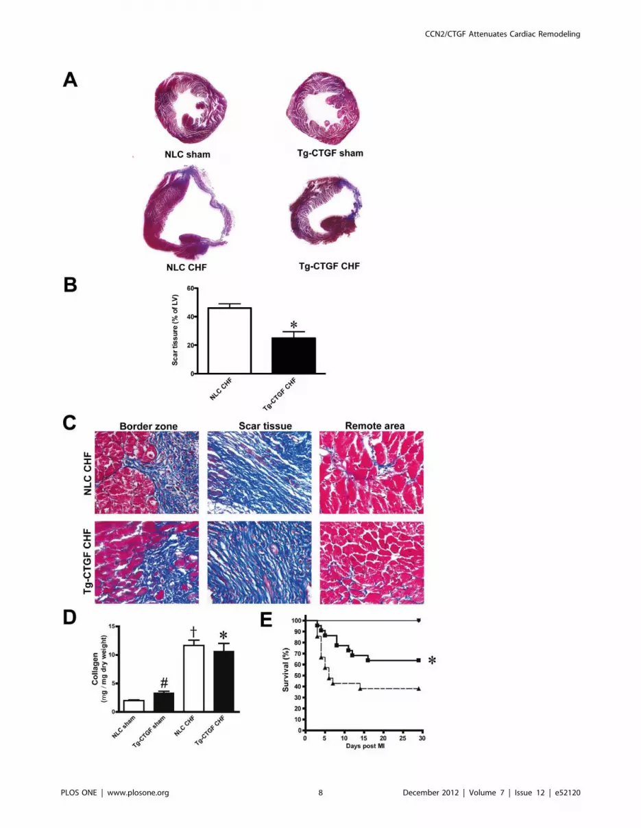
Masson’s trichrome staining of transverse myocardial sections at
the level of the papillary muscles revealed substantially lesser scar
tissue in Tg-CTGF versus NLC mice at study end-point (Fig. 4A–
B). Representative images of Masson’s trichrome-stained sections
from the border zone, scar tissue and remote area of Tg-CTGF
mice and NLC mice are displayed in Fig. 4C. Quantification of
myocardial fibrosis in the remote area by assay of myocardial
collagen (hydroxyproline), revealed similar collagen contents in
Tg-CTGF and NLC mice after MI (Fig. 4D). However, the
increase of myocardial collagen contents after MI relative to the
respective sham group was smaller in Tg-CTGF than in NLC
mice. Importantly, during the course of the study the survival rate
was significantly higher among Tg-CTGF mice than NLC mice
(64% vs. 38%, P,0.05) after MI (Fig. 4E).
Assessment of Cardiomyocyte Hypertrophy, MyocardialApoptosis, and Microvessel Densities in Tg-CTGF andNLC Mice after MI
Assessment of myocardial apoptosis by TUNEL-staining of
apoptotic nuclei revealed very rare events of cellular apoptosis in
the remote myocardium 24 hours after induction of MI. Cellular
apoptosis in remote myocardium at this time point was not
statistically different in Tg-CTGF and NLC mice (Fig. 5A–B).
However, 4 weeks after MI cellular apoptosis of remote
myocardial tissue was substantially increased. Yet, apoptosis of
remote myocardial tissue was significantly lower in Tg-CTGF
mice than in NLC mice at the later time point (Fig. 5A–B).
Consistent with the echocardiographic and hemodynamic data
demonstrating attenuated remodeling and preserved cardiac
function in Tg-CTGF versus NLC mice after MI, morphometric
analysis of transverse myocardial sections stained with rhodamin-
labeled WGA revealed decreased cross-sectional area of cardiac
myocytes of Tg-CTGF mice versus NLC (Fig. 6A–B). Reflecting
reduced signals for myocardial hypertrophy of Tg-CTGF mice
after MI, the increase of myocardial BNP and ANP mRNA levels
were also attenuated in Tg-CTGF mice, compared with that of
NLC mice after MI (Fig. 6C). Importantly, mRNA expression of
the rat-CTGF transgene in myocardial tissue of Tg-CTGF mice
was not altered after MI excluding alterations of a-MHC
promoter activities after MI (data not shown). Staining of
myocardial tissue with anti-CD31 antibody displayed slight
increase of microvessel density in the peri-infarct region of Tg-
CTGF mice (Fig. 6A–B).
Myocardial Tissue of Tg-CTGF Mice Display DecreasedContents of Inflammatory Cells after MI
Immunostaining against the macrophage marker CD68 and the
leucocyte marker CD45 also revealed decreased contents of
macrophages and leucocytes in peri-infarct tissue of hearts from
CCN2/CTGF Attenuates Cardiac Remodeling
PLOS ONE | www.plosone.org 3 December 2012 | Volume 7 | Issue 12 | e52120

Figure 1. Area at risk and infarct size in NLC and Tg-CTGF mice subjected to ligation of LAD. A. Photomicrographs of representativemyocardial sections of NLC and Tg-CTGF hearts perfused with Evans blue dye immediately after ligation of LAD. Histogram demonstrating area at riskin the left ventricles of NLC and Tg-CTGF mice determined by computerized planimetry after excision of the hearts. The data are mean6SEM of areaat risk as percent of total LV area of NLC (n = 8) and Tg-CTGF mice (n = 8), respectively (40.462.1% vs. 42.761.6%, P = 0.39). B. Photomicrographs ofrepresentative myocardial sections of NLC and Tg-CTGF hearts 24 hours after ligation of LAD and subsequent perfusion with Evans blue dye andmyocardial staining with TTC. Histogram demonstrating infarct size (area of necrosis relative to area at risk) 24 hours after ligation of LAD, determinedas described in the Materials and Methods section. The data are mean6SEM of NLC (n = 6) and Tg-CTGF hearts (n = 6). *P,0.05 vs. NLC group.doi:10.1371/journal.pone.0052120.g001
CCN2/CTGF Attenuates Cardiac Remodeling
PLOS ONE | www.plosone.org 4 December 2012 | Volume 7 | Issue 12 | e52120

Figure 2. Transthoracic echocardiography of NLC and Tg-CTGF mice at successive time points after MI. Successive echocardiographicrecordings of inter-ventricular septum thickness (IVS) and LV transverse diameter (LVID) at end-diastole and end-systole, as well as fractionalshortening (FS) and estimated ejection fraction (EF) in the total number of NLC CHF (m) and Tg-CTGF CHF mice (&), at baseline and 2 and 4 weeksafter ligation of LAD. Values are mean6SEM of NLC CHF and Tg-CTGF CHF mice. *P,0.05 vs. NLC group.doi:10.1371/journal.pone.0052120.g002
CCN2/CTGF Attenuates Cardiac Remodeling
PLOS ONE | www.plosone.org 5 December 2012 | Volume 7 | Issue 12 | e52120

CCN2/CTGF Attenuates Cardiac Remodeling
PLOS ONE | www.plosone.org 6 December 2012 | Volume 7 | Issue 12 | e52120

Tg-CTGF mice compared with hearts from NLC mice at study
end-point 4 weeks after MI (Fig. 7).
Myocardial Tissue of Tg-CTGF Mice Display IncreasedNumbers of c-kit+- and Ki-67+-Cells after MI
As shown in Fig. 7, the peri-infarct region of hearts from Tg-
CTGF mice revealed increased numbers of c-kit+- and Ki-67+-
cells compared with that of hearts from NLC mice 4 weeks after
MI, indicating increased stem cell activity and increased number
of proliferating cells in the myocardium of mice overexpressing
CTGF.
Serum CTGF Levels after MI in STEMI PatientsS-CTGF levels were determined in 42 patients with acute ST-
elevation MI, 2 days, one week, 2 months, and 1 year after PCI.
No overall changes of s-CTGF levels were observed throughout
the study period (Fig. 8A). However, significant intra-patient
changes were observed during the time course of the study.
Therefore, patients were stratified according to those in which s-
CTGF levels increased after MI versus those in which s-CTGF
levels decreased or remained unaltered. 21 patients displayed an
increase of s-CTGF levels, whereas 21 patients displayed
unchanged or lower s-CTGF levels after MI (Fig. 8B). These
groups were used to assess the relationship between s-CTGF and
cardiac remodeling as evaluated by CMR.
Similar Baseline Characteristics among Patients with andwithout Increased s-CTGF Levels after MI
Baseline characteristics for patient groups are shown in Table 1.
There were no statistical differences for any of the listed
parameters, except for cholesterol levels.
Increase in s-CTGF Levels in Patients after MI wasAssociated with Attenuation of CRP and GDF-15
In the early phase after myocardial infarction, patients with
increased levels of s-CTGF tended to have an attenuation of CRP
levels, although the differences did not reach statistical significance
(Fig. 8C). However, serum levels of GDF-15 were significantly
reduced during the study period in these patients, which was not
the case for patients with unchanged or decreased s-CTGF levels
after PCI (Fig. 8D).
Attenuation of Cardiac Remodeling in Patients withIncreased s-CTGF Levels after MI
Patients with increased s-CTGF levels after PCI displayed
significant reduction of indexed LV end-systolic volume (ESVi)
during the study period, as compared to patients with unchanged
or decreased levels (Fig. 9A). Indexed LV end-diastolic volume
(EDVi) was not significantly different between the groups and did
not display alterations during follow-up (Fig. 9B). Consistently,
LVEF 1 year after primary PCI was significantly improved in
patients with increased s-CTGF levels during follow-up (Fig. 9C).
Infarct size decreased significantly during the recovery and
study period of both patient sub-groups. Average infarct size, as
determined by CMR, on day 2 after PCI was similar between the
two groups (15.361.5g/m2 vs. 16.961.8g/m2, p = 0.51). After
1 year the infarct size was 8.061.1g/m2 in patients who
responded with increase in s-CTGF levels after MI versus
11.361.8g/m2 in patients who displayed unaltered or decreased
levels during follow-up (Fig. 9D). However, this difference did not
reach statistical significance (p = 0.11).
Discussion
The present study demonstrates increased survival, enhanced
infarct healing and attenuated LV remodeling after MI in
transgenic mice with cardiac-restricted overexpression of CTGF.
In accordance with the animal model, study of patients with ST-
elevation MI demonstrated attenuated LV remodeling in the
cohort that displayed increasing s-CTGF levels after MI. Thus, the
study provides novel translational evidence of enhanced infarct
healing and anti-remodeling activities of CTGF.
The area at risk following ligation of the left coronary artery was
found to be similar in Tg-CTGF mice and NLC mice. This
finding is congruent with previously reported data demonstrating
that regional myocardial blood flow was similar in Tg-CTGF mice
and NLC mice [7]. Consistent with these findings permanent
ligation of the left coronary artery in Tg-CTGF mice and NLC
mice generated myocardial infarctions of similar size after 24
hours of ischemia. Thus, the salutary actions of CTGF in post-MI
remodeling appear to rely on mechanisms that operate after this
time point. First, based on the cardioprotective actions of CTGF
due to its stimulation of the Akt/GSK-3b salvage kinase pathway
in cardiac myocytes recently reported from our laboratory [7],
CTGF might inhibit infarct expansion in the subacute phase after
MI. The reduced occurrence of cellular apoptosis in the remote
myocardium of Tg-CTGF mice after MI is consistent with this
hypothesis. Secondly, the anti-inflammatory properties reported
for CTGF may also attenuate pro-inflammatory responses
activated by ischemic tissue necrosis and thus, limit infarct
expansion [13]. To what extent such anti-inflammatory actions
of CTGF is mediated through the Akt/GSK-3 axis is currently
unknown. The reduced contents of CD68-positive macrophages,
and leucocytes (CD45-positive cells) in general, in the peri-infarct
region of Tg-CTGF mice reported in the current study, also
support a salutary anti-inflammatory role of CTGF. Consistent
with these findings, plasma GDF-15 levels, a TGF-b superfamily
cytokine and predictor of developing heart failure [14], as well as
plasma CRP levels, were both lower in patients that responded
with increasing serum CTGF levels after MI. However, whether
the reduced contents of leucocytes 4 weeks after induction of MI,
solely reflect anti-inflammatory actions of CTGF or to large extent
also enhanced infarct healing and differentiation of scar tissue are
yet to be resolved.
Another aspect of CTGF-mediated attenuation of myocardial
remodeling is the potential for an effect of CTGF on proliferation
of cardiac progenitors or cardiac stem cells, and ultimately, on
regeneration of myocardial tissue. Although myocardial tissue
contains resident progenitors or stems cells with some regenerative
capability, the significance of myocardial regeneration in the
natural cause of post-MI healing is limited [15]. However,
previous reports have indicated a paracrine factor-stimulated
Akt/GSK-3b pathway in proliferation and survival of resident
cardiac stem cells [16,17]. Thus, increased tissue concentrations of
Figure 3. Gravimetric analyses and assessments of left ventricular pressures in NLC and Tg-CTGF mice after MI. A. Histograms ofcardiac mass (HW) and dry lung weight (LW) normalized to body weight (BW) or tibia length (TL) in NLC CHF (n = 8) and Tg-CTGF CHF (n = 14) 4 weeksafter ligation of LAD and in corresponding NLC sham (n = 6) and Tg-CTGF sham (n = 5) animals. B. LV end-systolic pressure (ESP), end-diastolicpressure (EDP) and the contractile parameters (dP/dt)max and (dP/dt)min in NLC CHF (n = 8) vs. Tg-CTGF CHF mice (n = 11) subjected to LVcatheterization. Data are presented as mean6SEM. *P,0.05 vs. NLC CHF mice, #P,0.05 vs. NLC sham, {P,0.05 vs. corresponding sham group.doi:10.1371/journal.pone.0052120.g003
CCN2/CTGF Attenuates Cardiac Remodeling
PLOS ONE | www.plosone.org 7 December 2012 | Volume 7 | Issue 12 | e52120
CCN2/CTGF Attenuates Cardiac Remodeling
PLOS ONE | www.plosone.org 8 December 2012 | Volume 7 | Issue 12 | e52120

paracrine factors that enhance the activity of the Akt/GSK-3bpathway, like that of CTGF, may increase stem cell activity and
regeneration of myocardial tissue. In this study we demonstrate
increased number of c-kit+ cells as well as increased number of
cells in mitosis (Ki-67+ cells) in the peri-infarct region of Tg-CTGF
mice versus that of NLC mice after MI. Of particular interest in
this context is a recent report providing evidence of both
proliferation of c-kit+ stem cells in the peri-infarct region after
MI and contribution of these cells to new myocyte formation [18].
A tantalizing interpretation of our data is that the increased
contents of c-kit+ cells in the peri-infarct region of Tg-CTGF mice
may represent increased cardiac stem cell activity. However, c-kit
is a cell surface marker not only present on cardiac stem cells. C-kit
is widely expressed on hematopoietic stem cells and inflammatory
cells and mast cells of hematopoietic origin. On the other hand,
the reduced numbers of CD68-positive macrophages and CD45-
positive leucocytes (i.e. cells also expressing the surface marker c-
kit) in the peri-infarct region of Tg-CTGF would not be consistent
with the increased number c-kit+ cells in peri-infarct region being
inflammatory cells. Thus, the increased number of c-kit+ cells in
the peri-infarct region of Tg-CTGF mice may represent bona fide
cardiac stem cell activity. Supporting a putative role of CTGF-
mediated proliferation of cardiac stem cells/cardiac progenitors
after MI are recent data demonstrating that CTGF stimulates
Figure 4. Assessments of survival and myocardial fibrosis after MI in NLC and Tg-CTGF mice. A. Transverse sections of the left ventricle(plane of papillary muscle) of hearts from NLC CHF and Tg-CTGF CHF mice 4 weeks after MI, and of hearts from corresponding sham animals, stainedwith Massons trichrome stain. B. Representative images of Massons trichrome-stained sections from border zone, scar tissue and remote area in Tg-CTGF CHF and NLC CHF mice. Magnification: 6400. C. Histograms demonstrating area of scar tissue as percentage of left ventricular area in NLC CHF(n = 4) and Tg-CTGF CHF mice (n = 4) 4 weeks after MI. Data are presented as mean6SEM. *P,0.05 vs. NLC CHF mice. D. Histogram demonstratingquantification of myocardial fibrosis by assay of myocardial collagen (hydroxyproline) by quantitative HPLC of HCl-hydrolyzed non-ischemicmyocardial tissue 4 weeks after MI in NLC CHF (n = 6) vs. Tg-CTGF CHF mice (n = 11). Data are presented as mean6SEM. *P,0.05 vs. NLC CHF mice,#P,0.05 vs. NLC sham, {P,0.05 vs. corresponding sham group. E. Kaplan-Meier plot demonstrating survival rates of Tg-CTGF mice (&) versus NLCmice (m) after MI. There were no deaths among sham animals (.).doi:10.1371/journal.pone.0052120.g004
Figure 5. Morphometric analysis of myocardial cells undergoing apoptosis in NLC and Tg-CTGF mice after MI. A. Representativephotomicrographs of myocardial sections subjected to staining of cells undergoing apoptosis in remote myocardium of NLC CHF and Tg-CTGF CHFmice 24 hours and 4 weeks after MI. Sections are stained with TUNEL assay and Hoechst as detailed in the Materials and Methods section. Size barindicates 100 mm. B. Histograms of TUNEL positive nuclei in the remote myocardium of NLC CHF and Tg-CTGF CHF mice 24 hours and 4 weeks afterligation of LAD. Two visual fields/section and 3 sections per mice were analyzed. Data are mean6SEM of Tg-CTGF CHF (n = 4) and NLC CHF mice(n = 4). *P,0.05 vs. NLC CHF group.doi:10.1371/journal.pone.0052120.g005
CCN2/CTGF Attenuates Cardiac Remodeling
PLOS ONE | www.plosone.org 9 December 2012 | Volume 7 | Issue 12 | e52120

CCN2/CTGF Attenuates Cardiac Remodeling
PLOS ONE | www.plosone.org 10 December 2012 | Volume 7 | Issue 12 | e52120

proliferation of cardiosphere-derived cells in vitro [19]. However,
the implications of the latter findings, i.e. to what extent CTGF
may stimulate regeneration of myocardial tissue in vivo still remain
to be settled.
This study also indicates that reduced cellular apoptosis of the
remote myocardium Tg-CTGF mice may contribute to reduced
loss of myocardial tissue reflecting in decreased LV dilatation and
cardiac myocyte hypertrophy. Indeed, Tg-CTGF mice disclosed
reduced cross-sectional area of cardiac myocytes four weeks after
MI. However, the latter finding could also be due to direct CTGF-
engendered inhibition of myocardial hypertrophy through induc-
tion of anti-hypertrophic gene programs previously reported in
Tg-CTGF mice [7]. Thus, inhibition of myocardial hypertrophy
per se after MI may also contribute to attenuated LV remodeling.
The slight increase of microvessel density in the peri-infarct region
of Tg-CTGF mice 4 weeks after MI compared with that of NLC
mice is consistent with a previous report from our laboratory,
demonstrating increased microvessel densities in myocardial tissue
of Tg-CTGF mice not subjected to myocardial infarction [7]. The
significance of such an increase in myocardial microvessel densities
of Tg-CTGF mice is uncertain. However, despite the fact that
myocardial blood flow of Tg-CTGF mice was not found to be
statistically different from that of NLC mice [7], the increase of
myocardial microvessel densities of Tg-CTGF mice may provide
shorter distances of oxygen diffusion in the tissue, and thus,
improved transport of oxygen to the cells. The latter would be
particularly relevant under the increased workload of the
remaining myocardial tissue after myocardial infarction and
would also be consistent with the reduced incidence of cellular
apoptosis in remote myocardial tissue of Tg-CTGF versus that of
NLC mice reported in this study.
Mice with cardiac-restricted overexpression of CTGF displayed
subtle increase of myocardial collagen contents compared with
non-transgenic control mice [7]. Furthermore, the CTGF-
Figure 6. Cardiomyocyte cross-sectional area, microvessel densities, and mRNA expression markers of myocardial hypertrophyafter MI. A. Photomicrographs of representative transverse sections of NLC CHF and Tg-CTGF CHF mice 4 weeks after MI stained with WGA or anti-mouse CD31 IgG. B. Histograms demonstrating cardiac myocyte cross-sectional areas and microvessel densities in NLC CHF vs. Tg-CTGF CHF mice 4weeks after MI determined in transverse myocardial sections after staining with rhodamine-labeled WGA and immunohistochemical staining withanti-CD31, respectively. C. Myocardial BNP and ANP mRNA levels 4 weeks after MI in NLC CHF (n = 6) and Tg-CTGF CHF mice (n = 11) versus respectivesham groups. All values are presented as mean6SEM. *P,0.05 vs. NLC CHF mice, {P,0.05 vs. corresponding sham group.doi:10.1371/journal.pone.0052120.g006
Figure 7. Morphometric analyses of inflammatory cells and cells undergoing apoptosis in myocardial tissue after MI.Photomicrographs of immunohistochemical staining of CD68, CD45, c-Kit and Ki-67 in myocardial sections of hearts from NLC mice and Tg-CTGFmice 4 weeks after MI. Panels are from border zone of MI. Size bar indicates 50 mm. Arrows indicate examples of immunoreactive cells. Magnification:6400. B. Histograms of CD68+-cells, CD45+-cells, c-kit+-cells and Ki-67+-cells (immunoreactive cells/400x power field) in peri-infarct region of NLC CHFand Tg-CTGF CHF mice 4 weeks after ligation of LAD. 5 visual fields/section and 3 sections per mice were analyzed. Data are mean6SEM of Tg-CTGFCHF (n = 4) and NLC CHF mice (n = 4). *P,0.05 vs. NLC CHF group.doi:10.1371/journal.pone.0052120.g007
CCN2/CTGF Attenuates Cardiac Remodeling
PLOS ONE | www.plosone.org 11 December 2012 | Volume 7 | Issue 12 | e52120

engendered increase of myocardial collagen contents was minor
compared with that of non-ischemic myocardial tissue in ischemic
heart failure. Indeed, the increase of myocardial collagen contents
in ischemic heart failure was less in Tg-CTGF mice that that in
non-transgenic control mice, consistent with the attenuated signals
for LV remodeling under cardiac exposure to CTGF. An obvious
interpretation of the data is that CTGF, even under the settings of
ischemic heart failure, is not a major driver of myocardial fibrosis.
Such an interpretation may seem at odds with reports demon-
strating that myocardial CTGF expression is associated LV
remodeling in heart failure [20,21,22]. However, although
myocardial CTGF expression may reflect pathologic LV remod-
eling and myocardial fibrosis, no studies have yet reported that
CTGF is a principal driver of fibrosis. Thus, a function of CTGF
as a marker of myocardial damage and remodeling in heart failure
is not in contrast to a cardioprotective function of CTGF.
Figure 8. Serum levels of CTGF, CRP and GDF-15 in patients after myocardial infarction. A. Scatter-plot of serum CTGF levels at varioustime points after percutaneous coronary intervention in patients (n = 42) admitted for acute ST-elevation MI. Median serum CTGF levels andinterquartile range (25th to 75th percentile) at each time point are indicated. B. Time course of s-CTGF levels after stratification of the patient cohortinto two groups: patients that displayed increase in s-CTGF levels after MI (&; n = 21), and patients in which s-CTGF levels remained unaltered ordeclined after MI (m; n = 21). The figure demonstrates s-CTGF levels plotted as percent change from day 2 after PCI for each time point during theone-year follow-up after MI. Values are mean6SEM. *P,0.05 between groups. C–D. Panels demonstrating serial measurements of serum levels of CRPand GDF-15 in the two patient groups. Values are mean6SEM. *P,0.01 for intra-group comparisons.doi:10.1371/journal.pone.0052120.g008
CCN2/CTGF Attenuates Cardiac Remodeling
PLOS ONE | www.plosone.org 12 December 2012 | Volume 7 | Issue 12 | e52120

Increase of s-CTGF levels in patients after MI was associated
with similar anti-remodeling effects as in Tg-CTGF mice. Despite
the fact that relatively few patients were included, this study
represents a homogenous cohort of patients with acute ST-
elevation MI, due to single vessel thrombus, successfully
revascularized by PCI. The use of repeated CMR examinations
at discrete time-points allowed a precise description of both infarct
healing and LV function. Although the s-CTGF levels were not
statistically different at any time point during follow-up among the
groups, the statistical analysis revealed significant intra-patient
variation during infarct healing with the patient cohort segregating
into two sub-groups; one in which s-CTGF levels increased after
MI, and the other in which s-CTGF levels remained unaltered or
decreased. These striking differences in the intra-patient responses
of s-CTGF levels after MI allowed analyses of the putative
relations of quantitative parameters of LV remodeling and
function to that of the course of s-CTGF levels after MI.
Interestingly, only patients who responded with increase in s-
CTGF after MI displayed attenuated LV remodeling and
improved recovery of LV function. The wide inter-patient
variations in circulating CTGF levels are consistent with previous
reports and could have multiple explanations [10,23]. On the
other hand, the distinct intra-patient responses of s-CTGF levels
after MI could be due to single nucleotide polymorphisms in the
CTGF gene. Indeed, previous reports have disclosed single
nucleotide polymorphisms in the promoter region of CTGF at
binding sites for transcription factors [24,25]. Although s-CTGF
levels were not studied in relation to these specific polymorphisms,
patients homozygous for the G allele at -945 exhibited loss of Sp1
binding to the promoter and lack of Sp1-mediated repression of
the promoter [24]. Thus, the striking differences in s-CTGF
Table 1. Baseline characteristics of patients with or without increase of serum-CTGF levels after MI.
No s-CTGF increase after MI s-CTGF increase after MI
n = 21 n = 21 p
Age at inclusion (years) 5963 5862 ns
Male sex 17 (81%) 17 (81%) ns
Body mass index (kg/m2) 2761 2761 ns
Body surface area (m2) 2.0160.04 2.0060.05 ns
Systolic blood pressure (mmHg) 14267 13866 ns
Diastolic blood pressure (mmHg) 8864 8065 ns
Heart rate (min21) 7765 7063 ns
History
Pre-infarction angina 4 (19%) 9 (42%) ns
Current smoker 8 (38%) 11 (52%) ns
Diabetes mellitus 2 (9.5%) 1 (4.8%) ns
Hypertension 5 (24%) 5 (24%) ns
Labs
Cholesterol (mmol/L) 5.360.2 6.060.3 0.04
Troponin-T (mg/L) 0.1660.06 0.1560.05 ns
Creatinine (mmol/L) 7464 7563 ns
GFR (mL/min) 10067 9865 ns
Procedural data
LAD lesion 13 (62%) 8 (38%) ns
CX lesion 3 (14%) 3 (14%) ns
RCA lesion 5 (24%) 10 (48%) ns
Lesion location (proximal/mid/distal) 8/12/1 10/9/2 ns
Gp IIb/IIIa antagonist 16 (76%) 15 (71%) ns
Symptom-flow time (min) 263641 250640 ns
TIMI flow post-PCI (3/2/1) 18/3/0 17/4/0 ns
Discharge medications
ASA 21 (100%) 21 (100%) ns
Beta-blocker 9 (43%) 11 (52%) ns
ACEI/ARB 19 (91%) 19 (91%) ns
Statin 21 (100%) 21 (100%) ns
Aldosterone antagonist 5 (24%) 2 (10%) ns
Clopidogrel 21 (100%) 21 (100%) ns
doi:10.1371/journal.pone.0052120.t001
CCN2/CTGF Attenuates Cardiac Remodeling
PLOS ONE | www.plosone.org 13 December 2012 | Volume 7 | Issue 12 | e52120

responses to MI and post-MI remodeling in patients may be due to
polymorphisms in the promoter of CTGF.
In conclusion, the congruent findings of attenuated LV
remodeling after MI in mice with cardiac-restricted overexpression
of CTGF as well as in patients that respond with increased s-
CTGF levels after MI, support a beneficial role of CTGF in LV
remodeling and functional recovery of the heart after MI mediated
by attenuation of inflammatory responses and inhibition of
apoptosis.
Author Contributions
Conceived and designed the experiments: JG SØ OJK VNM KD TE HA
MSA. Performed the experiments: JG SØ OJK VNM CM MSA. Analyzed
the data: JG SØ OJK VNM CM TE HA MSA. Wrote the paper: JG HA
MSA. JG SØ OJK VNM CM KD TE HA MSA.
References
1. Ørn S, Manhenke C, Anand IS, Squire I, Nagel E, et al. (2007) Effect of left
ventricular scar size, location, and transmurality on left ventricular remodeling
with healed myocardial infarction. Am J Cardiol 99: 1109–1114.
2. Ahmed MS, Øie E, Vinge LE, Yndestad A, Andersen GØ, et al. (2004)
Connective tissue growth factor - a novel mediator of angiotensin II-stimulated
cardiac fibroblast activation in heart failure in rats. J Mol Cell Cardiol 36: 393–
404.
3. Chen MM, Lam A, Abraham JA, Schreiner GF, Joly AH (2000) CTGF
expression is induced by TGF- beta in cardiac fibroblasts and cardiac myocytes:
a potential role in heart fibrosis. J Mol Cell Cardiol 32: 1805–1819.
4. Koitabashi N, Arai M, Niwano K, Watanabe A, Endoh M, et al. (2008) Plasma
connective tissue growth factor is a novel potential biomarker of cardiac
dysfunction in patients with chronic heart failure. Eur J Heart Fail 10: 373–379.
5. Shi-Wen X, Leask A, Abraham D (2008) Regulation and function of connective
tissue growth factor/CCN2 in tissue repair, scarring and fibrosis. Cytokine &
growth factor reviews 19: 133–144.
Figure 9. Serial assessments of left ventricular volumes and infarct size in patients stratified according to s-CTGF levels after MI.Panels demonstrating end-systolic volume index (ESVi) (A), end-diastolic volume index (EDVi) (B), ejection fraction (EF) (C) and infarct size (D)determined by CMR at successive time points from 2 days to 1 year after PCI in patients who responded with increase in s-CTGF levels after MI (&;n = 21) compared with those in whom s-CTGF levels remained unaltered or decreased after MI (m; n = 21). Values are mean6SEM. *P,0.01 for intra-group comparisons.doi:10.1371/journal.pone.0052120.g009
CCN2/CTGF Attenuates Cardiac Remodeling
PLOS ONE | www.plosone.org 14 December 2012 | Volume 7 | Issue 12 | e52120

6. Panek AN, Posch MG, Alenina N, Ghadge SK, Erdmann B, et al. (2009)
Connective tissue growth factor overexpression in cardiomyocytes promotes
cardiac hypertrophy and protection against pressure overload. PLoS ONE 4:
e6743.
7. Ahmed MS, Gravning J, Martinov VN, von Lueder TG, Edvardsen T, et al.
(2011) Mechanisms of novel cardioprotective functions of CCN2/CTGF in
myocardial ischemia-reperfusion injury. Am J Physiol Heart Circ Physiol 300:
H1291–1302.
8. Michael LH, Entman ML, Hartley CJ, Youker KA, Zhu J, et al. (1995)
Myocardial ischemia and reperfusion: a murine model. Am J Physiol 269:
H2147–2154.
9. Ørn S, Manhenke C, Greve OJ, Larsen AI, Bonarjee VV, et al. (2009)
Microvascular obstruction is a major determinant of infarct healing and
subsequent left ventricular remodelling following primary percutaneous
coronary intervention. Eur Heart J 30: 1978–1985.
10. Bergestuen DS, Gravning J, Haugaa KH, Sahakyan LG, Aakhus S, et al. (2009)
Plasma CCN2/connective tissue growth factor is associated with right
ventricular dysfunction in patients with neuroendocrine tumors. BMC Cancer
10: 6.
11. Manhenke C, Ørn S, von Haehling S, Wollert KC, Ueland T, et al. (2011)
Clustering of 37 circulating biomarkers by exploratory factor analysis in patients
following complicated acute myocardial infarction. International journal of
cardiology.
12. Ørn S, Manhenke C, Ueland T, Damas JK, Mollnes TE, et al. (2009) C-reactive
protein, infarct size, microvascular obstruction, and left-ventricular remodelling
following acute myocardial infarction. European heart journal 30: 1180–1186.
13. Lin J, Liliensiek B, Kanitz M, Schimanski U, Bohrer H, et al. (1998) Molecular
cloning of genes differentially regulated by TNF-alpha in bovine aortic
endothelial cells, fibroblasts and smooth muscle cells. Cardiovasc Res 38: 802–
813.
14. Kempf T, von Haehling S, Peter T, Allhoff T, Cicoira M, et al. (2007)
Prognostic utility of growth differentiation factor-15 in patients with chronic
heart failure. J Am Coll Cardiol 50: 1054–1060.
15. Bergmann O, Bhardwaj RD, Bernard S, Zdunek S, Barnabe-Heider F, et al.
(2009) Evidence for cardiomyocyte renewal in humans. Science 324: 98–102.16. Gnecchi M, Zhang Z, Ni A, Dzau VJ (2008) Paracrine mechanisms in adult stem
cell signaling and therapy. Circ Res 103: 1204–1219.
17. Tateishi K, Ashihara E, Honsho S, Takehara N, Nomura T, et al. (2007)Human cardiac stem cells exhibit mesenchymal features and are maintained
through Akt/GSK-3beta signaling. Biochem Biophys Res Commun 352: 635–641.
18. Angert D, Berretta RM, Kubo H, Zhang H, Chen X, et al. (2011) Repair of the
injured adult heart involves new myocytes potentially derived from residentcardiac stem cells. Circ Res 108: 1226–1237.
19. Stastna M, Chimenti I, Marban E, Van Eyk JE (2010) Identification andfunctionality of proteomes secreted by rat cardiac stem cells and neonatal
cardiomyocytes. Proteomics 10: 245–253.20. Touvron M, Escoubet B, Mericskay M, Angelini A, Lamotte L, et al. (2012)
Locally expressed IGF1 propeptide improves mouse heart function in induced
dilated cardiomyopathy by blocking myocardial fibrosis and SRF-dependentCTGF induction. Disease models & mechanisms 5: 481–491.
21. Lakkisto P, Siren JM, Kyto V, Forsten H, Laine M, et al. (2011) Hemeoxygenase-1 induction protects the heart and modulates cellular and
extracellular remodelling after myocardial infarction in rats. Experimental
biology and medicine 236: 1437–1448.22. van Almen GC, Verhesen W, van Leeuwen RE, van de Vrie M, Eurlings C, et
al. (2011) MicroRNA-18 and microRNA-19 regulate CTGF and TSP-1expression in age-related heart failure. Aging cell 10: 769–779.
23. Gressner AM, Yagmur E, Lahme B, Gressner O, Stanzel S (2006) Connectivetissue growth factor in serum as a new candidate test for assessment of hepatic
fibrosis. Clin Chem 52: 1815–1817.
24. Fonseca C, Lindahl GE, Ponticos M, Sestini P, Renzoni EA, et al. (2007) Apolymorphism in the CTGF promoter region associated with systemic sclerosis.
N Engl J Med 357: 1210–1220.25. Wang B, Carter RE, Jaffa MA, Nakerakanti S, Lackland D, et al. (2010) Genetic
variant in the promoter of connective tissue growth factor gene confers
susceptibility to nephropathy in type 1 diabetes. J Med Genet 47: 391–397.
CCN2/CTGF Attenuates Cardiac Remodeling
PLOS ONE | www.plosone.org 15 December 2012 | Volume 7 | Issue 12 | e52120


