Embed Size (px)
Citation preview

Clinical Medicine Insights: Oncology 2013:7 41–51
doi: 10.4137/CMO.S10811
This article is available from http://www.la-press.com.
© the author(s), publisher and licensee Libertas Academica Ltd.
This is an open access article. Unrestricted non-commercial use is permitted provided the original work is properly cited.
Open AccessFull open access to this and thousands of other papers at
http://www.la-press.com.
Clinical Medicine Insights: Oncology
R e v I e w
Clinical Medicine Insights: Oncology 2013:7 41
cytoplasmic Overexpression of HeR2: a Key Factor in colorectal cancer
erik J Blok1, Peter JK Kuppen1, Jeroen eM van Leeuwen2 and Cornelis FM Sier1
1Department of Surgery, Leiden University Medical Center, Leiden, The Netherlands. 2Department of Cell and Applied Biology, Faculty of Science, Radboud University, Nijmegen, The Netherlands. Corresponding author email: [email protected]
Abstract: Trastuzumab, a humanized mouse monoclonal antibody directed against HER2 (Human Epidermal Growth Factor Receptor 2), is currently a keystone in the treatment of breast cancer. Meanwhile, trastuzumab has been validated for use in other types of cancer too. But the data on HER2 expression in colorectal cancer are ambiguous, with reported overexpression of HER2 varying between zero and 84%. In this review these studies are evaluated and compared. It shows that many factors influence the determination of HER2-expression, especially of the intracellular fraction of HER2. It is concluded that although membranous overexpression of HER2 is low in colorectal cancer with only 5% of all patients being positive, a significant proportion of the patients (30%) shows cytoplasmic HER2 overexpression. The clinical impact of enhanced intracellular HER2 is not known, because the nature and origin have not completely been revealed yet. Enlightening this process could be a stepping stone towards targeting of intracellular HER2 as a treatment option.
Keywords: HER2, ErbB2, Her2/neu, colorectal cancer, survival, membranous, cytoplasmic, immunohistochemistry, tissue microarray, biomarker, trastuzumab, herceptin

Blok et al
42 Clinical Medicine Insights: Oncology 2013:7
BackgroundColorectal cancer is a major worldwide health problem with an annual incidence of 1.2 million and an annual mortality of over 600,000 people.1,2 One of the reasons for the relatively high mortal-ity is high tumor stage at the time of diagnosis, caused by the lack of symptoms or the presence of aspecific symptoms during the initial stages of disease. With an increase in tumor stage, the risk of recurrence after resection rises and the chances of survival deteriorate. Chemotherapy has shown to be an efficient strategy for adjuvant therapy, but is still not capable of preventing recurrence in all patients.3 Therefore there is numerous ongoing research for alternative compounds to be used as adjuvant therapy. Monoclonal antibodies and other biologicals, targeting tumor-associated proteins and blocking essential processes of the tumor, are extensively studied. A crucial step in this process is the identification of tumor specific proteins that can be targeted by these compounds. One of these targets is HER2, which is primarily associated with breast cancer. This review provides an overview of all accessible studies that reported on HER2 over-expression in colorectal cancer, focusing especially on the differences between these studies.
HeR2Over the last two decades, the transmembrane tyrosine kinase receptor Human Epidermal Growth Factor Receptor 2 (HER2, also known as HER2/neu, ErbB2 or p185) has been shown to be an effective target for adjuvant therapy for especially breast cancer. HER2 is found to be upregulated in 20%–30% of all breast cancers. The 40–100 fold pro-tein upregulation is due to a genetic amplification to 25–50 copies.4 Slamon et al showed that tumors with overexpression of HER2 are more aggressive and, consequently, these patients have a worse prognosis compared to patients with HER2-negative tumors.5,6 Trastuzumab (Herceptin®), a monoclonal antibody against the extracellular domain of HER2, showed a dramatic improvement in survival as well as recur-rence rate when used as adjuvant therapy after breast cancer surgery. The recurrence risk dropped by 40% and the mortality rate by 30%.7,8 This novel HER2-targeted therapy required a standardized scoring of immunohistochemical HER2 tumor staining to select
the breast cancer patients that could benefit from trastuzumab. The scoring of HER2-status comprises four different outcomes: 0 (no staining or staining in ,10% of the tumor cells), 1+ (a faint or incom-plete membrane staining in .10% of the tumor cells), 2+ (a weak to moderate complete membrane stain-ing in .10% of the tumor cells) and 3+ (an intense and complete membrane staining in .30% of the tumor cells).9 There are multiple immunohistochemi-cal kits available for HER2-staining, but the poly-clonal antibody-based Herceptest (Dako, Glostrup, Denmark), which has been approved by the FDA (US Food and Drug Administration), is mostly used. Remarkably, it is reported that monoclonal antibody-based kits have a better agreement with HER2 gene amplification, as determined by FISH (fluorescence in situ hybridization).10,11 Generally, the 3+ cases have an excellent concordance with FISH, while the 2+ results are more equivocal.12 Current guidelines demand a FISH-analysis to determine the gene ampli-fication in 2+ cases.13
Colorectal HeR2 expressionAfter the breakthrough in breast cancer, HER2 has been evaluated as a target in other tumor types. After a 37% improvement in overall survival, this recently led to FDA-approval for the use of trastuzumab in HER2-positive metastatic gastric cancer.14
Several studies evaluating HER2 in colorectal cancer resulted in a large debate at the end of the last century because overexpression rates varied between zero and 84%.15 Also the clinical signifi-cance of HER2 in these publications was not con-sistent, with some publications associating HER2 overexpression to survival,16–18 while others failed to find such a correlation.19–21 Most researchers agreed that the differences in expression were likely due to differences in technical approaches, anti-bodies, and scoring protocols, but conclusive data were never presented. Since then a large number of immunohistochemical studies have emerged about HER2 in colorectal cancer. The majority showed membranous overexpression rates between zero and 15%.22–31 Remarkably, some studies reported both membranous and cytoplasmic overexpres-sion with much higher rates up to 60%.32–38 Up to now, there is no consistency about the proportion of HER2-overexpression in colorectal tumors.
HeR2 overexpression in colorectal cancer
Clinical Medicine Insights: Oncology 2013:7 43
Immunohistochemical detection of HeR2The major difficulty in comparing immunohistochem-ical studies is the variety in antibodies. Additionally, differences in technical procedures such as tissue fixa-tion, slide storage procedures, antigen retrieval, and incubation time could influence the staining outcome. The type of HER2-antibody used varied widely in the 27 analyzed studies in this review. In total, 15 differ-ent types of antibodies were reported, eight monoclo-nals and seven polyclonals (Table 1). The claim of Arnaout et al in an early study that polyclonal anti-bodies mainly stain cytoplasmic HER2, in contrast to monoclonal antibodies staining primarily membranous HER2, was not confirmed in more recent publications.41 For example, Half et al observed a cytoplasmic stain-ing in 64% of the cases using monoclonal antibod-ies CB11 and 3B5.23 Furthermore, there is evidence
that some antibodies might not be completely spe-cific for HER2, which could result in aberrant stain-ing patterns. Schrohl et al showed that monoclonal antibody clones 4B5 and CB11 (respectively from PATHWAY®; Ventana Medical Systems Inc, Tucson, AZ, USA and Oracle®; Leica Microsystems GmbH, Wetzlar, Germany) cross-reacted with HER4. The antibodies used in the much used Herceptest, did not cross-react with any other HER-receptors.47
Although differences in antigen specificity are pre-sumably the main cause for the differences between various reports, some studies using the same antibody still obtained varying results. For example, antibody NCL-CB11 (Novocastra Laboratories, Newcas-tle Upon Tyne, UK) is used in 5 different studies with a reported overexpression rate between 22% and 82%. Therefore, procedural variations might also substantially contribute to the variations found
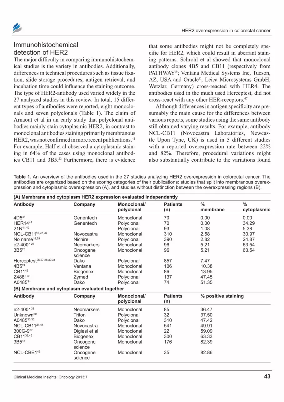
Table 1. An overview of the antibodies used in the 27 studies analyzing HeR2 overexpression in colorectal cancer. The antibodies are organized based on the scoring categories of their publications: studies that split into membranous overex-pression and cytoplasmic overexpression (A), and studies without distinction between the overexpressing regions (B).
(A) Membrane and cytoplasm HeR2 expression evaluated independentlyAntibody company Monoclonal/
polyclonalpatients (n)
% membrane
% cytoplasmic
4D541 Genentech Monoclonal 70 0.00 0.00HeR1441 Genentech Polyclonal 70 0.00 34.2921N41,42 Polyclonal 93 1.08 5.38NCL-CB1116,22,26 Novocastra Monoclonal 310 2.58 30.97No name18,29 Nichirei Polyclonal 390 2.82 24.87e2-400123 Neomarkers Monoclonal 96 5.21 63.543B523 Oncogene
scienceMonoclonal 96 5.21 63.54
Herceptest25,27,28,30,31 Dako Polyclonal 857 7.474B524 ventana Monoclonal 106 10.38CB1143 Biogenex Monoclonal 86 13.95Z488136 Zymed Polyclonal 137 47.45A048534 Dako Polyclonal 74 51.35(B) Membrane and cytoplasm evaluated togetherAntibody company Monoclonal/
polyclonalpatients (n)
% positive staining
e2-400138 Neomarkers Monoclonal 85 36.47Unknown20 Triton Polyclonal 32 37.50A048533,35 Dako Polyclonal 310 47.42NCL-CB1121,44 Novocastra Monoclonal 541 49.91300G-937 Digiesi et al Monoclonal 22 59.09CB1132,45 Biogenex Monoclonal 300 63.333B545 Oncogene
scienceMonoclonal 176 82.39
NCL-CBe146 Oncogene science
Monoclonal 35 82.86

Blok et al
44 Clinical Medicine Insights: Oncology 2013:7
in immunohistochemical HER2 studies. Incubation times of the antibodies in the various studies ranged from 60 minutes to overnight, and the antibody dilutions were between 1:20 and 1:250.16,21,22,26,44 However, these variations do not entirely explain the differences in HER2 staining as no consistent cor-relation is observed between incubation time and overexpression. Both Kountourakis et al and Sun et al report an overexpression of approximately 22% (cytoplasmic and membranous); Kountourakis et al used 1:250 dilution and 60 minutes incubation, while Sun et al used 1:40 with an overnight incubation.21,26 The two studies reported a highest overexpression of approximately 82%, incubation times of respectively 45 minutes and overnight respectively were used. In these studies, however, the comparability is ham-pered by the use of different antibodies.45,46 We per-formed a comparison of incubation times in our own laboratory, incubating paraffin-embedded colorectal tumor tissue using A0485, an anti-HER2 polyclonal antibody (DAKO Glostrup, Denmark) after antigen retrieval. As shown in Figure 1, an incubation time longer than the 30 minutes indicated in the manufac-turer’s protocol for breast cancer tissues gives rise to a stronger cytoplasmic staining pattern, allow-ing better comparison between different colorectal tumors. Because the absolute amount of stainable HER2 in colorectal cancers seems low when com-pared to breast cancer, it seems advisable for immu-nohistochemical analysis of HER2-overexpression
to adapt the protocols for breast cancer for use in col-orectal cancer.
Mirlacher et al showed a dramatic decrease in stain-ing intensity for HER2 in tissue microarrays (TMAs) between freshly cut and stored slides.39 We confirmed these results recently using our own TMA consisting of 500 colorectal cancer tissue samples obtained between 1991 and 2001. This TMA was produced using the same techniques as described earlier by our group for breast cancer.40 Slides were incubated with A0485. The operation date, and therefore the length of storage time, had a great impact particularly on cytoplasmic HER2 expression. In the group of patients operated on before 1996, 8.4% showed overexpression of cytoplas-mic HER2, while in the group of patients operated on in or after 1996, 42.7% expressed cytoplasmic over-expression. Tumors from patients operated on in 1998 or later showed the highest percentage of cytoplasmic staining (52%, manuscript in preparation). Therefore, it is likely that in studies having a long period of tissue collection the HER2 overexpression rate is underes-timated especially in the older samples, which could partly explain the variation between studies.
These data emphasize the importance of standard-ized staining procedures, as relatively insignificant procedure variations might lead to completely dif-ferent staining patterns and thereby different overex-pression rates.
scoring procedureA number of publications analyzed HER2- overexpression in colorectal tumors with genomic techniques like FISH, RT-PCR, Southern blotting, and Northern blotting. Kavanagh et al performed FISH on two 3+ cases and on nine 2+ cases. Both 3+ cases were confirmed to have gene amplification, but only one 2+ case was confirmed.24 Nathanson et al found approximately the same results, confirming three out of three 3+ cases and one out of two 2+ cases with both FISH and PCR.28 Ooi et al confirmed all 3+ and 2+ cases with FISH.29 Together these results confirmed that for colorectal cancers, the observations are similar for those found in breast cancer, ie, that 3+ overexpres-sion is conclusive for gene amplification, whereas 2+ staining is equivocal for gene amplification, and likely has other mechanisms of genetic overexpression.
When analyzing all accessible papers about HER2-overexpression on colon cancer tissue for their
1 hour
Overnight 2 hours
30 min
Figure 1. Paraffin-embedded colorectal cancer tissue was incubated with A0485 anti-HeR2 antibody with increasing incubation times of 30 minutes, 1 hour, 2 hours and overnight.note: with longer incubation times, an increase in cytoplasmic staining (brown) can be observed.

HeR2 overexpression in colorectal cancer
Clinical Medicine Insights: Oncology 2013:7 45
scoring methods, it was observed that most publica-tions follow the breast cancer protocol by examining only membranous overexpression,24,25,27–31,34,36,42,43 or split their scoring in membranous and cytoplasmic overexpression.16,18,22,23,26,41 In general, the mem-branous overexpression rates varied between 0% and 15%. Only Knösel et al and Park et al reported membranous overexpression of over 40%.34,36 However, Park et al only confirmed two out of 27 3+ cases with FISH, which puts their results in a dif-ferent perspective. Without these two outliers, the weighted average overexpression of membranous
HER2 was 5%. For cytoplasmic overexpression however, the rates varied between 0% and 66%, with a weighted average of 30% (Table 2).
A number of publications did not split their scores into membranous and cytoplasmic over-expression, but took them together to determine positivity.20,21,32,33,35,37,38,45,46 The observed overexpression rates varied between 22% and 83%, with a weighted average of 50% (Table 2). Unfortunately, no conclu-sions can be drawn about this percentage regarding the fraction of membranous or cytoplasmic overexpression. Based on the publications that used split results, it is
Table 2. An overview of all papers reporting about HeR2 overexpression in colorectal cancer. The papers are organized based on the scoring categories: split into membranous overexpression and cytoplasmic overexpression (A), or papers with-out distinguishing between the overexpressing regions (B). The weighted average was calculated with and without outliers. when one paper used more than one antibody, all antibodies were examined separately for the weighted average.
(A) Membraneous and cytoplasmic HeR2 overexpression evaluated separatelyAuthors Year Antibody patients (n) % membrane % cytoplasmKay et al16 1994 NCL-CB11 164 0 34Arnaout et al41 1992 4D5, 21N, HeR14 70 0 7–34Kim et al25 2004 Herceptest 185 1Osako et al18 1998 Nichirei 146 2 66Nathanson et al28 2003 Herceptest 169 3Ooi et al29 2004 Nichirei 244 3Schuell et al30 2006 Herceptest 77 4McCann et al42 1990 21N 23 4Half et al23 2004 e2-4001 and 3B5 96 5 64Ochs et al31 2004 Herceptest 109 6Kountourakis et al26 2006 NCL-CB11 106 6 17Gill et al22 2011 NCL-CB11 40 8 58Kavanagh et al24 2009 4B5 106 10Dursun et al43 2001 CB11 86 14Li et al27 2011 Herceptest 317 15Park et al36 2007 Z4881 137 47*Knosel et al34 2002 A0485 74 51*Total % positive membrane 9.22Total % positive membrane without outliers (*) 5.38Total % positive cytoplasm 30.58(B) Membrane and cytoplasm evaluated togetherAuthors Year Antibody patients (n) % positive stainingSun et al21 1995 NCL-CB11 293 23Demirbas et al32 2005 CB11 124 36Uner et al38 2005 e2-4001 85 36Kluftinger et al20 1992 Triton 32 38Kruszewski et al35 2010 A0485 202 47Jesus et al33 2005 A0485 108 48Porcelli et al37 2001 300G-9 22 59McKay et al44 2002 NCL-CB11 248 82Kapitanovic et al45 1997 3B5 and CB11 176 82Maurer et al46 1998 NCL-CBe1 35 83Total % positive staining 55.76

Blok et al
46 Clinical Medicine Insights: Oncology 2013:7
expected that the majority of overexpression in these publications is located in the cytoplasm, and only a minor fraction is overexpressed in the membrane.
Overall, it can be concluded that about 5% of col-orectal tumors have membranous overexpression of HER2, while the cytoplasmic overexpression varies strongly with an average around 30%. However, it is likely that this value is an underestimation due to the loss of HER2 antigen in older tissue samples as described earlier.
cytoplasmic HeR2The FDA-approved scoring system for breast cancer is based entirely on membranous HER-2 overexpression. Cytoplasmic overexpression in breast cancer occurs, but there are strict guidelines to ignore this.48,49 These guidelines are very specific as trastuzumab is only capable of binding the extracellular domain of membranous HER2. In other words, cytoplasmic HER2 would not have any consequence for clinical decisions about therapy. For years cytoplasmic overexpression of HER2 has been regarded as not relevant for breast cancer. Intracellular HER2 cannot be targeted by trastuzumab, it does not correlate with HER2 mRNA levels,51 it does not correlate to any clinical outcome,52 and it may even be a different protein than membranous HER2.
In contrast to breast cancer, there is evidence that in colorectal cancer cytoplasmic HER2 could in fact be associated with survival prognosis. Of 15 publi-cations that analyzed clinical prognostic parameters, six showed a worse patient survival with cytoplasmic tumor overexpression of HER2.16,18,22,32,34,45 Seven studies showed no correlation with any clinical parameter.20,21,26,33,35,44,46 Two other papers described a positive correlation between cytoplasmic overex-pression and well-differentiated tumors.23,38 Both publications hypothesize that enhanced cytoplasmic HER2 is an early marker in colorectal cancer, but decreases with progressing tumor stage and tumor dedifferentiation. This seems contradictory to the findings that patients with a HER2-cytoplasmic over-expressing tumor have a decreased survival rate as patients with progressed tumor (higher stage, dedif-ferentiation) would have less HER2 expression, but poorer prognosis. Moreover, one publication showed a worse differentiation and more lymph node metas-tases in cytoplasmic HER2-overexpressing tumors.32
When combining the distinction in scoring methods within those studies that report a clinical correlation, a remarkable pattern becomes clear. Of all seven studies that report a lack of clinical correlation, six of them do not split their scoring into membranous and cytoplasmic staining.20,21,33,35,44,46 In contrast, out of the six studies that report a clinical correlation, four of them do split their results in membranous and cyto-plasmic staining.16,18,22,34 Although this has never been confirmed in a large multi-center trial, these studies strongly suggest a clinical relevance of cytoplasmic HER2 overexpression in colorectal cancer.
Assuming that cytoplasmic HER2 has a prognostic role in colorectal cancer, it might also be involved in tumor pathogenesis like membranous HER2 in breast cancer. A plausible explanation would be that cyto-plasmic HER2 is forming homodimers, leading to an intracellular activation of the tyrosine kinase domain. Administration of trastuzumab will therefore not have any effect, as this antibody targets the extracel-lular domain only. Lapatinib, an intracellular tyrosine kinase inhibitor, has recently been approved for treatment of HER2-positive breast cancer patients.50 If cytoplasmic HER2 is indeed actively involved in colorectal carcinogenesis the administration of lapatinib, or other intracellular HER2-targeting com-pounds, could be a breakthrough in the treatment of especially this type of cancer.
Biology of cytoplasmic HeR2In breast cancer, it was suggested that cytoplasmic HER2 is a truncated or different protein, based on the fact that a 155 kD peptide was found, in contrast to the 185 kD HER2 found on the membrane. However, for colorectal cancer there have been ambiguous observations. Osako et al performed a western blot analysis on one membranous overexpressing speci-men, five membranous cytoplasmic specimens, and one negative sample. They observed 185 kD and 155 kD peptides in the membranous overexpressing specimen, but only 155 kD peptides in the cytoplas-mic overexpressing specimens.18 On the other hand, Kapitanovic et al performed a study in which they were able to split the cell lysate into a cytoplasmic and membranous fraction, and performed a western blot analysis with a different anti-HER2 antibody than Osako et al. They observed a 185 kD peptide in both fractions, and reported no 155 kD peptide. A western

HeR2 overexpression in colorectal cancer
Clinical Medicine Insights: Oncology 2013:7 47
blot using a P-Tyr antibody, detecting phosphorylated (activated) tyrosine kinase domains showed a 185 kD peptide in the intracellular fraction, indicating an intracellular activated HER2-receptor.45 This would suggest that the activated HER2 was internalized upon activation, or that HER2 was activated intracel-lularly eg, via homodimerization. Half et al analyzed the localization of cytoplasmic HER2 in colorectal cancer by electron microscopy after labeling with an anti-HER2 antibody and a secondary colloidal gold-labeled antibody. HER2 was located in the rough endoplasmatic reticulum (rER), consistent with newly synthesized peptides. It was also detected in cytosolic vacuoles, at the plasma membrane, and at desmosomes in the intercellular junctions. Particularly because of the localization at the rER, it is likely that this HER2 is derived from original peptide synthesis, and thus would be of genomic origin. Most likely, however, HER2 internalization and synthesis are occurring at the same time. Until these experiments are repeated using an antibody which has been validated to be solely specific for HER2, the origin of cytoplasmic HER2 will not be clarified.
Based on the hypothesis that cytoplasmic HER2 in colorectal cancer is actually newly synthesized rather than internalized, three different studies analyzed genomic amplification to determine the genomic background of HER2-overexpression. Half et al discovered membranous overexpression in only five out of 96 colorectal tumors (5%), while they determined cytoplasmic overexpression in 61 tumors (63%). Four out of five membranous HER2-overexpressing tumors showed amplification with FISH, while no cytoplasmic HER2-overexpressing tumors were confirmed with this technique. Moreover, RT-PCR was performed and showed a 12-fold higher mRNA expression in membranous overexpressing tumors compared to cytoplasmic overexpressing tumors.23 Pavlakis et al performed FISH-analysis on all cases described by Kountourakis et al. All 3+ cases of membranous overexpression were confirmed as being amplified, while only one of three 2+ cases was confirmed. No single cytoplasmic overexpressing tumor was confirmed.26,54 These results clearly indicate that membranous overexpression is associated with gene amplification, while cytoplasmic overexpression is due to other mechanisms. Maurer et al performed a northern blot and a Southern blot on the 29 cases
overexpressing HER2, without a distinction between membranous and cytoplasmic overexpression. Presumably this concerns cytoplasmic HER2 based on the high percentage of positive tumors (82%). They observed a 1.5 fold increase in mRNA via northern blot in tumors compared to healthy tissue, but the Southern blot did not show any gene amplification.46 Again, this indicates that cytoplasmic HER2 overexpression is based on mechanisms other than gene amplification.
For other types of cancer, it has been shown that overexpression of HER2 can also occur via mechanisms other than gene amplification, eg, via increased levels of promoter-binding proteins as was observed by Kameda et al in gastric cancer. He observed elevated levels of binding proteins to the TATA-box located in the promoter-region of the HER2-gene, which led to overexpression of HER2.55 Theoretically, mutations in downstream targets of HER2, for example KRAS, might also influence the expression of HER2 via affected feedback processes. These mechanisms could play a role in the observed cytoplasmic HER2 overexpression without any gene amplification. However, it is still unknown why HER2 does not migrate to the cell membrane, as it should being a transmembrane receptor. Whether it is a simple matter of quantities, and whether this tran-scriptional upregulation result in not enough HER2 to migrate to the cell membrane is unknown. It is con-troversial as the western blots from Kapitanovic et al and Osako et al exhibit a much denser signal when analyzing the cytoplasmic overexpressing cells, when compared to the membranous overexpress-ing cells.18,45 However, the 12-fold higher mRNA expression in membranous HER2 overexpressing tumors compared to cytoplasmic overexpression as discovered by Half et al would support this theory. Perhaps there is an unknown HER2-transporter pro-tein which is also regulated by the promoter-binding protein. Or perhaps the protein is in some way trun-cated when being overexpressed at a transcriptional level. Other possibilities include there being an actual 155 kD cleavage product incapable of migrat-ing to the membrane or an immune-evasive strategy by the tumor. One theory is based on the influence of the unfolded protein response (UPR). Upon cel-lular stress, this response initially stimulates multi-ple chaperone proteins in the endoplasmic reticulum

Blok et al
48 Clinical Medicine Insights: Oncology 2013:7
in order to increase the glycosylation and folding capacity of the endoplasmic reticulum, and induces apoptosis when this initial response is insufficient. UPR activation in breast cancer cells has been demonstrated.56 In normal, non-malignant cells there is a baseline expression of HER2, which leads to a HER2 on a cell membrane that cannot be detected by regular immunohistochemistry (Fig. 2A). A pos-sible theory is that in the case of overexpression of the HER2-gene, there is a surplus of HER2 mRNA which, after being translated into protein, cannot be handled properly in the ER and thus leads to aberrant glycosylation or misfolding of the immature protein (Fig. 2C). In this case, either the Golgi apparatus is not capable of maturing the protein or the protein
cannot be transported to the Golgi apparatus, both of which lead to accumulation of aberrant HER2 within the cytoplasm. This accumulation would trigger the UPR. The presence of an activated UPR will lead to an increase in properly glycosylated and folded protein, and the overexpression of HER2 by the cell at the membrane. However, for an unknown reason, gene amplification of HER2 in breast cancer leads to a more efficient UPR than HER2-overexpression in colorectal cancer (Fig. 2B). This difference in UPR could be influenced by other processes which affect cellular stress such as hypoxia, nutrient avail-ability, treatment, or other genetic instabilities. This may lead to the situation that UPR in breast cancer is efficient enough to process the surplus of immature
Normal cellA
Her2
GA
Nucleus
ERmRNA mRNA↑ ER
GA
Breast cancerB
Colorectal cancerC
mRNAER
Figure 2. A schematic representation of the difference in HeR2 expression between multiple cell types. In a normal cell (A), a single copy of the HeR2-gene is transcribed to low levels of mRNA, which is then translated to HeR2 which enters the endoplasmatic reticulum (eR). In the eR, HeR2 is processed (eg, glycosylated), and transported to the Golgi apparatus (GA). There it undergoes final processing before being transported to the membrane. In breast cancer (B), this process is basically identical except that gene amplification leads to higher mRNA and HER2 levels. The processing capacity of the ER is improved, most likely due to the unfolded protein response (UPR). In colorectal cancer however (c), there is no gene amplification, but gene expression is upregulated during transcription.notes: The eR cannot keep up with the high amount of protein, therefore immature HeR2 cannot to be completely processed in the eR, and can-not be transported to the GA and the cell membrane; instead incompletely folded HeR2 might be extracted from the eR via the eR associated deg-radation pathway, deglycosylated into a 155–160 kDa protein prior to it accumulation and ultimately proteasomal degradation in the cytoplasm.58

HeR2 overexpression in colorectal cancer
Clinical Medicine Insights: Oncology 2013:7 49
HER2, while in colorectal cancer the UPR fails to do so which leads to accumulation of misfolded and aberrantly glycosylated HER2. This unglycosylated form of HER2 might be identical to the p155 variant mentioned before.
conclusionsIt is clear that most of the debate around HER2-overexpression in colorectal cancer is due to differ-ences in staining/scoring techniques. When splitting the results in membranous and cytoplasmic overex-pression, a clear pattern appears with approximately 5% of all patients having membranous overexpres-sion, and about 30% of all patients having cytoplasmic overexpression. There is a strong correlation between genomic amplification and membranous overexpres-sion, whereas no genomic amplification is observed in cases with cytoplasmic HER2 expression.23,26,54
With around 5% of all colorectal cancers overex-pressing HER2 on their membrane, this would create a new treatment strategy with trastuzumab for approxi-mately 60,000 patients worldwide per year. Unfortu-nately, a clinical trial was ended due to a lack of patient accrual.57 A large, multicenter trial would be needed to include enough patients for sufficient analysis.
In contrast to membranous HER2 overexpres-sion, a significant proportion of colorectal tumors (30%–50%) show cytoplasmic HER2 overexpres-sion in most studies. The identity of cytoplasmic HER2 remains unclear, although there is some indi-rect evidence that it is derived from the upregulation of promoter-binding proteins leading to an increase in HER2 production. Additionally, the prognostic value of cytoplasmic HER2 is still unclear. An exten-sive study using standardized techniques on a large number of tumors is needed to elucidate this matter. If cytoplasmic HER2 has a pathophysiological role in colorectal cancer, intracellular HER2-targeting compounds, for example lapatinib, might be a new treatment option for the 30% of patients having cyto-plasmic HER2 overexpression. This would impact about 360,000 patients per year; a clear breakthrough in the treatment of colorectal cancer.
Author contributionsConceived and designed the experiments: EB, CS. Analysed the data: EB. Wrote the first draft of the manuscript: EB. Contributed to the writing of the
manuscript: EB, PK, CS. Agree with manuscript results and conclusions: EB, PK, JvL, CS. Jointly developed the structure and arguments for the paper: EB, PK, JvL, CS. Made critical revisions and approved final version: EB, PK, JvL, CS. All authors reviewed and approved of the final manuscript.
FundingThis research was performed within the framework of CTMM, the Center for Translational Molecular Medicine, project MUSIS (grant 03O-202).
competing InterestsAuthor(s) disclose no potential conflicts of interest.
Disclosures and ethicsAs a requirement of publication author(s) have pro-vided to the publisher signed confirmation of com-pliance with legal and ethical obligations including but not limited to the following: authorship and contributorship, conflicts of interest, privacy and confidentiality and (where applicable) protection of human and animal research subjects. The authors have read and confirmed their agreement with the ICMJE authorship and conflict of interest criteria. The authors have also confirmed that this article is unique and not under consideration or published in any other publication, and that they have permission from rights holders to reproduce any copyrighted material. Any disclosures are made in this section. The external blind peer reviewers report no conflicts of interest.
References1. Ferlay J, Shin H, Bray F, Forman D, Mathers C, Parkin D. GLOBOCAN
2008 v1.2, Cancer incidence and mortality worldwide. Available at: http://globocan.iarc.fr. Accessed Jun 12, 2012.
2. Winawer SJ. The multidisciplinary management of gastrointestinal cancer. colorectal cancer screening. Best Pract Res Clin Gastroenterol. 2007; 2(6)1:1031–48.
3. André T, Boni C, Mounedji-Boudiaf L, et al. Oxaliplatin, fluorouracil, and leucovorin as adjuvant treatment for colon cancer. N Engl J Med. 2004; 350(23):2343–51.
4. Gutierrez C, Schiff R. HER2: Biology, detection, and clinical implications. Arch Pathol Lab Med. 2011;135(1):55–62.
5. Slamon DJ, Godolphin W, Jones LA, et al. Studies of the HER-2/neu proto-oncogene in human breast and ovarian cancer. Science. 1989;244(4905): 707–12.
6. Slamon DJ, Clark GM, Wong SG, Levin WJ, Ullrich A, McGuire WL. Human breast cancer: correlation of relapse and survival with amplification of the HER-2/neu oncogene. Science. 1987;235(4785):177–82.
7. Slamon DJ, Leyland-Jones B, Shak S, et al. Use of chemotherapy plus a monoclonal antibody against HER2 for metastatic breast cancer that overex-presses HER2. N Engl J Med. 2001;344(11):783–92.

Blok et al
50 Clinical Medicine Insights: Oncology 2013:7
8. Smith I, Procter M, Gelber RD, et al. 2-year follow-up of trastuzumab after adjuvant chemotherapy in HER2-positive breast cancer: a randomised con-trolled trial. Lancet. 2006;369(9555):29–36.
9. Liu YH, Xu FP, Rao JY, et al. Justification of the change from 10% to 30% for the immunohistochemical her2 scoring criterion in breast cancer. Am J Clin Pathol. 2009;132(1):74–9.
10. Rhodes A, Jasani B, Anderson E, Dodson AR, Balaton AJ. Evaluation of HER-2/neu immunohistochemical assay sensitivity and scoring on formalin-fixed and paraffin-processed cell lines and breast tumors: a com-parative study involving results from laboratories in 21 countries. Am J Clin Pathol. 2002;118(3):408–17.
11. Seidman AD, Fornier MN, Esteva FJ, et al. Weekly trastuzumab and paclitaxel therapy for metastatic breast cancer with analysis of efficacy by HER2 immunophenotype and gene amplification. J Clin Oncol. 2001; 19(20):2587–95.
12. Gouvea AP, Milanezi F, Olson SJ, Leitao D, Schmitt FC, Gobbi H. Selecting antibodies to detect HER2 overexpression by immunohistochemistry in invasive mammary carcinomas. Appl Immunohistochem Mol Morphol. 2006;14(1):103–8.
13. Press MF, Sauter G, Bernstein L, et al. Diagnostic evaluation of HER-2 as a molecular target: an assessment of accuracy and reproducibility of labora-tory testing in large, prospective, randomized clinical trials. Clin Cancer Res. 2005;11(18):6598–607.
14. Bang YJ, Van Cutsem E, Feyereislova A, et al. Trastuzumab in combination with chemotherapy versus chemotherapy alone for treatment of HER2-positive advanced gastric or gastro-oesophageal junction cancer (ToGA): a phase 3, open-label, randomised controlled trial. Lancet. 1928;376(9742):687–97.
15. Ross JS. The HER-2/neu oncogene in tumors of the gastrointestinal tract. Cancer Invest. 2001;19(5):554–68.
16. Kay EW, Mulcahy H, Walsh CB, et al. Cytoplasmic c-erbB-2 protein expression correlates with survival in Dukes’ B colorectal carcinoma. Histopathology. 1994;25(5):455–61.
17. Lazaris AC, Theodoropoulos GE, Anastassopoulos P, et al. Prognostic sig-nificance of p53 and c-erbB-2 immunohistochemical evaluation in colorec-tal adenocarcinoma. Histol Histopathol. 1995;10(3):661–8.
18. Osako T, Miyahara M, Uchino S, Inomata M, Kitano S, Kobayashi M. Immunohistochemical study of c-erbB-2 protein in colorectal cancer and the correlation with patient survival. Oncology. 1998;55(6):548–55.
19. Berney CR, Fisher RJ, Yang J, Russell PJ, Crowe PJ. Protein markers in colorectal cancer: predictors of liver metastasis. Ann Surg. 1999;230(2): 179–84.
20. Kluftinger AM, Robinson BW, Quenville NF, Finley RJ, Davis NL. Correlation of epidermal growth factor receptor and c-erbB2 oncogene product to known prognostic indicators of colorectal cancer. Surg Oncol. 1992;1(1):97–105.
21. Sun XF, Carstensen JM, Stål O, Zhang H, Nordenskjöld B. c-erbB-2 oncopro-tein in relation to DNA ploidy and prognosis in colorectal adenocarcinoma. APMIS. 1995;103(4):309–15.
22. Gill MK, Manjari M, Jain K, et al. Expression of Her-2/neu in colon carci-noma and its correlation with the histological grades and the lymph nodes status. JCDR. 2011;5:1564–8.
23. Half E, Broaddus R, Danenberg KD, Danenberg PV, Ayers GD, Sinicrope FA. HER-2 receptor expression, localization, and activation in colorectal cancer cell lines and human tumors. Int J Cancer. 2004;108:540–8.
24. Kavanagh D, Chambers G, O’ Grady L, et al. Is overexpression of HER-2 a predictor of prognosis in colorectal cancer? BMC Cancer. 2009;9:1.
25. Kim JY, Lim SJ, Park K. Cyclooxygenase-2 and c-erbB-2 expression in col-orectal carcinoma assessed using tissue microarrays. Appl Immunohistochem Mol Morphol. 2004;12(1):67–70.
26. Kountourakis P, Pavlakis K, Psyrri A, et al. Clinicopathologic signifi-cance of EGFR and Her-2/neu in colorectal adenocarcinomas. Cancer J. 2006;12(3):229–36.
27. Li Q, Wang D, Li J, Chen P. Clinicopathological and prognostic significance of HER-2/neu and VEGF expression in colon carcinomas. BMC Cancer. 2011;11:277.
28. Nathanson DR, Culliford AT, Shia J, et al. HER 2/neu expression and gene amplification in colon cancer. Int J Cancer. 2003;105(6):796–802.
29. Ooi A, Takehana T, Li X, et al. Protein overexpression and gene amplifica-tion of HER-2 and EGFR in colorectal cancers: an immunohistochemical and fluorescent in situ hybridization study. Mod Pathol. 2004;17(8):895–904.
30. Schuell B, Gruenberger T, Scheithauer W, Zielinski CH, Wrba F. HER 2/neu protein expression in colorectal cancer. BMC Cancer. 2006;6:123.
31. Ochs AM, Wong L, Kakani V, et al. Expression of vascular endothelial growth factor and HER2/neu in stage II colon cancer and correlation with survival. Clin Colorectal Cancer. 2004;4(4):262–7.
32. Demirbas S, Sucullu I, Yildirim S, Celenk T. Influence of the c-erb B-2, nm23, bcl-2 and p53 protein markers on colorectal cancer. Turk J Gastroenterol. 2006;17(1):13–9.
33. Jesus EC, Matos D, Artigiani R, Waitzberg AF, Goldenberg A, Saad SS. Assessment of staging, prognosis and mortality of colorectal cancer by tumor markers: receptor erbB-2 and cadherins. Acta Cir Bras. 2005;20(6): 422–7.
34. Knösel T, Yu Y, Stein U, et al. Overexpression of c-erbB-2 protein corre-lates with chromosomal gain at the c-erbB-2 locus and patient survival in advanced colorectal carcinomas. Clin Exp Metastasis. 2002;19(5):401–7.
35. Kruszewski WJ, Rzepko R, Ciesielski M, et al. Expression of HER2 in colorectal cancer does not correlate with prognosis. Dis Markers. 2010; 29(5):207–12.
36. Park DI, Kang MS, Oh SJ, et al. HER-2/neu overexpression is an inde-pendent prognostic factor in colorectal cancer. Int J Colorectal Dis. 2007;22(5):491–7.
37. Porcelli B, Frosi B, Terzuoli L, et al. Expression of P185 and P53 in benign and malignant colorectal lesions. Histochem J. 2001;33(1):51–7.
38. Uner A, Ebinc FA, Akyurek N, Unsal D, Mentes BB, Dursun A. Vascular endothelial growth factor, c-erbB-2 and c-erbB-3 expression in colorectal adenoma and adenocarcinoma. Exp Oncol. 2005;27(3):225–8.
39. Mirlacher M, Kasper M, Storz M, et al. Influence of slide aging on results of translational research studies using immunohistochemistry. Mod Pathol. 2004;17(11):1414–20.
40. van Nes J, de Kruijf E, Faratian D, et al. COX2 expression in prognosis and in prediction to endocrine therapy in early breast cancer patients. Breast Cancer Res Treat. 2011;125(3):671–85.
41. Arnaout AH, Dawson PM, Soomro S, et al. HER2 (c-erbB-2) oncoprotein expression in colorectal adenocarcinoma: an immunohistological study using three different antibodies. J Clin Pathol. 1992;45(8):726–7.
42. McCann A, Dervan PA, Johnston PA, Gullick WJ, Carney DN. c-erbB-2 onco-protein expression in primary human tumors. Cancer. 1990;65(1):88–92.
43. Dursun A, Poyraz A, Suer O, Sezer C, Akyol G. Expression of Bcl-2 and c-ErbB-2 in colorectal neoplasia. Pathol Oncol Res. 2001;7(1):24–7.
44. McKay JA, Loane JF, Ross VG, et al. c-erbB-2 is not a major factor in the development of colorectal cancer. Br J Cancer. 2002;86(4):568–73.
45. Kapitanovic S, Radosevic S, Kapitanovic M, et al. The expression of p185(HER-2/neu) correlates with the stage of disease and survival in col-orectal cancer. Gastroenterology. 1997;112(4):1103–13.
46. Maurer CA, Friess H, Kretschmann B, et al. Increased expression of erbB3 in colorectal cancer is associated with concomitant increase in the level of erbB2. Hum Pathol. 1998;29(8):771–7.
47. Schrohl AS, Pedersen HC, Jensen SS, Nielsen SL, Brünner N. Human epidermal growth factor receptor 2 (HER2) immunoreactivity: specific-ity of three pharmacodiagnostic antibodies. Histopathology. 2011;59(5): 975–83.
48. Press MF, Hung G, Godolphin W, Slamon DJ. Sensitivity of HER-2/neu antibodies in archival tissue samples: potential source of error in immuno-histochemical studies of oncogene expression. Cancer Res. 1994;54(10): 2771–7.
49. Walker RA, Bartlett JMS, Dowsett M, et al. HER2 testing in the UK: further update to recommendations. J Clin Pathol. 2008;61(7):818–24.
50. Geyer CE, Forster J, Lindquist D, et al. Lapatinib plus capecitabine for HER2-positive advanced breast cancer. N Engl J Med. 2006;355(26):2733–43.
51. Taylor SL, Platt-Higgins A, Rudland PS, Winstanley JH, Barraclough R. Cytoplasmic staining of c-erbB-2 is not associated with the presence of detect-able c-erbB-2 mRNA in breast cancer specimens. Int J Cancer. 1998;76(4): 459–63.

HeR2 overexpression in colorectal cancer
Clinical Medicine Insights: Oncology 2013:7 51
52. Têtu B, Brisson J. Prognostic significance of HER-2/neu oncoprotein expression in node-positive breast cancer. The influence of the pattern of immunostaining and adjuvant therapy. Cancer. 1994;73(9):2359–65.
53. de Potter CR, van Daele S, van de Vijver MJ, et al. The expression of the neu oncogene product in breast lesions and in normal fetal and adult human tissues. Histopathology. 1989;15(4):351–62.
54. Pavlakis K, Kountourakis P, Stathopoulos E, et al. Her-2 protein expression, cellular localization, and gene amplification in colorectal carcinoma. Appl Immunohistochem Mol Morphol. 2007;15(4):441–5.
55. Kameda T, Yasui W, Yoshida K, et al. Expression of ERBB2 in human gas-tric carcinomas: relationship between p185erbb2 expression and the gene amplification. Cancer Res. 1990;50(24):8002–9.
56. Scriven P, Coulson S, Haines R, Balasubramanian S, Cross S, Wyld L. Activation and clinical significance of the unfolded protein response in breast cancer. Br J Cancer. 2009;101(10):1692–8.
57. Ramanathan RK, Hwang JJ, Zamboni WC, et al. Low overexpression of HER-2/Neu in advanced colorectal cancer limits the usefulness of trastu-zumab (herceptin) and irinotecan as therapy. A Phase II Trial. Cancer Invest. 2004;22(6):858–65.
58. Meijer IMJ, van Leeuwen JEM. ERBB2 is a target for USP8-mediated deubiquitination. Cell Signal. 2011;23(2):458–67.


