Embed Size (px)
Citation preview
Inhibition of Protein-Protein Interaction of HER2-EGFR andHER2–HER3 by a Rationally Designed Peptidomimetic
Sashikanth Banappagari1, Miriam Corti2, Seth Pincus2, and SeetharamaSatyanarayanajois*,1
1Department of Basic Pharmaceutical Sciences, College of Pharmacy, University of Louisiana atMonroe, Monroe, LA 712012Research Institute for Children, Children's Hospital, 200 Henry Clay Avenue, New Orleans, LA70118
AbstractProtein-protein interactions (PPI) play a crucial role in many biological processes, and modulationof PPI using small molecules to target hot spots has therapeutic value. As a model system we willuse PPI of human epidermal growth factor receptors (EGFRs). Among the four EGFRs, EGFR-HER2 and HER2–HER3 are well known in cancer. We have designed a small molecule that istargeted to modulate HER2-mediated signaling. Our approach is novel because the small moleculedesigned disrupts dimerization not only of EGFR-HER2 but also of HER2–HER3. In the presentstudy we have shown, using surface plasmon resonance (SPR) analysis, that a peptidomimetic,compound 5, binds specifically to HER2 protein extracellular domain and disrupts thedimerization of EGFRs. To evaluate the effect of compound 5 on HER2 signaling in vitro,Western blot and PathHunter assays were used. Results indicated that compound 5 inhibits thephosphorylation of HER2 kinase domain and inhibits the heterodimerization in a dose-dependentmanner. Molecular modeling methods were used to model the PPI of HER2–HER3 heterodimer.
KeywordsEGFR; HER2; Peptidomimetic; Homology modeling; Surface plasmon resonance; Protein-proteininteractions; PathHunter assay
IntroductionProtein-protein interactions (PPI) play a crucial role in many biological processes, includingtransmembrane signal transduction, cell regulation, and the immune response (Wells &McClenden, 2007; Reichmann et al., 2007). It has been established that human diseases canbe caused by aberrant protein-protein interactions (Ryan & Mathews, 2005), either by a lossof interactions or through the formation of complexes by overexpression of proteins on thecell surface. Understanding the structural details and modulation of protein-protein
*Address Correspondence to, Seetharama D. Satyanarayanajois, Assistant Professor, Department of Basic Pharmaceutical Sciences,University of Louisiana at Monroe, 1800 Bienville Drive, Monroe LA 71201 USA, Tel: (318)-342-1993; Fax: (318)-342-1737,[email protected].
Supplementary MaterialSupplementary materail consisting of HPLC, MS, and purity data along with SPR immobilization data, and control compound datacan be obtained free of charge from the authors.
Supporting Information Available: HPLC, MS, and purity data along with SPR immobilization data, control compound data. Thesecan be obtained free of charge from the authors or can be purchased from Adenine Press for _____
NIH Public AccessAuthor ManuscriptJ Biomol Struct Dyn. Author manuscript; available in PMC 2013 June 26.
Published in final edited form as:J Biomol Struct Dyn. 2012 ; 30(5): 594–606. doi:10.1080/07391102.2012.687525.
NIH
-PA Author Manuscript
NIH
-PA Author Manuscript
NIH
-PA Author Manuscript
interactions has significance in human health (Fuller, Burgoyne & Jackson, 2009;Sperandio, Reynes, Camproux, & Villoutriex, 2010). PPI have a wide range of affinities andpossess a high degree of specificity for carrying out cellular processes. Analysis of 3Dstructures of protein complexes has suggested that the PPI surfaces are large, covering 700–3000 Å2, and the binding surfaces are relatively flat (Bahadur, Chakrabarti, Rodier & Janin,2004). Detailed studies of PPI interaction surfaces and the amino acid residues by severalresearchers have indicated that there are “hot spots” on the protein-protein interface areawhere major contributions to binding free energy are provided. These hot spots have coreregions consisting mostly of aromatic amino acid residues such as Tyr, Trp, and Phe(Bahadur, Chakrabarti, Rodier & Janin, 2004; Moreira, Fernandes & Ramos, 2007; Chene,2006). All of these studies indicate that PPI are concentrated in crucial hot spots, which arehydrophobic in nature and small enough to accommodate only a small molecule. Modulationof PPI using small molecules to target hot spots has therapeutic value (Wells & McClenden,2007; Khan et al., 2011; Drozdov-Tikhomirov et al., 2001; Liang et al., 2000). Our interestis to design peptidomimetics to modulate the protein-protein interactions. As a modelsystem, we will use PPI of human epidermal growth factor receptors (EGFRs).
The human epidermal growth factor receptor (EGFRs/HERs) system of receptor tyrosinekinases plays an important role in cell growth and differentiation (Ferguson, 2008). Thereceptor system consists of four members: HER1 or EGFR and HER2–4. These receptorsalso play a key role in tumor progression. These proteins have an extracellular domain(ECD) (Burgess et al., 2003), a transmembrane helix, a cytoplasmic kinase domain, and aregulatory region. Ligand binding to EGFR or HER3 leads to heterodimerization of theseproteins, which leads to cell signaling. Different possible dimers such as EGFR-HER2,HER2–HER3, and HER2– HER4 have been proposed in the literature (Shankaran, Wiley &Resat, 2006; Tao & Maruyama, 2008). Among these, EGFR-HER2 and HER2–HER3 arewell known in cancer. HER2 overexpression and its dimerization leads to an aggressiveform of breast cancer (Landgraf, 2007; Nahta & Esteva, 2003; Hynes & Lane, 2005). It hasbeen reported that, apart from the EGFR-HER2 dimer, HER2–HER3 dimers are the majorcontributors to HER2-overexpressing breast cancer (Baselga & Swain, 2009; Lee-Hoeflichet al., 2008). Hence, the protein-protein interactions between HERs are a good model systemfor our study. The structure and functional properties of EGFR have been studied in depth,including the structural details of the homodimer of the ECD domain of EGFR (Ogiso et al.,2002). However, heterodimers of HERs have not been studied in atomic detail. Modelingthe PPI of HER2, HER3 and EGFR and understanding the hydrophobic hot spots in theseinteractions and PPI modulation by small molecules have potential clinical significance. Theinvolvement of extracellular domains of these proteins in dimerization and signaling hasbeen studied. However, at present, the crystal structure of heterodimers has not beenreported. Domains II and IV of ECD of EGFR and HER2 are known to be important in PPI(Berezov et al., 2002; Saxon & Lee, 1999), and domain II interactions have been studied inEGFR. Small molecules and antibodies have been targeted to HER2 (Lombardi et al., 2011;Huang et al., 2010; Chen, 2009; Arcangeli et al., 2008). The domain IV C-terminal is nearthe transmembrane domain, and the details of its interaction in dimerization have not beenelucidated. In the crystal structures of HER2, the electron density of the domain IV C-terminal is faded and not well defined. However, binding of the antibody trastuzumab todomain IV of HER2 is known to be clinically significant (Chang, 2007).
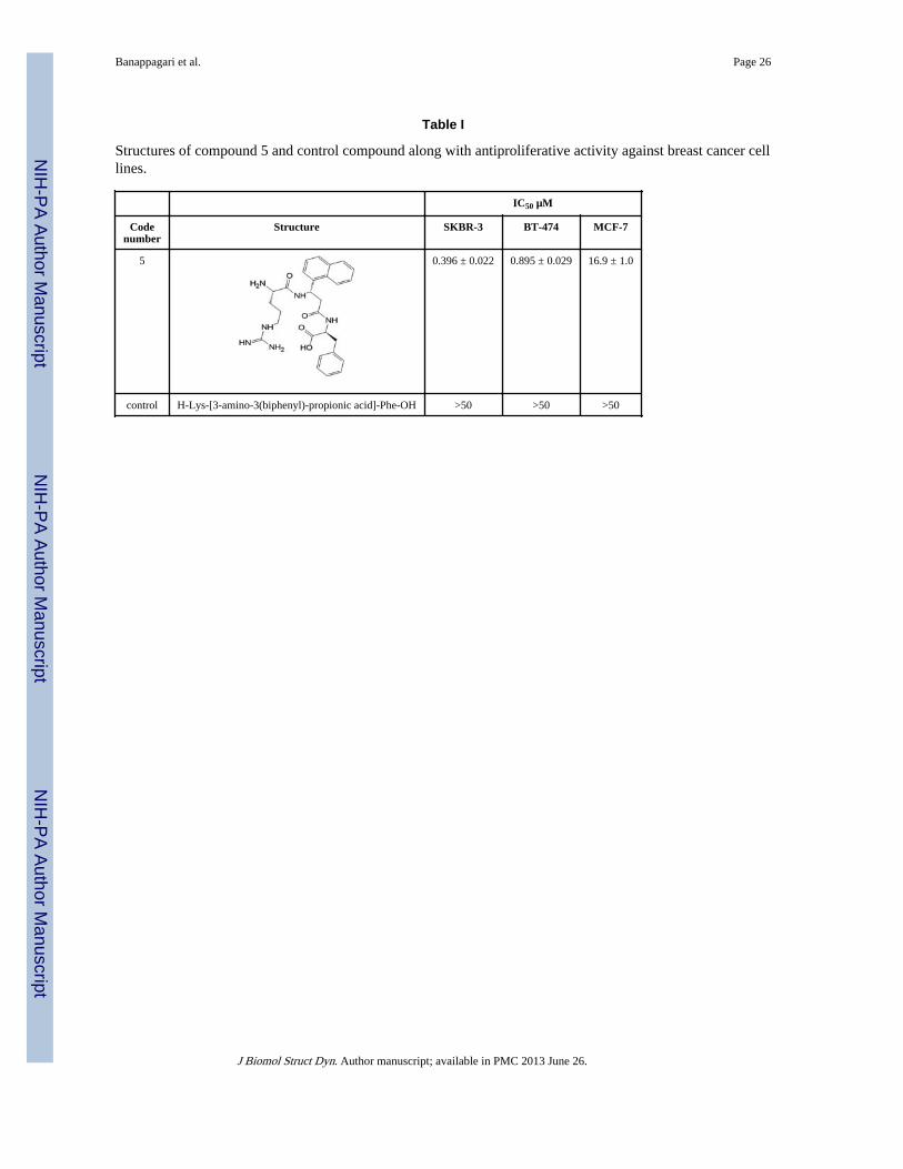
In our previous studies, we have reported the design of a small molecule, H-Arg-[3-amino-3(napthyl)-propionic acid]-Phe-OH (compound 5, Table I) (Satyanarayanajois,Villalba, Jianchao, & Lin, 2009; Banappagari, Satyanarayanajois & Ronald, 2010;Banappagari, Satyanarayanajois & Ronald, 2011) based on the protein-protein interaction inthe crystal structure of the HER2-antibody trastuzumab complex (Cho et al., 2003).Trastuzumab (anti-HER2) binds to HER2 on the C-terminal portion of domain IV. This
Banappagari et al. Page 2
J Biomol Struct Dyn. Author manuscript; available in PMC 2013 June 26.
NIH
-PA Author Manuscript
NIH
-PA Author Manuscript
NIH
-PA Author Manuscript
interaction is known to block proteolytic cleavage in HER2 and indirectly affectdimerization with other HERs that induce signaling pathways (Molina et al., 2001). Thus,blocking any interaction in domain IV on HER2 can disrupt signaling pathway(s) thatpromote cell proliferation and cancer (Franklin et al., 2004). The small molecule, compound5, showed antiproliferative activity against HER2-overexpressing breast cancer cell lineswith IC50 in the lower micromolar range (Satyanarayanajois, Villalba, Jianchao, & Lin,2009). Using fluorescence assay, competitive binding, and circular dichroism spectroscopy,we have shown that compound 5 binds to domain IV of the HER2 protein (Banappagari,Satyanarayanajois & Ronald, 2010). The interaction of domain IV of HER2 with EGFR orHER3 is not clear. Blocking domain IV of HER2 with antibodies is known to suppress thegrowth of cancer cells by indirectly inhibiting the dimerization mechanism of HER2 withother receptors. It is also known that the C-terminal portions of the domains interact withone another in EGFR and HER2, and that the absence of domain IV of HER2 changes theheteromeric signaling of EGFR (Kumagai et al., 2003). The aim of the present study is toshow that the designed peptidomimetic, compound 5, binds specifically to HER2 proteinand, in particular, to domain IV and disrupts the dimerization of EGFRs. We have used SPRanalysis, Western blot, and PathHunter assay to show that the compound 5 binds specificallyto HER2 domain IV and disrupts the protein-protein interactions of EGFR-HER2 as well asHER2–HER3. Molecular modeling methods were used to model the PPI of HER2–HER3heterodimer and propose the possible binding region of compound 5 to disrupt thedimerization process.
MethodsExtracellular domains of pure recombinant proteins HER2, EGFR and HER3 were obtainedfrom Bender MedSystems, eBioscience, San Diego, CA; Leinco Technologies, St. Louis,MO. Ligands for proteins EGFR and HER3, EGF and neuregulin 1 (NRG1) were obtainedfrom Abcam, Inc., (Cambridge, MA). Peptides were custom synthesized by New EnglandPeptides (Gardner, MA) or PolyPeptide Laboratories (San Diego, CA). The pure productswere analyzed by HPLC, electrospray ionization mass spectrometry (ESI-MS), and high-resolution mass spectrometry (HR-MS). HPLC chromatograms showed that the peptideswere > 95 % pure.
Surface Plasmon Resonance ExperimentsSurface plasmon resonance experiments were performed using Biacore 3000 (GE HealthSciences) at 25 °C (Komolov et al., 2010; Berezov, Zhang, Greene & Murali, 2001; Myszka,2000). Immobilization of compound 5, EGFR, HER2, HER3- extracellular domains anddomain IV-HER2 was done with the standard amine coupling procedure on a CM5 chip.Carboxyl groups on the sensor chip were activated by a solution containing 0.2 M N-ethyl-N-(dimethylaminopropyl) carbodiimide (EDC) and 0.05 M N-hydroxysuccinimide (NHS)(35 µL solution, flow rate 5 µL/min). Running buffer consisted of 0.01M HEPES, 0.15 MNaCl, 3 mM EDTA, 0.005% Tween at pH 7.5. Regeneration buffers were 50% acid cocktailand glycine 100 mM at pH 1.7. For immobilization, HER2 was injected onto the chipsurface at a rate of 10 µL/min until approximately 7000 response units (RU) was reached.Unreacted activated groups were blocked with 1 M ethanolamine for 7 min. A referencesurface was generated under the same conditions without compound 5 or HER2 proteininjection. The sensogram obtained was double referenced (reference surface was used asblank to correct for instrument and buffer artifacts and zero nM analyte buffer to avoid bulkshift). For binding, dimerization, and inhibition of dimerization studies, compound 5 orHER2, EGFR, HER3 were injected at various concentrations at a flow rate of 20 µL/min; forkinetics experiments (HER2-ECD was injected onto immobilized compound 5) HER2protein was injected at 30 µL/min for 2.5 min (association) and dissociated for 10 min. For
Banappagari et al. Page 3
J Biomol Struct Dyn. Author manuscript; available in PMC 2013 June 26.
NIH
-PA Author Manuscript
NIH
-PA Author Manuscript
NIH
-PA Author Manuscript
competitive studies, two analytes were injected sequentially using the coinject mode at aflow rate of 20 µL/min. A control peptidomimetic (an analog of compound 5, Table 1) thatdid not show antiproliferative activity in breast cancer cell lines was used to establishspecific binding of compound 5 (negative control).
Circular Dichroism ExperimentsCircular dichroism (CD) studies were carried out on a Jasco-J810 spectropolarimeterinstrument equipped with temperature control. The concentration of the proteins used was inthe range of 5 to 10 µM in deionized water. Spectra were collected from 360–190 nm usinga 1 mm path length rectangular quartz cell. Each spectrum was the average of three scanstaken at a scan rate of 50 nm/min with a spectral bandwidth of 0.1 nm. Calculation of theconcentration of protein was based on the fact that the human epidermal growth factorreceptors-ECD protein consists of 630 amino acid residues.
Western blot and phosphorylationBT474 and MCF7 cells were maintained in RPMI-1640 supplemented with penstrep 1 mL/100 mL, insulin 1 mg/100 mL, and 10% FBS. SKBR3 cells were maintained in McCoy’smedium supplemented with penstrep 1 mL/100 mL, insulin 1 mg/100 mL, and 10% FBS.After confluency, cells were trypsinized and triplicates were plated in T-75 flasks. After 24hrs the cells were treated with either AG-825 0.35 µM or compound 5 0.4 µM, or leftuntreated. Cells with compounds were incubated at 37 °C and 5% CO2. After 24 hrs, cellswere trypsinized, collected in 15 mL tubes, and centrifuged at 200 g. To the pellet, 200 µLof cell lysis buffer with 20 µL protease inhibitor and 20 µL phosphatase inhibitor wereadded. Tubes were vortexed for 1 min and then kept on ice for 10 min. This process isrepeated twice. Cells were subjected to centrifugation at high speed (450 g) and thesupernatant was collected into separate tubes and stored at −80 °C until further use. Proteinconcentration in the tubes was analyzed using Bradford’s assay. 40 µg of protein from eachsample was loaded on 10% SDS-polyacrylamide minigels. The gels were run at 30 mA, 125V and then transferred at 30 V for 12–16 h at 4 °C onto a nitrocellulose membrane. Aftertransfer, the membranes were blocked with 5% milk for 1 hr. The nitrocellulose membraneswere then probed with specific primary antibodies against p-HER2 diluted 1:3000 in 5%milk for 15 h at 4 °C. After incubation with primary antibody, the membranes were washedfive times with PBS and then incubated with secondary antibody to p-HER2 (anti-rabbitsecondary antibody) diluted 1:2000 in 5% milk with 0.001% Tween 20 for 1 h. Themembranes were then washed five times with PBS, and proteins were visualized using theSuper Signal enhanced chemiluminescence kit (Pierce, Rockford, IL). The Kodak GelLogic-1500 imaging system (Carestream Molecular Imaging, New Haven, CT) was used tovisualize the luminescent proteins. All experiments were repeated at least three times. Arepresentative Western blot image was used for final representation. The visualization of β-actin was used to ensure equal sample loading in each lane. Total HER2 protein wasdetected using an antibody to HER2 extracellular domain (Abcam).
PathHunter assayThe PathHunter assay kit was obtained from DiscoveRx Corporation (Fremont, CA). U2OScells expressing HER2–HER3 protein with C-terminal probe peptide were suspended in cellplating (CP) reagent and placed into wells of a 96-well plate at a density of 1 × 10⁴ cells perwell. After coating, the cells were incubated at 37 °C, 5% CO2 for 24 hours. Threefolddilutions of the agonist neuregulin 1(NRG1) were prepared from the stock of agonist in CPreagent. Cells were incubated with the agonist for 3 h. Then, 55 µL of detection reagent perwell was added and incubated at room temperature in the dark. After 60 min, luminescencefrom the cells was measured (Yin et al., 2009). EC80 concentration of the agonist was
Banappagari et al. Page 4
J Biomol Struct Dyn. Author manuscript; available in PMC 2013 June 26.
NIH
-PA Author Manuscript
NIH
-PA Author Manuscript
NIH
-PA Author Manuscript
measured from the dose-response curve of the agonist on PathHunter-expressed cells.Compound 5 (antagonist) and control (Table 1) dilutions were then prepared in CP reagentat threefold dilution and added to the remaining wells of the plate. The plate was incubatedfor 60 min at 37 °C. After 60 min, the EC80 dose of agonist (0.3 µM) was added to all thecells with compound 5 and control peptide. The plate was incubated for 3 hours at 37 °C.Then 55 µL of detection reagent per well was added and incubated for 60 min at roomtemperature. Luminescence from the plate was read using a BioTek microplate reader(BioTek, Winooski, VT). A plot of luminescence vs. concentration of agonist (NRG1) orantagonist (compound 5 and control peptide) was obtained. From the dose response curves,IC50 value of inhibition was obtained using GraphPad Prism software (GraphPad Software,Inc. La Jolla, CA).
ModelingThe crystal structure of the HER3 extracellular domain in closed conformation was obtainedfrom the Protein Data Bank (Cho & Leahy, 2002). Modeling of the HER3 extracellulardomain in open conformation was carried out using templates of HER2 crystal structure(Fisher et al., 2010) and EGFR extracellular domains (Ogiso et al., 2002; Lu et al., 2010)and domain IV of HER3 in closed conformation. SWISSMODEL was used to model thestarting structure (Arnold, Bordoli, Kopp & Schwede, 2010). The model was energyminimized using Insight II (Accelrys Inc. San Diego, CA) and evaluated for bond length,bond angle, and torsion angle using MolProbity (Chen et al., 2010). To generate the ligand-bound HER3 extracellular domain, EGFR extracellular domain in open conformation wasused (Ogiso et al., 2002). HER3 and EGFR extracellular domain structures were overlapped.The overlapped structures had EGF ligand positioned in the pocket between domains I andII of HER3. The EGF structure was replaced by heregulin structure, and the structure ofHER3 with heregulin-bound structure was minimized. The crystal structure of HER2extracellular domain in complex with trastuzumab was used for the heterodimer model ofthe HER2–HER3 complex (Cho et al., 2003). The crystal structure of HER2 in complexwith trastuzumab was missing residues 581–590, and 607–620 in domain IV. The crystalstructure of HER2 in complex with tryptophan-rich antibody has the structure of a loopregion covering residues 581–590 and 607–620 (Fisher et al., 2010). These missing residueswere added to the HER2 crystal structure from the trastuzumab complex to generate the full-length extracellular domain of HER2 protein. The generated model was evaluated for qualityby using Molprobity. The quality of the model was also evaluated by calculating solvent-accessible and solvent-excluded surface areas using autodock tools; the presence ofhydrophobic residues on the protein surface was determined by hydrophobic and hydrophilicsurface area calculations and representation using Maestro (Schrodinger LLC., Inc. Portland,OR).
To model the HER2–HER3 complex, the homodimer crystal structure of EGFR was used asa template (Ogiso et al., 2002). Structures of HER2 and HER3 over overlapped with EGFRdimer structure. Domains I to III of HER2 and HER3 were overlapped on the symmetricdimer of EGFR domains. In the overlapping position with the EGFR homodimer, HER2 andHER3 made contact with dimerization arm II and the C-terminal portion of domain IV. Themodels were compared with the recently reported crystal structure of the extracellulardomain of the homodimer of EGFR (Lu et al., 2010) and orientation of domain IV of twomolecules in the heterodimer was brought to close proximity using by overlapping with thehomodimer crystal structure.
Banappagari et al. Page 5
J Biomol Struct Dyn. Author manuscript; available in PMC 2013 June 26.
NIH
-PA Author Manuscript
NIH
-PA Author Manuscript
NIH
-PA Author Manuscript
ResultsSurface plasmon resonance analysis
Immobilization—In SPR experiments, one of the molecules (for example, HER2 protein,200 nM) is immobilized on the sensor surface and the analyte (EGFR or HER3, 200 nM)continuously flows over the surface of a microflow cell (Komolov et al., 2010; Berezov,Zhang, Greene & Murali, 2001; Myszka, 2000). The real-time interaction between the twoproteins is shown in a sensogram. For immobilization, HER2, EGFR, HER3, and domain IVof HER2 were injected onto the chip surface at a rate of 10 µL/min until response units ofapproximately 7000 RU were reached (immobilization of HER2, EGFR, HER3, and HER2domain IV are provided in Supporting Information). A stable surface of the immobilizedproteins 7500 RU for HER2-ECD, 9000 RU for HER3-ECD, 7000 RU for EGFR-ECD, and11,000 RU for domain IV-HER2 were obtained after injection.
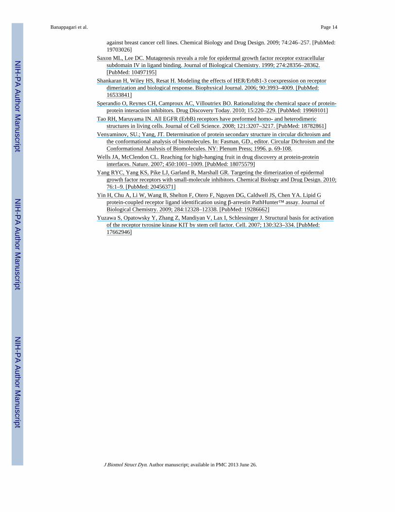
Compound 5 binds to HER2-ECD—The binding of compound 5 to HER2 ECD wasinvestigated by two methods: a) by immobilizing the HER2 ECD and using compound 5 asanalyte and b) by immobilizing compound 5 and using HER2 ECD as analyte. Curves weredouble referenced by subtracting the effects of control flow cell and 0.0 nM of analytebuffer control to avoid bulk shifts. When compound 5 was injected at differentconcentrations (0, 5, 10, 25, 50, 75, and 100 µM) into the flow-cell, concentration-dependentbinding to immobilized HER2-ECD was observed (Figure 1A).
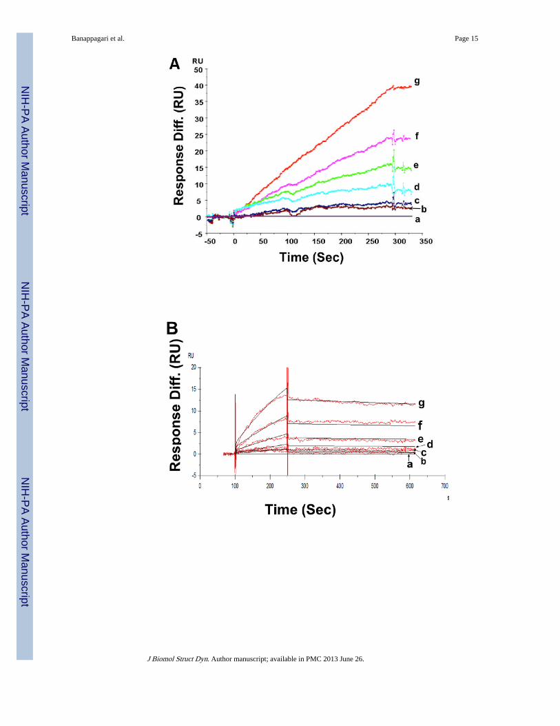
When compound 5 was immobilized on the CM5 chip surface and HER2 ECD was injectedat different concentrations (0, 5, 10, 20, 40, 80, and 160 µM) at a rate of 30 µL/min, bindingof HER2 ECD to compound 5 was observed (Figure 1B) exhibiting kinetics of associationand dissociation. The kinetic constant was estimated by global fitting analysis of the titrationcurve to the 1:1 Langmuir interaction model and the Kd value calculated was around 1.1 µM.The association rate constant was 2.1 × 10² M−1s−1 and the dissociation rate constant was2.31 × 10−4s.−1 As a reference, a control peptide was immobilized onto a CM5 chip, and aseries of concentrations of HER2-ECD was injected. No non-specific binding of the controlcompound to HER2 ECD was observed. Also, a peptidomimetic analog of compound 5(Table I) was also injected at a concentration of 100 µM onto immobilized HER2-ECD onflow cell 2. No binding was observed for control compound to HER2 ECD compared tocompound 5, indicating the specific binding of compound 5 to HER2 ECD (Figure 1C).
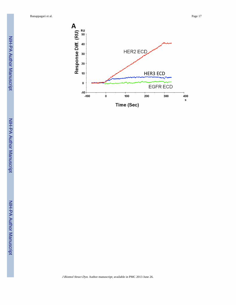
The next important experiment was to determine the specificity of compound 5 for HER2protein but not for the homologues receptors EGFR and HER3. To monitor the specificity,compound 5 was injected at a concentration of 100 µM onto immobilized HER2, EGFR, andHER3 on a CM5 sensor chip. Figure 2A shows the sensograms of flow cells 2, 3, and 4.Binding of compound 5 onto HER2-ECD was observed as shown by a 40 RU increase inresponse. However, no binding to EGFR and HER3 was observed, indicating that compound5 interacts specifically with HER2-ECD.
Compound 5 binds specifically to domain IV of HER2—HER2 extracellular domainIV is an important site for stabilizing the HER2-mediated dimers with the EGFR family ofreceptors such as EGFR and HER3. Also, domain IV is the binding site of the antibodytrastuzumab, which is used in treating breast cancer. Compound 5, a peptidomimetic, isdesigned to interact specifically with domain IV of HER2 protein. To demonstrate that thecompound 5 binds to domain IV of HER2, we immobilized domain IV-HER2 on a CM5sensor chip via an amine coupling process. A stable surface of approximately 11000 RU wasobtained in flow cell 2 of a CM5 sensor chip. Compound 5 was then injected onto domainIV-HER2 on a CM5 sensor chip at the rate of 10 µL/min to monitor the interaction of
Banappagari et al. Page 6
J Biomol Struct Dyn. Author manuscript; available in PMC 2013 June 26.
NIH
-PA Author Manuscript
NIH
-PA Author Manuscript
NIH
-PA Author Manuscript
compound 5. For comparison, trastuzumab, which is known to specifically interact withdomain IV of HER2, was also injected onto the CM5 sensor chip. Figures 2B and Crepresent the sensograms of compound 5 and trastuzumab binding to domain IV of HER2.The next important experiment was to evaluate whether compound 5 and trastuzumabcompete for the same binding site on domain IV-HER2. To monitor the competitive binding,trastuzumab and compound 5 were coinjected onto the immobilized domain IV-HER2. Inthe presence of trastuzumab, there was no interaction of compound 5 with HER2 domain IV(Supporting Information). This indicates that compound 5 may bind near the trastuzumabbinding site on domain IV-HER2.
Inhibition of HER2 mediated dimerization by compound 5—Dimerization ofEGFRs, in particular, EGFR-HER2 and HER2–HER3, plays an important role in activatingdownstream signaling (Ferguson, 2008; Burgess et al., 2003). The ultimate goal in designingthe compound 5 was to block HER2-mediated dimerization with the EGFR family ofreceptors EGFR and HER3 with HER2. To investigate the effect of compound 5 in blockingHER2-mediated dimerization process, HER2 was immobilized onto a CM5 chip and EGFRor HER3 was coinjected at 200 nM concentration with and without compound 5. EGFR andHER3 were pre-activated with EGF and NRG1 ligands by incubating at a concentration of20 nM for 15 min. Figures 3A and B represent the sensograms for coinjection experimentsof EGFR and HER3 with or without compound 5, respectively. Activated EGFR and HER3showed binding to HER2-ECD in the absence of compound 5, exhibiting heterodimerizationof the extracellular regions of the EGFR proteins. In the presence of compound 5,heterodimerization interaction was blocked. To validate the dimerization ability ofcommercially obtained protein extracellular domains (Bender MedSystems, eBioscience,San Diego, CA; Leinco Technologies, St. Louis, MO), proteins EGFR and HER3 werestudied using SPR in the absence of their ligands EGF and neuregulin (Abcam),respectively. EGFR and HER3 did not show any binding (no change in sensogram) toimmobilized HER2 in the absence of their ligands, suggesting an absence ofheterodimerization without the ligands. In the presence of ligands, EGFR and HER3exhibited changes in the sensograms (open conformation is needed for EGFR and HER3 todimerize with HER2) with kinetics of association and dissociation.
Circular DichroismCD experiments were performed to evaluate the secondary structure of the extracellulardomains of proteins HER2, EGFR, and HER3. Protein samples that were used in biacorestudies were commercially obtained of HER2, HER3, and EGFR. To verify that theseproteins have intact secondary structure, CD spectra of all the proteins were obtained inwater. CD spectra of proteins showed a negative band around 217 nm, suggesting β-sheetsecondary structure of the proteins (Venyaminov & Yang 1996) (Supporting Information).
Western blot analysis of phosphorylationTransphosphorylation of HER2-mediated dimers activate the downstream signaling cascadethat results in cell growth and proliferation (Ferguson, 2008; Burgess et al., 2003; Gijsen etal., 2010). Thus, inhibiting transphosphorylation of HER-mediated dimers plays a major rolein inhibiting cell growth and proliferation. SPR experiments provide information aboutdimerization of pure proteins. Furthermore, the proteins used had only ECD. To evaluate theeffect of compound 5 on the dimerization of full-length EGFRs in the native form in HER2-overexpressing breast cancer cell lines, the effect of compound 5 on the phosphorylation ofHER2 protein kinase domain in BT-474 and MCF-7 cells was investigated using Westernblot. If compound 5 inhibits the receptor heterodimerization/association in cells, then oneshould see a decrease in the phosphorylation of HER2 protein. Phosphorylation was detectedusing p-HER2, an antibody that specifically binds to phosphorylated HER2. Figures 4A&B
Banappagari et al. Page 7
J Biomol Struct Dyn. Author manuscript; available in PMC 2013 June 26.
NIH
-PA Author Manuscript
NIH
-PA Author Manuscript
NIH
-PA Author Manuscript
represents the Western blot analysis of the phosphorylation level in treated and untreatedBT-474 and MCF-7 cells respectively. Cells treated with compound 5 at 0.4 µMconcentration and AG-825 (a kinase inhibitor) (Gijsen et al., 2010; Levitzki & Gazit, 1995)showed a decreased level of p-HER2 compared to the level of p-HER2 in untreated cells.Comparison of the inhibition of phosphorylation of AG-825, a standard kinase inhibitor, andcontrol suggested that compound 5 showed significant inhibition of phosphorylation ofHER2. These results clearly suggest that compound 5 inhibits the full-length receptorassociation in the native form and, hence, phosphorylation of the kinase domain.
PathHunter assayThe PathHunter tyrosine kinase functional assay kit monitors dimerization and kinaseactivation of receptor tyrosine kinases (Yin et al., 2009). In the PathHunter assay(PathHunter technology is a registered trademark of DiscoveRx, Fremont, CA), The HER2–HER3 pair is expressed in U2OS cells by transfecting with cDNA for the EGFR proteinpairs. A small peptide epitope is expressed recombinantly on the intracellular C-terminalpart of HER2/HER3. An SH2 domain is co-expressed with a larger sequence, termedenzyme acceptor (EA), forming SH2-EA fusion protein. Activation of receptors results indimerization of receptor tyrosine kinase (HER2–HER3); the kinase domain isphosphorylated, and phosphorylated tyrosine acts as a recruitment site for downstreamsignaling molecules containing SH2 domains. The interaction of the C-terminal peptide andSH2-EA fusion protein forces complementation of the two enzyme fragments. This actionresults in the formation of fully complemented β-galactosidase, the activity of which ismeasured using a chemiluminescence detection reagent in the cellular environment(Kumagai et al., 2003). Addition of NRG1, a ligand for HER3, at different concentrationsinduces heterodimerization of HER2–HER3 and phosphorylation, which results in bindingof the SH2 domain protein and β-galactosidase activity. The activity of agonist NRG1 atdifferent concentration is shown in Figure 5. When compound 5 was incubated with cellsand NRG1 at a EC80 concentration (0.3 µM from the dose-response curve), the β-galactosidase activity was decreased, suggesting inhibition of dimerization of HER2–HER3in U2OS cells. The IC50 value of inhibition was 0.260 ± 0.002 µM for compound 5. Acontrol peptidomimetic that has structure similar to that of compound 5 was also evaluatedfor heterodimer inhibition activity. The control peptide did not exhibit any decrease in β-galactosidase activity, indicating the formation of heterodimers of HER2–HER3.
Modeling of heterodimersThe crystal structure of the homodimer of the ECD domain of EGFR has been reported.However, heterodimers of EGFRs have not been elucidated. It should be noted that modelsof homo- and –heterodimers of EGFRs proposed by different research groups is available inthe literature (Ferguson, 2008; Burgess et al., 2003; Shankaran, Wiley & Resat, 2006; Tao &Maruyama, 2008; Landgraf, 2007; Baselga & Swain, 2009; Ogiso et al., 2002). Our aim is tomodel the heterodimer of HER2–HER3 in the light of reported crystal structures andavailable data from the literature to explain the experimental results we have obtained.Based on SPR and Western blot analysis results, we proposed a model for EGFR-HER2,HER2–HER3 interactions and explored the importance of domain IV of HER2 in theheterodimerization and modulation of heterodimerization by compound 5. In an earlierreport we proposed a model for HER2-EGFR interaction (Banappagari, Satyanarayanajois &Ronald, 2010) that is consistent with experimentally observed results from the literature(Ferguson, 2008; Burgess et al., 2003). In the model of EGFR-HER2, the dimerization armsof domain II in both proteins interact with one another, stabilizing the dimer. In this report,we propose a model for the extracellular domain of HER2–HER3 interaction based onhomology modeling and using a template of EGFR homodimer structure. The structure ofopen conformation HER3 ECD is not available. Modeling of HER3 ECD was based on the
Banappagari et al. Page 8
J Biomol Struct Dyn. Author manuscript; available in PMC 2013 June 26.
NIH
-PA Author Manuscript
NIH
-PA Author Manuscript
NIH
-PA Author Manuscript
closed conformation of HER3 and the crystal structure of the open conformation of EGFRand HER2. EGFR, HER2 and HER3 share the sequence similarity (Burgess et al., 2003).The proposed model of ECD of HER3 in open conformation is shown in Figure 6. Theoverall conformation of HER3 ECD is similar to that of EGFR with domains I and IIIforming a cleft that accommodates the ligand (neuregulin/heregulin). Domain IV wasstretched away from domains I–III. The quality of the model was evaluated by Molprobity(Chen et al., 2010), which indicated that 87.5% of residues were in the favored region and97.8% of the residues were in the allowed region of the Ramachandran map. The quality ofthe model was also evaluated by calculating solvent-accessible and solvent-excluded surfaceareas; these areas were compared with those of the reported crystal structure of EGFR (Lu etal., 2010). The solvent-accessible surface area for HER3 ECD was 32,532 Å2, which wascomparable to that of the crystal structure EGFR ECD (32,094 Å2). The solvent-excludedsurface area for HER3 ECD was 27,188 (for the EGFR crystal structure 27,240 Å).Representation of the presence of hydrophobic residues on the surface of protein usinghydrophobic and hydrophilic surface area calculations (Maestro) suggested that the majorityof the surface area of the extracellular domain of HER3 is hydrophilic (SupportingInformation). Comparison of the crystal structure of EGFR ECD and the proposed model ofHER3 indicated that the packing of the core of the HER3 protein was similar to that ofEGFR (Lu et al., 2010).
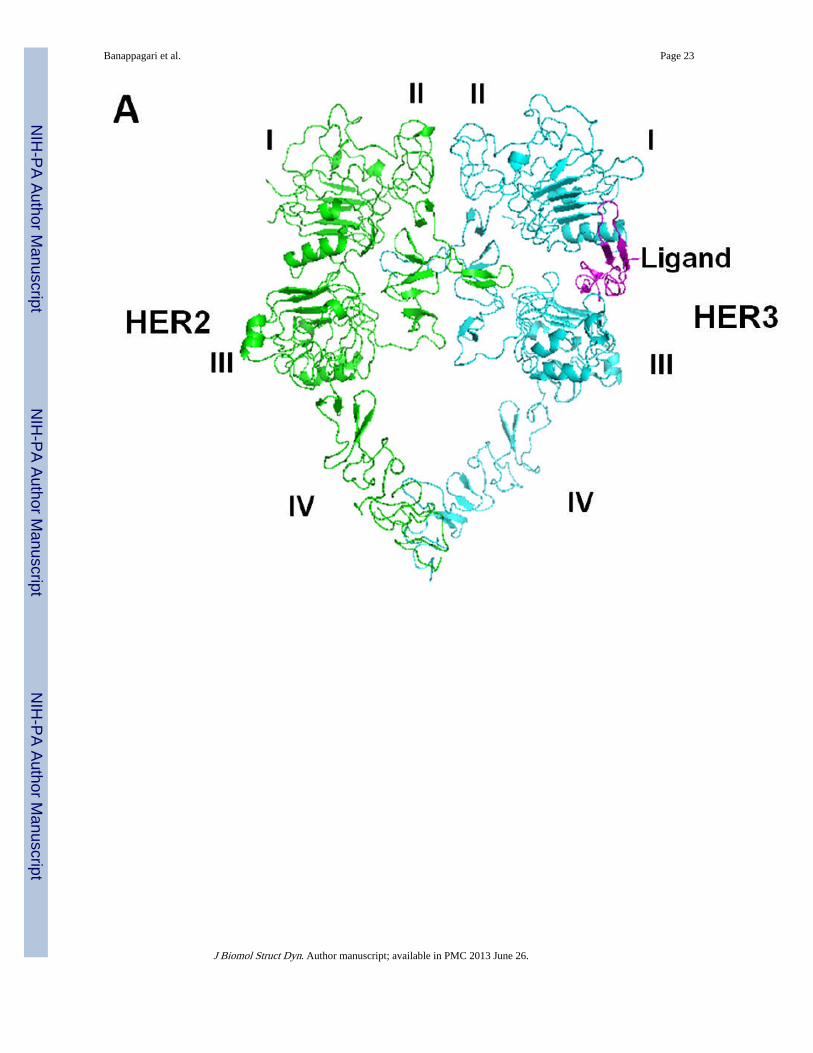
To model the heterodimer of HER2–HER3, the HER2 crystal structure from the trastuzumabantibody complex was used (Cho et al., 2003). However, the crystal structure did not havethe C-terminal part of domain IV well defined; hence, domain IV was modeled based on theHER2 crystal structure with a tryptophan-rich antibody (Fisher et al., 2010) and domain IVof EGFR in the homodimer of EGFR. Domain IV was added to the HER2 crystal structure.EGFR homodimers were used as templates to model HER2–HER3 heterodimers (Burgess etal., 2003). The resulting heterodimer model indicated that the C-terminal parts of domain IVof HER2 and HER3 interact with one another. The proposed heterodimer model for HER2–HER3 is shown in Figure 7A. Protein-protein interaction was observed in domain II anddomain IV of HER2 and HER3. The interaction of the dimerization arm of domain II wassimilar to that observed in the crystal structure of EGFR homodimer. Phe257 of HER2interacts with Tyr282 and Phe294 of HER3 on one side of the dimerization arm (Figure 7B).Phe268 and Leu291 of HER2 interact with Phe270 and Tyr265 of HER3 on the other side ofthe dimerization arm. There is also a hydrophobic interaction of Leu249 and Tyr267 ofHER2 with Gly318 of HER3, which stabilizes domain II of the heterodimer. In domain IV,Leu586 and Tyr588 of HER2 form hydrophobic interactions with Tyr601 and Pro599 ofHER3. There was a cation-Pi interaction between Lys583 of HER2 with Tyr601 of HER3. Ahydrogen-bonding interaction was observed between Asp585 of HER2 with Glu615,stabilizing the C-terminal portion of domain IV. When we compared the 3D structure of theEGFR homodimer with that of the HER2–HER3 heterodimer, the overall structures weresimilar (Lu et al., 2010). The difference was in the orientation of domains I and III, asexpected. HER2 does not have any known ligand and, hence, the ligand binding domains Iand II are close to each other. There was a difference in the structure of loops in the C-terminal part of domain IV.
DiscussionHER2-mediated heterodimerization plays a major role in cell growth and proliferation.Small molecules have been targeted to block the signaling by kinase domain inhibitor. Thereare few reports of small molecules or peptides targeting the extracellular domains of EGFRs(Berezov et al., 2002, Berezov, Zhang, Greene & Murali, 2001, Dakappagari et al., 2005;Allen et al., 2007; Yang et al., 2010). Among the extracellular domains, domain II is well-known in the dimerization process and is targeted for inhibition of dimerization in the case
Banappagari et al. Page 9
J Biomol Struct Dyn. Author manuscript; available in PMC 2013 June 26.
NIH
-PA Author Manuscript
NIH
-PA Author Manuscript
NIH
-PA Author Manuscript
of EGFR. The importance of domain IV of EGFRs is not very clear, although the antibodytrastuzumab is known to target domain IV of HER2 and has shown efficacy in treatment ofHER2-positive breast cancer patients (Huang et al., 2010; Arpino et al., 2007; Ahn & Vogel,2011). The mechanism of trastuzumab in blocking the signal for cell growth via domain IVis not clear, and trastuzumab does not block HER2–HER3 interaction. Our results clearlysuggest that a designed peptidomimetic binds to domain IV of HER2 protein and blocks theheterodimerization of EGFR-HER2 as well as HER2–HER3. SPR analysis clearly suggeststhat compound 5 binds specifically to domain IV and inhibits the heterodimerization processof EGFRs. Although SPR studies were conducted with pure proteins of extracellulardomains of EGFRs, our Western blot studies suggest that compound 5 inhibitsheterodimerization and phosphorylation in vitro in native cellular environments.Furthermore, PathHunter assay results suggested that compound 5 inhibits theheterodimerization of HER2–HER3 in a dose-dependent manner with an IC50 value of 0.260µM. This inhibition results in the inhibition of phosphorylation of the kinase domain. Tounderstand the molecular mechanism of blocking of signaling by binding of compound 5 todomain IV, molecular modeling studies were carried out. The heterodimer of HER2–HER3suggests that C-terminal parts of domain IV interact with one another apart from domain II.The C-terminal portion of domain IV is known to form weak interactions to stabilize theheterodimer of EGFRs. Modeling suggests that amino acid residues Phe573 to Trp592 ofdomain IV of HER2 may be involved in the interaction and stabilization ofheterodimerization. Binding of compound 5 to this region may destabilize the heterodimerand block the phosphorylation of kinase domain and, hence, the signal for cell growth. Thiscan be clearly seen the inhibition of phosphorylation of HER2 in the presence of compound5 in BT-474 cell lines that overexpress HER2 protein. Compound 5 was designed based onthe HER2-trastuzumab complex and interaction of trastuzumab with the C-terminal part ofdomain IV. Compound 5 inhibits the heterodimerization of EGFR-HER2, HER2-HER.However, trastuzumab does not directly inhibit the dimerization of EGFR-HER2. To explainthis, we compared the proposed model of EGFR-HER2, HER2–HER3 with a recentlyavailable crystal structure of a homodimer of EGFR with C-terminal domain IV (Lu et al.,2010). If we overlap the HER2 structure with the EGFR homodimer, it is clear thattrastuzumab binds to a site on domain IV that is slightly above the dimerization site ofdomain IV. Based on our modeling and SPR and Western blot studies, we propose thatcompound 5 binds near the dimerization surface of domain IV of HER2 as shown in Figure8. It should be noted that determination of exact binding site of compound 5 on HER2protein domain IV requires isotopic labeling of protein and NMR chemical shift perturbationstudies. In an earlier report we have carried out docking studies to indicate the possiblebinding site (Satyanarayanajois, Villalba, Jianchao & Lin, 2009; Banappagari,Satyanarayanajois & Ronald, 2010). Domain IV interaction in the EGFR monomer isdominated by hydrophobic residues, which clearly suggests that PPI in domain IV arehydrophobic in nature. The importance of domain IV in HER2 in the dimerization processand subsequent signaling is not well understood. Trastuzumab, which binds to domain IV ofHER2, does not disrupt the dimerization process. Studies related to domain IV of EGFRwhich is homologous to HER2 protein have been reported (Burgess et al., 2003; Ogiso et al.,2002; Cho et al., 2003; Lu et al., 2010). It is known that mutation of domain IV of EGFRECD can impair the ability of ligands to bind to it and induce phosphorylation (Saxon &Lee, 1999). It is also reported that domain IV contributes to the stabilization of ligand-induced dimer (Ferguson, 2008). Peptides modeled on disulfide-bonded modules of domainIV of EGFR and HER2 can disrupt EGFR dimerization (Berezov et al., 2002; Berezov,Zhang, Greene & Murali, 2001). However, there are controversial reports in the literatureabout the involvement of the C-terminal part of domain IV in the dimerization process. It isreported that mutation of domain IV in EGFR did not yield any conclusive results about theinvolvement of dimerization and its disruption (Yuzawa et al., 2007; Dawson et al., 2005).
Banappagari et al. Page 10
J Biomol Struct Dyn. Author manuscript; available in PMC 2013 June 26.
NIH
-PA Author Manuscript
NIH
-PA Author Manuscript
NIH
-PA Author Manuscript
The results presented here clearly suggest that disruption of domain IV interaction betweenEGFRs leads to disruption of heterodimerization and, hence, blocks the phosphorylation ofthe kinase domain. Compound 5 with hydrophobic groups in the center part of the moleculemay bind to the C-terminal hydrophobic part of HER2 and disrupt the dimerization process.The inhibition of the dimerization process of EGFR-HER2, HER2–HER3 has clinicalsignificance. It is known that, apart from EGFR-HER2, HER2–HER3 is important in breastcancer (Lee-Hoeflich et al., 2008). It has been reported that in HER2-overexpressing breastcancer HER2–HER3 is the important partner. Thus, 5 and analogs of 5 that target domain IVof HER2 and inhibit PPI of HER2–HER3, EGFR-HER3 are therapeutically useful.
ConclusionsPeptidomimetic 5 designed based on the HER2: trastuzumab crystal structure was shown tobe a dual inhibitor of EGFRs heterodimerization. Compound 5 binds specifically to ECD ofHER2 and, in particular, to domain IV, as shown by SPR analysis. Western blot analysisindicated that compound 5 inhibits the phosphorylation of HER2 protein, suggesting thatinhibition of dimerization by compound 5 results in blocking of signaling for cell growth.Modeling studies suggested that the C-terminal part of domain IV of EGFR, HER2, andHER3 is important in stabilizing the dimer and that compound 5 may bind to thehydrophobic pocket near the C-terminal of domain IV of HER2. Compound 5 acts as a dualinhibitor of heterodimerization of EGFRs. Further investigation is needed to evaluate theeffect of compound 5 on blocking of heterodimerization and its effect on tumor growthusing in vivo studies.
Supplementary MaterialRefer to Web version on PubMed Central for supplementary material.
AcknowledgmentsThe project described was supported by the National Center for Research Resources (5P20RR016456-11) and theNational Institute of General Medical Sciences (8 P20GM103424-11) from the National Institutes of Health.Banappagari was supported by LBRN summer research program to conduct SPR analaysis presented in thismanuscript. Surface plasmon resonance was performed in the Molecular Interaction Core of the Louisiana VaccineCenter, supported by the LA Board of Regents Post-Katrina Support Fund. The content is solely the responsibilityof the authors and does not necessarily represent the official views of the National Center for Research Resourcesor the National Institutes of Health. Herceptin was purchased at the pharmacy of Children's Hospital, New Orleans.
ReferencesAhn ER, Vogel CL. Dual HER2-targeted approaches in HER2-positive breast cancer. Breast Cancer
Research and Treatment. 2011; 131:371–383. [PubMed: 21956210]
Allen S, Garrett JT, Rawale SV, Jones AL, Philips G, Forni G, Morris JC, Oshima RG, Kaumaya TP.Peptide vaccines of the HER-2/neu dimerization loop are effective in inhibiting mammary tumorgrowth in vivo. Journal of Immunology. 2007; 179:472–482.
Arcangeli C, Cantale C, Galeffi P, Gianese G, Paparcone R, Rosato V. Understanding structural/functional properties of immunoconjugates for cancer therapy by computational approaches. Journalof Biomolecular Structure and Dynamics. 2008; 26:35–48. [PubMed: 18533724]
Arnold K, Bordoli L, Kopp J, Schwede T. The SWISS-MODEL Workspace: A web-basedenvironment for protein structure homology modeling. Bioinformatics. 2010; 22:195–201.[PubMed: 16301204]
Arpino G, Gutierrez C, Weiss H, Rimawi M, Massarweh S, Bharwani L, Placido SD, Osborne KC,Schiff R. Treatment of human epidermal growth factor receptor 2-overexpressing breast cancerxenografts with multiagent HER-targeted therapy. Journal of the National Cancer Institute. 2007;99:694–705. [PubMed: 17470737]
Banappagari et al. Page 11
J Biomol Struct Dyn. Author manuscript; available in PMC 2013 June 26.
NIH
-PA Author Manuscript
NIH
-PA Author Manuscript
NIH
-PA Author Manuscript
Bahadur RP, Chakrabarti P, Rodier F, Janin J. A dissection of specific and non-specific protein-proteininterfaces. Journal of Molecular Biology. 2004; 336:943–955. [PubMed: 15095871]
Burgess AW, Cho HS, Eigenbrot C, Ferguson KM, Garrett TP, Leahy DJ, Lemmon MA, SilwkowskiMX, Ward CW, Yokoyama S. An open-and-shut case? Recent insights into the activation of EGF/ErbB receptors. Molecular Cell. 2003; 12:541–552. [PubMed: 14527402]
Baselga J, Swain S. Novel anticancer targets: revisiting ERBB2 and discovering ERBB3. NatureReviews. Cancer. 2009; 9:463–475.
Berezov A, Zhang HT, Greene MI, Murali R. Disabling ErbB receptors with rationally designedexocyclic mimetics of antibodies: Structure-function analysis. Journal of Medicinal Chemistry.2001; 44:2565–2574. [PubMed: 11472210]
Berezov A, Chen J, Liu Q, Zhang HT, Greene MI, Murali R. Disabling receptor ensembles withrationally designed interface peptidomimetics. Journal of Biological Chemistry. 2002; 277:28330–28339. [PubMed: 12011054]
Banappagari S, Satyanarayanajois SD, Ronald S. A conformationally constrained peptidomimeticbinds to extracellular region of HER2 protein. Journal of Biomolecular Structure and Dynamics.2010; 28:289–308. [PubMed: 20919746]
Banappagari S, Ronald S, Satyanarayanajois SD. Structure-activity relationship of conformationallyconstrained peptidomimetics for antiproliferative activity in HER2-overexpressing breast cancercell lines. MedChemComm. 2011; 2:752–759. [PubMed: 21887403]
Chen VB, Arendall WB III, Headd JJ, Keedy DA, Immormino RM, Kapral GJ, Murray LW,Richardson JS, Richardson DC. MolProbity: all-atom structure validation for macromolecularcrystallography. Acta Crystallographica. 2010; D66:12–21.
Chen CYC. Weighted equation and rules--a novel concept for evaluating protein-ligand interaction.Journal of Biomolecular Structure and Dynamics. 2009; 3:271–282. [PubMed: 19795911]
Chene P. Drugs targeting protein-protein interactions. Chem Med Chem. 2006; 1:400–411. [PubMed:16892375]
Cho HS, Leahy DJ. Structure of the extracellular region of HER3 reveals an interdomain tether.Science. 2002; 297:1330–1333. [PubMed: 12154198]
Chang JC. HER2 inhibition: from discovery to clinical practice. Clinical Cancer Research. 2007; 13:1–3. [PubMed: 17200331]
Cho HS, Mason K, Ramyar KX, Stanley AM, Gabelll SB, Denney DW, Leahy DJ. Structure of theextracellular region of HER2 alone and in complex with herceptin Fab. Nature. 2003; 421:756–760. [PubMed: 12610629]
Dawson JP, Berger MB, Lin CC, Schlessinger J, Lemmon MA, Ferguson KM. Epidermal growthfactor receptor dimerization and activation require ligand-induced conformational changes in thedimer interface. Molecular and Cellular Biology. 2005; 25:7734–7742. [PubMed: 16107719]
Drozdov-Tikhomirov LN, Linde DM, Poroikov VV, Alexandrov AA, Skurida GI. Molecularmechanisms of protein-protein recognition: whether the surface placed charged residues determinethe recognition process? Journal of Biomolecular Structure and Dynamics. 2001; 19:279–284.[PubMed: 11697732]
Dakappagari NK, Lunte KD, Rawale S, Steele JT, Allen SD, Phillips G, Reilly RT, Kaumaya TP.Conformational HER-2/neu B-cell epitope peptide vaccine designed to incorporate two nativedisulfide bonds enhances tumor cell binding and antitumor activities. Journal of BiologicalChemistry. 2005; 280:54–63. [PubMed: 15507452]
Ferguson KM. Structure-based view of epidermal growth factor receptor regulation. Annual Review ofBiophysics. 2008; 37:353–373.
Fuller JC, Burgoyne NJ, Jackson RM. Predicting druggable binding sites at the protein-proteininterface. Drug Discovery Today. 2009; 14:155–161. [PubMed: 19041415]
Franklin MC, Carey KD, Vajdos FF, Leahy DJ, De Vos AM, Sliwkowski MX. Insights into ErbBsignaling from the structure of the ErbB2-pertuzumab complex. Cancer Cell. 2004; 5:317–328.[PubMed: 15093539]
Fisher RD, Ultsch M, Lingel A, Schaefer G, Shao L, Birtalan S, Sidhu SS, Eigenbrot C. Structure ofthe complex between HER2 and an antibody paratope formed by side chains from tryptophan andserine. Journal of Molecular Biology. 2010; 402:217–229. [PubMed: 20654626]
Banappagari et al. Page 12
J Biomol Struct Dyn. Author manuscript; available in PMC 2013 June 26.
NIH
-PA Author Manuscript
NIH
-PA Author Manuscript
NIH
-PA Author Manuscript
Gijsen M, King P, Perera T, Parker PJ, Harris AL, Larijani B, Kong A. HER2 phosphorylation ismaintained by a PKB negative feedback loop in response to anti-HER2 herceptin in breast cancer.PLoS Biology. 2010; 8 e1000563.
Hynes EN, Lane HA. ERBB receptors and cancer: the complexity of targeted inhibitors. NatureReviews, Cancer. 2005; 5:341–354.
Huang HJ, Lee KJ, Yu HW, Chen CY, Hsu CH, Chen HY, Tsai FJ, Chen CYC. Structure-Based andLigand-Based Drug Design for HER 2 Receptor. Journal of Biomolecular Structure and Dynamics.2010; 28:23–37. [PubMed: 20476793]
Khan SH, Ahmad F, Ahmad N, Flynn DC, Kumar R. Protein-Protein Interactions: Principles,Techniques, and their Potential Role in New Drug Development. Journal of BiomolecularStructure and Dynamics. 2011; 28:929–938. [PubMed: 21469753]
Komolov, KE.; Koch, KW. Application of surface plasmon resonance spectroscopy to study G-proteincoupled receptor signaling in Surface Plasmon Resonance. In: De Moi, NJ.; Fischer, MJE., editors.Methods in Molecular Biology. Vol. 627. NY: Springer Science; 2010.
Kumagai T, Katsumata M, Hasegawa A, Furuuchi K, Funakoshi T, Kawase I, Greene MI. Role ofextracellular subdomains of p185c-neu and epidermal growth factor receptor in ligand-independent association and transactivation. Proceedings of the National Academy of SciencesUSA. 2003; 100:9220–9225.
Landgraf R. HER2(ERBB2):functional diversity from structurally conserved building blocks. BreastCancer Research. 2007; 9:202–209. [PubMed: 17274834]
Lee-Hoeflich ST, Crocker L, Yao E, Pham T, Munroe X, Heoflich KP, Sliwkowski MX, Stern H. Acentral role of HER3 in HER2 amplified breast cancer: implications for targeting therapy. CancerResearch. 2008; 68:5878–5887. [PubMed: 18632642]
Levitzki A, Gazit A. Tyrosine kinase inhibition: an approach to drug development. Science. 1995;267:1782–1788. [PubMed: 7892601]
Liang S, Li W, Xiao L, Wang J, Lai L. Grafting of protein-protein interaction epitope. Journal ofBiomolecular Structure and Dynamics. 2000; 17:821–828. [PubMed: 10798527]
Lombardi A, Gianese G, Arcangeli C, Galeffi P, Sperandei M. Bacterial cytoplasm production of anEGFP-labeled single-chain Fv antibody specific for the HER2 human receptor. Journal ofBiomolecular Structure and Dynamics. 2011; 29:425–439. [PubMed: 22066531]
Lu C, Mi LZ, Grey MJ, Zhu J, Graef E, Yokoyama S, Springer TA. Structural evidence for looselinkage between ligand binding and kinase activation in the epidermal growth factor receptor.Molecular and Cellular Biology. 2010; 30:5432–5443. [PubMed: 20837704]
Molina MA, Codony-Servat J, Albancell J, Rojo F, Arribas J, Baselga J. Trastuzumab (herceptin), ahumanized anti-HER2 receptor monoclonal antibody, inhibits basal and activated HER2ectodomain cleavage in breast cancer cells. Cancer Research. 2001; 61:4744–4749. [PubMed:11406546]
Moreira IS, Fernandes PA, Ramos MJ. Hot spots—a review of the protein-protein interfacedeterminant amino acid residues. Proteins. 2007; 68:803–812. [PubMed: 17546660]
Myszka DG. Kinetic, equilibrium, and thermodynamic analysis of macromolecular interactions withBIACORE. Methods in Enzymology. 2000; 323:325–340. [PubMed: 10944758]
Nahta R, Esteva FJ. HER2 targeted therapy: lessions learned and future directions. Clinical CancerResearch. 2003; 9:5078–5084. [PubMed: 14613984]
Ogiso H, Ishitani R, Nureki O, Fukai S, Yamanaka M, Kim JH, Saito K, Sakamoto A, Inoue M,Shirouzu M, Yokoyama S. Crystal structure of the complex of human epidermal growth factor andreceptor extracellular domains. Cell. 2002; 110:775–787. [PubMed: 12297050]
Reichmann D, Rahat O, Cohen M, Neuvirth H, Schreiber G. The molecular architecture of protein-protein binding sites. Current Opinion in Structural Biology. 2007; 17:67–76. [PubMed:17239579]
Ryan DP, Mathews JM. Protein-protein interactions in human disease. Current Opinion in StructuralBiology. 2005; 15:441–446. [PubMed: 15993577]
Satyanarayanajois SD, Villalba S, Jianchao L, Lin GM. Design, synthesis, and docking studies ofpeptidomimetics based on HER2-herceptin binding site with potential antiproliferative activity
Banappagari et al. Page 13
J Biomol Struct Dyn. Author manuscript; available in PMC 2013 June 26.
NIH
-PA Author Manuscript
NIH
-PA Author Manuscript
NIH
-PA Author Manuscript
against breast cancer cell lines. Chemical Biology and Drug Design. 2009; 74:246–257. [PubMed:19703026]
Saxon ML, Lee DC. Mutagenesis reveals a role for epidermal growth factor receptor extracellularsubdomain IV in ligand binding. Journal of Biological Chemistry. 1999; 274:28356–28362.[PubMed: 10497195]
Shankaran H, Wiley HS, Resat H. Modeling the effects of HER/ErbB1-3 coexpression on receptordimerization and biological response. Biophysical Journal. 2006; 90:3993–4009. [PubMed:16533841]
Sperandio O, Reynes CH, Camproux AC, Villoutriex BO. Rationalizing the chemical space of protein-protein interaction inhibitors. Drug Discovery Today. 2010; 15:220–229. [PubMed: 19969101]
Tao RH, Maruyama IN. All EGFR (ErbB) receptors have preformed homo- and heterodimericstructures in living cells. Journal of Cell Science. 2008; 121:3207–3217. [PubMed: 18782861]
Venyaminov, SU.; Yang, JT. Determination of protein secondary structure in circular dichroism andthe conformational analysis of biomolecules. In: Fasman, GD., editor. Circular Dichroism and theConformational Analysis of Biomolecules. NY: Plenum Press; 1996. p. 69-108.
Wells JA, McClendon CL. Reaching for high-hanging fruit in drug discovery at protein-proteininterfaces. Nature. 2007; 450:1001–1009. [PubMed: 18075579]
Yang RYC, Yang KS, Pike LJ, Garland R, Marshall GR. Targeting the dimerization of epidermalgrowth factor receptors with small-molecule inhibitors. Chemical Biology and Drug Design. 2010;76:1–9. [PubMed: 20456371]
Yin H, Chu A, Li W, Wang B, Shelton F, Otero F, Nguyen DG, Caldwell JS, Chen YA. Lipid Gprotein-coupled receptor ligand identification using β-arrestin PathHunter™ assay. Journal ofBiological Chemistry. 2009; 284:12328–12338. [PubMed: 19286662]
Yuzawa S, Opatowsky Y, Zhang Z, Mandiyan V, Lax I, Schlessinger J. Structural basis for activationof the receptor tyrosine kinase KIT by stem cell factor. Cell. 2007; 130:323–334. [PubMed:17662946]
Banappagari et al. Page 14
J Biomol Struct Dyn. Author manuscript; available in PMC 2013 June 26.
NIH
-PA Author Manuscript
NIH
-PA Author Manuscript
NIH
-PA Author Manuscript
Banappagari et al. Page 15
J Biomol Struct Dyn. Author manuscript; available in PMC 2013 June 26.
NIH
-PA Author Manuscript
NIH
-PA Author Manuscript
NIH
-PA Author Manuscript
Figure 1.Sensograms of A) compound 5 interaction with immobilized HER2-ECD protein. Thesensograms show dose-dependent binding of compound 5 injected at concentrations of a)0.0, b) 5, c) 10, d) 25, e) 50, f) 75, and g) 100 µM. Injections were performed at the rate of50 µL/ 5 min. A dose-dependent binding of compound 5 onto HER2-ECD can be observed.B) HER2 ECD interaction with immobilized compound 5. The sensograms show dose-dependent binding of HER2 ECD injected at concentrations of a) 0.0, b) 5, c) 10, d) 20, e)40, f) 80, and g) 160 µM. Injections were performed at the rate of 30 µL/min. The kineticconstant was estimated by global fitting analysis of the titration curve to the 1:1 Langmuirinteraction model and the Kd value calculated was around 1.1 µM. C) the specific binding ofcompound 5 (100 µM) to HER2 ECD and control peptidomimetic (100 µM) that does notbind to HER2 ECD. Refer to Table I for the structure of the control peptidomimetic.
Banappagari et al. Page 16
J Biomol Struct Dyn. Author manuscript; available in PMC 2013 June 26.
NIH
-PA Author Manuscript
NIH
-PA Author Manuscript
NIH
-PA Author Manuscript
Banappagari et al. Page 17
J Biomol Struct Dyn. Author manuscript; available in PMC 2013 June 26.
NIH
-PA Author Manuscript
NIH
-PA Author Manuscript
NIH
-PA Author Manuscript
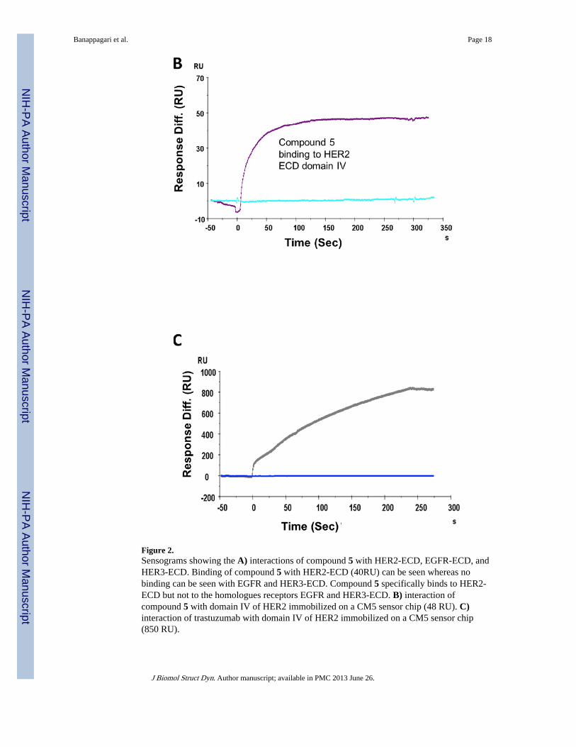
Figure 2.Sensograms showing the A) interactions of compound 5 with HER2-ECD, EGFR-ECD, andHER3-ECD. Binding of compound 5 with HER2-ECD (40RU) can be seen whereas nobinding can be seen with EGFR and HER3-ECD. Compound 5 specifically binds to HER2-ECD but not to the homologues receptors EGFR and HER3-ECD. B) interaction ofcompound 5 with domain IV of HER2 immobilized on a CM5 sensor chip (48 RU). C)interaction of trastuzumab with domain IV of HER2 immobilized on a CM5 sensor chip(850 RU).
Banappagari et al. Page 18
J Biomol Struct Dyn. Author manuscript; available in PMC 2013 June 26.
NIH
-PA Author Manuscript
NIH
-PA Author Manuscript
NIH
-PA Author Manuscript
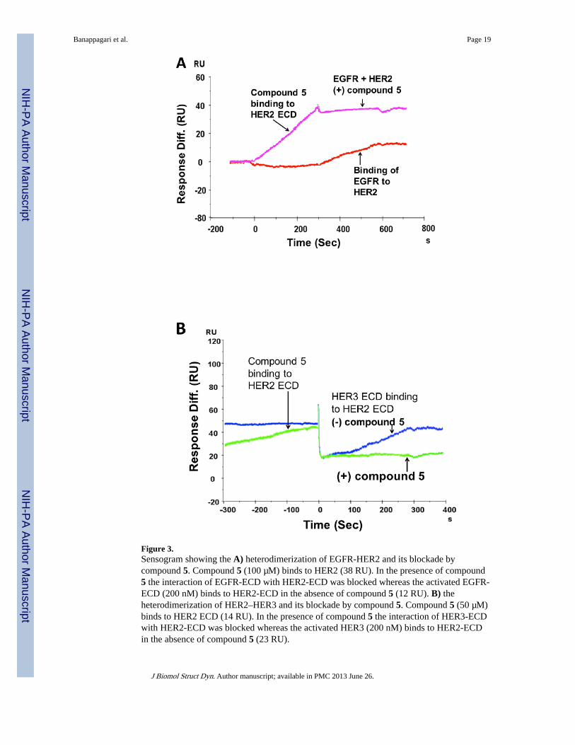
Figure 3.Sensogram showing the A) heterodimerization of EGFR-HER2 and its blockade bycompound 5. Compound 5 (100 µM) binds to HER2 (38 RU). In the presence of compound5 the interaction of EGFR-ECD with HER2-ECD was blocked whereas the activated EGFR-ECD (200 nM) binds to HER2-ECD in the absence of compound 5 (12 RU). B) theheterodimerization of HER2–HER3 and its blockade by compound 5. Compound 5 (50 µM)binds to HER2 ECD (14 RU). In the presence of compound 5 the interaction of HER3-ECDwith HER2-ECD was blocked whereas the activated HER3 (200 nM) binds to HER2-ECDin the absence of compound 5 (23 RU).
Banappagari et al. Page 19
J Biomol Struct Dyn. Author manuscript; available in PMC 2013 June 26.
NIH
-PA Author Manuscript
NIH
-PA Author Manuscript
NIH
-PA Author Manuscript
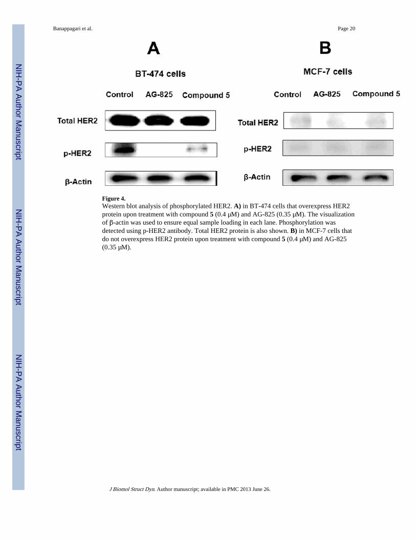
Figure 4.Western blot analysis of phosphorylated HER2. A) in BT-474 cells that overexpress HER2protein upon treatment with compound 5 (0.4 µM) and AG-825 (0.35 µM). The visualizationof β-actin was used to ensure equal sample loading in each lane. Phosphorylation wasdetected using p-HER2 antibody. Total HER2 protein is also shown. B) in MCF-7 cells thatdo not overexpress HER2 protein upon treatment with compound 5 (0.4 µM) and AG-825(0.35 µM).
Banappagari et al. Page 20
J Biomol Struct Dyn. Author manuscript; available in PMC 2013 June 26.
NIH
-PA Author Manuscript
NIH
-PA Author Manuscript
NIH
-PA Author Manuscript
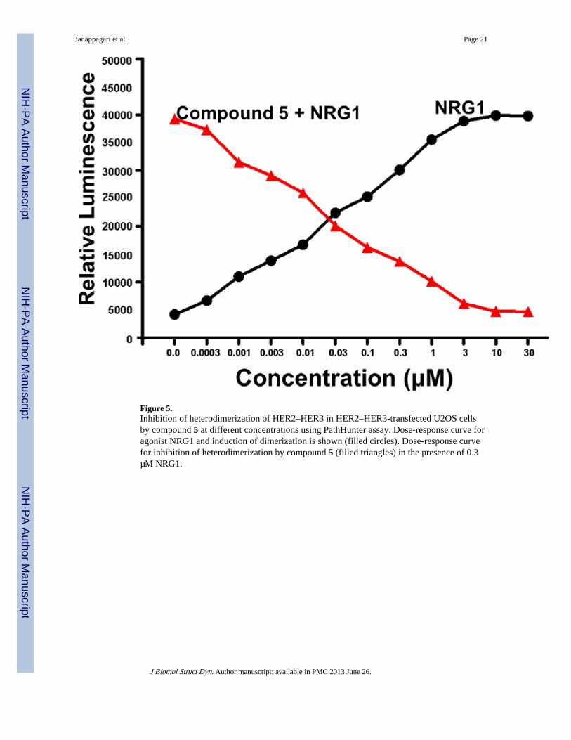
Figure 5.Inhibition of heterodimerization of HER2–HER3 in HER2–HER3-transfected U2OS cellsby compound 5 at different concentrations using PathHunter assay. Dose-response curve foragonist NRG1 and induction of dimerization is shown (filled circles). Dose-response curvefor inhibition of heterodimerization by compound 5 (filled triangles) in the presence of 0.3µM NRG1.
Banappagari et al. Page 21
J Biomol Struct Dyn. Author manuscript; available in PMC 2013 June 26.
NIH
-PA Author Manuscript
NIH
-PA Author Manuscript
NIH
-PA Author Manuscript

Figure 6.Proposed model for HER3 extracellular domain showing domains I to IV in openconformation.
Banappagari et al. Page 22
J Biomol Struct Dyn. Author manuscript; available in PMC 2013 June 26.
NIH
-PA Author Manuscript
NIH
-PA Author Manuscript
NIH
-PA Author Manuscript
Banappagari et al. Page 23
J Biomol Struct Dyn. Author manuscript; available in PMC 2013 June 26.
NIH
-PA Author Manuscript
NIH
-PA Author Manuscript
NIH
-PA Author Manuscript
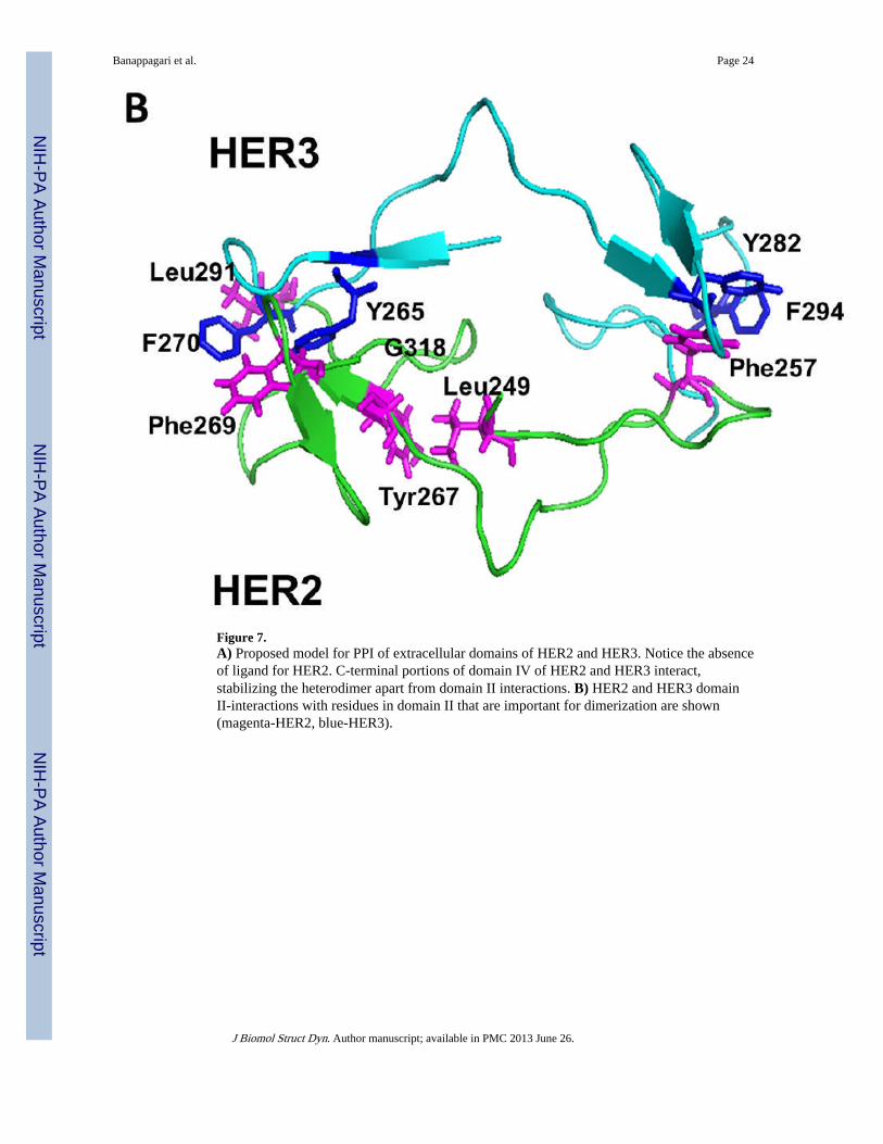
Figure 7.A) Proposed model for PPI of extracellular domains of HER2 and HER3. Notice the absenceof ligand for HER2. C-terminal portions of domain IV of HER2 and HER3 interact,stabilizing the heterodimer apart from domain II interactions. B) HER2 and HER3 domainII-interactions with residues in domain II that are important for dimerization are shown(magenta-HER2, blue-HER3).
Banappagari et al. Page 24
J Biomol Struct Dyn. Author manuscript; available in PMC 2013 June 26.
NIH
-PA Author Manuscript
NIH
-PA Author Manuscript
NIH
-PA Author Manuscript
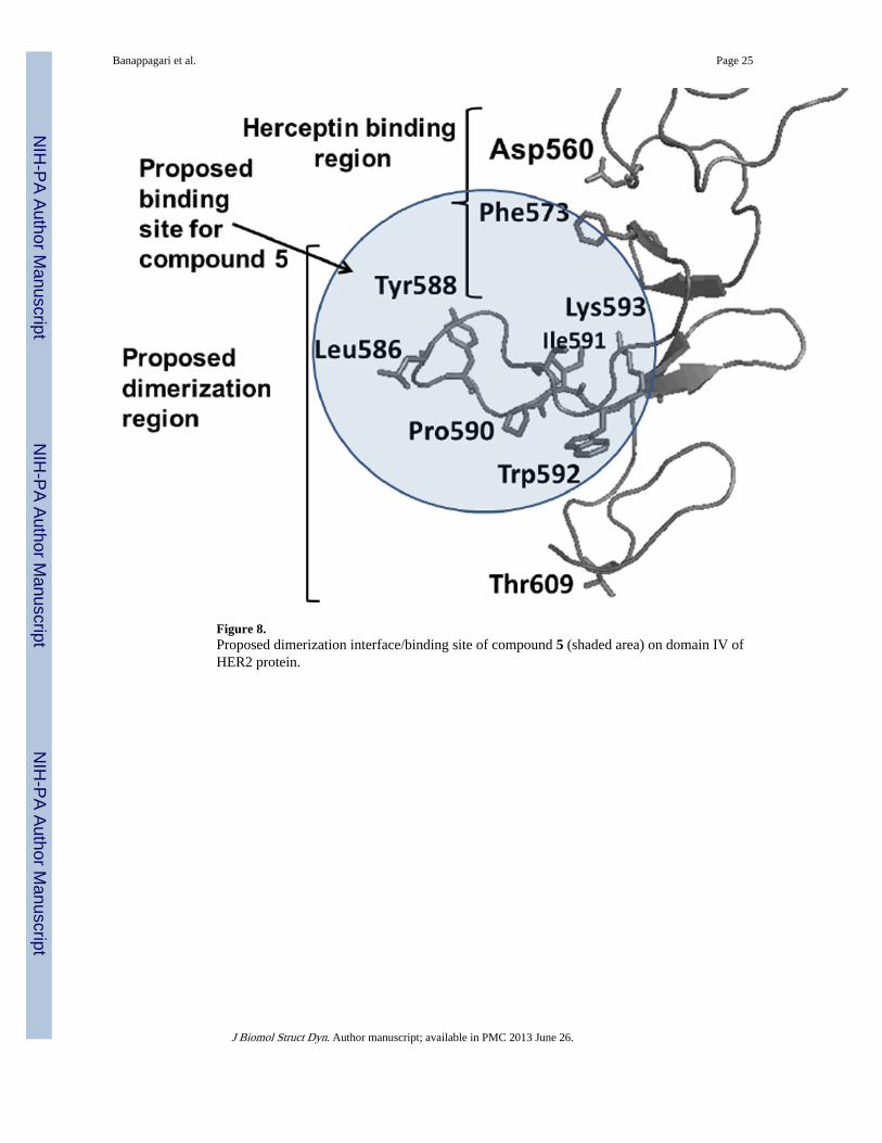
Figure 8.Proposed dimerization interface/binding site of compound 5 (shaded area) on domain IV ofHER2 protein.
Banappagari et al. Page 25
J Biomol Struct Dyn. Author manuscript; available in PMC 2013 June 26.
NIH
-PA Author Manuscript
NIH
-PA Author Manuscript
NIH
-PA Author Manuscript
NIH
-PA Author Manuscript
NIH
-PA Author Manuscript
NIH
-PA Author Manuscript
Banappagari et al. Page 26
Table I
Structures of compound 5 and control compound along with antiproliferative activity against breast cancer celllines.
IC50 µM
Codenumber
Structure SKBR-3 BT-474 MCF-7
5 0.396 ± 0.022 0.895 ± 0.029 16.9 ± 1.0
control H-Lys-[3-amino-3(biphenyl)-propionic acid]-Phe-OH >50 >50 >50
J Biomol Struct Dyn. Author manuscript; available in PMC 2013 June 26.


