Embed Size (px)
Citation preview

C Y T O T O X I C T C E L L S S P E C I F I C F O R
A N T I G E N S E X P R E S S E D O N
S U R F A C E I M M U N O G L O B U L I N - P O S I T I V E C E L L S *
By JAMES FORMAN, RICHARD CIAVARRA, AND ELLEN S. VITETTA
From the Department of Microbiology, University of Texas Health Science Center, Dallas, Texas 75235
The BCL1 tumor is a B cell neoplasm that causes massive splenomegaly and leukemia in BALB/c (Ig-1 a) mice (1). The tumor cells bear IgM and IgD on their surface and are phenotypically and functionally analogous to immature B cells (2). When mice from an allotype-congenic strain, C.B-20 (Ig-l~), are injected with 10 °- 107 BCL1 cells, they are resistant to tumor growth (3). These mice generate T cells that can adoptively transfer tumor immunity to sublethally irradiated C.B-20 recip- ients. Upon in vitro rechallenge of their spleen cells, cytotoxic effector cell activity is generated that is directed against antigens on BALB/c lymphoblast target cells as well as on BCLt cells (3).
Little information is available on the role of cytotoxic T cells in regulating B cell neoplasms. Abbas et al. (4) described effector cells that inhibited M O P C 315 secretion in vitro. These cells were phenotypically characteristic of cytotoxic T lymphocytes (CTL)) However, their specifcity was directed against the myeloma's ligand, trini- trophenol, rather than against immunoglobulin (Ig) itself. Most C T L activity that has been described is directed against viral antigens, minor H antigens, or haptens covalently coupled to cell membranes (5, 6). With the exception of trinitrophenylated proteins (7-9), C T L apparently display specificity for integral membrane proteins or molecules that can fuse with the plasma membrane (10). Accordingly, C T L could potentially be generated against surface Ig (sIg) determinants. In this regard, Rolink et al. (11) generated C T L using allotype congenic strains. However, the antigen(s) recognized by their effector cells were detected on a T cell tumor as well as both T and B lymphoblasts and presumably represent minor H antigens.
In this report we have determined the specifity of C.B-20 anti-BALB/c (anti-Ig H) cytotoxic effector cells as well as the tissue distribution of the antigen(s) recognized by these effector cells. The simplest interpretation of our data is that the effector cells recognize either the constant portion of the # and/or 8 heavy chain on Ig or a molecule coordinately expressed on sIg + cells.
Mate r ia l s a n d M e t h o d s Mice. Mice were initially obtained from Dr. Michael Potter at the National Institute of
Health (National Cancer Institute contract N01-CB094326). Mice were subsequently bred in our laboratories at the University of Texas Health Science Center.
* Supported by grants CA-23115, AI-12789, and CA-28149 from the National Institutes of Health. l Abbreviations used in this paper." Con A, concanavalin A; CTL, cytotoxic T cells; FACS, fluorescence-
activated cell sorter; FITC, fluoresceinated; LPS, lipopolysaccharide; NRIg, normal rabbit Ig; RAMIg, rabbit anti-mouse Ig; sIg, surface Ig.
J. Exp. MED. © The Rockefeller University Press • 0022-1007/81 / 11 / 1357/12 $1.00 13 5 7 Volume 154 November 1981 I357-1368
on February 19, 2016
jem.rupress.org
Dow
nloaded from
Published November 1, 1981

1358 CYTOTOXIC T CELLS SPECIFIC FOR Ig + CELLS
Generation of CTL. Mice were inoculated with 3 × 10 v spleen cells intraperitoneally. 1-6 mo later, spleens were removed from these injected mice and cultured in vitro as previously described (12) in order to generate cytotoxic effector cells.
Preparation of Target Cells. Coneanavalin A (Con A) lymphoblast targets were prepared as previously described (12). Lipopolysaccharide (LPS) lymphoblast targets were generated by culturing spleen cells at a concentration of 4 × 10⁶ cells/ml in RPMI 1640 with 10% fetal calf serum for 2 d, at which time 100/zg of LPS was added. The cells were then cultured for an additional 3 d before use.
Splenic T cells were prepared by passing spleen cells over nylon wool columns. The T cells were then cultured for 3 d in the presence of 5/zg/ml of Con A together with adherent cells. Splenic B cells were prepared by treating spleen cells with monoclonal anti-Thy-l.2 antibody (New England Nuclear, Boston, Mass.) and complement. The surviving cells were cultured for 3 d in the presence of LPS. On the day of assay, target cells were labeled with ~aehromium and centrifuged through an Isolymph solution (Gallard-Schlesinger Chemical Mfg. Corp., Carle Place, N. Y.) to remove nonviable cells.
Analysis of Thy-I + and Ig + Cells. In some experiments, target cells were analyzed for the percentage of Ig + cells by incubating the suspensions with fluoresceinated (FITC) rabbit anti- mouse Ig (F-RAMIg) (N. L. Cappel Laboratories, Cochranville, Pa.).
Thy-1 + cells were detected by incubation with monoclonal anti-Thy-l.2, followed by the F- RAMIg reagent. Similarly, aliquots were stained with F-RAMIg alone. The percentage of anti- Thy-1 + cells was thus calculated by subtraction.
Tumor Cells. BCL1 cells were originally obtained from Dr. Sam Strober, Stanford University Medical Center, Palo Alto, Calif. MOPC 315 was kindly provided by Dr. Richard Lynch, Washington University Medical School, St. Louis, Mo. MOPC 245, MOPC 300, TEPC 183, and P1798 were obtained from Dr. Michael Potter. MOPC 104E was provided for by Dr. Mel Bosma, Institute for Cancer Research, Philadelphia, Pa., and TEPC 15 was provided by Dr. John Cambier, Duke University, Durham, N. C. BCL1 × X63 is a hybridoma line resulting from the fusion of P3 × X63Ag8 (abbreviated X63) to BCL1 (13).
Assay for CTL. The assay for cytotoxic effector cell activity has been described previously (12). Net release of isotope represents the percent release of isotope from target cells in the presence of immune cells minus the percent release of isotope from target cells in the presence of control cells. Control cells represent cells from cultures lacking stimulator cells. The release of isotope from target cells in the presence of control cells ranged from 20 to 35%.
Indirect Fluorescence Analysis Using the Fluorescence-activated Cell Sorter (FACS). The in vivo (1) and in vitro (14) lines of BCL1 cells as well as the BCL1 × X63 hybridoma cells were stained with an affinity-purified polyvalent RAMIg or normal rabbit Ig (NRIg) control followed by FITC goat anti-rabbit Ig. Cells were analyzed on the FACS (B-D FACS Systems, Sunnyvale, Calif.) as described previously (15).
Resu l t s
Mice Challenged with Tumor Cells from an Ig H Chain Congenic Strain Generate Anti-Tumor CTL. We have previously reported (3) that BALB/e (Ig-1 a) mice inoculated with 106 or 107 BCL1 tumor cells die from the tumor after ~8 wk. O n the other hand, mice from an Ig H chain congenic strain, C.B-20 (Ig-1 b), are resistant to the same inoculum of BCL1 cells. T cells from the spleens of these tumor-rejector mice adoptik, ely transfer protection to irradiated C.B-20 animals.
When spleen cells from C.B-20 tumor-rejector mice were tested for their cytotoxic potential, they did not display activity against BCL1 or BALB/c lymphoblasts (data not shown). However, their spleen cells could be sensitized against either irradiated BCL1 or BALB/c cells in a secondary in vitro culture system so that cytotoxic activity was generated against both BCLx and B A L B / c target cells (Table I). Because these effector cells could be responsible for adoptive transfer of tumor immuni ty against
on February 19, 2016
jem.rupress.org
Dow
nloaded from
Published November 1, 1981

JAMES FORMAN, RICHARD CIAVARRA, AND ELLEN S. VITETrA
TABLE I Ability of C.B-20 Mice That Have Rejected BCLI Cells to Generate Effector Cell Activity
1359
Cells Net isotope release at E:T
Effectors Stimulators Targets 100:1 50:1 10:1
C.B-20* BCL1 BCL1 24.3 17.6 4.6 Anti-BCL~ BALB/c:~ 19,7 18.7 5.4
BALB/c BCLI 9.6 7.0 1.5 BALB/c 7.1 7.2 -3.8
* C.B-20 mice were inoculated with 10 e BCLI cells 46 d earlier. Their spleen cells were removed and cultured with irradiated stimulator cells for 5 d in vitro before testing. LPS-stimulated splenic lymphoblasts.
TABLE II
Cytotoxic Activity of CB-20 Anti-BALB/c Effector Cells
Cells Net isotope release at E/T
Effectors Targets* 100:1 50:1 10:1
C.B-20 anti-BALB/c BALB/c (Ig-l")~. 25.5 22.3 10.2 C.B-20 (Ig-I b) 1.3 0.4 2.5 B10.D2 (Ig-I b) 1.6 2.3 3.9 (B6 × BALB/c)F~ (Ig-la/Ig-I b) 25.4 21.6 9.7 BAB-14 (Ig-l"(v):Ig-lb(c)) 0.2 1.0 1.3
BALB/e anti-C.B-20 C.B-20 21.9 17.7 10.5 BALB/c 0.8 -0.3 - 1.3 B10.D2 16.9 10.6 4.5 (B6 × BALB)F~ 16. l 13.7 5.5 BI0 (lg-I b) -1.4 2.0 3.0
BAB-14 anti-BALB/c BALB/C 18.4 15.5 6.9
* LPS-stimulated splenic lymphoblasts. :~ Genotype.
BCLI, we fur ther invest igated the act ivi ty of effector cells genera ted agains t Ig heavy chain a l lo typic differences.
Specificity of CTL Generated from the Sensitization of lg H Chain Congenic Mice. C.B-20 mice were inocula ted with B A L B / c splenocytes. 2-6 mo later, thei r spleen cells were removed a n d the cells were cu l tu red in vi tro wi th i r r ad ia ted B A L B / c splenocytes for 5 d, at which t ime the cultures were assayed for cytotoxic ac t iv i ty against LPS lymphoblas ts . T h e d a t a presented in T a b l e II demons t ra te tha t these cells d isp lay a cytotoxic effect against B A L B / c and (BALB/c × B6)F1 (C57BL/6 is abb rev i a t ed as B6) target cells. O n the o ther hand , C.B-20 and B10.D2 (Ig-1 °) target cells were not lysed. This la t ter result was expected because B 10,D2 has the same Ig H chain l inkage group as C.B-20, and therefore should not be susceptible to lysis. T r e a t m e n t of the effector cells wi th monoclona l a n t i -T hy - l . 2 serum and complemen t ab roga ted their cytotoxic act ivi ty, ind ica t ing tha t the cytotoxic cells are T cells (da ta not shown).
B A L B / c ant i -C.B-20 effector cells were also t rea ted for their ac t iv i ty agains t a s imilar panel of target cells. C.B-20, (BA L B/c × B6)F1 and B10,D2 (H-2 d) target cells were kil led by these effector cells. T h e lysis of B 10.D2 target cells p r e sumab ly occurs because bo th C.B-20 and B10.D2 mice share the same alleles at their Ig H chain loci.
on February 19, 2016
jem.rupress.org
Dow
nloaded from
Published November 1, 1981

1360 C Y T O T O X I C T CELLS SPECIFIC FOR Ig + CELLS
The fact that H-2 congenic C57BL/10 (B10) (H-2 b, Ig-l~ target cells were not lysed indicates that the cytotoxic effector cells are H-2 restricted.
C.B-20 anti-BALB/c effector cells did not lyse BAB-14 target cells. Because BAB- 14 is a recombinant strain differing from C.B-20 at the V but not the C region of Ig H loci (16), this indicates that the specificity of the effector cells is directed against an antigen encoded for by a gene linked to the C region of the Ig H chain linkage group. Further evidence indicating that killer cells can be generated against an antigen controlled by an IgH C region-linked gene is the finding that BAB-14 animals sensitized against BALB/c spleen cells generate cytotoxic effector cells that lyse BALB/c targets (Table II). Thus, these data show that cytotoxic effector cells can be generated across the Ig H chain allotype barrier, and are in agreement with the previous report of Rolink et al. (1 I).
C.B-20 Anti-BALB/c Effector Cells Display Cross-reactive Lysis on Target Cells Carrying Different Ig H Alleles. The previous data indicate that C.B-20 and BALB/c mice can generate CTL with specificity for antigens controlled by genes that map to the Ig H chain linkage group. However, the specificity of these effector cells for other allotypes was not tested.
The data in Table III indicate that C.B-20 anti-BALB/c effector cells display cross- reactive lysis on C.AL-20 target cells, which carry the Ig-1 d allele. Further, these cross- reactive effector cells can be generated by in vitro stimulation with either BALB/c or C.AL-20 stimulator cells. In addition, C.B-20 effector cells also show cross-reactive lysis on H-2-matched DBA/2 (Ig-1 c) targets. These data suggest that either Ig-I a, Ig-I ~, and Ig-1 d strains express identical target antigens; or alternatively, there is a cross-reactive determinant in these three strains in addition to unique specificities. Experiments are currently in progress to distinguish between these possibilities.
Susceptibility of BCLt Cells to C.B-20 Anti-BALB/c Effector Cells. We previously demonstrated (3) that C.B-20 animals that had rejected a large inocula of BCL1 cells generated cytotoxic effector cells that lysed BCL1 and BALB/c target cells (Table I). In the following experiments, we ascertained whether effector cells produced by in vivo immunization of C.B-20 mice with BALB/c spleen cells would also generate effector cells that could recognize BCL1 target cells.
The data in Table IV demonstrate that C.B-20 anti-BALB/c effector cells are able to lyse BCL1 target cells. In this experiment, the in vivo BCL1 cells were obtained from the spleen, an organ that (in tumor-bearing animals) contains ~70% tumor cells (2). Because it is possible that BALB/c host cells rather than BCL1 cells are responsible
TABLE III
Specificity of C.B-20 Anti-BALB/c Effector Cells
Cells Net release at E:T
Effectors Stimulators Targets* 100:1 50:1 10:I
C.B-20 anti-BALB/c BALB/c BALB/c (Ig-l")~ 13.9 8.5 7.9 C.AL-20 (Ig-I d) 16.8 11.2 4.7
C.AL-20 BALB/c (lg-l") 17.5 11.0 1.4 C.AL-20 (lg-1 a) 23,6 17.4 4.9
BALB/c DBA/2 (lg-1 c) 28,1 26.5 10.0
* LPS-stimulated splenic lymphoblasts. :1: Genotype.
on February 19, 2016
jem.rupress.org
Dow
nloaded from
Published November 1, 1981

JAMES FORMAN, RICHARD CIAVARRA, AND ELLEN S. V I T E T T A
TABLE IV Susceptibility of BCL1 Cells to Anti-lg H Effector Cells
1361
Cells Net isotope release at E:T
Effectors Targets 100:1 50:1 10:1
C.B-20 anti-BALB/c BALB/c* 25.9 20.8 12.0 C.B-20* -3 .5 -2 .7 -1 .5 BCLI in vivo:~ 20.9 19.0 9.0 BCLa in vitro§ 22.7 26.4 21.5 BCL~ X X63]1 -4 .3 -3 .8 -2 .9
* LPS-stimulated splenic lymphoblasts. ~: Spleen cells from a tumor-bearing animal. § Line maintained by in vitro passage. [[ Hybridoma line (see text).
for the lytic effect observed, we also tested an in vitro-adapted BCL1 cell line (14). The data (Table IV) demonstrate that these in vitro-cultured cells are sensitive to lysis. Thus, the anti-Ig H region CTL do recognize antigenic determinants expressed by BCL1.
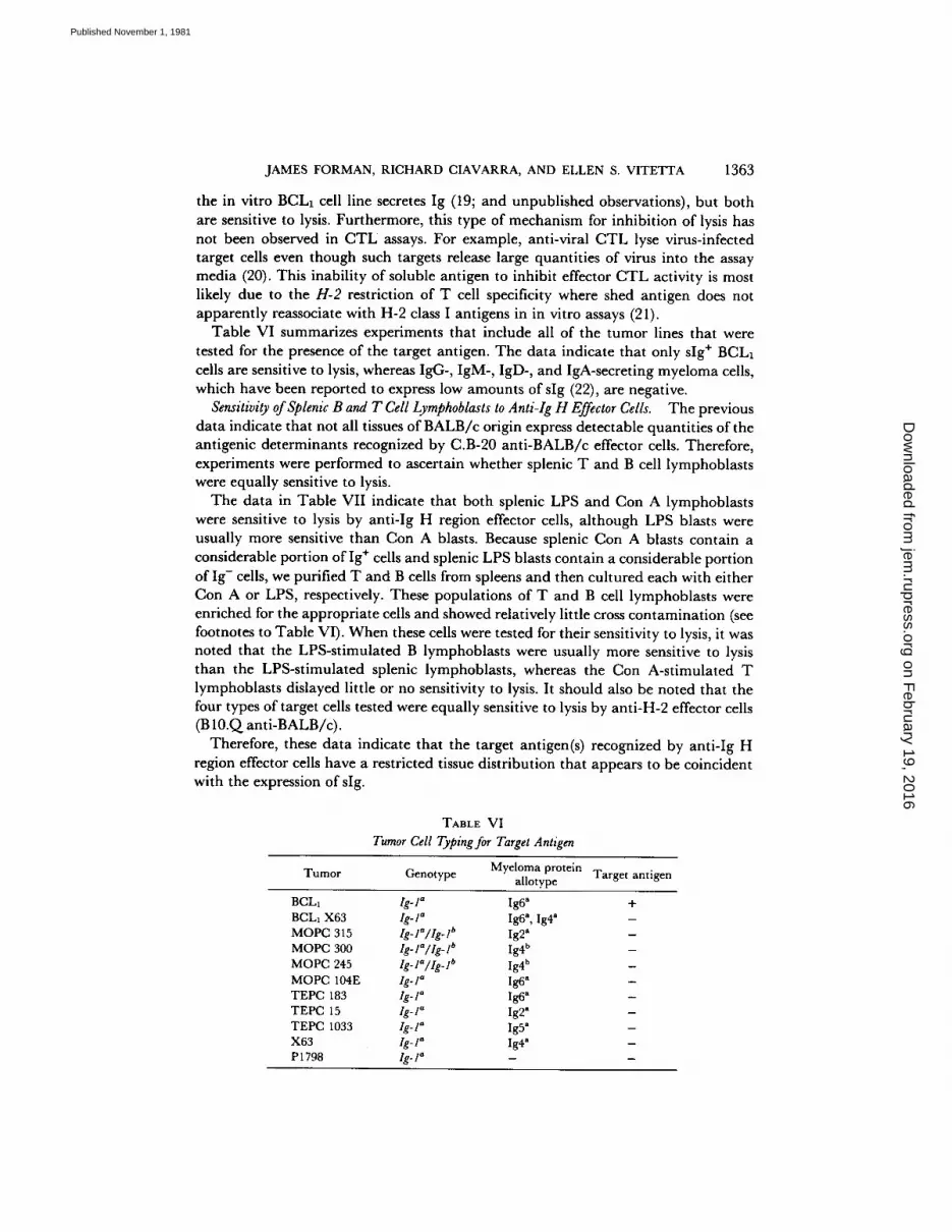
BCL1 cells have been fused to the myeloma line, X63. This hybridoma secretes IgM, which bears the same idiotypic determinants expressed on the IgM and IgD of the parental BCLx tumor cell, as well as the IgGlk from the X63 cells (13). However, when this hybridoma line was tested for its sensitivity to lysis, no cytotoxic effect was observed (Table IV).
These three BCL1 lines were also analyzed for cell surface Ig expression using the FACS. The data in Fig. 1 reveal that the two BCL1 lines contain a large percentage of sIg + cells (>50%), with a relatively heterogenous distribution of staining intensity. On the other hand, the BCLa hybridoma line contains very few sIg + cells, and these cells stain with a relatively low intensity.
Therefore, these results suggest that the expression of sIg is necessary for a target cell to be sensitive to lysis by anti-Ig H chain effector cells.
Susceptibility of Tumor Lines to Lysis by Anti-Ig H Effector Cells. Because the BCLa (sIg +) B cell leukemia was killed by C.B-20 anti-BALB/c effector cells, we next determined whether other BALB/c tumor cells also expressed the target antigen. Target cells included IgA-, IgGa-, and IgD-secreting myeloma cells and a thymic lymphoma (P 1798).
The data indicate that none of these tumor cells were sensitive to lysis by either C.B-20 anti-BALB/c or BALB/e anti-C.B-20 effector cells (Table V). The latter effector cells were used because three of the myelomas tested, MOPC 315, MOPC 300, and MOPC 245, are genotypically heterozygous at Ig-1; i.e., they were obtained from mice during backcrossing to generate the C.B-20 congenic line (17, 18) and could be sensitive to lysis as were (BALB/c × B6)F1 target cells (see Table II). The data also show that although the MOPC 315, MOPC 300, and P1798 cell lines are insensitive to lysis by C.B-20 anti-BALB/c effector cells, they are all sensitive to lysis by anti-H-2 CTL (B10.Q anti-BALB/c).
If Ig is the target antigen, then it is possible that secreted Ig from myeloma cells could adsorb onto unlabeled cells in the effector cell population and block or inhibit lysis. However, this alternative is unlikely. Thus, LPS induced B lymphoblasts and
on February 19, 2016
jem.rupress.org
Dow
nloaded from
Published November 1, 1981

1362 CYTOTOXIC T CELLS SPECIFIC FOR Ig + CELLS
x $
3
0 50 "100
5 ~ I BCL vitro
0 5O 100
I I BcL, xx63 1 5 I hybridOmO ]
0 50 ~00 ChQnnel Number
FIc. 1. Analysis of slg on BCLI and BCLI x X63 hybridoma cells. Cells were exposed to NRIg (solid lines) or RAMIg (dashed lines) followed by FITC goat anti-rabbit Ig. The cells were then analyzed using the FACS.
TABLE V
Susceptibility of Lyszs of BALB/c Tumor Cells to Anti-Ig H Effector Cells
Cells Myeloma Net isotope relea.sc at E:T Cell genotype protein
Eff¢ctors Targets allot ype 100: I 50: I 10: I
C.B-20 anti-BALB/c
BALB/c anti-C,B-20
BALB/c* Ig- l" (BALB/c X B6)F2* l ¢ - I ° / ( ¢ - I b C.B-20* Ig- I b
M O P C 315 l g - l~ / l g - I t' M O P C 300 Ig- l~/Ig- I b M O P C 245 l g - I~ / lg - I b
P3 x X63Ag8 lg-I ~
M O P C 315
M O P C 300
P1798 ,~e-/a BALB/c*
24.8 20.7 16.4 20.8 17.9 I 1.9
--0.8 --0.6 --0.2
lg-2 a 1.6 0.5 0.1 lg-4 h 5.7 4.3 1.4 Ig-4 b 3.7 2,5 0.5
Ig-4 a - 0 . 8 --2,6 --4.3
1.2 (25.7):~ 0,4 (24.3) 0.8 (14.4) - 0 . 8 (31.8) - 0 , 7 (27.5) - 0 . 5 (9,1)
0.3 (36.1) - 0 . 5 (35.7) - 4 . 5 (22.6) 25.3 (42.1) 25.9 (40.5) 17.1 (21.3)
xt BALB/c* 0.8 - 0 . 3 - 1 . 3 (BALB/c X B6)FI* 16.1 13.7 5.5
C.B-20* 21.9 17.7 10.5 MO P( : 300 -2 .6 - 4 . 6 - 7 . 0
M O P C 245 - 2 . I -3 .7 - 6 . 3 MO PC 315 -1 ,4 - 5 . 9 -10 .0
* I,PS-stimulated splenic lymphoblasts
:~ Values in parentheses represent isotope release from target cells in the presence of anti-H-2 C T L (BIO.Q anti-BAI,B/c).
on February 19, 2016
jem.rupress.org
Dow
nloaded from
Published November 1, 1981
JAMES FORMAN, RICHARD CIAVARRA, AND ELLEN S. VITETTA 1363
the in vitro BCLx cell line secretes Ig (19; and unpublished observations), but both are sensitive to lysis. Furthermore, this type of mechanism for inhibition of lysis has not been observed in C T L assays. For example, anti-viral C T L lyse virus-infected target cells even though such targets release large quantities of virus into the assay media (20). This inability of soluble antigen to inhibit effector C T L activity is most likely due to the H - 2 restriction of T cell specificity where shed antigen does not apparently reassociate with H-2 class I antigens in in vitro assays (21).
Table VI summarizes experiments that include all of the tumor lines that were tested for the presence of the target antigen. The data indicate that only sIg + BCL1 cells are sensitive to lysis, whereas IgG-, IgM-, IgD-, and IgA-secreting myeloma cells, which have been reported to express low amounts of sIg (22), are negative.
Sensitivity o f Splenic B and T Cell Lymphoblasts to Ant i - Ig H Effector Cells. The previous data indicate that not all tissues of BALB/c origin express detectable quantities of the antigenic determinants recognized by C.B-20 anti-BALB/c effector cells. Therefore, experiments were performed to ascertain whether splenic T and B cell lymphoblasts were equally sensitive to lysis.
The data in Table VII indicate that both splenic LPS and Con A lymphoblasts were sensitive to lysis by anti-Ig H region effector cells, although LPS blasts were usually more sensitive than Con A blasts. Because splenic Con A blasts contain a considerable portion of Ig ÷ cells and splenic LPS blasts contain a considerable portion of Ig- cells, we purified T and B cells from spleens and then cultured each with either Con A or LPS, respectively. These populations of T and B cell lymphoblasts were enriched for the appropriate cells and showed relatively little cross contamination (see footnotes to Table VI). When these cells were tested for their sensitivity to lysis, it was noted that the LPS-stimulated B lymphoblasts were usually more sensitive to lysis than the LPS-stimulated splenic lymphoblasts, whereas the Con A-stimulated T lymphoblasts dislayed little or no sensitivity to lysis. It should also be noted that the four types of target cells tested were equally sensitive to lysis by anti-H-2 effector cells (B 10.Q anti-BALB/c).
Therefore, these data indicate that the target antigen(s) recognized by anti-Ig H region effector cells have a restricted tissue distribution that appears to be coincident with the expression of sIg.
TABLE VI
Tumor Cell Typing for Target Antigen
Tumor Genotype Myeloma protein Target antigen allotype
BCL1 Ig-I a Ig6 a + BCL1 X63 Ig-I a Ig6 a, Ig4 a - MOPC 315 Ig-la/Ig-I b Ig2 a MOPC 300 Ig-l"/Ig-I b Ig4 b MOPC 245 Ig-l"/Ig.l b Ig4 b MOPC 104E Ig-l" Ig6 a - TEPC 183 Ig-l" Ig6 • - TEPC 15 Ig-I a Ig2" - TEPC 1033 Ig-l" Ig5" - X63 Ig-I '~ Ig4 a - P1798 Ig- l" - -
on February 19, 2016
jem.rupress.org
Dow
nloaded from
Published November 1, 1981

1364 C Y T O T O X I C T C E L L S S P E C I F I C F O R Ig + C E L L S
TABLE VII Sensitivity of Splenic Lymphoblasts to Anti-Ig H Effector Cells
Cells Net release at E :T = 100:1
Mi togen Experi- Experi- Experi- Experi- Effectors Targe ts
ment 1 merit 2 men t 3 merit 4
C.B-20 a n t i - B A L B / c B A L B / c WS LPS 16.1" 25.3 (42.1)~: - - - - WS Con A 14.8§ 7.6 (41.9) - - - -
B A L B / c B cells LPS 20.211 17.8 (42.7) 35.8 - - T cells Con A 8.8¶ 7.3 (39.0) 0.8 - -
B A L B / c anti-C.B-20 C.B-20 WS LPS - - - - - - 20.0* WS Con A - - - - - - 15.0§
C.B-20 B cells LPS - - - - - - 29.51[ T cells Con A - - - - - - 8.7¶
* WS, whole spleen; whole spleen LPS lymphoblas t s con ta ined 68-77% Ig ÷ cells and 15-18% Thy- l . 2 + cells.
:]: Numbers in parentheses represent lysis from target cells by B 10.Q a n t i - B A L B / c effector cells. § Whole spleen Con A lymphoblas t s con ta ined 19-24% Ig ÷ cells and 55-64% T h y - l . 2 + cells. [[ B cell lymphoblas t s con ta ined 85-95% Ig + cells and 2-7% Thy- l . 2 + cells. ¶ T cell lymphoblas t s con ta ined 12-14% Ig + cells and 75-78% T h y - l . 2 + cells.
Discussion
The data in this report provide the first direct evidence for the existence of CTL specific for antigenic determinants expressed on sIg + cells. The target antigens recognized by these CTL are encoded for by genes either linked to or identical with Ig H chain loci. Furthermore, because C.B-20 anti-BALB/c effector cells do not lyse BAB-14 lymphoblasts, the antigen recognized on BALB/c targets is encoded for by a gene linked to the C rather than the V region of the Ig-1 locus. Thus, our data demonstrate that the target antigens detected by the cytotoxic effector cells generated in this system are expressed on LPS-induced B cell lymphoblasts but not Con A- induced T cell lymphoblasts or a T cell leukemia. Although this indicates that the target antigens are expressed on cells of the B and not the T cell lineage, these antigens are not expressed on all cells of B cell origin. We were unable to detect target antigens on several myeloma cell lines that have been reported to have low levels of sIg (reviewed in 22). On the other hand, BCL1, an sIg + B cell leukemia was sensitive to lysis. Furthermore, a BCLa × X63 hybridoma line that secretes the same IgM as is expressed on the surface of the parental BCL1 line, but displays almost no sIg, was not lysed by the anti-Ig H effector cells. Taken together, these data suggest that the target antigen being detected by the CTL is either sIg itself or a determinant that is coordinately expressed on Ig + B cells.
These data provide a possible mechanism whereby CTL could regulate B cells expressing cell membrane allotypic (Ig) determinants. This type of activity would be consistent with previous studies indicating that other subpopulations of T lympho- cytes, viz., helper and suppressor T cells, can regulate the growth and differentiation of B cells possessing idiotypic or allotypic determinants on their membrane Ig. Thus, Herzenberg et al. (23, 24) have demonstrated allotype specific helper and suppressor T cells. Eichmann (25), Woodland and Cantor (26), and Bottomly and Maurer (27), have described helper cells that are idiotype specific. Rosenberg and Chiller (28) have evidence for an allotype (C region)-specific helper cell involved in the regulation of IgG responses. Both L'age-Stehr (29) and Nutt et al. (30) have data to indicate that
on February 19, 2016
jem.rupress.org
Dow
nloaded from
Published November 1, 1981

JAMES FORMAN, RICHARD CIAVARRA, AND ELLEN S. VITETTA 1365
antigen-primed B cells induce the expansion of helper T cell populations that recognize allotypes or idiotypes on B cells.
Rolink et al. (11) reported that CTL could be generated using allotype congenic strains that were similar but not identical to those used in this study. The effector cells they described detected an antigen expressed on a T cell tumor as well as both T and B cell lymphoblasts. In contrast, the antigen recognized by the cytotoxic cells generated here were restricted to sIg + B cells. This discrepancy between the two sets of data could be due to the fact that the BALB/c animals used by Rolink et al. (11) may differ at an/g-linked H-antigen locus from the BALB/c line used to derive the allotype congenic strains. Accordingly, the CTL generated by Rolink et al. (1 l) might be specific for such an H antigen(s). Alternatively, their culture system may allow for the generation of clones of effector cells that recognize additional non-Ig antigenic determinants on lymphoid cells. In this regard, we have noted that in some experi- ments where CTL from individual animals were tested on P815 (Ig-1 c, H-2 a) target cells, that some C.B-20 anti-BALB/c effector cells lysed this cell line. The activity of the CTL from these mice may be similar to that described by Rolink et al. (11) and is also consistent with the data of Riblet et al. (31), who demonstrated that allotype congenic strains reject skin grafts. Thus, those antigens might be analogous to minor H antigens that have been shown to elicit both skin graft rejection and to induce cytotoxie effeetor T cell activity (32). However, it is also possible that P815 cells express the same antigen that is detected on the sIg + cells. In this case, the target antigen would most likely be a differentiation antigen with coordinate expression on sIg + cells and P815 cells.
There are several molecules in addition to CTL target antigens that are controlled by Ig-1 linked loci. For example, Owen et al. (33) have immunized BALB/c animals with cells from allotype congenic cells C.AL-20 mice and were able to produce an antiserum that recognized a molecule found on Lyt-2 + suppressor cells. A hybridoma antibody that recognizes a second Ig-H-encoded antigen expressed on Lyt-1 ÷ suppres- sor inducer cells has been described (G. M. Spurll and F. L. Owen, personal communication). A. Finnegan and F. L. Owen (personal communication) have produced a C.B-20 anti-BALB/c antiserum that detects an antigen controlled by a gene linked to the V region of Ig-H and is expressed on CTL.
The role of cytotoxic and suppressor T cells in controlling growth of B cell neoplasms is not clear. Bosma and Bosma (34) have shown that anti-allotype sup- pressor cells can regulate the secretion of Ig by myeloma cells from Ig-1 b mice. Rohrer and Lynch (35) have demonstrated that suppression or enhancement of MOPC 315 tumor cell growth, myeloma stem cell production, and IgA secretion can be influenced by carrier-specific helper or suppressor T cells. Flood et al. (36) have demonstrated that mice immunized with purified myeloma proteins generate idiotype-specific T cells that regulate MOPC 315 growth.
Abbas et al. (4) have reported that Lyt-2 +, cyclophosphamide- and radiation- resistant T cells can regulate Ig secretion by MOPC 315 cells in vitro. The criterion for defining these cytotoxic cells was based on these phenotypic characteristics and by their ability to reduce the viability of the tumor cells over a 2-3-d culture period. However, these effector cells did not cause direct lysis of target cells in a 4-h chromium release assay. Further, unlike the specificity of the CTL described in this report, these
on February 19, 2016
jem.rupress.org
Dow
nloaded from
Published November 1, 1981

1366 CYTOTOXIC T CELLS SPECIFIC FOR Ig ÷ CELLS
cells were specific for the epitope which the MOPC 315 cells bind (trinitrophenol) rather than the Ig itself.
Although CTL could play a role in preventing B cell tumors, they may not be relevant in regulating myeloma cell growth because the level of surface Ig on these cells is relatively small (22). On the other hand, tumor cells such as BCL1 may be more amenable to regulation by CTL because these cells generally have a high density of sIg (2), which could be recognized as a tumor-specific antigen.
It is not known whether the ability of C.B-20 mice to resist BCL1 is due to the activity of CTL. We previously reported (3) that the tumor rejection antigen maps to the V region rather than the C region of the Ig gene complex because BAB-14 mice were tumor susceptible. However, recent data (R. Ciavarra and J. Forman, unpub- lished data) suggest that for tumor rejection to occur, mice need to recognize two determinants, one in the V region and the other in the C region. A similar conclusion has been drawn in the anti-Qa-1 CTL system (37). If this interpretation is correct, then the anti-allotype CTL described here could be relevant anti-tumor effector cells. 2
S u m m a r y
C.B-20 mice were immunized with splenocytes or B leukemia cells (BCL1) from Ig H chain allotype congenic strains. Spleen cells from these immunized mice were rechallenged in vitro to generate H-2-restricted cytotoxic T cells that were specific for target antigens controlled by genes linked to the Ig H chain locus.
The anti-Ig H cytotoxic T cells detected an antigen(s) expressed only on surface Ig ~- cells. Thus, T cell lymphoblasts, eight BALB/c myeloma cell lines, and a T cell lymphoma were not lysed by the effector cells. In contrast, B cell lymphoblasts and the surface Ig + BCL1 cells were sensitive to lysis. A surface Ig- hybridoma (which secretes the IgM from the BCL1 cells) generated by fusing BCL1 cells to X63 myeloma cells was not killed by the effector cells.
These data indicate that cytotoxic T cells specific for antigenic determinants on either surface IgM or IgD or on a molecule that is coordinately expressed on IgM + or IgD + cells can be generated and that such cells might play a role in regulating the growth of normal B cells or surface Ig ÷ tumor cells in vivo.
We thank Ms. J. Tsan, Ms. M. Wyatt, and Ms. S. Byers for their excellent technical assistance. We thank Ms. B. J. Washington for secretarial help.
Received for publication 7July 1981.
References 1. Slavin, S., and S. Strober. 1978. Spontaneous murine B cell leukemia. Nature (Lond.). 272:
624. 2. Krolick, K. A., P. C. Isakson, J. W. Uhr, and E. S. Vitetta. 1979. BCL1, a murine model for
chronic lymphocytic leukemia: use of the surface immunoglobulin idiotype for the detection and treatment of tumor. Immunol. Rev. 48:81.
2 Similar results demonstrating anti-Ig CTL have been reported in Snodgrass, H. R., D. B. Wilson, and M. J. Bosma. 1981. T lymphocytes specific for immunoglobulin allotype. I. Igh-lb-specific T cells demonstrated by suppression in vivo and cytotoxicity in vitro..]. Exp. Med. 154"480; and Snodgrass, H. R., M. J. Bosma, and D. B. Wilson. 1981. T lymphocytes specific for immunoglobulin allotype. If. Cloned Igh- lb-specific cytotoxic T cells.J. Exp. Med. 154:491.
on February 19, 2016
jem.rupress.org
Dow
nloaded from
Published November 1, 1981

JAMES FORMAN, RICHARD CIAVARRA, AND ELLEN S. VITETTA 1367
3. Ciavarra, R., and J. Forman. 1981. Influence of IgH V-region genes on the growth kinetics of a murine B cell leukemia (BCL1).J. Immunol. 126:54.
4. Abbas, A. K., S. E. Ratnofsky, and S.J. Burakoff. 1980. T lymphocyte-mediated suppression of myeloma function in vitro. II. Evidence for regulation of hapten-binding myelomas by syngeneic hapten-specific cytolytic T lymphocytes. J. Exp. Med. 152:306.
5. Zinkernagel, R., and P. C. Doherty. 1979. MHC-restricted cytotoxic T cells: studies on the biological role of polymorphic major transplantation antigens determining T cell restriction- specificity function and responsiveness. Adv. Immunol. 97:51.
6. Ciavarra, R., and J. Forman. 1981. Cell membrane antigens recognized by anti-rival and anti-trinitrophenyl cytotoxic T lymphocytes. Immunol. Rev. In press.
7. Schmitt-Verhulst, A., C. B. Pettinelli, P. A. Henkart, J. K. Lunney, and G. M. Shearer. 1978. H-2 restricted cytotoxic effectors generated in vitro by the addition of trinitrophenyl- conjugated soluble proteins.J. Exp. Med. 147:352.
8. Ozato, K., and C. S. Henney. 1978. Studies on lymphocyte-mediated cytolysis. XII. Hapten transferred to cell surfaces by interaction with liposomes is recognized by antibody but not by hapten-specific H-2 restricted cytotoxic T cells.J. Immunol. 121:6.
9. Ciavarra, R., and J. Forman. 1980. Cells treated with trinitrobenzene sulfonic acid express an antigenic determinant recognized by cytotoxic effector cells that is not detected on cells coated with trinitrophenylated proteins. J. Immunol. 124:713.
10. Gething, M. J., U. Koszinowski, and M. Waterfield. 1978. Fusion of Sendai virus with the target cell membrane is required for T cell cytotoxicity. Nature (Lond.). 274:689.
11, Rolink, T., K. Eichmann, and M. M. Simon. 1978. Detection of two allotype (Ig-l)-linked minor histocompatibility loci by the use of H-2 restricted cytotoxic lymphocytes in congenic mice. Immunogenetics. 7:321.
12, Forman, J., and J. W. Streilein. 1979. T cells recognize minor histoeompatibility antigens on H-2 allogeneic cells.J, Exp. Med. 150:1001.
13. Krolick, K. A., C. Villemez, P. Isakson, J. W. Uhr, and E. S. Vitetta. 1980. Selective killing of normal or neoplastic B cells by antibodies coupled to the A chain of ricin. Proc. Natl. Acad. Sci. U. S. A. 7715419.
14. Gronowicz, E. S., C. A. Doss, F. D. Howard, D. C. Morrison, and S. Strober. 1980. An in vitro line of the B cell tumor BCLI can be activated by LPS to secrete IgM. J. Immunol. 125: 976.
15. Krolick, K. A., P. C. Isakson, and E. S. Vitetta. 1979. Murine B cell leukemia (BCLI): organ distribution and kinetics of growth as determined by fluorescence analysis with an anti-idiotypic antibody. J. Immunol. 123:1928.
16, Claflin, J. L., J. Wolfe, and V. Ruppert. 1979. Structural evidence for recombination at the Igh (H chain) complex in BAB-14 mice.J. Immunol. 123:2088.
17. Potter, M., and R. Lieberman. 1967. Genetic studies of immunoglobulins in mice. Cold Spring Harbor Symp. Quant. Biol. 32:187.
18, Eisen, H. N., E, S. Simms, and M. Potter. 1968. Mouse myeloma proteins with antihapten antibody activity. The protein produced by plasma cell tumor MOPC-315. Biochemistry. 7: 4126.
19, Andersson, J., O. Sjoberg, and G. Moiler. 1972. Mitogens as probes for immunocyte activation and cellular cooperation, Transplant. Rev. 11:131.
20, Doherty, P. C,, R, V, Blanden, and R. M. Zinkernagel. 1976. Specificity of virus-immune effector T cells for H-2K or H-2D compatible interactions: implications for H-antigen diversity. Transplant. Rev. 29:89.
21. Bevan, M.J . 1976. Minor H antigens introduced on H-2 different stimulating cells cross- react at the cytotoxic T cell level during in vivo priming.,]. Immunol. 117:2233.
22, Katz, D, H. 1977. In Lymphocyte Differentiation, Recognition, and Regulation. Academic Press, Inc., New York. 164.
on February 19, 2016
jem.rupress.org
Dow
nloaded from
Published November 1, 1981

1368 CYTOTOXIC T CELLS SPECIFIC FOR Ig ÷ CELLS
23. Herzenberg, L. A., K. Okumura, and C. M. Metzler. 1975. Regulation of immunoglobulin and antibody production by allotype suppressor T cells in mice. Transplant. Rev. 27:57.
24. Herzenberg, L. A., K. Okumura, H. Cantor, V. L. Sato, F.-W. Shen, E. A. Boyse, and L. A. Herzenberg. 1976. T-cell regulation of antibody responses: demonstration of allotype- specific helper T cells and their specific removal by supressor T cells.J. Exp. Med. 144:330.
25. Eichman, K. 1978. Expression and function of idiotypes on lymphocytes. Adv. Immunol. 26: 195.
26. Woodland, R. T., and H. Cantor. 1978. Idiotype specific T helper cells are required to induce idiotype-positive B memory cells to secrete antibody. Fur. J. Immunol. 8:600.
27. Bottomly, K., and P. H. Maurer. 1980. Antigen-specific helper T cells required for dominant production of an idiotype (ThId) are not under immune response (Ir) gene control.J. Exp. Med. 152:1571.
28. Rosenberg, Y. J'., and J. M. Chiller. 1979. Ability of antigen-specific helper cells to effect a class-restricted increase in total Ig-secreting cells in spleens after immunization with the antigen.dr. Exp. Med. 150:517.
29. L'age-Stehr, J. 1981. Priming of T helper cells by antigen-activated B cells. B cell-primed Lyt-1 + helper cells are restricted to cooperate with B cells expressing the IgvH phenotype of the priming B cells.J. Exp. Med. 153:1236.
30. Nutt, N., Haber, J., and H. H. Wortis. 1981. Influenceof Igh-linked gene products on the generation o f T helper cells in the response to sheep erythrocytes. ]. Exp. Med. 153:1225.
31. Riblet, R., and C. Congleton. 1977. A possible allotype linked histocompatibility gene. Immunogenetics. 5:511.
32. Simpson, E., and R. D. Gordon. 1977. Responsiveness to H-Y antigen, Ir gene complemen- ration and target cell specificity. Irnrnunol. Rev. 35:59.
33. Owen, F. L., R. Riblet, and B. A. Taylor. 1981. The T suppressor cell alloantigen Tsu a maps near immunoglobulin allotype genes and may be a heavy chain constant-region marker on a T cell receptor.J. Exp. Med. 153:801.
34. Bosma, M. J., and C. G. Bosma. 1977. Prevention of IgG2a production as a result of allotype-specific interaction between T and B cells.dr. Exp. Med. 145:743.
35. Rohrer, J. W., and R. G. Lynch. 1979. Immunoregulation of localized and disseminated murine myeloma; antigen-specific regulation of MOPC-315 stemm cell proliferation and secretory cell differentiation. J. Irnmunol. 123:1083.
36. Flood, P. M., C. Phillips, M. Taupier, and H. Schreider. 1980. Regulation of myeloma growth in vitro by idiotype specific T lymphocytes.dr. Imrnunol. 124:424.
37. Keene, J., and .J. Forman. 1981. The role of helper cells in the generation of anti-Qa-1 cytotoxic T cells. Fed. Proc. 40:1062. (Abstr.)
on February 19, 2016
jem.rupress.org
Dow
nloaded from
Published November 1, 1981


