Embed Size (px)
Citation preview

UchidaYasuyuki Kitaura, Tsukasa Matsuda and Koji Kawai, Shoichi Maruyama, Shinya Toyokuni,Shibata, Hiroaki Miyashita, Yoshichika Miho Chikazawa, Natsuki Otaki, Takahiro OF ANTIGENSOF ELECTRONEGATIVE POTENTIAL Glycation End Products: INVOLVEMENTAntibodies Raised against Advanced Multispecificity of Immunoglobulin MMolecular Bases of Disease:
doi: 10.1074/jbc.M113.452177 originally published online March 29, 20132013, 288:13204-13214.J. Biol. Chem.
10.1074/jbc.M113.452177Access the most updated version of this article at doi:
.JBC Affinity SitesFind articles, minireviews, Reflections and Classics on similar topics on the
Alerts:
When a correction for this article is posted•
When this article is cited•
to choose from all of JBC's e-mail alertsClick here
Supplemental material:
http://www.jbc.org/content/suppl/2013/03/29/M113.452177.DC1.html
http://www.jbc.org/content/288/19/13204.full.html#ref-list-1
This article cites 42 references, 22 of which can be accessed free at
at Nagoya U
niversity Library on A
ugust 20, 2014http://w
ww
.jbc.org/D
ownloaded from
at N
agoya University L
ibrary on August 20, 2014
http://ww
w.jbc.org/
Dow
nloaded from

Multispecificity of Immunoglobulin M Antibodies Raisedagainst Advanced Glycation End ProductsINVOLVEMENT OF ELECTRONEGATIVE POTENTIAL OF ANTIGENS*□S
Received for publication, January 9, 2013, and in revised form, February 21, 2013 Published, JBC Papers in Press, March 29, 2013, DOI 10.1074/jbc.M113.452177
Miho Chikazawa‡, Natsuki Otaki‡, Takahiro Shibata‡, Hiroaki Miyashita‡, Yoshichika Kawai‡, Shoichi Maruyama§,Shinya Toyokuni¶, Yasuyuki Kitaura�, Tsukasa Matsuda**, and Koji Uchida‡1
From the Laboratories of ‡Food and Biodynamics, �Nutritional Biochemistry, and **Molecular Bioregulation, Graduate School ofBioagricultural Sciences and the §Department of Nephrology, Graduate School of Medical Sciences, Nagoya University, Nagoya464-8601, Japan and the ¶Department of Pathology and Biological Responses, Graduate School of Medical Sciences, NagoyaUniversity, Nagoya 466-8550, Japan
Background: Advanced glycation end products (AGEs) can act as neoantigens to trigger immune responses.Results:Natural IgM antibodies against AGEs recognizemultiplemolecules, including DNA and chemically modified proteins.Conclusion: There is a close relationship between the formation of AGEs and innate immune responses.Significance:Our findings highlight AGEs and related modified proteins as a source of multispecific natural antibodies.
Advanced glycation end products (AGEs) are a heterogeneousand complex group of compounds that are formedwhen reducingsugars, such as dehydroascorbic acid, react in a nonenzymatic waywith amino acids in proteins and othermacromolecules. AGEs areprevalent in the diabetic vasculature and contribute to the devel-opment of atherosclerosis. The presence and accumulation ofAGEs inmanydifferent cell types affect the extracellular and intra-cellular structureand function. In thepresent study,westudied theimmune response to the dehydroascorbic acid-derived AGEs andprovide multiple lines of evidence suggesting that the AGEs couldbe an endogenous source of innate epitopes recognized by naturalIgMantibodies.Prominent IgMtiters to theAGEsweredetected inthe sera of normal mice and were significantly accelerated by theimmunizationwith theAGEs. Patientswith systemic lupus erythe-matosus (SLE), a potentially fatal systemic autoimmune diseasecharacterized by the increased production of autoantibodies,showedsignificantlyhigherserumlevelsof theIgMtiteragainst theAGEs than healthy individuals. A progressive increase in the IgMresponse against the AGEs was also observed in the SLE-pronemice. Strikingly, a subset of monoclonal antibodies, showing aspecificity toward the AGEs, prepared from normal mice immu-nizedwith theAGEs and from the SLEmice cross-reactedwith thedouble-strandedDNA.Moreover, theyalsocross-reactedwith sev-eral other modified proteins, including the acetylated proteins,suggesting that the multiple specificity of the antibodies might beascribed, at least in part, to the increased electronegative potentialof the proteins. These findings suggest that the protein modifica-tion by the endogenous carbonyl compounds, generating electro-negative proteins, could be a source of multispecific naturalantibodies.
Advanced glycation end products (AGEs)2 are a heterogene-ous group of molecules formed from the nonenzymatic reac-tion of reducing sugars with the free amino groups of proteins,lipids, and nucleic acids (1). The initial product of this reactionis called a Schiff base, which spontaneously rearranges itselfinto an Amadori product. The formation and accumulation ofAGEs has been implicated in a variety of age-related diseases,including diabetic complications and atherosclerosis (2, 3).AGEs can initiate a wide range of responses in cells and tissues,such as the inappropriate expression of growth factors, altera-tions in growth dynamics, accumulation of the extracellularmatrix, promotion of vasoregulatory dysfunction, and initia-tion of death pathways. AGEs can also act as neoantigens totrigger proinflammatory cellular immune responses. A reser-voir of soluble AGEs, derived from proteins during the circula-tion and degradation of tissue-bound AGE, has been shown tocontribute to pathogenesis by interacting with vascular tissuesvia specific receptors, including the receptor for AGEs andscavenger receptors (4). Indeed,N�-(carboxymethyl)lysine, oneof the major AGEs, binds the receptor for AGEs andmodulatescellular properties (5). Ligation of AGEs to the receptors medi-ates the uptake and triggers the cellular activation or prolifera-tion, leading to inflammation and tissue destruction (4).Dehydroascorbic acid (DHA), an oxidized form of vitamin C,
is known to be an endogenous dicarbonyl compound, whichcan serve as an excellent intermediate of the glycation reaction.As a powerful antioxidant, vitamin C can neutralize harmfulfree radicals, and it aids in neutralizing pollutants and toxins.Although the direct antioxidant protection afforded by vitaminC is limited to water-soluble environments, it plays an antioxi-dant role in lipids through its regeneration of the fat-solubleantioxidant vitamin E (�-tocopherol).When used as an antiox-idant, vitamin C is readily oxidized to DHA. In addition, DHAcan be rearranged into additional degradation products, such as
* This work was supported by the project “Signaling Functions of ReactiveOxygen Species (ROS Signal),” Grants-in-Aid for Scientific Research onInnovative Areas (Research in a Proposed Research Area), Ministry of Edu-cation, Culture, Sports, Science, and Technology, Japan.
□S This article contains supplemental Figs. S1–S7.1 To whom correspondence should be addressed: Laboratory of Food and
Biodynamics, Graduate School of Bioagricultural Sciences, Nagoya Univer-sity, Nagoya 464-8601, Japan. Tel.: 81-52-789-4127; Fax: 81-52-789-5296;E-mail: [email protected].
2 The abbreviations used are: AGE, advanced glycation end product; DHA,dehydroascorbic acid; SLE, systemic lupus erythematosus; Ab, antibody;KLH, keyhole limpet hemocyanin.
THE JOURNAL OF BIOLOGICAL CHEMISTRY VOL. 288, NO. 19, pp. 13204 –13214, May 10, 2013© 2013 by The American Society for Biochemistry and Molecular Biology, Inc. Published in the U.S.A.
13204 JOURNAL OF BIOLOGICAL CHEMISTRY VOLUME 288 • NUMBER 19 • MAY 10, 2013
at Nagoya U
niversity Library on A
ugust 20, 2014http://w
ww
.jbc.org/D
ownloaded from

2,3-diketogulonic acid, 3-deoxythreosone, xylosone, andthreosone (6). These vitamin C-derived products can cova-lently modify proteins through nonenzymatic glycation to gen-erate AGEs (7–9). It has been suggested that, because the vita-min C levels are high in a variety of tissues, DHA and itsdegradation products could be causally involved in the forma-tion of AGEs in vivo.Modified forms, such as AGEs, generated on self-antigens
could be an important target of the immune response. Theexistence of antibodies (Abs) againstAGEswas indeed reportedby Witztum et al. (10), demonstrating that plasma frompatients with diabetes could react with glycated proteins. Sub-sequently, N�-(carboxymethyl)lysine was identified as a majortarget for these Abs (11). They also reported increased levels ofAbs against N�-(carboxymethyl)lysine in patients with diabeticnephropathy. More recently, Engelbertsen et al. (12) observedan association between high levels of IgM against the methyl-glyoxal-modified apolipoprotein B100 and reduced coronaryartery calcification in patients with type 2 diabetes and sug-gested that the IgMagainst themethylglyoxal-modified proteinmay be protective in diabetic vasculopathy. However, the link-age between AGEs and innate immunity, especially focusing onthe production of natural Abs, has never been studied. More-over, the exact nature of the anti-AGEs Abs remains to be elu-cidated. In the present study, we studied the innate immuneresponse to the DHA-derived AGEs and provided multiplelines of evidence suggesting that the AGEs could be an endog-enous source of innate epitopes recognized by natural antibod-ies. In addition, based on the findings that the natural Abscross-reactedwith dsDNA and several othermodified proteins,including the acetylated proteins, we suggest a mechanism, inwhich the electronegative potential of antigens might beinvolved, at least in part, in the recognition by the natural Abs.
EXPERIMENTAL PROCEDURES
Materials—DHA, methylglyoxal, and calf thymus dsDNAwere obtained from Sigma-Aldrich. BSA was obtained fromWako Pure Chemical Industries, Ltd. (Osaka, Japan). All ofother reagents used in the study were of analytical grade andobtained from commercial sources.Animals—Balb/c mice were purchased from the Japan SLC
(Hamamatsu, Japan). Female MRL-lpr and MRL-MpJ micewere purchased from Chubu Kagaku Shizai Co., Ltd. (Nagoya,Japan). All animal protocols were approved by the AnimalExperiment Committee in the Graduate School of Bioagricul-tural Sciences of Nagoya University.Plasma Samples—Plasma samples were obtained from 5
healthy individuals, 20 patients with IgA nephropathy, and 26patients with SLE who underwent diagnostic evaluation at theNagoya University Hospital (Nagoya, Japan). The antibodytiters against dsDNA and AGEs in the plasma samples weremeasured by ELISA using calf thymus dsDNA and DHA-mod-ified BSA, respectively, as the coating antigens. This study wasapproved by the Ethical Committee of the Nagoya UniversitySchool of Medicine.Preparation of Modified Proteins in Vitro—Modification of
the protein by DHA was performed by incubating BSA (1.0mg/ml) with DHA (25.0 mM) in PBS buffer (pH 7.4) at 37 °C
under atmospheric oxygen. After 7 days, aliquots were col-lected and dialyzed against PBS. The oxidized LDL was pre-pared as previously described (13). The acetylated BSAwas pre-pared according to a published procedure (14).Statistical Analysis—Differences were analyzed by the
unpaired two-tailed Student’s t test or Welch’s t test as appro-priate, and p values � 0.05 were considered significant.ELISA—We used direct antigen ELISAs to measure the anti-
body reactivity. The calf thymus DNA and native and modifiedproteins were used as the antigens. A 100-�l aliquot of the anti-gen solution (50 �g/ml) was added to each well of a 96-wellmicrotiter plate and incubated for 20 h at 4 °C. The antigensolution was then removed, and the plate was washed with PBScontaining 0.5% Tween 20 (PBS/Tween). Each well was incu-bated with 200 �l of 4% Blockace (Yukijirushi, Sapporo, Japan)in PBS/Tween for 60 min at 37 °C to block the unsaturatedplastic surface. The plate was then washed three times withPBS/Tween. A 100-�l aliquot of a 500� dilution of serum wasadded to eachwell and incubated for 2 h at 37 °C. After discard-ing the supernatants andwashing three times with PBS/Tween,100 �l of a 5 � 103 dilution of goat anti-mouse IgG or IgMconjugated to horseradish peroxidase in PBS/Tween wasadded. After incubation for 1 h at 37 °C, the supernatant wasdiscarded, and the plates were washed three times with PBS/Tween. The enzyme-linked Ab bound to the well was revealedby adding 100�l/well of 1,2-phenylenediamine (0.5mg/ml) in a0.1 M citrate/phosphate buffer (pH 5.5) containing 0.003%hydrogen peroxide. The reaction was terminated by the addi-tion of 2 M sulfuric acid (50 �l/well), and the absorbance at 492nm was read using a micro-ELISA plate reader. The signalswere within the dynamic range of the assays with respect to Ablevels.Polyacrylamide Gel Electrophoresis—Mobility shifts of mod-
ified proteinswere demonstrated in a nondenaturing polyacryl-amide gel (8% acrylamide) in 2� TAE buffer (80 mM Tris ace-tate, 2mMEDTA, pH 8.0). Tris-glycine (pH 8.4) was used as therunning buffer. 10�l of 1mg/ml protein wasmixed with 2�l of6� loading buffer (0.03% bromophenol blue, 10 mM Tris-HCl,pH 7.6, 60 mM EDTA, 60% glycerol) and electrophoresed in thegel at 100 V. After electrophoresis, the gel was stained withCoomassie Brilliant Blue.Preparation of IgM mAbs against AGEs—Spleen cells from
the SLE-prone MRL-lpr mice were fused with P3/U1 murinemyeloma cells and cultured in hypoxanthine/aminopterin/thy-midine selection medium. The culture supernatants of thehybridoma were screened using an ELISA, employing pairs ofwells in microtiter plates on which were absorbed calf thymusdsDNA and DHA-treated BSA (MRL-lpr mice) as antigens (5�g of protein or DNA/well). After incubation with 100�l of thehybridoma supernatants, and with intervening washes withPBS/Tween, the wells were incubated with horseradish perox-idase goat anti-mouse IgG in PBS/Tween, followed by a sub-strate solution containing 100�l/well of 1,2-phenylenediamine(0.5 mg/ml) in a 0.1 M citrate/phosphate buffer (pH 5.5) con-taining 0.003% hydrogen peroxide. Hybridoma cells, corre-sponding to the supernatants that were positive on eitherdsDNA or DHA-modified BSA and negative on native BSA,were then cloned by limited dilution. After repeated screenings,
Multispecificity of IgM against AGEs
MAY 10, 2013 • VOLUME 288 • NUMBER 19 JOURNAL OF BIOLOGICAL CHEMISTRY 13205
at Nagoya U
niversity Library on A
ugust 20, 2014http://w
ww
.jbc.org/D
ownloaded from

three clones showing the most distinctive recognition of DNAand two clones showing the most distinctive recognition ofboth DHA-modified BSAs were obtained.The IgMmAbs were also prepared from Balb/c mice immu-
nized with DHA-modified proteins. The immunogen was pre-pared by incubating keyhole limpet hemocyanin (KLH; 1.0mg/ml) with 25 mM DHA in 5 ml of PBS (pH 7.4) at 37 °C for 7days. We immunized the female Balb/c mice on day 1 withcomplete Freund adjuvant and 0.05 mg of immunogen (DHA-modified KLH) and boosted on days 7, 17, and 27 with incom-plete Freund adjuvant, by emulsifying and intraperitonealinjection. The IgM mAbs were prepared from the immunizedmice following the same protocol as for the SLE-prone mice.Antibody Sequence Analysis—Immunoglobulin variable
region genes were cloned and sequenced following amplifica-tion by PCR. The total RNA was prepared from 5 � 106hybridoma cells by the phenol-guanidine isothiocyanatemethod (TRIzol reagent; Invitrogen) according to themanufac-turer’s protocol. The first strand cDNA synthesis was per-formed with RevertAid reverse transcriptase (Thermo Scien-tific) using the manufacturer’s protocol. A 5-�g sample of thetotal RNA was primed with 10 pmol of random primers. Vari-able region genes were amplified using degenerate sense prim-ers homologous to the mouse heavy and light chain leadersequences and antisense constant primers (Novagen), as previ-ously described (15). The amplification products were ligatedinto the pGEM-T Easy Vector (Promega) using standard pro-tocols, and both strands of inserts were sequenced using anautomated dye chain termination DNA sequencer. The BasicLocal Alignment Search Tool (BLAST) protocol was used tosearch the GenBankTM database to determine the homologywith the V regions of other murine Abs that have beensequenced (16).B Cell Responses after Immunization—Six-week-old Balb/c
mice were immunized with 50 �g of KLH or DHA-modifiedKLH in complete Freund’s adjuvant by intraperitoneal injec-tion. The mice were then boosted every 2 weeks with 50 �g ofthe immunogens in incomplete Freund’s adjuvant by emulsify-ing and intraperitoneal injection. TheAbs used for the lympho-cyte stainings were anti-mouse PE/Cy5 CD5 (clone 53-7.3; Bio-Legend) and anti-mouse CD45R/B220 (clone RA3-6B2;LifeSpan BioSciences). The anti-mouse CD45R/B220 waslabeled by Alexa Fluor 488 carboxylic acid, succinimidyl ester,
and mixed isomers (Molecular Probes). Mouse peritoneal cellswere isolated by peritoneal lavage with 5 ml of blocking buffer(PBS with 3% FBS) (17). To block the Fc receptors, 1 � 106/50�l cells were incubated in Blocking Buffer for 15 min at 4 °C.The cells were stained in staining buffer (1:100 anti-CD5 Ab,1:20 anti-CD5 antibody in blocking buffer) for 30 min at 4 °C.Flow cytometry was performed on a FACS JSAN (Bay Biosci-ence). The flow cytometric profiles were analyzed using FlowJosoftware (Tree Star).Analysis of Antigen-Antibody Interaction—Dynabeads
M-270 carboxylic acid (Dynal; Invitrogen) were coupled withBSA and blocked according to instructions from the manufac-turer. Subsequently, the BSA-coupled beads were incubatedwith 25mMDHA in 0.2 M sodium phosphate buffer (pH 7.4) for7 days to obtain the AGE-BSA-coupled beads. Independently,the acetylated BSA-coupled beads were prepared according tothe published procedure (14). The beads (2 � 107) were thenadded to microcentrifuge tubes and incubated with 30 �g ofnormal mouse IgM (ab18400; Abcam, Cambridge, UK) orBDM1 in 1ml of PBS/Tween for 1 h at room temperature. Afterwashing three times with PBS/Tween, the beads were stainedwith 100 �l of a 5 � 102 dilution of the Alexa Fluor 488-conju-gated antibody against mouse IgM (A21042; Invitrogen) for 30min at room temperature. Flow cytometry was performed on aFACS JSAN (Bay Bioscience). Flow cytometric profiles wereanalyzed using FlowJo software (Tree Star). A total of 20,000events/sample were counted and analyzed.
RESULTS
Innate Immune Response to AGEs—To characterize theimmune response to theAGEs, specific Ab titers to theAGEs inthe sera from the Balb/cmice that had not received experimen-tal immunization or in vitro stimulation were assessed. Promi-nent IgM titers to the DHA-modified BSA, in addition to thenative dsDNA and oxidized LDL, were detected in the sera,whereas the IgM titers to the native BSA andLDLwereminimalor undetectable (Fig. 1A). The IgG responses to the dsDNA andAGEs were also detected, but their titers were severalfold lowerthan the IgM titers (Fig. 1, B and C). Similar IgM titers to thedsDNA andAGEs were observed in the sera from other normalmice, such as the C57BL/6 and MRL-MpJ mice (supplementalFig. S1). Specific pathogen free-maintained mice containedIgM titers to the dsDNA and AGEs compared with titers
FIGURE 1. Presence of natural Abs against AGEs in normal mice. A, immunoreactivity of the Balb/c mice sera with the AGEs and other endogenous antigens.The IgM titer in the mouse sera was examined by an ELISA employing pairs of wells in microtiter plates on which were absorbed native BSA, DHA-modified BSA,dsDNA, LDL, and oxidized LDL (oxLDL) as the antigens. B, the titer of IgM Abs in the Balb/c mice sera to the endogenous antigens. C, the titer of IgG Abs in theBalb/c mice sera to the endogenous antigens. In B and C, native BSA (open circle), DHA-modified BSA (closed circle), and dsDNA (open triangle) were used as theantigens.
Multispecificity of IgM against AGEs
13206 JOURNAL OF BIOLOGICAL CHEMISTRY VOLUME 288 • NUMBER 19 • MAY 10, 2013
at Nagoya U
niversity Library on A
ugust 20, 2014http://w
ww
.jbc.org/D
ownloaded from
against the native protein (supplemental Fig. S2), suggestingthat the basal IgM titers against the dsDNA and AGEs arelargely independent of noncommensal exposure to microbialpathogens. These data and the fact that the post-translationalmodification of proteins, such as glycation, is enhanced in agingand stressed cells and occurs under physiological conditionssuggest the existence of an association between the formationof AGEs and innate immune responses.Stimulation of Innate Immune Response by AGEs—We then
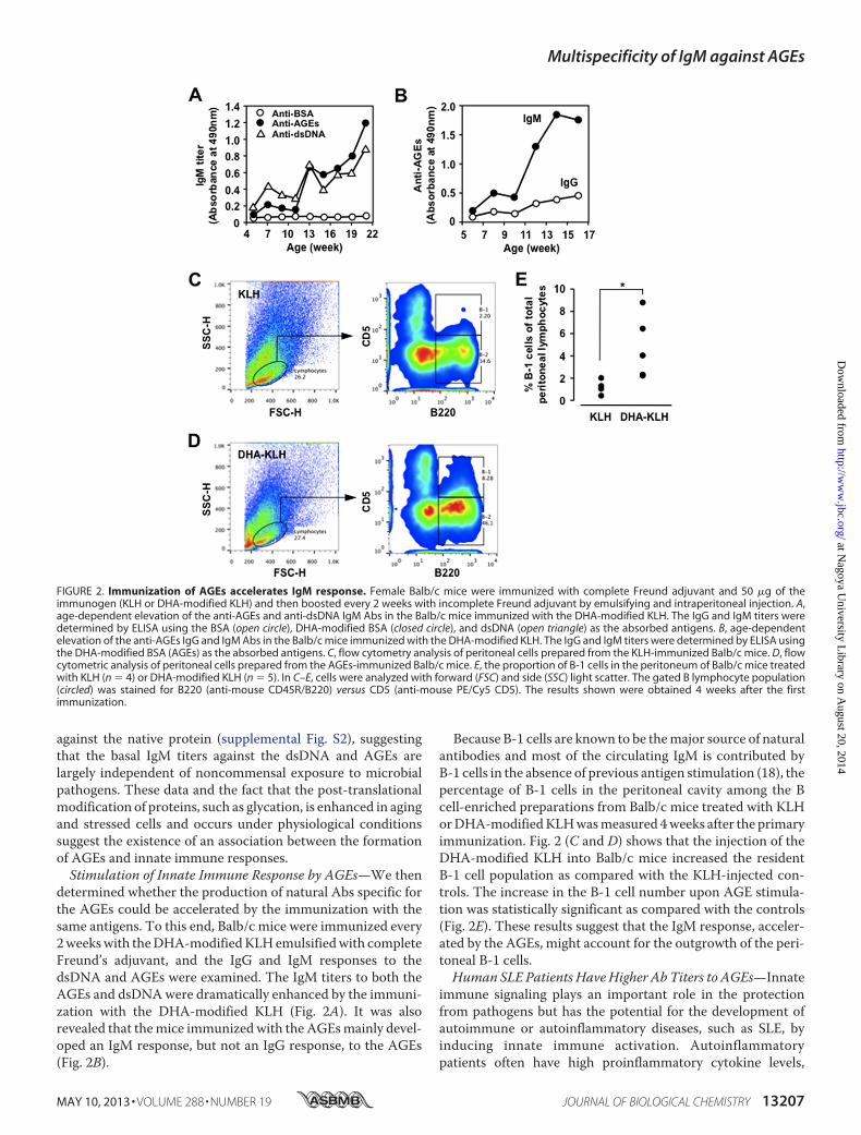
determined whether the production of natural Abs specific forthe AGEs could be accelerated by the immunization with thesame antigens. To this end, Balb/c mice were immunized every2weekswith theDHA-modifiedKLHemulsifiedwith completeFreund’s adjuvant, and the IgG and IgM responses to thedsDNA and AGEs were examined. The IgM titers to both theAGEs and dsDNAwere dramatically enhanced by the immuni-zation with the DHA-modified KLH (Fig. 2A). It was alsorevealed that themice immunized with the AGEsmainly devel-oped an IgM response, but not an IgG response, to the AGEs(Fig. 2B).
Because B-1 cells are known to be themajor source of naturalantibodies and most of the circulating IgM is contributed byB-1 cells in the absence of previous antigen stimulation (18), thepercentage of B-1 cells in the peritoneal cavity among the Bcell-enriched preparations from Balb/c mice treated with KLHorDHA-modifiedKLHwasmeasured 4weeks after the primaryimmunization. Fig. 2 (C and D) shows that the injection of theDHA-modified KLH into Balb/c mice increased the residentB-1 cell population as compared with the KLH-injected con-trols. The increase in the B-1 cell number upon AGE stimula-tion was statistically significant as compared with the controls(Fig. 2E). These results suggest that the IgM response, acceler-ated by the AGEs, might account for the outgrowth of the peri-toneal B-1 cells.Human SLE Patients Have Higher Ab Titers to AGEs—Innate
immune signaling plays an important role in the protectionfrom pathogens but has the potential for the development ofautoimmune or autoinflammatory diseases, such as SLE, byinducing innate immune activation. Autoinflammatorypatients often have high proinflammatory cytokine levels,
FIGURE 2. Immunization of AGEs accelerates IgM response. Female Balb/c mice were immunized with complete Freund adjuvant and 50 �g of theimmunogen (KLH or DHA-modified KLH) and then boosted every 2 weeks with incomplete Freund adjuvant by emulsifying and intraperitoneal injection. A,age-dependent elevation of the anti-AGEs and anti-dsDNA IgM Abs in the Balb/c mice immunized with the DHA-modified KLH. The IgG and IgM titers weredetermined by ELISA using the BSA (open circle), DHA-modified BSA (closed circle), and dsDNA (open triangle) as the absorbed antigens. B, age-dependentelevation of the anti-AGEs IgG and IgM Abs in the Balb/c mice immunized with the DHA-modified KLH. The IgG and IgM titers were determined by ELISA usingthe DHA-modified BSA (AGEs) as the absorbed antigens. C, flow cytometry analysis of peritoneal cells prepared from the KLH-immunized Balb/c mice. D, flowcytometric analysis of peritoneal cells prepared from the AGEs-immunized Balb/c mice. E, the proportion of B-1 cells in the peritoneum of Balb/c mice treatedwith KLH (n � 4) or DHA-modified KLH (n � 5). In C–E, cells were analyzed with forward (FSC) and side (SSC) light scatter. The gated B lymphocyte population(circled) was stained for B220 (anti-mouse CD45R/B220) versus CD5 (anti-mouse PE/Cy5 CD5). The results shown were obtained 4 weeks after the firstimmunization.
Multispecificity of IgM against AGEs
MAY 10, 2013 • VOLUME 288 • NUMBER 19 JOURNAL OF BIOLOGICAL CHEMISTRY 13207
at Nagoya U
niversity Library on A
ugust 20, 2014http://w
ww
.jbc.org/D
ownloaded from

which may come from activation of the innate immunity (19).SLE is a potentially fatal systemic autoimmune disease charac-terized by the increased production of auto-Abs, immune com-plex deposition in the microvasculature, leukocyte infiltration,and, ultimately, tissue damage in a number of organs. Of themultiple auto-Abs described in this disease, the Abs against thenative DNA are among the most characteristic, yet the trigger-ing antigen in the disease is still unknown. The appearance ofthese Abs in humans and in murine models of lupus correlateswith the progression of the disease, and by comparison with allthe other lupus auto-Abs, those against the dsDNA are thoughtto be the most pathogenic and involved in the development ofrenal pathology. To evaluate the presence of IgM Abs in theautoimmune diseases, we measured the plasma Ab titersdirected against the dsDNA and AGEs. The patients with SLEindeed exhibited significant increases in the Ab titers to theDHA-modified proteins (Fig. 3B), as well as to the dsDNA (Fig.3A) compared with the controls and the patients with IgAnephropathy. A high correlation was observed between theanti-dsDNA IgM and anti-AGEs IgM titers in the SLE patients(Fig. 3C).Elevated Immune Response to AGEs in SLE-prone MRL-lpr
Mice—The presence of natural IgM Abs against the AGEs wasalso evaluated in the SLE-prone mice. We first determinedwhether the AGEs could be recognized by the sera from theMRL-lprmice, which carry a defective Fas gene and develop aspontaneous lupus-like disease as they age, and from the con-trol MRL-MpJ mice. The AGEs prepared upon incubation ofBSA with DHA showed a significant cross-reactivity with the
sera from the MRL-lpr mice, whereas no cross-reactivity withthe sera from the control MRL-MpJ mice was observed (sup-plemental Fig. S3). When the age-dependent change in the Abtiters was measured in the sera from the MRL-lpr mice andcontrol MRL-MpJ mice, only the MRL-lpr mice displayed aspontaneous age-dependent elevation of the IgG and IgMresponses to both dsDNA and AGEs (Fig. 4). Specificity of Absthat deposited in the glomeruli of the MRL-lpr mice alsoshowed that the titers of the IgG and IgMAbs against the AGEswere significantly higher in theMRL-lprmice than in theMRL-MpJ mice (supplemental Fig. S4). These clinical and animaldata suggest that the Ab response against the AGEs may be animmunological characteristic common to SLE.Sequence Analysis of mAbs against AGEs—To establish the
mAbs that recognize the AGEs, we sought to isolate thehybridoma clones, producing the Abs specific for the AGEs,from the Balb/c mice immunized with the AGEs and from theMRL-lpr mice. Three IgM mAbs, clones BDM1, BDM2, andBDM3, showing a recognition specificity toward the AGEs, wasestablished from the immunized Balb/c mice after screeningbased on the specific binding to the corresponding antigen(AGEs). We also established three hybridoma clones, ADL7,ADL19, and DDL17, producing the IgM Abs, and threehybridoma clones, ADL13, DDL18, and DDL20, producing theIgG Abs, from the MRL-lprmice.The VH and VL nucleotide sequences and the corresponding
gene families were determined for the six anti-AGEs IgMmAbs(Fig. 5). V region gene usage and sequence alignment of theanti-AGEs mAbs are shown in A and B of Fig. 5, respectively.With the exception of the two IgM mAbs, clones DDL17 andBDM2, selected for the J558 and 3609 gene expressions, respec-tively, these alignments clearly showed preferential VH usage.
FIGURE 3. Elevation of innate immune response to the AGEs in autoim-mune diseases. A, the immunoreactivity of human plasma with the dsDNA.B, the immunoreactivity of human plasma with DHA-modified proteins. C,linear regression analysis between serum anti-dsDNA IgM and anti-DHA-modified protein IgM titers in SLE patients. The plasma samples were pre-pared from 5 healthy individuals, 20 patients with IgA nephropathy (IgA-N),and 26 patients with SLE. The levels of the IgM Abs against the dsDNA andAGEs in the plasma samples were measured by ELISA using calf thymusdsDNA and DHA-modified protein, respectively, as the coating antigens. Thecalculated p values were obtained by Welch’s t test analysis.
FIGURE 4. Age-dependent elevation of IgG and IgM responses to bothdsDNA and AGEs in SLE-prone MRL-lpr mice compared with those in thewild-type MRL-MpJ mice. A, anti-AGEs IgG. B, anti-AGEs IgM. C, anti-dsDNAIgG. D, anti-dsDNA IgM. The antibody titers were determined by ELISA usingthe DHA-modified BSA and dsDNA as the absorbed antigens. The IgG and IgMtiters were determined by ELISA using the dsDNA and DHA-modified proteinas the absorbed antigens. Each point represents the average value of sixanimals.
Multispecificity of IgM against AGEs
13208 JOURNAL OF BIOLOGICAL CHEMISTRY VOLUME 288 • NUMBER 19 • MAY 10, 2013
at Nagoya U
niversity Library on A
ugust 20, 2014http://w
ww
.jbc.org/D
ownloaded from

Most notably, two IgM mAbs, ADL7 and ADL19, arising fromtheMRL-lprmice and two IgMmAbs, BDM1 and BDM3, aris-ing from the Balb/c mice immunized with the AGEs are allencoded by the VH7183 subfamilies. In addition, all of theseIgM mAbs indeed showed a high nucleotide homology of90–96% with the VH7183 subfamilies. The increased VH7183family usage is known to be reminiscent of the signatures ofnatural Abs typically produced by B-1 cells (20–22). Thus,these IgM mAbs cloned from these mice might represent anexpansion of the population of natural Abs ubiquitouslyexpressed in healthy individuals.Multispecificity of Anti-AGEs mAbs—Because IgM titers to
the native dsDNA, in addition to theAGEs,were detected in thesera from the Balb/c mice (Fig. 1), the cross-reactivity of theanti-AGEs IgM mAbs toward the dsDNA were evaluated. As
expected, the IgM mAbs, BDM1, BDM2, and BDM3, estab-lished from the Balb/c mice immunized with the AGEs, cross-reacted not only with the DHA-modified BSA but also with thedsDNA (Fig. 6A). In addition, the IgG mAbs, ADL13, DDL18,and DDL20, and the IgM mAbs, ADL7, ADL19, and DDL17,cloned from the SLE mice, also cross-reacted with both theAGEs and dsDNA (Fig. 6B). Thus, a subset of IgG and IgMmAbs, showing a specificity toward the AGEs, were found to bethe multispecific mAbs that could recognize the dsDNA as analternative antigen.Involvement of Electronegative Potential of Antigens in the
Recognition by the Anti-AGEs mAbs—It is well known that theAbs to dsDNA often bind to other antigens in addition todsDNA. The additional reactivities provide a view of antibody-antigen interactions that may partly depend on the structural
FIGURE 5. Sequence analysis of mAbs against AGEs. A, V region gene usage of the IgM mAbs directed against the AGEs. B, sequence alignment of thehypervariable regions of anti-AGEs mAbs. The completely conserved residues are indicated by an asterisk (*), and the essentially conserved ones are indicatedby a period (.) or a colon (:). The sequences were aligned using program ClustalW (version 1.82) and manually modified. The residues are colored according tothe standard ClustalX color scheme. The sequences have the following DDBJ accession numbers: ADL7_L, AB793279; DDL17_L, AB793506; ADL19_L,AB793508; ADL19_H, AB793509; BDM1_L, AB793510; BDM1_H, AB793511; BDM2_L, AB793512; BDM2_H, AB793513; BDM3_L, AB793514; and BDM3_H,AB793515.
Multispecificity of IgM against AGEs
MAY 10, 2013 • VOLUME 288 • NUMBER 19 JOURNAL OF BIOLOGICAL CHEMISTRY 13209
at Nagoya U
niversity Library on A
ugust 20, 2014http://w
ww
.jbc.org/D
ownloaded from

similarity of the antigens (molecular mimicry). Thus, we spec-ulated that the multispecificity of the Abs directed against theAGEs might arise through a molecular and/or immunologicmimicry among the antigens, including the dsDNA and AGEs.To gain an insight into this mechanism, we examined the effectof the DHA treatment on the mobility of the protein by nativegel electrophoresis and observed an enhanced anodic mobilitycompared with the native protein (supplemental Fig. S5). Thedata implicate that DHA gave rise to the increased negativecharge of the protein probably because of the modification ofthe �-amino group of the lysine residues. The data also raisedthe possibility that the electronegative potential of antigensmight be responsible for the recognition by the anti-AGEsmAbs. It has indeed been suggested that a large number ofproteins, including the anti-DNA Abs, may recognize and beguided to their binding sites on the dsDNA helix through spe-cific arginines reading the electronegative potential in theminor groove (23). Hence, using a variety of carbonyl com-pounds, we prepared several modified proteins and evaluatedthe correlation between the electronegative potential of themodified proteins and their cross-reactivity with the anti-AGEsmAbs. Among the carbonyl compounds tested, the lipid per-oxidation products, such as 4-oxo-2-hexenal and 4-oxo-2-non-enal, and most of the glycolytic aldehydes, such as glyoxal,methylglyoxal, glyceraldehyde, glycolaldehyde, and dihydroxy-acetone, gave rise to a significant anodic mobility shift in theprotein on the native gel electrophoresis (Fig. 7,A andB), whichwas well correlated with the cross-reactivity of the Ab againstthe AGEs (Fig. 7C). Of interest, despite the fact that 2-alkenals(C3–C10) are strong electrophiles, which could readily reactwith lysine residues of proteins, a slight mobility shift of theprotein bands was observed. The data suggest that the covalentbinding of 2-alkenals hardly affect the charge on the protein.To provide further insight on the involvement of the electro-
negative potential of the antigens in the recognition by the anti-AGEs Abs, we tested whether the sera from the SLEmice couldrecognize electronegative molecules, such as acetylated pro-teins. As expected, the ELISA analysis showed that the SLE seracross-reactedwith the acetylated BSA (Fig. 8A). In addition, theacetylated protein was recognized by the anti-AGEsmAbs IgMBDM1 (Fig. 8B). We also observed that the SLE sera and anti-AGEs IgM showed a remarkable specificity toward other elec-
tronegativemolecules, including nucleic acids and phospholip-ids (supplemental Figs. S6 and S7).To obtain more direct and convincing evidence for the rec-
ognition of the electronegative proteins by the anti-AGEsmAbs, we analyzed the antibody-antigen interaction by flowcytometry assays. The assay using the solid phase paramagneticbeads coupled to the antigens (BSA, AGEs, and acetylated BSA)as the probes showed that the binding of the normal IgM tothese antigens was negligible, whereas the anti-AGEsmAb spe-cifically recognized the AGEs and acetylated protein (Fig. 8C).These data support our hypothesis that the electronegativepotential of the antigens might be involved, at least in part, inthe recognition by the natural mAbs directed against the AGEs.
DISCUSSION
Natural Abs, predominantly IgMs, are produced at tightlyregulated levels in the complete absence of external antigenicstimulation. They provide immediate, early, and broad protec-tion against pathogens, making them a crucial nonredundantcomponent of the humoral immune system (24, 25). They havealso been shown to play an important function in the hostresponse to the consequences of oxidative stress during oxida-tive events that occur when cells undergo apoptosis (reviewedin Ref. 26). These Abs are mainly produced by a subset of long-lived, self-replenishing B cells termed B-1 cells. It has beenshown that B-1 cells can also respond to certain antigenic stim-uli, e.g., 1,3-dextran, when presented on an appropriate carrieror in an appropriate immunization vehicle (reviewed inRef. 27).In our current study, we detected prominent IgM titers not onlyto the dsDNA and oxidized LDL, but also to the AGEs in thesera of normal, conventionally housedmice (Fig. 1) and specificpathogen-free-maintained mice (supplemental Fig. S2). Theapparent high prevalence of the AGEs as targets of natural Abslikely reflects the ubiquitous presence of these epitopes conse-quent to glycation events. The observation (Fig. 2,A andB) thatthe immunization of Balb/c mice with the AGEs in adjuvantstrongly induced the IgM response, whereas virtually no IgGresponse was observed, suggests that the immunization ofAGEsmay not induce the typical B cellmemory. Rather, the B-1cells of the appropriate specificity may respond to the immuni-zation of the AGEs by IgM production and limited isotypeswitching but do not produce high affinity Abs that might effi-
FIGURE 6. Multispecificity of the anti-AGEs mAbs. A, the immunoreactivity of anti-AGEs mAbs prepared from the Balb/c mice immunized with the DHA-modified KLH. Three hybridoma clones, BDM1, BDM2, and BDM3, producing IgM Abs specific for the AGEs were prepared from the mice. B, the immunoreac-tivity of the anti-AGEs mAbs prepared from MRL-lpr mice against the AGEs. Three hybridoma clones, ADL7, ADL19, and DDL17, producing the IgM Abs specificfor the AGEs, and three hybridoma clones, ADL13, DDL18, and DDL20, producing IgG Abs specific for the AGEs, were prepared from the female MRL-lpr mice(20 weeks old). The IgM titers were determined by ELISA using dsDNA and native and DHA-modified proteins as the absorbed antigens.
Multispecificity of IgM against AGEs
13210 JOURNAL OF BIOLOGICAL CHEMISTRY VOLUME 288 • NUMBER 19 • MAY 10, 2013
at Nagoya U
niversity Library on A
ugust 20, 2014http://w
ww
.jbc.org/D
ownloaded from

ciently react with the self-antigens that selected these Abs intothe B-1 repertoire. This hypothesis can be supported by theobservations that the treatment of Balb/c mice with the AGEsintraperitoneally enriched B-1 cells in the peritoneal cavity
after stimulation (Fig. 2, C–E). The mechanism(s) for elevationof the IgM titers in the Balb/cmice immunizedwith theAGEs ispresently unknown. However, it may not be unlikely that theIgM Abs found associated with aging and oxidative stress rep-resent an expansion of the population of theseAbs ubiquitouslyexpressed in normal healthy mice or, alternatively, representthe products of an antigen-selectedB lymphocyte population. Ithas indeed been noted that the established B-1 cell clones canbe expanded by antigen exposure, leading to the increased IgMlevels in the plasma (28).Based on the finding that the high IgM titers to the AGEs
were observed in normal and SLEmice sera, we isolated severalhybridoma clones, producing the Abs specific for the AGEsfrom theAGEs-immunizedBalb/c andMRL-lprmice and char-acterized their genetic and structural origin and specificity indetail. V gene analysis revealed several striking features of theseanti-AGEsmAbs, most notably the frequent use of the VH7183gene subfamilies by four IgMmAbs established from theAGEs-immunized Balb/c mice and disease-associated MRL-lpr mice(Fig. 5A). It is clear that immunization with the AGEs results inthe expansion of the VH7183 encoded B cell pool, suggestingthat the immune responses have a similar B cell origin. VH7183is the smallest and most JH-proximal of the mouse VH familiesand is expressed in the early repertoire encoding for a widerange of polyreactive and autoreactive specificities (15). Thus,there is a very strong indication that the anti-AGEs specificity ispreferentially encoded by VH7183, although it is also clear thatthe anti-AGEs IgMmAbs can on occasion be encoded by otherVH gene families, such as J558 and 3609.Of interest, most of the anti-AGEs IgG and IgMmAbs estab-
lished from these SLE-prone mice cross-reacted with thedsDNA. In addition, similar multispecific IgM Abs were
FIGURE 7. Electronegative potential of the aldehyde-modified proteins and their cross-reactivity with the anti-AGEs IgM mAb. The modified proteinswere prepared by incubating BSA (1.0 mg/ml) with 1 mM lipid peroxidation-derived aldehydes in 1 ml of PBS (pH 7.4) at 37 °C for 24 h (A) or with 25 mM glycolyticaldehydes (B) in 5 ml of PBS (pH 7.4) at 37 °C for 7 days. The anti-AGEs IgM mAb BDM1 prepared from the Balb/c mice immunized with the DHA-modified KLHwas used. Mobility shifts of modified proteins were examined in a nondenaturing polyacrylamide gel. The native BSA gave three bands, representing monomer(m), dimer (d), and trimer (t) of the protein. The IgM antibody titer was determined by ELISA using the DHA-modified BSA as the absorbed antigens. Thealdehydes used were: C3, acrolein; C4, crotonaldehyde; C5, 2-pentenal; C6, 2-hexenal; C7, 2-heptenal; C8, 2-octenal; C9, 2-nonenal; C10, 2-decenal; HNE,4-hydroxy-2-nonenal; ONE, 4-oxo-2-nonenal; OHE, 4-oxo-2-hexenal; DHA, dehydroascorbic acid; GO, glyoxal; MG, methylglyoxal; GA, glycolaldehyde; GLA,glyceraldehyde; DA, dihydroxyacetone. C, correlation between relative mobility of the modified proteins and their cross-reactivity with the anti-AGEs IgM. Therelative mobility value was obtained by dividing the protein migration distance by the dye front distance in the gel electrophoresis test (A and B). The Ab titerwas determined by ELISA using the DHA-modified BSA (AGEs) as the absorbed antigens.
FIGURE 8. Involvement of electronegative potential of antigens in therecognition by the anti-AGEs mAbs. A, cross-reactivity of the sera from theMRL-MpJ and MRL-lpr mice with native BSA (open bar) and acetylated BSA(AcBSA, closed bar). B, cross-reactivity of the anti-AGEs IgM BDM1 establishedfrom the Balb/c mice immunized with the DHA-modified KLH with acetylatedBSA. The antibody titers were determined by ELISA using the native BSA andDHA-modified and acetylated BSA as the absorbed antigens. C, flow cytomet-ric analysis of antigen-antibody interaction. The solid phase paramagneticbeads coupled to the antigens, BSA (black line), AGEs (blue line), and acety-lated BSA (red line), were used as the probes. The control mouse IgM (leftpanel) and representative anti-AGEs IgM BDM1 (right panel) were tested forbinding to the protein-coupled beads. The number of binding events is plot-ted against fluorescence intensity.
Multispecificity of IgM against AGEs
MAY 10, 2013 • VOLUME 288 • NUMBER 19 JOURNAL OF BIOLOGICAL CHEMISTRY 13211
at Nagoya U
niversity Library on A
ugust 20, 2014http://w
ww
.jbc.org/D
ownloaded from
obtained from the Balb/c mice immunized with the AGEs. Thesix anti-AGEs IgM mAbs differ in both their heavy and lightchain sequences (Fig. 5B). Therefore, the relative importance ofthe heavy and light chains in the actual binding to the antigens,such as AGEs and dsDNA, remains to be clarified. One possi-bility is that different VH and VL germ line genes are used toproduce similar combining sites. A second possibility is that thedifferent combinations result from the difference in the fineantigenic specificities of the Abs. It is also notable that the oneIgM mAb BDM2 presented here differs in its specificity fordsDNA. The mAb possesses only low avidity to dsDNA,whereas other anti-AGEs IgM mAbs bound dsDNA with rela-tively high affinity. Thismay be associated with the observationthat the amino acid sequence of the mAb BDM2 VH gene haslimited identity (40–42%) with that of other mAb VH genes. Itis therefore likely that amutation in themAb BDM2may inter-fere with the recognition of dsDNA. Several different antigeniccross-reactivities have been identified for the anti-DNA Abs(29). These Abs share structural similarities with the Absagainst bacterial polysaccharide, and some cross-react with thebacterial polysaccharide and protect mice against a lethal bac-terial infection (30, 31). Other studies have also demonstrated across-reactivity of the anti-DNA Abs with microbial proteinantigens, non-nucleic acid autoantigens, cell membranes andextracellular matrix components (32, 33). These cross-reactiv-ities for an endogenous self- or neoself-antigen and an exoge-nous pathogen have also been described as a characteristic ofnatural Abs (34). A pattern of broad reactivity of a preformedpool of Abs has been suggested to be required for the rapid andimmediate recognition and protection against invading patho-gens. Recognition of multiple epitopes by the same Ab is ofvalue to an organism, because this may result in an increase inthe Ab repertoire and immune diversity without the need foradditional lymphocytes expressing distinct Abs.The present data suggest that the multispecificity of the nat-
ural Abs toward two or more distinct antigenic determinantscould arise from antigens associated with the covalent modifi-cation of proteins by the reactive molecules that originatedfrom the glycation and lipid peroxidation reactions. It is notunlikely that, because these reactive molecules are known togenerate a variety of structures, some specific adducts gener-ated in the modified proteins may be associated with the pro-duction of the natural Abs that simultaneously recognize otherdistinct molecules, such as dsDNA. However, such adductscommonly produced uponmodification of protein by the reac-tive molecules that originated from the glycation and lipid per-oxidation reactions are not known. In addition, we found thatthe anti-AGEs mAbs recognized a variety of electronegativemolecules, including the acetylated proteins, nucleic acids, andphospholipids (Fig. 8 and supplemental Figs. S6 and S7). Thesedata suggest an alternative mechanism in which the electrone-gative potential could be involved, at least in part, in the multi-ple cross-reactivity of the mAbs. This hypothesis may besupported by themechanism for the cross-reactivity of the anti-DNAAbs, in which there are flexible phosphodiester polymers,such as RNA, teichoic acid, or othermolecules, whose distribu-tion of phosphates or similar negatively charged epitopes con-
formwith the available contacts in the anti-dsDNAAb combin-ing site (35).It has been shown that the natural IgM Abs could facilitate
the removal of damaged cells in vivo. Chang et al. (36) demon-strated that the IgM Abs against the lipid peroxidation-modi-fied proteins showed specific binding to the surface of apoptoticcells, but not to the surface of normal cells. They have alsoshown that the Abs inhibited the macrophage uptake of apo-ptotic cells, whereas the control IgM did not. Based on thesedata, they made a hypothesis that oxidized lipids and/or oxi-dized lipid-protein adducts on the surface of apoptotic cells,presumably derived from membrane peroxidation that occursduring apoptosis, could be ligands for the phagocytosis of apo-ptotic cells. We have also observed that the natural IgM Absagainst the AGEs established from the AGEs-immunizedBalb/c and MRL-lpr mice could bind the apoptosis-inducedcells.3 Of interest, it has been suggested that, by altering thesurface charge, the lipid changes that accompany apoptosismay cause redistribution of cationic proteins, potentially con-tributing to the execution of apoptotic cells. Indeed, the accu-mulation of anionic lipids in certain cellular membranes hasbeen shown to serve to target and retain proteins with polyca-tionic motifs (37). Thus, the binding of the Abs against theAGEs to the surface of apoptotic cells may also be associatedwith the increased electronegative potential of the cells.The clearance of dying cells is one of the most essential
responsibilities of the immune system,which is required to pre-vent uncontrolled inflammation and autoimmunity. In themurine immune system, natural IgM Abs that recognize apo-ptotic cells have been shown to enhance the phagocytic clear-ance of dead and dying cells and to suppress innate immunesignaling pathways. In patients with SLE, the IgM auto-Abs,which bind to the neo-epitopes on apoptotic cells, have beendemonstrated to be present at significantly higher levels inpatients with a lower disease activity and with less severe organdamage (38). On the other hand,Witte (39) proposed a possiblemechanism of protection by the IgM auto-Abs against thedsDNA, in which the IgM Abs may improve the clearance ofpathogenic immune complexes containing IgG. Others havealso suggested that the IgM Abs remove cellular debris andthereforemay prevent the formation of IgGAbs (40). Indeed, inthe absence of secreted IgM, normal mice spontaneouslydevelop autoreactive IgG specific for the dsDNA (41). It has alsobeen suggested that the IgM Abs have a protective role againstthe development of glomerulonephritis (42). In addition, in theSLE-proneMRL-lprmice, the absence of secreted IgM acceler-ates the development of IgG auto-Abs and glomerulonephritis,and the mice succumb to the disease at an earlier age. Thesefindings demonstrate that the secreted IgM can suppress thedevelopment of IgG auto-Abs and autoimmune disease underphysiological conditions. Because autoantigenic epitopeswould be generated even in healthy individuals, this IgM func-tion is important for maintaining homeostasis. Thus, the highprevalence of the IgM Abs against the AGEs in the SLE-pronemice and SLE patients likely reflects the highest reactivity of
3 M. Chikazawa and K. Uchida, unpublished data.
Multispecificity of IgM against AGEs
13212 JOURNAL OF BIOLOGICAL CHEMISTRY VOLUME 288 • NUMBER 19 • MAY 10, 2013
at Nagoya U
niversity Library on A
ugust 20, 2014http://w
ww
.jbc.org/D
ownloaded from

DHA and its metabolites toward protein and the ubiquitouspresence of such AGEs consequent to oxidative events.In conclusion, the results of this study raised the possibility
that the AGEsmay be a ubiquitous target of natural Abs in bothmice and humans. In addition, the apparent high prevalence ofthe AGEs as targets of the natural Abs likely reflects the ubiq-uitous presence of these epitopes consequent to glycationevents. These findings offer an attractive hypothesis that theAGEs could be an important trigger of innate immunity andtherefore contribute to the protection against certain exoge-nous invading pathogens and endogenous damage-associatedmolecules.
Acknowledgments—We thank Dr. Sohei Ito (University of Shizuoka)for the antibody sequence analysis and Dr. Takaaki Kojima (NagoyaUniversity) for flow cytometric analysis. We also thank Yuki Hondofor excellent editorial support.
REFERENCES1. Monnier, V. M., Sell, D. R., and Genuth, S. (2005) Glycation products as
markers and predictors of the progression of diabetic complications.Ann.N.Y. Acad. Sci. 1043, 567–581
2. Brownlee,M., Cerami, A., and Vlassara, H. (1988) Advanced glycosylationend products in tissue and the biochemical basis of diabetic complications.N. Engl. J. Med. 318, 1315–1321
3. Baynes, J. W., and Monnier, V. M. (1989) Prog. Clin. Biol. Res. 304, 1–414. Vlassara, H. (2001) The AGE-receptor in the pathogenesis of diabetic
complications. Diabetes Metab. Res. Rev. 17, 436–4435. Kislinger, T., Fu, C., Huber, B., Qu,W., Taguchi, A., Du Yan, S., Hofmann,
M., Yan, S. F., Pischetsrieder, M., Stern, D., and Schmidt, A. M. (1999)N�-(Carboxymethyl)lysine adducts of proteins are ligands for receptor foradvanced glycation end products that activate cell signaling pathways andmodulate gene expression. J. Biol. Chem. 274, 31740–31749
6. Nemet, I., andMonnier, V.M. (2011)VitaminCdegradation products andpathways in the human lens. J. Biol. Chem. 286, 37128–37136
7. Dunn, J. A., Ahmed, M. U., Murtiashaw, M. H., Richardson, J. M., Walla,M. D., Thorpe, S. R., and Baynes, J. W. (1990) Reaction of ascorbate withlysine and protein under autoxidizing conditions: formation of N�-(car-boxymethyl)lysine by reaction between lysine and products of autoxida-tion of ascorbate. Biochemistry 29, 10964–10970
8. Nagaraj, R. H., Sell, D. R., Prabhakaram, M., Ortwerth, B. J., and MonnierV.M. (1991) High correlation between pentosidine protein crosslinks andpigmentation implicates ascorbate oxidation in human lens senescenceand cataractogenesis. Proc. Natl. Acad. Sci. U.S.A. 88, 10257–10261
9. Tessier, F., Obrenovich, M., and Monnier, V. M. (1999) Structure andmechanism of formation of human lens fluorophore LM-1. Relationshipto vesperlysine A and the advanced Maillard reaction in aging, diabetes,and cataractogenesis. J. Biol. Chem. 274, 20796–20804
10. Witztum, J. L., Steinbrecher, U. P., Kesaniemi, Y. A., and Fisher, M. (1984)Autoantibodies to glucosylated proteins in the plasma of patients withdiabetes mellitus. Proc. Natl. Acad. Sci. U.S.A. 81, 3204–3208
11. Shibayama, R., Araki, N., Nagai, R., and Horiuchi, S. (1999) Autoantibodyagainst N�-(carboxymethyl)lysine. An advanced glycation end product ofthe Maillard reaction. Diabetes 48, 1842–1849
12. Engelbertsen, D., Anand, D. V., Fredrikson, G. N., Hopkins, D., Corder, R.,Shah, P. K., Lahiri, A., Nilsson, J., and Bengtsson, E. (2012) High levels ofIgM against methylglyoxal-modified apolipoprotein B100 are associatedwith less coronary artery calcification in patients with type 2 diabetes.J. Intern. Med. 271, 82–89
13. Shibata, T., Shimozu, Y., Wakita, C., Shibata, N., Kobayashi, M., Machida,S., Kato, R., Itabe, H., Zhu, X., Sayre, L. M., and Uchida, K. (2011) Lipidperoxidationmodification of protein generatesN�-(4-oxononanoyl)lysineas a pro-inflammatory ligand. J. Biol. Chem. 286, 19943–19957
14. Basu S. K., Goldstein J. L., Anderson G. W., and Brown M. S. (1976)
Degradation of cationized low density lipoprotein and regulation of cho-lesterol metabolism in homozygous familial hypercholesterolemia fibro-blasts. Proc. Natl. Acad. Sci. U.S.A. 73, 3178–3182
15. Antone, S. M., Adderson, E. E., Mertens, N. M., and Cunningham, M. W.(1997) Molecular analysis of V gene sequences encoding cytotoxic anti-streptococcal/anti-myosin monoclonal antibody 36.2.2 that recognizesthe heart cell surface protein laminin. J. Immunol. 159, 5422–5430
16. Altschul, S. F., Madden, T. L., Schäffer, A. A., Zhang, J., Zhang, Z., Miller,W., and Lipman, D. J. (1997) Gapped BLAST and PSI-BLAST. A newgeneration of protein database search programs. Nucleic Acids Res. 25,3389–3402
17. Wardemann, H., Boehm, T., Dear, N., and Carsetti, R. (2002) B-1a B cellsthat link the innate and adaptive immune responses are lacking in theabsence of the spleen. J. Exp. Med. 195, 771–780
18. Hayakawa, K., and Hardy, R. R. (2000) Development and function of B-1cells. Curr. Opin. Immunol. 12, 346–353
19. Kawasaki, T., Kawai, T., and Akira, S. (2011) Recognition of nucleic acidsby pattern-recognition receptors and its relevance in autoimmunity. Im-munol. Rev. 243, 61–73
20. Diaw, L., Magnac, C., Pritsch, O., Buckle, M., Alzari, P. M., and Dighiero,G. (1997) Structural and affinity studies of IgM polyreactive natural au-toantibodies. J. Immunol. 158, 968–976
21. Holmberg, D. (1987) High connectivity, natural antibodies preferentiallyuse 7183 and QUPC 52 VH families. Eur. J. Immunol. 17, 399–403
22. Pennell, C. A., Arnold, L. W., Haughton, G., and Clarke, S. H. (1988)Restricted Ig variable region gene expression among Ly-1�B cell lympho-mas. J. Immunol. 141, 2788–2796
23. Lindemose, S., Nielsen, P. E., Hansen, M., and Møllegaard, N. E. (2011) ADNAminor groove electronegative potential genome map based on pho-to-chemical probing. Nucleic Acids Res. 39, 6269–6276
24. Miller, Y. I., Choi, S. H., Wiesner, P., Fang, L., Harkewicz, R., Hartvigsen,K., Boullier, A., Gonen, A., Diehl, C. J., Que, X., Montano, E., Shaw, P. X.,Tsimikas, S., Binder, C. J., and Witztum, J. L. (2011) Oxidation-specificepitopes are danger-associated molecular patterns recognized by patternrecognition receptors of innate immunity. Circ. Res. 108, 235–248
25. Weismann, D., and Binder, C. J. (2012) The innate immune response toproducts of phospholipid peroxidation. Biochim. Biophys. Acta 1818,2465–2475
26. Grönwall, C., Vas, J., and Silverman, G. J. (2012) Protective roles of naturalIgM antibodies. Front. Immunol. 3, 66
27. Baumgarth, N. (2011) The double life of a B-1 cell. Self-reactivity selectsfor protective effector functions. Nat. Rev. Immunol. 11, 34–46
28. Hartvigsen, K., Chou, M. Y., Hansen, L. F., Shaw, P. X., Tsimikas, S.,Binder, C. J., and Witztum, J. L. (2009) The role of innate immunity inatherogenesis. J. Lipid Res. 50, S388–393
29. Spatz, L., Iliev, A., Saenko, V., Jones, L., Irigoyen,M.,Manheimer-Lory, A.,Gaynor, B., Putterman, C., Bynoe, M., Kowal, C., Kuo, P., Newman, J., andDiamond, B. (1997) Studies on the structure, regulation, and pathogenicpotential of anti-dsDNA antibodies.Methods 11, 70–78
30. Kowal, C., Weinstein, A., and Diamond, B. (1999) Molecular mimicrybetween bacterial and self antigen in a patient with systemic lupus erythe-matosus. Eur. J. Immunol. 29, 1901–1911
31. Limpanasithikul, W., Ray, S., and Diamond, B. (1995) Cross-reactive an-tibodies have both protective and pathogenic potential. J. Immunol. 155,967–973
32. Chan, T. M., Leung, J. K., Ho, S. K., and Yung, S. (2002) Mesangial cell-binding anti-DNA antibodies in patients with systemic lupus erythema-tosus. J. Am. Soc. Nephrol. 13, 1219–1229
33. Jacob, L., Lety,M. A., Choquette, D., Viard, J. P., Jacob, F., Louvard, D., andBach, J. F. (1987) Presence of antibodies against a cell-surface protein,cross-reactive with DNA, in systemic lupus erythematosus. A marker ofthe disease. Proc. Natl. Acad. Sci. U.S.A. 84, 2956–2959
34. Thompson, K. M., Sutherland, J., Barden, G., Melamed, M. D., Wright,M. G., Bailey, S., and Thorpe, S. J. (1992) Human monoclonal antibodiesspecific for blood group antigens demonstrate multispecific propertiescharacteristic of natural autoantibodies. Immunology 76, 146–157
35. Radic, M. Z., and Weigert, M. (1994) Genetic and structural evidence forantigen selection of anti-DNA antibodies. Annu. Rev. Immunol. 12,
Multispecificity of IgM against AGEs
MAY 10, 2013 • VOLUME 288 • NUMBER 19 JOURNAL OF BIOLOGICAL CHEMISTRY 13213
at Nagoya U
niversity Library on A
ugust 20, 2014http://w
ww
.jbc.org/D
ownloaded from

487–52036. Chang, M. K., Bergmark, C., Laurila, A., Hörkkö, S., Han, K. H., Friedman,
P., Dennis, E. A., andWitztum, J. L. (1999)Monoclonal antibodies againstoxidized low-density lipoprotein bind to apoptotic cells and inhibit theirphagocytosis by elicited macrophages. Evidence that oxidation-specificepitopes mediate macrophage recognition. Proc. Natl. Acad. Sci. U.S.A.96, 6353–6358
37. Yeung T, Terebiznik M, Yu L, Silvius J, Abidi, W.M., Philips M, Levine T,Kapus A, and Grinstein S. (2006) Receptor activation alters inner surfacepotential during phagocytosis. Science 313, 347–351
38. Grönwall, C., Akhter, E., Oh, C., Burlingame, R. W., Petri, M., and Silver-man, G. J. (2012) IgM autoantibodies to distinct apoptosis-associated an-
tigens correlate with protection from cardiovascular events and renal dis-ease in patients with SLE. Clin. Immunol. 142, 390–398
39. Witte, T. (2008) IgM antibodies against dsDNA in SLE. Clin. Rev. AllergyImmunol. 34, 345–347
40. Hahn, B. H. (1998) Antibodies to DNA. N. Engl. J. Med. 338, 1359–136841. Boes, M., Schmidt, T., Linkemann, K., Beaudette, B. C., Marshak-Roth-
stein, A., and Chen, J. (2000) Accelerated development of IgG autoanti-bodies and autoimmune disease in the absence of secreted IgM.Proc. Natl.Acad. Sci. U.S.A. 97, 1184–1189
42. Shoenfeld, Y., and Toubi, E. (2005) Protective autoantibodies. Role in ho-meostasis, clinical importance, and therapeutic potential. ArthritisRheum. 52, 2599–2606
Multispecificity of IgM against AGEs
13214 JOURNAL OF BIOLOGICAL CHEMISTRY VOLUME 288 • NUMBER 19 • MAY 10, 2013
at Nagoya U
niversity Library on A
ugust 20, 2014http://w
ww
.jbc.org/D
ownloaded from


![[Aging: role and control of glycation]](https://img.pdfslide.net/doc/110x75/635f0913ac6942764f03d5ce/aging-role-and-control-of-glycation.jpg)


