Embed Size (px)
Citation preview
RESEARCH ARTICLE
D-Ribosylated Tau forms globular aggregateswith high cytotoxicity
Lan Chen Æ Yan Wei Æ Xueqing Wang ÆRongqiao He
Received: 27 April 2009 / Accepted: 25 May 2009 / Published online: 11 June 2009
� Birkhauser Verlag, Basel/Switzerland 2009
Abstract Although the glycation of Tau that is involved in
paired helical filament formation in Alzheimer’s disease has
been widely studied, little attention has been paid to the role
of D-ribose in the glycation of Tau. Here, we show that Tau is
rapidly glycated in the presence of D-ribose, resulting in
oligomerization and polymerization. Glycated derivatives
appeared after 24 h incubation. Western blotting indicated
the formation of advanced glycation end-products (AGEs)
during initial stages of glycation. Thioflavin T-positive
(ThT-positive) aggregations that appeared from day 4 indi-
cated the globular-like features. Atomic force microscopy
revealed that the surface morphology of ribosylated Tau40
was globular-like. Kinetic studies suggested that D-ribosy-
lated Tau is slowly oligomerized and rapidly polymerized
with ThT-positive features. Moreover, D-ribosylated Tau
aggregates were highly toxic to SHSY5Y cells and resulted
in both apoptosis and necrosis. This work has demonstrated
that D-ribose reacted with Tau protein rapidly, producing
ThT-positive aggregations which had high cytotoxicity.
Keywords D-Ribose � Tau protein � Glycation �Aggregation � Cytotoxicity
Introduction
Tau is a major microtubule-binding protein that is impor-
tant for the assembly and stabilization of microtubules [1],
which are required for axonal transport and morphogenesis
[2, 3]. In a normal neuron, Tau is localized in the axons,
neuronal soma [4–6] and nuclei [7], and its function is
regulated by phosphorylation [8]. The discovery that Tau is
the major protein subunit of paired helical filaments
(PHFs)/neurofibrillary tangles in Alzheimer’s disease (AD)
has markedly stimulated interest in understanding the
structure and function of this protein [9–11]. Furthermore,
PHF-Tau is not only related to hyperphosphorylation
but also to other post-translational modifications such as
glycation [12–14].
Glycated Tau protein has been found in PHFs from the
brain tissues of Alzheimer’s patients [15]. In AD, non-
enzymatically glycated Tau induces neuronal oxidant
stress resulting in cytokine gene expression and release of
amyloid beta-peptide [16]. Kuhla and coworkers have
observed the promotive effects of reactive carbonyl
compounds (RCCs) on Tau aggregation and filament
formation [17]. As an in vitro study shows, glucose-
glycated Tau has been found to promote fibrillization by
shifting the equilibrium toward the fibrillized state, but
does not promote filament nucleation [18]. However, it is
still unclear whether glycation causes Tau to form amy-
loid-like aggregates. Thus, the relationship between
glycation and Tau misfolding is worth studying to clar-
ify the role of glycation in protein aggregation and
cytotoxicity.
Electronic supplementary material The online version of thisarticle (doi:10.1007/s00018-009-0058-7) contains supplementarymaterial, which is available to authorized users.
L. Chen � Y. Wei � X. Wang � R. He (&)
State Key Laboratory of Brain and Cognitive Sciences,
Institute of Biophysics, Chinese Academy of Sciences,
15 Datun Road, Chaoyang District, 100101 Beijing, China
e-mail: [email protected]
Y. Wei
Graduate University of Chinese Academy of Sciences,
19A Yu Quan Road, Shijingshan District, 100049 Beijing, China
R. He
Key Laboratory of Mental Health, Institute of Psychology,
Chinese Academy of Sciences, 100101 Beijing, China
Cell. Mol. Life Sci. (2009) 66:2559–2571
DOI 10.1007/s00018-009-0058-7 Cellular and Molecular Life Sciences
So far, most work has focused on the role of D-glucose
in the glycation of Tau [18–21]. D-Glucose exists in solu-
tion as an intramolecular hemiacetal in which the free
hydroxyl group at C-5 reacts with the aldehydic C-1, ren-
dering the latter asymmetric and producing a stereoisomer.
The six-membered aldopyranose ring is much more stable
than the aldofuranose five-membered ring because the
pentose ring of D-ribose is not planar but occurs in one of a
variety of conformations generally described as ‘‘puck-
ered’’ [22]. The unstable aldofuranose ring is vulnerable to
reaction with an amino group. Therefore, D-glucose is not
as efficient in glycating a protein as reducing furanoses
such as D-ribose.
D-Ribose is a naturally occurring pentose monosaccha-
ride present in all living cells and is an essential component
for energy production in the body. It is used to synthesize
nucleotides, nucleic acids, glycogen, and other important
metabolic products. D-Ribose is also formed in the body
from conversion of D-glucose via the pentose phosphate
pathway. Thus, D-ribose is continually present both intra-
cellularly and extracellularly, and has opportunities to react
with proteins and produce glycated derivatives. For this
reason, glycation of Tau protein with D-ribose needs to be
addressed and investigated.
Glycation of a protein causes fluorescence at 410 nm (or
450 nm) [23–26] that is thought to indicate the formation
of advanced glycation end-products (AGEs). However, few
authors have studied the relationship between the forma-
tion of the fluorescence and Tau protein aggregation, that
is, whether the formation of the fluorescence is positively
related to the polymerization of a protein, especially the
appearance of ThT-positive aggregations during glycation.
Clarification of this problem will be helpful for using
fluorescence to study the structure–function relationship of
a protein during glycation.
It is well known that glycation affects the structure and
function of proteins such as hemoglobin [27] and albumin
[28, 29]. Glycation of hemoglobin results in a marked loss of
its function in transportation of oxygen in the blood and in
releasing carbon dioxide in the lung [30]. In this laboratory,
we have also observed that glycation induced inactivation
and conformational change in D-glyceraldehyde-3-phos-
phate dehydrogenase [31, 32]. Rapid in vitro glycation of
a-synuclein with D-glucose (requiring less than 7 days),
however, did not result in distinct conformational changes
or inactivation of the protein [33]. Furthermore, our
unpublished data showed that extracellular ribose was able
to induce intracellular proteins including tau to be glycated.
We have also found that Tau protein aggregates in the
presence of formaldehyde at low concentrations and forms
amyloid-like deposits [34]. Here, we show that glycation of
Tau protein in the presence of D-ribose generates globular
ThT-positive cytotoxic deposits.
Materials and methods
Expression and purification of Tau40
Human wild type tau40 was expressed in Escherichia coli
BL21 (DE3) using the pRK172-tau40 plasmid (a kind gift
from Dr. Goedert of the University of Cambridge, UK) and
purified as described before [35, 36]. The purified Tau40
protein showed a single band on 12% SDS-PAGE gels with
a purity of over 95%. The identity of the Tau40 protein
band was confirmed by Western blotting using Tau-1
monoclonal antibody (Sigma, USA) (data not shown). The
purified Tau protein was lyophilized and stored at -70�C
before use.
Preparation of D-ribosylated protein
Tau40 (final concentration 0.2 mM) was freshly dissolved
in 20 mM Tris–HCl (pH 7.4), mixed with 1 M D-ribose,
and incubated for different time intervals at 37�C. Tau40 in
the absence of D-ribose and D-ribose alone were used
as controls. All solutions were filtered with 0.22 lm
membranes (Millipore, USA). A Bicinchoninic acid pro-
tein-assay kit (Pierce Biotechnology, USA) was used to
determine protein concentration [37].
NBT colorimetric fructosamine assay
The extent of glycation of individual Tau40 preparations
was assessed using the nitroblue tetrazolium (NBT) assay
as described previously [29, 38]. This method is based on a
color change correlated with the reduction of NBT to
monoformazan by Amadori rearrangement products in
alkaline buffer [39]. Along with 10 ll of the sample or
standard, 200 ll of 0.75 mM NBT (Ameresco, USA) was
added to a 96-well microplate. The kinetics of the reduc-
tion of NBT by fructosamine groups (0.1 M carbonate
buffer, pH 10.35) were measured at 540 nm using an MK3
microplate reader (Thermo, USA) after incubating for
30 min at 37�C. Standard curves were generated by addi-
tion of 10 ll of 1-deoxy-1-morpholino-D-fructose (1-DMF;
Sigma-Aldrich, USA). Fructosamine formation was moni-
tored by comparison with standard curves (R2 [ 0.99).
Measurement of fluorescence
Intrinsic fluorescence of Tau40 (final concentration 2 lM)
was monitored on an F4500 fluorescence spectrophotom-
eter (Hitachi, Japan). The emission spectrum from 290 to
500 nm was recorded by excitation at 280 nm at 25�C. To
assess the fluorescence of AGEs derived from glycated
protein, we scanned the emission spectrum from 350 to
500 nm (kex = 320 nm) as described previously [40].
2560 L. Chen et al.
Measurement of ThT-binding fluorescence
Tau40 (final concentration 2 lM) and ThT (30 lM; Sigma)
were mixed at 25�C, and fluorescence was subsequently
measured (kex 450 nm; kem 485 nm) as described [34].
Observation of protein aggregation with atomic force
microscopy (AFM)
The conditions for D-ribosylation of Tau were as described
above. All of the solutions used were filtered through a
0.22-lm filter. The glycated protein was diluted to the
desired concentration using Tris–HCl buffer (pH 7.4).
Samples (10 ll) were kept at room temperature for 5 min
to allow the protein to absorb onto the mica. Observation
under an atomic force microscope (Mutiplemode-I; Digital
Instruments, USA) was performed as described previously
[41].
Circular Dichroism (CD) measurements
Far-UV CD measurements were taken with a circular
dichrograph (Jasco J-720; Japan). Samples in 1-mm quartz
cuvettes were maintained at 25�C using a circulating water
bath. Spectra of D-ribose-glycated Tau40 (final concentra-
tion 20 lM) were measured (195–260 nm) with a step size
of 1.0 nm. All measurements were replicated 10 times and
averaged. The background of the corresponding buffers in
the absence of protein and D-ribose was subtracted for all
samples.
Cell culture
SHSY5Y cells were cultured in Dulbecco’s modified
Eagle’s medium (DMEM; Invitrogen, USA) containing
10% fetal bovine serum (Hyclone, USA), 100 IU/ml pen-
icillin and 100 lg/ml streptomycin (Sigma) at 37�C in a
humidified 5% CO2 incubator. Cells were grown to
70–80% confluence in 25-mm-diameter dishes and fed
every fourth day. The culture was replaced with serum-free
medium before addition of glycated protein during exper-
iments. Tau protein was incubated with D-ribose and
aliquots were taken at different time intervals to incubate
with cells for 8 h. After that, the medium was changed to
DMEM with 10% fetal bovine serum.
Cell viability test
To determine cell viability, we used the standard 3-(4,5-
dimethylthiazol-2-yl)-2,5-diphenyl tetrazolium bromide
(MTT; Sigma) test, with the slight modifications suggested
by Mayo and Stein [42]. SHSY5Y cells were seeded on a
96-well plate at a concentration of 105 cells per well with
or without exposure to glycated Tau40 (3-day incubation)
at various concentrations for 8 h. After 24 or 48 h, MTT
(final concentration 0.5 mg/ml) was added and plates were
incubated at 37�C for 4 h. The reaction was stopped by
replacement of the MTT-containing medium with 150 ll
dimethysulfoxide, and absorbance at 540 nm was mea-
sured on a Multiscan MK3 spectrophotometer (Thermo
Electron Corporation, USA).
Flow cytometric analysis
Cells undergoing apoptosis were detected by using double
staining with Annexin V-FITC/PI (propidium iodide) in the
dark according to the manufacturer’s instructions. Briefly,
cells attached to plastic dishes were harvested using 0.25%
trypsin and washed twice with cold PBS. Cell pellets were
suspended in 19 binding buffer (10 mM HEPES/NaOH,
pH 7.4, 140 mM NaCl, 2.5 mM CaCl2) at a concentration
of 1 9 106 cells/ml. Cells were then incubated with
AnnexinV-FITC and PI for 15 min (22–25�C) in the dark.
The stained cells were immediately analyzed on a flow
cytometer (FAC Svantage SE, USA). Each measurement
was replicated at least three times.
Data analysis
All values reported are means ± standard errors (SE),
except where otherwise indicated. Data were analyzed by
employing Origin 6.0 statistical software. Differences
between experimental groups were considered to be sig-
nificant if the probability was \0.05 in two-tailed tests.
Kinetic data were analyzed as described by Tsou [43].
Results
Glycation of Tau40 in the presence of D-ribose
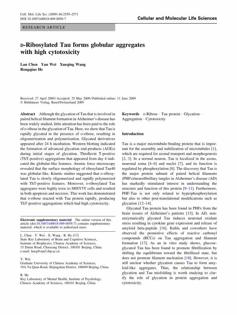
During glycation, Tau40 was incubated with D-ribose and
aliquots were taken for 12% SDS-PAGE at different time
intervals (Fig. 1, panels A and B). While only one low
molecular weight protein band was present in the ungly-
cated Tau40 control, two high molecular weight protein
bands appeared in samples containing glycated Tau40,
demonstrating Tau protein aggregation. One of the bands
was smeared in appearance suggesting that Tau monomers
oligomerized to dimers or octamers (oligomers); and the
other smearing of the high molecular weight band indicated
polymerization to decamers (polymers). Our results thus
indicate that Tau began to oligomerize from 1 day after the
start of incubation, and polymerization occurred markedly
D-Ribosylated Tau with high cytotoxicity 2561
from day 2. The Tau monomer band was retarded in the
presence of D-ribose with increasing incubation time,
suggesting that the apparent molecular mass of the glycated
Tau protein increased with time. However, significant
polymerization and band retardation were not detected for
Tau alone in the absence of D-ribose (panels C and D,
Fig. 1). The difference between the apparent molecular
masses suggested that *51 ribose units bound to one Tau
protein.
In our kinetic studies, the increase in grey density of the
bands of oligomers and polymers underwent a relaxation
and a biphasic process (slow and fast phases). Density of
oligomers and polymers increased from day 1 and 2,
respectively, and more rapidly on further incubation. The
data were analyzed according to Tsou’s method [43]. The
first order rates of the fast phase for oligomers and poly-
mers were five- and three-fold higher than those of the slow
phases, respectively (Table 1). Tau monomers likewise
decreased in slow and fast phases as oligomers and poly-
mers increased. This suggests that glycation with D-ribose
leads to a slow oligomerization of ribosylated Tau protein
after a relaxation time, followed by fast polymerization.
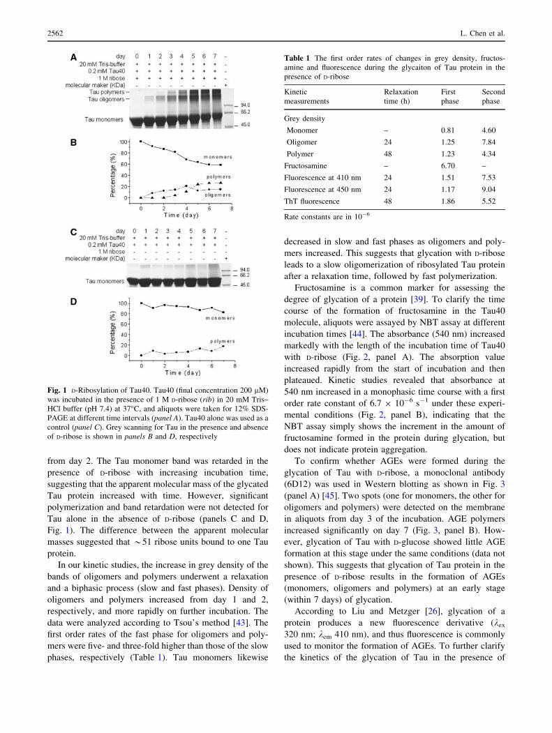
Fructosamine is a common marker for assessing the
degree of glycation of a protein [39]. To clarify the time
course of the formation of fructosamine in the Tau40
molecule, aliquots were assayed by NBT assay at different
incubation times [44]. The absorbance (540 nm) increased
markedly with the length of the incubation time of Tau40
with D-ribose (Fig. 2, panel A). The absorption value
increased rapidly from the start of incubation and then
plateaued. Kinetic studies revealed that absorbance at
540 nm increased in a monophasic time course with a first
order rate constant of 6.7 9 10-6 s-1 under these experi-
mental conditions (Fig. 2, panel B), indicating that the
NBT assay simply shows the increment in the amount of
fructosamine formed in the protein during glycation, but
does not indicate protein aggregation.
To confirm whether AGEs were formed during the
glycation of Tau with D-ribose, a monoclonal antibody
(6D12) was used in Western blotting as shown in Fig. 3
(panel A) [45]. Two spots (one for monomers, the other for
oligomers and polymers) were detected on the membrane
in aliquots from day 3 of the incubation. AGE polymers
increased significantly on day 7 (Fig. 3, panel B). How-
ever, glycation of Tau with D-glucose showed little AGE
formation at this stage under the same conditions (data not
shown). This suggests that glycation of Tau protein in the
presence of D-ribose results in the formation of AGEs
(monomers, oligomers and polymers) at an early stage
(within 7 days) of glycation.
According to Liu and Metzger [26], glycation of a
protein produces a new fluorescence derivative (kex
320 nm; kem 410 nm), and thus fluorescence is commonly
used to monitor the formation of AGEs. To further clarify
the kinetics of the glycation of Tau in the presence of
Fig. 1 D-Ribosylation of Tau40. Tau40 (final concentration 200 lM)
was incubated in the presence of 1 M D-ribose (rib) in 20 mM Tris–
HCl buffer (pH 7.4) at 37�C, and aliquots were taken for 12% SDS-
PAGE at different time intervals (panel A). Tau40 alone was used as a
control (panel C). Grey scanning for Tau in the presence and absence
of D-ribose is shown in panels B and D, respectively
Table 1 The first order rates of changes in grey density, fructos-
amine and fluorescence during the glycaiton of Tau protein in the
presence of D-ribose
Kinetic
measurements
Relaxation
time (h)
First
phase
Second
phase
Grey density
Monomer – 0.81 4.60
Oligomer 24 1.25 7.84
Polymer 48 1.23 4.34
Fructosamine – 6.70 –
Fluorescence at 410 nm 24 1.51 7.53
Fluorescence at 450 nm 24 1.17 9.04
ThT fluorescence 48 1.86 5.52
Rate constants are in 10-6
2562 L. Chen et al.
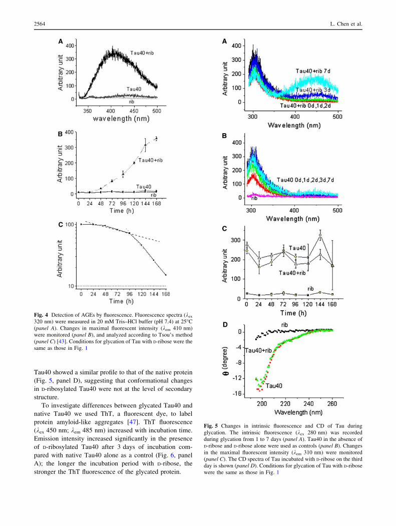
D-ribose, changes in fluorescence were measured as
shown in Fig. 4. The fluorescence emission intensity
(410 nm) of Tau40 incubated with D-ribose was much
stronger than that of Tau40 alone as a control (Fig. 4,
panel A). The fluorescence of glycated polymers
(collected from PAGE) was markedly stronger than that
of glycated monomers (data not shown), suggesting that
fluorescence at 410 nm resulted mainly from D-ribosylat-
ed polymers.
To confirm that 410 nm emission is related to protein
aggregation, the data shown in Fig. 4 (panel B) were
analyzed according to Tsou’s method [43]. The kinetic
increment of the emission intensity underwent a relaxation
period (24 h) and followed a biphasic time course (a slow
and a fast phase) (Fig. 4, panel C). First order rate con-
stants obtained from analysis of 410 nm fluorescence
emission increments were similar to those obtained by
SDS-PAGE analysis of the polymerization of D-ribosylated
Tau40 (Fig. 1 and Table 1), indicating that the increase in
the emission at 410 nm is related to the aggregation of
D-ribosylated Tau protein.
Conformational changes of Tau protein
during ribosylation
To investigate the conformational changes of Tau protein
during glycation, we measured the intrinsic fluorescence of
the ribosylated protein by excitation at 280 nm. As shown
in Fig. 5 (panel C), intrinsic fluorescence at 310 nm shows
a slight change with incubation time. However, the change
in fluorescence was not significant compared to that of Tau
in the absence of D-ribose as a control. Changes in intrinsic
fluorescence represent microenvironmental variations at
Tyr residues where solvent molecules collide with the
fluorophore and consume the energy of fluorescence [46].
Notably, a new fluorescent derivative appeared around
450 nm from day 3 of the glycation process (Fig. 5, panel
A). The appearance of fluorescence at 450 nm also repre-
sented the formation of AGEs in Tau polymers. Most
interestingly, an energy transfer between Tyr residues and
ribosylated fluorescent derivatives (around 450 nm) was
observed by excitation at 280 nm. Control Tau in the
absence of D-ribose did not show this energy transfer
(Fig. 5, panel B), suggesting that the spatial localization of
Tyr residues is close to the glycated fluorescent derivative
in Tau polymers.
To detect changes in the secondary structure of Tau40 in
the presence of D-ribose, CD spectra of Tau40 were scan-
ned during glycation. However, the spectrum of ribosylated
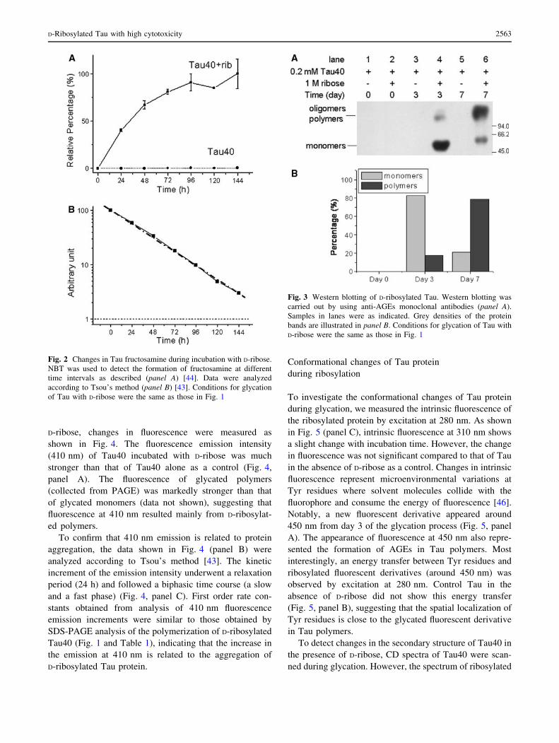
Fig. 2 Changes in Tau fructosamine during incubation with D-ribose.
NBT was used to detect the formation of fructosamine at different
time intervals as described (panel A) [44]. Data were analyzed
according to Tsou’s method (panel B) [43]. Conditions for glycation
of Tau with D-ribose were the same as those in Fig. 1
Fig. 3 Western blotting of D-ribosylated Tau. Western blotting was
carried out by using anti-AGEs monoclonal antibodies (panel A).
Samples in lanes were as indicated. Grey densities of the protein
bands are illustrated in panel B. Conditions for glycation of Tau with
D-ribose were the same as those in Fig. 1
D-Ribosylated Tau with high cytotoxicity 2563
Tau40 showed a similar profile to that of the native protein
(Fig. 5, panel D), suggesting that conformational changes
in D-ribosylated Tau40 were not at the level of secondary
structure.
To investigate differences between glycated Tau40 and
native Tau40 we used ThT, a fluorescent dye, to label
protein amyloid-like aggregates [47]. ThT fluorescence
(kex 450 nm; kem 485 nm) increased with incubation time.
Emission intensity increased significantly in the presence
of D-ribosylated Tau40 after 3 days of incubation com-
pared with native Tau40 alone as a control (Fig. 6, panel
A); the longer the incubation period with D-ribose, the
stronger the ThT fluorescence of the glycated protein.
Fig. 4 Detection of AGEs by fluorescence. Fluorescence spectra (kex
320 nm) were measured in 20 mM Tris–HCl buffer (pH 7.4) at 25�C
(panel A). Changes in maximal fluorescent intensity (kem 410 nm)
were monitored (panel B), and analyzed according to Tsou’s method
(panel C) [43]. Conditions for glycation of Tau with D-ribose were the
same as those in Fig. 1
Fig. 5 Changes in intrinsic fluorescence and CD of Tau during
glycation. The intrinsic fluorescence (kex 280 nm) was recorded
during glycation from 1 to 7 days (panel A). Tau40 in the absence of
D-ribose and D-ribose alone were used as controls (panel B). Changes
in the maximal fluorescent intensity (kem 310 nm) were monitored
(panel C). The CD spectra of Tau incubated with D-ribose on the third
day is shown (panel D). Conditions for glycation of Tau with D-ribose
were the same as those in Fig. 1
2564 L. Chen et al.
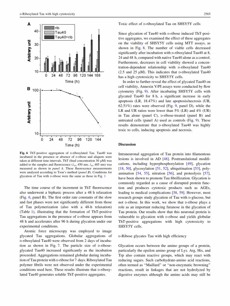
The time course of the increment in ThT fluorescence
also underwent a biphasic process after a 48 h relaxation
(Fig. 6, panel B). The first order rate constants of the slow
and fast phases were not significantly different from those
of Tau polymerization (also with a 48-h relaxation)
(Table 1), illustrating that the formation of ThT-positive
Tau aggregations in the presence of D-ribose appears from
48 h and accelerates after 96 h during glycation under our
experimental conditions.
Atomic force microscopy was employed to image
glycated Tau aggregations. Globular aggregations of
D-ribosylated Tau40 were observed from 2 days of incuba-
tion as shown in Fig. 7. The particle size of D-ribose-
glycated Tau40 increased significantly as the incubation
proceeded. Aggregations remained globular during incuba-
tion of Tau protein with D-ribose for 7 days. Ribosylated Tau
polymer fibrils were not observed under the experimental
conditions used here. These results illustrate that D-ribosy-
lated Tau40 generates soluble ThT-positive aggregates.
Toxic effect of D-ribosylated Tau on SHSY5Y cells
Since glycation of Tau40 with D-ribose induced ThT-posi-
tive aggregates, we examined the effect of these aggregates
on the viability of SHSY5Y cells using MTT assays, as
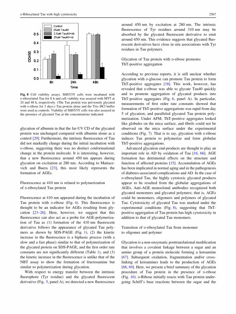
shown in Fig. 8. The number of viable cells decreased
significantly after incubation with D-ribosylated Tau40 at 8,
24 and 48 h, compared with native Tau40 alone as a control.
Furthermore, decreases in cell viability showed a concen-
tration-dependent relationship with D-ribosylated Tau40
(2.5 and 25 lM). This indicates that D-ribosylated Tau40
has a high cytotoxicity to SHSY5Y cells.
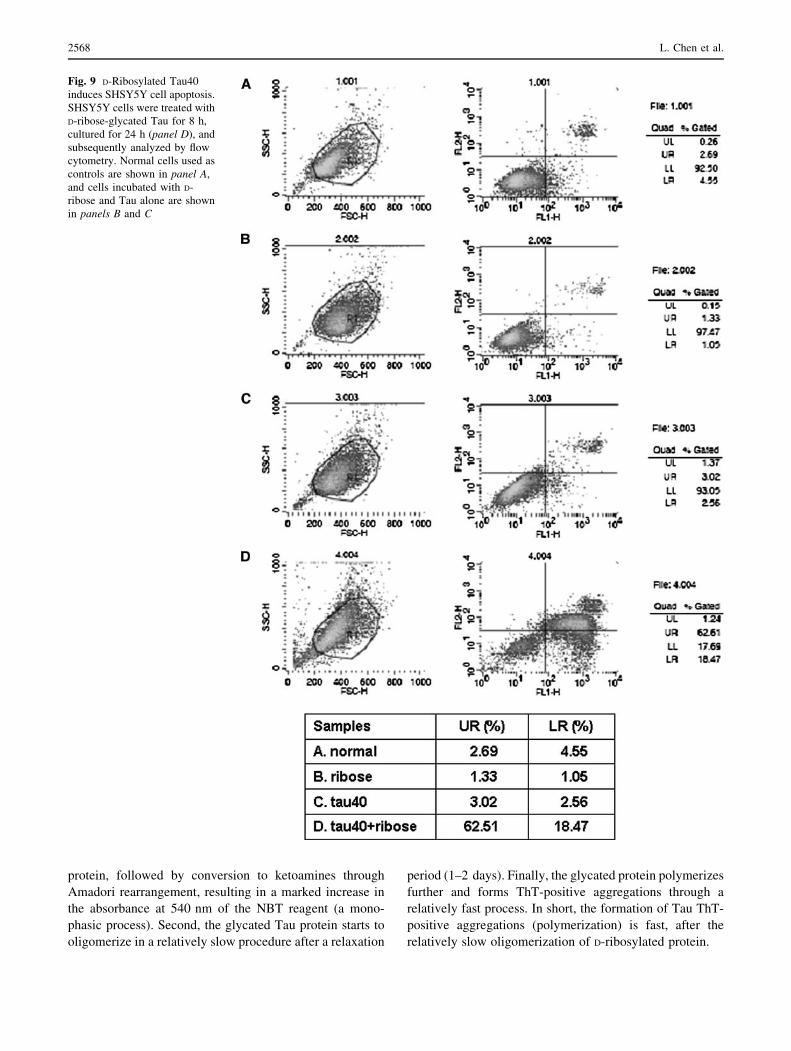
In order to further reveal the effect of glycated Tau40 on
cell viability, Annexin V/PI assays were conducted by flow
cytometry (Fig. 9). After incubating SHSY5Y cells with
glycated Tau40 for 8 h, a significant increase in early
apoptosis (LR, 18.47%) and late apoptosis/necrosis (UR,
62.51%) rates were observed (Fig. 9, panel D), while the
LR and UR ratios were lower than 5% (LR) and 4% (UR)
in Tau alone (panel C), D-ribose-treated (panel B) and
untreated cells (panel A) used as controls (Fig. 9). These
results demonstrate that D-ribosylated Tau40 was highly
toxic to cells, inducing apoptosis and necrosis.
Discussion
Intraneuronal aggregation of Tau protein into filamentous
lesions is involved in AD [48]. Posttranslational modifi-
cations, including hyperphosphorylation [49], glycation
[18, 50], glycosylation [51, 52], ubiquitination [53], poly-
amination [54, 55], nitration [56], and proteolysis [57],
have been shown to promote Tau fibrillization. Glycation is
commonly regarded as a cause of disrupted protein func-
tion and produces cytotoxic products such as AGEs,
leading to medical complications [58, 59]. However, most
research groups study glycation of Tau with D-glucose, but
not D-ribose. In this work, we show that D-ribose plays a
role as an important reducing furanose in the glycation of
Tau protein. Our results show that this neuronal protein is
vulnerable to glycation with D-ribose and yields globular
ThT-positive aggregations with high cytotoxicity to
SHSY5Y cells.
D-Ribose glycates Tau with high efficiency
Glycation occurs between the amino groups of a protein,
particularly the epsilon amino group of Lys. Arg, His, and
Trp also contain reactive groups, which may react with
reducing sugars. Such carbohydrate-amino acid reactions,
often termed as ‘‘Maillard’’ or ‘‘non-enzymatic browning’’
reactions, result in linkages that are not hydrolyzed by
digestive enzymes although the amino acids may still be
Fig. 6 ThT-positive aggregation of D-ribosylated Tau. Tau40 was
incubated in the presence or absence of D-ribose and aliquots were
taken at different time intervals. ThT (final concentration 30 lM) was
added to the samples and fluorescence (kex 450 nm; kex 485 nm) was
measured as shown in panel A. These fluorescence measurements
were analyzed according to Tsou’s method (panel B). Conditions for
glycation of Tau with D-ribose were the same as those in Fig. 1
D-Ribosylated Tau with high cytotoxicity 2565
recovered from the protein by acid hydrolysis [60]. The
results from SDS-PAGE suggest that approximately 51
D-ribose units bind to one molecule of Tau protein. As
Tau40 contains 44 Lys, 12 His and 14 Arg residues, which
can react with D-ribose, our results suggest that most of the
available amino acid residues react with D-ribose.
D-Ribose, an efficient glycator, is a naturally occurring
pentose monosaccharide and an essential component for
energy production in the body. D-Ribose exists in all living
cells and in blood, and is present in the human brain [61].
Furthermore, Tau has an unstable conformation with a
flexible peptidyl chain. Schweers and colleagues showed
that the Tau protein exists in a ‘‘worm-like’’ conformation
[62], having the irregular structure of an unfolded protein
[63]. Thus, most Lys residues are exposed on the exterior
of the Tau molecule because of the hydrophilic nature of
the e-amino group. Thus, the worm-like Tau protein has
more opportunities to react with reducing sugars than
globular-like proteins, explaining why Tau protein is rap-
idly glycated in the presence of D-ribose.
Although Tau40 can be glycated with D-glucose in vitro,
it is still unclear whether D-glucose is the most important
sugar in Tau glycation. It is well known that D-glucose has
a lower reducing potential than D-ribose [15, 64]. Necular
and Kuret [18] have reported that glycated Tau is not able
to promote filament nucleation. Similarly, glycation of
alpha-synuclein with D-glucose and D-fructose does not
induce amyloidosis products [33]. Our recent data also
show that glycation of BSA with D-ribose is much faster
than with D-glucose [65]. This is probably due to the low
glycation efficiency of D-glucose compared with D-ribose.
As mentioned above, the far-UV CD spectra (Fig. 5,
panel D) showed little change in the secondary structure of
Tau protein. Similar results have been observed during
Fig. 7 Atomic force microscopic image of D-ribosylated Tau.
Aliquots of Tau40 incubated with D-ribose were observed by AFM
(panel A–H). Sizes (nm) of the particles of Tau40 incubated with
D-ribose are shown (panel I). Conditions for glycation of Tau with
D-ribose were the same as those in Fig. 1. The scale bar equals
100 nm
2566 L. Chen et al.
glycation of albumin in that the far-UV CD of the glycated
protein was unchanged compared with albumin alone as a
control [29]. Furthermore, the intrinsic fluorescence of Tau
did not markedly change during the initial incubation with
D-ribose, suggesting there was no distinct conformational
change in the protein molecule. It is interesting, however,
that a new fluorescence around 450 nm appears during
glycation on excitation at 280 nm. According to Matiace-
vich and Buera [23], this most likely represents the
formation of AGEs.
Fluorescence at 410 nm is related to polymerization
of D-ribosylated Tau protein
Fluorescence at 410 nm appeared during the incubation of
Tau protein with D-ribose (Fig. 4). This fluorescence is
thought to be an indicator for AGEs resulting from gly-
cation [23–26]. Here, however, we suggest that this
fluorescence can also act as a probe for AGE-polymeriza-
tion of Tau as (1) formation of the 410 nm fluorescent
derivative follows the appearance of glycated Tau poly-
mers as shown by SDS-PAGE (Fig. 1), (2) the kinetic
increase in the fluorescence is a biphasic process (with a
slow and a fast phase) similar to that of polymerization of
the glycated protein on SDS-PAGE, and the first order rate
constants are not significantly different (Table 1), and (3)
the kinetic increase in the fluorescence is unlike that of the
NBT assay to show the formation of fructosamine but
similar to polymerization during glycation.
With respect to energy transfer between the intrinsic
fluorophore (Tyr residue) and the glycated fluorescent
derivative (Fig. 5, panel A), we detected a new fluorescence
around 450 nm by excitation at 280 nm. The intrinsic
fluorescence of Tyr residues around 310 nm may be
absorbed by the glycated fluorescent derivative to emit
around 450 nm. This evidence suggests that glycated fluo-
rescent derivatives have close in situ associations with Tyr
residues in Tau polymers.
Glycation of Tau protein with D-ribose promotes
ThT-positive aggregation
According to previous reports, it is still unclear whether
glycation with D-glucose can promote Tau protein to form
ThT-positive aggregates [18]. This work, however, has
revealed that D-ribose was able to glycate Tau40 quickly
and to promote aggregation of glycated products into
ThT-positive aggregates (Fig. 6, panel A). In particular,
measurements of first order rate constants showed that
formation of ThT-positive aggregations was rapid from day
5 of glycation, and paralleled glycated Tau protein poly-
merization. Under AFM, ThT-positive aggregates looked
like globules on the mica surface, and fibrils could not be
observed on the mica surface under the experimental
conditions (Fig. 7). That is to say, glycation with D-ribose
induces Tau protein to polymerize and form globular
ThT-positive aggregations.
Advanced glycation end-products are thought to play an
important role in AD by oxidation of Tau [16, 66]. AGE
formation has detrimental effects on the structure and
function of affected proteins [15]. Accumulation of AGEs
has been implicated in normal aging and in the pathogenesis
of diabetes-associated complications and AD. In the case of
D-ribosylated Tau, the highly cytotoxic glycated products
appear to be resulted from the globular aggregations of
AGEs. Anti-AGE monoclonal antibodies recognized both
glycated monomers and glycated polymers; that is, AGEs
could be monomers, oligomers and polymers of glycated
Tau. Cytotoxicity of glycated Tau was marked under the
experimental conditions (Fig. 8), suggesting that ThT-
positive aggregation of Tau protein has high cytotoxicity in
addition to that of glycated Tau monomers.
Transition of D-ribosylated Tau from monomer
to oligomer and polymer
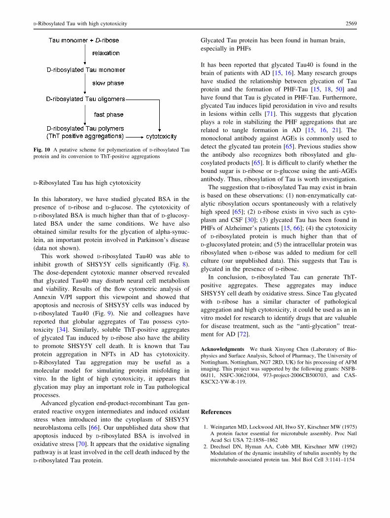
Glycation is a non-enzymatic posttranslational modification
that involves a covalent linkage between a sugar and an
amino group of a protein molecule forming a ketoamine
[67]. Subsequent oxidation, fragmentation and/or cross-
linking of ketoamines leads to the production of AGEs
[68, 69]. Here, we present a brief summary of the glycation
procedure of Tau protein in the presence of D-ribose
(Fig. 10). D-Ribose initially reacts with Tau protein under-
going Schiff’s base reactions between the sugar and the
Fig. 8 Cell viability assays. SHSY5Y cells were incubated with
D-ribosylated Tau for 8 h and cell viability was assayed with MTT at
24 and 48 h, respectively. (The Tau protein was previously glycated
with D-ribose for 3 days.) Tau protein alone and the Tris–HCl buffer
were used as controls. Viability of SHSY5Y cells was also assayed in
the presence of glycated Tau at the concentrations indicated
D-Ribosylated Tau with high cytotoxicity 2567
protein, followed by conversion to ketoamines through
Amadori rearrangement, resulting in a marked increase in
the absorbance at 540 nm of the NBT reagent (a mono-
phasic process). Second, the glycated Tau protein starts to
oligomerize in a relatively slow procedure after a relaxation
period (1–2 days). Finally, the glycated protein polymerizes
further and forms ThT-positive aggregations through a
relatively fast process. In short, the formation of Tau ThT-
positive aggregations (polymerization) is fast, after the
relatively slow oligomerization of D-ribosylated protein.
Fig. 9 D-Ribosylated Tau40
induces SHSY5Y cell apoptosis.
SHSY5Y cells were treated with
D-ribose-glycated Tau for 8 h,
cultured for 24 h (panel D), and
subsequently analyzed by flow
cytometry. Normal cells used as
controls are shown in panel A,
and cells incubated with D-
ribose and Tau alone are shown
in panels B and C
2568 L. Chen et al.
D-Ribosylated Tau has high cytotoxicity
In this laboratory, we have studied glycated BSA in the
presence of D-ribose and D-glucose. The cytotoxicity of
D-ribosylated BSA is much higher than that of D-glucosy-
lated BSA under the same conditions. We have also
obtained similar results for the glycation of alpha-synuc-
lein, an important protein involved in Parkinson’s disease
(data not shown).
This work showed D-ribosylated Tau40 was able to
inhibit growth of SHSY5Y cells significantly (Fig. 8).
The dose-dependent cytotoxic manner observed revealed
that glycated Tau40 may disturb neural cell metabolism
and viability. Results of the flow cytometric analysis of
Annexin V/PI support this viewpoint and showed that
apoptosis and necrosis of SHSY5Y cells was induced by
D-ribosylated Tau40 (Fig. 9). Nie and colleagues have
reported that globular aggregates of Tau possess cyto-
toxicity [34]. Similarly, soluble ThT-positive aggregates
of glycated Tau induced by D-ribose also have the ability
to promote SHSY5Y cell death. It is known that Tau
protein aggregation in NFTs in AD has cytotoxicity.
D-Ribosylated Tau aggregation may be useful as a
molecular model for simulating protein misfolding in
vitro. In the light of high cytotoxicity, it appears that
glycation may play an important role in Tau pathological
processes.
Advanced glycation end-product-recombinant Tau gen-
erated reactive oxygen intermediates and induced oxidant
stress when introduced into the cytoplasm of SHSY5Y
neuroblastoma cells [66]. Our unpublished data show that
apoptosis induced by D-ribosylated BSA is involved in
oxidative stress [70]. It appears that the oxidative signaling
pathway is at least involved in the cell death induced by the
D-ribosylated Tau protein.
Glycated Tau protein has been found in human brain,
especially in PHFs
It has been reported that glycated Tau40 is found in the
brain of patients with AD [15, 16]. Many research groups
have studied the relationship between glycation of Tau
protein and the formation of PHF-Tau [15, 18, 50] and
have found that Tau is glycated in PHF-Tau. Furthermore,
glycated Tau induces lipid peroxidation in vivo and results
in lesions within cells [71]. This suggests that glycation
plays a role in stabilizing the PHF aggregations that are
related to tangle formation in AD [15, 16, 21]. The
monoclonal antibody against AGEs is commonly used to
detect the glycated tau protein [65]. Previous studies show
the antibody also recognizes both ribosylated and glu-
cosylated products [65]. It is difficult to clarify whether the
bound sugar is D-ribose or D-glucose using the anti-AGEs
antibody. Thus, ribosylation of Tau is worth investigation.
The suggestion that D-ribosylated Tau may exist in brain
is based on these observations: (1) non-enzymatically cat-
alytic ribosylation occurs spontaneously with a relatively
high speed [65]; (2) D-ribose exists in vivo such as cyto-
plasm and CSF [30]; (3) glycated Tau has been found in
PHFs of Alzheimer’s patients [15, 66]; (4) the cytotoxicity
of D-ribosylated protein is much higher than that of
D-glucosylated protein; and (5) the intracellular protein was
ribosylated when D-ribose was added to medium for cell
culture (our unpublished data). This suggests that Tau is
glycated in the presence of D-ribose.
In conclusion, D-ribosylated Tau can generate ThT-
positive aggregates. These aggregates may induce
SHSY5Y cell death by oxidative stress. Since Tau glycated
with D-ribose has a similar character of pathological
aggregation and high cytotoxicity, it could be used as an in
vitro model for research to identify drugs that are valuable
for disease treatment, such as the ‘‘anti-glycation’’ treat-
ment for AD [72].
Acknowledgments We thank Xinyong Chen (Laboratory of Bio-
physics and Surface Analysis, School of Pharmacy, The University of
Nottingham, Nottingham, NG7 2RD, UK) for his processing of AFM
imaging. This project was supported by the following grants: NSFB-
06J11, NSFC-30621004, 973-project-2006CB500703, and CAS-
KSCX2-YW-R-119.
References
1. Weingarten MD, Lockwood AH, Hwo SY, Kirschner MW (1975)
A protein factor essential for microtubule assembly. Proc Natl
Acad Sci USA 72:1858–1862
2. Drechsel DN, Hyman AA, Cobb MH, Kirschner MW (1992)
Modulation of the dynamic instability of tubulin assembly by the
microtubule-associated protein tau. Mol Biol Cell 3:1141–1154
Fig. 10 A putative scheme for polymerization of D-ribosylated Tau
protein and its conversion to ThT-positive aggregations
D-Ribosylated Tau with high cytotoxicity 2569
3. Friede RL, Ho KC (1977) The relation of axonal transport of
mitochondria with microtubules and other axoplasmic organelles.
J Physiol 265:507–519
4. Binder LI, Frankfurter A, Rebhun LI (1985) The distribution of
tau in the mammalian central nervous system. J Cell Biol
101:1371–1378
5. Khatoon S, Grundke-Iqbal I, Iqbal K (1994) Levels of normal and
abnormally phosphorylated tau in different cellular and regional
compartments of Alzheimer disease and control brains. FEBS
Lett 351:80–84
6. Papasozomenos SC, Binder LI (1987) Phosphorylation deter-
mines two distinct species of Tau in the central nervous system.
Cell Motil Cytoskeleton 8:210–226
7. Wei Y, Qu MH, Wang XS, Chen L, Wang DL, Liu Y, Hua Q, He
RQ (2008) Binding to the minor groove of the double-strand, tau
protein prevents DNA from damage by peroxidation. PLoS ONE
3:e2600
8. Lindwall G, Cole RD (1984) Phosphorylation affects the ability
of tau protein to promote microtubule assembly. J Biol Chem
259:5301–5305
9. Iqbal K, Grundke-Iqbal I, Zaidi T, Merz PA, Wen GY, Shaikh
SS, Wisniewski HM, Alafuzoff I, Winblad B (1986) Defective
brain microtubule assembly in Alzheimer’s disease. Lancet
2:421–426
10. Grundke-Iqbal I, Iqbal K, Tung YC, Quinlan M, Wisniewski HM,
Binder LI (1986) Abnormal phosphorylation of the microtubule-
associated protein tau (tau) in Alzheimer cytoskeletal pathology.
Proc Natl Acad Sci USA 83:4913–4917
11. Grundke-Iqbal I, Iqbal K, Quinlan M, Tung YC, Zaidi MS,
Wisniewski HM (1986) Microtubule-associated protein tau. A
component of Alzheimer paired helical filaments. J Biol Chem
261:6084–6089
12. Smith MA, Taneda S, Richey PL, Miyata S, Yan SD, Stern D,
Sayre LM, Monnier VM, Perry G (1994) Advanced Maillard
reaction end products are associated with Alzheimer disease
pathology. Proc Natl Acad Sci USA 91:5710–5714
13. Horie K, Miyata T, Yasuda T, Takeda A, Yasuda Y, Maeda K,
Sobue G, Kurokawa K (1997) Immunohistochemical localization
of advanced glycation end products, pentosidine, and carboxy-
methyllysine in lipofuscin pigments of Alzheimer’s disease and
aged neurons. Biochem Biophys Res Commun 236:327–332
14. Sasaki N, Fukatsu R, Tsuzuki K, Hayashi Y, Yoshida T, Fujii N,
Koike T, Wakayama I, Yanagihara R, Garruto R, Amano N,
Makita Z (1998) Advanced glycation end products in Alzhei-
mer’s disease and other neurodegenerative diseases. Am J Pathol
153:1149–1155
15. Ko LW, Ko EC, Nacharaju P, Liu WK, Chang E, Kenessey A,
Yen SH (1999) An immunochemical study on tau glycation in
paired helical filaments. Brain Res 830:301–313
16. Yan SD, Yan SF, Chen X, Fu J, Chen M, Kuppusamy P, Smith
MA, Perry G, Godman GC, Nawroth P (1995) Non-enzymatically
glycated tau in Alzheimer’s disease induces neuronal oxidant
stress resulting in cytokine gene expression and release of amy-
loid beta-peptide. Nat Med 1:693–699
17. Kuhla B, Haase C, Flach K, Luth HJ, Arendt T, Munch G
(2007) Effect of pseudophosphorylation and cross-linking by
lipid peroxidation and advanced glycation end product precur-
sors on tau aggregation and filament formation. J Biol Chem
282:6984–6991
18. Necula M, Kuret J (2004) Pseudophosphorylation and glycation
of tau protein enhance but do not trigger fibrillization in vitro.
J Biol Chem 279:49694–49703
19. Gonzalez C, Farias G, Maccioni RB (1998) Modification of tau to
an Alzheimer’s type protein interferes with its interaction with
microtubules. Cell Mol Biol (Noisy-le-grand) 44:1117–1127
20. Ledesma MD, Bonay P, Colaco C, Avila J (1994) Analysis of
microtubule-associated protein tau glycation in paired helical
filaments. J Biol Chem 269:21614–21619
21. Nacharaju P, Ko L, Yen SH (1997) Characterization of in vitro
glycation sites of tau. J Neurochem 69:1709–1719
22. Nelson DL, Cox MM (2004) Lehninger principle of biochemis-
try, 3rd edn. Worth, New York, pp 297–324
23. Matiacevich SB, Buera MP (2006) A critical evaluation of fluo-
rescence as a potential marker for the Maillard reaction. Food
Chem 95:423–430
24. Moreaux V, Birlouez-Aragon I (1997) Degradation of tryptophan
in heated ß-lactoglobulin-lactose mixtures is associated with
intense Maillard reaction. J Agric Food Chem 45:1905–1910
25. Ferrer E, Alegria A, Farre R, Clemente G, Calvo C (2005)
Fluorescence, browning index, and color in infant formulas
during storage. J Agric Food Chem 53:4911–4917
26. Liu X, Metzger LE (2007) Application of fluorescence spec-
troscopy for monitoring changes in nonfat dry milk during
storage. J Dairy Sci 90:24–37
27. Garlick RL, Mazer JS, Higgins PJ, Bunn HF (1983) Character-
ization of glycosylated hemoglobins. Relevance to monitoring of
diabetic control and analysis of other proteins. J Clin Invest
71:1062–1072
28. Shaklai N, Garlick RL, Bunn HF (1984) Nonenzymatic glyco-
sylation of human serum albumin alters its conformation and
function. J Biol Chem 259:3812–3817
29. Mendez DL, Jensen RA, McElroy LA, Pena JM, Esquerra RM
(2005) The effect of non-enzymatic glycation on the unfolding of
human serum albumin. Arch Biochem Biophys 444:92–99
30. James PE, Lang D, Tufnell-Barret T, Milsom AB, Frenneaux MP
(2004) Vasorelaxation by red blood cells and impairment in
diabetes: reduced nitric oxide and oxygen delivery by glycated
hemoglobin. Circ Res 94:976–983
31. He RQ, Yang MD, Zheng X, Zhou JX (1995) Isolation and some
properties of glycated D-glyceraldehyde-3-phosphate dehydroge-
nase from rabbit muscle. Biochem J 309(Pt 1):133–139
32. He RQ, Li YG, Wu XQ, Li L (1995) Inactivation and con-
formation changes of the glycated and non-glycated
D-glyceraldehyde-3-phosphate dehydrogenase during guanidine-
HCl denaturation. Biochim Biophys Acta 1253:47–56
33. Sheng Z, Liu Y, Chen L, He R (2008) Nonenzymatic glycation of
a-synuclein and changes in its conformation. Prog Biochem
Biophys 35:1202–1208
34. Nie CL, Wei Y, Chen X, Liu YY, Dui W, Liu Y, Davies MC,
Tendler SJ, He RG (2007) Formaldehyde at low concentration
induces protein tau into globular amyloid-like aggregates in vitro
and in vivo. PLoS ONE 2:e629
35. Crowther RA, Olesen OF, Smith MJ, Jakes R, Goedert M (1994)
Assembly of Alzheimer-like filaments from full-length tau pro-
tein. FEBS Lett 337:135–138
36. Hua Q, He RQ (2003) Tau could protect DNA double helix
structure. Biochim Biophys Acta 1645:205–211
37. Morton RE, Evans TA (1992) Modification of the bicinchoninic
acid protein assay to eliminate lipid interference in determining
lipoprotein protein content. Anal Biochem 204:332–334
38. Baker JR, Metcalf PA, Johnson RN, Newman D, Rietz P (1985)
Use of protein-based standards in automated colorimetric deter-
minations of fructosamine in serum. Clin Chem 31:1550–1554
39. Baker JR, Zyzak DV, Thorpe SR, Baynes JW (1993) Mechanism
of fructosamine assay: evidence against role of superoxide as
intermediate in nitroblue tetrazolium reduction. Clin Chem
39:2460–2465
40. Coussons PJ, Jacoby J, McKay A, Kelly SM, Price NC, Hunt JV
(1997) Glucose modification of human serum albumin: a struc-
tural study. Free Radic Biol Med 22:1217–1227
2570 L. Chen et al.
41. Nie CL, Wang XS, Liu Y, Perrett S, He RQ (2007) Amyloid-like
aggregates of neuronal tau induced by formaldehyde promote
apoptosis of neuronal cells. BMC Neurosci 8:9
42. Mayo L, Stein R (2007) Characterization of LPS and interferon-
gamma triggered activation-induced cell death in N9 and primary
microglial cells: induction of the mitochondrial gateway by nitric
oxide. Cell Death Differ 14:183–186
43. Tsou CL (1988) Folding of the nascent peptide chain into a
biologically active protein. Biochemistry 27:1809–1812
44. Xu YJ, Wu XQ, Liu W, Lin XH, Chen JW, He RQ (2002) A
convenient assay of glycoserum by nitroblue tetrazolium with
iodoacetamide. Clin Chim Acta 325:127–131
45. Ikeda K, Higashi T, Sano H, Jinnouchi Y, Yoshida M, Araki T,
Ueda S, Horiuchi S (1996) N (epsilon)-(carboxymethyl)lysine
protein adduct is a major immunological epitope in proteins
modified with advanced glycation end products of the Maillard
reaction. Biochemistry 35:8075–8083
46. Swamy MJ, Surolia A (1989) Studies on the tryptophan residues
of soybean agglutinin. Involvement in saccharide binding. Biosci
Rep 9:189–198
47. Naiki H, Higuchi K, Hosokawa M, Takeda T (1989) Fluorometric
determination of amyloid fibrils in vitro using the fluorescent dye,
thioflavin T1. Anal Biochem 177:244–249
48. Buee L, Bussiere T, Buee-Scherrer V, Delacourte A, Hof PR
(2000) Tau protein isoforms, phosphorylation and role in
neurodegenerative disorders. Brain Res Brain Res Rev 33:95–
130
49. Gong CX, Liu F, Grundke-Iqbal I, Iqbal K (2005) Post-transla-
tional modifications of tau protein in Alzheimer’s disease.
J Neural Transm 112:813–838
50. Harrington CR, Colaco CA (1994) Alzheimer’s disease. A
glycation connection. Nature 370:247–248
51. Wang JZ, Grundke-Iqbal I, Iqbal K (1996) Glycosylation of
microtubule-associated protein tau: an abnormal posttranslational
modification in Alzheimer’s disease. Nat Med 2:871–875
52. Robertson LA, Moya KL, Breen KC (2004) The potential role of
tau protein O-glycosylation in Alzheimer’s disease. J Alzheimers
Dis 6:489–495
53. Bancher C, Grundke-Iqbal I, Iqbal K, Fried VA, Smith HT,
Wisniewski HM (1991) Abnormal phosphorylation of tau pre-
cedes ubiquitination in neurofibrillary pathology of Alzheimer
disease. Brain Res 539:11–18
54. Murthy SN, Wilson JH, Lukas TJ, Kuret J, Lorand L (1998)
Cross-linking sites of the human tau protein, probed by reactions
with human transglutaminase. J Neurochem 71:2607–2614
55. Tucholski J, Kuret J, Johnson GV (1999) Tau is modified by
tissue transglutaminase in situ: possible functional and metabolic
effects of polyamination. J Neurochem 73:1871–1880
56. Horiguchi T, Uryu K, Giasson BI, Ischiropoulos H, LightFoot R,
Bellmann C, Richter-Landsberg C, Lee VM, Trojanowski JQ
(2003) Nitration of tau protein is linked to neurodegeneration in
tauopathies. Am J Pathol 163:1021–1031
57. Wang JZ, Gong CX, Zaidi T, Grundke-Iqbal I, Iqbal K (1995)
Dephosphorylation of Alzheimer paired helical filaments by
protein phosphatase-2A and -2B. J Biol Chem 270:4854–4860
58. Hasegawa Y, Suehiro A, Higasa S, Namba M, Kakishita E (2002)
Enhancing effect of advanced glycation end products on seroto-
nin-induced platelet aggregation in patients with diabetes
mellitus. Thromb Res 107:319–323
59. Koga K, Yamagishi S, Okamoto T, Inagaki Y, Amano S,
Takeuchi M, Makita Z (2002) Serum levels of glucose-derived
advanced glycation end products are associated with the severity
of diabetic retinopathy in type 2 diabetic patients without renal
dysfunction. Int J Clin Pharmacol Res 22:13–17
60. Scott ML, Nesheim MC, Young RJ (1982) Nutrition of the
chicken. Scott, Ithaca, pp 100–102
61. Pliml W, von Arnim T, Stablein A, Hofmann H, Zimmer HG,
Erdmann E (1992) Effects of ribose on exercise-induced
ischaemia in stable coronary artery disease. Lancet 340:507–510
62. Schweers O, Schonbrunn-Hanebeck E, Marx A, Mandelkow E
(1994) Structural studies of tau protein and Alzheimer paired
helical filaments show no evidence for beta-structure. J Biol
Chem 269:24290–24297
63. Rosenberg KJ, Ross JL, Feinstein HE, Feinstein SC, Israelachvili
J (2008) Complementary dimerization of microtubule-associated
tau protein: implications for microtubule bundling and tau-med-
iated pathogenesis. Proc Natl Acad Sci USA 105:7445–7450
64. Grandhee SK, Monnier VM (1991) Mechanism of formation of
the Maillard protein cross-link pentosidine. Glucose, fructose,
and ascorbate as pentosidine precursors. J Biol Chem 266:11649–
11653
65. Wei Y, Chen L, Chen J, Ge L, He RQ (2009) Rapid glycation
with D-ribose induces globular amyloid-like aggregations of BSA
with high cytotoxicity to SH-SY5Y cells. BMC Cell Biol 10:10
66. Yan SD, Chen X, Schmidt AM, Brett J, Godman G, Zou YS,
Scott CW, Caputo C, Frappier T, Smith MA (1994) Glycated tau
protein in Alzheimer disease: a mechanism for induction of
oxidant stress. Proc Natl Acad Sci USA 91:7787–7791
67. Day JF, Thorpe SR, Baynes JW (1979) Nonenzymatically glu-
cosylated albumin. In vitro preparation and isolation from normal
human serum. J Biol Chem 254:595–597
68. Degenhardt TP, Thorpe SR, Baynes JW (1998) Chemical modi-
fication of proteins by methylglyoxal. Cell Mol Biol (Noisy-
le-grand) 44:1139–1145
69. McCance DR, Dyer DG, Dunn JA, Bailie KE, Thorpe SR, Baynes
JW, Lyons TJ (1993) Maillard reaction products and their relation
to complications in insulin-dependent diabetes mellitus. J Clin
Invest 91:2470–2478
70. Alikhani M, Maclellan CM, Raptis M, Vora S, Trackman PC,
Graves DT (2007) Advanced glycation end products induce
apoptosis in fibroblasts through activation of ROS, MAP kinases,
and the FOXO1 transcription factor. Am J Physiol Cell Physiol
292:C850–C856
71. Rofina JE, Singh K, Skoumalova-Vesela A, van Ederen AM, van
Asten AJ, Wilhelm J, Gruys E (2004) Histochemical accumula-
tion of oxidative damage products is associated with Alzheimer-
like pathology in the canine. Amyloid 11:90–100
72. Munch G, Kuhla B, Luth HJ, Arendt T, Robinson SR (2003)
Anti-AGEing defences against Alzheimer’s disease. Biochem
Soc Trans 31:1397–1399
D-Ribosylated Tau with high cytotoxicity 2571
![Michel parameters and [tau] neutrino helicity from decay correlations in Z--\u003e[tau]+[tau]](https://img.pdfslide.net/doc/110x75/6350c359f55d98549a09e91e/michel-parameters-and-tau-neutrino-helicity-from-decay-correlations-in-z-u003etautau.jpg)


![Upper Limits on the Branching Ratios Tau-]Mu-Gamma and Tau-]E-Gamma](https://img.pdfslide.net/doc/110x75/63619cb540b666b8ec0dfe62/upper-limits-on-the-branching-ratios-tau-mu-gamma-and-tau-e-gamma.jpg)

