Embed Size (px)
Citation preview
Journal of Materials Science: Materials in Medicine (2020) 31:132https://doi.org/10.1007/s10856-020-06462-x
CLINICAL APPLICATIONS OF BIOMATERIALS
Original Research
Decellularized tissue-engineered heart valves calcification:what do animal and clinical studies tell us?
Adel F. Badria1,2 ● Petros G. Koutsoukos3 ● Dimosthenis Mavrilas2
Received: 3 April 2020 / Accepted: 31 October 2020 / Published online: 5 December 2020© The Author(s) 2020
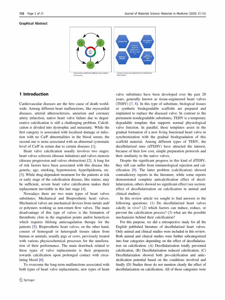
AbstractCardiovascular diseases are the first cause of death worldwide. Among different heart malfunctions, heart valve failure dueto calcification is still a challenging problem. While drug-dependent treatment for the early stage calcification could slowdown its progression, heart valve replacement is inevitable in the late stages. Currently, heart valve replacements involvemainly two types of substitutes: mechanical and biological heart valves. Despite their significant advantages in restoring thecardiac function, both types of valves suffered from serious drawbacks in the long term. On the one hand, the mechanicalone showed non-physiological hemodynamics and the need for the chronic anticoagulation therapy. On the other hand, thebiological one showed stenosis and/or regurgitation due to calcification. Nowadays, new promising heart valve substituteshave emerged, known as decellularized tissue-engineered heart valves (dTEHV). Decellularized tissues of different typeshave been widely tested in bioprosthetic and tissue-engineered valves because of their superior biomechanics,biocompatibility, and biomimetic material composition. Such advantages allow successful cell attachment, growth andfunction leading finally to a living regenerative valvular tissue in vivo. Yet, there are no comprehensive studies that arecovering the performance of dTEHV scaffolds in terms of their efficiency for the calcification problem. In this review article,we sought to answer the question of whether decellularized heart valves calcify or not. Also, which factors make themcalcify and which ones lower and/or prevent their calcification. In addition, the review discussed the possible mechanismsfor dTEHV calcification in comparison to the calcification in the native and bioprosthetic heart valves. For this purpose, wedid a retrospective study for all the published work of decellularized heart valves. Only animal and clinical studies wereincluded in this review. Those animal and clinical studies were further subcategorized into 4 categories for each dependingon the effect of decellularization on calcification. Due to the complex nature of calcification in heart valves, other in vitro andin silico studies were not included. Finally, we compared the different results and summed up all the solid findings ofwhether decellularized heart valves calcify or not. Based on our review, the selection of the proper heart valve tissue sources(no immunological provoking residues), decellularization technique (no damaged exposed residues of the decellularizedtissues, no remnants of dead cells, no remnants of decellularizing agents) and implantation techniques (avoiding suturingduring the surgical implantation) could provide a perfect anticalcification potential even without in vitro cell seeding oradditional scaffold treatment.
* Adel F. [email protected]
1 Department of Fiber and Polymer Technology, Division ofCoating Technology, KTH Royal Institute of Technology,Stockholm, Sweden
2 Department of Mechanical Engineering and Aeronautics,Division of Applied Mechanics, Technology of Materials andBiomechanics, University of Patras, Patras, Greece
3 Department of Chemical Engineering, University of Patras,Patras University Campus, 26504 Patras, Greece
1234
5678
90();,:
1234567890();,:
Graphical Abstract
1 Introduction
Cardiovascular diseases are the first cause of death world-wide. Among different heart malfunctions, like myocardialdiseases, arterial atherosclerosis, aneurism and coronaryartery infarction, native heart valve failure due to degen-erative calcification is still a challenging problem. Calcifi-cation is divided into dystrophic and metastatic. While thefirst category is associated with localized damage or infec-tion with no Ca/P abnormalities in the blood serum, thesecond one is more associated with an abnormal systematiclevel of Ca/P in serum due to certain diseases [1].
Heart valve calcification usually involves two stages:heart valves sclerosis (disease initiation) and valves stenosis(disease progression and valves obstruction) [2]. A long listof risk factors have been associated with this disease likegenetic, age, smoking, hypertension, hyperlipidemia, etc.[3]. While drug-dependent treatment for the patients at riskor early stage of the calcification disease, like statins, maybe sufficient, severe heart valve calcification makes theirreplacement inevitable in this late stage [4].
Nowadays there are two main types of heart valvessubstitutes: Mechanical and Bioprosthetic heart valves.Mechanical valves are mechanical devices from metals and/or polymers working as non-return flow valves. The maindisadvantage of this type of valves is the formation ofthrombotic clots in the stagnation points and/or hemolysiswhich requires lifelong anticoagulation therapy for thepatients [5]. Bioprosthetic heart valves, on the other hand,consist of homograft or heterograft tissues taken fromhuman or animals, usually pigs or cows, previously treatedwith various physicochemical processes for the ameliora-tion of their performance. The main drawback related tothese types of valve replacements is their propensitytowards calcification upon prolonged contact with circu-lating blood [6].
To overcome the long-term malfunctions associated withboth types of heart valve replacements, new types of heart
valve substitutes have been developed over the past 20years, generally known as tissue-engineered heart valves(TEHV) [7, 8]. In this type of substitute, biological tissuesor synthetic biodegradable scaffolds are prepared andimplanted to replace the diseased valve. In contrast to thepermanent nondegradable substitutes, TEHV is a temporarydegradable template that supports normal physiologicalvalve function. In parallel, these templates assist in thegradual formation of a new living functional heart valve insynchronization with the gradual biodegradation of thisscaffold material. Among different types of TEHV, thedecellularized ones (dTEHV) have attracted the interest,because of their low cost, simple preparation protocols andtheir similarity to the native valves.
Despite the significant progress in this kind of dTEHV,they still can suffer from immunological rejection and cal-cification [9]. The latter problem (calcification) showedcontradictory reports in the literature; while some reportsdemonstrated complete anticalcification effect of decel-lularization, others showed no significant effect (see section:effect of decellularization on calcification in animal andclinical studies).
In this review article we sought to find answers to thefollowing questions: (1) Do decellularized heart valvescalcify in vivo? (2) which factors can induce, reduce, orprevent the calcification process? (3) what are the possiblemechanisms behind their calcification?
For this purpose, we did a retrospective study for all theEnglish published literature of decellularized heart valves.Only animal and clinical studies were included in this review.Both animal and clinical studies were further subcategorizedinto four categories depending on the effect of decellulariza-tion on calcification: (A) Decellularization totally preventedcalcification, (B) Decellularization reduced calcification, (C)Decellularization showed both pro-calcification and antic-alcification potential based on the conditions involved andfinally (D) Studies those do not mention clearly the effect ofdecellularization on calcification. All of those categories were
132 Page 2 of 21 Journal of Materials Science: Materials in Medicine (2020) 31:132
tabulated and compared to each other’s depending on the hostanimal (in the case of animal studies), the type of heart valvesinvolved, the technique of decellularization, recellularizationand type of cells used, duration of the study follow-up, the sizeof the group involved and finally the techniques used toevaluate calcification occurrence. Due to the complex nature ofcalcification of heart valves, other in vitro and in silico studieswere not included in this study. Finally, we compared thedifferent results and summed up all the solid findings ofwhether decellularized heart valves calcify or not.
2 Calcification of native heart valves
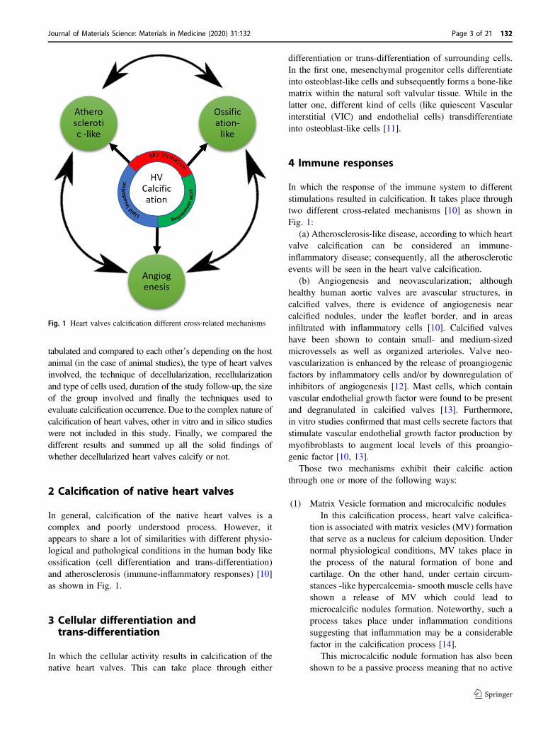
In general, calcification of the native heart valves is acomplex and poorly understood process. However, itappears to share a lot of similarities with different physio-logical and pathological conditions in the human body likeossification (cell differentiation and trans-differentiation)and atherosclerosis (immune-inflammatory responses) [10]as shown in Fig. 1.
3 Cellular differentiation andtrans-differentiation
In which the cellular activity results in calcification of thenative heart valves. This can take place through either
differentiation or trans-differentiation of surrounding cells.In the first one, mesenchymal progenitor cells differentiateinto osteoblast-like cells and subsequently forms a bone-likematrix within the natural soft valvular tissue. While in thelatter one, different kind of cells (like quiescent Vascularinterstitial (VIC) and endothelial cells) transdifferentiateinto osteoblast-like cells [11].
4 Immune responses
In which the response of the immune system to differentstimulations resulted in calcification. It takes place throughtwo different cross-related mechanisms [10] as shown inFig. 1:
(a) Atherosclerosis-like disease, according to which heartvalve calcification can be considered an immune-inflammatory disease; consequently, all the atheroscleroticevents will be seen in the heart valve calcification.
(b) Angiogenesis and neovascularization; althoughhealthy human aortic valves are avascular structures, incalcified valves, there is evidence of angiogenesis nearcalcified nodules, under the leaflet border, and in areasinfiltrated with inflammatory cells [10]. Calcified valveshave been shown to contain small- and medium-sizedmicrovessels as well as organized arterioles. Valve neo-vascularization is enhanced by the release of proangiogenicfactors by inflammatory cells and/or by downregulation ofinhibitors of angiogenesis [12]. Mast cells, which containvascular endothelial growth factor were found to be presentand degranulated in calcified valves [13]. Furthermore,in vitro studies confirmed that mast cells secrete factors thatstimulate vascular endothelial growth factor production bymyofibroblasts to augment local levels of this proangio-genic factor [10, 13].
Those two mechanisms exhibit their calcific actionthrough one or more of the following ways:
(1) Matrix Vesicle formation and microcalcific nodulesIn this calcification process, heart valve calcifica-
tion is associated with matrix vesicles (MV) formationthat serve as a nucleus for calcium deposition. Undernormal physiological conditions, MV takes place inthe process of the natural formation of bone andcartilage. On the other hand, under certain circum-stances -like hypercalcemia- smooth muscle cells haveshown a release of MV which could lead tomicrocalcific nodules formation. Noteworthy, such aprocess takes place under inflammation conditionssuggesting that inflammation may be a considerablefactor in the calcification process [14].
This microcalcific nodule formation has also beenshown to be a passive process meaning that no active
Fig. 1 Heart valves calcification different cross-related mechanisms
Journal of Materials Science: Materials in Medicine (2020) 31:132 Page 3 of 21 132
cellular mechanisms are involved. This is supportedby the presence of amorphous and crystalline calcificdeposits at the sites of cell death, either by apoptosisor necrosis. Therefore, the residuals of the dead cells’debris may be involved in the formation andpropagation of calcific deposits. However, the under-lying mechanism is not yet well understood [10].Conclusively, the valve calcification through eitherpassive or cellular mechanisms is very likely to becross related. Also, it is possible that these osteoblast-like cells may actively generate vesicles or undergoapoptosis resulting in the accumulation of calciumand the subsequent development of microcalcificnodules [15].
(2) ECM remodeling-mediated calcificationA mentioned earlier, one of the mechanisms
involved in heart valve calcification is the abnormalremodeling of valvular tissue ECM. This is often aconsequence of increased expression of some metal-loproteinases and cathepsins, which may result in thedegradation of collagen and elastin and the formationof pro-inflammatory peptides, provoking the inflam-matory response and allowing the expansion ofcalcific deposits. Additional evidence for this cellularmechanism is the observed activation of VIC in thecalcified heart valves. It leads to abnormal secretion ofcollagen, hyaluronan, and other extracellular compo-nents, consequently affecting the stiffness of thevalves and modulating the transition of VIC intoosteoblast-like cells [10].
(3) Lipid insudation
Atherosclerotic-like mechanism of calcification involveslipid accumulation on the formed calcific deposits, becauseof hypercholesterolemia. A significant relation has beenfound between the high levels of proatherogenic oxidizedlow-density lipoprotein and fibrocalcific deposits formationon aortic heart valves [16].
Besides, it has been shown that the lipid level reductionin the blood after pharmaceutical treatment with HMG-CoAreductase inhibitors (statins), which decreased the osteo-genic pathways signaling, led to reduced aortic valve cal-cification in hypercholesterolemic animal models, comparedwith control, non-treated animals [16]. However, in con-trary to this suggested mechanism, two different clinicaltrials using statins (Simvastatin and Ezetimibe) failed toshow this inhibitory effect of statins regarding the pro-gression of aortic valve calcification [17, 18]. The contra-dictory results between those different studies suggestedthat there are two possible explanations: (a) Statin antic-alcification effect is a disease stage-dependent. Hence whileit may help slowing down the progression of calcification inthe later stage of the disease, it might not have the same
effects in the early stages. (b)Statins have two opposingmechanisms concerning the calcification process. On theone hand, it works as HMG-CoA reductase inhibitorsleading to reduction of calcification. On the other hand,statins have shown to induce osteogenic differentiation.Thus, in a valve where VIC have been differentiatedinto osteoblast-like cells, statins may have a procalcifyingeffect [10].
5 Calcification of bioprosthetic heart valves
The pathogenesis of bioprosthetic heart valve calcificationis even less understood than that of the native aortic valve.Still, the general mechanisms of both valves seem to beclose [19].
Like in native valves, bioprosthetic calcification involvespassive and active mechanisms. Concerning the activemechanism, there is a close similarity between both nativeand bioprosthetic heart valves like the valvular infiltrationof cellular elements (smooth muscle cells, T-lymphocytes,monocyte macrophages, and mesenchymal cells) and theexpression of noncollagenous matrix proteins [20]. Theassociation of local cellular expression of noncollagenousmatrix proteins with valvular calcification suggests thatvalvular mineralization may be an actively regulated pro-cess in both valve types.
Concerning the passive mechanism, calcification of theECM structural proteins, collagen and elastin, has beenreported in clinical and experimental bioprosthetic valves ina rat subdermal model. Usually, collagen and elastic fibers,provide the major active sites for nucleation and crystalgrowth of calcium-phosphate phases [21]. The extent ofcalcification is usually promoted by the usage of cross-linkers like e.g. glutaraldehyde and/or formaldehyde[22, 23]. In the native valve calcification mechanism(without crosslinking), the interaction between calcium (inthe extracellular fluid) with phosphorus (in the cell mem-brane remnants) leads to primary nucleation of differentcalcium-phosphate crystals phases [22, 23]. Similarly, in thepresence of crosslinkers, it was suggested that the disruptionof calcium pumps in cell membrane increases, which causesa sharp increase of Ca2+ concentration inside the cells andraises the chance for further interaction with phosphatepresent at different sites of the cell (like cell membranes andDNA), thus forming the critical nuclei needed for theinitiation of the process [22, 23]. Calcification proceeds andresults in the formation of crystal aggregates appearing asnodules, which weaken the respective tissue and causebioprostheses malfunction. Paradoxically, it has beenshown that calcification is not a function of higher cross-linker concentrations. Glutaraldehyde at higher concentra-tion of 3% in the fixation solution resulted in less
132 Page 4 of 21 Journal of Materials Science: Materials in Medicine (2020) 31:132
calcification in comparison with the observed in the pre-sence of 0.6% w/w concentration [21].
Another similarity between calcification in native andbioprosthetic heart valves is the effect of flexion points onheart valves calcification. The sites of intense mechanicaldeformations generated during the cardiac cycle in the heartvalves (like the commissures and the base of the leaflets atthe aortic ring) are the favorable positions for mineralsdeposition in both types [21].
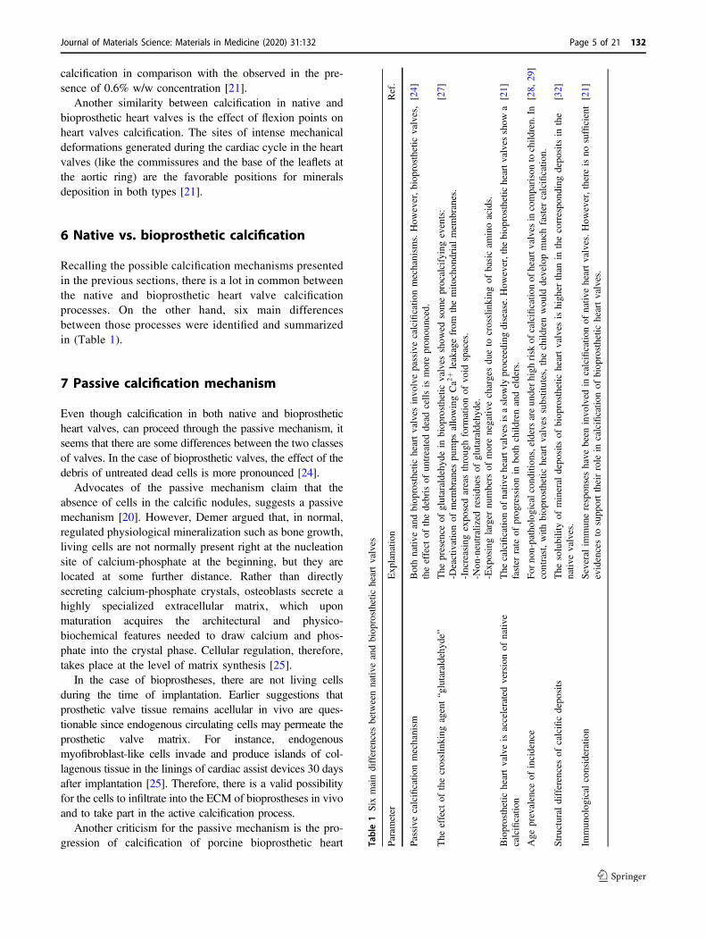
6 Native vs. bioprosthetic calcification
Recalling the possible calcification mechanisms presentedin the previous sections, there is a lot in common betweenthe native and bioprosthetic heart valve calcificationprocesses. On the other hand, six main differencesbetween those processes were identified and summarizedin (Table 1).
7 Passive calcification mechanism
Even though calcification in both native and bioprostheticheart valves, can proceed through the passive mechanism, itseems that there are some differences between the two classesof valves. In the case of bioprosthetic valves, the effect of thedebris of untreated dead cells is more pronounced [24].
Advocates of the passive mechanism claim that theabsence of cells in the calcific nodules, suggests a passivemechanism [20]. However, Demer argued that, in normal,regulated physiological mineralization such as bone growth,living cells are not normally present right at the nucleationsite of calcium-phosphate at the beginning, but they arelocated at some further distance. Rather than directlysecreting calcium-phosphate crystals, osteoblasts secrete ahighly specialized extracellular matrix, which uponmaturation acquires the architectural and physico-biochemical features needed to draw calcium and phos-phate into the crystal phase. Cellular regulation, therefore,takes place at the level of matrix synthesis [25].
In the case of bioprostheses, there are not living cellsduring the time of implantation. Earlier suggestions thatprosthetic valve tissue remains acellular in vivo are ques-tionable since endogenous circulating cells may permeate theprosthetic valve matrix. For instance, endogenousmyofibroblast-like cells invade and produce islands of col-lagenous tissue in the linings of cardiac assist devices 30 daysafter implantation [25]. Therefore, there is a valid possibilityfor the cells to infiltrate into the ECM of bioprostheses in vivoand to take part in the active calcification process.
Another criticism for the passive mechanism is the pro-gression of calcification of porcine bioprosthetic heart Ta
ble1Six
maindifferencesbetweennativ
eandbiop
rosthetic
heartvalves
Param
eter
Exp
lanatio
nRef.
Passive
calcificatio
nmechanism
Bothnativ
eandbiop
rosthetic
heartvalves
invo
lvepassivecalcificatio
nmechanism
s.How
ever,biop
rosthetic
valves,
theeffect
ofthedebrisof
untreateddead
cells
ismorepron
ounced.
[24]
The
effect
ofthecrosslinking
agent“glutaraldehy
de”
The
presence
ofglutaraldehy
dein
biop
rosthetic
valves
show
edsomeprocalcifyingevents:
-Deactivationof
mem
branes
pumps
allowingCa2
+leakagefrom
themito
chon
drialmem
branes.
-Increasingexpo
sedareasthroug
hform
ationof
void
spaces.
-Non
-neutralized
residu
esof
glutaraldehy
de.
-Exp
osinglarger
numbers
ofmorenegativ
echargesdu
eto
crosslinking
ofbasicam
inoacids.
[27]
Bioprosthetic
heartvalveisacceleratedversionof
nativ
ecalcificatio
nThe
calcificatio
nof
nativ
eheartv
alvesisaslow
lyproceeding
disease.How
ever,the
biop
rosthetic
heartv
alvesshow
afaster
rate
ofprog
ressionin
both
child
renandelders.
[21]
Age
prevalence
ofincidence
For
non-patholog
icalcond
ition
s,eldersareun
derhigh
risk
ofcalcificatio
nof
heartv
alvesin
comparisonto
child
ren.In
contrast,with
biop
rosthetic
heartvalves
substitutes,thechild
renwou
lddevelopmuchfaster
calcificatio
n.[28,
29]
Structuraldifferencesof
calcificdepo
sits
The
solubilityof
mineral
depo
sitsof
biop
rosthetic
heartvalves
ishigh
erthan
inthecorrespo
ndingdepo
sitsin
the
nativ
evalves.
[32]
Immun
olog
ical
consideration
Several
immun
erespon
seshave
been
invo
lved
incalcificatio
nof
nativ
eheartvalves.How
ever,thereisno
sufficient
evidencesto
supp
orttheirrole
incalcificatio
nof
biop
rosthetic
heartvalves.
[21]
Journal of Materials Science: Materials in Medicine (2020) 31:132 Page 5 of 21 132
valves upon implantation. Assuming that the main reasonunderlying calcification is the presence of calcifyingpotential of the original valvular tissue. This means theyshould calcify also in the animals from which they wereextracted (e.g., pigs) [25]. However, they apparently do notcalcify under normal physiological conditions. Two differ-ent possible explanations may be given: (a) Alteration of thetype and structure of the ECM proteins caused by glutar-aldehyde treatment; (b) Structural alterations in the lipidsbecause of redox processes taking place [26]. It is suggestedhowever that such oxidized lipids and cross-linked proteinsare necessary but not sufficient for calcification [25].
To sum up, based on the previous argument, Demer et al.1997 suggested that bioprosthetic valve calcification shouldnot be considered as passive, but as endochondral calcifi-cation. In both cases, mineralization takes place in acellulartissues mainly at the stage of extracellular matrix organi-zation and maturation [25].
8 The effect of cross-linking agent
In native heart valves, there is no presence of crosslinkers(glutaraldehyde) contributing to the calcification mechan-isms. On the other hand, the presence of glutaraldehyde inbioprosthetic heart valves is believed to contribute in fourdifferent ways in the calcification process [27]:
(a) In the passive calcification: it is taking place throughthe deactivation of membranes pumps (consequently dama-ging Ca–P electrochemical gradient across the membrane andallowing the Ca2+ leakage from the mitochondrial mem-branes, resulting to the formation of calcific deposits) [27];
(b) The formation of void space, resulting in the expo-sure of active sites or physical niche for calcification.
(c) Glutaraldehyde unreacted residues may promotecalcification,
(d) Glutaraldehyde crosslinking with the basic amino acidsin collagen helices, thus impairing charges balances andexposing larger numbers of more negative charges which mayprovide active sites for Ca2+ binding and hence with theconcomitant development of nucleation sites for calcification.
Thus, in contrary to those bioprostheses, the native heartvalves do not have any glutaraldehyde. Hence, all the possibleeffects can only take place in the bioprostheses HV.
9 The bioprosthetic heart valve is anaccelerated version of native heartcalcification
According to Demer, calcific aortic valve disease is avariable severity disorder, advancing at a relatively low rate.It may be divided into two main stages: (a) Aortic sclerosis,
involves mild valve thickening, in which there is no bloodflow obstruction. (b) Aortic stenosis, accompanied bysevere calcification, because of which the movement ofleaflets is impaired. In general, the rates of bioprostheticvalve calcification are significantly higher in comparisonwith the respective process of native valves calcification[25]. It should also be noted that the rate of progression ofcalcification is markedly accelerated in younger patients.Children and adolescents have an especially acceleratedcourse while elderly patients demonstrate a lower rate ofbioprosthetic valve degeneration [21].
10 Age prevalence of incidence
As in native aortic valves, calcific changes in bioprostheticvalves is a prominent feature of primary valve failure.However, the prevalence of calcification and the biopros-thetic valve failure appears to decrease with the age incontrast to native valves. In several studies, younger ageswere more vulnerable to a bioprosthetic valve failure and aneed for reoperation [28, 29]. This finding indicated that thecalcification process of bioprosthetic valves may be differ-ent from the observed in native valves [29]. The possibleexplanation for such a correlation is that remodeling rate isdifferent between children and adults, In young ages, highrate of remodeling is accompanied with the differentiationof VIC into myofibroblast due to chronic stress [30]. Thosemyofibroblast express osteoblast markers (osteocalcin andbone sialoprotein), leading finally to the formation ofnodules-like bone-type calcific deposits inside the valvulartissue structure [31].
11 The structural differences of calcificdeposits
Physicochemical characterization of the calcific depositswith the powder X-ray diffraction and Fourier TransformInfrared spectroscopy showed that the crystalline phases ofhuman heart valves calcific deposits collected from differentpathological cases, were similar in terms of chemicalcomposition, crystal structure, morphology and solubility[32, 33]. However, the solubility of mineral deposits ofbioprosthetic heart valves was higher in the correspondingdeposits on native valves [32]. A possible explanation forthis difference may be due to shorter residence and ageingtime of the mineral deposits in the bioprosthetic valves,including transient crystal phases, which eventually dis-solve easier compared to more stable phases formed in thenative ones [34]. Further confirmations for this possibilitycame from the finding that the structure of the calcificdeposits developed on bioprosthetic valves are less
132 Page 6 of 21 Journal of Materials Science: Materials in Medicine (2020) 31:132
crystalline, in comparison with the corresponding forma-tions on the natural heart valves deposits [32].
12 Immunologic consideration
The role of immunological responses in the heart valvecalcification is a very controversial and broad topic. Asmentioned in the previous sections, several immuneresponses have been involved in calcification of nativeheart valves. However, Schoen and Levy argued the suf-ficiency of the pieces of evidence for the role of theimmune system in the calcification of bioprosthetic heartvalves as follows; (a) animal immunological sensitizationis not only limited to fresh tissues but also cross-linkedtissues, (b) in different valve dysfunction disorders, anti-bodies were detected after, not before the dysfunction-ality, (c) very often mononuclear inflammatory cells werefound in the failed tissues valves. However, this finding isnot enough to consider it as immunologic rejection.Accordingly, there is no solid evidence for the hypothesisof the role of inflammatory cells for heart valve calcifi-cation processes. This opinion was further supported bythe finding that there was no difference in calcificationpotential of the heart valve cusps between groups, whichwere enclosed in a filter chamber (isolated from host cellscontact) and a control group [21].
Despite the six mentioned differences stated abovebetween native and bioprosthetic heart valves, comparisonbetween native and bioprosthetic calcification is a quitechallenging issue. The reason for such difficulty is attrib-uted to the wide variety of bioprosthetic valves with each ofthem having their own characteristics. However, literaturereports mostly focus on the glutaraldehyde-treated porcineor glutaraldehyde-treated bovine pericardial valves.
13 Calcification of decellularized heartvalves
Although bioprosthetic heart valves present a better hemo-dynamic profile and anticoagulation free post-implantationlifespan, at the present, calcification restricts the patients’profile to elderly or people noncompatible with antic-oagulation therapies (e.g., pregnant women) [35]. More-over, considering the previously mentioned drawbacks,associated with different types of heart valves bio-prostheses, a new class of substitutes emerged, known asTEHV. The idea behind this new type is to use them astemporary scaffolds, while in parallel to restore the normalfunction of the native valve, enhance host cells infiltrationin vivo and support their response to mechano-transductionto regenerate normal live valvular tissue [8]. Upon
proceeding of this new tissue, the scaffold material isexpected to be gradually biodegraded until its total repla-cement by the generated new living tissue valve. Amongdifferent types of TEHV, the decellularized ones (dTEHV)have attracted the interest, because of their low cost, simplepreparation protocols and their similarity to the nativevalves. Till 2020 more than 80 articles and reviews haveintroduced and evaluated the use of these dTEHV [36–44].
Despite the relatively poor knowledge of calcificationpossible mechanisms in those types of heart valves, severalresearch groups pointed to the role of immunogenicity andcell remnants as the main culprits in the calcification [10].Hence the decellularization process is expected to reduce oreliminate the calcification.
So far, many animal and clinical studies have beenpublished since 2000, in which decellularized heart valves,were used for the replacement of defective ones (Tables 2and 4). Up to present, there is no comprehensive reviewrepresenting and discussing those data in the light of theircalcification potential.
In the present review, we focus mainly on the calcifica-tion potential of those implanted dTEHV scaffolds fromboth animal and human heart valves through clinical andanimal studies. pericardial tissue-based decellularized heartvalves [45] and decellularized synthetic-seeded scaffolds[46] will not be covered in this review.
14 The effect of the decellularization on thecalcification from animal studies
Since the emergence of tissue decellularization concept as apromising option for s+caffolds preparation in the tissueengineering field, wide variations of techniques have beenutilized. The purpose of all those techniques is alwaysthe same:
(1) Preservation of the structural integrity of the originaltissue ECM; to restore its mechanical and biologicalproperties.
(2) Preservation of and/or enrichment with importantgrowth factors.
(3) Removal of any cell debris and any immunetriggering parts.
Theoretically, based on the calcification possiblemechanisms discussed earlier, successful decellularizationwould prevent calcification without a need for any furthertreatment.
In general, the history of acellular tissue in cardiovas-cular application backs to the work of Vesely in vitro studyin 1992, regarding in vitro studies [8, 47].
Since then, many animal studies have been conducted ondecellularized tissues, in which different kinds of animalmodels (Sullfok Sheep, Dog, Rats, Kangaroo, Pigs, chacma
Journal of Materials Science: Materials in Medicine (2020) 31:132 Page 7 of 21 132
Table 2 Animal studies conducted to date with decellularized heart valves and their correlation to calcification
Calcificationprevention
Xenogeneic/Allogeneic
Graft Source Decellularization Technique Cells Model Max.Implantationduration
sample size Characterization Aortic/Pulmonary
Ref.
Yes XenogeneicAllogeneic
PigSheep
Chemical/EnzymaticSynerGraft Tech(Hypotonic,RNAse, DNase)
Unseeded Sheep 11 m 9 Pig4 Sheep
Gross examination A & P [84]
Yes Xenogeneic Pig Chemical24 h in 0.1%DOA, thenstored in Hanks buffer withantibiotics
Vascular Ecs Juvenile sheep 8 m 8 X-ray; flame atomicabsorption spectrometry,Von Kossa
P [85]
Yes Xenogeneic Pig Chemical(AutoTissue Ltd)1%DOA/ 70% EtOH, thenkept in RPMI nutrientmedium up to 30 daysbefore implantation
Unseeded Juvenile sheep 11 m 4 X-ray; flame atomicabsorption spectrometry,Von Kossa; gross exam
P [58]
Yes Allogeneic Sheep Chemical/EnzymaticFor 24 h tissues were treatedwith a solution of 0.5% SOCand 0.5% SDS for 24 h.Then for 72 h (6 cycles)with PBS supplemented.Finally, tissues were treatedby a DNase for 4 h at 37 °C
12 ECs9 Unseeded
Merino lambs 3 m 21 Von Kossa staining, grossexamination
P [64]
Yes Xenogeneic Pig ChemicalValves were treated for 14 hwith 1% DOA inphysiological saline at 37 °Cthen washed withphysiological saline.
Unseeded Juvenile sheep 2 y 11 ECG P [59]
Yes Xenogeneic Pig Chemical/Physicalvalves were immersed inPEG solution containingantibiotics for 168 h understirrer. The PEG solutionwas changed every 48 h.The valves were exposed to100 kGy gamma irradiationsin room air. The valves werewashed in normal salinesolution for 24 h, thentransferred into DNasesolution for 48 h at 37 °C.The DNase solution thenvalves were washed withnormal saline solution for3 h.
Unseeded RatsDogs
Rats: 2 mDogs: 6 m
20 Rats9 Dogs
Von Kossa staining A [63]
Yes Allogeneic Sheep ChemicalValves were decellularizedat room temperature in asolution of 0.5% SDC and0.5% SDS for 48 h,followed by 2 wash cycles(12 h each) in distilledwater, then six wash cycleswith PBS under continuousshaking. Then valves werestored in PBS at 4 C forone day.
Unseeded Sheep 9 m 12 Von Kossa staining; Grossexamination
A [55]
Yes Allogeneic Sheep Chemical/PhysicalBefore decellularizationvalves were kept in RPMI1640 containing 10%DMSO and 10% FBS andcryopreserved for 48 h.At room temperature, tissueswere treated with an N-lauroyl sarcosinate in a Trisbuffer solution for 24 hcontaining a recombinantendonuclease andantibiotics. Then the tissueswere rinsed by recirculatingwater through a bed of resinfor 24 h. The valves weredivided into 2 groups:1- No further processing andplaced into isotonic salinewith polymyxin B thenstored at 1 °C to10 °C2- Glycerolized for 24 hthen stored at −80 °C
Unseeded Juvenile sheep 1 y 1) 52) 5
Gross examination; HR-X-ray; EGG; MRI;Angiography
p [68]
132 Page 8 of 21 Journal of Materials Science: Materials in Medicine (2020) 31:132
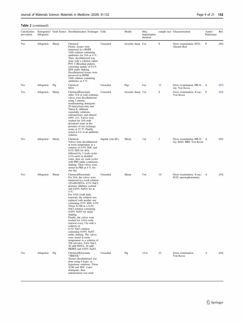
Table 2 (continued)
Calcificationprevention
Xenogeneic/Allogeneic
Graft Source Decellularization Technique Cells Model Max.Implantationduration
sample size Characterization Aortic/Pulmonary
Ref.
Yes Allogeneic Sheep ChemicalFirstly, tissues wereimmersed in a RMPI1640 solution containingantibiotics for 24 h at 4 °C.Then, decellularized wasdone with a solution calledPUC I (Brazilian patent),consisting mainly of 0.1%SDS under shaking.Decellularized tissues werepreserved in RPMI1640 solution containingantibiotics at 4 °C.
Unseeded Juvenile sheep 6 m 8 Gross examination; ECG;Alizarin Red
P [86]
Yes Allogeneic Pig ChemicalDOA
Unseeded Pigs 6 m 12 Gross examination; HR-X-ray; Von Kossa
A [87]
Yes Allogeneic Sheep Chemical/EnzymaticAfter 72 h of cold ischemia,valves were decellularizedusing 2 anionic,nondenaturing detergents(N-lauroylsarcosine andTriton-X, differentosmolality solutions,endonuclease, and ethanol(40% v/v). Valves werewashed for 24 h withdeionized water in thepresence of ion exchangeresins at 21 °C. Finallystored at 4 C in an antibioticsolution.
Unseeded Juvenile sheep 5 m 8 Gross examination; X-ray;Von Kossa
P [62]
Yes Allogeneic Sheep ChemicalValves were decellularizedat room temperature in asolution of 0.5% SDC and0.5% SDS for 48 h,followed by 2 wash cycles(12 h each) in distilledwater, then six wash cycleswith PBS under continuousshaking. Then valves werestored in PBS at 4 °C forone day
Jugular vein ECs Sheep 3 m 5 Gross examination; HR-X-ray; EGG; MRI; Von Kossa
A [69]
Yes Allogeneic Sheep Chemical/EnzymaticFor 24 h, the valves wereimmersed in a wash solution(20 mM EDTA, 0.3% NaCl,protease inhibitor cocktailand 0.05% NaN3) for at4 °C.For 144 h (with dailyrenewal), the solution wasreplaced with another onecontaining 0.5% SDS, 0.5%Triton X-100 in a 0.3%NaCl solution containing0.05% NaN3 for undershaking.Finally, the valves werewashed for 144 h (withrenewal every 3 h) with asolution of0.3% NaCl solutioncontaining 0.05% NaN3under shaking. The valveswere stored at roomtemperature in a solution of200 ml/valve, 0.6% NaCl,20 mM EDTA, 10 mMHEPES and 0.05% NaN3.
Unseeded Sheep 5 m 14 Gross examination, X-ray;ECG; spectrophotometry
A [54]
Yes Allogeneic Pig Chemical/Enzymatic“TRICOL”Tissues decellularized wasdone using b hypo- orhypertonic solutions, TritonX100 and SDC. Later,detergents, thenendonuclease was used.
Unseeded Pig 15 m 22 Gross examination;Von Kossa
A [60]
Journal of Materials Science: Materials in Medicine (2020) 31:132 Page 9 of 21 132
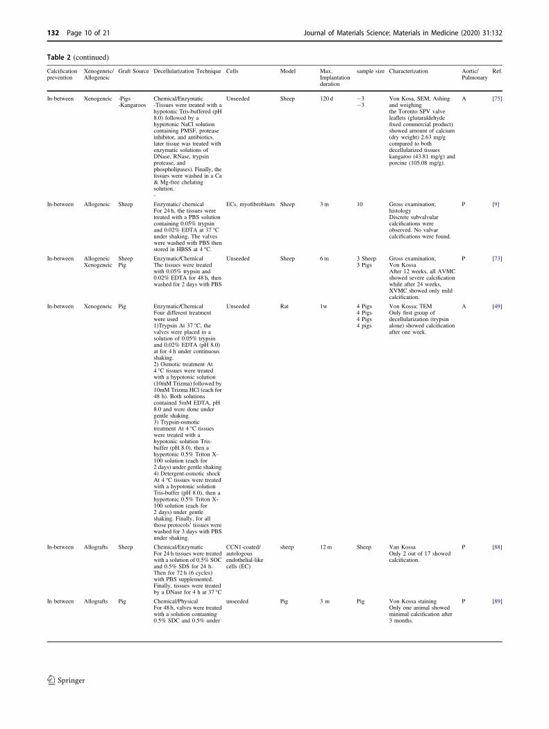
Table 2 (continued)
Calcificationprevention
Xenogeneic/Allogeneic
Graft Source Decellularization Technique Cells Model Max.Implantationduration
sample size Characterization Aortic/Pulmonary
Ref.
In-between Xenogeneic -Pigs-Kangaroos
Chemical/Enzymatic-Tissues were treated with ahypotonic Tris-buffered (pH8.0) followed by ahypertonic NaCl solutioncontaining PMSF, proteaseinhibitor, and antibiotics.later tissue was treated withenzymatic solutions ofDNase, RNase, trypsinprotease, andphospholipases). Finally, thetissues were washed in a Ca& Mg-free chelatingsolution.-
Unseeded Sheep 120 d −3−3
Von Kosa, SEM, Ashingand weighingthe Toronto SPV valveleaflets (glutaraldehydefixed commercial product)showed amount of calcium(dry weight) 2.63 mg/gcompared to bothdecellularized tissueskangaroo (43.81 mg/g) andporcine (105.08 mg/g).
A [75]
In-between Allogeneic Sheep Enzymatic/ chemicalFor 24 h, the tissues weretreated with a PBS solutioncontaining 0.05% trypsinand 0.02% EDTA at 37 °Cunder shaking. The valveswere washed with PBS thenstored in HBSS at 4 °C.
ECs, myofibroblasts Sheep 3 m 10 Gross examination;histologyDiscrete subvalvularcalcifications wereobserved. No valvarcalcifications were found.
P [9]
In-between AllogeneicXenogeneic
SheepPig
Enzymatic/ChemicalThe tissues were treatedwith 0.05% trypsin and0.02% EDTA for 48 h, thenwashed for 2 days with PBS
Unseeded Sheep 6 m 3 Sheep3 Pigs
Gross examination;Von KossaAfter 12 weeks, all AVMCshowed severe calcificationwhile after 24 weeks,XVMC showed only mildcalcification.
P [73]
In-between Xenogeneic Pig Enzymatic/ChemicalFour different treatmentwere used1)Trypsin At 37 °C, thevalves were placed in asolution of 0.05% trypsinand 0.02% EDTA (pH 8.0)at for 4 h under continuousshaking.2) Osmotic treatment At4 °C tissues were treatedwith a hypotonic solution(10mM Trizma) followed by10mM Trizma HCl (each for48 h). Both solutionscontained 5mM EDTA, pH8.0 and were done undergentle shaking.3) Trypsin-osmotictreatment At 4 °C tissueswere treated with ahypotonic solution Tris-buffer (pH 8.0), then ahypertonic 0.5% Triton X-100 solution (each for2 days) under gentle shaking4) Detergent-osmotic shockAt 4 °C tissues were treatedwith a hypotonic solutionTris-buffer (pH 8.0), then ahypertonic 0.5% Triton X-100 solution (each for2 days) under gentleshaking. Finally, for allthose protocols’ tissues werewashed for 3 days with PBSunder shaking.
Unseeded Rat 1w 4 Pigs4 Pigs4 Pigs4 pigs
Von Kossa; TEMOnly first group ofdecellularization (trypsinalone) showed calcificationafter one week.
A [49]
In-between Allografts Sheep Chemical/EnzymaticFor 24 h tissues were treatedwith a solution of 0.5% SOCand 0.5% SDS for 24 h.Then for 72 h (6 cycles)with PBS supplemented.Finally, tissues were treatedby a DNase for 4 h at 37 °C
CCN1-coated/autologousendothelial-likecells (EC)
sheep 12 m Sheep Van KossaOnly 2 out of 17 showedcalcification.
P [88]
In between Allografts Pig Chemical/PhysicalFor 48 h, valves were treatedwith a solution containing0.5% SDC and 0.5% under
unseeded Pig 3 m Pig Von Kossa stainingOnly one animal showedminimal calcification after3 months.
P [89]
132 Page 10 of 21 Journal of Materials Science: Materials in Medicine (2020) 31:132
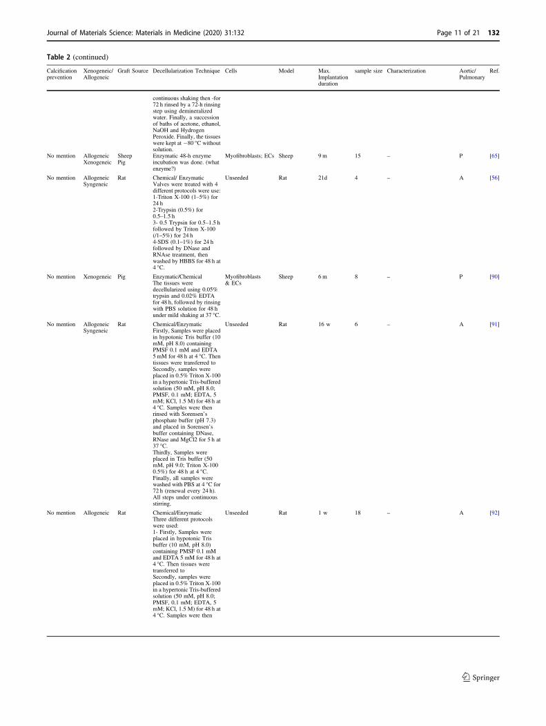
Table 2 (continued)
Calcificationprevention
Xenogeneic/Allogeneic
Graft Source Decellularization Technique Cells Model Max.Implantationduration
sample size Characterization Aortic/Pulmonary
Ref.
continuous shaking then -for72 h rinsed by a 72-h rinsingstep using demineralizedwater. Finally, a successionof baths of acetone, ethanol,NaOH and HydrogenPeroxide. Finally, the tissueswere kept at −80 °C withoutsolution.
No mention AllogeneicXenogeneic
SheepPig
Enzymatic 48-h enzymeincubation was done. (whatenzyme?)
Myofibroblasts; ECs Sheep 9 m 15 – P [65]
No mention AllogeneicSyngeneic
Rat Chemical/ EnzymaticValves were treated with 4different protocols were use:1-Triton X-100 (1–5%) for24 h2-Trypsin (0.5%) for0.5–1.5 h3- 0.5 Trypsin for 0.5–1.5 hfollowed by Triton X-100(/1–5%) for 24 h4-SDS (0.1–1%) for 24 hfollowed by DNase andRNAse treatment, thenwashed by HBBS for 48 h at4 °C.
Unseeded Rat 21d 4 – A [56]
No mention Xenogeneic Pig Enzymatic/ChemicalThe tissues weredecellularized using 0.05%trypsin and 0.02% EDTAfor 48 h, followed by rinsingwith PBS solution for 48 hunder mild shaking at 37 °C.
Myofibroblasts& ECs
Sheep 6 m 8 – P [90]
No mention AllogeneicSyngeneic
Rat Chemical/EnzymaticFirstly, Samples were placedin hypotonic Tris buffer (10mM, pH 8.0) containingPMSF 0.1 mM and EDTA5mM for 48 h at 4 °C. Thentissues were transferred toSecondly, samples wereplaced in 0.5% Triton X-100in a hypertonic Tris-bufferedsolution (50 mM, pH 8.0;PMSF, 0.1 mM; EDTA, 5mM; KCl, 1.5 M) for 48 h at4 °C. Samples were thenrinsed with Sorensen’sphosphate buffer (pH 7.3)and placed in Sorensen’sbuffer containing DNase,RNase and MgCl2 for 5 h at37 °C.Thirdly, Samples wereplaced in Tris buffer (50mM, pH 9.0; Triton X-1000.5%) for 48 h at 4 °C.Finally, all samples werewashed with PBS at 4 °C for72 h (renewal every 24 h).All steps under continuousstirring.
Unseeded Rat 16 w 6 – A [91]
No mention Allogeneic Rat Chemical/EnzymaticThree different protocolswere used:1- Firstly, Samples wereplaced in hypotonic Trisbuffer (10 mM, pH 8.0)containing PMSF 0.1 mMand EDTA 5 mM for 48 h at4 °C. Then tissues weretransferred toSecondly, samples wereplaced in 0.5% Triton X-100in a hypertonic Tris-bufferedsolution (50 mM, pH 8.0;PMSF, 0.1 mM; EDTA, 5mM; KCl, 1.5 M) for 48 h at4 °C. Samples were then
Unseeded Rat 1 w 18 – A [92]
Journal of Materials Science: Materials in Medicine (2020) 31:132 Page 11 of 21 132
baboons, merino lamb), immunotype of the used heartvalves (Xenografts, Homografts and Allografts) and decel-lularization methods (Enzymatic, detergents, combined),cell-seeded or unseeded have been used. The mineralization
process was monitored using different characterization toolsseparately or together (Gross examination, Echocardio-graphy, MRI, Electron Microscopy, Atomic absorption, andHistology) (Table 2).
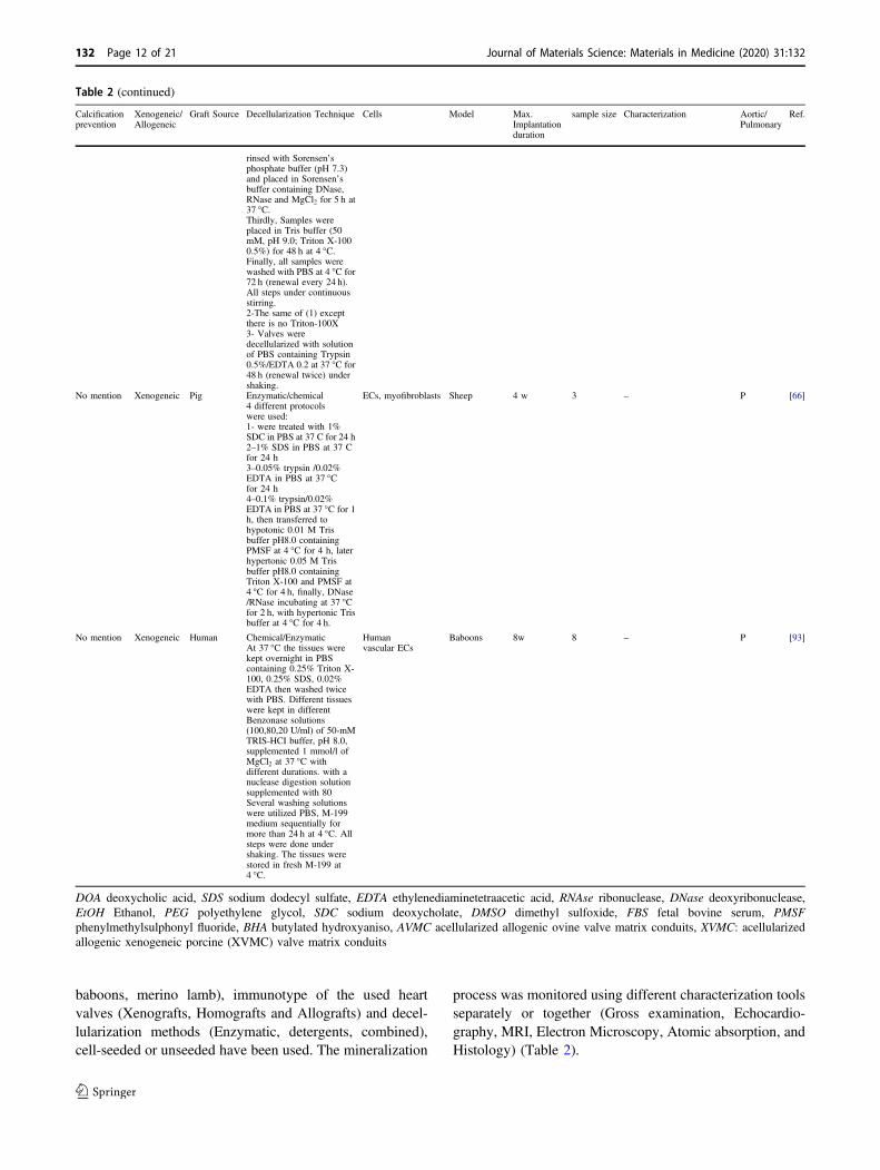
Table 2 (continued)
Calcificationprevention
Xenogeneic/Allogeneic
Graft Source Decellularization Technique Cells Model Max.Implantationduration
sample size Characterization Aortic/Pulmonary
Ref.
rinsed with Sorensen’sphosphate buffer (pH 7.3)and placed in Sorensen’sbuffer containing DNase,RNase and MgCl2 for 5 h at37 °C.Thirdly, Samples wereplaced in Tris buffer (50mM, pH 9.0; Triton X-1000.5%) for 48 h at 4 °C.Finally, all samples werewashed with PBS at 4 °C for72 h (renewal every 24 h).All steps under continuousstirring.2-The same of (1) exceptthere is no Triton-100X3- Valves weredecellularized with solutionof PBS containing Trypsin0.5%/EDTA 0.2 at 37 °C for48 h (renewal twice) undershaking.
No mention Xenogeneic Pig Enzymatic/chemical4 different protocolswere used:1- were treated with 1%SDC in PBS at 37 C for 24 h2–1% SDS in PBS at 37 Cfor 24 h3–0.05% trypsin /0.02%EDTA in PBS at 37 °Cfor 24 h4–0.1% trypsin/0.02%EDTA in PBS at 37 °C for 1h, then transferred tohypotonic 0.01 M Trisbuffer pH8.0 containingPMSF at 4 °C for 4 h, laterhypertonic 0.05 M Trisbuffer pH8.0 containingTriton X-100 and PMSF at4 °C for 4 h, finally, DNase/RNase incubating at 37 °Cfor 2 h, with hypertonic Trisbuffer at 4 °C for 4 h.
ECs, myofibroblasts Sheep 4 w 3 – P [66]
No mention Xenogeneic Human Chemical/EnzymaticAt 37 °C the tissues werekept overnight in PBScontaining 0.25% Triton X-100, 0.25% SDS, 0.02%EDTA then washed twicewith PBS. Different tissueswere kept in differentBenzonase solutions(100,80,20 U/ml) of 50-mMTRIS-HCI buffer, pH 8.0,supplemented 1 mmol/l ofMgCl2 at 37 °C withdifferent durations. with anuclease digestion solutionsupplemented with 80Several washing solutionswere utilized PBS, M-199medium sequentially formore than 24 h at 4 °C. Allsteps were done undershaking. The tissues werestored in fresh M-199 at4 °C.
Humanvascular ECs
Baboons 8w 8 – P [93]
DOA deoxycholic acid, SDS sodium dodecyl sulfate, EDTA ethylenediaminetetraacetic acid, RNAse ribonuclease, DNase deoxyribonuclease,EtOH Ethanol, PEG polyethylene glycol, SDC sodium deoxycholate, DMSO dimethyl sulfoxide, FBS fetal bovine serum, PMSFphenylmethylsulphonyl fluoride, BHA butylated hydroxyaniso, AVMC acellularized allogenic ovine valve matrix conduits, XVMC: acellularizedallogenic xenogeneic porcine (XVMC) valve matrix conduits
132 Page 12 of 21 Journal of Materials Science: Materials in Medicine (2020) 31:132
The first published in vivo study for the evaluation ofdecellularized heart valves was by O’Brien and colleaguesin 1999. In this work, porcine aortic valves were decel-lularized by a process designed to remove all the leafletcells. The treated valves were next implanted in weanlingsheep. After 150 days, the grafts were explanted andassessed histologically and analyzed for calcium content bythe atomic absorption spectrometry. All valves werehemodynamically functional at explant. Histologicalexamination of the explanted valvular tissues showedstructurally intact leaflets with in-growth of host fibro-blastoid cells without evidence of calcification. The calciumcontent in porcine leaflets was unaltered over the durationof the implant. The lack of calcification of acellular aorticleaflets suggested that prolonged durability of such valves isattainable, without the use of cross-linking agents [48].
Since then, large numbers of animal and clinical studieshave been published. In this review, those animal studiesare sorted and further subcategorized into four differentcategories (Tables 2 and 4) for each, depending on the effectof decellularization on calcification: (A) Decellularizationtotally prevented calcification, (B) Decellularizationreduced calcification, (C) Decellularization showed bothpro-calcification and anticalcification potential based on theconditions involved and finally (D) Studies did not mentionclearly the effect of decellularization on calcification. All ofthose categorized were tabulated and compared to eachother’s depending on the host animal (in the case of animalstudies), the type of heart valves involved, the technique ofdecellularization, cell seeding or not and type of cells used,duration of the study follow-up, the size of the groupinvolved and finally the techniques used to evaluate calci-fication occurrence). Also, so far nearly around five specificarticles comparing different decellularization techniqueseffect on calcification have been published; in vivo studies[49–51] and in vitro studies [52, 53].
14.1 Categories A&B: decellularization results incalcification reduction or prevention
Different approaches showed a successful anticalcificationeffect of the used decellularization techniques: Some ofthem are without any further post-decellularization process(only decellularization) as SDS [54], SDS/osmotic shock[42, 55], SDS/ RNAse/DNase [56], DOA [57], DOA/EtOH[58, 59], DOA/triton x-100/endonucleases [60], N-laur-oylsarcosine/cryo [61], N-lauroylsarcosine/Triton-X/EtOH[62], PEG/gamma radiation [63].
Other reports had post-decellularization treatment as cellseeding (endothelial cells [57, 64], endothelial and myofi-broblast cells [65, 66]), proteins and/or growth factorstreatments as fibronectin [57, 67], and fibronectin/SDF-1[54] have been used. Although both treatments improved
coverage of the decellularized tissue with endothelial cells,they showed no difference regarding anticalcificationtreatment (both treated and untreated decellularized ovineaortic allograft did not calcify).
Different post-decellularized preservations also havebeen investigated like cryopreservation [62, 68, 69].According to those three publications, a clear antic-alcification effect of the decellularized group in comparisonto bioprosthetic valves (either xenografts or allografts) hasbeen shown.
Noteworthy, in this category (A&B) the slight forma-tion—in some cases—of calcific deposits were attributedto the immune response of the injuries related to suturing.Therefore, the use of sutureless or transcatheter would—theoretically—overcome this problem. Some animal stu-dies showed promising results for using percutaneousdTEHV implantation (no calcification and good remodel-ing) [70]. On the other hand, a report showed a differentcomplicated scenario when evaluating the effect of thistechnique on the calcification on general. This study dis-cussed the effect of the severity and distribution pattern ofcalcific deposits of heart valves on the percutaneousimplantation success [71].
14.2 Category C: decellularization results inconditional calcification
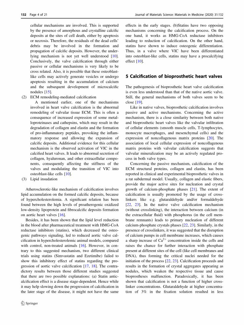
In contrary to the reports that showed the absence or justsuture line calcification, few ones showed that decellular-ization would not prevent calcification (Fig. 2).
For example, Steinhoff and associates showed thatdecellularized allogeneic heart valves implanted in sheepmodel demonstrated calcification after using EDTA/trypsindecellularization technique with autologous endothelial andmyofibroblast. The calcification mainly took place aroundthe cell remnants [9]. It was also associated with a strongleukocyte infiltration early after implantation, suggestingthat an inflammatory reaction was responsible for theunderlying mechanism of early-stage calcification. Thissuggested mechanism is with an agreement with a pre-viously published explanation of calcification in bloodvessel’s wall by Andrews et al. [72]. They suggested thatthe wall would simulate the generation of activated plateletaggregates or thrombus acceleration, because of glycopro-tein binding availability in collagen. The adhered and acti-vated platelets might also interact with inflammatoryleukocytes as well, facilitating leukocyte-endothelial celladhesion, leading finally to calcification [72]. The conclu-sions from this work further corroborate the suggestion ofan immuno-mediated calcification [9]. It is noteworthy hereto mention that the sensitivity response for any injuriesbetween different animals’ models is an important factorwhen studying calcification, as showed that sheep injuries
Journal of Materials Science: Materials in Medicine (2020) 31:132 Page 13 of 21 132
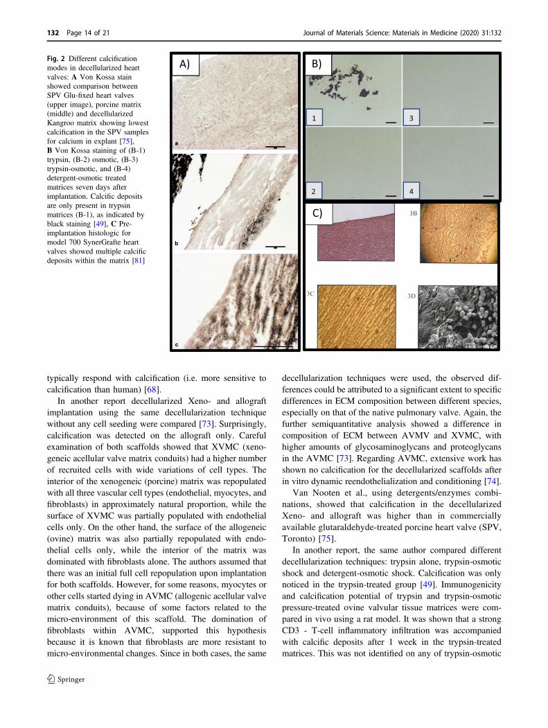
typically respond with calcification (i.e. more sensitive tocalcification than human) [68].
In another report decellularized Xeno- and allograftimplantation using the same decellularization techniquewithout any cell seeding were compared [73]. Surprisingly,calcification was detected on the allograft only. Carefulexamination of both scaffolds showed that XVMC (xeno-geneic acellular valve matrix conduits) had a higher numberof recruited cells with wide variations of cell types. Theinterior of the xenogeneic (porcine) matrix was repopulatedwith all three vascular cell types (endothelial, myocytes, andfibroblasts) in approximately natural proportion, while thesurface of XVMC was partially populated with endothelialcells only. On the other hand, the surface of the allogeneic(ovine) matrix was also partially repopulated with endo-thelial cells only, while the interior of the matrix wasdominated with fibroblasts alone. The authors assumed thatthere was an initial full cell repopulation upon implantationfor both scaffolds. However, for some reasons, myocytes orother cells started dying in AVMC (allogenic acellular valvematrix conduits), because of some factors related to themicro-environment of this scaffold. The domination offibroblasts within AVMC, supported this hypothesisbecause it is known that fibroblasts are more resistant tomicro-environmental changes. Since in both cases, the same
decellularization techniques were used, the observed dif-ferences could be attributed to a significant extent to specificdifferences in ECM composition between different species,especially on that of the native pulmonary valve. Again, thefurther semiquantitative analysis showed a difference incomposition of ECM between AVMV and XVMC, withhigher amounts of glycosaminoglycans and proteoglycansin the AVMC [73]. Regarding AVMC, extensive work hasshown no calcification for the decellularized scaffolds afterin vitro dynamic reendothelialization and conditioning [74].
Van Nooten et al., using detergents/enzymes combi-nations, showed that calcification in the decellularizedXeno- and allograft was higher than in commerciallyavailable glutaraldehyde-treated porcine heart valve (SPV,Toronto) [75].
In another report, the same author compared differentdecellularization techniques: trypsin alone, trypsin-osmoticshock and detergent-osmotic shock. Calcification was onlynoticed in the trypsin-treated group [49]. Immunogenicityand calcification potential of trypsin and trypsin-osmoticpressure-treated ovine valvular tissue matrices were com-pared in vivo using a rat model. It was shown that a strongCD3 - T-cell inflammatory infiltration was accompaniedwith calcific deposits after 1 week in the trypsin-treatedmatrices. This was not identified on any of trypsin-osmotic
C)
B)
1
2
3
4
A)Fig. 2 Different calcificationmodes in decellularized heartvalves: A Von Kossa stainshowed comparison betweenSPV Glu-fixed heart valves(upper image), porcine matrix(middle) and decellularizedKangroo matrix showing lowestcalcification in the SPV samplesfor calcium in explant [75],B Von Kossa staining of (B-1)trypsin, (B-2) osmotic, (B-3)trypsin-osmotic, and (B-4)detergent-osmotic treatedmatrices seven days afterimplantation. Calcific depositsare only present in trypsinmatrices (B-1), as indicated byblack staining [49], C Pre-implantation histologic formodel 700 SynerGrafte heartvalves showed multiple calcificdeposits within the matrix [81]
132 Page 14 of 21 Journal of Materials Science: Materials in Medicine (2020) 31:132
treated leaflet matrix. A possible explanation could be thatthe collagenous matrix of trypsin-treated implants wasprobably more damaged in comparison with trypsin-osmotic pressure-treated leaflets. Mineralization of biolo-gical valves usually begins at the sites of disrupted collagenfibers in the damaged ECM (where VIC are activated totake part in the natural remodeling procedure). It seems thattheir activation and function is related to the calcificationmechanism [76]. The loss of GAGs (Glycosaminoglycans)in these scaffolds was not responsible for calcification. Itwas previously speculated that the presence of negativelycharged GAG molecules within the ECM of cuspal tissuemay reduce calcification, by chelating calcium ions, therebyreducing supersaturation and preventing hydroxyapatitenucleation and growth. However, in this study, the GAG-depleted trypsin-osmotic matrices did not form calcificdeposits [49].
Finally, the presence of decellularizing agent remnants(e.g., DOA) after completion of the decellularization pro-cess would contribute to the formation of calcific depositsthrough the passive mechanism. In such a case, the ineffi-cient washing techniques after decellularization is a suffi-cient cause to calcification [52].
Based on the previous reports, the causes of calcificationin the decellularized heart valves can be classified into fourmain reasons (Table 3).
15 The effect of the decellularization on thecalcification from clinical studies
Since 2002 till now (2020), around 30 publications and fourmain reviews [39, 42, 77, 78] have been publishedregarding decellularized heart valves implantation inhumans. Some of them are case studies, while others areclinical trials.
The first published study was done by Dohmen et al.,reported that a 43-year-old patient suffering from aorticvalve stenosis underwent a Ross operation using pulmonarycryopreserved allograft (following DOA decellularization
and in vitro EC seeding) to reconstruct the RVOT (rightventricular outflow tract). Multislice computed tomographyexamination showed that there were no calcifications inboth heart valves after 1 year [79].
The diversity of the different techniques used for decel-lularization, cell seeding, and additional treatment are verysimilar to the one that took place in animal studies, aspresented in Table 4.
Based on the previous table results, most of those clinicalstudies showed a promising anticalification effect upondecellularization of the heart valves.
16 Commercial products
To the best of our knowledge, there are two main productsof decellularized heart valves and one from decellularizedporcine sheets (Table 5) [80].
The first commercially available product in the marketwas Synergraft® (CryoLife Inc., USA), which showed ser-ious problems in its early models [81]. However, recentmodels of the product showed very promising results con-cerning calcification [82]. Matrix P® (AutoTissue GmbH,Germany), a decellularized porcine heart valve used since2002 in nearly 200 cases. Another product of the samecompany, Matrix P plus N™ was recently approved [60].Lately, a clinical trial in 2019 showed promising perfor-mance of a newly developed product called ESPOIR®(Corlife, Germany) after a two-year follow-up study [83].
17 Conclusions and Future trend
The different species of decellularized valves, decellular-ization techniques, recellularization or not, storage condi-tions, characterization tools, duration of follow-up,experimental population and even the confusion in the usedterminology, constitute a vast body of complex data, fromwhich it is very difficult to draw solid conclusions. How-ever, still few conclusions may be drawn:
Table 3 Causes of the calcification in decellularized heart valves possible
Possible cause Possible explanation Reference
Damaged ECM Mineralization of biological valves usually begins at the sites of disrupted collagenfibers. (why)
[49]
Immunological response It takes place against immune triggering materials like cell remnants in inefficientdecellularization, death of implanted or in vivo recruited cells or in the presence of α-gal(in porcine valves). Usually, immunological events, differentiation and trans-differentiation took place.
[9, 68, 72, 94]
Species individual differences ofextracellular matrix composition
Heart valves from different species has different ECM structure and components whichinfluence their interaction with different types of host cells.
[64, 73]
Residual of detergent Under physiological conditions some decellularizing agents like deoxycholic acidsresidues may enhance the formation of calcific deposits inside ECM.
[52]
Journal of Materials Science: Materials in Medicine (2020) 31:132 Page 15 of 21 132
Table 4 Clinical studies conducted to date with decellularized heart valves and their correlation to calcification
Preventcalcification
Xenogeneic/Allogeneic
Graft source Decellularization technique Cells Max.implantationduration
Sample size Characterization Aortic/Pulmonary
Ref.
Yes Allogeneic Human ChemicalAt 37 °C, the valves were treated with 1% DOA,followed by an extensive wash in normal saline.
Unseeded 1 y 1 Case MRI; ECG P [79]
Yes Allogeneic Human Synergraft Unseeded 15 m 22 ECG A [95]
Yes AllogeneicXenogeneic
HumanPig
1) Cryolife2) Autotissue
ECs 3 y 1) 112) 12
ECG; MRI P [96]
Yes Allogeneic Human ChemicalFor 10 days, the valves were treated with SDS, andstored in medium RPMI medium at 4 °C
Unseeded 25 m 51 ECG A [97]
Yes Allogeneic Human SynerGraft Unseeded 5y 342 ECG P [82]
Yes Allogeneic Human 1- ChemicalTwo different protocols were used:The tissues were treated with DOA2- The valves were treated with SDS
1)Unseeded2) ECs
11y 1) 392) 44
ECG P [98]
Yes Allogeneic Human ChemicalTissues were treated with SDS 0.1% for 24 h undercontinuous shaking, then washed for 10 days withRinger Lactate solution
Unseeded 5y 41 ECG; MRI; CT A [99]
Yes Xenogeneic Pig Matrix P plus Unseeded 17 m 16 ECG P [100]
Yes Allogeneic Human AutoTissue ECs 10 y 11 ECG; MRI P [101]
Yes Allogeneic Human ChemicalAt room temperature, tissues were treated with 0.5%SDC and 0.5% SDS for 36 h. Finally, Tissues werewashed with NaCl 0.9% solution and stored at 4 °C
Unseeded 5 y 17 MRI; ECG P [102]
Yes Homograft Human ESPOIR unseeded 7 y 5 CT P [103]
No Allogeneic Human Synergraft Unseeded 2 y 1 IHC; Grossexamination
A [104]
No Xenogeneic Pig Matrix P plus Unseeded 1 y 106 IHC P [105]
No Allogeneic Human Synergraft Unseeded 1 y 4 ECG; Grossexamination;Histo; SEM
P [81]
No mention Allogeneic Human SynerGaft Unseeded 3 y 41 -ECG P [106]
No mention Allogeneic Human Chemical/ EnzymaticFirst tissues were treated with hypotonic solutions thenby enzymatic solution containing RNAse/ DNase.
Unseeded 5 y 47 -ECG P [107]
No mention Allogeneic Human AutoTissue Unseeded 18 m 11 – P [58]
No mention Allogeneic Human Synergraft Unseeded 19 m 26 – P [108]
No mention Allogeneic Human Enzymatic/ChemicalFor 48 h, tissues were treated with a PBS solutioncontaining 0.5% Trypsin and 0.2% EDTA at 37 °C (2cycles).
EPCs 40 m 2 – P [109]
No mention Allogeneic Human ChemicalTwo different protocols were used:1- (AutoTissue®)The tissues were treated with DOA, 1%, andethanol, 80%2- The tissues were treated with SDS, 0.1%.
Unseeded 4 y 1)352)33
- P [110]
No mention Xenogeneic Pig Matrix P plus Unseeded 15 m 11 – P [111]
No mention Allogeneic Human SynerGraft Unseeded 9 y 29 – P [112]
No mention Allogeneic Human SynerGraft Unseeded 10 y 39 – P [113]
No mention Allogeneic Human ChemicalThe tissues were treated with SDS 0.1%
Unseeded 3 m 6 – P & A [114]
No mention Allogeneic Human ChemicalAt room temperature, tissues were treated with asolution of 0.5% SDC and 0.5% SDS for 48 h, thenwashed with distilled water 24 h, 12 h each followed bytwo washing process in distilled water, and PBS. Allsteps were done under shaking. Finally, tissues werestored in PBS at 4 °C.
Unseeded 3 y 47 – P [69]
No mention Allograft Human ESPOIR Unseeded 3 y 6 cases – P [115]
No mention homograft Human ChemicalAt room temperature, tissues were treated with 0.5%SDC and 0.5% SDS for 36 h. Finally, Tissues werewashed with NaCl 0.9% solution and stored at 4 °C
unseeded 10 y – P [83]
DOA deoxycholic acid, SDS sodium dodecyl sulfate, EDTA ethylenediaminetetraacetic acid, RNAse ribonuclease, DNase deoxyribonuclease,EtOH Ethanol, PEG polyethylene glycol
132 Page 16 of 21 Journal of Materials Science: Materials in Medicine (2020) 31:132
● Different clinical and animal studies showed successfulearly, mid, and long-term performance of decellularizedpulmonary heart valve allografts without calcificationfor Ross operation.
● Different clinical and animals’ studies showed success-ful early, mid, and long-term results studying thefunction of decellularized aortic heart valvesallograftswithout calcification.
● Despite the limited number of successful decellularizedheart valves xenografts in animal models, only one shortand mid-term clinical study with successful results havebeen reported.
● Although the immunological factor in heart valvecalcification is controversial in the literature, in mostof the clinical and animal studies it was deemed as animportant factor for calcification.
● In the case of calcification of decellularized heart valves,there was no unique pathway different from thecalcification mechanisms taking place in native andglutaraldehyde fixed ones.
● The careful selection of the proper heart valve tissuesources (no immunological provoking residues), decel-lularization technique (no damaged exposed residues ofthe decellularized tissues, no remnants of dead cells, noremnants of decellularizing agents) and implantationtechniques (avoiding suturing during the surgicalimplantation) could provide a perfect anticalcificationpotential even with no need for in vitro cell seeding oradditional scaffold treatment.
Acknowledgements I would like to thank Dr. Ahmad Rashad Else-bahy for his final revision and valuable comments on this review. Also,I would like to thank Prof. Farid Badria for his valuable comments.
Funding Open access funding provided by Royal Institute ofTechnology.
Compliance with ethical standards
Conflict of interest The authors declare that they have no conflict ofinterest.
Ethical approval This article does not contain any studies with humanor animal participants performed by any of the authors.
Publisher’s note Springer Nature remains neutral with regard tojurisdictional claims in published maps and institutional affiliations.
Open Access This article is licensed under a Creative CommonsAttribution 4.0 International License, which permits use, sharing,adaptation, distribution and reproduction in any medium or format, aslong as you give appropriate credit to the original author(s) and thesource, provide a link to the Creative Commons license, and indicate ifchanges were made. The images or other third party material in thisarticle are included in the article’s Creative Commons license, unlessindicated otherwise in a credit line to the material. If material is notincluded in the article’s Creative Commons license and your intendeduse is not permitted by statutory regulation or exceeds the permitteduse, you will need to obtain permission directly from the copyrightholder. To view a copy of this license, visit http://creativecommons.org/licenses/by/4.0/.
References
1. Booth AL, Li CQ, Al-Dossari GA, Stevenson HL. Abundantdystrophic calcifications mimicking aortic valve abscess in apatient undergoing elective aortic valve replacement. BMJ CaseRep. 2017. https://doi.org/10.1136/bcr-2017-220368
2. Otto CM, Prendergast, B. Aortic-valve stenosis - From patientsat risk to severe valve obstruction. N Engl J Med. 2014. https://doi.org/10.1056/NEJMra1313875
3. Rajamannan NM, Evans FJ, Aikawa E, Grande-Allen KJ, DemerLL, Heistad DD, et al. Calcific aortic valve disease: Not simply adegenerative process: a review and agenda for research from thenational heart and lung and blood institute aortic stenosisworking group. Circulation. 2011. https://doi.org/10.1161/CIRCULATIONAHA.110.006767
4. Steinberg DH. Aortic valve calcification: moving toward the rootof the problem. J Am Coll Cardiol. 2019. https://doi.org/10.1016/j.jacc.2018.11.016
5. Mavrilas D, Apostolakis, E, Koutsoukos P. Prosthetic aorticvalves: a surgical and bioengineering approach. In Aortic valvesurgery; 2011. https://doi.org/10.5772/20860
6. Harris C, Croce B, Cao C. Tissue and mechanical heart valves.Ann Cardiothorac Sur. 2015;4:399 https://doi.org/10.3978/j.issn.2225-319X.2015.07.01
7. Filová E, Straka F, Ejovský TMIŘ, Mašín J, Áková LBAČ.Tissue-engineered heart valves; 2009;58:S145–6.
8. Vesely, I. Heart valve tissue engineering. Circ Res. 2005. https://doi.org/10.1161/01.RES.0000185326.04010.9f
Table 5 Commercially available decellularized heart valve products and their calcification potential
Commercial name Company Source of valve Calcification potential Ref
CryoValve Synergraft® family (Aorticvalves and SG pulmonary valves)
CryoLifeInc., USA
Human pulmonaryand aortic valves
-No mention-No calcification-11% of the explanted valves showedcalcification, 0.3% of the total population
[113][106][82]
Matrix P® and Matrix P plus N® (AutoTissueGmbH, Germany)
Porcine tissue sheets -No mention-No mention
[100][116]
Arise AV®and Espoir PV®
(Corlife,Germany)
Human pulmonaryand aortic valves
-No mention-No calcification
[117][118]
Journal of Materials Science: Materials in Medicine (2020) 31:132 Page 17 of 21 132
9. Steinhoff G, Stock U, Karim N, Mertsching H, Timke A, Rolf R,et al. Tissue engineering of pulmonary heart valves on allogenicacellular matrix conduits in vivo restoration of valve tissue.Circulation. 2000;102:Iii-50–Iii-55. https://doi.org/10.1161/01.CIR.102.suppl
10. Leopold JA. Cellular mechanisms of aortic valve calcification.Circ Cardiovas Interv. 2012;5:605–14. https://doi.org/10.1161/CIRCINTERVENTIONS.112.971028
11. Hortells L, Sur S, St. Hilaire C. Cell phenotype transitions incardiovascular calcification. Fron Cardiovas Med. 2018. https://doi.org/10.3389/fcvm.2018.00027
12. Sponder M, Fritzer-Szekeres M, Litschauer B, Binder T,Strametz-Juranek J. Endostatin and osteopontin are elevated inpatients with both coronary artery disease and aortic valve cal-cification. IJC Metab Endocr. 2015. https://doi.org/10.1016/j.ijcme.2015.08.002
13. Syväranta S, Helske S, Laine M, Lappalainen J, Kupari M,Mäyränpää MI, et al. Vascular endothelial growth factor-secreting mast cells and myofibroblasts: a novel self-perpetuating angiogenic pathway in aortic valve stenosis.Arterioscler Thromb Vascul Biol. 2010. https://doi.org/10.1161/ATVBAHA.109.198267
14. Golub EE. Role of matrix vesicles in biomineralization. BiochimBiophys Acta. 2009. https://doi.org/10.1016/j.bbagen.2009.09.006
15. Chen J, Peacock JR, Branch J, David Merryman W . Biophysicalanalysis of dystrophic and osteogenic models of valvular calci-fication. J Biomech Eng. 2014. https://doi.org/10.1115/1.4029115
16. Singh RB, Mengi SA, Xu YJ, Arneja AS, Dhalla NS. Patho-genesis of atherosclerosis: a multifactorial process. Exp CliniCardiol. 2002;7:40–53.
17. Chan KL, Teo K, Dumesnil JG, Ni A, Tam J. Effect of lipidlowering with rosuvastatin on progression of aortic stenosis:results of the aortic stenosis progression observation: Measuringeffects of rosuvastatin (Astronomer) trial. Circulation.2010;121:306–14. https://doi.org/10.1161/CIRCULATIONAHA.109.900027
18. Cowell SJ, Newby DE, Prescott RJ, Bloomfield P, Reid J,Northridge DB, et al. A randomized trial of intensive lipid-lowering therapy in calcific aortic stenosis. N Engl Med.2005;352:2389–97. https://doi.org/10.1056/NEJMoa043876
19. David TE, Ivanov, J. Is degenerative calcification of the nativeaortic valve similar to calcification of bioprosthetic heart valves?J Thor Cardiovas Surg. 2003. https://doi.org/10.1016/S0022
20. Srivatsa SS, Harrity PJ, Maercklein PB, Kleppe L, Veinot J,Edwards WD, et al. Increased cellular expression of matrixproteins that regulate mineralization is associated with calcifi-cation of native human and porcine xenograft bioprosthetic heartvalves. J Clin Investig. 1997;99:996–1009. https://doi.org/10.1172/JCI119265
21. Schoen FJ, Levy RJ. Calcification of tissue heart valve sub-stitutes: progress toward understanding and prevention. TheAnnp Thor Surg. 2005;79:1072–80. https://doi.org/10.1016/j.athoracsur.2004.06.033
22. Glimcher MJ. Mechanism of calcification: role of collagen fibrilsand collagen‐phosphoprotein complexes in vitro and in vivo.Anat Record. 1989. https://doi.org/10.1002/ar.1092240205
23. Veis A, Schlueter RJ. The macromolecular organization ofdentine matrix collagen. I. Characterization of Dentine Collagen.Biochemistry. 1964. https://doi.org/10.1021/bi00899a009
24. Simionescu DT. Prevention of calcification in bioprosthetic heartvalves: challenges and perspectives. Exp Opin Biol Ther. 2004.https://doi.org/10.1517/14712598.4.12.1971
25. Demer LL. Lipid hypothesis of cardiovascular calcification.Circulation. 1994;95:297–8.
26. Srinivasan M, Sedmak D, Jewell S (2002). Effect of fixatives andtissue processing on the content and integrity of nucleic acids.Am J Pathol. https://doi.org/10.1016/S0002-9440(10)64472-0
27. Kim KM, Herrera GA, Battarbee HD. Role of glutaraldehyde incalcification of porcine aortic valve fibroblasts. Am J Pathol.1999;154:843–52. https://doi.org/10.1016/S0002-9440(10)65331-X
28. Freeman RV, Otto CM. Spectrum of calcific aortic valve disease:Pathogenesis, disease progression, and treatment strategies. Cir-culation. 2005. https://doi.org/10.1161/CIRCULATIONAHA.104.486738
29. Ruel M, Kulik A, Rubens FD, Bédard P, Masters RG, Pipe AL,et al. Late incidence and determinants of reoperation in patientswith prosthetic heart valves. Eur J Cardio-thorac Surg.2004;25:364–70. https://doi.org/10.1016/j.ejcts.2003.12.013
30. Van Geemen D, Soares ALF, Oomen PJA, Driessen-Mol A,Janssen-Van Den Broek MWJT, Van Den Bogaerdt AJ, et al.Age-dependent changes in geometry, tissue composition andmechanical properties of fetal to adult cryopreserved humanheart valves. PLoS ONE. 2016. https://doi.org/10.1371/journal.pone.0149020
31. Benton JA, Kern HB, Leinwand LA, Mariner PD, Anseth KS.Statins block calcific nodule formation of valvular interstitialcells by inhibiting α-smooth muscle actin expression. Arter-ioscler Thromb Vasc Biol. 2009. https://doi.org/10.1161/ATVBAHA.109.195271
32. Tomazic BB, Edwards WD, Schoen FJ. Physicochemical char-acterization of natural and bioprosthetic heart valve calcificdeposits: Implications for prevention. Ann Thorac Surg. 1995;60Suppl. 2. https://doi.org/10.1016/0003-4975(95)00205-Y
33. Cottignoli V, Cavarretta E, Salvador L, Valfré C, Maras A.morphological and chemical study of pathological deposits inhuman aortic and mitral valve stenosis: a biomineralogical con-tribution. Pathol Res Int. 2015. https://doi.org/10.1155/2015/342984
34. Rokidi S, Mavrilas D, Koutsoukos PG. Calcification of bioma-terials. In Mineral scales and deposits: scientific and technolo-gical approaches. United Kingdom: Elsevier; 2015. https://doi.org/10.1016/B978-0-444-63228-9.00016-4
35. Li KYC. Bioprosthetic heart valves: upgrading a 50-year oldtechnology. Front Cardiovasc Med. 2019. https://doi.org/10.3389/fcvm.2019.00047
36. Naso F, Gandaglia A. Different approaches to heart valvedecellularization: a comprehensive overview of the past 30 years.Xenotransplantation. 2018:1–10. https://doi.org/10.1111/xen.12354
37. Boroumand S, Asadpour S, Akbarzadeh A, Faridi-majidi R.Heart valve tissue engineering: an overview of heart valvedecellularization processes. Regen. Med. 2018. https://doi.org/10.2217/rme-2017-0061C?
38. Bouten CVC, Smits AIPM, Baaijens FPT, Hutcheson JD. Canwe grow valves inside the heart? Perspective on material- basedin situ heart valve tissue engineering. Front Cardiovas Med.2018;5:1–10. https://doi.org/10.3389/fcvm.2018.00054
39. Copeland KM, Wang B, Shi X, Simionescu DT, Hong Y, BajonaP, et al. Decellularization in Heart Valve Tissue Engineering. In:Sacks M., Liao J. (eds) Advances in Heart Valve Biomechanics.Cham: Springer; 2018. https://doi.org/10.1007/978-3-030-01993-8_12
40. Schmidt D, Stock Ua, Hoerstrup SP. Tissue engineering of heartvalves using decellularized xenogeneic or polymeric startermatrices. Philosoph Trans R Soc London. Ser B Biol Sci.2007;362:1505–12. https://doi.org/10.1098/rstb.2007.2131
41. Taylor PM, Cass AEG, Yacoub MH. Extracellular matrix scaf-folds for tissue engineering heart valves. Prog Pediatric Cardiol.2006;21:219–25. https://doi.org/10.1016/j.ppedcard.2005.11.010
132 Page 18 of 21 Journal of Materials Science: Materials in Medicine (2020) 31:132
42. Neumann A, Cebotari S, Tudorache I, Haverich A, Sarikouch S.Heart valve engineering: decellularized allograft matrices inclinical practice. Biomed Tech. 2013;2013:6–9. https://doi.org/10.1515/bmt-2012-0115
43. Knight RL, Wilcox HE, Korossis SA, Fisher J, Ingham E. Theuse of acellular matrices for the tissue engineering of cardiacvalves. J. Eng Med. 2008;222:129–43. https://doi.org/10.1243/09544119JEIM230
44. Stock Ua, Schenke-Layland K. Performance of decellularizedxenogeneic tissue in heart valve replacement. Biomaterials.2006;27:1–2. https://doi.org/10.1016/j.biomaterials.2005.05.100
45. Smit FE, Botes L, van den Heever JJ, Laker L, Dohmen PM.Decellularized sterile-stabilized bovine pericardial (DSBP)scaffolds: recellularization, comparative strength and calcifica-tion compared to glutaraldehyde crosslinked pericardial patchesin the Juvenile Ovine Model. Structural Heart. 2019. https://doi.org/10.1080/24748706.2019.1589332
46. Bruder L, Spriestersbach H, Brakmann K, Stegner V, Sigler M,Berger F, et al. Transcatheter decellularized tissue-engineeredheart valve (dTEHV) grown on polyglycolic acid (PGA) scaffoldcoated with P4HB shows improved functionality over 52 weeksdue to polyether-ether-ketone (PEEK) insert. J Funct Biomater.2018. https://doi.org/10.3390/jfb9040064
47. Vesely I, Noseworthy R. Culture of human aortic fibroblasts onporcine bioprosthetic valves. Can J Cardiol. 1992;8(SupplB):65B.
48. O’Brien M. Frf Goldstein S, Walso S, Black KS, Elkins R,Clarke D. The SynerGraft valve: a new acellular (non-glutaraldehyde-fixed) tissue heart valve for autologous recellu-larization first experimental studies before clinical implantation.Semin Thorac Cardiovasc Surg. 1999. S1043067999000787[pii].
49. Somers P, de Somer F, Cornelissen M, Thierens H, Van NootenG. Decellularization of heart valve matrices: search for the idealbalance. Artif Cells Blood Substit Biotechnol. 2012;40:151–62.https://doi.org/10.3109/10731199.2011.637925
50. van Steenberghe M, Schubert T, Gerelli S, Bouzin C, Guiot Y,Xhema D, et al. Porcine pulmonary valve decellularization withNaOH-based vs detergent process: preliminary in vitro andin vivo assessments. J Cardiothorac Surg. 2018. https://doi.org/10.1186/s13019-018-0720-y
51. Meyer SR, Chiu B, Churchill TA, Zhu L, Lakey JRT, Ross DB.Comparison of aortic valve allograft decellularization techniquesin the rat. J Biomed Mater Res. 2006:254–62. https://doi.org/10.1002/jbm.a
52. Dettin M, Zamuner A, Naso F, Monteleone A, Spina M, GerosaG. Natural scaffolds for regenerative medicine: decellularizedheart valves. BioMed Res Int. 2017.
53. Haupt J, Lutter G, Gorb SN, Simionescu DT, Frank D, Seiler J,et al. Detergent-based decellularization strategy preserves macro-and microstructure of heart valves. Interact Cardiovasc ThoracSurg. 2018;26:230–6. https://doi.org/10.1093/icvts/ivx316
54. Flameng, W, De Visscher, G, Mesure, L, Hermans, H, Jashari, R,& Meuris, B. Coating with fibronectin and stromal cell-derivedfactor-1α of decellularized homografts used for right ventricularoutflow tract reconstruction eliminates immune response-relateddegeneration. J Thorac Cardiovas Surg. 2014; 147. https://doi.org/10.1016/j.jtcvs.2013.06.022
55. Baraki H, Tudorache I, Braun M, Höffler K, Görler A, Lich-tenberg A, et al. Orthotopic replacement of the aortic valve withdecellularized allograft in a sheep model. Biomaterials.2009;30:6240–6. https://doi.org/10.1016/j.biomaterials.2009.07.068
56. Grauss R, Hazekamp M, van Vliet S, Gittenberger-de Groot a,DeRuiter M. Decellularization of rat aortic valve allograftsreduces leaflet destruction and extracellular matrix remodeling. J
Thorac Cardiovas Surg. 2003;126:2003–10. https://doi.org/10.1016/S0022-5223(03)00956-5
57. Dohmen PM, Ozaki S, Nitsch R, Yperman J, Flameng W,Konertz W. A tissue engineered heart valve implanted in ajuvenile sheep model. Med Sci Monit. 2003;9:BR97–BR104.3299 [pii].
58. da Costa FDA, Dohmen PM, Duarte D, von Glenn C, Lopes SV,Filho HH, et al. Immunological and echocardiographic evalua-tion of decellularized versus cryopreserved allografts during theRoss operation. Eur J Cardio-Thorac Surg. 2005;27:572–8.https://doi.org/10.1016/j.ejcts.2004.12.057
59. Erdbrügger W, Konertz W, Dohmen PM, Posner S, Ellerbrok H,Brodde O-E, et al. Decellularized xenogenic heart valves revealremodeling and growth potential in vivo. Tissue Eng.2006;12:2059–68. https://doi.org/10.1089/ten.2006.12.2059
60. Iop L, Bonetti A, Naso F, Rizzo S, Cagnin S, Bianco R, et al.Decellularized allogeneic heart valves demonstrate self-regeneration potential after a long-term preclinical evaluation.PLoS ONE. 2014;9. https://doi.org/10.1371/journal.pone.0099593
61. Acharya G, Armstrong M, McFall C, Quinn RW, Hilbert SL,Converse, GL, et al. Calcium and phosphorus concentrations innative and decellularized semilunar valve tissues. J Heart ValveDis. 2014;23:259–70.
62. Quinn RW, Hilbert SL, Bert AA, Drake BW, Bustamante JA,Fenton JE, et al. Performance and morphology of decellularizedpulmonary valves implanted in juvenile sheep. Ann Thorac Surg.2011;92:131–7. https://doi.org/10.1016/j.athoracsur.2011.03.039
63. Ota T, Taketani S, Iwai S, Miyagawa S, Furuta M, Hara M, et al.Novel method of decellularization of porcine valves usingpolyethylene glycol and gamma irradiation. Ann Thorac Surg.2007;83:1501–7. https://doi.org/10.1016/j.athoracsur.2006.11.083
64. Lichtenberg A, Tudorache I, Cebotari S, Ringes-Lichtenberg S,Sturz G, Hoeffler K, et al. In vitro re-endothelialization ofdetergent decellularized heart valves under simulated physiolo-gical dynamic conditions. Biomaterials. 2006;27:4221–9. https://doi.org/10.1016/j.biomaterials.2006.03.047
65. Teebken O, Mertsching H, Haverich A. Modification of heartvalve allografts and xenografts by means of tissue engineering.Transplant Proc. 2002;34:2333 http://scholar.google.com/scholar?hl=en&btnG=Search&q=intitle:Modification+of+heart+valve+allografts+and+xenografts+by+means+of+Tissue+Engineering#0
66. Zhou J, Fritze O, Schleicher M, Wendel H-P, Schenke-LaylandK, Harasztosi C, et al. Impact of heart valve decellularization on3-D ultrastructure, immunogenicity and thrombogenicity. Bio-materials. 2010;31:2549–54. https://doi.org/10.1016/j.biomaterials.2009.11.088
67. Dohmen PM, da Costa F, Yoshi S, Lopes SV, da Souza FP,Vilani R, et al. Histological evaluation of tissue-engineered heartvalves implanted in the juvenile sheep model: is there a need forin-vitro seeding? J Heart Valve Dis. 2006;15:823–9.
68. Hopkins RA, Jones AL, Wolfinbarger L, Moore MA, Bert AA,Lofland GK. Decellularization reduces calcification whileimproving both durability and 1-year functional results of pul-monary homograft valves in juvenile sheep. J Thorac CardiovasSurg. 2009;137:907–13. https://doi.org/10.1016/j.jtcvs.2008.12.009. 913e1-4.
69. Tudorache I, Calistru A, Baraki H, Meyer T, Höffler K, Sar-ikouch S, et al. Orthotopic replacement of aortic heart valveswith tissue-engineered grafts. Tissue Eng A. 2013;19:1686–94.https://doi.org/10.1089/ten.TEA.2012.0074
70. Schmitt, B, Spriestersbach, H, O H-Icí, D, Radtke, T, Bartosch,M, Peters, H, et al. Percutaneous pulmonary valve replacementusing completely tissue-engineered off-the-shelf heart valves:
Journal of Materials Science: Materials in Medicine (2020) 31:132 Page 19 of 21 132
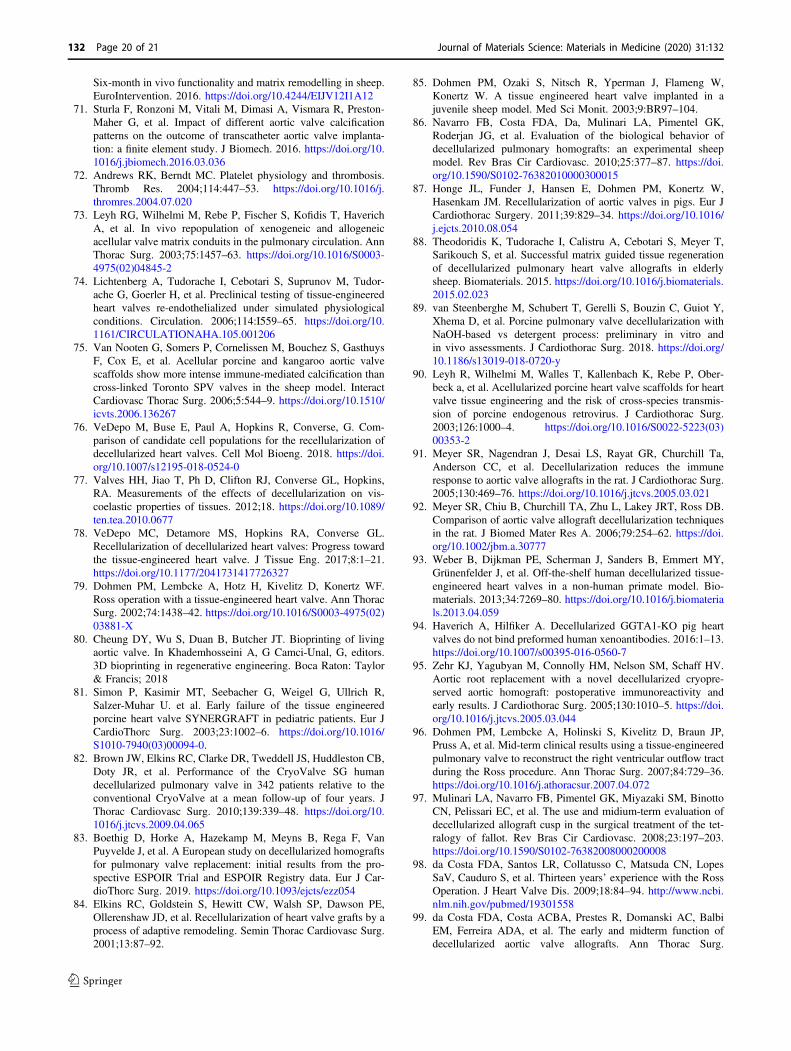
Six-month in vivo functionality and matrix remodelling in sheep.EuroIntervention. 2016. https://doi.org/10.4244/EIJV12I1A12
71. Sturla F, Ronzoni M, Vitali M, Dimasi A, Vismara R, Preston-Maher G, et al. Impact of different aortic valve calcificationpatterns on the outcome of transcatheter aortic valve implanta-tion: a finite element study. J Biomech. 2016. https://doi.org/10.1016/j.jbiomech.2016.03.036
72. Andrews RK, Berndt MC. Platelet physiology and thrombosis.Thromb Res. 2004;114:447–53. https://doi.org/10.1016/j.thromres.2004.07.020
73. Leyh RG, Wilhelmi M, Rebe P, Fischer S, Kofidis T, HaverichA, et al. In vivo repopulation of xenogeneic and allogeneicacellular valve matrix conduits in the pulmonary circulation. AnnThorac Surg. 2003;75:1457–63. https://doi.org/10.1016/S0003-4975(02)04845-2
74. Lichtenberg A, Tudorache I, Cebotari S, Suprunov M, Tudor-ache G, Goerler H, et al. Preclinical testing of tissue-engineeredheart valves re-endothelialized under simulated physiologicalconditions. Circulation. 2006;114:I559–65. https://doi.org/10.1161/CIRCULATIONAHA.105.001206
75. Van Nooten G, Somers P, Cornelissen M, Bouchez S, GasthuysF, Cox E, et al. Acellular porcine and kangaroo aortic valvescaffolds show more intense immune-mediated calcification thancross-linked Toronto SPV valves in the sheep model. InteractCardiovasc Thorac Surg. 2006;5:544–9. https://doi.org/10.1510/icvts.2006.136267
76. VeDepo M, Buse E, Paul A, Hopkins R, Converse, G. Com-parison of candidate cell populations for the recellularization ofdecellularized heart valves. Cell Mol Bioeng. 2018. https://doi.org/10.1007/s12195-018-0524-0
77. Valves HH, Jiao T, Ph D, Clifton RJ, Converse GL, Hopkins,RA. Measurements of the effects of decellularization on vis-coelastic properties of tissues. 2012;18. https://doi.org/10.1089/ten.tea.2010.0677
78. VeDepo MC, Detamore MS, Hopkins RA, Converse GL.Recellularization of decellularized heart valves: Progress towardthe tissue-engineered heart valve. J Tissue Eng. 2017;8:1–21.https://doi.org/10.1177/2041731417726327
79. Dohmen PM, Lembcke A, Hotz H, Kivelitz D, Konertz WF.Ross operation with a tissue-engineered heart valve. Ann ThoracSurg. 2002;74:1438–42. https://doi.org/10.1016/S0003-4975(02)03881-X
80. Cheung DY, Wu S, Duan B, Butcher JT. Bioprinting of livingaortic valve. In Khademhosseini A, G Camci-Unal, G, editors.3D bioprinting in regenerative engineering. Boca Raton: Taylor& Francis; 2018
81. Simon P, Kasimir MT, Seebacher G, Weigel G, Ullrich R,Salzer-Muhar U. et al. Early failure of the tissue engineeredporcine heart valve SYNERGRAFT in pediatric patients. Eur JCardioThorc Surg. 2003;23:1002–6. https://doi.org/10.1016/S1010-7940(03)00094-0.
82. Brown JW, Elkins RC, Clarke DR, Tweddell JS, Huddleston CB,Doty JR, et al. Performance of the CryoValve SG humandecellularized pulmonary valve in 342 patients relative to theconventional CryoValve at a mean follow-up of four years. JThorac Cardiovasc Surg. 2010;139:339–48. https://doi.org/10.1016/j.jtcvs.2009.04.065
83. Boethig D, Horke A, Hazekamp M, Meyns B, Rega F, VanPuyvelde J, et al. A European study on decellularized homograftsfor pulmonary valve replacement: initial results from the pro-spective ESPOIR Trial and ESPOIR Registry data. Eur J Car-dioThorc Surg. 2019. https://doi.org/10.1093/ejcts/ezz054
84. Elkins RC, Goldstein S, Hewitt CW, Walsh SP, Dawson PE,Ollerenshaw JD, et al. Recellularization of heart valve grafts by aprocess of adaptive remodeling. Semin Thorac Cardiovasc Surg.2001;13:87–92.
85. Dohmen PM, Ozaki S, Nitsch R, Yperman J, Flameng W,Konertz W. A tissue engineered heart valve implanted in ajuvenile sheep model. Med Sci Monit. 2003;9:BR97–104.
86. Navarro FB, Costa FDA, Da, Mulinari LA, Pimentel GK,Roderjan JG, et al. Evaluation of the biological behavior ofdecellularized pulmonary homografts: an experimental sheepmodel. Rev Bras Cir Cardiovasc. 2010;25:377–87. https://doi.org/10.1590/S0102-76382010000300015
87. Honge JL, Funder J, Hansen E, Dohmen PM, Konertz W,Hasenkam JM. Recellularization of aortic valves in pigs. Eur JCardiothorac Surgery. 2011;39:829–34. https://doi.org/10.1016/j.ejcts.2010.08.054
88. Theodoridis K, Tudorache I, Calistru A, Cebotari S, Meyer T,Sarikouch S, et al. Successful matrix guided tissue regenerationof decellularized pulmonary heart valve allografts in elderlysheep. Biomaterials. 2015. https://doi.org/10.1016/j.biomaterials.2015.02.023
89. van Steenberghe M, Schubert T, Gerelli S, Bouzin C, Guiot Y,Xhema D, et al. Porcine pulmonary valve decellularization withNaOH-based vs detergent process: preliminary in vitro andin vivo assessments. J Cardiothorac Surg. 2018. https://doi.org/10.1186/s13019-018-0720-y
90. Leyh R, Wilhelmi M, Walles T, Kallenbach K, Rebe P, Ober-beck a, et al. Acellularized porcine heart valve scaffolds for heartvalve tissue engineering and the risk of cross-species transmis-sion of porcine endogenous retrovirus. J Cardiothorac Surg.2003;126:1000–4. https://doi.org/10.1016/S0022-5223(03)00353-2
91. Meyer SR, Nagendran J, Desai LS, Rayat GR, Churchill Ta,Anderson CC, et al. Decellularization reduces the immuneresponse to aortic valve allografts in the rat. J Cardiothorac Surg.2005;130:469–76. https://doi.org/10.1016/j.jtcvs.2005.03.021
92. Meyer SR, Chiu B, Churchill TA, Zhu L, Lakey JRT, Ross DB.Comparison of aortic valve allograft decellularization techniquesin the rat. J Biomed Mater Res A. 2006;79:254–62. https://doi.org/10.1002/jbm.a.30777
93. Weber B, Dijkman PE, Scherman J, Sanders B, Emmert MY,Grünenfelder J, et al. Off-the-shelf human decellularized tissue-engineered heart valves in a non-human primate model. Bio-materials. 2013;34:7269–80. https://doi.org/10.1016/j.biomaterials.2013.04.059
94. Haverich A, Hilfiker A. Decellularized GGTA1-KO pig heartvalves do not bind preformed human xenoantibodies. 2016:1–13.https://doi.org/10.1007/s00395-016-0560-7
95. Zehr KJ, Yagubyan M, Connolly HM, Nelson SM, Schaff HV.Aortic root replacement with a novel decellularized cryopre-served aortic homograft: postoperative immunoreactivity andearly results. J Cardiothorac Surg. 2005;130:1010–5. https://doi.org/10.1016/j.jtcvs.2005.03.044
96. Dohmen PM, Lembcke A, Holinski S, Kivelitz D, Braun JP,Pruss A, et al. Mid-term clinical results using a tissue-engineeredpulmonary valve to reconstruct the right ventricular outflow tractduring the Ross procedure. Ann Thorac Surg. 2007;84:729–36.https://doi.org/10.1016/j.athoracsur.2007.04.072
97. Mulinari LA, Navarro FB, Pimentel GK, Miyazaki SM, BinottoCN, Pelissari EC, et al. The use and midium-term evaluation ofdecellularized allograft cusp in the surgical treatment of the tet-ralogy of fallot. Rev Bras Cir Cardiovasc. 2008;23:197–203.https://doi.org/10.1590/S0102-76382008000200008
98. da Costa FDA, Santos LR, Collatusso C, Matsuda CN, LopesSaV, Cauduro S, et al. Thirteen years’ experience with the RossOperation. J Heart Valve Dis. 2009;18:84–94. http://www.ncbi.nlm.nih.gov/pubmed/19301558
99. da Costa FDA, Costa ACBA, Prestes R, Domanski AC, BalbiEM, Ferreira ADA, et al. The early and midterm function ofdecellularized aortic valve allografts. Ann Thorac Surg.
132 Page 20 of 21 Journal of Materials Science: Materials in Medicine (2020) 31:132
2010;90:1854–60. https://doi.org/10.1016/j.athoracsur.2010.08.022
100. Rüffer A, Purbojo A, Cicha I, Glöckler M, Potapov S, Dittrich S,et al. Early failure of xenogenous de-cellularised pulmonaryvalve conduits-a word of caution! Eur J Cardiothorac Surg.2010;38:78–85. https://doi.org/10.1016/j.ejcts.2010.01.044
101. Dohmen PM, Lembcke A, Holinski S, Pruss A, Konertz W. Tenyears of clinical results with a tissue-engineered pulmonaryvalve. Ann Thorac Surg. 2011;92:1308–14. https://doi.org/10.1016/j.athoracsur.2011.06.009
102. Cebotari S, Tudorache I, Ciubotaru A, Boethig D, Sarikouch S,Goerler A, et al. Use of fresh decellularized allografts for pulmonaryvalve replacement may reduce the reoperation rate in children andyoung adults: early report. Circulation. 2011;124:S115–23. https://doi.org/10.1161/CIRCULATIONAHA.110.012161
103. Bobylev, D, Sarikouch, S, Tudorache, I, Cvitkovic, T, Söylen, B,Boethig, D, et al. Double semilunar valve replacement in com-plex congenital heart disease using decellularized homografts.Interact Cardiovasc Thorac Surg. 2019. https://doi.org/10.1093/icvts/ivy212
104. Miller DV, Edwards WD, Zehr KJ. Endothelial and smoothmuscle cell populations in a decellularized cryopreserved aortichomograft (SynerGraft) 2 years after implantation. J ThoracCardiovasc Surg. 2006;132:175–6. https://doi.org/10.1016/j.jtcvs.2006.02.038
105. Bloch O, Sc M, Golde P, Dohmen PM, Ph D, Posner S, et al.Immune response in patients receiving a bioprosthetic heartvalve: lack of response with decellularized valves. Tissue Eng.2011;17:2399–405. https://doi.org/10.1089/ten.tea.2011.0046
106. Konuma T, Devaney EJ, Bove EL, Gelehrter S, Hirsch JC,Tavakkol Z, et al. Performance of CryoValve SG decellularizedpulmonary allografts compared with standard cryopreservedallografts. Ann Thorac Surg. 2009;88:849–54. https://doi.org/10.1016/j.athoracsur.2009.06.003. Discussion 554-5.
107. Burch PT, Kaza AK, Lambert LM, Holubkov R, Shaddy RE,Hawkins JA. Clinical performance of decellularized cryopreservedvalved allografts compared with standard allografts in the rightventricular outflow tract. Ann Thorac Surg. 2010;90:1301–5.https://doi.org/10.1016/j.athoracsur.2010.05.024
108. Tavakkol Z, Gelehrter S, Goldberg CS, Bove EL, Devaney EJ,Ohye RG. Superior durability of SynerGraft pulmonary allograftscompared with standard cryopreserved allografts. Ann Thorac Surg.2005;80:1610–4. https://doi.org/10.1016/j.athoracsur.2005.04.017
109. Cebotari S, Lichtenberg A, Tudorache I, Hilfiker A, MertschingH, Leyh R, et al. Clinical application of tissue engineered human
heart valves using autologous progenitor cells. Circulation.2006;114:I132–7. https://doi.org/10.1161/CIRCULATIONAHA.105.001065
110. Costa F, Dohmen P, Vieira E, Lopes SV, Colatusso C, PereiraEWL, et al. Ross Operation with decellularized pulmonary allo-grafts: medium-term results. Rev Bras Cir Cardiovasc.2007;22:454–62. http://www.ncbi.nlm.nih.gov/pubmed/18488113
111. Cicha I, Rüffer A, Cesnjevar R, Glöckler M, Agaimy A, DanielWG, et al. Early obstruction of decellularized xenogenic valvesin pediatric patients: involvement of inflammatory and fibro-proliferative processes. Cardiovasc Pathol. 2011;20:222–31.https://doi.org/10.1016/j.carpath.2010.04.006
112. Ruzmetov M, Shah JJ, Geiss DM, Fortuna RS. Decellularizedversus standard cryopreserved valve allografts for right ven-tricular outflow tract reconstruction: a single-institution com-parison. J Thorac Cardiovasc Surg. 2012;143:543–9. https://doi.org/10.1016/j.jtcvs.2011.12.032
113. Brown JW, Ruzmetov M, Eltayeb O, Rodefeld MD, TurrentineMW. Performance of SynerGraft decellularized pulmonaryhomograft in patients undergoing a Ross procedure. Ann ThoracSurg. 2011;91:416–22. https://doi.org/10.1016/j.athoracsur.2010.10.069. Discussion 422-3.
114. Kneib C, von Glehn CQC, Costa FDa, Costa MTBa, Susin MF.Evaluation of humoral immune response to donor HLA afterimplantation of cellularized versus decellularized human heartvalve allografts. Tissue Antigens. 2012;80:165–74. https://doi.org/10.1111/j.1399-0039.2012.01885.x
115. Ozawa H, Ueno T, Taira M, Toda K, Kuratani T, Sawa Y.Application of a fresh decellularized pulmonary allograft forpulmonary valve replacement in Japan. Circ. J. 2018. https://doi.org/10.1253/circj.cj-18-0150
116. Christ, T, Paun, AC, Grubitzsch, H, Holinski, S, Falk, V, Dushe,S. Long-term results after the Ross procedure with the decel-lularized AutoTissue Matrix P® bioprosthesis used for pulmon-ary valve replacement. Eur J CardioThorc Surg. 2018. https://doi.org/10.1093/ejcts/ezy377
117. Sarikouch, S, Horke, A, Tudorache, I, Beerbaum, P, Westhoff-Bleck, M, Boethig, D, et al. Decellularized fresh homografts forpulmonary valve replacement: a decade of clinical experience.Eur J CardioThorc Surg. 2016. https://doi.org/10.1093/ejcts/ezw050
118. Tudorache, I, Horke, A, Cebotari, S, Sarikouch, S, Boethig, D,Breymann, T, et al. Decellularized aortic homografts for aorticvalve and aorta ascendens replacement. Eur J Cardiothorc Surg.2016. https://doi.org/10.1093/ejcts/ezw013
Journal of Materials Science: Materials in Medicine (2020) 31:132 Page 21 of 21 132


![[Vascular calcification in chronic kidney disease]](https://img.pdfslide.net/doc/110x75/634cb3ac1983efcda6059151/vascular-calcification-in-chronic-kidney-disease-1700101186.jpg)


