Embed Size (px)
Citation preview
Page 1/28
Deciphering Core-Phyllomicrobiome of Rice Genotypes Grown in ContrastingMountain and Island Agroclimatic Zones: Implications for MicrobiomeEngineering Against Blast DiseaseKuleshwar Prasad Sahu
Indian Agricultural Research InstituteKumar Aundy ( [email protected] )
Indian Agricultural Research Institute https://orcid.org/0000-0002-7401-9885Sakthivel Krishnan
ICAR Central Agricultural Research InstituteBhaskar Reddy
Indian Agricultural Research InstituteMukesh Kumar
Indian Agricultural Research InstituteAsharani Patel
Indian Agricultural Research InstituteNeelam Sheoran
Indian Agricultural Research InstituteGopalakrishnan Subbaiyan
Indian Agricultural Research InstitutePrakash Ganesan
IARI: Indian Agricultural Research InstituteRajeev Rathour
CSK HPKV: Himachal Pradesh Agricultural UniversityRaj K Gautam
Indian Agricultural Research Institute
Research
Keywords: Antibiosis, Bacterial Volatiles, Blast, Core-Microbiome, Defense genes, Magnaporthe oryzae, Phyllomicrobiome, Phyllosphere, Rice
Posted Date: February 9th, 2021
DOI: https://doi.org/10.21203/rs.3.rs-173841/v1
License: This work is licensed under a Creative Commons Attribution 4.0 International License. Read Full License
Page 2/28
AbstractBackground
The fundamental role and contributions of phyllosphere habitat in shaping plant functional ecology are poorly investigated, and often underestimated.Phyllosphere -the harsh and dynamic foliar-photosynthetic-habitat is continuously exposed to vagaries of changing weather events during the entire plant life.With its adapted microbiota, the phyllosphere-niche brings microbial diversity to the plant-holobiont pool and potentially modulates a multitude of plant andagronomic traits. The phyllomicrobiome structure and the consequent ecological functions are vulnerable to a host of biotic (Genotypes) and abiotic-factors(Environment) which is further compounded by agronomic-transactions on domesticated agricultural crops. However, the ecological forces driving thephyllomicrobiome assemblage and functions are among the most under-studied aspects of plant biology. Despite the reports on the occurrence of diverseprokaryotic phyla such as Proteobacteria, Firmicutes, Bacteroides, and Actinobacteria on phyllosphere habitat, the functional characterization leading to theirutilization for agricultural sustainability is not yet adequately explored.
Currently, the metagenomic-Next-Generation-Sequencing (mNGS) technique scanning the conserved V3-V4 region of ribosomal RNA gene is a widely adoptedstrategy for microbiome-investigations. However, the structural and functional validation of mNGS annotations by microbiological methods is not integratedinto the microbiome exploration-programs. In the present study, we combined the high throughput mNGS approach with conventional microbiologicalmethods to decipher the core-functional-phyllomicrobiome of contrasting rice genotypes varying in their response to blast disease grown in contrastingagroclimatic zones in India. We, further, scanned the rice phyllosphere by electron microscopy to show the microbial communities on leaf.
Magnaporthe oryzae -the phyllosphere pathogen inciting necrotic lesion on cereal crops is managed by the deployment of ‘non-durable’ blast resistance genesand ‘toxic’ fungicidal molecules. Nowadays, there is a growing consensus for devising an alternative strategy for mitigating blast owing to a recent ban on theuse of most commonly used fungicidal molecule, tricyclazole. In the present work, we further identi�ed phyllosphere- core-functional microbial groups leadingto the proposal of phyllomicrobiome assisted rice blast management strategy. Multi-pronged activities of phyllomicrobiome against Magnaporthe oryzae(antifungal activity), rice innate immunity (defense elicitation), and rice blast disease (disease suppression) have been elaborated for effective managementof blast by phyllomicrobiome re-engineering.
Results
Rice phyllomicrobiome of tropical “Island-Zone” displayed marginally more bacterial community diversity than that of temperate ‘Mountain-Zone’. Principalcoordinate analysis based on Bray Curtis and ANoSIM method indicated nearly converging-phyllomicrobiome pro�les on two contrasting rice genotypesgrown in the same agroclimatic zone. However, the rice genotype grown in the contrasting Mountain-zone and Island-zone displayed diverse-phyllomicrobiome pro�les indicating a strong in�uence of environmental factors rather than the genotype on phyllomicrobiome structure and assembly. Thepredominance of Phyla such as Proteobacteria, Actinobacteria, and Firmicutes was observed on the rice phyllosphere irrespective of the genotypes andenvironmental conditions. The core-microbiome analysis showed multi-microbiota-core consisting of Acidovorax, Arthrobacter, Bacillus, Clavibacter,Clostridium, Cronobacter, Curtobacterium, Deinococcus, Erwinia, Exiguobacterium, Hymenobacter, Kineococcus, Klebsiella, Methylobacterium, Methylocella,Microbacterium, Nocardioides, Pantoea, Pedobacter, Pseudomonas, Salmonella, Serratia, Sphingomonas and Streptomyces on phyllosphere of rice genotypesgrown in contrasting agroclimatic zones. The linear discriminant analysis (LDA) effect size (LEfSe) method revealed ten and two distinct bacterial genera inblast-resistant and -susceptible genotypes, respectively. The analysis further indicated 15 and 16 climate-zone speci�c bacterial genera for Mountain andIsland zone, respectively. SparCC based network analysis of phyllomicrobiome showed hundreds of complex intra-microbial cooperative or competitiveinteractions on the rice genotypes and agroclimatic zones. Our microbiological validation of mNGS data further con�rmed the presence of residentAcinetobacter, Aureimonas, Curtobacterium, Enterobacter, Exiguobacterium, Microbacterium, Pantoea, Pseudomonas, and Sphingomonas on the ricephyllosphere. Strikingly, the two contrasting agroclimatic zones displayed genetically identical bacterial isolates on the phyllosphere that could be attributedto the spatio-temporal transmission of core-phyllomicrobiome, perhaps, aided by rice seeds. A total of 59 distinct bacterial isolates were obtained, identi�ed,and evaluated for their functional attributes on Magnaporthe oryzae and rice plant. The phyllomicrobiome associated core-bacterial communities showedsecreted-metabolite and volatile-compound mediated antifungal activity on M. oryzae. Upon phyllobacterization (a term coined for spraying of bacterial cellson the phyllosphere), the core bacterial species such as Acinetobacter baumannii, Aureimonas sp., Pantoea ananatis, P. eucrina, and Pseudomonas putidaelicited plant defense and contributed signi�cantly to blast disease suppression. Transcriptional analysis by qPCR indicated induction of rice innate immunityassociated genes such as OsPR1.1, OsNPR1, OsPDF2.2, OsFMO, OsPAD4, OsCEBiP, and OsCERK1 in phyllobacterized rice seedlings.
Conclusions
The rice genotypes growing in a particular agroclimatic zone showed a convergent phyllomicrobiome assemblage and composition. Conversely, divergingphyllomicrobiome assembly was observed on rice genotype cultivated in the contrasting agroclimatic zones. Agroclimatic zones and the associated climatic-factors rather than plant-genotypes per se appeared to drive phyllomicrobiome structure and composition on the rice genotypes. Our integrated mNGSmethod and microbiological validation divulged Acinetobacter, Aureimonas, Curtobacterium, Enterobacter, Exiguobacterium, Microbacterium, Pantoea,Pseudomonas, and Sphingomonas as core phyllomicrobiome of rice. Genetically identical bacterial communities belonging to Pantoea intercepted on thephyllosphere of rice grown in the two contrasting agroclimatic zones are suggestive of spatio-temporal transmission of phyllomicrobiome aided by seed. Thecore-microbiome mediated phyllobacterization showed potential for blast disease suppression by direct-antibiosis and defense elicitation. The identi�cationof phyllosphere adapted functional core-bacterial communities in our study and their co-occurrence dynamics presents an opportunity to devise novelstrategies for rice blast management through phyllomicrobiome reengineering in the future.
Background
Page 3/28
Plant microbiota is believed to have an evolutionary-association with higher plants, and together they function as meta-organism in the environment. Thetotal microbiota colonizing the plants is termed as plant holobiont which, often, gives a functional extension and metabolic �exibility to the plant genomes [1,2]. Microbial members of the microbiomes interact dynamically among them as well as with the plant species displaying cooperative or competitiverelationships. Hence, the intra-microbial interaction is believed to impact not only the composition of the microbiomes but also the physiological andecological functions of the host plants in general.
The phyllosphere, a subset of the phytosphere, is touted as a harsh plant-associated habitat for diverse microbiota that host phyllomicrobiome. The totalglobal phyllosphere is predicted to represent 109 square kilometers that could harbor 1026 bacterial cells [3]. On the phyllosphere niche, the microbiome is notonly affected by biotic and abiotic factors but also by nutrient depletion [4, 5]. Unlike rhizosphere and endophytic microbiome, the phyllomicrobiome is notextensively investigated in many crop plants. However, the prokaryotic microbial association on rice phyllosphere and their complex-interactions modulatingplant growth, and protection against microbial pathogens are reported. It is widely perceived that the phyllosphere prokaryotic diversity and their populationsize are large enough to play a pivotal role in plant growth [6, 7] and defense against pathogens [8, 9]. Microbial interactions with their host plants can also beneutral and commensal [10]. The plant genotype, climate, geographical location, edaphic factors, and agronomic practices are among the key factors shapingthe phyllosphere microbiota composition and their ecological functions [3, 11-13]. Although predicted and highlighted in many publications, the key drivers ofphyllomicrobiome composition and their functions are not completely understood.
Qualitatively, majority of the microbiota of phyllomicrobiome are non-pathogenic bacteria that belong to a few phyla such as Proteobacteria, Firmicutes,Bacteroides, and Actinobacteria [14, 15]. At lower taxonomic hierarchy, bacterial genera frequently encountered on phyllosphere are Kineococcus,Hymenobacter,Acinetobacter, Bacillus, Citrobacter, Curtobacterium, Enterobacter, Erwinia, Frigoribacterium, Methylobacterium, Pantoea, Pseudomonas, andSphingomonas [15-20]. For epiphytic-colonization, microbes have evolved adaptive traits such as dark pigmented cells, extracellular polymeric substances,biosurfactants, bio�lms, and utilization of plant/ microbial volatile compounds [21]. Furthermore, the epiphytic bacterial communities presumed to survive onsugar-photosynthates sourced from the leaf interior diffused through the cuticle to the epiphytic surface [22, 23].
The rice phyllosphere is also a habitat for foliar pathogens like Magnaporthe and Xanthomonas that cause leaf-diseases. Blast disease caused byMagnaporthe oryzae (anamorph Pyricularia oryzae Sacc.) remains a global production constraint and a threat to food security in developing nations [24-27].Blast disease of rice accounts for nearly 30 % production loss, which is enough to feed 60 million world’s population if managed preemptively [28]. Currently,blast management depends heavily on fungicides and host-resistance; both are inadequate to combat the production losses during epidemics. While thefungicides are not compatible with the environment and trade, the host plant resistance is non-durable owing to the emergence of new pathotypes [29]. It is,further, reported that the blast resistance conferred by host resistance genes often breaks down within 3-5 years of rice cultivation due to the preexistingvirulence diversity of M. oryzae [30]. Therefore, there is a need for the development of a sustainable and durable blast management strategy for rice. Bespokemicrobiome therapy is proposed as NextGen-Crop-care strategy to ensure eco-friendly crop disease management [31]. Microbial strains with desired functionscan be selected and engineered to form synthetic microbiomes for agricultural applications [32]. The perceived advantage is the ability of synthetic-microbiome to buffer against environmental perturbations. However, the development of such synthetic microbiomes is, often, hampered by our limitedunderstanding of the core functional microbiome. Harnessing the potential of naturally occurring phyllomicrobiome for foliar disease and crop managementhas not been attempted till date. Since the phyllosphere microbiomes have been reported to play a pivotal role in growth, development, and defense againstbiotic and abiotic stress, pro�ling the phyllomicrobiome for deciphering the functions assumes signi�cance.
With this background, the current investigation was conducted to identify the core-phyllomicrobiome of rice and its potential to suppress blast disease. We,further, attempted to decipher the major driver(s) of phyllosphere microbiome composition using the integrated metagenomic Next Generation Sequencing(mNGS) approach and conventional microbiological methods. For this purpose, phyllomicrobiome samples were generated from two contrasting ricegenotypes differing for a single resistance-gene, Pi2 conferring complete resistance to blast, thereby varying for their reaction to blast disease, grown in two-contrasting agro-climatic zones in India separated by more than 2800 Km. The agroclimatic zones represented Mountain-zone in the Himalayan region(Palampur) and Island-zone in Andaman Island in the Bay-of-Bengal, India (Port Blair). While the mountain-zone in Palampur is an endemic-location for blastdisease, the island-zone in Port Blair is non-endemic.
We identi�ed the core-phyllomicrobiome of rice genotypes in the combined and comparative mNGS and microbiological data. The results indicated theassociation of complex microbial assemblages displaying diverse-functions on the rice phyllosphere for rice blast management. Our in-vitro screening ofphyllomicrobiome against M.oryzae and in-planta evaluation trial against rice blast disease further con�rmed the potential of functional-microbial groups forphyllomicrobiome assisted rice cultivation in the future.
MethodsStudy location and sampling for phyllomicrobiome assessment
Metagenomic NGS (mNGS), microbiological and microscopic experiments were performed on rice phyllomicrobiome sampled from the rice plots of twocontrasting agroclimatic-zones of India. The experimental sites were, (i). blast-endemic mountain-zone at Palampur, Himachal Pradesh, India [32°6'4.7"N,76°32'39.79"E; altitude 1275 meter above mean sea level (MSL); mean temperature 22-23 °C; mean rainfall 700-1000 mm; relative humidity (RH) 60.0 %;sunshine hours 300-350 h]; and (ii). blast non-endemic Island-zone in Port Blair, Andaman Island, India [11°38'07.0"N, 92°39'12.7"E); altitude 16 meters aboveMSL, mean temperature 26-28 °C, mean rainfall 3060 mm; RH 80.0 % (https://en.climate-data.org; (www.worldweatheronline.com)]. Both experiments wereconducted during rice cultivation seasons in August - September 2016 at Palampur and March - April 2017 in Port Blair. Blast disease susceptible genotype,PRR 78 and its near-isogenic line Pusa 1602 introgressed with Pi2 gene [33] conferring complete resistance to blast disease were planted and grown inparallel rows with spacing of 20 cm by adopting standard agronomic practices. Phyllosphere samples were collected aseptically in sterilized falcon-tubes on
Page 4/28
15 and 30 days post sowing. Phyllosphere samples were collected aseptically in sterilized falcon-tubes on 15- and 30-days post sowing. Thus collectedsamples in two replications were transported to the laboratory in cool-containers maintained at 4 oC + 0.5 °C, and processed for microbiomes pro�ling within48 hours.
mNGS based pro�ling of phyllomicrobiome
Extraction of phyllosphere microbial genomic DNA: Leaf (5.0 g) samples collected from the two rice genotypes in two replications were shaken with 50 ml ofsterile phosphate buffer saline [PBS, g L-1 NaCl 8; KCl 0.2; Na2HPO4 1.44; KH2PO4 0.24; pH-7.4] amended with 0.1 % Tween-20 (PBS-T) to dislodge thephyllomicrobiome. Thus, collected phyllosphere samples were serially extracted six times in 50 ml of PBS buffer by vigorous shaking for 30 minutes at 250-rpm followed by vortexing for 10 s. Thus separated phyllomicrobiome suspension (300 mL) was collected aseptically in a pre-sterilized container andcentrifuged at 12K g force for 60 min at 4.0 ºC to collect the phyllomicrobiome pellets. The pellet obtained was subjected to total microbial community DNAextraction by Cetyl Trimethyl Ammonium Bromide (CTAB) method previously described by Moore et al [34]. The quality and yield of microbial community DNAwere assessed electrophoretically, spectrophotometrically (Nanodrop 2000, Thermo Scienti�c, USA), and �uorometrically (Qubit dsDNA BR Assay; ThermoFisher Scienti�c Inc., Qubit® 2.0).
Preparation of mNGS libraries for 2 x 300 bp Sequencing Chemistry: The amplicon-libraries were prepared using Nextera XT Index Kit (Illumina Inc. San Diego,CA, USA) as prescribed for the 16S rRNA gene-sequence based Metagenomic Sequencing Library Preparation Protocol (Part # 15044223 Rev. B). Primers forthe ampli�cation of the 490 -bp hyper-variable V3-V4 region of 16S rRNA gene of Eubacteria and Archaea were synthesized and used. The sequences of thePCR primers are V3F: 5'-CCTACGGGNGGCWGCAG-3’ and V4R: 5’-GACTACHVGGGTATCTAATCC-3'. The target-amplicons were generated using a fusion-primerthat consists of Illumina adaptors and multiplex-index sequence as per the manufacturer’s instructions (Illumina Inc. San Diego, CA, USA). The amplicon-libraries were puri�ed by 1X AMpureXP beads and checked on Agilent High Sensitivity (HS) chip on Bioanalyzer 2100 and quanti�ed on �uorometer usingQubit dsDNA HS Assay kit (Life Technologies, California, USA). Quality passed libraries were equimolar pooled and then sequenced using the Illumina MiSeqplatform with 300 × 2 pair-end sequencing chemistry following the manufacturer’s protocols (Illumina, San Diego, CA, USA).
Metagenomic bioinformatic analysis
Initially, the sequenced raw forward-reads (R1) and reverse-reads (R2) from all samples were visualized using the FastQC version [35] to screen the qualitystatistics of the 16S rRNA gene amplicon reads (https://www.bioinformatics.babraham.ac.uk/projects/fastqc/). The raw-reads were, then, curated to removepoor-quality reads to obtain high-quality reads using Trimmomatic v0.35 [36] with parameters to i) remove adapter sequences, and ii) curate ambiguousreads (reads with unknown nucleotides “N” larger than 5 %), low-quality sequences (reads with more than 10 % quality threshold (QV) < 20 Phred score)(http://www.usadellab.org/cms/?page=trimmomatic). The �nal quality passed read-pairs were joined using PEAR (Paired-End reAd mergeR) version 0.9.8(https://cme.h-its.org/exelixis/web/software/pear/) with default parameters. The joined paired-reads were processed for the downstream taxonomicclassi�cation; the unpaired reads were discarded. The taxonomic classi�cation of the �nal high-quality reads was performed using MG-RAST v4.0(https://www.mg-rast.org/), wherein 1) 16S rRNA featured reads were sorted using SortmeRNA, 2) sorted reads were clustered at ≥97 % similarity using CD-HIT method, and then 3) clustered reads were taxonomically classi�ed against SILVA SSU database (https://www.arb-silva.de/). The classi�ed reads/ taxonabundance downloaded >100 bases and 90 % similarity through best hit classi�cation.
Metagenome statistical analysis
Statistical Analysis of Metagenomic Pro�le (STAMP; V 2.9) (https://beikolab.cs.dal.ca/software/STAMP) was referred to determine microbial diversity andabundance in the phyllosphere. Welch-T-test and Post-Hoc Test at a con�dence interval of ≥ 95 % was followed. Further, Microbiome Analyst [37] was utilizedfor the determination of α-diversity, and β- diversity, as well as to identify core-phyllomicrobiome (https://www.microbiomeanalyst.ca/). For this, initially, readswere rare�ed on minimum library size (18000 reads, minimum classi�ed read in a sample), and then total sum scaling (TSS) was applied for datanormalization. α- diversity signi�cance was calculated using ANOVA test; Principal Coordinate Analysis (PCoA) was performed using Analysis of similarities(ANoSIM) based on Bray-Curtis method. The biomarker features were determined through the Linear discriminant analysis (LDA) combined with effect sizemeasurements (LDA-LEfSe) approach at signi�cance P<0.05 and LDA score >2.0 (http://huttenhower.sph.harvard.edu/lefse/). Bacterial genera co-occurrencenetwork was analysed using SparCC method with the signi�cance of P<0.05 and strong correlation coe�cient R2 >0.60 or <-0.6(http://github.com/scwatts/FastSpar).
Microscopic visualization of rice phyllomicrobiome
Scanning Electron Microscopy: Scanning electron microscopy (SEM) was adopted for visualization of rice phyllomicrobiome following the method ofBozzola [38]. For SEM, rice leaves were cut into small pieces (3 mm2) and �xed in 2.5 % glutaraldehyde for 12 h at 4.0 °C, rinsed in phosphate buffer saline(PBS-0.1 M, pH 7.2) for 10 min. Leaves were then dehydrated through graded series of 70, 80, 90, 95, and 100 % acetone and then dried with a chemical dryer.The leaf preparations were, then, mounted on aluminum stubs using silver adhesive tape and sputter-coated with gold: palladium alloy (18 nm) for 30 minconsisting of 10 cycles of three min each for uniform coating (SC 7620 Emitech sputter-coater with a pressure of 10−1 mbar). Thus prepared leaf sampleswere examined and visualized under Scanning Electron Microscope (Zeiss EVO MA 10; Oxford Technologies) at 20.00 kV and magni�cations ranging from 4KX to16 KX. The entire leaf surface was screened and searched for the possible presence of bacterial cells and images were captured.
Culturing of phyllomicrobiome by microbiological methods
Isolation and characterization of the cultivable phyllomicrobiome of rice: Another set of the leaf samples (500 mg) collected from the two rice genotypes weresubjected to phyllomicrobiome isolation on nutrient agar [NA, gL-1 Peptone 5.0; Beef extract 3.0; NaCl 5.0; Agar 15.0; pH 7.0 ±0.2 ] and M9 minimal media [2
Page 5/28
mM MgSO4; 0.1 mM CaCl2; 0.3 % Glucose; 1.5 % Agar; 1×M9 salts (5×M9 salts gL-1 Na2HPO4.7H2O 64.0; KH2PO4 15.0, NaCl 2.5; NH4Cl 5.0)]. Brie�y, the leafwas shaken with 50 ml of sterile phosphate buffer saline amended with 0.1 % tween-20 (PBS-T) for 30 minutes at 250 revolutions per minute followed byvortexing for 10 seconds. The aliquot, thus, obtained was decimally diluted up to 10-5. Aliquots of 1.0 ml at 10-3, 10-4, and 10-5 from each sample were pourplated into nutrient agar and M9 minimal media supplemented with 2, 3, 5 triphenyl tetrazolium chloride (50 mg L-1) to assist the morphotyping of thebacterial communities. The plates were incubated at 28 °C+2 °C for 72 hours. The experiment was conducted with three biological and three technicalreplications. The bacterial colonies were counted and isolated based on their morphology (size, shape, colour, texture, and margin). Later on, a singlerepresentative colony of each-morphotype was sub-cultured, puri�ed and frozen-way in -80 °C and -20 °C as glycerol stock (30 % V/V). Species richness andthe Shannon-Wiener diversity index (H) were determined for the cultured bacterial communities.
Molecular diversity analysis and identi�cation of phyllomicrobiome associated bacterial species
BOX-PCR DNA �ngerprinting: Genomic DNA of each of the bacterial isolates was isolated by the CTAB method prescribed by Moore et al [34]. Isolated andpuri�ed genomic DNA was quantitated and quality checked electrophoretically and spectrophotometrically (NanoDrop 2000, ThermoScienti�c, USA). Finally,the genomic DNA was reconstituted at 100 ng µl-1 and used as a template in PCR ampli�cation. Box-PCR based DNA-�ngerprinting was performed fordiversity analysis as well to eliminate the duplicate isolates from the collection [39]. The BOX-PCR amplicon pro�ling technique speci�cally ampli�es the non-coding conserved sequences in the bacterial genome and is considered a highly discriminatory DNA �ngerprinting technique for bacteria [40, 41]. Ampliconswere resolved in 1.0 % agarose gel at 30 volts for 10-12 hours and image-captured (QuantityOne, BioRad Laboratories, USA). Isolates showing identicalamplicon pro�les were presumed to be duplicates and represented one BOX-Amplicon Group. One representative isolate from each BOX-Amplicon Group waseventually used in the downstream work.
16S rRNA gene sequencing: Ampli�cation of 16S rRNA gene was performed using universal primers 27F (27F: 5’-AGAGTTTGATCCTGGCTCAG-3’) and 1492R(1492R: 5’-GGTTACCTTGTTACGACTT-3’) to amplify the 1465 bp region to establish bacterial identity [42, 43]. Then, the PCR products resolved in 1.0 %agarose gel were excised from the agarose gel and eluted using a gel elution kit (Wizard® SV Gel and PCR Clean-Up System) according to the manufacturer’sinstructions (Promega Corporation, USA). The cycle-sequencing reaction was performed using 20–30 ng of the puri�ed amplicon using the ABI PRISM BigDyeTerminators v3.1 cycle sequencing kit (Applied Biosystems Foster City, CA, USA) according to the manufacturer's instruction. The puri�ed product wassequenced bi-directionally to obtain maximum coverage of the spacer region. The sequences were end trimmed, edited, and contig assembled using DNA-baser (http://www.dnabaser.com/download/DNA-Baser-sequence-assembler/). The curated sequences were, further, subjected to Basic Local AlignmentSearch Tool analysis (NCBI nucleotide BLAST) to establish their identity by closest match (https://www.ncbi.nlm.nih.gov/nucleotide/). All curated 16S rRNAgene sequences of phyllosphere bacterial species were submitted to GenBank database and assigned accession numbers.
Functional screening of phyllosphere bacterial communities
In vitro antifungal activity on Magnaporthe oryzae: Volatile and secretory metabolite mediated antagonistic assay of bacterial isolates were conducted on M.oryzae (isolate 1637) by dual-culture confrontation method. The percent inhibition of mycelial growth over mock was estimated by adopting the methodsdescribed by Sheoran et al [42] and Munjal et al [43]. Additionally, the fungicidal or fungistatic nature of the bacterial volatiles on M. oryzae was alsodetermined. Brie�y, bacterial isolates found completely inhibiting the growth of M. oryzae were further allowed to reestablish mycelial-growth. Based on the re-growth of the mycelium, the bacterial volatile were either categorized as fungicidal or fungistatic.
The radial growth of the fungus was measured and percent inhibition of growth over control was calculated with the help of the following formula
Where I = percent inhibition
C = Colony diameter in control
T = Colony diameter in treatment
In planta blast suppressive activity: The bacterial isolates signi�cantly antagonistic to blast fungus in vitro were selected for this assay. Blast susceptible ricegenotype, Pusa Basmati-1, was allowed to germinate in bacterial cell suspension (2×10⁷ CFU mL-1) for �ve days. Upon germination, the transplants were,further, grown in a climate-controlled greenhouse set at temperature 28°C ±2 °C/ RH 90+10 % /Light/dark cycles 14/10 h. Seedlings were foliar sprayed withphyllosphere bacterial suspension (Phyllobacterization; 107 CFU mL-1) and challenged with a conidial-suspension of M. oryzae 1637 (2 × 10⁵ conidia mL-1)three weeks post sowing according to the protocols of Rajashekara et al [44]. Blast disease index was determined seven days post-inoculation using a 0–5disease rating-scale where 0= no evidence of infection; 1.0= brown specks smaller than 0.5 mm in diameter; 2.0= brown-specks of 0.5-1.0 mm in diameter;3.0= roundish to elliptical lesions of about 1.0-3.0 mm in diameter; 4.0= typical spindle-shaped blast lesion, 3 mm or longer with little or no coalescence of thelesion; 5.0= same as 4.0 but half or more leaves killed by coalescence of lesions. Plants rated 0.0-2.0 were considered resistant, 3.0 as moderatelysusceptible, and 4.0-5.0 were considered susceptible [45]. The disease severity was calculated using the following formula.
Page 6/28
Further, the percent reduction in disease severity as compared to control was estimated using the following formula.
Where RDS = Reduction in Disease Severity (%)
C = Disease Severity in control
T = Disease Severity in treatment.
In planta rice defense gene(s) activation: Having observed the antifungal blast suppressive potential of phyllosphere core-bacterial communities on rice,qPCR experiments were conducted to decipher the leaf phyllobacterization effects on transcriptional changes in defense pathways in rice. Six phyllospherebacterial isolates namely, Pantoea ananatis OsEp-Plm-30P3;Pantoea ananatisOsEp-Plm-30P21;Pantoea ananatisOsEp-AN-30A8;Aureimonas sp. OsEp-Plm-30P7;Pantoea eucrinaOsEp-Plm-30P10 andPseudomonas putidaOsEp-Plm-15P11 showing signi�cant blast disease suppression were selected for the study.Brie�y, whole seedlings of Pusa Basmati-1 bacterized with 2×10⁷ CFU mL-1 sampled at 24, 48, and 72 hours were immediately snap-frozen using liquid-nitrogen (to arrest all the cellular activity) and then stored instantly at -80°C till further use. Total RNA was isolated using the SV Tool RNA Isolation Systemaccording to the manufacturer's instruction (Promega, Madison, USA). The quality and quantity of RNA were assessed spectrophotometrically (NanoDrop2000, ThermoScienti�c, USA) as well as by agarose gel electrophoresis. The experiment was repeated two times with three technical replications.
Candidate defense genes: Putative defense genes, OsCEBiP [46], OsCERK1 [47], OsPAD4 [48], OsEDS1 [49], OsNPR1 [50], OsPDF2.2 [51], OsFMO1 [52, 53] andOsPR1.1 [54] were chosen; PCR primers targeting the above defense genes are presented (Supplementary Table 1-2). The qPCR experiment was conducted ina Real-Time PCR instrument (Light Cycler 96, Roche Life Science, Switzerland) using GoTaq® 1-Step RT-qPCR System; qPCR reaction conditions were asfollows; one cycle of reverse transcription at 37 °C for 15 minutes followed by reverse-transcriptase inactivation step of 95 °C for 10 minutes followed by 30cycles of 95 °C for 10 seconds, annealing at 58 °C for 30 seconds and extension at 72 °C for 30 sec followed by three-step melting of 95°C for 10 seconds, 63°C for 60 seconds and 97 °C for 1 second and then �nal cooling at 37 °C for 30 seconds. The expression levels of all eight defense-genes were calculated withreference to the expression of a housekeeping gene, OsActin, for normalization in different samples. Then, the qPCR data were analysed usingLightCycler®96 Roche SW 1.1 software, and the mean Ct values were considered for calculation of 2-ΔΔCT to estimate the fold changes in gene -expression.The fold-change data were interpreted as value 1.0 for no change, ≥ 2.0 represents signi�cant upregulation, ≤ 1.0 is down-regulation, and ≤ 0.5 forsigni�cant down-regulation.
Statistical analyses
All the experimental data were analyzed using the data analysis tool available in MS o�ce excel 2007. The data obtained were subjected to signi�canceanalysis by analysis of variance (ANOVA) at p ≤ 0.05 level of signi�cance. Further, various parameters like the standard error of the mean (SEm), standarderror of the difference between two means (SEd), the critical difference (CD), coe�cient of variation (CV) were calculated. For �gures and tables, the valuesare represented as the mean of all biological and technical replicates.
ResultsRice phyllomicrobiome samples, metagenome read statistics and diversity-indices
Phyllomicrobiome pro�les of PRR 78 (Blast susceptible) and Pusa 1602 (Blast resistant) grown in two contrasting agro-climatic zones were analysed anddecoded by integrated mNGS and microbiological methods (Fig. 1). A total of eight-samples, namely, (i). Palampur-PRR 78-2016 (PRR 78-Plm1 & PRR 78-Plm2); (ii). Palampur-Pusa 1602-2016 (Pusa 1602- Plm1 & Pusa 1602- Plm2); (iii). ANI-PRR 78- 2017 (PRR 78- ANI1 & PRR 78- ANI2); (iv). ANI-Pusa 1602-2017(Pusa 1602- ANI1 & Pusa 1602- ANI2) were generated and subjected to microbiome analysis (Supplementary Table 3). The alpha-diversity indices ofphyllosphere-microbial diversity determined using the mNGS data are furnished in Table 1. While the Shannon diversity index ranged from 2.12 to 3.15, theSimpson and Chao1 are in the range of 0.729 to 0.896 and 128.11 to 300.61, respectively. The observed species was in the range of 111-267. The maximumdiversity and maximum number of OTUs were found in most of the samples generated from the Island zone (Fig. 2; Table 1).
PCoA based Bray Curtis and ANoSIM
PCoA of metagenome reads of contrasting rice genotypes, PRR 78, and Pusa 1602 by Bray-Curtis and ANoSIM revealed converging and shared microbiomeassemblage on rice genotypes when grown in the same agroclimatic-zone. The same genotype, either PRR 78 or Pusa 1602, showed diverging-microbiomecomposition when grown in another agroclimatic zone, either Mountain or Island-zone (Fig. 3).
Linear discriminant analysis (LDA) effect size (LEfSe) analysis
The LDA-LEfSe score calculated at 2.0 signi�cance level revealed microbial-biomarker pro�les for rice genotypes and agroclimatic zones. The result showed atotal of 10 biomarkers for Pusa 1602 and two for PRR 78. Klebsiella and Exiguobacterium were found to be a unique microbial biomarker for PRR 78 whileMethylobacterium, Janibacter, Frankia, Macrococcus, Leptolyngbya, Shigella, Pseudacidovorax, Anoxybacillus, and Cellulosimicrobium were predicted to be abiomarker of Pusa 1602. For the geographical location, a total of 15 biomarkers for the mountain zone at Palampur and 16 for the Island zone for Port Blairsamples were deciphered. Pantoea, Arthrobacter, Acidovorax, Erwinia, Microbacterium, Shewanella, Acinetobacter, Sphingobacterium, Pseudoalteromonas,Herbaspirillum, Psychrobacter, Candidatus-Koribacter, Mesorhizobium, Variovarax, and Roseateles were found to be a biomarker for mountain zone while,
Page 7/28
Lysinibacillus, Alkaliphilus, Cylindrospermum, Enterococcus, Bi�dobacterium, Arthrospira, Leptolyngbya, Candidatus-Aquiluna, Agromyces, Lactobacillus,Leifsonia, Clostridium, Streptomyces, Bacillus, and Curtobacterium were identi�ed as a biomarker for island zone (Supplementary Fig. 1).
SparCC network of variety and location
Network analysis showed the positive (cooperative) and negative (competitive) interactions within the phyllomicrobiome members on the phyllosphere. Inagroclimatic zones and rice genotypes, as many as 68 bacterial genera were predicted to interacting among themselves showing positive and negativeinteractions on the phyllosphere (Supplementary Table 4; Supplementary Fig. 2). SparCC based network analysis of phyllomicrobiome showed 128 and 127cooperative interactions on the rice genotypes and agro-climatic zones, respectively; as many as 104 and 108competitive interactions were also predicted onthe genotypes and climatic zones.
Comparative mNGS analysis of contrasting rice genotypes
The bacterial taxa can be considered as a member of “core microbiota” if it is “consistently” associated with all genotypes of a particular species. All otherbacterial species may belong to “satellite microbiota” members. Comparative mNGS analysis of rice genotypes revealed the dominance of Proteobacteria,Firmicutes, and Actinobacteria on both the rice genotypes. A total of 11 phyla were found predominated in Pusa 1602 compared to PRR 78; they wereDeinococcus-Thermus, Aqui�cae, Gemmantimonadetes, Chloro�exi, Acidobacteria, Planctomycetes, Verucomicrobia, Actinobacteria, Proteobacteria,Bacteroidetes, and Nitrospirae. On the other hand, only three phyla Firmicutes, Fusobacteria, and Cyanobacteria were found predominated in PRR 78(Supplementary Fig. 3). Phyllomicrobiome pro�les of all taxonomic hierarchies are furnished in Supplementary Fig. 3. Phyllomicrobiome at genus levelshowed primarily Pantoea followed byCurtobacterium, Methylobacterium, Exiguobacterium, andBacillus on Pusa 1602; PRR 78 showed the dominance ofExiguobacterium followed by Pantoea, Sphingomonas, Curtobacterium, and Arthrobacter (Table 2; Fig. 4).
Comparative mNGS analysis of contrasting agroclimatic zones
Comparative mNGS analysis of rice genotypes of two climatic-zones at mountain and island zones revealed the dominance of Proteobacteria, Firmicutes,and Actinobacteria over other phyla on the rice phyllosphere (Supplementary Fig. 4; Supplementary Fig. 5). The comparative mNGS analysis further revealedthe dominance of seven phyla each in Port Blair and Palampur samples. Whereas Actinobacteria, Aqui�cae, Chloro�exi, Cyanobacteria, Nitrospirae,Planctomycetes, and Verucomicrobia were found on the Island zone, the mountain zone showed the presence of Acidobacteria, Bacteroidetes Deinococcus-Thermus, Gemmantimonadetes, Firmicutes, Fusobacteria, and Proteobacteria (Supplementary Fig. 4; Supplementary Fig. 5). Unique phyllomicrobiomepro�les for mountain and island agroclimatic zones showing taxonomic hierarchies such as class, order, and family are presented in Supplementary Fig. 4and Supplementary Fig. 5. The genera-level comparative microbial pro�le revealed the predominance of Bacillus, Curtobacterium, Exiguobacterium, Pantoea, and Sphingomonas at the Island zone while the rice phyllosphere in the mountain zone was dominated by Arthrobacter, Exiguobacterium, Methylobacterium,and Pantoea over other bacterial genera (Table 2; Fig. 4; Fig. 5; Supplementary Fig. 6).
Core microbiome analysis
Core-microbiome at the genus level was analyzed for rice genotypes as well as for the agroclimatic zones. Core microbiome of blast susceptible genotype,PRR 78 was found consisting of 17 bacterial genera with a maximum prevalence of Pantoea, Klebsiella, and Exiguobacterium. Blast resistant genotype Pusa1602 showed core microbiota composed of 19 genera with the maximum prevalence of Pantoea, Methylobacterium, and Exiguobacterium. For agroclimaticzones, the core phyllomicrobiome at the mountain zone was found comprising of 20 genera with the high representation of Pantoea, MicrobacteriumExiguobacterium, and Arthrobacter. Similarly, the core phyllomicrobiome at the Island zone displayed 16 genera with the maximum prevalence of Pantoea,Methylobacterium, Exiguobacterium, Curtobacterium, and Bacillus.
Overall, the core phyllomicrobiome of rice deduced from all sets of samples revealed 26 bacterial genera with the maximum prevalence of PantoeaandExiguobacterium. The other member of rice core phyllomicrobiome were Methylobacterium, Curtobacterium, Sphingomonas, Microbacterium, Bacillus,Klebsiella, Arthrobacter, Hymenobacter, Deinococcus, Pseudomonas, Nocardioides, Kineococcus, Erwinia, Cronobacter, Clavibacter, Acidovorax, Streptomyces,Serratia, Salmonella, Pedobacter, Methylocella, and Clostridium (Table 3).
Scanning Electron Microscopic imaging of phyllomicrobiome
The SEM imaging of rice phyllomicrobiome revealed the physical presence of bacterial cells aggregates of 5-8 cells, and unevenly distributed solitarybacterial-cells on the phyllosphere of rice genotypes. The Eukaryotic cells and hyphal fragments were also found scattered among the prokaryotic cells (Fig.6).
Culturable phyllomicrobiome analysis
Enumeration, characterization, and identi�cation of rice phyllomicrobiome associated bacterial communities: Susceptible genotype (3.127 - 4.313 CFU g-1)recorded the higher epiphytic bacterial population as compared to resistant genotypes (2.945 - 3.317 CFU g-1) in both locations (Supplementary Table 5;Supplementary Table 6). A total of 78 distinct morphotypes of cultured bacterial communities were isolated from both locations. A relatively more bacterialpopulation was found on 30 days old phyllosphere (45 morphotypes) as compared to 15 days (33 morphotypes). The results of diversity indices indicatedthat the blast susceptible genotype and 30 days old phyllosphere recorded signi�cantly more bacterial diversity than the resistant genotype and 15 days oldphyllosphere. The Shannon diversity index ranged from 1.12 to 1.8 for all the cultured phyllosphere microbiome. The diversity indices of epiphytic bacteriaisolate colonized rice phyllosphere representing three locations are presented in Table 4. BOX-PCR DNA �ngerprinting of all 78 morphotypes culminated in 59distinct BOX Amplicon Groups. At least in one BOX-amplicon group, the amplicon pro�les were found perfectly identical for isolates OsEp-Plm-15P4; OsEp-
Page 8/28
Plm-15P8; OsEp-Plm-15P9; OsEp-Plm-15P10; OsEp-Plm-15P13; OsEp-Plm-15P15 from mountain zone, and for isolates, OsEp-AN-15A10; OsEp-AN-15A11;OsEp-AN-15A17; OsEp-AN-15A18 representing island zone. One of each isolates, OsEp-Plm-15P9 and OsEp-AN-15A10, representing the mountain zone andisland zone, respectively were selected for further work (Supplementary Figure 7). Isolates with identical amplicon pro�les were considered duplicates. 16SrRNA gene sequence-based database searches for isolated bacterial species revealed the high-frequency occurrence of Acidovorax (3), Acinetobacter (6),Aureimonas (2), Curtobacterium (5), Enterobacter (6), Exiguobacterium (4), Microbacterium (2), Pantoea (16), Pseudomonas (5) andSphingomonas (7) on ricephyllosphere (Supplementary Figure 8;Supplementary Table 7). Six bacterial isolates from the mountain zone and four from the island zone (represented byOsEp-Plm-15P9 and OsEp-AN-15A10) which shared all BOX PCR amplicons (genetically identical isolates) were identi�ed as Pantoea ananatis.
Microbiological validation of phyllomicrobiome pro�le and isolation of core microbiome
A total of 59 bacterial species belonging to 14 diverse bacterial genera such as Acidovorax, Acinetobacter, Agrobacterium, Aureimonas, Curtobacterium,Enterobacter, Enterococcus, Erwinia, Exiguobacterium, Microbacterium, Micrococcus, Pantoea, Pseudomonas, andSphingomonas were cultured, isolated, andpreserved from rice phyllomicrobiome (Supplementary Figure 9a-9m). All cultured bacterial �ora were also found among the mapped reads in the mNGS data.Further, comparative analysis of phyllomicrobiome of rice samples con�rmed the consistent association of Acinetobacter, Curtobacterium, Enterobacter,Exiguobacterium, Pantoea, Pseudomonas, and Sphingomonas in Mountain and Island agroclimatic zones in both the mNGS and microbiological approaches(Data not shown). Bacterial genera such as Acinetobacter, Curtobacterium, Enterobacter, Exiguobacterium, Pantoea, Pseudomonas, andSphingomonas wereconsistently associated with both the genotypes in all samples (data not shown).
Activity screening for identi�cation of functional core-phyllomicrobiome
Screening for antifungal activity: Dual plate confrontation assay showed inhibition of mycelial growth of M. oryzae by both volatiles and secretedmetabolites produced by bacterial species. Among the 59 bacteria evaluated, 14 phyllosphere-associated bacterial isolates (23.7%) displayed over 40.0 %inhibition of mycelial growth by their secreted metabolites (Table 5; Supplementary Fig. 10). The antagonistic bacterial isolates represented species such asAcinetobacter baumannii; Acinetobacter soli; Erwinia tasmaniensis; Exiguobacterium indicum; Pantoea agglomerans; Pantoea ananatis; Pantoea dispersa;Pantoea eucrina; andPseudomonas oryzihabitans. Similarly, a total of 15 of them (25.4 %) inhibited the growth of M. oryzae completely by airborne bacterialvolatile organic compounds (BVCs) (Table 5; Supplementary Fig. 11). The antifungal volatile releasing bacterial isolates represented the species such asAcinetobacter baumannii; Acinetobacter soli; Aureimonas sp.; Pantoea agglomerans; Pantoea ananatis; Pantoea dispersa; Pantoea eucrina; Pseudomonasparafulva, Pseudomonas putida; and Pseudomonas oryzihabitans. Further, the BVCs of �ve bacterial isolates were found to show fungicidal activity while theremaining 10 were fungistatic on M. oryzae(SupplementaryTable 6; Supplementary Fig. 12).
Screening for blast disease suppression: Blast susceptible rice cultivar, Pusa Basmati 1, was used for evaluating the anti-blast activity of ricephyllomicrobiome associated bacterial communities on blast disease incited by M. oryzae. A total of 20 bacterial strains were selected based on in vitroinhibition of M. oryzae. The isolates represented bacterial genera such as Pantoea (12 strains), Pseudomonas (2), Acinetobacter (3), Aureimonas (1), Erwinia(1), andExiguobacterium (1). Rice seeds germinated in the presence of bacterial cells (2×10⁷ CFU mL-1) were allowed to grow in a climate-controlled green-house and challenged with M. oryzae. Before pathogen challenge inoculation, a booster dose of bacterial cell suspension was sprayed onto the leaf lamina.Blast incidence and severity were scored as per the blast score chart recommended by Mackill and Bonman [43]. Most of the bacterial isolates were found toreduce the blast disease development in the plants of the susceptible rice cultivar. Maximum reduction in disease severity was shown by Pantoea ananatisOsEp-Plm-30P3 (74.3%), Pantoea ananatis OsEp-Plm-30P21 (74.2%), Pantoea ananatis OsEp-AN-30A8 (73.0%.), Aureimonas sp.OsEp-Plm-30P7(73.0%),Pantoea eucrina OsEp-Plm-30P10 (71.5%),Pseudomonas putida OsEp-Plm-15P11 (51.8 %), Pantoea ananatis OsEp-Plm-15P9 (49.7%), andAcinetobacter baumannii OsEp-Plm-30P11 (47.3%) (Table 6; Fig. 7;Supplementary Fig. 13).
Phyllosphere bacteria-induced expression of defense genes in rice
Seven candidate plant defense genes i.e. OsCEBiP, OsCERK1, OsPAD4, OsNPR1, OsPDF2.2, OsFMO1, andOsPR1.1 showed marginal to a high level ofexpression in phyllobacterised rice seedlings as compared to the reference gene, OsActin. Interestingly, all six phyllosphere bacterial species such as Pantoeaananatis OsEp-Plm-30P3; Aureimonas sp. OsEp-Plm-30P7; Pantoea eucrina OsEp-Plm-30P10; Pantoea ananatis OsEp-Plm-30P21; Pseudomonas putidaOsEp-Plm-15P11 and Pantoea ananatis OsEp-AN-30A8 triggered consistent over-expression of OsCEBiP in rice seedlings. Signi�cant induction of OsCEBiP,OsCERK1, andOsPAD4 was observed in rice seedlings sprayed with Pantoea or Aureimonas. Strikingly, Aureimonas sp. OsEp-Plm-30P7 showed signi�cantand sustained over-expression of OsCEBiPin 24, 48, and 72 hpi. The epiphytic bacteria-inoculation mediated activation of defense genes was morepronounced during early time points peaking at 48 hpi with a sharp drop at 72 h of bacterial interaction (Fig. 8; Supplementary Fig. 14; Supplementary Table9).
DiscussionPlant microbiological explorations in the past have revealed highly complex microbial 'assemblages and networks' associated with different plants andspeci�c plant niches termed as plant holobiont. The plant physiological and ecological functions are, therefore, modulated holistically by plant holobiont (andthe plant microbiomes). The microbiome in the rhizosphere, phyllosphere, spermosphere, and endosphere has vital ecological functions supporting plant life.It is also believed that the plants continuously recruit and renew their microbial partners on epiphytic and endophytic niches. Herein, microbial succession ispredicted to play a contributory role in plant ecology. Metagenome, the total genomic contents of microbiota, and that of plant-genome are presumed topossess diverse metabolic capabilities usually not found in plants per se.
Plant microbiome plays a versatile ecosystem function by their competitive and cooperative activities leading to nutrient-cycling, plant growth, and health [3,55-59]. Mills et al [59] proposed a concept of keystone microbial species which is central to the microbial community assemblage and the sustainability of
Page 9/28
the ecological niche. Adapted microbial communities developing an intimate association with that of plant species during their co-evolution are termed ascore-microbiome (or core-microbiota) which is speculated to be vertically transmitted in successive generations of plants [60]. Nevertheless, microbiomecomposition on a plant niche is in�uenced by plant genotype, habitat, ecosystem, as well as macro and micro-climatic conditions [61]. It is further reportedthat long-term seasonal patterns related to climatic variations serve a vital role in shaping the phyllosphere microbiome as compared to short-term weather�uctuations during crop season [62].
The plant phyllosphere is one of the habitats for diverse microorganisms that are adapted to survive intra-day vagaries of weather as well as the nutrient-depleted niches. However, the major drivers of phyllosphere microbiome structure and composition are not adequately understood. Although speculated fromthe microbiome pro�le of multiple genotypes, the core-phyllomicrobiome of rice is not elucidated thoroughly. We attempted to integrate both mNGS andmicrobiological strategies to characterize the core phyllomicrobiome of the rice genotype. For this, �rst, we sequenced phyllosphere metagenome of two ricegenotypes contrasting for their reaction to blast disease grown in two contrasting agroclimatic zones of India namely, the Mountain zone in the HimalayanHill and the Island zone in the Andaman Island situated in the Bay-of-Bengal. Uniquely, the phyllomicrobiome in our study represented blast susceptiblegenotype PRR 78, and Pusa 1602 -the near-isogenic line of PRR 78, introgressed with Pi2 gene conferring complete resistance to blast disease. Most of thephyllomicrobiome studies, till now, focused mostly on pro�ling of microbiome using mNGS methods alone. Furthermore, very few attempts have been madeto exploit the phyllomicrobiome for crop production and protection. Therefore, the ultimate goal of our investigation was to decipher the functional core-phyllomicrobiome of rice for exploiting phyllomicrobiome assisted rice cultivation with a focus on blast disease management. While the blast mitigationstrategy by R-genes is threatened by new pathotypes, the fungicide is environmentally unsafe and is no longer accepted in trade [30, 63]. Hence, there is aneed for alternative approaches for blast disease management preferably through eco-friendly strategies.
mNGS-survey coupled with culturing-based validation revealed diverse bacterial �ora on the phyllosphere of rice. Members belong to phyla such asProteobacteria, Firmicutes, Actinobacteria, and Bacteroidetes were found consistently on phyllosphere of both the resistant and susceptible genotypes grownMountain and Island zones; dominance phylum - Proteobacteria on phyllosphere is reported by many workers [64-66]. Recently, Roman-Reyna et al [67] foundregion-speci�c microbial hubs belonging to diverse bacterial families in rice after studying 3024 accessions. Families such as Bacillaceae,Comamonadaceae, Enterobacteriaceae, Enterococcaceae, Kineosporiaceae, Methylobacteriaceae, Microbacteriaceae, Micrococcaceae, Moraxellaceae,Nocardiaceae, Paenibacillaceae, Pseudoalteromonadaceae, Pseudomonadaceae, Rhizobiaceae, Sphingomona -daceae, and Xanthomonadaceae contributedgenera such as Acinetobacter, Arthrobacter, Bacillus, Curtobacterium, Enterobacter, Exiguobacterium, Kineococcus, Methylobacterium, Microbacterium,Paenibacillus, Pantoea, Pseudoalteromonas, Pseudomonas, Rhodococcus and Sphingomonas on the phyllosphere of both the genotypes in both thecontrasting agro-climatic zones indicating their speci�c association with rice plant; they may represent core phyllomicrobiome of rice. Bacterial genera suchas Curtobacterium, Enterobacter, Methylobacterium, Microbacterium, and Sphingomonas are frequently reported as the core-spermosphere microbiome of rice[68, 69]. Kim et al [70] also reported dominance of Pantoea (42.5 %), Methlyobacterium (11.8 %), Curtobacterium (9.3 %), Pseudomonas (8.7 %),andSphingomonas (8.6 %) on rice spermosphere. They also observed that the seed microbiome appeared to be highly stable and protected owing to theirnatural encapsulation in the seed coat that enables them to be inherited, known as vertical transmission.
The core-phyllomicrobiome assemblage observed in our study seems to be less or unaffected by local climatic conditions of either hill ecosystem or coastalecosystem and genotype differences. Therefore, it is concluded that the spermosphere of PRR 78 and Pusa 1602 harboured a core-phyllomicrobiomeconsisting of Acinetobacter, Arthrobacter, Bacillus, Curtobacterium, Enterobacter, Exiguobacterium, Kineococcus, Methylobacterium, Microbacterium,Paenibacillus, Pantoea, Pseudoalteromonas, Pseudomonas, Rhodococcus, and Sphingomonas. According to Eyre et al [69] an ideal core microbiome isde�ned as the microbiota shared between genotypes grown in geographical areas that do not share common environmental conditions. The genotypes, PRR78, and Pusa 1602 grown in contrasting agroclimatic zones representing the lower-Himalayan region and coastal Island region showed the consistentpresence of bacterial genera that are reported as core seed microbiome. Along with the recent shreds of evidence from rice seed microbiomes, it is furtherspeculated that the rice seeds played a carrier of microbiome which enabled its spatio-temporal transmission across diverse geographical locations andseasons.
Bacterial families Actinomycetaceae, Aerococcaceae, Burkholderiaceae, Caulobacteraceae, Corynebacteriaceae, Dietziaceae, Sphingobacteriaceae, andStaphylococcaceae were observed only in resistant rice genotype -Pusa 1602 in both the locations but not in blast susceptible genotype, PRR 78. Further, PRR78 showed bacterial families Clostridiaceae, Intrasporangiaceae, and Oxalobacteraceae that were not observed in blast-resistant type, Pusa 1602. Therefore,they may be considered as genotype-speci�c phyllomicrobiome.
The impact of disease resistance conferring-gene (R- gene) introgression in cultivated crops on phyllomicrobiome composition and assemblage is recentlyreported [67]. The rice line IR24 introgressed with Xa4 gene conferring resistance to bacterial blight caused by Xanthomonas oryzae pv. oryzae showed areduction in the abundance of Actinobacteria, but an increase in Proteobacteria and Firmicutes compared to IR24. Similarly, the rice line R711+SAox had adecrease in the abundance of Firmicutes and an increase in Proteobacteria. A signi�cant in�uence of plant genotype on rhizosphere and endospheremicrobiome is also reported by several workers [71-73].
Bacterial communities identi�ed on rice phyllomicrobiome by mNGS were further validated by culture-based microbiological methods which yielded 78bacterial morphotypes. More number of morphotypes was isolated from 30 days old rice seedlings as compared to 15 days old seedlings suggestive of theexpansion of microbial biomass on plant niches upon aging. These isolates were further characterized using BOX-AIR-PCR �ngerprinting that resulted in 59discrete isolates based on the amplicon pro�le of the isolates. BOX-PCR is one of the frequently used molecular tools in bacterial typing and biogeographystudies of microbial isolates [39, 74]. The BOX-PCR �ngerprinted 59 phyllosphere bacterial isolates represented 13 genera and 29 species based closestmatch of 16S rRNA gene sequence in multiple databases. Interestingly, as many as six bacterial morphotypes from mountain-zone and four from tropicalisland-zone were found sharing all BOX-PCR amplicons; they can be considered as genetically identical isolates. The most frequented bacterial species in thecultivated phyllomicrobiome belonged to Acinetobacter, Acidovorax, Curtobacterium, Enterobacter, Pantoea, Pseudomonas, and Sphingomonas. We observed
Page 10/28
genetically identical Pantoea ananatis in the phyllomicrobiome obtained from the two agroclimatic zones. Interception of genetically identical OsEp-Plm-15P9 and OsEp-AN-15A10 identi�ed as Pantoea ananatisrepresenting contrasting and well-separated agroclimatic zones is indicative of vertical transmissionof phyllomicrobiome. The evidence generated for vertical transmission of phyllomicrobiome may be attributed to rice seeds. Recently Charishma [75] reportedhigh-frequency occurrence of Pantoea ananatis on rice spermosphere and phyllosphere of Pusa Basmati-1 and VLD85 by adopting dual mNGS andmicrobiological methods. Spermosphere microbiome seems to have spread to rice phyllomicrobiome pool during seedling emergence and further plantgrowth. Our data on seed transmission of phyllomicrobiome supported the observations of Kim et al [70]. Altogether, it may be concluded that ricespermosphere is among the primary sources of the core phyllomicrobiome, and the rice grown in contrasting geographical locations may have acquired thephyllomicrobiome from the spermosphere as well.
The core bacterial genera such as Acinetobacter (pale brown), Aeromonas (dark brown), Aureimonas(yellow), Curtobacterium (yellow; red), Exiguobacterium(yellow; orange), Methylobacterium (pink), Microbacterium (yellow), Micrococcus (yellow; red), Pantoea (yellow), and Sphingomonas (yellow) foundconsistently on phyllosphere are frequently reported pigment-producing species. Dark pigmentation is one of the adaptive traits of bacteria and othermicrobes in the phyllosphere [61, 76]. The pigmentation of many Aeromonas species is attributed to the production of L-3, 4-dihydroxyphenylalanine (L-DOPA) based melanin [77]. Rice foliar niche is frequently cited habitat for pink-pigmented–facultative -methylotrophic (PPFM) bacteria and yellow-pigmentedPantoea that is tolerant of harmful -ray radiation as well as nutritional and moisture stress [76]. Recently, Carvalho and Castillo [78] reported the signi�cantrole of sunlight in shaping the microbiome of the phyllosphere. The intimate association of Pantoea ananatis with the phyllosphere of many plants includingrice plants is reported [79, 80]. Microbacterium testaceum is reported to degrade N-acyl-homoserine lactone on a potato leaf and is considered as anaggressive plant colonizer involved in natural biocontrol against plant pathogen [81]. Microbacterium has been reported in the rice phyllosphere andspermosphere [68, 82, 83]. The phyllomicrobiome data further revealed horizontal microbiome transmission from insects like Anopheles stephensi to rice asevident from the interception of Asaia-a mosquito-associated bacteria on phyllosphere samples from Andaman Island that is endemic for malaria [84].
Techniques such as �uorescent in situ hybridization (FISH) and SEM are among the frequently used methods to visualize native microbial cells as well as toanalyse the spatial distribution of microbial cells on phyllosphere [85, 86]. Our SEM imaging indicated the physical presence of bacterial cells aggregates of5-8 cells, and unevenly distributed solitary bacterial cells on the rice phyllosphere. The formation of aggregates or bio�lms by bacterial communities is toutedas one of the adaptive mechanisms on the nutrient-depleted harsh plant habitat like phyllosphere [10, 87].
The cultured core-phyllomicrobiome (59 isolates) displayed secretary compounds (SCs) or bacterial volatile compounds (BVCs) mediated antifungal activityon M. oryzae. Whereas Acinetobacter, Pantoea, and Pseudomonas inhibited M. oryzae by SCs and BVCs, the Aureimonas, Erwinia, andExiguobacteriumshowed SC mediated antagonism. The antagonistic potential of these bacterial genera is frequently cited against diverse phytopathogens (Acinetobacterbaumannii [88], Pantoea ananatis [89], Pantoea agglomerans [90], Pseudomonas oryzihabitans [91-93], Pseudomonas putida [42, 94]). While the majority isyet to be �eld-tested against plant diseases, the apple strain of Pantoea vagans C9-1 is registered as BlightBan C9-1 by Nufarms America Inc., Burr Ridge, IL,USA for biocontrol of �re blight caused by Erwinia amylovora. Signi�cant reduction of blast disease in rice was observed with phyllobiome associatedPantoea, Aureimonas, Pseudomonas, and Acinetobacter applied as a prophylactic phyllobacterization which can be attributed to antifungal antibiosis. Riceblast suppression by rhizospheric Bacillus, Streptomyces, Pseudomonas, Pantoea, Paenibacillus, Burkholderia Enterobacter, and Paraburkholderia isreported [95, 96]. Reduction of leaf blast severity by phyllosphere actinomycetes is also recently reported by Harsonowati et al [97].
Phyllobacterized plants showed an elevated expression of defense genes such as OsCEBiP, OsCERK, OsPR1.1, OsNPR1,OsPDF2.2, OsFMO, andOsPAD4;signi�cant induction of OsCEBiP, OsCERK1, andOsPAD4 was observed in rice seedlings sprayed with Pantoea or Aureimonas. OsCEBiP and OsCERK1 areknown to interact with chitin to activate MAMP Triggered Immune (MTI) responses in plants [46]. OsCERK1 is a receptor-like kinase (RLK) believed to perceivefungal-chitin and bacterial-peptidoglycan [47]. OsPAD4 and OsEDS1 play an important role in jasmonic acid-mediated induced systemic resistance. Theaccumulation of rice phytoalexin mamilactone-A is reported to be modulated by the expression of the OsPAD4 gene and is known to govern blast resistance[48, 49, 98]. Marginal induction of OsNPR1, OsFMO, OsPDF2.2, and OsPR1.1 was observed in bacterized seedlings. OsNPR1 is the central regulator ofsalicylic acid (SA)-mediated defense signaling which is also responsible for the reallocation of energy and resources during the defense response [50].Similarly, OsFMO1 is also an essential component for induced systemic acquired resistance [52, 53]. OsPDF2.2 is a plant defensin responsible for theinhibition of the growth of fungi [51]. OsPR1.1 is an acidic pathogenesis-related protein, and a marker for salicylic acid-mediated SAR [54].
Black pepper endophyte, Pseudomonas putida BP25 has been recently reported to induce defense in rice plants against blast disease [94]. Similarly,Arabidopsis thaliana genes governing SA-mediated defense and growth promotion were found up-regulated by P. putida BP25 [99] and Bacillus megateriumBP17 [100]{Vibhuti, 2017 #61; Akamatsu, 2013 #44}. Species belonging to Microbacterium and Stenotrophomonas have also been recently reported to elicitdefense against rice blast disease [101]. Patel et al [102] recently reported the antifungal and defense elicitation activity by BVC belongs to pyrazine againstthe rice blast disease.
ConclusionA converging phyllomicrobiome assemblage was observed on rice genotypes grown in a particular agroclimatic zone. Conversely, rice genotype grown incontrasting agroclimatic zones displayed divergent phyllomicrobiome assemblage. Agroclimatic zone and the associated climatic factors rather than host-genotype per se appears to drive phyllomicrobiome structure and composition on the rice genotypes. Our integrated approach revealed Acinetobacter,Aureimonas, Curtobacterium, Enterobacter, Exiguobacterium, Microbacterium, Pantoea, Pseudomonas, andSphingomonas as core phyllomicrobiome of rice.Genetically identical bacterial communities intercepted on the phyllosphere of rice grown in the contrasting agroclimatic zone are suggestive of spatio-temporal transmission of phyllomicrobiome aided by seed. The core microbiome mediated phyllobacterization showed potential for blast diseasesuppression which could be attributed to direct-antibiosis as well as indirect- elicitation of innate immunity in rice. The identi�cation of phyllosphere adapted
Page 11/28
functional core-bacterial communities in our study and their co-occurrence dynamics presents an opportunity to devise novel strategies for rice blastmanagement through phyllomicrobiome reengineering in the future.
AbbreviationsANI: Andaman and Nicobar Islands
ANoSIM: ANalysis of SIMilarities
ANOVA: Analysis of variance
BVC: Bacterial Volatile Compounds
CD: Critical Difference
CFU: Colony Forming Units
CTAB: Cetyl Trimethyl Ammonium Bromide
CV: Coe�cient of variation
Km: Kilometer
LDA-LEfSe: Linear discriminant analysis (LDA) effect size (LEfSe) method
MG-RAST: Metagenomic Rapid Annotations using Subsystems Technology
mNGS: Metagenomic Next-Generation Sequencing
NA: Nutrient Agar
NextGen-Crop-care: Next-Generation technology for crop health management
OTU: Operational Taxonomic Units
PBS: Phosphate Buffered Saline
PBS-T: Tween 20 amended Phosphate Buffered Saline
PCoA: Principal Coordinate analysis
PEAR: Paired-End reAd mergeR
Phyllobacterization: A term coined for spraying of bacterial cell suspension on phyllosphere
Phyllomicrobiome: Microbiome adapted on above-ground plant foliar parts including leaf
Phytosphere: Plant associated niche including epi and endophytic niches
Plm: Palampur, India
qPCR: Quantitative Real-Time PCR
RDS: Reduction in Disease Severity
SC: Secreted Compounds
SEd: Standard Error of the difference between two means
SEM: Scanning Electron Microscopy
SEm: Standard Error of the mean
STAMP: Statistical Analysis of Metagenomic Pro�le
TSS: Total Sum Scaling
DeclarationsEthics approval and consent to participate
Page 12/28
Our manuscript entitled “Deciphering core-phyllomicrobiome of rice genotypes grown in contrasting mountain and island agroclimatic zones: Implications formicrobiome engineering against blast disease” complies with the Ethical Rules applicable for the journal.
Consent for publication
All authors have read the manuscript and consented to the publication
Availability of data and material
Data sets were submitted to NCBI GenBank with BioProject ID PRJNA681302. The data sets were also uploaded in MG-RAST server under project IDmgp94842 with following sample name and deposition numbers; PRR 78_Plm1 (mgm4895994.3); PRR 78_Plm2 (mgm4895995.3); Pusa 1602_Plm1(mgm4895999.3); Pusa 1602_Plm2 (mgm4896000.3); PRR 78_ANI1 (mgm4895998.3); PRR 78_ANI2 (mgm4896001.3); Pusa 1602_ANI1 (mgm4895997.3);Pusa 1602_ANI2 (mgm4895996.3). All bacterial cultures and fungal isolate are available in the Division of Plant Pathology, ICAR-IARI, New Delhi.
Competing interests
The authors declared no con�ict of interest
Funding
Kuleshwar Prasad Sahu offers sincere thanks to the Council of Scienti�c and Industrial Research (CSIR) for �nancial support in the form of Junior and SeniorResearch Fellowships (File No: 09/083(0367)/2016-EMR-I) for the Ph. D program. Kuleshwar Prasad Sahu and A. Kumar are grateful to NAHEP-CAAST on"Genomics assisted crop improvement and management" for �nancial assistance.
Authors' contributions
KPS and AK -Conceptualization, Methodology, Resources, and Validation; KPS, KS, RR, RKG -Carried out the �eld planting in Palampur and Port Blair; SG, AK -Assisted in procuring rice genotypes and plant analysis; NS, MK, AP, GP, AK -assisted KPS in various lab experiments; AK- Supervised the work on a regularinterval; BR, KPS, AK –Metagenome data analysis; AK, KPS- Data analysis and Manuscript preparation; All authors read and approved the �nal manuscript.
Acknowledgments
We thank the Director, IARI, and Dean, PG School, Indian Council of Agricultural Research-Indian Agricultural Research Institute, New Delhi for logistic supportand encouragement. We gratefully acknowledge the research facilities provided by NAHEP-CAAST, ICAR-IARI, New Delhi.
References1. Sessitsch A, Hardoim P, Döring J, Weilharter A, Krause A, Woyke T, et al. Functional characteristics of an endophyte community colonizing rice roots as
revealed by metagenomic analysis. Mol Plant-Microbe Interact. 2012; 25(1): 28-36. https://doi.org/10.1094/mpmi-08-11-0204.
2. Bulgarelli D, Schlaeppi K, Spaepen S, Van Themaat EVL, Schulze-Lefert P. Structure and functions of the bacterial microbiota of plants. Annu Rev PlantBiol. 2013; 64:807-838. https://doi.org/10.1146/annurev-arplant-050312-120106.
3. Vorholt JA. Microbial life in the phyllosphere. Nat Rev Microbiol. 2012; 10(12): 828-840. https://doi.org/10.1038/nrmicro2910.
4. Lindow SE, Leveau JH. Phyllosphere microbiology. Curr Opin Biotechnol. 2002; 13(3): 238-243. https://doi.org/10.1016/s0958-1669(02)00313-0.
5. Bringel F, Couée I. Pivotal roles of phyllosphere microorganisms at the interface between plant functioning and atmospheric trace gas dynamics. FrontMicrobiol. 2015; 6: 486. https://doi.org/10.3389/fmicb.2015.00486.
�. Chinnadurai C, Balachandar D, Sundaram SP. Characterization of 1-aminocyclopropane-1-carboxylate deaminase producing methylobacteria fromphyllosphere of rice and their role in ethylene regulation. World J Microbiol Biotechnol. 2009; 25(8): 1403-1411. https://doi.org/10.1007/s11274-009-0027-1.
7. Janarthine S, Eganathan P. Plant growth promoting of endophytic Sporosarcina aquimarina SjAM16103 isolated from the pneumatophores of Avicenniamarina L. Int J Microbiol. 2012; 2012. https://doi.org/10.1155/2012/532060.
�. De Costa DM, Samarasinghe SST, Dias HRD, Dissanayake DMN. Control of rice sheath blight by phyllosphere epiphytic microbial antagonists.Phytoparasitica. 2008; 36(1): 52-65. https://doi.org/10.1007/bf02980748.
9. Balint-Kurti P, Simmons SJ, Blum JE, Ballaré CL, Stapleton AE. Maize leaf epiphytic bacteria diversity patterns are genetically correlated with resistance tofungal pathogen infection. Mol Plant-Microbe Interact. 2010; 23(4): 473-484. https://doi.org/10.1094/mpmi-23-4-0473.
10. Lindow SE, Brandl MT. Microbiology of the phyllosphere. Appl Environ Microbiol. 2003; 69(4): 1875-1883. https://doi.org/10.1128/aem.69.4.1875-1883.2003.
11. Redford AJ, Bowers RM, Knight R, Linhart Y, Fierer N. The ecology of the phyllosphere: geographic and phylogenetic variability in the distribution ofbacteria on tree leaves. Environ Microbiol. 2010; 12(11): 2885-2893. https://doi.org/10.1111/j.1462-2920.2010.02258.x.
12. Finkel OM, Burch AY, Lindow SE, Post AF, Belkin S. Geographical location determines the population structure in phyllosphere microbial communities of asalt-excreting desert tree. Appl Environ Microbiol. 2011; 77(21): 7647-7655. https://doi.org/10.1128/aem.05565-11.
13. Rastogi G, Coaker GL, Leveau JH. New insights into the structure and function of phyllosphere microbiota through high-throughput molecularapproaches. FEMS Microbiol Lett. 2013; 348(1): 1-10. https://doi.org/10.1111/1574-6968.12225.
Page 13/28
14. Kembel SW, O’Connor TK, Arnold HK, Hubbell SP, Wright SJ, Green JL. Relationships between phyllosphere bacterial communities and plant functionaltraits in a neotropical forest. Proc Natl Acad Sci. 2014; 111(38): 13715-13720. https://doi.org/10.1073/pnas.1216057111.
15. Durand A, Maillard F, Alvarez-Lopez V, Guinchard S, Bertheau C, Valot B, et al. Bacterial diversity associated with poplar trees grown on a Hg-contaminated site: Community characterization and isolation of Hg-resistant plant growth-promoting bacteria. Sci Total Environ. 2018; 622: 1165-1177.https://doi.org/10.1016/j.scitotenv.2017.12.069.
1�. Kecskeméti E, Berkelmann-Löhnertz B, Reineke A. Are epiphytic microbial communities in the carposphere of ripening grape clusters (Vitis vinifera L.)different between conventional, organic, and biodynamic grapes? PloS one. 2016; 11(8): e0160852. https://doi.org/10.1371/journal.pone.0160852.
17. Aleklett K, Hart M, Shade A. The microbial ecology of �owers: an emerging frontier in phyllosphere research. Botany. 2014; 92(4): 253-266.https://doi.org/10.1139/cjb-2013-0166.
1�. Steven B, Huntley RB, Zeng Q. The in�uence of �ower anatomy and apple cultivar on the apple �ower phytobiome. Phytobiomes. 2018;2(3):171-179.https://doi.org/10.1094/pbiomes-03-18-0015-r.
19. Madhaiyan M, Poonguzhali S, Sa TM. In�uence of plant species and environmental conditions on epiphytic and endophytic pink-pigmented facultativemethylotrophic bacterial populations associated with �eld-grown rice cultivars. J Microbiol Biotechnol. 2007; 17(10): 1645-1654.https://doi.org/10.1099/ijs.0.64603-0.
20. Madhaiyan M, Poonguzhali S, Kwon SW, Sa TM. Methylobacterium phyllosphaerae sp. nov., a pink-pigmented, facultative methylotroph from thephyllosphere of rice. Int J Syst Evol Microbiol. 2009; 59(1): 22-27. https://doi.org/10.1099/ijs.0.001693-0.
21. Sivakumar N, Sathishkumar R, Selvakumar G, Shyamkumar R, Arjunekumar K. Phyllospheric Microbiomes: Diversity, Ecological Signi�cance, andBiotechnological Applications. Plant Microbiomes for Sustainable Agriculture: Springer; 2020. p. 113-172. https://doi.org/10.1007/978-3-030-38453-1_5.
22. Schreiber L, Krimm U, Knoll D, Sayed M, Auling G, Kroppenstedt RM. Plant–microbe interactions: identi�cation of epiphytic bacteria and their ability toalter leaf surface permeability. New Phytol. 2005; 166(2): 589-594. https://doi.org/10.1111/j.1469-8137.2005.01343.x.
23. Van der Wal A, Leveau JH. Modelling sugar diffusion across plant leaf cuticles: the effect of free water on substrate availability to phyllosphere bacteria.Environ Microbiol. 2011; 13(3): 792-797. https://doi.org/10.1111/j.1462-2920.2010.02382.x.
24. Dean RA, Talbot NJ, Ebbole DJ, Farman ML, Mitchell TK, Orbach MJ, et al. The genome sequence of the rice blast fungus Magnaporthe grisea. Nature.2005; 434(7036): 980-986. https://doi.org/10.1038/nature03449.
25. Sharma TR, Rai AK, Gupta SK, Vijayan J, Devanna BN, Ray S. Rice blast management through host-plant resistance: retrospect and prospects. Agric Res.2012; 1(1): 37-52. https://doi.org/10.1007/s40003-011-0003-5.
2�. Yasuda N, Mitsunaga T, Hayashi K, Koizumi S, Fujita Y. Effects of pyramiding quantitative resistance genes pi21, Pi34, and Pi35 on rice leaf blastdisease. Plant Dis. 2015; 99(7): 904-909. https://doi.org/10.1094/pdis-02-14-0214-re.
27. Mehta S, Singh B, Dhakate P, Rahman M, Islam MA. Rice, marker-assisted breeding, and disease resistance. Disease Resistance in Crop Plants: Springer;2019. p. 83-111. https://doi.org/10.1007/978-3-030-20728-1_5.
2�. Scheuermann KK, Raimondi JV, Marschalek R, de Andrade A, Wickert E. Magnaporthe oryzae genetic diversity and its outcomes on the search for durableresistance. Mol Basis Plant Genet Divers. 2012: 331-356. https://doi.org/10.5772/33479.
29. Nalley L, Tsiboe F, Durand-Morat A, Shew A, Thoma G. Economic and environmental impact of rice blast pathogen (Magnaporthe oryzae) alleviation inthe United States. PloS one. 2016; 11(12): e0167295. https://doi.org/10.1371/journal.pone.0167295.
30. Devi SJSR, Singh K, Umakanth B, Vishalakshi B, Renuka P, Sudhakar KV, et al. Development and identi�cation of novel rice blast resistant sources andtheir characterization using molecular markers. Rice Science. 2015; 22(6): 300-308. https://doi.org/10.1016/j.rsci.2015.11.002.
31. Gopal M, Gupta A, Thomas GV. Bespoke microbiome therapy to manage plant diseases. Front Microbiol. 2013;4:355.https://doi.org/10.3389/fmicb.2013.00355.
32. Foo JL, Ling H, Lee YS, Chang MW. Microbiome engineering: Current applications and its future. Biotechnol J. 2017; 12(3): 1600099.https://doi.org/10.1002/biot.201600099.
33. Singh VK, Singh A, Singh SP, Ellur RK, Choudhary V, Sarkel S, et al. Incorporation of blast resistance into “PRR78”, an elite Basmati rice restorer line,through marker assisted backcross breeding, Field Crops Res. 2012; 128:8-16. https://doi.org/10.1016/j.fcr.2011.12.003.
34. Moore E, Arnscheidt A, Krüger A, Strömpl C, Mau M. Simpli�ed protocols for the preparation of genomic DNA from bacterial cultures. Molecular microbialecology manual. 1999; 1(1): 1-15. https://doi.org/10.1007/978-1-4020-2177-0_101.
35. Andrews S. FastQC: a quality control tool for high throughput sequence data. Babraham Bioinformatics, Babraham Institute, Cambridge, United Kingdom.2018.
3�. Bolger AM, Lohse M, Usadel B. Trimmomatic: a �exible trimmer for Illumina sequence data. Bioinformatics. 2014; 30(15): 2114-2120.https://doi.org/10.1093/bioinformatics/btu170.
37. Dhariwal A, Chong J, Habib S, King IL, Agellon LB, Xia J. MicrobiomeAnalyst: a web-based tool for comprehensive statistical, visual, and meta-analysis ofmicrobiome data. Nucleic Acids Res. 2017; 45(W1): W180-W188. https://doi.org/10.1093/nar/gkx295.
3�. Bozzola JJ, Russell LD. Electron microscopy: principles and techniques for biologists. Jones & Bartlett Learning; 1999. https://doi.org/10.1086/417649.
39. Versalovic J, Schneider M, De Bruijn FJ, Lupski JR. Genomic �ngerprinting of bacteria using repetitive sequence-based polymerase chain reaction.Methods Mol Cell Biol. 1994; 5(1): 25-40. https://doi.org/10.1007/978-1-4615-6369-3_34.
40. Kumar A, Sarma YR, Anandaraj M. Evaluation of genetic diversity of Ralstonia solanacearum causing bacterial wilt of ginger using REP–PCR and PCR–RFLP. Curr Sci. 2004: 1555-1561.
Page 14/28
41. Eke P, Kumar A, Sahu KP, Wakam LN, Sheoran N, Ashajyothi M, et al. Endophytic bacteria of desert triangular spurge (Euphorbia antiquorumL.) conferdrought tolerance and induce growth promotion in tomato (Solanum lycopersicum L.). Microbiol Res. 2019; 228:126302.https://doi.org/10.1016/j.micres.2019.126302.
42. Sheoran N, Nadakkakath AV, Munjal V, Kundu A, Subaharan K, Venugopal V, et al. Genetic analysis of plant endophytic Pseudomonas putida BP25 andchemo-pro�ling of its antimicrobial volatile organic compounds. Microbiol Res. 2015; 173:66-78. https://doi.org/10.1016/j.micres.2015.02.001.
43. Munjal V, Nadakkakath AV, Sheoran N, Kundu A, Venugopal V, Subaharan K, et al. Genotyping and identi�cation of broad spectrum antimicrobial volatilesin black pepper root endophytic biocontrol agent, Bacillus megaterium BP17. Biol Control. 2016; 92: 66-76.https://doi.org/10.1016/j.biocontrol.2015.09.005.
44. Rajashekara H, Ellur RK, Khanna A, Nagarajan M, Gopalakrishnan S, Singh A, et al. Inheritance of blast resistance and its allelic relationship with �vemajor R genes in a rice landrace ‘Vanasurya’. Indian Phytopathol. 2014; 67(4): 365-369. https://doi.org/10.5958/0975-6906.2014.00846.3.
45. Mackill D, Bonman J. Inheritance of blast resistance in near-isogenic lines of rice. Phytopathology. 1992; 82(7): 746-749. https://doi.org/10.1094/phyto-82-746.
4�. Akamatsu A, Wong HL, Fujiwara M, Okuda J, Nishide K, Uno K, et al. An OsCEBiP/OsCERK1-OsRacGEF1-OsRac1 module is an essential early componentof chitin-induced rice immunity. Cell Host Microbe. 2013; 13(4): 465-476. https://doi.org/10.1016/j.chom.2013.03.007.
47. Kouzai Y, Mochizuki S, Nakajima K, Desaki Y, Hayafune M, Miyazaki H, et al. Targeted gene disruption of OsCERK1 reveals its indispensable role in chitinperception and involvement in the peptidoglycan response and immunity in rice. Mol Plant-Microbe Interact. 2014; 27(9): 975-982.https://doi.org/10.1094/mpmi-03-14-0068-r.
4�. Ke Y, Liu H, Li X, Xiao J, Wang S. Rice OsPAD 4 functions differently from Arabidopsis A t PAD 4 in host-pathogen interactions. The Plant Journal. 2014;78(4): 619-631. https://doi.org/10.1111/tpj.12500.
49. Ke Y, Kang Y, Wu M, Liu H, Hui S, Zhang Q, et al. Jasmonic acid-involved OsEDS1 signaling in Rice-bacteria interactions. Rice. 2019; 12(1): 25.https://doi.org/10.1186/s12284-019-0283-0.
50. Sugano S, Jiang CJ, Miyazawa SI, Masumoto C, Yazawa K, Hayashi N, et al. Role of OsNPR1 in rice defense program as revealed by genome-wideexpression analysis. Plant Mol Biol. 2010;74(6):549-562. https://doi.org/10.1007/s11103-010-9695-3.
51. Thomma BP, Cammue BP, Thevissen K. Plant defensins. Planta. 2002; 216(2): 193-202. https://doi.org/10.1007/s00425-002-0902-6.
52. Mishina TE, Zeier J. The Arabidopsis �avin-dependent mono-oxygenase FMO1 is an essential component of biologically induced systemic acquiredresistance. Plant Physiol. 2006; 141(4): 1666-1675. https://doi.org/10.1104/pp.106.081257.
53. Koch M, Vorwerk S, Masur C, Shari�-Sirchi G, Olivieri N, Schlaich NL. A role for a �avin-containing mono-oxygenase in resistance against microbialpathogens in Arabidopsis. The Plant Journal. 2006; 47(4): 629-639. https://doi.org/10.1111/j.1365-313x.2006.02813.x.
54. Breen S, Williams SJ, Outram M, Kobe B, Solomon PS. Emerging insights into the functions of pathogenesis-related protein 1. Trends Plant Sci. 2017;22(10): 871-879. https://doi.org/10.1016/j.tplants.2017.06.013.
55. Brader G, Compant S, Vescio K, Mitter B, Trognitz F, Ma LJ, et al. Ecology and genomic insights into plant-pathogenic and plant-nonpathogenicendophytes. Annu Rev Phytopathol. 2017; 55: 61-83. https://doi.org/10.1146/annurev-phyto-080516-035641.
5�. Lemanceau P, Blouin M, Muller D, Moënne-Loccoz Y. Let the core microbiota be functional. Trends Plant Sci. 2017; 22(7): 583-595.https://doi.org/10.1016/j.tplants.2017.04.008.
57. Reinhold-Hurek B, Bünger W, Burbano CS, Sabale M, Hurek T. Roots shaping their microbiome: global hotspots for microbial activity. Annu RevPhytopathol. 2015; 53: 403-424. https://doi.org/10.1146/annurev-phyto-082712-102342.
5�. Hardoim PR, Van Overbeek LS, Berg G, Pirttilä AM, Compant S, Campisano A, et al. The hidden world within plants: ecological and evolutionaryconsiderations for de�ning functioning of microbial endophytes. Microbiol Mol Biol Rev. 2015; 79(3): 293-320. https://doi.org/10.1128/mmbr.00050-14.
59. Mills LS, Soulé ME, Doak DF. The keystone-species concept in ecology and conservation. BioScience. 1993;43(4):219-224.https://doi.org/10.2307/1312122.
�0. Toju H, Peay KG, Yamamichi M, Narisawa K, Hiruma K, Naito K, et al. Core microbiomes for sustainable agroecosystems. Nat Plants. 2018;4(5):247-257.https://doi.org/10.1038/s41477-018-0139-4.
�1. Jacobs JL, Carroll TL, Sundin GW. The role of pigmentation, ultraviolet radiation tolerance, and leaf colonization strategies in the epiphytic survival ofphyllosphere bacteria. Microb Ecol. 2005; 49(1): 104-113. https://doi.org/10.1007/s00248-003-1061-4.
�2. Stone BW, Jackson CR. Seasonal Patterns Contribute More Towards Phyllosphere Bacterial Community Structure than Short-Term Perturbations, MicrobEcol. 2020:1-11. https://doi.org/10.1007/s00248-020-01564-z.
�3. Li WT, Chern MS, Yin JJ, Wang J, Chen Xw. Recent advances in broad-spectrum resistance to the rice blast disease. Curr Opin Plant Biol. 2019; 50: 114-120. https://doi.org/10.1016/j.pbi.2019.03.015.
�4. Yasmin S, Hakim S, Zaheer A, Mirza B, Mirza MS. Metagenomic Analysis of Bacterial Community Associated with Rhizosphere and Phyllosphere ofBasmati Rice. bioRxiv. 2020. https://doi.org/10.1101/2020.04.09.034009.
�5. Ren G, Zhang H, Lin X, Zhu J, Jia Z. Response of phyllosphere bacterial communities to elevated CO2 during rice growing season. Appl MicrobiolBiotechnol. 2014; 98(22): 9459-9471. https://doi.org/10.1007/s00253-014-5915-0.
��. Knief C, Delmotte N, Chaffron S, Stark M, Innerebner G, Wassmann R, et al. Metaproteogenomic analysis of microbial communities in the phyllosphereand rhizosphere of rice. The ISME journal. 2012; 6(7): 1378-1390. https://doi.org/10.1038/ismej.2011.192.
Page 15/28
�7. Roman-Reyna V, Pinili D, Borjaa FN, Quibod I, Groen SC, Mulyaningsih ES, et al. The rice leaf microbiome has a conserved community structure controlledby complex host-microbe interactions. Cell-Host-Microbe-D-19-00340. 2019. https://doi.org/10.2139/ssrn.3382544.
��. Midha S, Bansal K, Sharma S, Kumar N, Patil PP, Chaudhry V, et al. Genomic resource of rice seed associated bacteria. Front in Microbiol. 2016;6:1551.https://doi.org/10.3389/fmicb.2015.01551.
�9. Eyre AW, Wang M, Oh Y, Dean RA. Identi�cation and characterization of the core rice seed microbiome. Phytobiomes Journal. 2019; 3(2): 148-157.https://doi.org/10.1094/pbiomes-01-19-0009-r.
70. Kim H, Lee KK, Jeon J, Harris WA, Lee YH. Domestication of Oryza species eco-evolutionarily shapes bacterial and fungal communities in rice seed.Microbiome. 2020; 8(1): 1-17. https://doi.org/10.1186/s40168-020-00805-0.
71. Edwards J, Johnson C, Santos-Medellín C, Lurie E, Podishetty NK, Bhatnagar S, et al. Structure, variation, and assembly of the root-associatedmicrobiomes of rice. Proc Natl Acad Sci. 2015; 112(8): E911-E920. https://doi.org/10.1073/pnas.1414592112.
72. Xu J, Zhang J, Zhu C, Zhu J, Lin X, Feng Y. In�uence of rice cultivars on soil bacterial microbiome under elevated carbon dioxide. J Soils Sed. 2019; 19(5):2485-2495. https://doi.org/10.1007/s11368-018-2220-z.
73. Simonin M, Dasilva C, Terzi V, Ngonkeu EL, Diouf D, Kane A, et al. In�uence of plant genotype and soil on the wheat rhizosphere microbiome: Evidencesfor a core microbiome across eight African and European soils. FEMS Microbiol Ecol. 2020; 96(6): �aa067. https://doi.org/10.1093/femsec/�aa067.
74. Brusetti L, Malkhazova I, Gtari M, Tamagnini I, Borin S, Merabishvili M, et al. Fluorescent-BOX-PCR for resolving bacterial genetic diversity, endemism andbiogeography. BMC Microbiol. 2008; 8(1): 1-13. https://doi.org/10.1186/1471-2180-8-220.
75. Charishma K. Comparative analysis of spermosphere and phyllosphere bacteriome of rice and their effect on foliar pathogens, Magnaporthe oryzae andXanthomonas oryzae pv. oryzae: Division of Plant Pathology ICAR-Indian Agricultural Research Institute New Delhi.https://krishikosh.egranth.ac.in/handle/1/5810143449.
7�. Green PN. The genus Methylobacterium. The prokaryotes. 1992. https://doi.org/10.1007/0-387-30745-1_14.
77. Lewis FG, Antony MG. Melanin and novel melanin precursors from Aeromonas media. FEMS Microbiol Lett. 1998; 169: 261-268.https://doi.org/10.1111/j.1574-6968.1998.tb13327.x.
7�. Carvalho SD, Castillo JA. In�uence of light on plant–phyllosphere interaction. Front Plant Sci. 2018;9:1482. https://doi.org/10.3389/fpls.2018.01482.
79. Cottyn B, Debode J, Regalado E, Mew TW, Swings J. Phenotypic and genetic diversity of rice seed-associated bacteria and their role in pathogenicity andbiological control. J Appl Microbiol. 2009; 107(3): 885-897. https://doi.org/10.1111/j.1365-2672.2009.04268.x.
�0. Cother EJ, Noble DH, Van De Ven RJ, Lanoiselet V, Ash G, Vuthy N, et al. Bacterial pathogens of rice in the Kingdom of Cambodia and description of anew pathogen causing a serious sheath rot disease. Plant Pathol. 2010; 59(5): 944-953. https://doi.org/10.1111/j.1365-3059.2010.02310.x.
�1. Morohoshi T, Wang WZ, Someya N, Ikeda T. Genome sequence of Microbacterium testaceum StLB037, an N-acylhomoserine lactone-degradingbacterium isolated from potato leaves. Am Soc Microbiol; 2011; 2072-2073. https://doi.org/10.1128/jb.00180-11.
�2. Leveau JH, Lindow SE. Appetite of an epiphyte: quantitative monitoring of bacterial sugar consumption in the phyllosphere. Proc Natl Acad Sci. 2001;98(6): 3446-3453. https://doi.org/10.1073/pnas.061629598.
�3. Kaku H, Subandiyah S, Ochiai H. Red stripe of rice [Oryza sativa] is caused by a bacterium Microbacterium sp. J Gen Plant Pathol (Japan). 2000.https://doi.org/10.1007/pl00012937.
�4. Favia G, Ricci I, Marzorati M, Negri I, Alma A, Sacchi L, et al. Bacteria of the genus Asaia: a potential paratransgenic weapon against malaria.Transgenesis and the management of vector-borne disease: Springer; 2008. p. 49-59. https://doi.org/10.1007/978-0-387-78225-6_4.
�5. Morris CE, Monier J, Jacques M. Methods for observing microbial bio�lms directly on leaf surfaces and recovering them for isolation of culturablemicroorganisms, Appl Environ Microbiol. 1997;63(4):1570-1576. https://doi.org/10.1128/aem.63.4.1570-1576.1997.
��. Remus‐Emsermann MN, Lücker S, Müller DB, Potthoff E, Daims H, Vorholt JA. Spatial distribution analyses of natural phyllosphere‐colonizing bacteriaon A rabidopsis thaliana revealed by �uorescence in situ hybridization, Environ Microbiol. 2014;16(7):2329-2340. https://doi.org/10.1111/1462-2920.12482.
�7. Monier JM, Lindow S. Frequency, size, and localization of bacterial aggregates on bean leaf surfaces, Appl Environ Microbiol. 2004;70(1):346-355.https://doi.org/10.1128/aem.70.1.346-355.2004.
��. Liu CH, Chen X, Liu TT, Lian B, Gu Y, Caer V, et al. Study of the antifungal activity of Acinetobacter baumannii LCH001 in vitro and identi�cation of itsantifungal components. Appl Microbiol Biotechnol. 2007; 76(2): 459-466. https://doi.org/10.1007/s00253-007-1010-0.
�9. Gasser F, Cardinale M, Schildberger B, Berg G. Biocontrol of Botrytis cinerea by successful introduction of Pantoea ananatis in the grapevinephyllosphere. Int J Wine Res. 2012; 4: 53-63. https://doi.org/10.2147/ijwr.s31339.
90. Sadik S, Mazouz H, Benbouazza ABA, Achbani E. Biological control of bacterial onion diseases using a bacterium, Pantoeaagglomerans 2066-7. Int J SciRes. 2015; 4(1): 103-111. https://doi.org/10.15414/jmbfs.2016.5.6.612-616.
91. Horuz S, Aysan Y. Biological control of watermelon seedling blight caused by Acidovorax citrulli using antagonistic bacteria from the generaCurtobacterium, Microbacterium, and Pseudomonas. Plant Prot Sci. 2018; 54(3): 138-146. https://doi.org/10.17221/168/2016-pps.
92. Rariz G, Ferrando L, Echegoyen N, Scavino AF. Antagonism between Azospirillumbrasilense Az39 and Pseudomonas oryzihabitans, a seed-borneendophyte, in growing rice plants. Rev Agronómica del Noroeste Argentino. 2017;31:45-56.
93. Vagelas I, Gowen SR. Control of Fusarium oxysporum and root-knot nematodes (Meloidogyne spp.) with Pseudomonas oryzihabitans. Pak JPhytopathol. 2012; 24(1): 32-38.
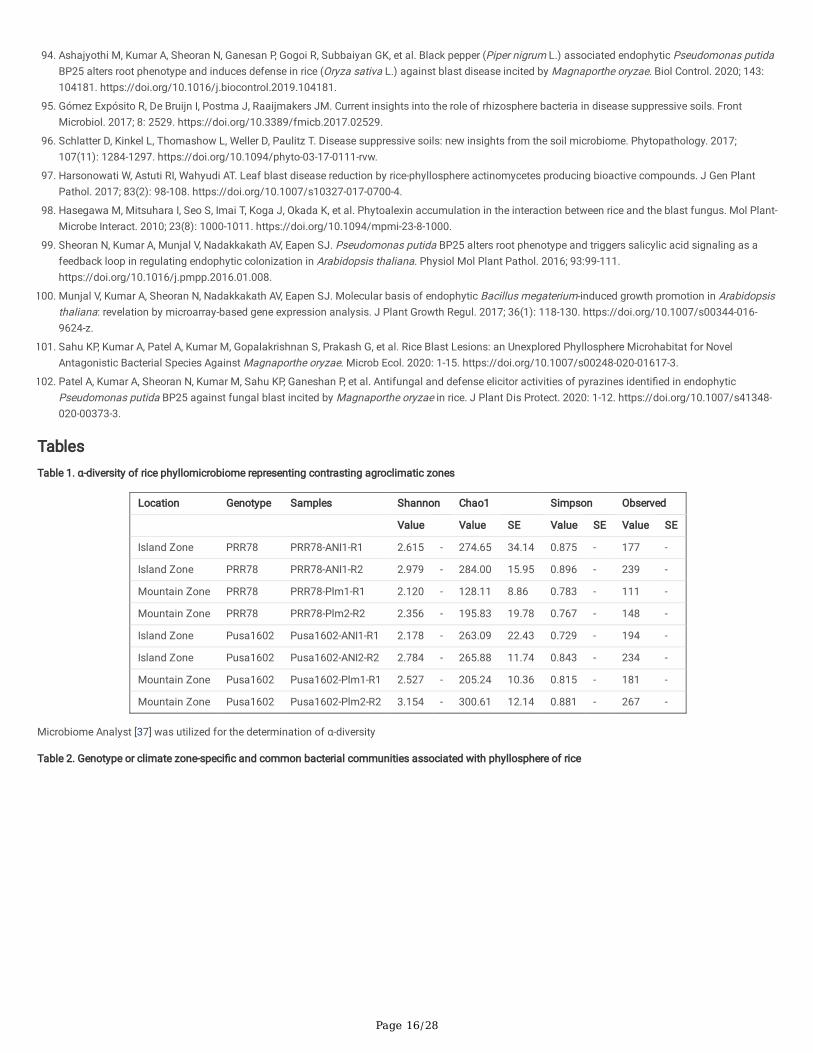
Page 16/28
94. Ashajyothi M, Kumar A, Sheoran N, Ganesan P, Gogoi R, Subbaiyan GK, et al. Black pepper (Piper nigrum L.) associated endophytic Pseudomonas putidaBP25 alters root phenotype and induces defense in rice (Oryza sativa L.) against blast disease incited by Magnaporthe oryzae. Biol Control. 2020; 143:104181. https://doi.org/10.1016/j.biocontrol.2019.104181.
95. Gómez Expósito R, De Bruijn I, Postma J, Raaijmakers JM. Current insights into the role of rhizosphere bacteria in disease suppressive soils. FrontMicrobiol. 2017; 8: 2529. https://doi.org/10.3389/fmicb.2017.02529.
9�. Schlatter D, Kinkel L, Thomashow L, Weller D, Paulitz T. Disease suppressive soils: new insights from the soil microbiome. Phytopathology. 2017;107(11): 1284-1297. https://doi.org/10.1094/phyto-03-17-0111-rvw.
97. Harsonowati W, Astuti RI, Wahyudi AT. Leaf blast disease reduction by rice-phyllosphere actinomycetes producing bioactive compounds. J Gen PlantPathol. 2017; 83(2): 98-108. https://doi.org/10.1007/s10327-017-0700-4.
9�. Hasegawa M, Mitsuhara I, Seo S, Imai T, Koga J, Okada K, et al. Phytoalexin accumulation in the interaction between rice and the blast fungus. Mol Plant-Microbe Interact. 2010; 23(8): 1000-1011. https://doi.org/10.1094/mpmi-23-8-1000.
99. Sheoran N, Kumar A, Munjal V, Nadakkakath AV, Eapen SJ. Pseudomonas putida BP25 alters root phenotype and triggers salicylic acid signaling as afeedback loop in regulating endophytic colonization in Arabidopsis thaliana. Physiol Mol Plant Pathol. 2016; 93:99-111.https://doi.org/10.1016/j.pmpp.2016.01.008.
100. Munjal V, Kumar A, Sheoran N, Nadakkakath AV, Eapen SJ. Molecular basis of endophytic Bacillus megaterium-induced growth promotion in Arabidopsisthaliana: revelation by microarray-based gene expression analysis. J Plant Growth Regul. 2017; 36(1): 118-130. https://doi.org/10.1007/s00344-016-9624-z.
101. Sahu KP, Kumar A, Patel A, Kumar M, Gopalakrishnan S, Prakash G, et al. Rice Blast Lesions: an Unexplored Phyllosphere Microhabitat for NovelAntagonistic Bacterial Species Against Magnaporthe oryzae. Microb Ecol. 2020: 1-15. https://doi.org/10.1007/s00248-020-01617-3.
102. Patel A, Kumar A, Sheoran N, Kumar M, Sahu KP, Ganeshan P, et al. Antifungal and defense elicitor activities of pyrazines identi�ed in endophyticPseudomonas putida BP25 against fungal blast incited by Magnaporthe oryzae in rice. J Plant Dis Protect. 2020: 1-12. https://doi.org/10.1007/s41348-020-00373-3.
TablesTable 1. α-diversity of rice phyllomicrobiome representing contrasting agroclimatic zones
Location Genotype Samples Shannon Chao1 Simpson Observed
Value Value SE Value SE Value SE
Island Zone PRR78 PRR78-ANI1-R1 2.615 - 274.65 34.14 0.875 - 177 -
Island Zone PRR78 PRR78-ANI1-R2 2.979 - 284.00 15.95 0.896 - 239 -
Mountain Zone PRR78 PRR78-Plm1-R1 2.120 - 128.11 8.86 0.783 - 111 -
Mountain Zone PRR78 PRR78-Plm2-R2 2.356 - 195.83 19.78 0.767 - 148 -
Island Zone Pusa1602 Pusa1602-ANI1-R1 2.178 - 263.09 22.43 0.729 - 194 -
Island Zone Pusa1602 Pusa1602-ANI2-R2 2.784 - 265.88 11.74 0.843 - 234 -
Mountain Zone Pusa1602 Pusa1602-Plm1-R1 2.527 - 205.24 10.36 0.815 - 181 -
Mountain Zone Pusa1602 Pusa1602-Plm2-R2 3.154 - 300.61 12.14 0.881 - 267 -
Microbiome Analyst [37] was utilized for the determination of α-diversity
Table 2. Genotype or climate zone-speci�c and common bacterial communities associated with phyllosphere of rice
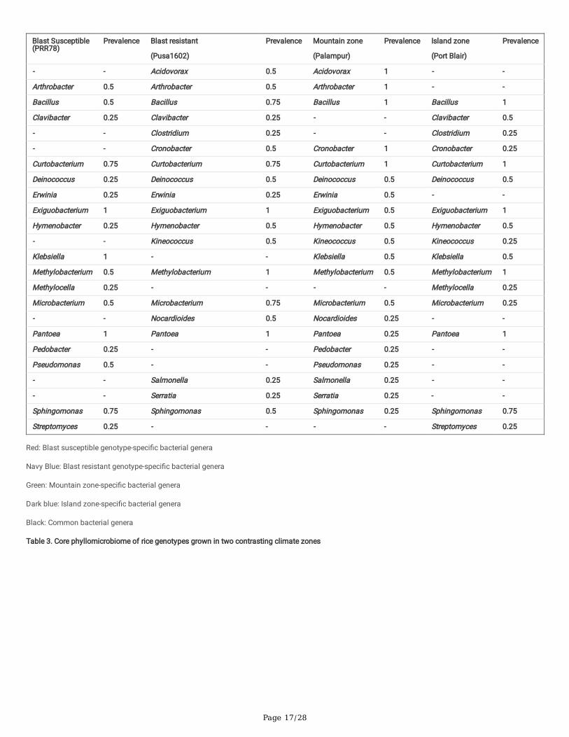
Page 17/28
Blast Susceptible (PRR78)
Prevalence Blast resistant
(Pusa1602)
Prevalence
Mountain zone
(Palampur)
Prevalence Island zone
(Port Blair)
Prevalence
- - Acidovorax 0.5 Acidovorax 1 - -
Arthrobacter 0.5 Arthrobacter 0.5 Arthrobacter 1 - -
Bacillus 0.5 Bacillus 0.75 Bacillus 1 Bacillus 1
Clavibacter 0.25 Clavibacter 0.25 - - Clavibacter 0.5
- - Clostridium 0.25 - - Clostridium 0.25
- - Cronobacter 0.5 Cronobacter 1 Cronobacter 0.25
Curtobacterium 0.75 Curtobacterium 0.75 Curtobacterium 1 Curtobacterium 1
Deinococcus 0.25 Deinococcus 0.5 Deinococcus 0.5 Deinococcus 0.5
Erwinia 0.25 Erwinia 0.25 Erwinia 0.5 - -
Exiguobacterium 1 Exiguobacterium 1 Exiguobacterium 0.5 Exiguobacterium 1
Hymenobacter 0.25 Hymenobacter 0.5 Hymenobacter 0.5 Hymenobacter 0.5
- - Kineococcus 0.5 Kineococcus 0.5 Kineococcus 0.25
Klebsiella 1 - - Klebsiella 0.5 Klebsiella 0.5
Methylobacterium 0.5 Methylobacterium 1 Methylobacterium 0.5 Methylobacterium 1
Methylocella 0.25 - - - - Methylocella 0.25
Microbacterium 0.5 Microbacterium 0.75 Microbacterium 0.5 Microbacterium 0.25
- - Nocardioides 0.5 Nocardioides 0.25 - -
Pantoea 1 Pantoea 1 Pantoea 0.25 Pantoea 1
Pedobacter 0.25 - - Pedobacter 0.25 - -
Pseudomonas 0.5 - - Pseudomonas 0.25 - -
- - Salmonella 0.25 Salmonella 0.25 - -
- - Serratia 0.25 Serratia 0.25 - -
Sphingomonas 0.75 Sphingomonas 0.5 Sphingomonas 0.25 Sphingomonas 0.75
Streptomyces 0.25 - - - - Streptomyces 0.25
Red: Blast susceptible genotype-speci�c bacterial genera
Navy Blue: Blast resistant genotype-speci�c bacterial genera
Green: Mountain zone-speci�c bacterial genera
Dark blue: Island zone-speci�c bacterial genera
Black: Common bacterial genera
Table 3. Core phyllomicrobiome of rice genotypes grown in two contrasting climate zones
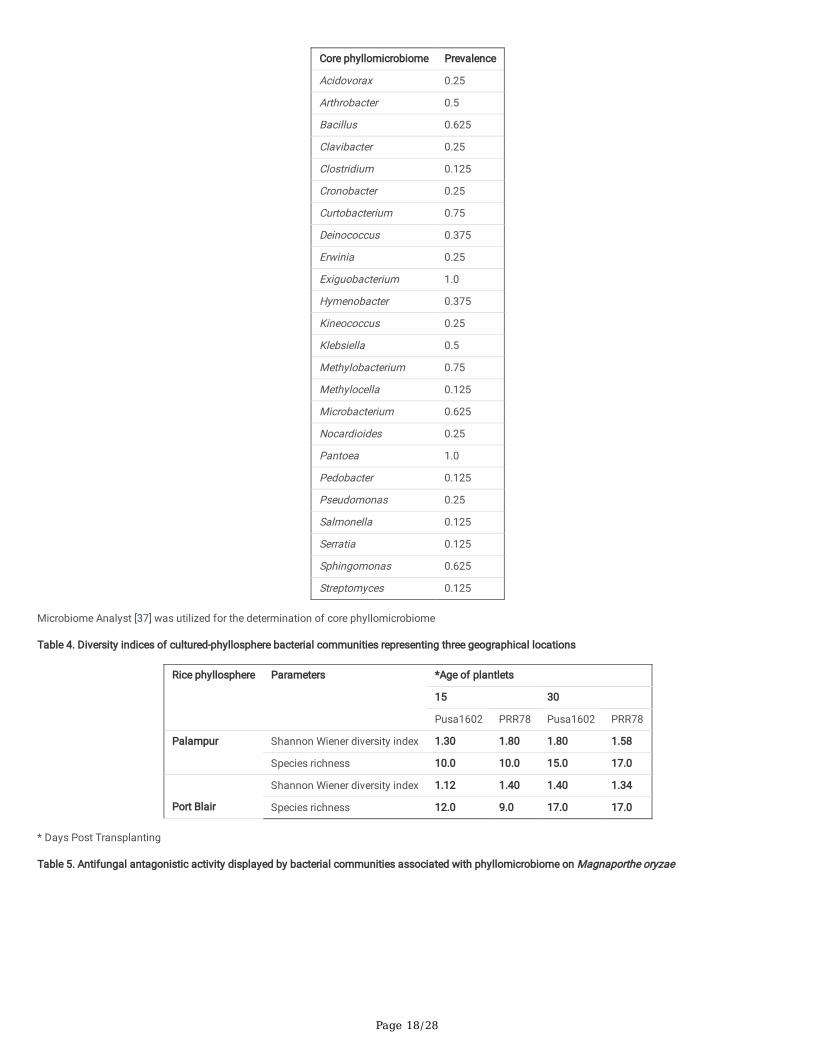
Page 18/28
Core phyllomicrobiome Prevalence
Acidovorax 0.25
Arthrobacter 0.5
Bacillus 0.625
Clavibacter 0.25
Clostridium 0.125
Cronobacter 0.25
Curtobacterium 0.75
Deinococcus 0.375
Erwinia 0.25
Exiguobacterium 1.0
Hymenobacter 0.375
Kineococcus 0.25
Klebsiella 0.5
Methylobacterium 0.75
Methylocella 0.125
Microbacterium 0.625
Nocardioides 0.25
Pantoea 1.0
Pedobacter 0.125
Pseudomonas 0.25
Salmonella 0.125
Serratia 0.125
Sphingomonas 0.625
Streptomyces 0.125
Microbiome Analyst [37] was utilized for the determination of core phyllomicrobiome
Table 4. Diversity indices of cultured-phyllosphere bacterial communities representing three geographical locations
Rice phyllosphere Parameters *Age of plantlets
15 30
Pusa1602 PRR78 Pusa1602 PRR78
Palampur Shannon Wiener diversity index 1.30 1.80 1.80 1.58
Species richness 10.0 10.0 15.0 17.0
Port Blair
Shannon Wiener diversity index 1.12 1.40 1.40 1.34
Species richness 12.0 9.0 17.0 17.0
* Days Post Transplanting
Table 5. Antifungal antagonistic activity displayed by bacterial communities associated with phyllomicrobiome on Magnaporthe oryzae
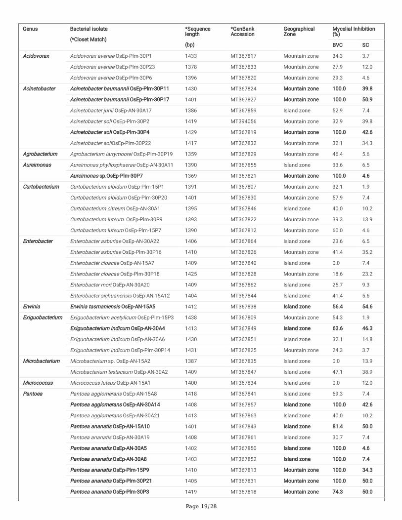
Page 19/28
Genus Bacterial isolate
(*Closet Match)
*Sequencelength
(bp)
*GenBankAccession
GeographicalZone
Mycelial Inhibition(%)
BVC SC
Acidovorax Acidovorax avenae OsEp-Plm-30P1 1433 MT367817 Mountain zone 34.3 3.7
Acidovorax avenae OsEp-Plm-30P23 1378 MT367833 Mountain zone 27.9 12.0
Acidovorax avenae OsEp-Plm-30P6 1396 MT367820 Mountain zone 29.3 4.6
Acinetobacter Acinetobacter baumannii OsEp-Plm-30P11 1430 MT367824 Mountain zone 100.0 39.8
Acinetobacter baumannii OsEp-Plm-30P17 1401 MT367827 Mountain zone 100.0 50.9
Acinetobacter junii OsEp-AN-30A17 1386 MT367859 Island zone 52.9 7.4
Acinetobacter soli OsEp-Plm-30P2 1419 MT394056 Mountain zone 32.9 39.8
Acinetobacter soli OsEp-Plm-30P4 1429 MT367819 Mountain zone 100.0 42.6
Acinetobacter soliOsEp-Plm-30P22 1417 MT367832 Mountain zone 32.1 34.3
Agrobacterium Agrobacterium larrymoorei OsEp-Plm-30P19 1359 MT367829 Mountain zone 46.4 5.6
Aureimonas Aureimonas phyllosphaerae OsEp-AN-30A11 1390 MT367855 Island zone 33.6 6.5
Aureimonas sp.OsEp-Plm-30P7 1369 MT367821 Mountain zone 100.0 4.6
Curtobacterium Curtobacterium albidum OsEp-Plm-15P1 1391 MT367807 Mountain zone 32.1 1.9
Curtobacterium albidum OsEp-Plm-30P20 1401 MT367830 Mountain zone 57.9 7.4
Curtobacterium citreum OsEp-AN-30A1 1395 MT367846 Island zone 40.0 10.2
Curtobacterium luteum OsEp-Plm-30P9 1393 MT367822 Mountain zone 39.3 13.9
Curtobacterium luteum OsEp-Plm-15P7 1390 MT367812 Mountain zone 60.0 4.6
Enterobacter Enterobacter asburiae OsEp-AN-30A22 1406 MT367864 Island zone 23.6 6.5
Enterobacter asburiae OsEp-Plm-30P16 1410 MT367826 Mountain zone 41.4 35.2
Enterobacter cloacae OsEp-AN-15A7 1409 MT367840 Island zone 0.0 7.4
Enterobacter cloacae OsEp-Plm-30P18 1425 MT367828 Mountain zone 18.6 23.2
Enterobacter mori OsEp-AN-30A20 1409 MT367862 Island zone 25.7 9.3
Enterobacter sichuanensis OsEp-AN-15A12 1404 MT367844 Island zone 41.4 5.6
Erwinia Erwinia tasmaniensis OsEp-AN-15A5 1412 MT367838 Island zone 56.4 54.6
Exiguobacterium Exiguobacterium acetylicum OsEp-Plm-15P3 1438 MT367809 Mountain zone 54.3 1.9
Exiguobacterium indicum OsEp-AN-30A4 1413 MT367849 Island zone 63.6 46.3
Exiguobacterium indicum OsEp-AN-30A6 1430 MT367851 Island zone 32.1 14.8
Exiguobacterium indicum OsEp-Plm-30P14 1431 MT367825 Mountain zone 24.3 3.7
Microbacterium Microbacterium sp. OsEp-AN-15A2 1387 MT367835 Island zone 0.0 13.9
Microbacterium testaceum OsEp-AN-30A2 1409 MT367847 Island zone 47.1 38.9
Micrococcus Micrococcus luteus OsEp-AN-15A1 1400 MT367834 Island zone 0.0 12.0
Pantoea Pantoea agglomerans OsEp-AN-15A8 1418 MT367841 Island zone 69.3 7.4
Pantoea agglomerans OsEp-AN-30A14 1408 MT367857 Island zone 100.0 42.6
Pantoea agglomerans OsEp-AN-30A21 1413 MT367863 Island zone 40.0 10.2
Pantoea ananatis OsEp-AN-15A10 1401 MT367843 Island zone 81.4 50.0
Pantoea ananatis OsEp-AN-30A19 1408 MT367861 Island zone 30.7 7.4
Pantoea ananatis OsEp-AN-30A5 1402 MT367850 Island zone 100.0 4.6
Pantoea ananatis OsEp-AN-30A8 1403 MT367852 Island zone 100.0 7.4
Pantoea ananatis OsEp-Plm-15P9 1410 MT367813 Mountain zone 100.0 34.3
Pantoea ananatis OsEp-Plm-30P21 1405 MT367831 Mountain zone 100.0 50.0
Pantoea ananatis OsEp-Plm-30P3 1419 MT367818 Mountain zone 74.3 50.0
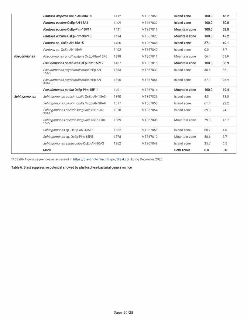
Page 20/28
Pantoea dispersa OsEp-AN-30A18 1412 MT367860 Island zone 100.0 48.2
Pantoea eucrina OsEp-AN-15A4 1409 MT367837 Island zone 100.0 50.0
Pantoea eucrina OsEp-Plm-15P14 1421 MT367816 Mountain zone 100.0 52.8
Pantoea eucrina OsEp-Plm-30P10 1414 MT367823 Mountain zone 100.0 47.2
Pantoea sp. OsEp-AN-15A15 1400 MT367845 Island zone 57.1 49.1
Pantoea sp. OsEp-AN-15A9 1402 MT367842 Island zone 0.0 3.7
Pseudomonas Pseudomonas oryzihabitans OsEp-Plm-15P6 1398 MT367811 Mountain zone 56.4 51.9
Pseudomonas parafulva OsEp-Plm-15P12 1407 MT367815 Mountain zone 100.0 38.9
Pseudomonas psychrotolerans OsEp-AN-15A6
1383 MT367839 Island zone 38.6 36.1
Pseudomonas psychrotolerans OsEp-AN-30A13
1396 MT367856 Island zone 57.1 26.9
Pseudomonas putida OsEp-Plm-15P11 1401 MT367814 Mountain zone 100.0 19.4
Sphingomonas Sphingomonas paucimobilis OsEp-AN-15A3 1390 MT367836 Island zone 4.3 13.0
Sphingomonas paucimobilis OsEp-AN-30A9 1377 MT367853 Island zone 61.4 22.2
Sphingomonas pseudosanguinis OsEp-AN-30A10
1378 MT367854 Island zone 59.3 24.1
Sphingomonas pseudosanguinis OsEp-Plm-15P2
1389 MT367808 Mountain zone 79.3 15.7
Sphingomonas sp. OsEp-AN-30A15 1362 MT367858 Island zone 60.7 4.6
Sphingomonas sp. OsEp-Plm-15P5 1378 MT367810 Mountain zone 58.6 3.7
Sphingomonas yabuuchiae OsEp-AN-30A3 1362 MT367848 Island zone 35.7 6.5
Mock Both zones 0.0 0.0
*16S rRNA gene sequences as accessed in https://blast.ncbi.nlm.nih.gov/Blast.cgi during December 2020
Table 6. Blast suppressive potential showed by phyllosphere bacterial genera on rice
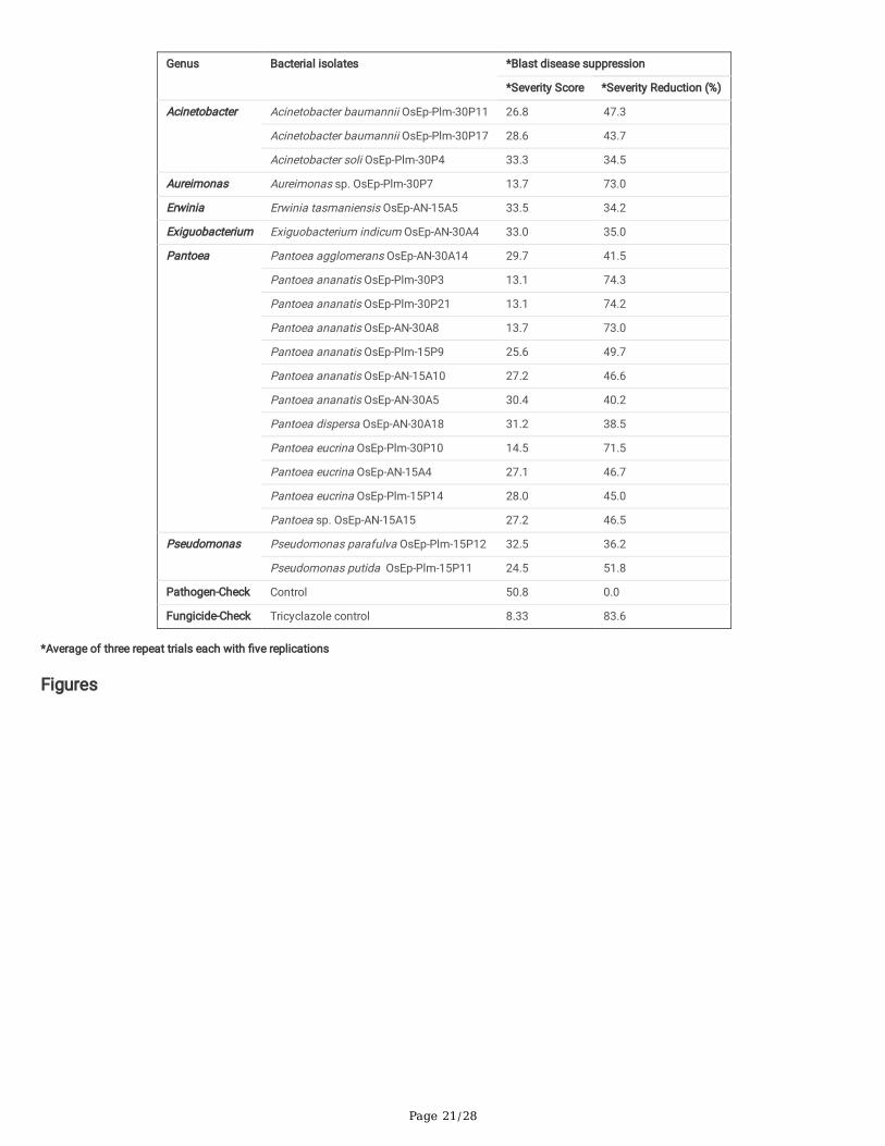
Page 21/28
Genus Bacterial isolates *Blast disease suppression
*Severity Score *Severity Reduction (%)
Acinetobacter
Acinetobacter baumannii OsEp-Plm-30P11 26.8 47.3
Acinetobacter baumannii OsEp-Plm-30P17 28.6 43.7
Acinetobacter soli OsEp-Plm-30P4 33.3 34.5
Aureimonas Aureimonas sp. OsEp-Plm-30P7 13.7 73.0
Erwinia Erwinia tasmaniensis OsEp-AN-15A5 33.5 34.2
Exiguobacterium Exiguobacterium indicum OsEp-AN-30A4 33.0 35.0
Pantoea Pantoea agglomerans OsEp-AN-30A14 29.7 41.5
Pantoea ananatis OsEp-Plm-30P3 13.1 74.3
Pantoea ananatis OsEp-Plm-30P21 13.1 74.2
Pantoea ananatis OsEp-AN-30A8 13.7 73.0
Pantoea ananatis OsEp-Plm-15P9 25.6 49.7
Pantoea ananatis OsEp-AN-15A10 27.2 46.6
Pantoea ananatis OsEp-AN-30A5 30.4 40.2
Pantoea dispersa OsEp-AN-30A18 31.2 38.5
Pantoea eucrina OsEp-Plm-30P10 14.5 71.5
Pantoea eucrina OsEp-AN-15A4 27.1 46.7
Pantoea eucrina OsEp-Plm-15P14 28.0 45.0
Pantoea sp. OsEp-AN-15A15 27.2 46.5
Pseudomonas Pseudomonas parafulva OsEp-Plm-15P12 32.5 36.2
Pseudomonas putida OsEp-Plm-15P11 24.5 51.8
Pathogen-Check Control 50.8 0.0
Fungicide-Check Tricyclazole control 8.33 83.6
*Average of three repeat trials each with �ve replications
Figures
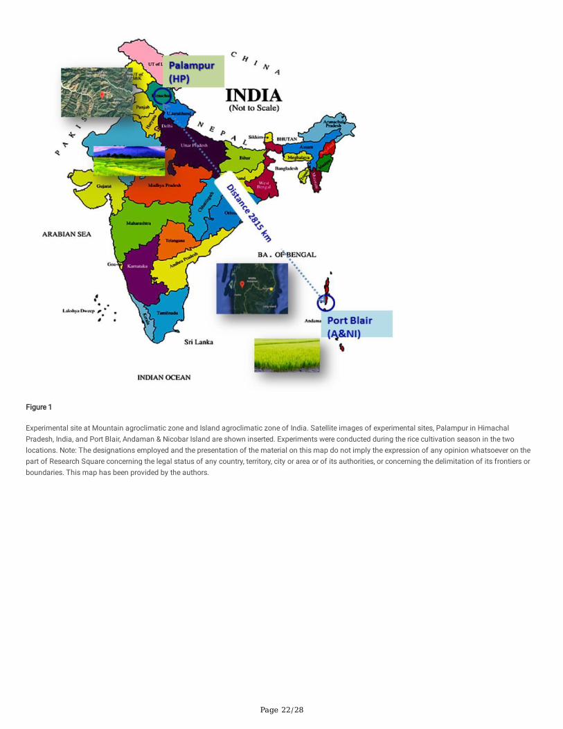
Page 22/28
Figure 1
Experimental site at Mountain agroclimatic zone and Island agroclimatic zone of India. Satellite images of experimental sites, Palampur in HimachalPradesh, India, and Port Blair, Andaman & Nicobar Island are shown inserted. Experiments were conducted during the rice cultivation season in the twolocations. Note: The designations employed and the presentation of the material on this map do not imply the expression of any opinion whatsoever on thepart of Research Square concerning the legal status of any country, territory, city or area or of its authorities, or concerning the delimitation of its frontiers orboundaries. This map has been provided by the authors.
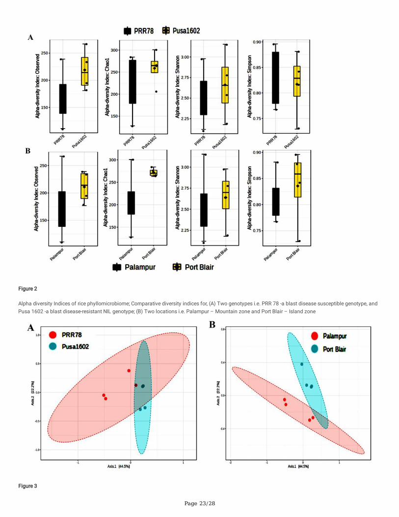
Page 23/28
Figure 2
Alpha diversity Indices of rice phyllomicrobiome; Comparative diversity indices for, (A) Two genotypes i.e. PRR 78 -a blast disease susceptible genotype, andPusa 1602 -a blast disease-resistant NIL genotype; (B) Two locations i.e. Palampur – Mountain zone and Port Blair – Island zone
Figure 3
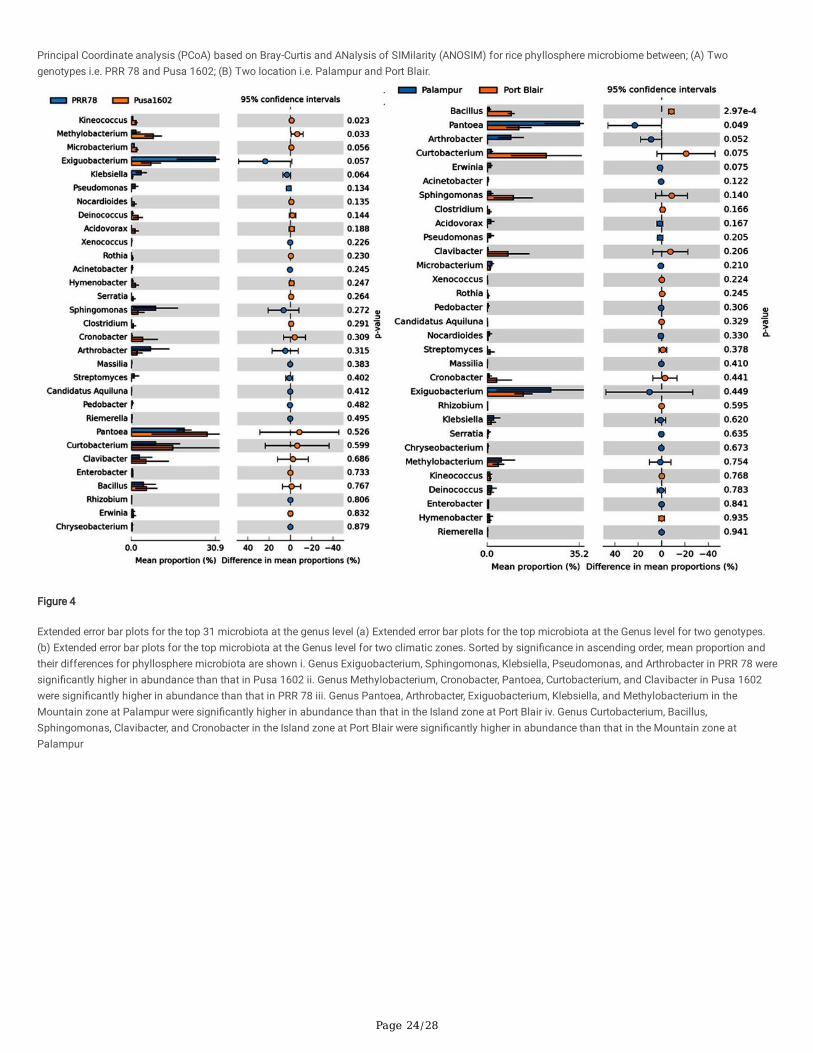
Page 24/28
Principal Coordinate analysis (PCoA) based on Bray-Curtis and ANalysis of SIMilarity (ANOSIM) for rice phyllosphere microbiome between; (A) Twogenotypes i.e. PRR 78 and Pusa 1602; (B) Two location i.e. Palampur and Port Blair.
Figure 4
Extended error bar plots for the top 31 microbiota at the genus level (a) Extended error bar plots for the top microbiota at the Genus level for two genotypes.(b) Extended error bar plots for the top microbiota at the Genus level for two climatic zones. Sorted by signi�cance in ascending order, mean proportion andtheir differences for phyllosphere microbiota are shown i. Genus Exiguobacterium, Sphingomonas, Klebsiella, Pseudomonas, and Arthrobacter in PRR 78 weresigni�cantly higher in abundance than that in Pusa 1602 ii. Genus Methylobacterium, Cronobacter, Pantoea, Curtobacterium, and Clavibacter in Pusa 1602were signi�cantly higher in abundance than that in PRR 78 iii. Genus Pantoea, Arthrobacter, Exiguobacterium, Klebsiella, and Methylobacterium in theMountain zone at Palampur were signi�cantly higher in abundance than that in the Island zone at Port Blair iv. Genus Curtobacterium, Bacillus,Sphingomonas, Clavibacter, and Cronobacter in the Island zone at Port Blair were signi�cantly higher in abundance than that in the Mountain zone atPalampur
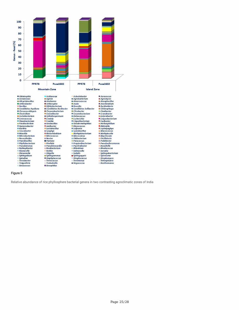
Page 25/28
Figure 5
Relative abundance of rice phyllosphere bacterial genera in two contrasting agroclimatic zones of India
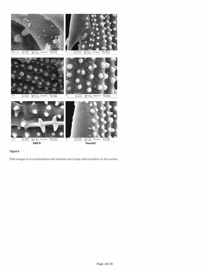
Page 26/28
Figure 6
SEM images of rice phyllosphere with bacterial and fungal cells/mycelium on the surface
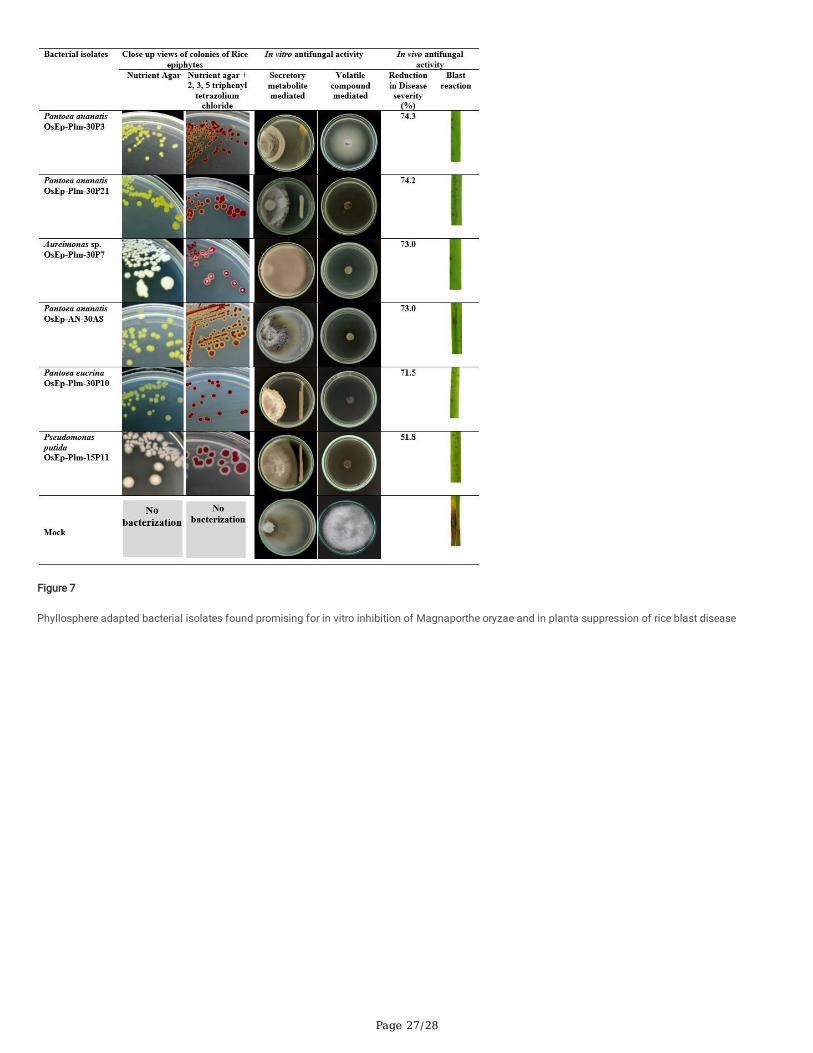
Page 27/28
Figure 7
Phyllosphere adapted bacterial isolates found promising for in vitro inhibition of Magnaporthe oryzae and in planta suppression of rice blast disease
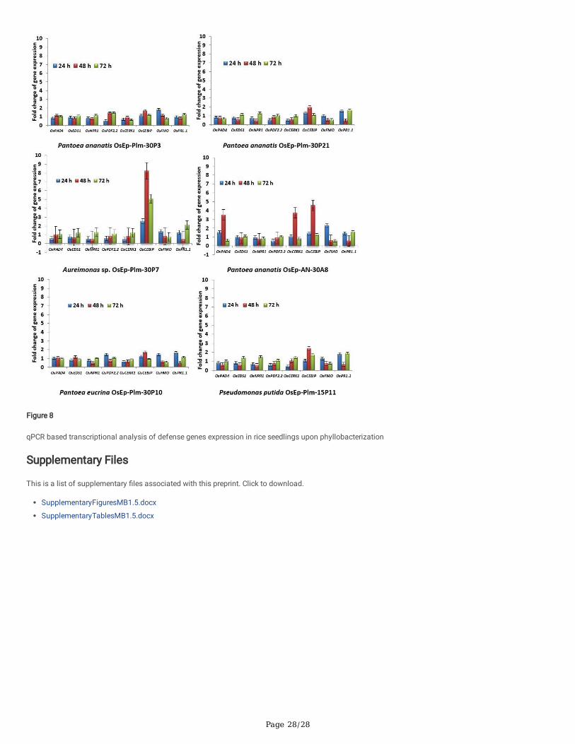
Page 28/28
Figure 8
qPCR based transcriptional analysis of defense genes expression in rice seedlings upon phyllobacterization
Supplementary Files
This is a list of supplementary �les associated with this preprint. Click to download.
SupplementaryFiguresMB1.5.docx
SupplementaryTablesMB1.5.docx