Embed Size (px)
Citation preview

Decorin Inhibition of PDGF-Stimulated VascularSmooth Muscle Cell Function
Potential Mechanism for Inhibition of Intimal Hyperplasia afterBalloon Angioplasty
Nafiseh Nili,* Asim N. Cheema,*Frank J. Giordano,† Alan W. Barolet,*Saeid Babaei,* Reed Hickey,†
Mohammad R. Eskandarian,* Mirjam Smeets,‡
Jagdish Butany,§ Gerard Pasterkamp,‡ andBradley H. Strauss*From the Roy and Ann Foss Interventional Cardiology Research
Program,* Terrence Donnelly Heart Centre, St. Michael’s Hospital
and University Health Network,§ University of Toronto, Toronto,
Ontario, Canada; the Cardiovascular Gene Therapy Program,†
Yale University, New Haven, Connecticut; and the Experimental
Cardiology Laboratory,‡ University Medical Center, Utrecht,
The Netherlands
Decorin is a small proteoglycan that binds to transform-ing growth factor-� (TGF-�) and inhibits its activity.However, its interaction with platelet-derived growthfactor (PDGF), involved in arterial repair after injury, isnot well characterized. The objectives of this study wereto assess decorin-PDGF and decorin-PDGF receptor(PDGFR) interactions, the in vitro effects of decorin onPDGF-stimulated smooth muscle cell (SMC) functionsand the in vivo effects of decorin overexpression onarterial repair in a rabbit carotid balloon-injury model.Decorin binding to PDGF was demonstrated by solid-phase binding and affinity cross-linking assays. Decorinpotently inhibited PDGF-stimulated PDGFR phosphory-lation. Pretreatment of rabbit aortic SMC with decorinsignificantly inhibited PDGF-stimulated cell migration,proliferation, and collagen synthesis. Decorin overex-pression by adenoviral-mediated gene transfection inballoon-injured carotid arteries significantly decreasedintimal cross-sectional area and collagen content by�50% at 10 weeks compared to �-galactosidase-trans-fected or balloon-injured, non-transfected controls.This study shows that decorin binds to PDGF and inhib-its its stimulatory activity on SMCs by preventing PDGFRphosphorylation. Decorin overexpression reduces inti-mal hyperplasia and collagen content after arterialinjury. Decorin may be an effective therapy for theprevention of intimal hyperplasia after balloon angio-plasty. (Am J Pathol 2003, 163:869–878)
Previous studies have shown that smooth muscle cell(SMC) activation and extracellular matrix (ECM) deposi-tion play important roles in intimal hyperplasia after bal-loon injury.1,2 Coincident with an arterial injury, quiescentSMC are exposed to cytokines and growth factors thatprofoundly alter their behavior. Platelet-derived growthfactor (PDGF) is a potent growth factor produced byplatelets, SMCs, and endothelial cells in the injured ves-sel wall.3 It initiates a multitude of biological effectsthrough the activation of intracellular signal transductionpathways that contribute to SMC proliferation, migration,and collagen synthesis.3–5 The importance of PDGF indevelopment of intimal hyperplasia has been establishedin arterial-injury models.6,7
Decorin is a small proteoglycan that consists of asingle glycosaminoglycan side chain linked to a coreprotein containing leucine-rich repeats of about 24 aminoacids.8,9 It is found in the ECM of a variety of tissues andcell types.8,9 Decorin interacts with a variety of proteinsthat are involved in matrix assembly,10 and in the regu-lation of fundamental biological functions such as cellattachment,11 migration,12 and proliferation.13,14 Decorininhibits cell proliferation in several cell types, includingcancer cells.13 It mediates its regulatory effects eitherdirectly by up-regulating cyclin-dependent kinase inhib-itors such as p21 and p27, or through its ability to interactwith growth factors.15,16 It is well established that decorinbinds to TGF-�17 and inhibits its biological activity in anumber of cell types, including rat aortic SMCs.16 How-ever, it is not known whether decorin may also exert itsregulatory effects by interacting with and modulating theactivity of PDGF, another prominent growth factor in-volved in arterial repair. Therefore, the objectives of thisstudy were to assess decorin-PDGF and decorin-PDGFreceptor (PDGFR) interactions, the effects of decorin onPDGFR phosphorylation and PDGF-stimulated SMC
Supported by the Heart and Stroke Foundation of Ontario and dedicatedto the memory of Robyn Strauss Albert.
N. N. and A. N. C. contributed equally to the manuscript.
Accepted for publication May 12, 2003.
Address reprint requests to Dr. Bradley Strauss, Terrence Donnelly HeartCentre, Division of Cardiology, St. Michael’s Hospital, 30 Bond Street,Toronto, Ontario, Canada M5B 1W8. E-mail: [email protected].
American Journal of Pathology, Vol. 163, No. 3, September 2003
Copyright © American Society for Investigative Pathology
869

functions in vitro, and effects of decorin overexpressionon arterial repair in a rabbit carotid balloon-injury modelthat is characterized by PDGF up-regulation.6,7
Materials and Methods
Decorin Binding to PDGF
Solid-solid-phase binding and affinity cross-linking as-says were performed to assess PDGF and decorin inter-actions.
Solid-Phase Radioligand-Binding of 125I-PDGF toDecorin
Bovine articular cartilage decorin (Sigma, St. Louis,MO) was coated (1 �g/well) onto the surface of a 96-wellplate (Nalge Nunc International, Denmark).17 Nonspe-cific binding was blocked with 1% bovine serum albumin(BSA) in binding buffer for 3 hours at 37°C. Immobilizeddecorin was then allowed to bind to 4 � 104 cpm of125I-PDGF-BB (Amersham Biosciences Corp., Piscat-away, NJ) in the presence or absence of increasingconcentrations of chondroitin sulfate (Sigma), BSA, orunlabeled PDGF-BB (R & D Systems Inc., Minneapolis,MN) as competitors. After a 6-hour incubation at 37°C,bound 125I-PDGF was solubilized in 0.3 mol/L NaOH, 1%Triton X-100 for 30 minutes and the radioactivity of eachwell was measured using a gamma counter (WALLAC,1470 WIZARD). Nonspecific binding was determined byadding 100-fold excess unlabeled PDGF to the incuba-tion mixture.
Affinity Cross-Linking of Decorin with PDGF andImmunoprecipitation Using Decorin Antibody
Decorin (1 �g) was mixed with PDGF-BB (0.01, 0.1, or1.0 �g) in 200 �l of binding buffer18 and incubated at37°C for 3 hours. The proteins were then cross-linked with0.3 mmol/L disuccinimidyl suberate (DSS; Pierce, Rock-ford, IL, USA) at 37°C for 30 minutes and the reaction wasterminated by adding TBS buffer (10 mmol/L Tris-basebuffer, pH 8.0, 150 mmol/L NaCl). Cross-linked proteinswere immunoprecipitated with anti-decorin monoclonalantibody 6D619 (generous gift from Dr. Paul G. Scott,University of Alberta, Edmonton, Alberta) using protein Gsepharose (Amersham Biosciences Corp) at room tem-perature for 1 hour. The suspension was centrifuged andbeads were washed four times with ice-cold TBS buffer.To recover specifically bound proteins, beads were sus-pended in 50 �l of 1X SDS-PAGE sample-loading buffer,mixed for 30 minutes at room temperature, and centri-fuged. Proteins in 25 �l of supernatant were analyzed by4 to 20% SDS and electrotransferred onto a nitrocellulosemembrane (Bio-Rad, Hercules, CA, USA). Decorin-PDGFcomplex was detected with anti-decorin and anti-PDGFantibodies as described below.
Immunoblotting of Decorin-PDGF Complex UsingDecorin and PDGF Antibodies
The membrane was immunoblotted with anti-decorinmonoclonal antibody 6D6.19 Anti-mouse IgG-HRP (SantaCruz Biotechnology, Inc., Santa Cruz, CA, USA) wasused for detection of primary antibody. The membranewas stripped and re-immunoblotted with goat anti-PDGFantibody (R & D Systems). Anti-goat IgG-HRP (SantaCruz) was used for detection of primary antibody. Detec-tion was performed using a chemiluminescence peroxi-dase detection system (Sigma) and exposure to BioMaxfilm (Kodak).
In Vitro Studies
Cell Culture
Rabbit aortic SMC from passages 5 and 6 were cul-tured in Dulbecco’s modified Eagle’s medium (DMEM),supplemented with 2% penicillin-streptomycin and 10%fetal calf serum. For DNA synthesis and cell proliferationassays, cells were initially seeded at 5 � 104 per well into24-well plates and at 104 per well into 96-well plates,respectively. For all other experiments, cells were grownto confluency. In each assay, cells were serum-starvedfor 48 hours and then pretreated for 30 minutes with theindicated concentration of decorin followed by treatmentwith PDGF-BB (10 ng/ml) for 24 hours.
PDGFR Expression
Confluent monolayers of cells were incubated in se-rum-free medium containing PDGF-BB in the presence orabsence of decorin. After 24 hours of incubation period,cells were washed with PBS and lysed in 1X SDS-PAGEsample-loading buffer. Samples containing 50 �g of pro-teins were separated by 4 to 20% SDS-PAGE and elec-trotransferred onto a nitrocellulose membrane. Mem-branes were immunoblotted with goat anti-humanPDGFR-� or -� antibodies (R&D Systems). Anti-goat IgG-HRP (Santa Cruz) was used for detection of primaryantibody and revealed using chemiluminescence detec-tion system followed by autoradiography.
PDGFR � Phosphorylation
Confluent monolayers of cells were incubated in se-rum-free medium containing PDGF-BB in the presence orabsence of decorin for 1 hour at 4°C. Cells were ex-tracted as previously described.20 Lysates were incu-bated at 4°C for 6 hours with 2 �g of goat anti-humanPDGFR-� antibody and then 30 �l protein of G Sepha-rose (Pharmacia Biotech) was added and mixed at 4°Covernight. Immunoprecipitated receptors were releasedfrom protein G pellets by boiling for 5 minutes in 1XSDS-PAGE sample-loading buffer containing 2-mercap-toethanol and separated by 8% SDS-PAGE. Proteinswere electrotransferred onto a nitrocellulose membraneand immunoblotted with anti-phosphotyrosine 4G10
870 Nili et alAJP September 2003, Vol. 163, No. 3

monoclonal antibody (Upstate Biotechnology, LakePlacid, NY). Goat anti-mouse secondary antibody wasused for detection of primary antibody. The membranewas stripped and re-immunoblotted with goat anti-humanPDGFR-� antibody. Detection was performed usingchemiluminescence detection system followed by auto-radiography.
Binding of 125I-PDGF to Cells or PDGF Receptors
Cell binding assay and affinity cross-linking were per-formed as described previously.18 Briefly, monolayers ofconfluent cells were washed three times with cold bind-ing buffer and incubated in binding buffer for 30 minutesat 4°C. Confluent cells were pretreated for 30 minuteswith decorin (30 �g/ml) in binding buffer, followed byadding 125I-PDGF (0.1 nmol/L). 125I-PDGF was alsoadded to two other groups that were not pretreated withdecorin. One group was given unlabeled PDGF (10nmol/L) as a competitor to show specificity of the bindingwhile the other group (control group) did not receiveunlabeled PDGF. All groups were incubated at 4°C for 4hours. Cells were then washed four times with bindingbuffer. For binding assay, cells were solubilized with 1XSDS-PAGE sample- loading buffer. The radioactivity ofaliquots of soluble fractions was measured using agamma counter. For affinity cross-linking assay, cellswere cross-linked with DSS (0.3 mmol/L) at 4°C for 15minutes and lysed in 1X SDS-PAGE sample-loadingbuffer. Soluble fractions containing 50 �g protein wereanalyzed by 4 to 12% SDS-PAGE. The gel was driedfollowed by autoradiography using BioMax film.
Cell Necrosis and Apoptosis
Cells were incubated in serum-free medium containing30 �g/ml of decorin for 24 hours. For cell necrosis, lactatedehydrogenase (LDH) was measured in the medium us-ing CytoTox 96 Non-Radioactive Cytotoxicity Assay kit(Promega Corporation, Madison, WI) according to themanufacturer’s instructions.
For cell apoptosis, cells were washed with PBS, lysedin 1X SDS-PAGE sample-loading buffer and centrifuged.Supernatants containing 50 �g protein were analyzed by4 to 20% SDS-PAGE and electroblotted onto a nitrocel-lulose membrane. The membrane was immunoblottedwith rabbit polyclonal anti-caspase-3 (CPP32) Ab-4 (Neo-Marker, Fremont, CA). Anti-rabbit IgG-HRP (Santa Cruz)was used for detection of primary antibody. The mem-brane was then stripped and re-immunoblotted with anti-mouse monoclonal anti-� actin antibody (Sigma). Goatanti-mouse secondary antibody (Sigma) was used. De-tection was performed using chemiluminescence detec-tion system (Sigma) followed by autoradiography.
Growth Assays
For DNA synthesis, 1.0 �Ci/ml of 3H-thymidine (Amer-sham) was added to the cultures 24 hours after treatingcells with PDGF-BB in the presence or absence of
decorin and incubated at 37°C for 6 hours. DNA synthe-sis was measured as previously described.16 For cellproliferation, cells were incubated with PDGF-BB (10 ng/ml) in the presence and absence of decorin (20 �g/ml)for the indicated time periods and cell density was de-termined as described by Hou et al21 Briefly, cells werefixed with 4% paraformaldehyde and stained with 0.5%toluidine blue in 4% paraformaldehyde for 5 minutes.Cells were rinsed in water twice and solubilized with 100�l of 1% SDS. Absorbance was measured in a microtiterplate reader (Molecular Devices) at 595 nm.
Migration Assay
Neuro Probe 48-well microchemotaxis chambers(Costar, Corning Inc., Corning, NY) with PVP-free poly-carbonate filter (8.0 �m pore size) were used. The bottomwell of the Boyden chamber was filled with 250 �l ofserum-free medium containing PDGF-BB (10 ng/ml). Qui-escent cells were trypsinized and resuspended in serum-free medium with or without decorin and incubated for 30minutes at 37°C. Cells (2 � 104) were then added to theupper well of the microchemotaxis chamber and incu-bated for 5 hours at 37°C. Cells that migrated to the lowerside of the filters were fixed and stained with the Diff-Quick staining kit (VWR Laboratory). The filters weremounted on glass slides and counted by light micros-copy using �100 magnification. The results were ex-pressed as number of migrated cells per high power field(HPF).
Collagen Synthesis Assay
Ascorbic acid (50 �g/ml) and 14C-proline (5 �Ci/ml)were added to the cultures 24 hours after treating cellswith PDGF-BB in the presence or absence of decorin andincubated at 37°C for 6 hours. Collagen synthesis wasmeasured in the culture medium using a bacterial colla-genase digestion method.22
In Vivo Effects of Decorin Overexpression inBalloon-Injured Arteries
Carotid Artery Model
The animal experiments were performed in accor-dance with guidelines set out by the University of Torontoand approved by the St. Michael’s Hospital Animal CareCommittee. A double-injury carotid artery model wasdone in 48 normolipemic male New Zealand White rab-bits weighing 3.5 to 4.0 kg as previously described.23
Both carotid arteries were injured with a 3.5-mm diameterballoon angioplasty catheter. At 3 weeks, a second bal-loon injury was performed. Immediately after second in-jury, the arteries were surgically exposed and the bal-loon-injured segments were temporarily isolated withligatures. Gene transfer to these arterial segments wasachieved using recombinant adenovirus vectors pre-pared by homologous recombination as previously de-scribed.24 Briefly, a 1.5-kb rabbit decorin cDNA was
Decorin, A Potent Inhibitor of VSMC Function 871AJP September 2003, Vol. 163, No. 3

cloned into the shuttle vector pCMVPLPA in which ex-pression of the cDNA is driven by the CMV promoter.Recombination between this shuttle vector and the ade-novirus 5 genomic vector pJM17 produced recombinantadenovirus encoding the decorin expression cassette. Acontrol �-galactosidase encoding adenovirus was pre-pared similarly. In vivo gene transfer was achieved byinjecting 109 pfu of adenovirus in a 100-�l volume into thevessel lumen to distend the balloon-injured arterial seg-ment. Ligatures were positioned on either side of a20-mm segment to prevent drainage and the volume wasallowed to dwell for 20 minutes before repairing the arte-rial puncture and restoring perfusion. The contralateralartery was treated in a similar fashion. A third groupunderwent the two serial balloon injuries without genetransfection to serve as balloon-injured, non-transfectedcontrols. The animals were sacrificed at three time points:24 hours for transgene detection, 1 week for decorinimmunostaining and cell proliferation, and 10 weeks formeasurement of collagen content, intimal hyperplasia,and immunohistochemistry. At sacrifice, treated arterieswere divided into two (15 mm and 5 mm) segments. The15-mm segments were used for collagen measurementand the 5-mm segments were fixed in 10% bufferedformalin. Arteries removed at 24 hours were snap-frozenin liquid nitrogen and stored at �80°C until use.
Decorin Transgene Detection by PCR
DNA was extracted from rabbit carotid arteries usingDNeasy Tissue kit (Qiagen Inc.). The polymerase chainreaction (PCR) mixture contained 35 pmol each of senseprimer (GTA CGG TGG GAG GTC TAT AT) from the CMVpromoter and antisense primer (CCA CTG AAC ACA GATT) from the rabbit decorin gene. The inclusion of the CMVpromoter sequence in the sense primer ensured speci-ficity for identifying exogenous decorin cDNA. PCR wasstarted by adding Master Mix (TaqPCR Master Mix kit,Qiagen, Inc) and carried out for 42 cycles with a dena-turation step of 60 seconds at 94°C, an annealing step of60 seconds at 57°C, and extension step of 105 secondsat 72°C. An aliquot of each sample was separated by2.0% agarose gel electrophoresis and visualized byethidium bromide staining.
Cell Proliferation
Proliferating cells in arteries at 1 week following ballooninjury were identified by immunohistochemistry using an-ti-Mib-1 antibody25 (1:500 dilution, DAKO Diagnostics).Mib-1-positive cells were counted in the intima, media,and adventitia of injured arteries using an image analysissystem (Scion Image, Scion Corp) under �20 magnifica-tion. Proliferation rates were expressed as total Mib-1-positive cells/arterial cross-section.
Assessment of Inflammation
Arterial cross-sections from both 1-week and 10-weekanimals were stained for the presence of neutrophils and
macrophages. Mouse monoclonal antibodies againstrabbit neutrophils (1:50 dilution, Serotec Inc., NC) andrabbit macrophages, RAM11 (1:50 dilution, Dako, CA)were used on paraffin-embedded sections and examinedunder light microscopy. The total number of neutrophilsand macrophages were counted at �40 magnification intwo to three representative sections of each artery. Thesections with the maximum number of neutrophils ormacrophages were used for analysis.
Immunostaining for Decorin
Cross-sections from 1-week post-angioplasty arterieswere immunostained with the anti-decorin antibody, at adilution of 1:100.
Immunostaining for Fibronectin
Identification of fibronectin in 10-week post-angio-plasty arteries was performed with a mouse monoclonalanti-human cellular fibronectin antibody (Chemicon Inter-national, Temecula, CA, 1:50 dilution). Paraffin-embed-ded, formalin-fixed arterial cross-sections were treatedby microwave heating for 10 minutes in citrate buffer andendogenous peroxidase was blocked with 0.6% hydro-gen peroxide for 45 minutes. Large bowel was used as apositive control. Negative controls for the arterial sectionswere done with omission of the primary antibody. Allslides were reviewed by one of the authors (J.B.) withoutknowledge of the treatment group. Each layer of thevessel wall was semi-quantitatively scored using a 0 (nostaining) to 4 (intense staining) scale.
Morphometric Analysis
Serial cross-sections (four to five per vessel) wereobtained from the treated arteries and stained with Mo-vat-pentachrome stain. Computerized morphometry (Sci-on Image, Scion Corp) was performed to determine inti-mal cross-sectional area (CSA) and intimal thickness.
Collagen Content and Density Measurement
Collagen content was determined biochemically usinga hydroxyproline assay as previously described.2 Quan-tification of collagen density was performed in intima,media, and adventitia using picro sirius red staining anddigital image microscopy with circularly polarized light asdescribed by Sierevogel et al26 In short, a picro sirius redimage was converted into a gray value image. Regions ofinterest (ROI) were drawn to select the different arteriallayers. The total amount of gray values in each layer wasdetermined for collagen content. Collagen density, ex-pressed as gray-values (arbitrary unit)/�m2, was calcu-lated by dividing the collagen content in each layer by thecross-sectional area of this layer.
Statistical Analysis
All measurements are expressed as mean � SD. Stu-dent’s t-test was used for comparison between two
872 Nili et alAJP September 2003, Vol. 163, No. 3
groups while analysis of variance (ANOVA) was performedfor multiple comparisons. All statistical analyses were per-formed using SPSS software (SPSS Inc., Chicago, IL). Sta-tistical significance was defined as P � 0.05.
Results
Decorin Binding to PDGF
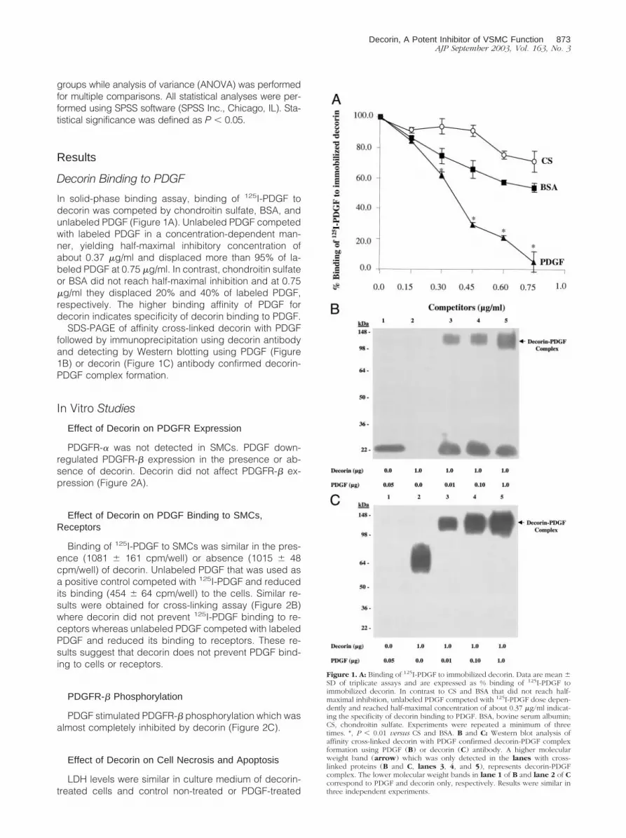
In solid-phase binding assay, binding of 125I-PDGF todecorin was competed by chondroitin sulfate, BSA, andunlabeled PDGF (Figure 1A). Unlabeled PDGF competedwith labeled PDGF in a concentration-dependent man-ner, yielding half-maximal inhibitory concentration ofabout 0.37 �g/ml and displaced more than 95% of la-beled PDGF at 0.75 �g/ml. In contrast, chondroitin sulfateor BSA did not reach half-maximal inhibition and at 0.75�g/ml they displaced 20% and 40% of labeled PDGF,respectively. The higher binding affinity of PDGF fordecorin indicates specificity of decorin binding to PDGF.
SDS-PAGE of affinity cross-linked decorin with PDGFfollowed by immunoprecipitation using decorin antibodyand detecting by Western blotting using PDGF (Figure1B) or decorin (Figure 1C) antibody confirmed decorin-PDGF complex formation.
In Vitro Studies
Effect of Decorin on PDGFR Expression
PDGFR-� was not detected in SMCs. PDGF down-regulated PDGFR-� expression in the presence or ab-sence of decorin. Decorin did not affect PDGFR-� ex-pression (Figure 2A).
Effect of Decorin on PDGF Binding to SMCs,Receptors
Binding of 125I-PDGF to SMCs was similar in the pres-ence (1081 � 161 cpm/well) or absence (1015 � 48cpm/well) of decorin. Unlabeled PDGF that was used asa positive control competed with 125I-PDGF and reducedits binding (454 � 64 cpm/well) to the cells. Similar re-sults were obtained for cross-linking assay (Figure 2B)where decorin did not prevent 125I-PDGF binding to re-ceptors whereas unlabeled PDGF competed with labeledPDGF and reduced its binding to receptors. These re-sults suggest that decorin does not prevent PDGF bind-ing to cells or receptors.
PDGFR-� Phosphorylation
PDGF stimulated PDGFR-� phosphorylation which wasalmost completely inhibited by decorin (Figure 2C).
Effect of Decorin on Cell Necrosis and Apoptosis
LDH levels were similar in culture medium of decorin-treated cells and control non-treated or PDGF-treated
Figure 1. A: Binding of 125I-PDGF to immobilized decorin. Data are mean �SD of triplicate assays and are expressed as % binding of 125I-PDGF toimmobilized decorin. In contrast to CS and BSA that did not reach half-maximal inhibition, unlabeled PDGF competed with 125I-PDGF dose depen-dently and reached half-maximal concentration of about 0.37 �g/ml indicat-ing the specificity of decorin binding to PDGF. BSA, bovine serum albumin;CS, chondroitin sulfate. Experiments were repeated a minimum of threetimes. *, P � 0.01 versus CS and BSA. B and C: Western blot analysis ofaffinity cross-linked decorin with PDGF confirmed decorin-PDGF complexformation using PDGF (B) or decorin (C) antibody. A higher molecularweight band (arrow) which was only detected in the lanes with cross-linked proteins (B and C, lanes 3, 4, and 5), represents decorin-PDGFcomplex. The lower molecular weight bands in lane 1 of B and lane 2 of Ccorrespond to PDGF and decorin only, respectively. Results were similar inthree independent experiments.
Decorin, A Potent Inhibitor of VSMC Function 873AJP September 2003, Vol. 163, No. 3

cells (data not shown). There was also no evidence of activecaspase-3 in decorin-treated, control non-treated or PDGF-treated cells in the cell apoptosis assay (Figure 3).
Effect of Decorin on DNA Synthesis and CellProliferation
Cells treated with PDGF showed a sixfold increase inDNA synthesis compared to control non-treated cells.Decorin inhibited PDGF-stimulated DNA synthesis in adose-dependent manner (Figure 4A). To determinewhether decorin had an effect on cell proliferation, thegrowth of SMCs were determined at 24 and 48 hours.Decorin significantly inhibited PDGF-stimulated cell pro-liferation at both time points (Figure 4B).
Effect of Decorin on Cell Migration
An approximately twofold increase was observed in cellmigration for the PDGF-treated cells compared to control
non-treated cells. Decorin caused a dose-dependent inhi-bition of PDGF-stimulated cell migration (Figure 4C).
Effect of Decorin on Collagen Synthesis
A fourfold increase was observed in the rate of colla-gen synthesis for the PDGF-treated cells compared tocontrol non-treated cells. Decorin caused a dose-depen-dent inhibition of PDGF-stimulated collagen synthesis(Figure 4D).
In Vivo Studies
Detection of Decorin Transgene
At 24 hours following injury and gene transfection, a870-bp decorin cDNA fragment was identified in thedecorin-transfected arteries but not detected in injured�-gal-transfected, non-transfected injured or uninjuredarteries (Figure 5A).
Decorin Immunostaining
At 1 week after balloon injury, decorin was detected inthe media of decorin-transfected arteries (Figure 5B), butnot in �-gal-transfected, non-transfected injured or unin-jured arteries.
Intimal Hyperplasia
The intimal CSA was significantly reduced by �50% indecorin-transfected arteries (Figures 6 and 7) comparedto �-gal-transfected and non-transfected injured controlarteries (P � 0.04). Similarly, the maximum intimal thick-ness was also reduced by �50% in decorin-transfected(0.09 � 0.03 mm) compared to �-gal-transfected (0.19 �0.15 mm) and non-transfected injured controls (0.17 �0.10 mm) (P � 0.04).
Figure 2. A: Western blot analysis of PDGFR-�. PDGF inhibited PDGFR-�expression. Decorin did not affect PDGFR-� expression. B: Autoradiographof a gel loaded with extracts containing 50 �g of protein from cells incubatedwith 125I-PDGF in the presence or absence of unlabeled PDGF or decorin.Decorin did not affect binding of 125I-PDGF to PDGF receptor. UnlabeledPDGF competed with 125I-PDGF and prevented its binding to receptorsindicating specificity of binding. C: Western blot analysis of phospho-PDGFR-�. Decorin inhibited PDGF-stimulated PDGFR-� phosphorylation(top). Immunoblotting with anti-PDGFR-� shows (bottom) that similaramounts of receptor were present in all samples. Results were similar in threeindependent experiments.
Figure 3. Western blot analysis of caspase-3 after treating cells with decorin(30 �g/ml) or PDGF (10 ng/ml) for 24 hours. Each lane contains 50 �g ofcellular proteins. Western blots were probed with anti-caspase-3 (top) oranti �-actin (bottom) to verify equal loading of protein in each lane. Positivecontrol was from human Jurkat cells (tonsil). There was no evidence of cellapoptosis in decorin-treated cells as well as control non-treated or PDGF-treated cells. Results were similar in three independent experiments.
874 Nili et alAJP September 2003, Vol. 163, No. 3

Cell Proliferation
The cell proliferation rates at 1 week post-angioplastywere not significantly different between the three groups(decorin-transfected arteries: 10 � 8; �-gal-transfected14 � 7; non-transfected injured: 9 � 5 Mib-1-positivecells/arterial cross-section). In addition, the cellular pro-liferation rates were also similar for the three (intima,
media, and adventitia) layers of the vessel wall betweengroups.
Inflammation and Cellular Infiltrate
Lack of active inflammation at both time points (1 weekand 10 weeks) was evident by an absence of neutrophils
Figure 4. PDGF-stimulated (A) DNA synthesis, (B) cell proliferation, (C) cell migration, and (D) collagen synthesis were significantly inhibited by decorin in adose-dependent manner. PDGF-stimulated cell proliferation was significantly inhibited by decorin at 24 hours and 48 hours. Experiments were performed intriplicate and repeated a minimum of three times. †, P � 0.01 versus control non-treated cells; *, P � 0.02 versus PDGF-treated cells.
Figure 5. A: PCR of DNA from rabbit carotid arteries at 24 hours after transfection. Lanes 1 and 2: decorin-transfected arteries; lanes 3 and 4: �-gal-transfectedarteries; lanes 5 and 6: injured non-transfected arteries; lanes 7 and 8: uninjured arteries and far left lane (�X174): DNA standards. In the decorin-transfectedarteries, a 870-bp fragment was identified by using primers for the CMV promotor sequence and the rabbit decorin gene. B: Decorin (brown staining)demonstrated in medial layer by immunohistochemistry at 1 week after angioplasty and transfection. Arrowhead, external elastic lamina; arrow, internal elasticlamina; l, lumen; m, media; a, adventitia.
Decorin, A Potent Inhibitor of VSMC Function 875AJP September 2003, Vol. 163, No. 3

or macrophages in the majority of treated arteries. In onlyone �-gal-transfected artery at 10 weeks, six macro-phages were present. Four neutrophils were detected inone decorin-transfected artery at 1 week.
Collagen Content and Density
At 10 weeks, there was a significant decrease in col-lagen content in decorin-transfected arteries comparedto �-gal-transfected and injured, non-transfected arter-ies, (228 � 98 versus 343 � 109 and 411 � 331 �ghydroxyproline/arterial segment, respectively, P � 0.03).Based on the relationship between collagen content andintimal area, the decrease in collagen content in thedecorin-treated arteries appeared to be proportional to
the reduction in intimal area. This was supported by thecollagen density data. There was no statistically signifi-cant difference in collagen density between the threegroups in the adventitia (decorin: 101 � 30 gray values/�m2; injured, non-transfected: 107 � 23 gray values/�m2; �-gal: 97 � 21 gray values/�m2) or the intima(decorin: 5.9 � 1.9 gray values/�m2; injured, non-trans-fected: 5.7 � 4.2 gray values/�m2; �-gal 7.7 � 5.4 grayvalues/�m2).
Fibronectin Immunostaining
At 10 weeks, there was intense staining in the adven-titia in all three groups with no or mild intimal staining in allthree groups. There were no statistical differences in theintimal staining (decorin: 0.33�.55; injured, non-trans-fected 0.78�.83; �-gal: 0.92�.80).
Discussion
PDGF is released after arterial injury and plays an impor-tant role in regulating cellular and extracellular responsesin vascular repair.27 It has also been implicated in thedevelopment of intimal hyperplasia following angio-plasty3,27 and post-transplant arteriopathy.28 Previousstudies have shown that targeting PDGF with anti-PDGFantibody29 or its receptor expression with antisense oli-gonucleotides30 reduces intimal hyperplasia. We now
Figure 6. Intimal cross-sectional area at 10 weeks after angioplasty. *, P �0.04 versus �-gal and control.
Figure 7. Movat-pentachrome-stained sections of injured carotid arteries at 10 weeks after angioplasty. A: Control (injured non-transfected). B: �-gal-transfected.C: Decorin-transfected. Marked inhibition of intimal hyperplasia seen in decorin-transfected arteries. Arrowhead, external elastic lamina; arrow, internal elasticlamina; l, lumen; i, intima; m, media; a, adventitia.
876 Nili et alAJP September 2003, Vol. 163, No. 3

report for the first time that decorin protein has a potentand dose-dependent inhibitory effect on PDGF stimula-tion of three major SMC functions including cell prolifer-ation, migration, and collagen synthesis. Only onestudy16 has examined the effects of decorin on PDGF-stimulated SMC proliferation and did not show a pro-longed inhibitory effect. This discrepancy may be due todifferent experimental conditions and models used. Wehave also shown in an animal arterial balloon-injury modelcharacterized by PDGF up-regulation that decorin signif-icantly inhibited intimal hyperplasia and collagen accu-mulation. It seems reasonable to conclude that the inhib-itory effects of decorin demonstrated in the in vivo studyare at least in part due to decorin neutralization of PDGFactivity.
The inhibitory effect of decorin on PDGF-stimulatedSMC functions was not due to cell apoptosis or necrosis.The novel mechanisms of decorin interactions with PDGFin our study were direct decorin binding to PDGF andacting as a potent inhibitor of PDGF receptor phosphor-ylation in response to PDGF. There was no effect onPDGF receptor-� expression. The specificity of decorinbinding to PDGF was demonstrated by a solid-phasebinding assay in which unlabeled PDGF competed with125I-PDGF for binding to decorin. This was also confirmedby affinity cross-linking using both PDGF and decorinantibodies which indicated PDGF binding to decorin in aconcentration-dependent manner.
Despite important regulatory effects on ECM deposi-tion, decorin expression is not up-regulated in humancoronary restenosis specimens31 or after balloon injury inanimal models,32 a finding confirmed in the present studyin the �-gal-transfected and the injured, non-transfectedarteries. In decorin-transfected arteries, there wasdecorin expression evident at 1 week after transfectionand a marked reduction in intimal cross-sectional area at10 weeks. Since intimal area was reduced without achange in cell proliferation, reduced collagen content(50% compared to �-gal-transfected and 80% comparedto injured non-transfected arteries) appears to be amechanism responsible for the inhibition of intimal thick-ening by decorin. This conclusion is also supported by invitro results that showed complete inhibition of collagensynthesis in SMCs. An anti-fibrotic effect of decorin hasbeen previously demonstrated in experimental lung33
and glomerulonephritis models.34 This is the first report ofan anti-fibrotic effect of decorin overexpression in a vas-cular-injury model. In a rat carotid balloon-injury model,Fischer et al32 showed that cell-mediated decorin trans-fection decreased neointimal volume and increased inti-mal collagen density, based only on immunostaining. Incontrast, our studies using a quantitative measurement ofcollagen content and collagen density showed de-creased collagen content with no significant differencesin collagen density in the intima or the adventitia. More-over, we did not find any significant differences in fi-bronectin immunostaining in the intima between the threegroups. This suggests that the intimal extracellular matrixis not qualitatively different between the decorin-trans-fected and the two control groups.
The mechanism by which decorin regulates ECM andexerts its anti-fibrotic effect is not clearly known. Previousstudies have shown that decorin binds to TGF-�17 andinhibits its activity in vitro16 and in vivo.33,35 Decorin-PDGFbinding and inhibition of PDGFR phosphorylation bydecorin reported in this study indicate an additionalmechanism of ECM regulation by decorin. Decorin is alsoknown to bind to collagen36–38 and inhibit collagen fibril-logenesis.39 These diverse effects of decorin make it anattractive therapeutic agent for several pathological arte-rial conditions characterized by intimal hyperplasia.
In conclusion, our study shows a novel interaction ofdecorin with PDGF, based on decorin-PDGF complexformation and inhibition of PDGF-stimulated PDGFRphosphorylation and SMC activities. Decorin overexpres-sion significantly reduced both intimal hyperplasia andcollagen content after balloon injury, demonstrating apotent beneficial anti-fibrotic effect for decorin.
References
1. Schwartz R, Holmes D, Topol E: The restenosis paradigm revisited:an alternate proposal for cellular mechanisms. J Am Coll Cardiol1992, 20:1284–1293
2. Strauss BH, Chisholm RJ, Keeley FW, Gotlieb AI, Logan RA, Arm-strong PW: Extracellular matrix remodeling after balloon-angioplastyinjury in a rabbit model of restenosis. Circ Res 1994, 75:650–658
3. Jawien A, Bowen-Pope DF, Lindner V, Schwartz SM, Clowes AW:Platelet-derived growth factor promotes smooth muscle migrationand intimal thickening in a rat model of balloon angioplasty. J ClinInvest 1992, 89:507–511
4. Jiang B, Yamamura S, Nelson PR, Mureebe L, Kent KC: Differentialeffects of platelet-derived growth factor isotypes on human smoothmuscle cell proliferation and migration are mediated by distinct sig-naling pathways. Surgery 1996, 120:427–431, 432
5. Amento EP, Ehsani N, Palmer H, Libby P: Cytokines and growthfactors positively and negatively regulate interstitial collagen geneexpression in human vascular smooth muscle cells. ArteriosclerThromb 1991, 11:1223–1230
6. Miyauchi K, Aikawa M, Tani T, Nakahara K, Kawai S, Nagai R, OkadaR, Yamaguchi H: Effect of probucol on smooth muscle cell prolifera-tion and dedifferentiation after vascular injury in rabbits: possible roleof PDGF. Cardiovasc Drugs Ther 1998, 12:251–260
7. Majesky M, Reidy M, Bowen-Pope D, Hart C, Wilcox J, Schwartz S:PDGF ligand and receptor gene expression during repair of arterialinjury. J Cell Biol 1990, 111:2149–2158
8. Hocking A, Shinomura T, McQuillan D: Leucine rich repeat glycopro-teins of the extracellular matrix. Matrix Biol 1998, 17:1–19
9. Krusius T, Ruoslathi E: Primary structure of an extracellular matrixproteoglycan core protein deduced from cloned cDNA. Proc NatlAcad Sci USA 1986, 83:7683–7687
10. Thieszen S, Rosenquist T: Expression of collagens and decorin dur-ing aortic arch artery development: implications for matrix patternformation. Matrix Biol 1995, 14:573–582
11. Merle B, Malaval L, Lawler J, Delmas P, Clezardin P: Decorin inhibitscell attachment to thrombospondin-1 by binding to a KKTR-depen-dent cell adhesive site present within the N-terminal domain of throm-bospondin-1. J Cell Biochem 1997, 67:75–83
12. Merle B, Durussel L, Delmas PD, Clezardin P: Decorin inhibits cellmigration through a process requiring its glycosaminoglycan sidechain. J Cell Biochem 1999, 75:538–546
13. Yamaguchi Y, Ruoslahti E: Expression of human proteoglycan inChinese hamster ovary cells inhibits cell proliferation. Nature 1988,336:244–246
14. Nash MA, Loercher AE, Freedman RS: In vitro growth inhibition ofovarian cancer cells by decorin: synergism of action between decorinand carboplatin. Cancer Res 1999, 59:6192–6196
15. De Luca A, Santra M, Baldi A, Giordano A, Iozzo RV: Decorin-induced
Decorin, A Potent Inhibitor of VSMC Function 877AJP September 2003, Vol. 163, No. 3
growth suppression is associated with up-regulation of p21, an inhib-itor of cyclin-dependent kinases. J Biol Chem 1996, 271:18961–18965
16. Fischer J, Kinsella M, Levkau B, Clowes A, Wight T: Retroviral over-expression of decorin differentially affects the response of arterialsmooth muscle cells to growth factors. Arterioscle Thromb Vasc Biol2001, 21:777–784
17. Hildebrand A, Romaris M, Rasmussen L, Heinegard D, Twardzik D,Border W, Ruoslahti E: Interaction of the small interstitial proteogly-cans biglycan, decorin, and fibromodulin with transforming growthfactor �. Biochem J 1994, 302:527–534
18. Kaname S, Ruoslahti E: Betaglycan has multiple binding sites fortransforming growth factor-� 1. Biochem J 1996, 315:815–820
19. Scott P, Dodd C, Pringle G: Mapping the locations of the epitopes offive monoclonal antibodies to the core protein of dermatan sulfateproteoglycan II (decorin). J Biol Chem 1993, 268:11558–11564
20. Baxter RM, Secrist JP, Vaillancourt RR, Kazlauskas A: Full activationof the platelet-derived growth factor �-receptor kinase involves mul-tiple events. J Biol Chem 1998, 273:17050–17055
21. Hou G, Mulholland D, Gronska MA, Bendeck MP: Type VIII collagenstimulates smooth muscle cell migration and matrix metalloproteinasesynthesis after arterial injury. Am J Pathol 2000, 156:467–476
22. Kulik TJ, Alvarado SP: Effect of stretch on growth and collagensynthesis in cultured rat and lamb pulmonary arterial smooth musclecells. J Cell Physiol 1993, 157:615–624
23. Barolet AW, Nili N, Cheema A, Robinson R, Natarajan MK, O’BlenesS, Li J, Eskandarian MR, Sparkes J, Rabinovitch M, Strauss BH:Arterial elastase activity after balloon angioplasty and effects of elafin,an elastase inhibitor. Arterioscler Thromb Vasc Biol 2001, 21:1269–1274
24. Giordano FJ, He H, McDonough P, Meyer M, Sayen MR, DillmannWH: Adenovirus-mediated gene transfer reconstitutes depressedsarcoplasmic reticulum Ca2�-ATPase levels and shortens prolongedcardiac myocyte Ca2� transients. Circulation 1997, 96:400–403
25. Aoyagi M, Yamamoto M, Wakimoto H: Immunohistochemical detec-tion of Ki-67 in replicative smooth muscle cells of rabbit carotidarteries after balloon denudation. Stroke 1995, 26:2328–2331
26. Sierevogel MJ, Velema E, van der Meer FJ, Nijhuis MO, Smeets M, deKleijn DP, Borst C, Pasterkamp G: Matrix metalloproteinase inhibitionreduces adventitial thickening and collagen accumulation followingballoon dilation. Cardiovasc Res 2002, 55:864–869
27. Leppanen O, Janjic N, Carlsson MA, Pietras K, Levin M, Vargeese C,Green LS, Bergqvist D, Ostman A, Heldin CH: Intimal hyperplasia
recurs after removal of PDGF-AB and -BB inhibition in the rat carotidartery-injury model. Arterioscler Thromb Vasc Biol 2000, 20:E89–E95
28. de Groot-Kruseman HA, Baan CC, Mol WM, Niesters HG, Maat AP,Balk AH, Weimar W: Intragraft platelet-derived growth factor-� andtransforming growth factor-�1 during the development of acceleratedgraft vascular disease after clinical heart transplantation. TransplImmunol 1999, 7:201–205
29. Rutherford C, Martin W, Salame M, Carrier M, Anggard E, Ferns G:Substantial inhibition of neo-intimal response to balloon injury in therat carotid artery using a combination of antibodies to platelet-derivedgrowth factor-BB and basic fibroblast growth factor. Atherosclerosis1997, 130:45–51
30. Sirois MG, Simons M, Edelman ER: Antisense oligonucleotide inhibi-tion of PDGFR-� receptor subunit expression directs suppression ofintimal thickening. Circulation 1997, 95:669–676
31. Riessen R, Isner J, Blessing E, Loushin C, Nikol S, Wight T: Regionaldifferences in the distribution of the proteoglycans biglycan anddecorin in the extracellular matrix of atherosclerotic and restenotichuman coronary arteries. Am J Pathol 1994, 144:962–974
32. Fischer J, Kinsella M, Clowes M, Lara S, Clowes A: Local expressionof bovine decorin by cell-mediated gene transfer reduces neointimalformation after balloon injury in rats. Circ Res 2000, 86:676–683
33. Giri S, Hyde D, Braun R, Gaarde W: Antifibrotic effect of decorin in ableomycin hamster model of lung fibrosis. Biochem Pharmacol 1997,54:1205–1216
34. Border W, Noble N, Yamamoto T, Harper J: Natural inhibitor of trans-forming growth factor-� protects against scarring in experimentalkidney disease. Nature 1992, 360:361–364
35. Isaka Y, Brees D, Ikegaya K, Kaneda Y, Imai E, Noble N: Genetherapy by skeletal muscle expression of decorin prevents fibroticdisease in rat kidney. Nat Med 1996, 2:418–423
36. Schonherr E, Hausser H, Beavan L, Kresse H: Decorin-type I collageninteraction: presence of separate core protein-binding domains.J Biol Chem 1995, 270:8877–8883
37. Bidanset DJ, Guidry C, Rosenberg LC, Choi HU, Timpl R, Hook M:Binding of the proteoglycan decorin to collagen type VI. J Biol Chem1992, 267:5250–5256
38. Keene DR, San Antonio JD, Mayne R, McQuillan DJ, Sarris G, SantoroSA, Iozzo RV: Decorin binds near the C terminus of type I collagen.J Biol Chem 2000, 275:21801–21804
39. Neame PJ, Kay CJ, McQuillan DJ, Beales MP, Hassell JR: Indepen-dent modulation of collagen fibrillogenesis by decorin and lumican.Cell Mol Life Sci 2000, 57:859–863
878 Nili et alAJP September 2003, Vol. 163, No. 3


![Stimulated Brillouin scattering slow light in optical fibers [Invited]](https://img.pdfslide.net/doc/110x75/634dc52e024fe175900b1734/stimulated-brillouin-scattering-slow-light-in-optical-fibers-invited.jpg)


