Embed Size (px)
Citation preview

Decreased tumorigenic potential of EphA2-overexpressing breastcancer cells following treatment with adenoviral vectors thatexpress EphrinA1
Loren W Noblitt,1,2 Dinesh S Bangari,1,2 Shruti Shukla,1,2 Deborah W Knapp,2
Sulma Mohammed,2 Michael S Kinch,3 and Suresh K Mittal1,2
1Laboratory of Gene Therapy, Purdue University, West Lafayette, Indiana 47907, USA; 2Purdue UniversityCancer Center, Purdue University, West Lafayette, Indiana 47907, USA; and 3MedImmune, Inc.,Gaithersburg, Maryland 20878, USA.
The EphA2 receptor tyrosine kinase is frequently overexpressed in invasive breast cancer cells. Moreover, these malignant cells
have unstable cell–cell contacts, which preclude EphA2 from interacting with its ligand, EphrinA1, which is anchored to the
membrane of adjacent cells. This defect is important because ligand binding causes EphA2 to transmit signals that negatively
regulate tumor cell growth and survival, whereas the absence of ligand binding favors these same behaviors. In our present study,
human adenoviral type 5 (HAd) vectors were engineered to express secreted-forms of EphrinA1. These vectors were used to infect
MDA-MB-231 human breast cancer cells, or MCF-10A human breast epithelial cells providing matched controls. Infection with
HAd-EphrinA1-Fc (HAd vector expressing extracellular domain of human EphrinA1 attached to Fc portion of human IgG1 heavy
chain) caused increased EphA2 activation and turnover and consequently decreased tumor cell viability in soft agar assays.
Consistent with this observation, infection of MDA-MB-231 cells with HAd-EphrinA1-Fc prevented tumor formation in xenograft
models. Furthermore, therapeutic modeling via intratumoral inoculation revealed that HAd-EphrinA1-Fc significantly inhibited
subsequent tumor growth as compared to matched controls. These results suggest that targeting of EphA2 with adenoviral vectors
may have therapeutic value.
Cancer Gene Therapy (2004) 11, 757–766. doi:10.1038/sj.cgt.7700761
Published online 10 September 2004
Keywords: EphA2; EphrinA1; breast cancer; adenoviral vectors; delivery vehicle
Changes in the expression or function of receptortyrosine kinases have been linked to many types of
cancers, most notably breast cancer.1 Tyrosine kinasestransmit powerful signals that are essential for thegrowth, survival and invasiveness of aggressive cancercells. For instance, the EphA2 receptor tyrosine kinase isoverexpressed in many highly aggressive breast cancers.2
High levels of EphA2 have been documented in manydifferent cancers2–6 and this overexpression has beenattributed to many different transcriptional and post-translational modifications that frequently arise in cancer.For example, EphA2 expression is governed in part byp53 and Ras, both of which are frequently altered incancer.7,8 These changes are important because high levelsof EphA2 are sufficient to promote a tumorigenicphenotype,2 which clearly demonstrate a role for EphA2overexpression in cancer.
Much recent evidence has shown that EphA2 functionsvery differently in normal versus malignant cells.9,10 Forexample, EphA2 may function in normal cells as anegative regulator of cell growth and migration tomediate contact inhibition at high cell density. Theseoutcomes appear to arise from the fact that EphA2 bindsa ligand, known as EphrinA1,11–13 which is anchored tothe membrane of adjacent cells. The biochemical con-sequences of ligand binding include EphA2 autopho-sphorylation,11–13 which mediates active signaling thatdecreases extracellular matrix (ECM) attachment andimportantly, increases EphA2 turnover. Consequently,normal epithelial cells generally have low levels of EphA2protein, which is itself tyrosine phosphorylated. However,tumor cells generally have unstable cell–cell contacts andthis decreases EphA2–EphrinA1 binding.14 Consequently,the EphA2 in malignant cells is not phosphorylated,which alters the subcellular localization and function ofEphA2 in a manner that favors tumor cell growth andsurvival.2 Compounding this, decreased ligand-mediatedturnover causes EphA2 to accumulate at the cell surface.However, restoration of EphA2 stimulation can beachieved using artificial ligands15 and monoclonal
Received March 9, 2004.
Address correspondence and reprint requests to: Dr Suresh K
Mittal, Laboratory of Gene Therapy, 1290 Lynn Hall, PurdueUniversity, West Lafayette, IN 47907, USA.
E-mail: [email protected]
Cancer Gene Therapy (2004) 11, 757–766r 2004 Nature Publishing Group All rights reserved 0929-1903/04 $30.00
www.nature.com/cgt
antibodies16 and these treatments negatively regulatetumor cell growth and survival.Human adenovirus (HAd)-based vectors have been the
focus of considerable interest in the last few years, basedon their potential application as delivery vehicles forhuman gene therapy.17–20 The results of animal studiesand clinical trials in humans for cancer therapy havefurther increased interest in adenoviral-based therapy.21–26
Based on the differential ligand binding that EphA2demonstrates in normal versus malignant cells, wedescribe herein the engineering of HAd type 5 (HAd5)vectors that express secreted forms of EphrinA1 and showthat these vectors can negatively regulate tumor cellgrowth in vitro and in vivo.
Results
Generation of HAd5 vector containing ephrinA1-Fcor ephrinA1-Sc
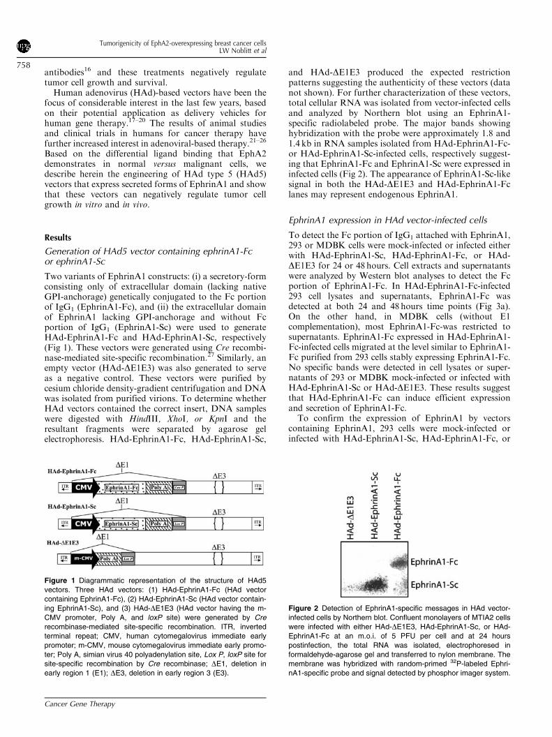
Two variants of EphrinA1 constructs: (i) a secretory-formconsisting only of extracellular domain (lacking nativeGPI-anchorage) genetically conjugated to the Fc portionof IgG1 (EphrinA1-Fc), and (ii) the extracellular domainof EphrinA1 lacking GPI-anchorage and without Fcportion of IgG1 (EphrinA1-Sc) were used to generateHAd-EphrinA1-Fc and HAd-EphrinA1-Sc, respectively(Fig 1). These vectors were generated using Cre recombi-nase-mediated site-specific recombination.27 Similarly, anempty vector (HAd-DE1E3) was also generated to serveas a negative control. These vectors were purified bycesium chloride density-gradient centrifugation and DNAwas isolated from purified virions. To determine whetherHAd vectors contained the correct insert, DNA sampleswere digested with HindIII, XhoI, or KpnI and theresultant fragments were separated by agarose gelelectrophoresis. HAd-EphrinA1-Fc, HAd-EphrinA1-Sc,
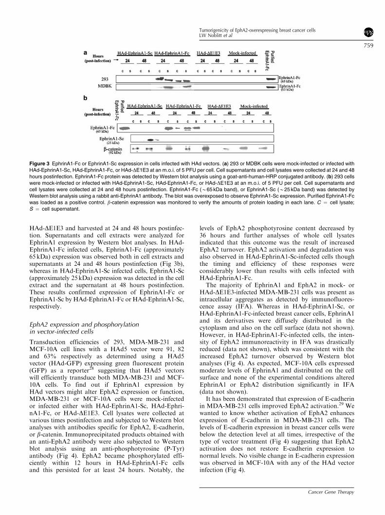
and HAd-DE1E3 produced the expected restrictionpatterns suggesting the authenticity of these vectors (datanot shown). For further characterization of these vectors,total cellular RNA was isolated from vector-infected cellsand analyzed by Northern blot using an EphrinA1-specific radiolabeled probe. The major bands showinghybridization with the probe were approximately 1.8 and1.4 kb in RNA samples isolated from HAd-EphrinA1-Fc-or HAd-EphrinA1-Sc-infected cells, respectively suggest-ing that EphrinA1-Fc and EphrinA1-Sc were expressed ininfected cells (Fig 2). The appearance of EphrinA1-Sc-likesignal in both the HAd-DE1E3 and HAd-EphrinA1-Fclanes may represent endogenous EphrinA1.
EphrinA1 expression in HAd vector-infected cells
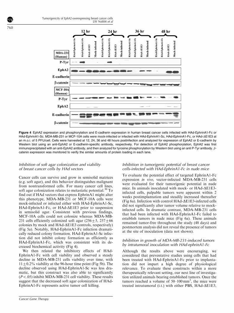
To detect the Fc portion of IgG1 attached with EphrinA1,293 or MDBK cells were mock-infected or infected eitherwith HAd-EphrinA1-Sc, HAd-EphrinA1-Fc, or HAd-DE1E3 for 24 or 48hours. Cell extracts and supernatantswere analyzed by Western blot analyses to detect the Fcportion of EphrinA1-Fc. In HAd-EphrinA1-Fc-infected293 cell lysates and supernatants, EphrinA1-Fc wasdetected at both 24 and 48hours time points (Fig 3a).On the other hand, in MDBK cells (without E1complementation), most EphrinA1-Fc-was restricted tosupernatants. EphrinA1-Fc expressed in HAd-EphrinA1-Fc-infected cells migrated at the level similar to EphrinA1-Fc purified from 293 cells stably expressing EphrinA1-Fc.No specific bands were detected in cell lysates or super-natants of 293 or MDBK mock-infected or infected withHAd-EphrinA1-Sc or HAd-DE1E3. These results suggestthat HAd-EphrinA1-Fc can induce efficient expressionand secretion of EphrinA1-Fc.To confirm the expression of EphrinA1 by vectors
containing EphrinA1, 293 cells were mock-infected orinfected with HAd-EphrinA1-Sc, HAd-EphrinA1-Fc, or
Figure 1 Diagrammatic representation of the structure of HAd5
vectors. Three HAd vectors: (1) HAd-EphrinA1-Fc (HAd vector
containing EphrinA1-Fc), (2) HAd-EphrinA1-Sc (HAd vector contain-
ing EphrinA1-Sc), and (3) HAd-DE1E3 (HAd vector having the m-
CMV promoter, Poly A, and loxP site) were generated by Cre
recombinase-mediated site-specific recombination. ITR, inverted
terminal repeat; CMV, human cytomegalovirus immediate early
promoter; m-CMV, mouse cytomegalovirus immediate early promo-
ter; Poly A, simian virus 40 polyadenylation site, Lox P, loxP site for
site-specific recombination by Cre recombinase; DE1, deletion in
early region 1 (E1); DE3, deletion in early region 3 (E3).
Figure 2 Detection of EphrinA1-specific messages in HAd vector-
infected cells by Northern blot. Confluent monolayers of MTIA2 cells
were infected with either HAd-DE1E3, HAd-EphrinA1-Sc, or HAd-
EphrinA1-Fc at an m.o.i. of 5 PFU per cell and at 24 hours
postinfection, the total RNA was isolated, electrophoresed in
formaldehyde-agarose gel and transferred to nylon membrane. The
membrane was hybridized with random-primed 32P-labeled Ephri-
nA1-specific probe and signal detected by phosphor imager system.
Tumorigenicity of EphA2-overexpressing breast cancer cellsLW Noblitt et al
758
Cancer Gene Therapy
HAd-DE1E3 and harvested at 24 and 48 hours postinfec-tion. Supernatants and cell extracts were analyzed forEphrinA1 expression by Western blot analyses. In HAd-EphrinA1-Fc infected cells, EphrinA1-Fc (approximately65kDa) expression was observed both in cell extracts andsupernatants at 24 and 48 hours postinfection (Fig 3b),whereas in HAd-EphrinA1-Sc infected cells, EphrinA1-Sc(approximately 25kDa) expression was detected in the cellextract and the supernatant at 48 hours postinfection.These results confirmed expression of EphrinA1-Fc orEphrinA1-Sc by HAd-EphrinA1-Fc or HAd-EphrinA1-Sc,respectively.
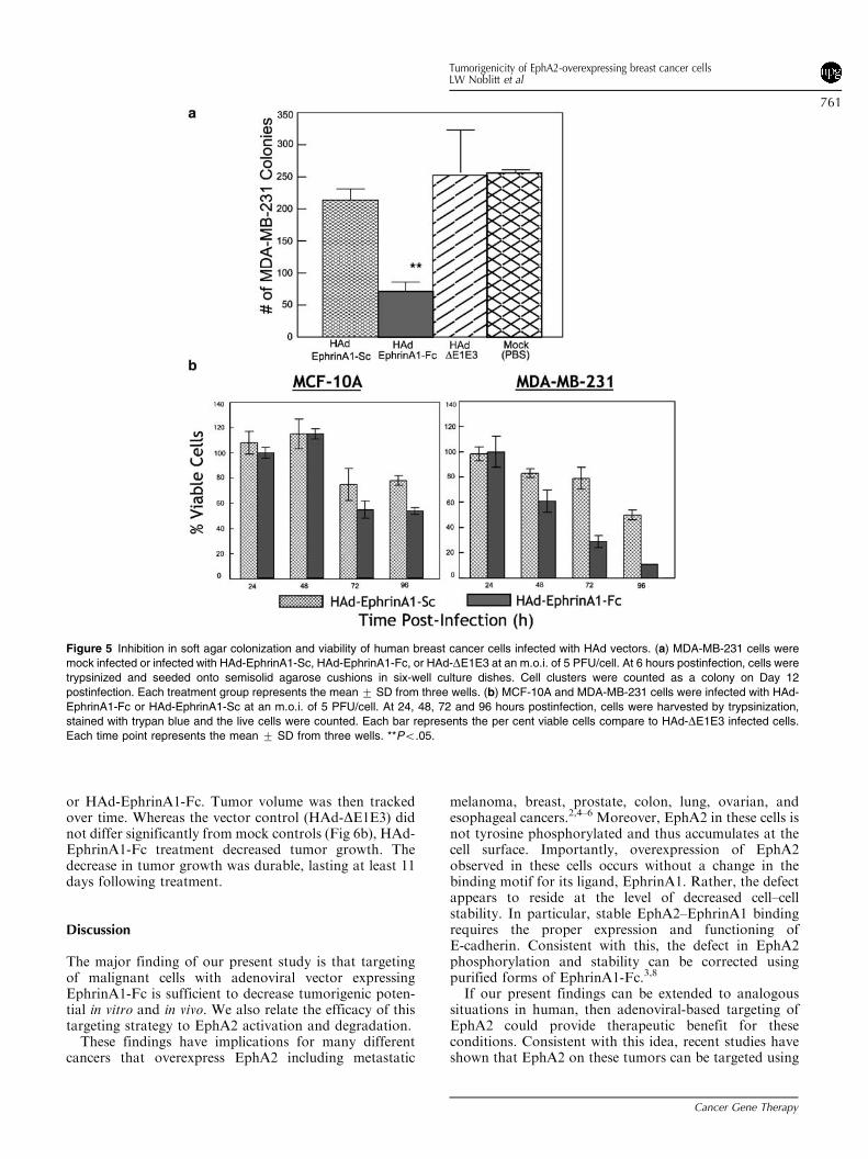
EphA2 expression and phosphorylationin vector-infected cells
Transduction efficiencies of 293, MDA-MB-231 andMCF-10A cell lines with a HAd5 vector were 91, 82and 63% respectively as determined using a HAd5vector (HAd-GFP) expressing green fluorescent protein(GFP) as a reporter28 suggesting that HAd5 vectorswill efficiently transduce both MDA-MB-231 and MCF-10A cells. To find out if EphrinA1 expression byHAd vectors might alter EphA2 expression or function,MDA-MB-231 or MCF-10A cells were mock-infectedor infected either with HAd-EphrinA1-Sc, HAd-Ephri-nA1-Fc, or HAd-DE1E3. Cell lysates were collected atvarious times postinfection and subjected to Western blotanalyses with antibodies specific for EphA2, E-cadherin,or b-catenin. Immunoprecipitated products obtained withan anti-EphA2 antibody were also subjected to Westernblot analysis using an anti-phosphotyrosine (P-Tyr)antibody (Fig 4). EphA2 became phosphorylated effi-ciently within 12 hours in HAd-EphrinA1-Fc cellsand this persisted for at least 24 hours. Notably, the
levels of EphA2 phosphotyrosine content decreased by36 hours and further analyses of whole cell lysatesindicated that this outcome was the result of increasedEphA2 turnover. EphA2 activation and degradation wasalso observed in HAd-EphrinA1-Sc-infected cells thoughthe timing and efficiency of these responses wereconsiderably lower than results with cells infected withHAd-EphrinA1-Fc.The majority of EphrinA1 and EphA2 in mock- or
HAd-DE1E3-infected MDA-MB-231 cells was present asintracellular aggregates as detected by immunofluores-cence assay (IFA). Whereas in HAd-EphrinA1-Sc, orHAd-EphrinA1-Fc-infected breast cancer cells, EphrinA1and its derivatives were diffusely distributed in thecytoplasm and also on the cell surface (data not shown).However, in HAd-EphrinA1-Fc-infected cells, the inten-sity of EphA2 immunoreactivity in IFA was drasticallyreduced (data not shown), which was consistent with theincreased EphA2 turnover observed by Western blotanalyses (Fig 4). As expected, MCF-10A cells expressedmoderate levels of EphrinA1 and distributed on the cellsurface and none of the experimental conditions alteredEphrinA1 or EphA2 distribution significantly in IFA(data not shown).It has been demonstrated that expression of E-cadherin
in MDA-MB-231 cells improved EphA2 activation.29 Wewanted to know whether activation of EphA2 enhancesexpression of E-cadherin in MDA-MB-231 cells. Thelevels of E-cadherin expression in breast cancer cells werebelow the detection level at all times, irrespective of thetype of vector treatment (Fig 4) suggesting that EphA2activation does not restore E-cadherin expression tonormal levels. No visible change in E-cadherin expressionwas observed in MCF-10A with any of the HAd vectorinfection (Fig 4).
Figure 3 EphrinA1-Fc or EphrinA1-Sc expression in cells infected with HAd vectors. (a) 293 or MDBK cells were mock-infected or infected with
HAd-EphrinA1-Sc, HAd-EphrinA1-Fc, or HAd-DE1E3 at an m.o.i. of 5 PFU per cell. Cell supernatants and cell lysates were collected at 24 and 48
hours postinfection. EphrinA1-Fc protein was detected by Western blot analysis using a goat-anti-human-HRP conjugated antibody. (b) 293 cells
were mock-infected or infected with HAd-EphrinA1-Sc, HAd-EphrinA1-Fc, or HAd-DE1E3 at an m.o.i. of 5 PFU per cell. Cell supernatants and
cell lysates were collected at 24 and 48 hours postinfection. EphrinA1-Fc (B65 kDa band), or EphrinA1-Sc (B25 kDa band) was detected by
Western blot analysis using a rabbit anti-EphrinA1 antibody. The blot was overexposed to observe EphrinA1-Sc expression. Purified EphrinA1-Fc
was loaded as a positive control. b-catenin expression was monitored to verify the amounts of protein loading in each lane. C ¼ cell lysate;
S ¼ cell supernatant.
Tumorigenicity of EphA2-overexpressing breast cancer cellsLW Noblitt et al
759
Cancer Gene Therapy
Inhibition of soft agar colonization and viabilityof breast cancer cells by HAd vectors
Cancer cells can survive and grow in semisolid matrices(e.g. soft agar), and this behavior distinguishes malignantfrom nontransformed cells. For many cancer cell lines,soft agar colonization relates to metastatic potential.30 Tofind out if HAd vectors that express EphrinA1 might alterthis phenotype, MDA-MB-231 or MCF-10A cells weremock-infected or infected either with HAd-EphrinA1-Sc,HAd-EphrinA1-Fc, or HAd-DE1E3 prior to suspensionin semisolid agar. Consistent with previous findings,MCF-10A cells could not colonize whereas MDA-MB-231 cells efficiently colonized soft agar (25675, 257766colonies by mock and HAd-DE1E3 controls, respectively)(Fig 5a). Notably, HAd-EphrinA1-Fc infection dramati-cally reduced colony formation. HAd-EphrinA1-Sc infec-tion did not inhibit colony formation as efficiently asHAd-EphrinA1-Fc, which was consistent with its de-creased biochemical activity (Fig 4).We then related the inhibitory effects of HAd-
EphrinA1-Fc with cell viability and observed a steadydecline in MDA-MB-231 cells viability over time, with1170.2% viability at the 96-hour time point (Fig 5b). Thedecline observed using HAd-EphrinA1-Sc was less dra-matic, but this construct was also able to significantly(Po.05) inhibit MDA-MB-231 cell viability. These resultssuggest that the decreased soft agar colonization of HAd-EphrinA1-Fc represents active tumor cell killing.
Inhibition in tumorigenic potential of breast cancercells-infected with HAd-EphrinA1-Fc in nude mice
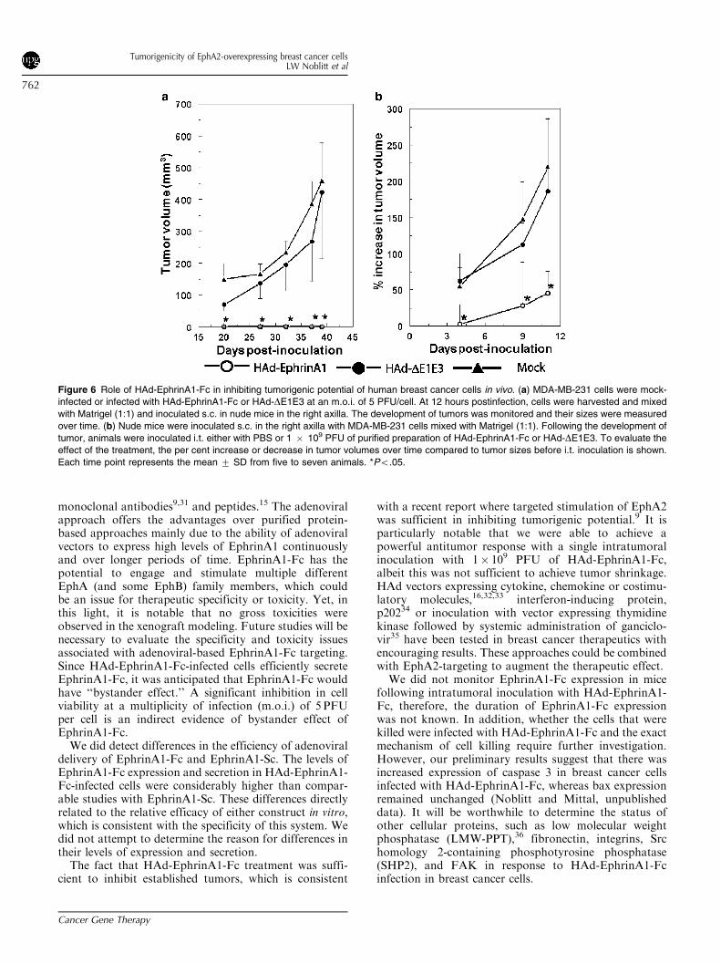
To evaluate the potential effect of targeted EphrinA1-Fcexpression in vivo, vector-infected MDA-MB-231 cellswere evaluated for their tumorigenic potential in nudemice. In animals inoculated with mock- or HAd-DE1E3-infected cells, palpable tumors were apparent within 2weeks postimplantation and steadily increased thereafter(Fig 6a). Infection with control HAd-DE1E3-infected cellsdid not significantly alter tumor volume relative to mock-infected cells. In dramatic contrast, MDA-MB-231 cellsthat had been infected with HAd-EphrinA1-Fc failed toestablish tumors in nude mice (Fig 6a). These animalsremained tumor-free throughout the study (48 days) andpostmortem analysis did not reveal the presence of tumorsat the site of inoculation (data not shown).
Inhibition in growth of MDA-MB-231-induced tumorsby intratumoral inoculation with HAd-ephrinA1-Fc
Although the results above were encouraging, weconsidered that preventative studies using cells that hadbeen treated with HAd-EphrinA1-Fc prior to implanta-tion did not impart a high degree of physiologicalrelevance. To evaluate these constructs within a moretherapeutically relevant setting, our next line of investiga-tion utilized animals bearing established tumors. Once thetumors reached a volume of 50–100mm3, the mice weretreated intratumoral (i.t.) with either PBS, HAd-DE1E3,
Figure 4 EphA2 expression and phosphorylation and E-cadherin expression in human breast cancer cells infected with HAd-EphrinA1-Fc or
HAd-EphrinA1-Sc. MDA-MB-231 or MCF-10A cells were mock-infected or infected with HAd-EphrinA1-Sc, HAd-EphrinA1-Fc, or HAd-DE1E3 at
an m.o.i. of 5 PFU/cell. Cells were harvested at 12, 24, 36 and 48 hours postinfection and analyzed for expression of EphA2 or E-cadherin by
Western blot using an anti-EphA2 or E-cadherin-specific antibody, respectively. For detection of EphA2 phosphorylation, EphA2 was first
immunoprecipitated with an anti-EphA2 antibody, and then analyzed for tyrosine phosphorylation by Western blot using an anti P-Tyr antibody. b-
catenin expression was monitored to verify the similar amounts of protein loading in each lane.
Tumorigenicity of EphA2-overexpressing breast cancer cellsLW Noblitt et al
760
Cancer Gene Therapy
or HAd-EphrinA1-Fc. Tumor volume was then trackedover time. Whereas the vector control (HAd-DE1E3) didnot differ significantly from mock controls (Fig 6b), HAd-EphrinA1-Fc treatment decreased tumor growth. Thedecrease in tumor growth was durable, lasting at least 11days following treatment.
Discussion
The major finding of our present study is that targetingof malignant cells with adenoviral vector expressingEphrinA1-Fc is sufficient to decrease tumorigenic poten-tial in vitro and in vivo. We also relate the efficacy of thistargeting strategy to EphA2 activation and degradation.These findings have implications for many different
cancers that overexpress EphA2 including metastatic
melanoma, breast, prostate, colon, lung, ovarian, andesophageal cancers.2,4–6 Moreover, EphA2 in these cells isnot tyrosine phosphorylated and thus accumulates at thecell surface. Importantly, overexpression of EphA2observed in these cells occurs without a change in thebinding motif for its ligand, EphrinA1. Rather, the defectappears to reside at the level of decreased cell–cellstability. In particular, stable EphA2–EphrinA1 bindingrequires the proper expression and functioning ofE-cadherin. Consistent with this, the defect in EphA2phosphorylation and stability can be corrected usingpurified forms of EphrinA1-Fc.3,8
If our present findings can be extended to analogoussituations in human, then adenoviral-based targeting ofEphA2 could provide therapeutic benefit for theseconditions. Consistent with this idea, recent studies haveshown that EphA2 on these tumors can be targeted using
Figure 5 Inhibition in soft agar colonization and viability of human breast cancer cells infected with HAd vectors. (a) MDA-MB-231 cells were
mock infected or infected with HAd-EphrinA1-Sc, HAd-EphrinA1-Fc, or HAd-DE1E3 at an m.o.i. of 5 PFU/cell. At 6 hours postinfection, cells were
trypsinized and seeded onto semisolid agarose cushions in six-well culture dishes. Cell clusters were counted as a colony on Day 12
postinfection. Each treatment group represents the mean 7 SD from three wells. (b) MCF-10A and MDA-MB-231 cells were infected with HAd-
EphrinA1-Fc or HAd-EphrinA1-Sc at an m.o.i. of 5 PFU/cell. At 24, 48, 72 and 96 hours postinfection, cells were harvested by trypsinization,
stained with trypan blue and the live cells were counted. Each bar represents the per cent viable cells compare to HAd-DE1E3 infected cells.
Each time point represents the mean 7 SD from three wells. **Po.05.
Tumorigenicity of EphA2-overexpressing breast cancer cellsLW Noblitt et al
761
Cancer Gene Therapy
monoclonal antibodies9,31 and peptides.15 The adenoviralapproach offers the advantages over purified protein-based approaches mainly due to the ability of adenoviralvectors to express high levels of EphrinA1 continuouslyand over longer periods of time. EphrinA1-Fc has thepotential to engage and stimulate multiple differentEphA (and some EphB) family members, which couldbe an issue for therapeutic specificity or toxicity. Yet, inthis light, it is notable that no gross toxicities wereobserved in the xenograft modeling. Future studies will benecessary to evaluate the specificity and toxicity issuesassociated with adenoviral-based EphrinA1-Fc targeting.Since HAd-EphrinA1-Fc-infected cells efficiently secreteEphrinA1-Fc, it was anticipated that EphrinA1-Fc wouldhave ‘‘bystander effect.’’ A significant inhibition in cellviability at a multiplicity of infection (m.o.i.) of 5 PFUper cell is an indirect evidence of bystander effect ofEphrinA1-Fc.We did detect differences in the efficiency of adenoviral
delivery of EphrinA1-Fc and EphrinA1-Sc. The levels ofEphrinA1-Fc expression and secretion in HAd-EphrinA1-Fc-infected cells were considerably higher than compar-able studies with EphrinA1-Sc. These differences directlyrelated to the relative efficacy of either construct in vitro,which is consistent with the specificity of this system. Wedid not attempt to determine the reason for differences intheir levels of expression and secretion.The fact that HAd-EphrinA1-Fc treatment was suffi-
cient to inhibit established tumors, which is consistent
with a recent report where targeted stimulation of EphA2was sufficient in inhibiting tumorigenic potential.9 It isparticularly notable that we were able to achieve apowerful antitumor response with a single intratumoralinoculation with 1� 109 PFU of HAd-EphrinA1-Fc,albeit this was not sufficient to achieve tumor shrinkage.HAd vectors expressing cytokine, chemokine or costimu-latory molecules,16,32,33 interferon-inducing protein,p20234 or inoculation with vector expressing thymidinekinase followed by systemic administration of ganciclo-vir35 have been tested in breast cancer therapeutics withencouraging results. These approaches could be combinedwith EphA2-targeting to augment the therapeutic effect.We did not monitor EphrinA1-Fc expression in mice
following intratumoral inoculation with HAd-EphrinA1-Fc, therefore, the duration of EphrinA1-Fc expressionwas not known. In addition, whether the cells that werekilled were infected with HAd-EphrinA1-Fc and the exactmechanism of cell killing require further investigation.However, our preliminary results suggest that there wasincreased expression of caspase 3 in breast cancer cellsinfected with HAd-EphrinA1-Fc, whereas bax expressionremained unchanged (Noblitt and Mittal, unpublisheddata). It will be worthwhile to determine the status ofother cellular proteins, such as low molecular weightphosphatase (LMW-PPT),36 fibronectin, integrins, Srchomology 2-containing phosphotyrosine phosphatase(SHP2), and FAK in response to HAd-EphrinA1-Fcinfection in breast cancer cells.
Figure 6 Role of HAd-EphrinA1-Fc in inhibiting tumorigenic potential of human breast cancer cells in vivo. (a) MDA-MB-231 cells were mock-
infected or infected with HAd-EphrinA1-Fc or HAd-DE1E3 at an m.o.i. of 5 PFU/cell. At 12 hours postinfection, cells were harvested and mixed
with Matrigel (1:1) and inoculated s.c. in nude mice in the right axilla. The development of tumors was monitored and their sizes were measured
over time. (b) Nude mice were inoculated s.c. in the right axilla with MDA-MB-231 cells mixed with Matrigel (1:1). Following the development of
tumor, animals were inoculated i.t. either with PBS or 1 � 109 PFU of purified preparation of HAd-EphrinA1-Fc or HAd-DE1E3. To evaluate the
effect of the treatment, the per cent increase or decrease in tumor volumes over time compared to tumor sizes before i.t. inoculation is shown.
Each time point represents the mean 7 SD from five to seven animals. *Po.05.
Tumorigenicity of EphA2-overexpressing breast cancer cellsLW Noblitt et al
762
Cancer Gene Therapy
Materials and methods
Cell culture and viruses
293 cells, HAd5 E1-transformed human embryonic kidneycells that support E1-deleted HAd replication,37 were usedprimarily to grow recombinant E1-deleted HAd vectors.293Cre cells (a gift from Merck, Inc., Whitehouse Station,NJ) not only support replication of E1-deleted HAd butalso produce Cre-recombinase,38 which catalyzes site-specific homologous recombination in the presence ofloxP sites.27 The MT1A2 cell line was derived frompolyoma virus middle T antigen (PyMidT)-inducedmammary adenocarcinoma in transgenic mice.32 This cellline was kindly provided by Dr William Muller, (Depart-ment of Biology, McMaster University, Hamilton, Ontar-io, Canada). 293, 293Cre, MT1A2, MDBK (Madin DarbyBovine Kidney cell line), and MDA-MB-231 (aggressivehuman breast cancer cell line) were grown as monolayercultures in Eagle’s minimum essential medium (MEM)(Gibco BRL, Gaithersburg, MD) supplemented with 5–10% reconstituted fetal bovine serum (FetalClone III;Hyclone, Logan, UT), 50 mg/ml gentamicin sulfate (FisherScientific, Pittsburgh, PA) and 50mg/ml amphotericin B(Fisher Scientific). MCF-10A (nontransformed humanbreast epithelial cell line) cells were grown as monolayercultures in MEM supplemented with 5% FetalClone III,50mg/ml gentamicin sulfate, 50mg/ml amphotericin B,10mg/ml insulin (Sigma-Aldrich, Inc., St. Louis, MO),50mg/ml epidermal growth factor (Upstate Biotechnol-ogy, Lake Placid, NY) and 0.50mg/ml hydrocortisone(Sigma-Aldrich). HAd5 vectors were grown in 293 cellsand virus-infected cell pellets were used for preparation ofvirus stocks. For in vivo studies, purified virus prepara-tions generated by cesium chloride-density gradientcentrifugation.39 Virus stocks and purified virus prepara-tions were titrated in 293 cells by plaque assay.
Antibodies
The EphA2-specific monoclonal antibody (D7) was gener-ated by Dr MS Kinch et al40 and anti-EphrinA1 polyclonalantibody was purchased from Santa Cruz Biotechnology,(Santa Cruz, CA). An antibody specific for P-Tyr wasacquired from Upstate Biological, Inc. (Waltham, MA)and b-catenin-specific, and E-cadherin-specific antibodieswere obtained from Transduction Laboratories, (SanDiego, CA). FITC-labeled antibodies: goat-anti-mouse-FITC and goat-anti-rabbit-FITC were procured fromSouthern Biotechnology (Birmingham, AL) and Sigma-Aldrich, respectively. HRP-conjugated antibodies: goatanti-mouse-HRP and goat anti-rabbit-HRP were fromBio Rad (Hercules, CA), whereas, a rabbit anti-mouseantibody was acquired from Chemicon (Temecula, CA).
Construction of E1 shuttle plasmids containingEphrinA1-Fc or EphrinA1-Sc and generation ofHAd5 vectors
Restriction endonucleases, T4 DNA ligase, T4 polymer-ase, and their appropriate buffers were purchased from
New England Biolabs (Beverly, MA). Calf intestinealkaline phosphatase and its appropriate buffer wereobtained from Life Technologies, Inc. (Gaithersburg,MD). Plasmid DNA were purified using Qiagen PlasmidMaxi Kit (Qiagen Inc., Valencia, CA) following themanufacturer’s protocol.A 2.8 kb NruI and XbaI fragment containing the
EphrinA1-Fc gene along with the cytomegalovirus(CMV) immediate early promoter was excised frompCDNA3-EphrinA1-Fc, pCDNA3 containing the Ephri-nA1 attached with Fc portion of IgG1 (kindly provided byDr BC Wang, Case Western Reserve University, Cleve-land, OH). In order to obtain the EphrinA1-Sc (EphrinA1without the transmembrane anchor and Fc portion ofIgG1) construct along with the CMV promoter,pCDNA3-EphrinA1-Fc was digested with NruI andBamH1 and a 1.2 kb fragment was isolated.For generation of HAd5 vectors, the Cre recombinase-
mediated site-specific recombination system was used.27
To generate HAd5 E1 shuttle plasmids containingEphrinA1-Fc (2.8 kb NruI–BamH1fragment), or Ephri-nA1-Sc (1.2 kb NruI–BamH1fragment), these DNAfragments were inserted at an appropriate site in theshuttle vector pDC316 (plasmid containing the left end ofHAd5 (4 kb) with 3.1 kb E1 deletion, a loxp site for site-specific recombination in the presence of Cre recombi-nase, and an intact packaging signal (C))27 to producepDC316-EphrinA1-Fc and pDC316-EphrinA1-Sc, re-spectively. 293Cre cells were cotransfected withpDC316-EphrinA1-Fc or pDC316-EphrinA1-Sc andpBHGloxDE1,3Cre (plasmid containing almost entireHAd5 genome except the C, E1 and E3 deletions, andthe addition of a loxp site for site-specific recombination)using the calcium-phosphate technique41 to generateHAd-EphrinA1-Fc and HAd-EphrinA1-Sc, respectivelyby Cre recombinase-mediated site-specific recombination.The pDC316 and pBHGloxDE1,3Cre were obtained fromMicrobix, Inc. (Toronto, Ontario, Canada). Similarly, avector having E1 and E3 deletions (HAd-DE1E3) was alsoconstructed. HAd5 vectors were plaque purified in 293cells.
Northern blot analysis
Total cellular RNA was isolated 24 hours postinfectionusing TRI reagent (Sigma-Aldrich) according to themanufacturer’s protocol. Approximately 10mg RNAwas electrophoresed through 1.5% agarose-formaldehydegel and transferred to a Zeta-probe membrane (BioRadLaboratories) by capillary transfer.42 The membrane wasincubated at 801C for 1 hour and prehybridized at 421Cfor 2 hours. Subsequently, the membrane was hybridizedovernight at 421C with an EphrinA1-specific probelabeled with [a32P]-dCTP (Amersham Biosciences Corp.,Piscataway, NJ) using random labeling kit (Invitrogen,Carlsbad, CA). The membrane was washed, exposed witha Phosphor Imager screen, scanned by Cyclone storagephosphor system and analyzed with OptiQuant imageanalysis software (Packard Instrument Company,Meriden, CT).
Tumorigenicity of EphA2-overexpressing breast cancer cellsLW Noblitt et al
763
Cancer Gene Therapy
Western blot analysis
Cell lysates were fractionated on 10–12% SDS-polyacry-lamide gels and transferred to nitrocellulose membranes(BioRad) by electrophoresis as described elsewhere.42
To avoid non-specific binding, membranes were blockedwith phosphate-buffered saline (PBS), pH 7.2 containing0.05% Tween 20 and 5% dry skimmed milk. Fordetecting phosphorylation of immunoprecipitatedEphA2, membranes were blocked using 2% cold fishgelatin and 0.1% bovine serum albumin instead of dryskim milk. Membranes were probed with one of thefollowing primary antibodies, specific for: D7 (EphA2),4G10 (P-Tyr), E-cadherin or EphrinA1. After extensivewashing, the membranes were then probed with eithergoat anti-mouse IgG antibody-conjugated with HRP orgoat anti-rabbit IgG-HRP as appropriate. For detectionof EphrinA1-Fc, a goat anti-human IgG antibody-HRPwas used as a primary antibody. The membranes weresoaked in a 1:1 ratio of Super Signal West Pico StablePeroxide Solution: Super Signal West Pico Luminol/Enhancer (Pierce Co., Rockland, IL) for chemilumines-cence and emitted light was captured by autoradiography(Kodak X-OMAT; Kodak, Rochester, NY) or enhancedchemiluminescence (Kodak Image Station; Kodak).Membranes were stripped and reprobed with a b-catenin-specific antibody to verify equal loading, andwhen feasible, membranes were cut into two pieces toavoid stripping.
Immunoprecipitation
Protein A sepharose (PAS) beads were treated with arabbit anti-murine (RAM) IgG antibody to prepareRAM-attached to PAS (RAMPAS) as described.16 Celllysates and D7 antibody were added into microfuge tubescontaining RAMPAS to allow specific binding of D7-EphA2 complexes to RAMPAS. Beads were washedextensively to remove the unbound proteins of the celllysate. Immunoprecipitated protein samples were loadedand fractionated by SDS-PAGE, transferred to anitrocellulose membrane and Western blot analysis wasperformed as described above.
Soft agar assay
MDA-MB-231 or MCF-10A cell monolayers at approxi-mately 80–90% confluency were infected with HAd-EphrinA1-Sc, HAd-EphrinA1-Fc, or HAd-DE1E3 at anm.o.i. of 5 PFU/cell. Following 6 hours of infection, cellswere harvested by trypsinization and resuspended in 2�MEM supplemented with 10% FetalClone III. Warmed0.6% agarose in water was then added in a 1:1 ratio to2� MEM containing cells and the mixture was pouredonto 0.3% agarose cushions in six-well dishes. Theagarose was allowed to solidify at room temperaturefollowed by incubation at 371C in a CO2 incubator. Everythird day, each dish alternatively received 250ml/well of1� MEM supplemented with 10% FetalClone III or1ml/well of 2� MEM and 0.6% agarose (1:1 ratio). At
12 days postinfection, colonies were assessed microscopi-cally and defined as clusters of contiguous cells. The meanand standard deviation were calculated from dataobtained from three independent samples.
Cell viability assay
MDA-MB-231 or MCF-10A cells in 12-well culture plateswere mock-infected or infected with HAd-EphrinA1-Sc,HAd-EphrinA1-Fc, or HAd-DE1E3 at an m.o.i. of5 PFU/cell. At 24, 48, 72 and 96 hours postinfection,cells were harvested by trypsinization, stained withTrypan blue and the live cells were counted microscopi-cally using a calibrated grid. Each bar represents the percent viable cells compare to HAd-DE1E3 infected cells.Each time point represents the mean 7 SD from threewells.
In vivo studies
Athymic BALB/c mice, 10–11 weeks old, were obtainedfrom the National Cancer Institute. All animals wereacclimated for 7 days before inoculation. Mice (Sevenanimals per group) were injected s.c. in the right axillawith 4� 106 MDA-MB-231 cells infected with HAd-EphrinA1-Fc or HAd-DE1E3 at an m.o.i. of 5 PFU percell, harvested 12 hours postinfection and mixed withMatrigel (1:1). Mice inoculated with mock-infectedMDA-MB-231 cells provided matched controls.In another experiment, nude mice (seven animals/
group) were inoculated s.c. in the right axilla with4� 106 MDA-MB-231 cells mixed with Matrigel (1:1)for development of MDA-MB-231-induced tumors.Following the development of 50–100mg3 tumors, i.t.injections of PBS, or 1� 109 PFU of purified preparationof HAd-EphrinA1-Fc or HAd-DE1E3 were performed.Tumor volumes were calculated using the formula, Tumorvolume ¼ {Length(Width)}2/2, for both control andtreated mice to evaluate the extent of tumor size reductionor increase over time. Mice were killed when the tumorsreached a volume of approximate 700mm3 or on Day 48postinoculation.
Statistical analysis
Statistical analyses of in vitro and in vivo data wereperformed using Student’s t-distribution, defining signifi-cance as Po.05.
Acknowledgments
We thank Jane Stewart, Shaji Abraham, Rebecca Pratt,and Keith Kikawa for their technical advice, ElizabethBruckheimer for critical reading of the manuscript andJane Kovach for excellent secretarial assistance. Thiswork was partially supported by grants from PurdueResearch Foundation, National Cancer Institute (U01CA91318) and the US Army Medical Research Acquisi-tion Activity.
Tumorigenicity of EphA2-overexpressing breast cancer cellsLW Noblitt et al
764
Cancer Gene Therapy
References
1. Dickson RB, Lippman ME. Growth factors in breastcancer. Endocr Rev. 1995;16:559–589.
2. Zelinski DP, Zantek ND, Stewart JC, et al. EphA2overexpression causes tumorigenesis of mammary epithelialcells. Cancer Res. 2001;61:2301–2306.
3. Rosenberg IM, Goke M, Kanai M, et al. Epithelial cellkinase-B61: an autocrine loop modulating intestinal epithe-lial migration and barrier function. Am J Physiol. 1997;273:G824–G832.
4. Easty DJ, Guthrie BA, Maung K, et al. Protein B61 as anew growth factor: expression of B61 and up-regulation ofits receptor epithelial cell kinase during melanoma progres-sion. Cancer Res. 1995;55:2528–2532.
5. Andres AC, Zuercher G, Djonov V, et al. Protein tyrosinekinase expression during the estrous cycle and carcino-genesis of the mammary gland. Int J Cancer. 1995;63:288–296.
6. Walker-Daniels J, Coffman K, Azimi M, et al. Over-expression of the EphA2 tyrosine kinase in prostate cancer.Prostate. 1999;41:275–280.
7. Dohn M, Jiang J, Chen X. Receptor tyrosine kinase EphA2is regulated by p53-family proteins and induces apoptosis.Oncogene. 2001;20:6503–6515.
8. Miao H, Wei BR, Peehl DM, et al. Activation of EphAreceptor tyrosine kinase inhibits the Ras/MAPK pathway.Nat Cell Biol. 2001;3:527–530.
9. Coffman KT, Hu M, Carles-Kinch K, et al. DifferentialEphA2 epitope display on normal versus malignant cells.Cancer Res. 2003;63:7907–7912.
10. Kinch MS, Carles-Kinch K. Overexpression and functionalalterations of the EphA2 tyrosine kinase in cancer. Clin ExpMetast. 2003;20:59–68.
11. Bartley TD, Hunt RW, Welcher AA, et al. B61 is a ligandfor the ECK receptor protein-tyrosine kinase. Nature.1994;368:558–560.
12. Zisch AH, Pazzagli C, Freeman AL, et al. Replacing twoconserved tyrosines of the EphB2 receptor with glutamicacid prevents binding of SH2 domains without abrogatingkinase activity and biological responses. Oncogene. 2000;19:177–187.
13. Kullander K, Mather NK, Diella F, et al. Kinase-depen-dent and kinase-independent functions of EphA4 receptorsin major axon tract formation in vivo. Neuron. 2001;29:73–84.
14. Walker-Daniels J, Hess AR, Hendrix MJC, et al. Differ-ential regulation of EphA2 in normal and malignant cells[Review]. Am J Pathol. 2003;162:1037–1042.
15. Koolpe M, Dail M, Pasquale EB. An ephrin mimeticpeptide that selectively targets the EphA2 receptor. J BiolChem. 2002;277:46974–46979.
16. Stewart AK, Lassam NJ, Quirt IC, et al. Adenovector-mediated gene delivery of interleukin-2 in metastatic breastcancer and melanoma: results of a phase 1 clinical trial. GeneTherapy. 1999;6:350–363.
17. Curiel DT. The development of conditionally replicativeadenoviruses for cancer therapy. Clin Cancer Res. 2000;6:3395–3399.
18. Hitt MM, Graham FL. Adenovirus vectors for human genetherapy. Adv Virus Res. 2000;55:479–505.
19. Liu Y, Huang H, Saxena A, et al. Intratumoral coinjectionof two adenoviral vectors expressing functional interleukin-18 and inducible protein-10, respectively, synergizes to
facilitate regression of established tumors. Cancer GeneTher. 2002;9:533–542.
20. St George JA. Gene therapy progress and prospects:adenoviral vectors [Review]. Gene Therapy. 2003;10:1135–1141.
21. Liu Y, Zhang X, Zhang W, et al. Adenovirus-mediatedCD40 ligand gene-engineered dendritic cells elicit enhancedCD8(+) cytotoxic T-cell activation and antitumor immu-nity. Cancer Gene Ther. 2002;9:202–208.
22. Ambar BB, Frei K, Malipiero U, et al. Treatmentof experimental glioma by administration of adenoviralvectors expressing Fas ligand. Hum Gene Ther. 1999;10:1641–1648.
23. Parks R, Evelegh C, Graham F. Use of helper-depen-dent adenoviral vectors of alternative serotypes permitsrepeat vector administration. Gene Therapy. 1999;6:1565–1573.
24. Trudel S, Li Z, Dodgson C, et al. Adenovector engineeredinterleukin-2 expressing autologous plasma cell vaccinationafter high-dose chemotherapy for multiple myeloma — aphase 1 study. Leukemia. 2001;15:846–854.
25. Wen XY, Mandelbaum S, Li ZH, et al. Tricistronic viralvectors co-expressing interleukin-12 (1L-12) and CD80 (B7-1) for the immunotherapy of cancer: preclinical studies inmyeloma. Cancer Gene Ther. 2001;8:361–370.
26. Akbulut H, Zhang L, Tang Y, et al. Cytotoxic effect ofreplication-competent adenoviral vectors carrying L-plastinpromoter regulated E1A and cytosine deaminase genes incancers of the breast, ovary and colon. Cancer Gene Ther.2003;10:388–395.
27. Ng P, Parks RJ, Cummings DT, et al. A high-efficiencyCre/loxP-based system for construction of adenoviralvectors. Hum Gene Ther. 1999;10:2667–2672.
28. Bangari DS, Mittal SK. Porcine adenoviral vectors evadepreexisting humoral immunity to adenoviruses and effi-ciently infect both human and murine cells in culture. VirusRes. 2004, in press.
29. Zantek ND, Azimi M, Fedor-Chaiken M, et al. E-cadherinregulates the function of the EphA2 receptor tyrosinekinase. Cell Growth Differ. 1999;10:629–638.
30. Price JE. Clonogenicity and experimental metastatic poten-tial of spontaneous mouse mammary neoplasms. J NatlCancer Inst. 1986;77:529–535.
31. Carles-Kinch K, Kilpatrick KE, Stewart JC, et al. Anti-body targeting of the EphA2 tyrosine kinase inhi-bits malignant cell behavior. Cancer Res. 2002;62:2840–2847.
32. Addison CL, Braciak T, Ralston R, et al. Intratumoralinjection of an adenovirus expressing interleukin 2 inducesregression and immunity in a murine breast cancer model.Proc Natl Acad Sci USA. 1995;92:8522–8526.
33. Palmer K, Hitt M, Emtage PC, et al. Combined CXCchemokine and interleukin-12 gene transfer enhances anti-tumor immunity. Gene Therapy. 2001;8:282–290.
34. Ding Y, Wen Y, Spohn B, et al. Proapoptotic and antitumoractivities of adenovirus-mediated p202 gene transfer. ClinCancer Res. 2002;8:3290–3297.
35. Vlachaki MT, Chhikara M, Aguilar L, et al. Enhancedtherapeutic effect of multiple injections of HSV-TK + GCVgene therapy in combination with ionizing radiation in amouse mammary tumor model. Int J Radiat Oncol BiolPhys. 2001;51:1008–1017.
36. Kikawa KD, Vidale DR, Van Etten RL, et al. Regulation ofthe EphA2 kinase by the low molecular weight tyrosine
Tumorigenicity of EphA2-overexpressing breast cancer cellsLW Noblitt et al
765
Cancer Gene Therapy

phosphatase induces transformation. J Biol Chem. 2002;277:39274–39279.
37. Graham FL, Smiley J, Russell WC, et al. Characteristics ofa human cell line transformed by DNA from humanadenovirus type 5. J Gen Virol. 1977;36:59–74.
38. Chen L, Anton M, Graham FL. Production and charac-terization of human 293 cell lines expressing the site-specific recombinase Cre. Somat Cell Mol Genet. 1996;22:477–488.
39. Graham FL,, Prevec L. Manipulation of adenovirus vectors.In: Murray EJ, ed. Methods of Molecular Biology: Gene
Transfer and Expression Protocols. Totowa: Humana Press;1991: 109–128.
40. Kinch MS, Kilpatrick KE, Zhong C. Identification oftyrosine phosphorylated adhesion proteins in human cancercells. Hybridoma. 1998;17:227–235.
41. Graham FL, van der Eb AJ. A new technique for the assayof infectivity of human adenovirus 5 DNA. Virology.1973;52:456–467.
42. Sambrook J, Russell DW. Molecular Cloning: A LaboratoryManual. Cold Spring Harbor, New York: Cold HarborPress; 2001.
Tumorigenicity of EphA2-overexpressing breast cancer cellsLW Noblitt et al
766
Cancer Gene Therapy


