Embed Size (px)
Citation preview

Determinants of GBP Recruitment to Toxoplasma gondiiVacuoles and the Parasitic Factors That Control ItSebastian Virreira Winter1¤a, Wendy Niedelman2, Kirk D. Jensen2, Emily E. Rosowski2, Lindsay Julien2,
Eric Spooner1, Kacey Caradonna3, Barbara A. Burleigh3, Jeroen P. J. Saeij2, Hidde L. Ploegh1,2*, Eva-
Maria Frickel1*¤b
1 Whitehead Institute for Biomedical Research, Cambridge, Massachusetts, United States of America, 2 Department of Biology, Massachusetts Institute of Technology,
Cambridge, Massachusetts, United States of America, 3 Department of Immunology and Infectious Diseases, Harvard School of Public Health, Boston, Massachusetts,
United States of America
Abstract
IFN-c is a major cytokine that mediates resistance against the intracellular parasite Toxoplasma gondii. The p65 guanylate-binding proteins (GBPs) are strongly induced by IFN-c. We studied the behavior of murine GBP1 (mGBP1) upon infectionwith T. gondii in vitro and confirmed that IFN-c-dependent re-localization of mGBP1 to the parasitophorous vacuole (PV)correlates with the virulence type of the parasite. We identified three parasitic factors, ROP16, ROP18, and GRA15 thatdetermine strain-specific accumulation of mGBP1 on the PV. These highly polymorphic proteins are held responsible for alarge part of the strain-specific differences in virulence. Therefore, our data suggest that virulence of T. gondii in animals mayrely in part on recognition by GBPs. However, phagosomes or vacuoles containing Trypanosoma cruzi did not recruitmGBP1. Co-immunoprecipitation revealed mGBP2, mGBP4, and mGBP5 as binding partners of mGBP1. Indeed, mGBP2 andmGBP5 co-localize with mGBP1 in T. gondii-infected cells. T. gondii thus elicits a cell-autonomous immune response in micewith GBPs involved. Three parasitic virulence factors and unknown IFN-c-dependent host factors regulate this complexprocess. Depending on the virulence of the strains involved, numerous GBPs are brought to the PV as part of a large,multimeric structure to combat T. gondii.
Citation: Virreira Winter S, Niedelman W, Jensen KD, Rosowski EE, Julien L, et al. (2011) Determinants of GBP Recruitment to Toxoplasma gondii Vacuoles and theParasitic Factors That Control It. PLoS ONE 6(9): e24434. doi:10.1371/journal.pone.0024434
Editor: Silvia N. Moreno, Univ. Georgia, United States of America
Received May 24, 2011; Accepted August 9, 2011; Published September 8, 2011
Copyright: � 2011 Virreira Winter et al. This is an open-access article distributed under the terms of the Creative Commons Attribution License, which permitsunrestricted use, distribution, and reproduction in any medium, provided the original author and source are credited.
Funding: This research was supported by grants from the NIH (to JPJS & HLP), a Massachusetts Life Sciences Center New Investigator Award (to JPJS), a Pre-Doctoral Grant in the Biological Sciences (5-T32-GM007287-33) (to EER) and by stipends from the German National Academic Foundation (to SVW), the CancerResearch Institute (to KDJ), the Cleo and Paul Schimmel Fund (to EER), Bayer HealthCare (to KC), and the Human Frontier Science Program (to EMF). The fundershad no role in study design, data collection and analysis, decision to publish, or preparation of the manuscript.
Competing Interests: Commercial funding has been received from Bayer HealthCare. This does not alter the authors’ adherence to all the PLoS ONE policies onsharing data and materials. The authors have no patents or products in development or marketed products to declare.
* E-mail: [email protected] (HLP); [email protected] (E-MF)
¤a Current address: Department for Cellular Microbiology, Max Planck Institute for Infection Biology, Berlin, Germany¤b Current address: Division of Parasitology, MRC National Institute for Medical Research, London, United Kingdom
Introduction
Type II interferon or IFN-c is essential to control survival and
proliferation of intracellular pathogens and is the most important
regulatory cytokine that combats infection with Toxoplasma gondii.
IFN-c upregulates the expression of hundreds of genes [1,2], some
of the most abundant of which are four families of GTPases: Mx
proteins, very large inducible GTPases (VLIG), p47 immunity-
related GTPases (IRGs), and p65 guanylate-binding proteins
(GBPs). A common feature of the IFN-c-inducible GTPases may
be the mediation of innate resistance to many intracellular
pathogens [3]. For instance, Mx proteins confer resistance to
influenza virus [4]. Additionally, mice that lack certain p47 IRG
genes show increased susceptibility to various bacteria and
protozoa [5,6]. Family members of the IRGs localize to the
parasitophorous vacuole (PV) of T. gondii in IFN-c-induced cells in
vitro and mediate killing of the parasite by disruption of the PV [7–
9]. However, virulent type I strains can evade vacuolar disruption
and some IRGs are localized only to vacuoles that contain the less
virulent type II or type III but not type I parasites [10,11].
Toxoplasma secretes proteins from three secretory organelles,
the micronemes, the rhoptries and the dense granules, into the
host cell upon invasion [12,13]. Some of these secreted proteins
traffic back to the PV membrane (PVM) and it is likely that is
where they exert their function [14]. Indeed, recent studies
reported that the rhoptry protein kinase ROP18 of type I strains
can phosphorylate the nucleotide binding site of the switch loop 1
of certain IRGs, which prevents subunit oligomerization and GTP
hydrolysis, which in turn inhibits accumulation on the PVM and
protects the parasite from being destroyed [15,16].
Whereas the mouse genome has 23 copies of p47 GTPases, the
human genome contains only two, both of which lack any IFN-cresponse elements [17]. In contrast, one other main IFN-c-
inducible GTPase family, the conserved p65 GBPs, is represented
with 13 copies in the mouse genome and seven genes in the human
genome [18]. Detailed structural and mechanistic information is
available for some family members [19–21].
Most GBPs are believed to be isoprenylated, endowing them
with the ability to associate with intracellular membranes [22,23].
Some links to antiviral activity [24,25], as well as an antiprolif-
PLoS ONE | www.plosone.org 1 September 2011 | Volume 6 | Issue 9 | e24434
erative function in endothelial cells and fibroblasts have been
suggested [26–28]. Various GBPs are induced upon infection with
T. gondii in vivo and localize to the PV of type II and III T. gondii
strains in infected cells in vitro [29]. Furthermore, hGBP1
potentiates the anti-chlamydia effects of IFN-c [30], in support
of an antimicrobial role. However, how virulent T. gondii can
evade accumulation of the GBPs at its PV is unclear. How
recruitment of the GBPs is regulated and whether other proteins
are involved is also not known.
We studied the behavior of mGBP1 in vitro in the course of
infection of murine cells with various strains of T. gondii that differ
in their virulence in mice. We conclude that the recognition of a
parasite vacuole by mGBP1 is dependent on interferon-inducible
host factors, the nucleotide-bound state of the GTPase, multi-
merization of the GTPase, and on the strain of the parasite. We
further determined that mGBP1 exerts its function in conjunction
with other members of the GBP family, namely GBP2 and GBP5.
Three T. gondii-derived proteins, rhoptry protein 16 (ROP16),
ROP18, and dense granule antigen 15 (GRA15), are partly
responsible for the strain-specific accumulation of mGBP1 at the
PV. Our study posits the function of mGBP1 to be that of a cell-
autonomous immunity factor that distinguishes between T. gondii
of different virulence. Along with the IRGs, GBPs may be
considered as key factors mediating resistance to T. gondii.
Results
Virulent T. gondii mostly evades recognition of itsparasitophorous vacuole by mGBP1
GBPs differentially localize to PVs formed by varying strains of
T. gondii [29], but the molecular mechanisms that underlie this
specific targeting are unknown. We investigated the decoration of
the PV harboring T. gondii parasites that differ in virulence with
mGBP1. To explore the kinetics of recruitment of mGBP1 as well
as the percentage of mGBP1-positive vacuoles that contain type I,
II or III T. gondii parasites, we produced a rabbit polyclonal
antiserum to recombinant mGBP1. We also generated MEFs that
stably overexpress either N-terminally FLAG-tagged mGBP1 or
eGFP-mGBP1. In uninfected cells, both variants co-localized with
endogenous mGBP1 upon stimulation with IFN-c and were
localized to punctate structures in the cytoplasm (Fig. S1).
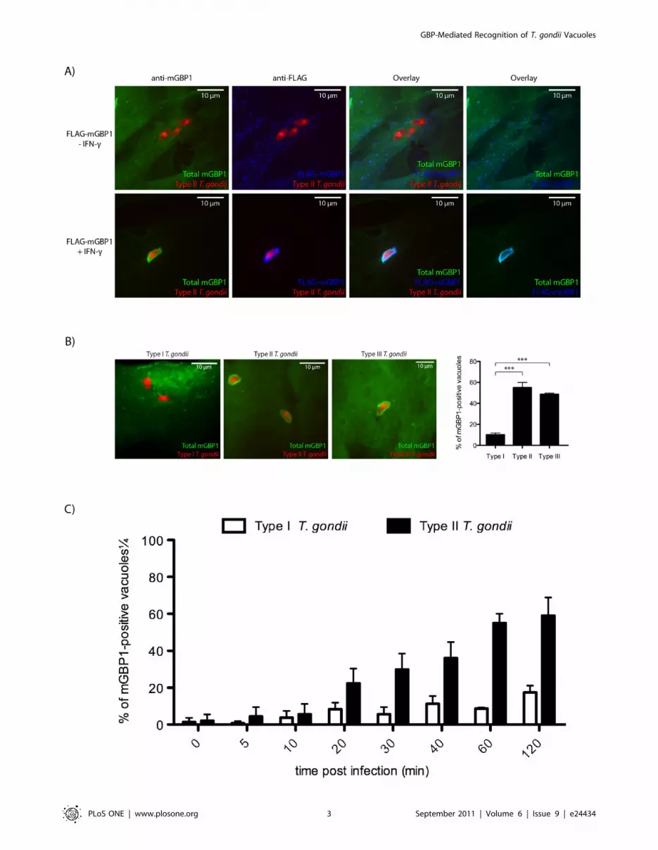
We observed recruitment of endogenous and FLAG-tagged
mGBP1 to the vacuole of type II tachyzoites only when MEFs
were pre-stimulated with IFN-c (Fig. 1a), as reported [29]. This
finding suggests the presence of either an IFN-c-inducible co-
factor or an IFN-c-dependent modification of mGBP1 or its co-
factors essential for vacuolar recognition by mGBP1. IFN-cpretreated MEFs infected subsequently with T. gondii of different
virulence types showed distinct patterns of recruitment 1 hour
post-infection. We extended current studies to include type III
(CEP) T. gondii, and while most type II (Pru) and type III (CEP) T.
gondii showed ring-like accumulation of mGBP1 around the (PV),
virulent type I (RH)-containing vacuoles are predominantly
mGBP1-negative (Fig. 1b). Only 11% of type I vacuoles were
decorated with mGBP1, as opposed to 55% of type II and 49% of
type III vacuoles 1 hour post-infection (Fig. 1b).
We performed live cell imaging by confocal microscopy to track
the extent and kinetics of eGFP-mGBP1 recruitment in MEFs.
Unexpectedly, in some cases, virulent type I T. gondii escaped from
vacuoles that had already recruited mGBP1 (Video S1, S2, S3). We
never observed this phenomenon for vacuoles containing type II or
III T. gondii. After type I escaped from vacuoles with an established,
circular accumulation of mGBP1, two types of outcomes were
observed: either the parasite stripped off mGBP1 and resided within
the same cell without any new recruitment of mGBP1, or the
parasite left the cell after escaping from the GTPase-decorated
vacuole while the host cell died. Is mGBP1 recruitment to type I T.
gondii rare and specific to a sub-fraction of these vacuoles, or do type
I parasites rapidly strip off mGBP1 from the PV to escape immune
detection? We analyzed the frequency of mGBP1-positive PVs for
types I and II over time and recorded a noticeable difference of
mGBP1-positive vacuoles only after approximately 20 min. At
2 hours post-infection, 63% of type II vacuoles are mGBP1-positive,
versus only 20% of type I vacuoles (Fig. 1c). Taken together, we
conclude that type I and II vacuoles are both generally capable to
recruit mGBP1 and that this process takes place within approxi-
mately 30 minutes post-infection. Invasion in this assay is not
synchronized and thus mGBP1 accumulates at a growing number
of intracellular type II parasites. However, type I-containing
vacuoles are able to strip off the GTPase at a timepoint 20 minutes
or later post-infection. This process is fast and complete within a few
minutes. Thus for type I parasites continuous recruitment to newly
invaded parasites seems to be counteracted by active stripping and
evasion strategies with the eventual result of only 20% of PVs being
decorated by mGBP1 at 2 hours post-infection.
How recruitment is initiated, and whether or not the GBP-
mediated response is specific for T. gondii or rather represents a
more general reaction to intracellular microbes remain open
questions. We therefore explored co-localization of mGBP1 with
phagosomes and PVs of Trypanosoma cruzi. No recruitment of
mGBP1 to phagocytosed zymosan or T. cruzi trypomastigotes was
detectable 1 hour post-infection (Fig. S2 and Procedures S1). To
determine if T. cruzi is not targeted by mGBP1 at all or if it can
rapidly evade recruitment, localization of mGBP1 after infection
was monitored over time. At no time did we detect targeting of
mGBP1 to the PV of T. cruzi (data not shown).
Virulence factors, including ROP16, ROP18 and GRA15,interfere with recruitment of mGBP1
What are underlying causes for type I T. gondii’s ability to evade
mGBP1 recruitment and killing? We tested if virulent T. gondii could
degrade mGBP1 upon invasion as a means to prevent recruitment
of GBPs. IFN-c-stimulated RAW264.7 cells were infected with
fluorescent type I or type II T. gondii. Infected cells were separated
from uninfected cells by FACS, and presence and levels of mGBP1
protein were analyzed by immunoblot (IB). No degradation of
mGBP1 was observed by IB two hours post-infection in cells
infected with either type I or type II T. gondii (Fig. 2a).
Next, we examined whether specific T. gondii factors mediate the
difference in recruitment behavior of mGBP1 to the vacuole. We
recently identified two T. gondii proteins that are delivered into the
host cell cytosol upon invasion. These proteins activate host
signaling pathways in a strain-specific manner: the rhoptry protein
ROP16 is a tyrosine kinase, of which the type I and III copy can
directly phosphorylate STAT3/6 [31], and the type II dense
granule protein GRA15 activates the NFkB pathway [13]. Both
play a role in the strain-specific differences in virulence. We
therefore tested if these two T. gondii factors contribute to delivery
and persistence of mGBP1 to the T. gondii PV. Recruitment of
mGBP1 to the vacuole of the type II PruA7 T. gondii strain,
transgenic for the type I version of ROP16 (PruA7+ROP16I),
versus the parental type II PruA7 strain, was ,15%, and 53%
respectively (Fig. 2b). A deletion of GRA15 in the same type II
parental strain (PruA7DGRA15) led to a reduction of mGBP1
recruitment to ,30% (Fig. 2b). Both modified type II strains
exhibit a significant reduction of mGBP1 on the PV compared to
wild-type parental type II. However, type I and type III strains
both have a ROP16 allele that can activate STAT3/6 and an
GBP-Mediated Recognition of T. gondii Vacuoles
PLoS ONE | www.plosone.org 2 September 2011 | Volume 6 | Issue 9 | e24434
GBP-Mediated Recognition of T. gondii Vacuoles
PLoS ONE | www.plosone.org 3 September 2011 | Volume 6 | Issue 9 | e24434

allele of GRA15 that does not activate the NFkB pathway and
therefore another factor different between type I and type III must
exist to explain the difference in accumulation of mGBP1 on the
PVM of these strains. Recently, it was shown that type I ROP18
can directly phosphorylate two threonines in the switch 1 loop of
the IRGs, thereby inactivating them. Type III strains do not
express ROP18 and to determine if type I ROP18 has a role in the
evasion of mGBP1 recruitment we expressed type I ROP18 in a
type III strain (CEP+ROP18I) and compared its recruitment of
mGBP1 to a wild-type type III strain (CEP). Recruitment of
mGBP1 to the vacuole of CEP+ROP18I versus CEP, was ,7%,
and 51%, respectively, a significant difference (Fig. 2b). Thus,
three polymorphic secreted Toxoplasma proteins determine the
strain differences in recruitment of mGBP1 to the vacuole.
GTP-binding and nucleotide-dependent multimerizationare essential for re-localization of mGBP1
To investigate how activation and localization of mGBP1 are
regulated by the host, and to explore which structural require-
ments are needed for a proper targeting to the PV of T. gondii, we
made use of mGBP1 point mutants. GBPs consist of two structural
domains. The globular N-terminal nucleotide-binding domain
contains the evolutionarily conserved G1–G5 motifs of the G-
domain, whereas the helical GBP C-terminal domain is more
diverse. We introduced two mutations into the GTP-binding motif
G1, where either arginine 48 or lysine 51 was changed to alanine.
These two point mutants were based on the structure and
properties of the homologous human GBP1 (hGBP1). Both
mutations cause a 100–1000-fold decrease of the GTP hydrolysis
rate in hGBP1 [20]. The R48A mutant is preferentially GTP-
bound, since it binds the GTP analogue mant-GppNHp with
higher affinity compared to the wild-type protein. The K51A
mutant exhibits an up to ,50-fold reduction in its affinity for
nucleotide and is predominantly nucleotide-free and monomeric
[20]. In addition, we generated a CaaX motif mutant by changing
cysteine to alanine to abolish potential farnesylation of the protein.
We investigated which of these altered mGBP1 versions would still
be recruited to type II-containing vacuoles in IFN-c-pretreated
MEFs.
The R48A mutant was still targeted to the parasite-containing
vacuoles in an IFN-c-dependent manner (Fig. 3). Both intensity of
accumulation and frequency of mGBP1-positive vacuoles were
Figure 2. T. gondii cannot degrade mGBP1 but parasitic virulence factors interfere with recruitment of mGBP1. (A) Degradation ofmGBP1 after invasion of T. gondii is not detectable by IB. RAW264.7 cells were induced with 200 U/ml IFN-c overnight and infected with mCherry-expressing T. gondii (Pru or RH). Infected cells were separated from uninfected cells by FACS 2 h post-infection. For each lysate, 50 mg of protein,which was equivalent to 300,000 cells, were loaded per lane. The mGBP1 protein was detected by rabbit polyclonal anti-mGBP1 antiserum (*). (B)Type II strains with deleted GRA15, type II strains transgenic for the type I version of ROP16, and type III strains transgenic for the type I version ofROP18 show less mGBP1-positive vacuoles compared to the parental strain. MEFs that overexpress eGFP-mGBP1 were induced with 200 U/ml IFN-cand infected with PruA7, a Pru type II T. gondii strain, PruA7 either lacking GRA15 or transgenic for the type I version of ROP16, CEP, a type II strain, orCEP transgenic for the type I version of ROP18. For each strain, at least 100 vacuoles of invaded T. gondii were checked for recruitment of mGBP1.Data represent four independent experiments. Decrease of mGBP1 recruitment is significant with ** p,0.01 and *** p,0.001.doi:10.1371/journal.pone.0024434.g002
Figure 1. mGBP1 preferentially accumulates on type II (Pru) and III (CEP) but not on type I (RH) vacuoles. (A) MEFs that overexpressFLAG-mGBP1 were stimulated with 200 U/ml IFN-c overnight and infected with mCherry-expressing type II T. gondii at an MOI between 5 and 10 for1 h. (B) Representative pictures of IFN-c-induced wild-type MEFs infected with either type I, II or III 1 h post-infection show that mGBP1 ispreferentially recruited to nonvirulent types II and III vacuoles. The right panel shows the frequencies of mGBP1-positive vacuoles. However, whenmGBP1 was recruited to the virulent type I (11% of the vacuoles), the intensity of recruitment was comparable to the recruitment to types II and III.The differences in extent of recruitment of mGBP1 to PVs between type I and types II and III are significant with *** p,0.001. Error bars representstandard deviations of three independent experiments. (C) The frequency of mGBP1-positive vacuoles of type I and type II T. gondii was determinedat various time points. MEFs that overexpress FLAG-mGBP1 were induced with 200 U/ml IFN-c overnight and infected with type I or type II T. gondii atan MOI between 5 and 10 before they were fixed after the indicated time. For each strain, at least 100 vacuoles of invaded T. gondii were checked forrecruitment of mGBP1. Error bars represent standard deviations of three independent experiments. A rabbit polyclonal anti-mGBP1 antiserum and amouse monoclonal anti-FLAG antibody (Sigma-Aldrich) were used for stainings. Pictures were taken with a spinning disk confocal microscope.doi:10.1371/journal.pone.0024434.g001
GBP-Mediated Recognition of T. gondii Vacuoles
PLoS ONE | www.plosone.org 4 September 2011 | Volume 6 | Issue 9 | e24434

indistinguishable from wild-type mGBP1. In contrast, the K51A
mutant was unable to target to the T. gondii-containing vacuoles
(Fig. 3).
The abolition of farnesylation does not impact mGBP1’s ability
to recognize a vacuole that contains type II T. gondii (Fig. 3), at
least not in IFN-c-pretreated MEFs. It has recently been shown
that human GBPs can homo- and heterodimerize in tissue culture
cells with the proteins always localizing to the compartment of the
prenylated partner [32]. It is thus conceivable that recombinant
mGBP1 is targeted to the PV by a prenylated endogenous GBP
family member.
A recent study reports the critical role of the Toll-like receptor
(TLR) -trafficking protein UNC93B1 in host resistance to T. gondii,
possibly by direct exertion of its function at the PVM [33]. We
therefore investigated whether TLR-mediated signaling had an
impact on mGBP1 recruitment to the PVM. Since recruitment of
the GTPase in both MyD88/TRIF knock-out macrophages, as
well as macrophages overexpressing a mutant UNC93B1 deficient
in TLR trafficking was not impaired, we concluded that signaling
via TLRs prior to activation of mGBP1 was not required (Fig. S3).
Why does mGBP1 recruitment to T. gondii vacuoles require pre-
stimulation with IFN-c, also in cells that constitutively overexpress
mGBP1? We tested if mGBP1 is modified in an IFN-c-dependent
manner in any form detectable by SDS-PAGE. When we followed
the fate of metabolically labeled mGBP1 for 120 min, we failed to
observe a size difference of the immunoprecipitated protein (Fig.
S4 and Procedures S1). We therefore consider it less likely that
mGBP1 is extensively modified in an IFN-c-dependent manner,
and favor the possible presence of an IFN-c-induced auxiliary
factor that helps recruit mGBP1 to the PV. The identity of this
factor remains to be determined.
mGBP1 acts together with mGBP2 and mGBP5 at the PVEven though GBPs were identified as IFN-c-regulated genes
more than 25 years ago, their immunological function remains
enigmatic and only two binding partners of the GBPs have been
identified. NIK/HGK binds to hGBP3 and the p110 subunit of
PI3K binds to mGBP2 [21,34]. A step towards defining the
molecular function of this class of GTPases in their function as
anti-parasitic agents is a determination of the proteins that
mGBP1 interacts with, both in the cytoplasm and on the vacuole
of T. gondii. Furthermore, the identification of interactors would
help in understanding how GBPs are activated and targeted to the
PV of T. gondii. We generated RAW264.7 cells that stably
overexpressed FLAG-tagged mGBP1 wild-type protein as well as
the K51A and the R48A mutants. Cells were stimulated with IFN-
c, and whole cell lysates were used for immunoprecipitation (IP)
with an anti-FLAG antibody. Co-immunoprecipitated proteins
were separated by SDS-PAGE gel electrophoresis, and their
identities were determined by MS/MS (Fig. 4a). We performed
two independent immunoprecipitation experiments, using differ-
ent lysis buffers, to confirm potential binding partners. Peptides
from three other GBPs were recovered in both experiments,
namely mGBP2, mGBP4 and mGBP5 (Fig. 4a). For all three
proteins, peptides were only recovered in IPs of FLAG-mGBP1
and FLAG-mGBP1(R48A) but not when FLAG-mGBP1(K51A)
Figure 3. Nucleotide-dependent multimerization is required while farnesylation is dispensable for recruitment of mGBP1 to the PV.(A) Confocal pictures show MEFs that overexpress FLAG-tagged mutants of mGBP1 stimulated with 200 U/ml IFN-c overnight and infected withmCherry-expressing type II T. gondii (Pru) at an MOI between 5 and 10 for 1 h. The R48A and C586S mGBP1 mutants were targeted to the PV like thewild-type counterpart. In contrast, the K51A mGBP1 mutant was unable to accumulate around the PV. Rabbit polyclonal anti-mGBP1 antiserum and amouse monoclonal anti-FLAG antibody were used for stainings.doi:10.1371/journal.pone.0024434.g003
GBP-Mediated Recognition of T. gondii Vacuoles
PLoS ONE | www.plosone.org 5 September 2011 | Volume 6 | Issue 9 | e24434
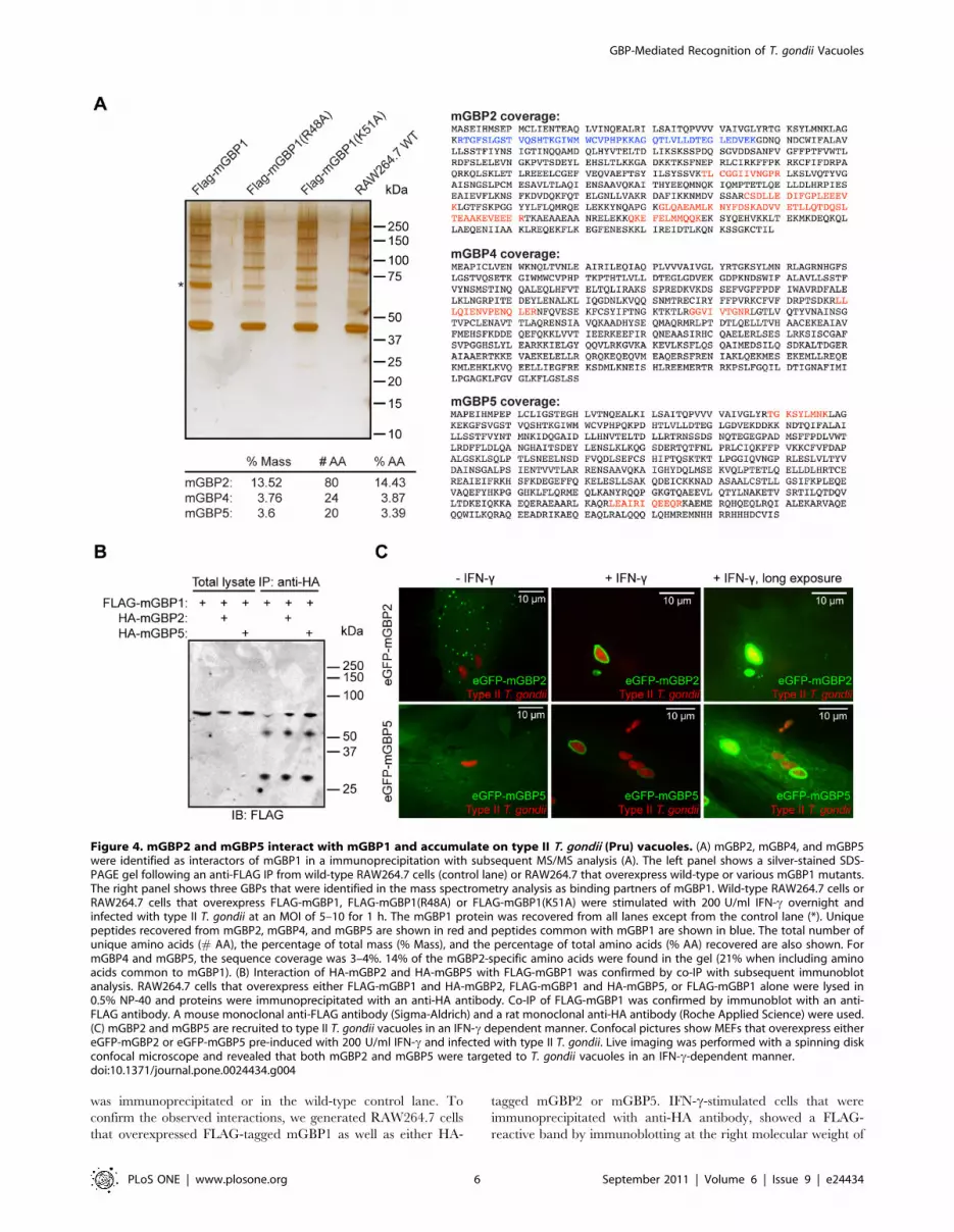
was immunoprecipitated or in the wild-type control lane. To
confirm the observed interactions, we generated RAW264.7 cells
that overexpressed FLAG-tagged mGBP1 as well as either HA-
tagged mGBP2 or mGBP5. IFN-c-stimulated cells that were
immunoprecipitated with anti-HA antibody, showed a FLAG-
reactive band by immunoblotting at the right molecular weight of
Figure 4. mGBP2 and mGBP5 interact with mGBP1 and accumulate on type II T. gondii (Pru) vacuoles. (A) mGBP2, mGBP4, and mGBP5were identified as interactors of mGBP1 in a immunoprecipitation with subsequent MS/MS analysis (A). The left panel shows a silver-stained SDS-PAGE gel following an anti-FLAG IP from wild-type RAW264.7 cells (control lane) or RAW264.7 that overexpress wild-type or various mGBP1 mutants.The right panel shows three GBPs that were identified in the mass spectrometry analysis as binding partners of mGBP1. Wild-type RAW264.7 cells orRAW264.7 cells that overexpress FLAG-mGBP1, FLAG-mGBP1(R48A) or FLAG-mGBP1(K51A) were stimulated with 200 U/ml IFN-c overnight andinfected with type II T. gondii at an MOI of 5–10 for 1 h. The mGBP1 protein was recovered from all lanes except from the control lane (*). Uniquepeptides recovered from mGBP2, mGBP4, and mGBP5 are shown in red and peptides common with mGBP1 are shown in blue. The total number ofunique amino acids (# AA), the percentage of total mass (% Mass), and the percentage of total amino acids (% AA) recovered are also shown. FormGBP4 and mGBP5, the sequence coverage was 3–4%. 14% of the mGBP2-specific amino acids were found in the gel (21% when including aminoacids common to mGBP1). (B) Interaction of HA-mGBP2 and HA-mGBP5 with FLAG-mGBP1 was confirmed by co-IP with subsequent immunoblotanalysis. RAW264.7 cells that overexpress either FLAG-mGBP1 and HA-mGBP2, FLAG-mGBP1 and HA-mGBP5, or FLAG-mGBP1 alone were lysed in0.5% NP-40 and proteins were immunoprecipitated with an anti-HA antibody. Co-IP of FLAG-mGBP1 was confirmed by immunoblot with an anti-FLAG antibody. A mouse monoclonal anti-FLAG antibody (Sigma-Aldrich) and a rat monoclonal anti-HA antibody (Roche Applied Science) were used.(C) mGBP2 and mGBP5 are recruited to type II T. gondii vacuoles in an IFN-c dependent manner. Confocal pictures show MEFs that overexpress eithereGFP-mGBP2 or eGFP-mGBP5 pre-induced with 200 U/ml IFN-c and infected with type II T. gondii. Live imaging was performed with a spinning diskconfocal microscope and revealed that both mGBP2 and mGBP5 were targeted to T. gondii vacuoles in an IFN-c-dependent manner.doi:10.1371/journal.pone.0024434.g004
GBP-Mediated Recognition of T. gondii Vacuoles
PLoS ONE | www.plosone.org 6 September 2011 | Volume 6 | Issue 9 | e24434

67 kDa suggesting that mGBP1 interacts with mGBP2 and
mGBP5 (Fig. 4b).
We next investigated the recruitment of mGBP2 and mGBP5 to
the vacuole of type II T. gondii in MEFs that express either eGFP-
mGBP2 or eGFP-mGBP5. Both eGFP fusion proteins of mGBP2
and mGBP5 were overexpressed in MEFs and localized to
punctate structures that resembled the mGBP1 structures. We
established that mGBP2 and mGBP5 were also brought to the PV
in an IFN-c-dependent manner (Fig. 4c). Localization of mGBP2
and mGBP5 to punctate structures of varying degrees was still
observed when GBPs accumulated at T. gondii vacuole (Fig. 4c,
right panel). The extent of recruitment was similar to that
observed for mGBP1, and mGBP2 and mGBP5 co-localized with
mGBP1 at the PV of type II T. gondii (Fig. S5). Furthermore, the
absence of the C-terminal cysteine that would carry the farnesyl
moiety does not impact the recruitment of mGBP2 and mGBP5 to
the PV (Fig. S6).
Our findings suggest that various GBPs act jointly at the
interface of host pathogen interaction to fight T. gondii. Further
studies will be needed to analyze the exact regulatory mechanisms
underlying the recruitment of these large GTPases.
mGBP1 co-localizes with TGTP after infection with T.gondii in vitro
The IRG TGTP (Irgb6) is recruited to T. gondii vacuoles and is
involved in GTPase-mediated disruption of the PV [7,11]. Except
for their common transcriptional regulation by IFN-c, neither a
functional link nor co-localization of GBPs with IRGs has been
found. When investigated by confocal microscopy, both mGBP1
and TGTP always co-localized to identical PVs after infection with
T. gondii in vitro (Fig. 5). Furthermore, the intensity and hence the
amount of accumulated mGBP1 was comparable to the amount of
TGTP detected around the PV.
This finding suggests that GBPs and IRGs act jointly at the
interface of host pathogen interaction to fight T. gondii. Further
studies will be needed to analyze the exact regulatory mechanisms
underlying the recruitment of these two families of large GTPases.
Discussion
Various IRG p47 GTPases accumulate at the PV to regulate
growth and survival of T.gondii [6–8,35]. However, many questions
remain about the relevant activation pathways and mechanisms of
this means of host defense. The general function of the other
family of IFN-c-inducible GTPases, the p65 GTPases, remains
equally mysterious. What are the exact host and pathogen
activation signals that trigger the accumulation of the GTPases
on the parasitic vacuole? Why are the p47 GTPases such a large
family of proteins in the mouse genome, and yet they are without
an obvious functional counterpart in the human genome? Do the
p65 GTPases, more equally represented in mouse and human
genomes, exert a similar anti-parasitic function as do the p47
GTPases?
The immune responses elicited by the mGBP1 inducers TLR-9
and IFN-c are important for resistance to T. gondii infection
[29,36–38], an observation consistent with a role of mGBP1 in the
host response to T. gondii. mGBP1 localizes to discrete, vesicle-like
structures in the cytoplasm and is recruited to the PV of non-
virulent T. gondii in infected cells [29]. We confirmed these findings
by demonstrating the targeting of mGBP1 to the PV after T. gondii
infection, but only when host cells were pre-stimulated with IFN-c.
Thus, IFN-c induces an as yet uncharacterized factor necessary for
targeting of mGBP1 to the PV. Certain IRGs may be among these
essential IFN-c-dependent factors. The observation that mGBP1
was preferentially recruited to the PV of nonvirulent types II and
III of T. gondii, and not to the virulent type I strain, as do certain
IRGs, supports this hypothesis [10,11]. An IFN-c-dependent
modification of mGBP1 would be a distinct possibility, but this
suggestion lacks experimental support. Accumulation of mGBP1
at the PV was initiated already a few minutes after invasion. This
demonstrates a possible role of the GBPs as part of an early
cellular response to T. gondii infection and implies that they may be
key factors of an initial cell-autonomous innate immune response.
Surprisingly, we found that virulent type I parasites, for the most
part, not only suppressed recruitment of mGBP1, but could also
escape from a vacuole to which mGBP1 had already been
recruited.
Why certain vacuoles are decorated with mGBP1, whereas
others are not, is not fully understood. This applies to the family of
GBPs as it does to the IRGs. However, two recent studies reported
that T. gondii type I kinase ROP18 can directly phosphorylate the
p47 IRGs IIGP1 (Irga6) and TGTP (Irgb6), which then prevents
the GTPases from being recruited to the vacuole [15,16].
Expression of ROP16 of the virulent type I T. gondii in a
nonvirulent type II strain reduces recruitment of mGBP1 to the
vacuole by more than 60%. We hypothesize that ROP16, also a
kinase, may phosphorylate mGBP1 or an as yet unknown host
substrate, which then impedes recruitment of mGBP1 to the PV.
However, expression of ROP16 of the virulent type I and III T.
gondii in a nonvirulent type II strain reduces virulence in vivo [39].
This disparity between our in vitro experiments and previous in vivo
observations might be explained by two separate effects of ROP16
occurring in vivo. Since both type II and III parasites recruit
mGBP1 to their vacuoles, it is likely that STAT3/6 activation by
Figure 5. mGBP1 co-localizes with TGTP after infection with T. gondii. Confocal pictures show that mGBP1 co-localizes with TGTP afterinfection with T. gondii in vitro (B). MEFs that overexpress eGFP-mGBP1 were stimulated with 200 U/ml IFN-c overnight and infected with GFP-expressing Type II T. gondii at a MOI between 5 and 10 for 1 h. A goat polyclonal anti-TGTP antibody was used for staining.doi:10.1371/journal.pone.0024434.g005
GBP-Mediated Recognition of T. gondii Vacuoles
PLoS ONE | www.plosone.org 7 September 2011 | Volume 6 | Issue 9 | e24434

ROP16 of type I and III T.gondii and subsequent inhibition of pro-
inflammatory cytokine secretion or other STAT3/6-mediated
effects decrease virulence and trump interference with the GBP-
mediated cell-autonomous immunity. Deletion of GRA15 in a
nonvirulent type II strain also diminishes efficient recognition of
the vacuole by mGBP1. GRA15 is localized to the cytoplasmic
side of the vacuole and most likely exerts its effect directly on
mGBP1, possibly as a stabilizing factor. GRA15 of type II
parasites activates NFkB, however since recruitment of mGBP1 is
observed in less than one hour and GRA15-mediated NFkB
activation is only seen after four hours, we find it unlikely that a
NFkB-mediated effect plays a role. Mice infected with a type II
strain lacking GRA15 show a significantly increased parasite
burden than mice infected with the parental type II strain [13].
Virulence of T. gondii in mice may therefore be partly caused by
interference with the GBP-mediated host defense against the
parasite. Further studies are needed to analyze the exact molecular
mechanism of the recognition function of these two T. gondii
factors. We also found that ROP18 plays a significant role in the
evasion of GBP1 recruitment as a type III strain expressing type I
ROP18 (CEP+ROP18I) is able to evade recruitment. In fact, the
recruitment of III+ROP18I is similar to the recruitment of type I
strain suggesting that ROP18 is the only factor mediating the
difference in recruitment between type I and type III strains. How
ROP18 mediates its effects on mGBP1 recruitment remains to be
investigated. It is possible that ROP18 directly phosphorylates
mGBP1 and thereby affects its recruitment or the effect could be
indirect. For example if recruitment of the IRGs is necessary for
the GBPs to be recruited to the vacuole then ROP18 inhibition of
IRG recruitment could also affect GBP recruitment.
Three point mutants of mGBP1, R48A, K51A, and C586S,
were used to investigate how activation and localization of mGBP1
are regulated by the host. mGBP1(R48A) is still recruited to the
vacuole, despite having a more than 100-fold reduced rate of GTP
hydrolysis. This mutant is probably GTP-bound, implying that the
nucleotide-bound state and nucleotide-dependent multimerization
are important, while GTP hydrolysis is dispensable for targeting of
mGBP1 to the PV membrane. The inability of the nucleotide-
empty and monomeric K51A mutant to re-localize to the PV
supports this hypothesis. In contrast to the properties reported for
human GBP1 [22], we find that farnesylation is dispensable for a
correct subcellular localization of the GBPs. However, since GBPs
can form dimers and experiments were performed in wild-type
cells, the farnesylation mutants may still dimerize with wild-type,
farnesylated proteins to explain their recruitment to the PV.
Considering that only 15% of mGBP1 is farnesylated [40], other
posttranslational modifications might be present and be important
for proper targeting of the GTPase. However, we did not detect
obvious signs of modification of GBPs in pulse-chase labeling
experiments upon IFN-c stimulation of cells. Also, activation of
mGBP1 did not require prior engagement of TLRs.
We did not observe localization of mGBP1 to the PV of T. cruzi
or to phagosomes containing zymosan A. The GBPs are therefore
most likely a specific response to subset of intracellular pathogens,
including T. gondii, rather than serve as general regulators of
phagocytosis and phagosomal degradation. The T. gondii-specific
re-localization of mGBP1, mGBP2, and mGBP5 suggests that
certain GBPs act in concert to fulfill their functions after induction
by IFN-c. However, variations in the C-terminal parts of the GBPs
suggest that each GBP has a specific role in this complex to
mediate resistance against certain intracellular pathogens.
IP data confirmed interactions between members of the mouse
GBP family, as reported also for the IRGs [41] and human GBPs
[32]. mGBP1 interacts with mGBP2, mGBP5, and possibly with
mGBP4. Both mGBP2 and mGBP5 are brought to the PV of T.
gondii in a manner similar to mGBP1, with farnesylation
dispensable for targeting to the PV. The recruitment of mGBP5
to the T. gondii type II PV is a finding at variance with published
negative results [29], which could be the result of the antibody
employed.
The highly conserved p65 GBPs join the p47 IRGs as elements
of cell-autonomous immunity against certain intracellular patho-
gens. Several GBPs are re-localized to the PV of T. gondii minutes
after invasion to counteract the parasite. Virulent strains of T.
gondii have evolved mechanisms to evade the recognition by the
host cell and interfere with activation of the GBPs through
injection of virulence factors like ROP16 and ROP18. Further
investigations along the lines of our findings are needed to clearly
define the role of all GBP family members, their regulation and
interaction with the p47 IRGs specifically. Identification of the first
parasitic factors that are able to control cellular localization of the
GBPs, as well as knowing that the family members form complexes
with each other provides a strong starting point for these studies.
Materials and Methods
Immunochemical ReagentsThe following immunological reagents were used for immuno-
blotting, immunoprecipitation and immunofluorescence: mouse
monoclonal anti-FLAG antibody (clone M2, Sigma-Aldrich), rat
monoclonal anti-HA antibody (clone 3F10, Roche Applied
Science), goat polyclonal anti-T. gondii antibody (Abcam), and
rabbit polyclonal anti-mGBP1 antiserum (raised against recombi-
nant full-length mGBP1). Mouse IFN-c was purchased from
PeproTech Inc.
CloningFull-length cDNA clones from Open Biosystems encoding
mGBP1 (BC108990), mGBP2 (BC011336), and mGBP5
(BC058555) were cloned into pMSCV vectors for retroviral
expression. N-terminal FLAG- and HA-epitope tags were
introduced by primers. Site-directed mutagenesis was performed
using the QuikChange II Site-Directed Mutagenesis Kit (Agilent
Technologies Inc.) to introduce the R48A and K51A mutations
into mGBP1. The C586S and C587S mutations were introduced
to the coding sequences by primers.
Primer sequencesSee Table S1.
Cell culture and parasite strainsHuman foreskin fibroblasts (HFFs), RAW264.7 cells, LLcMK2
cells, and mouse immortalized macrophages were cultivated in
Dulbecco’s modified Eagle medium (DMEM) containing 10%
fetal calf serum (FCS), 2 mM L-glutamine, and penicillin-
streptomycin. 129/SvEv6C57BL/6 mouse embryonic fibroblasts
(MEFs) were cultivated in DMEM supplemented with 15% FCS,
2 mM L-glutamine, 0.1 mM non-essential amino acids, 115 mM
b-mercaptoethanol and penicillin-streptomycin. Immortalized
macrophages from wild-type and MyD88/TRIF double-knockout
mice were a kind gift from Douglas Golenbock (University of
Massachusetts Medical School, Worcester, MA). Immortalization
of macrophages has been described earlier [42]. 3d mice that
overexpress a mutant Unc93B1 were a kind gift of Dr. Bruce
Beutler (The Scripps Research Institute, La Jolla, CA) [43]. T.
gondii strains CEP, Pru, PruA7, and RH were propagated in HFF
monolayers as described previously [44]. All four strains were
genetically modified to drive cytosolic expression of a fluorescent
GBP-Mediated Recognition of T. gondii Vacuoles
PLoS ONE | www.plosone.org 8 September 2011 | Volume 6 | Issue 9 | e24434

protein. The Pru Dhxgprt fLUCGFP strain (PruA7) [45] was used
to construct PruA7 GRA15 gene deletion and ROP16 transgenic
parasite strains. The generation of PruA7DGRA15 has been
described recently [13]. To generate PruA7+ROP16I parasites
and CEP+ROP18I parasites, a hemagglutinin-tagged ROP16I or
ROP18I expression construct containing 2 kb of upstream
genomic region flanking the start codon of the type I ROP16/
ROP18 gene was cloned and shuttled by LR recombination to a
Gateway (Invitrogen) compatible destination vector pTKO-att
[13], which flanks inserts with the Toxoplasma GRA2 39 UTR
and contains the selectable hxgprt gene. To generate PruA7+R-
OP16I and CEP+ROP18I parasites, the linearized ROP16I/
ROP18I pTKO-att expression construct was transfected by
electroporation into PruA7 or CEPhpt2 parasites. Stable in-
tegrants were selected with mycophenolic acid/xanthine and
cloned by limiting dilution [46]. Successful integration, expression
in the rhoptries, and functionality of ROP16I/ROP18I was
confirmed by immunofluorescence staining for the HA tag and
nuclear translocation of phosphorylated STAT6 in infected HFFs
(PruA7+ROP16I) or enhancement of in vivo virulence (CE-
P+ROP18I). T. cruzi, Tulahuen strain, was maintained in
LLcMK2 cells.
Co-ImmunoprecipitationIFN-c-induced RAW264.7 cells were lysed for 30 min at 4uC in
0.5% NP-40 lysis buffer (25 mM Tris HCl pH 7.4, 150 mM
NaCl, 5 mM MgCl2, cOmplete EDTA-free Protease Inhibitor
Cocktail (Roche) and 0.5 mM GTP) and nuclear fraction was
removed by centrifugation. Supernatants were pre-cleared for 3 h
at 4uC with Protein G beads and subsequently incubated for 2 h at
4uC with a rat monoclonal anti-HA antibody (Roche Applied
Science) and Protein G beads. Bound proteins were eluted from
the washed beads by boiling at 95uC in Laemmli buffer and
subjected to SDS–PAGE and IB.
Large-scale immunoprecipitation and mass spectrometryRAW264.7 cells were stimulated with 200 U/ml IFN-c
overnight, infected with T. gondii Pru for 1 h at an MOI of 5–
10. Proteins were co-immunoprecipitated with a mouse monoclo-
nal anti-FLAG antibody (Sigma-Aldrich) as described above.
Bound proteins were eluted from the washed beads with 200 mg/
ml FLAG peptide and subjected to SDS–PAGE and silver
staining. For mass spectrometry analysis, single bands were
excised from each lane of a silver stained SDS-PAGE gel
encompassing the entire molecular weight range. Trypsin digested
extracts were analyzed by reversed phase HPLC and a Thermo-
Fisher LTQ linear ion trap mass spectrometer. Peptides were
identified from the MS data using SEQUEST algorithms44 that
searched a species-specific database generated from NCBI’s non-
redundant (nr.fasta) database.
Immunofluorescence and live-cell imagingImmunofluorescence was performed in Lab-TekTM II - CC2TM
Chamber SlideTM System slides (Nalge Nunc International) and
cells were grown in standard growth media at 37uC and 5% CO2.
Cells were stimulated with 200 U/ml IFN-c overnight and
infected with T. gondii at an MOI of 5–10. After the indicated
time, cells were fixed in 2% PFA for 30 min at RT. Washed cells
were permeabilized in 0.1% Triton X-100/PBS for 10 min. Non-
fluorescent proteins were visualized by antibody staining. Samples
were imaged with a spinning disk confocal microscope. The
frequency of mGBP1-positive vacuoles was determined by
counting at least 100 T. gondii-containing PVs. A vacuole was
counted as mGBP1-positive when a distinct and clear circular
accumulation of mGBP1 around the PV was visible. Live-cell
imaging was performed in Lab-TekTM II Chambered Coverglass
slides (Nalge Nunc International). Cells were treated as described
above, but not fixed. Instead, cells were imaged live at 37uC and
5% CO2 using a spinning disk confocal microscope.
Supporting Information
Figure S1 mGBP1 is localized to punctate structures inthe cytoplasm. MEFs that overexpress FLAG-mGBP1 or
eGFP-mGBP1 were stimulated with 200 U/ml IFN-c overnight
and stained with an anti-mGBP1 antiserum to detect the
endogenous and tagged protein. Following IFN-c stimulation,
the mGBP1 protein was localized to punctate structures within the
cytoplasm. Most of the punctae stained by anti-mGBP1 were also
detected with the anti-FLAG antibody, indicating co-localization
of tagged mGBP1 with the endogenous mGBP1 protein pool.
Similar results were obtained for the eGFP fusion protein of
mGBP1. Localization patterns for both constructs and the
endogenous mGBP1 were similar in cells without IFN-c pre-
stimulation (data not shown). Rabbit polyclonal anti-mGBP1
antiserum and a mouse monoclonal anti-FLAG antibody (Sigma-
Aldrich) were used for stainings. Pictures were taken with a
spinning disk confocal microscope.
(TIF)
Figure S2 mGBP1 is not recruited to other compart-ments within the host cell. (A) mGBP1 was not recruited to
zymosan-containing phagosomes. Mouse immortalized macro-
phages were stimulated with 200 U/ml IFN-c overnight and
incubated with zymosan-Alexa 647 for 1 h to allow phagocytosis of
labeled zymosan A. Rabbit polyclonal anti-mGBP1 antiserum was
used for staining. Arrowheads point to diffuse accumulation of
mGBP1 in the vicinity of the phagosome. (B) mGBP1 is not
recruited to the PV of T. cruzi. MEFs were stimulated with 200 U/
ml IFN-c overnight and infected with Trypanosoma cruzi for 1 h.
Rabbit polyclonal anti-mGBP1 antiserum and DAPI were used for
stainings. T. cruzi was visualized by DAPI stain that also labeled
nuclear DNA of the host cell. Arrows point to T. cruzi. Pictures
were taken with a spinning disk confocal microscope.
(TIF)
Figure S3 mGBP1 recruitment to T. gondii is indepen-dent from TLR signaling and Unc93B1. Confocal pictures
show MEFs from 3d mice that overexpress mutant Unc93B1 and
mouse immortalized macrophages from MyD88/TRIF double-
knockout mice that were infected with type II T. gondii (Pru) and
stained for mGBP1. MEFs from 3d mice and mouse immortalized
macrophages from MyD88/TRIF double-knockout mice were
stimulated with 200 U/ml IFN-c overnight and infected with
mCherry expressing type II T. gondii at an MOI of 5 to 10 for 1 h.
Rabbit polyclonal anti-mGBP1 antiserum was used to detect
endogenous mGBP1. Neither expression of dominant negative
Unc93B1 or abolition of MyD88/TRIF prevents recruitment of
mGBP1 to the PV.
(TIF)
Figure S4 mGBP1 is not extensively modified uponstimulation with IFN-c. SDS-PAGE gel shows whole cell
lysates of radioactively labeled RAW264.7 after IP with anti-
FLAG and anti-mGBP1 antibodies. RAW264.7 cells were
stimulated with 200 U/ml IFN-c overnight, starved for 1 h,
metabolically labeled with [35S] for 10 min, and chased for the
indicated time. At the end of each time point, cells were lysed in
0.5% NP-40 lysis buffer and IPs and re-IPs were performed with a
rabbit polyclonal anti-mGBP1 antiserum and a mouse monoclonal
GBP-Mediated Recognition of T. gondii Vacuoles
PLoS ONE | www.plosone.org 9 September 2011 | Volume 6 | Issue 9 | e24434

anti-FLAG antibody (Sigma-Aldrich). Immunoprecipitated sam-
ples were separated on a 10% SDS-PAGE gel, dried on a
Whatman paper and analyzed by fluorography. FLAG-tagged
mGBP1 was visible at 68 kDa (*), and endogenous mGBP1 was
detected at 66 kDa (**). An IFN-c-independent, unidentified
protein was co-immunoprecipitated by both antibodies (***).
(TIF)
Figure S5 mGBP2/5 co-localize with mGBP1 at the PVof type II T. gondii upon IFN-c pre-stimulation. Pictures
were taken with a spinning disk confocal microscope. MEFs that
overexpress either FLAG-mGBP1/HA-mGBP2 or FLAG-
mGBP1/HA-mGBP2 were stimulated with 200 U/ml IFN-covernight and infected with mCherry-expressing type II T. gondii
(Pru) at an MOI between 5 and 10 for 1 h. A mouse monoclonal
anti-FLAG antibody (Sigma-Aldrich) and a rat monoclonal anti-
HA antibody (Roche Applied Science) were used to stain for
FLAG-mGBP1, HA-mGBP2, and HA-mGBP5. HA-mGBP2 and
HA-mGBP5 co-localized with mGBP1 at the PV of type II T.
gondii upon stimulation with IFN-c.
(TIF)
Figure S6 Farnesylation of mGBP2 and mGBP5 isdispensable for targeting to type II T. gondii vacuoles.Confocal pictures show MEFs that overexpress either HA-
mGBP2(C586S) or HA-mGBP5(C587S) infected with type II T.
gondii (Pru). MEFs that overexpress HA-mGBP2(C586S) or HA-
mGBP5(C587S) were stimulated with 200 U/ml IFN-c overnight
and infected with type II T. gondii for 1 h. Cells were stained with a
rat monoclonal anti-HA antibody (Roche Applied Science).
mGBP2 and mGBP5 were able to localize to PVs without any
modification by prenyltransferases.
(TIF)
Table S1 Primer sequences. Primer sequences for cloning
primers and quikchange primers are listed. In addition to the
sequences, the corresponding restriction enzymes used for
digestion, the introduced mutations, the binding sites or the
product lengths are shown.
(TIF)
Procedures S1 Supplemental Experimental Procedures.(DOC)
Video S1 MEFs that overexpress eGFP-mGBP1 were stimulated
with 200 U/ml IFN-c overnight and infected with mCherry-
expressing type I T. gondii at an MOI between 5 and 10. Video was
started 40 min post-infection, and images were collected every 20
seconds. This video shows 6 frames per second.
(MP4)
Video S2 MEFs that overexpress eGFP-mGBP1 were stimulated
with 200 U/ml IFN-c overnight and infected with mCherry-
expressing type I T. gondii at an MOI between 5 and 10. Video was
started 90 min post-infection, and images were collected every 45
seconds. This video shows 3 frames per second.
(MP4)
Video S3 MEFs that overexpress eGFP-mGBP1 were stimulated
with 200 U/ml IFN-c overnight and infected with mCherry-
expressing type I T. gondii at an MOI between 5 and 10. Video was
started 180 min post-infection, and images were collected every 30
seconds. This video shows 15 frames per second.
(MP4)
Acknowledgments
We thank Dr. Boris Striepen (University of Georgia, Athens, GA) for
providing the mCherry-expressing Pru T. gondii strain, Dr. Marc-Jan
Gubbels (Boston College, Chestnut Hill, MA) for providing the mCherry-
expressing RH T. gondii strain and for continuous thoughtful discussions.
We thank Dr. Douglas Golenbock (University of Massachusetts Medical
School, Worcester, MA) for providing immortalized macrophages from
wild-type and MyD88/TRIF double-knockout mice, and Dr. Bruce
Beutler (The Scripps Research Institute, La Jolla, CA) for providing 3d
mice. We are thankful to Joern Coers for critically reading the manuscript
and to all members of the Ploegh lab for critical discussions.
Author Contributions
Conceived and designed the experiments: SVW JPJS HLP E-MF.
Performed the experiments: SVW WN KDJ EER LJ ES KC E-MF.
Analyzed the data: SVW JPJS HLP E-MF. Contributed reagents/
materials/analysis tools: BAB. Wrote the paper: SVW HLP E-MF.
References
1. Ehrt S, Schnappinger D, Bekiranov S, Drenkow J, Shi S, et al. (2001)
Reprogramming of the macrophage transcriptome in response to interferon-
gamma and Mycobacterium tuberculosis: signaling roles of nitric oxide synthase-
2 and phagocyte oxidase. J Exp Med 194: 1123–1140.
2. de Veer MJ, Holko M, Frevel M, Walker E, Der S, et al. (2001) Functional
classification of interferon-stimulated genes identified using microarrays.
J Leukoc Biol 69: 912–920.
3. MacMicking JD (2004) IFN-inducible GTPases and immunity to intracellular
pathogens. Trends Immunol 25: 601–609. doi:10.1016/j.it.2004.08.010.
4. Staeheli P, Danielson P, Haller O, Sutcliffe JG (1986) Transcriptional activation
of the mouse Mx gene by type I interferon. Mol Cell Biol 6: 4770–4774.
5. Taylor GA (2007) IRG proteins: key mediators of interferon-regulated host
resistance to intracellular pathogens. Cell Microbiol 9: 1099–1107. doi:10.1111/
j.1462-5822.2007.00916.x.
6. Zhao YO, Rohde C, Lilue JT, Konen-Waisman S, Khaminets A, et al. (2009)
Toxoplasma gondii and the Immunity-Related GTPase (IRG) resistance system
in mice: a review. Mem Inst Oswaldo Cruz 104: 234–240.
7. Martens S, Parvanova I, Zerrahn J, Griffiths G, Schell G, et al. (2005)
Disruption of Toxoplasma gondii parasitophorous vacuoles by the mouse
p47-resistance GTPases. PLoS Pathog 1: e24. doi:10.1371/journal.
ppat.0010024.
8. Butcher BA, Greene RI, Henry SC, Annecharico KL, Weinberg JB, et al. (2005)
p47 GTPases regulate Toxoplasma gondii survival in activated macrophages.
Infect Immun 73: 3278–3286. doi:10.1128/IAI.73.6.3278-3286.2005.
9. Ling YM, Shaw MH, Ayala C, Coppens I, Taylor GA, et al. (2006) Vacuolar
and plasma membrane stripping and autophagic elimination of Toxoplasma
gondii in primed effector macrophages. J Exp Med 203: 2063–2071.
doi:10.1084/jem.20061318.
10. Zhao YO, Khaminets A, Hunn JP, Howard JC (2009) Disruption of the
Toxoplasma gondii parasitophorous vacuole by IFNgamma-inducible immuni-
ty-related GTPases (IRG proteins) triggers necrotic cell death. PLoS Pathog 5:
e1000288. doi:10.1371/journal.ppat.1000288.
11. Zhao Y, Ferguson DJP, Wilson DC, Howard JC, Sibley LD, et al. (2009)
Virulent Toxoplasma gondii evade immunity-related GTPase-mediated parasite
vacuole disruption within primed macrophages. J Immunol 182: 3775–3781.
doi:10.4049/jimmunol.0804190.
12. Hakansson S, Charron AJ, Sibley LD (2001) Toxoplasma evacuoles: a two-step
process of secretion and fusion forms the parasitophorous vacuole. EMBO J 20:
3132–3144. doi:10.1093/emboj/20.12.3132.
13. Rosowski EE, Lu D, Julien L, Rodda L, Gaiser RA, et al. (2011) Strain-specific
activation of the NF-{kappa}B pathway by GRA15, a novel Toxoplasma gondii
dense granule protein. J Exp Med, Available: http://www.ncbi.nlm.nih.gov/
pubmed/21199955. Accessed 10 Jan 2011.
14. Lecordier L, Mercier C, Sibley LD, Cesbron-Delauw MF (1999) Transmem-
brane insertion of the Toxoplasma gondii GRA5 protein occurs after soluble
secretion into the host cell. Mol Biol Cell 10: 1277–1287.
15. Fentress SJ, Behnke MS, Dunay IR, Mashayekhi M, Rommereim LM, et al.
(2010) Phosphorylation of Immunity-Related GTPases by a Toxoplasma gondii-
Secreted Kinase Promotes Macrophage Survival and Virulence. Cell Host
Microbe 8: 484–495. doi:10.1016/j.chom.2010.11.005.
16. Steinfeldt T, Konen-Waisman S, Tong L, Pawlowski N, Lamkemeyer T, et al.
(2010) Phosphorylation of Mouse Immunity-Related GTPase (IRG) Resistance
Proteins Is an Evasion Strategy for Virulent Toxoplasma gondii. PLoS Biol 8:
e1000576. doi:10.1371/journal.pbio.1000576.
17. Bekpen C, Hunn JP, Rohde C, Parvanova I, Guethlein L, et al. (2005) The
interferon-inducible p47 (IRG) GTPases in vertebrates: loss of the cell
GBP-Mediated Recognition of T. gondii Vacuoles
PLoS ONE | www.plosone.org 10 September 2011 | Volume 6 | Issue 9 | e24434

autonomous resistance mechanism in the human lineage. Genome Biol 6: R92.
doi:10.1186/gb-2005-6-11-r92.18. Kresse A, Konermann C, Degrandi D, Beuter-Gunia C, Wuerthner J, et al.
(2008) Analyses of murine GBP homology clusters based on in silico, in vitro and
in vivo studies. BMC Genomics 9: 158. doi:10.1186/1471-2164-9-158.19. Prakash B, Renault L, Praefcke GJ, Herrmann C, Wittinghofer A (2000)
Triphosphate structure of guanylate-binding protein 1 and implications fornucleotide binding and GTPase mechanism. EMBO J 19: 4555–4564.
doi:10.1093/emboj/19.17.4555.
20. Praefcke GJK, Kloep S, Benscheid U, Lilie H, Prakash B, et al. (2004)Identification of residues in the human guanylate-binding protein 1 critical for
nucleotide binding and cooperative GTP hydrolysis. J Mol Biol 344: 257–269.doi:10.1016/j.jmb.2004.09.026.
21. Messmer-Blust AF, Balasubramanian S, Gorbacheva VY, Jeyaratnam JA,Vestal DJ (2010) The interferon-gamma-induced murine guanylate-binding
protein-2 inhibits rac activation during cell spreading on fibronectin and after
platelet-derived growth factor treatment: role for phosphatidylinositol 3-kinase.Mol Biol Cell 21: 2514–2528. doi:10.1091/mbc.E09-04-0344.
22. Modiano N, Lu YE, Cresswell P (2005) Golgi targeting of human guanylate-binding protein-1 requires nucleotide binding, isoprenylation, and an IFN-
gamma-inducible cofactor. Proc Natl Acad Sci USA 102: 8680–8685.
doi:10.1073/pnas.0503227102.23. Vestal DJ, Gorbacheva VY, Sen GC (2000) Different subcellular localizations for
the related interferon-induced GTPases, MuGBP-1 and MuGBP-2: implicationsfor different functions? J Interferon Cytokine Res 20: 991–1000. doi:10.1089/
10799900050198435.24. Anderson SL, Carton JM, Lou J, Xing L, Rubin BY (1999) Interferon-induced
guanylate binding protein-1 (GBP-1) mediates an antiviral effect against
vesicular stomatitis virus and encephalomyocarditis virus. Virology 256: 8–14.doi:10.1006/viro.1999.9614.
25. Carter CC, Gorbacheva VY, Vestal DJ (2005) Inhibition of VSV and EMCVreplication by the interferon-induced GTPase, mGBP-2: differential require-
ment for wild-type GTP binding domain. Arch Virol 150: 1213–1220.
doi:10.1007/s00705-004-0489-2.26. Guenzi E, Topolt K, Cornali E, Lubeseder-Martellato C, Jorg A, et al. (2001)
The helical domain of GBP-1 mediates the inhibition of endothelial cellproliferation by inflammatory cytokines. EMBO J 20: 5568–5577. doi:10.1093/
emboj/20.20.5568.27. Guenzi E, Topolt K, Lubeseder-Martellato C, Jorg A, Naschberger E, et al.
(2003) The guanylate binding protein-1 GTPase controls the invasive and
angiogenic capability of endothelial cells through inhibition of MMP-1expression. EMBO J 22: 3772–3782. doi:10.1093/emboj/cdg382.
28. Gorbacheva VY, Lindner D, Sen GC, Vestal DJ (2002) The interferon (IFN)-induced GTPase, mGBP-2. Role in IFN-gamma-induced murine fibroblast
proliferation. J Biol Chem 277: 6080–6087. doi:10.1074/jbc.M110542200.
29. Degrandi D, Konermann C, Beuter-Gunia C, Kresse A, Wurthner J, et al.(2007) Extensive characterization of IFN-induced GTPases mGBP1 to mGBP10
involved in host defense. J Immunol 179: 7729–7740.30. Tietzel I, El-Haibi C, Carabeo RA (2009) Human guanylate binding proteins
potentiate the anti-chlamydia effects of interferon-gamma. PLoS ONE 4: e6499.doi:10.1371/journal.pone.0006499.
31. Saeij JP, Coller S, Boyle JP, Jerome ME, White MW, et al. (2007) Toxoplasma
co-opts host gene expression by injection of a polymorphic kinase homologue.Nature 445: 324–327. doi:10.1038/nature05395.
32. Britzen-Laurent N, Bauer M, Berton V, Fischer N, Syguda A, et al. (2010)
Intracellular trafficking of guanylate-binding proteins is regulated by hetero-
dimerization in a hierarchical manner. PLoS ONE 5: e14246. doi:10.1371/
journal.pone.0014246.
33. Melo MB, Kasperkovitz P, Cerny A, Konen-Waisman S, Kurt-Jones EA, et al.
(2010) UNC93B1 Mediates Host Resistance to Infection with Toxoplasma
gondii. PLoS Pathog 6: e1001071. doi:10.1371/journal.ppat.1001071.
34. Luan Z, Zhang Y, Liu A, Man Y, Cheng L, et al. (2002) A novel GTP-binding
protein hGBP3 interacts with NIK/HGK. FEBS Lett 530: 233–238.
35. Khaminets A, Hunn JP, Konen-Waisman S, Zhao YO, Preukschat D, et al.
(2010) Coordinated loading of IRG resistance GTPases on to the Toxoplasma
gondii parasitophorous vacuole. Cell Microbiol 12: 939–961. doi:10.1111/
j.1462-5822.2010.01443.x.
36. Minns LA, Menard LC, Foureau DM, Darche S, Ronet C, et al. (2006) TLR9 is
required for the gut-associated lymphoid tissue response following oral infection
of Toxoplasma gondii. J Immunol 176: 7589–7597.
37. Suzuki Y, Orellana MA, Schreiber RD, Remington JS (1988) Interferon-
gamma: the major mediator of resistance against Toxoplasma gondii. Science
240: 516–518.
38. Egan CE, Sukhumavasi W, Butcher BA, Denkers EY (2009) Functional aspects
of Toll-like receptor/MyD88 signalling during protozoan infection: focus on
Toxoplasma gondii. Clin Exp Immunol 156: 17–24. doi:10.1111/j.1365-
2249.2009.03876.x.
39. Saeij JPJ, Boyle JP, Coller S, Taylor S, Sibley LD, et al. (2006) Polymorphic
secreted kinases are key virulence factors in toxoplasmosis. Science 314:
1780–1783. doi:10.1126/science.1133690.
40. Stickney JT, Buss JE (2000) Murine guanylate-binding protein: incomplete
geranylgeranyl isoprenoid modification of an interferon-gamma-inducible
guanosine triphosphate-binding protein. Mol Biol Cell 11: 2191–2200.
41. Hunn JP, Koenen-Waisman S, Papic N, Schroeder N, Pawlowski N, et al. (2008)
Regulatory interactions between IRG resistance GTPases in the cellular
response to Toxoplasma gondii. EMBO J 27: 2495–2509. doi:10.1038/
emboj.2008.176.
42. Hornung V, Bauernfeind F, Halle A, Samstad EO, Kono H, et al. (2008) Silica
crystals and aluminum salts activate the NALP3 inflammasome through
phagosomal destabilization. Nat Immunol 9: 847–856. doi:10.1038/ni.1631.
43. Tabeta K, Hoebe K, Janssen EM, Du X, Georgel P, et al. (2006) The Unc93b1
mutation 3d disrupts exogenous antigen presentation and signaling via Toll-like
receptors 3, 7 and 9. Nat Immunol 7: 156–164. doi:10.1038/ni1297.
44. Roos DS, Donald RG, Morrissette NS, Moulton AL (1994) Molecular tools for
genetic dissection of the protozoan parasite Toxoplasma gondii. Methods Cell
Biol 45: 27–63.
45. Kim S-K, Karasov A, Boothroyd JC (2007) Bradyzoite-specific surface antigen
SRS9 plays a role in maintaining Toxoplasma gondii persistence in the brain
and in host control of parasite replication in the intestine. Infect Immun 75:
1626–1634. doi:10.1128/IAI.01862-06.
46. Donald RG, Roos DS (1998) Gene knock-outs and allelic replacements in
Toxoplasma gondii: HXGPRT as a selectable marker for hit-and-run
mutagenesis. Mol Biochem Parasitol 91: 295–305.
47. Bonner WM, Laskey RA (1974) A film detection method for tritium-labelled
proteins and nucleic acids in polyacrylamide gels. Eur J Biochem 46: 83–88.
GBP-Mediated Recognition of T. gondii Vacuoles
PLoS ONE | www.plosone.org 11 September 2011 | Volume 6 | Issue 9 | e24434


