Embed Size (px)
Citation preview

Sensors 2014, 14, 346-355; doi:10.3390/s140100346
sensors ISSN 1424-8220
www.mdpi.com/journal/sensors
Article
Development of an Antigen-DNAzyme Based Probe for a Direct
Antibody-Antigen Assay Using the Intrinsic DNAzyme Activity
of a Daunomycin Aptamer
Noorsharmimi Omar 1, Qiuting Loh
1, Gee Jun Tye
1, Yee Siew Choong
1, Rahmah Noordin
1,
Jörn Glökler 2 and Theam Soon Lim
1,*
1 Institute for Research in Molecular Medicine, Universiti Sains Malaysia, Minden 11800, Penang,
Malaysia; E-Mails: [email protected] (N.O.); [email protected] (Q.L.);
[email protected] (G.J.T.); [email protected] (Y.S.C.); [email protected] (R.N.) 2 Department of Molecular Biotechnology and Functional Genomics, Technical University of
Applied Sciences Wildau, Bahnhofstr. 1, Wildau 15745, Germany; E-Mail: [email protected]
* Author to whom correspondence should be addressed; E-Mail: [email protected];
Tel.: +604-653-4801; Fax: +604-653-4803.
Received: 31 October 2013; in revised form: 9 December 2013 / Accepted: 13 December 2013 /
Published: 27 December 2013
Abstract: G-Quadruplex (G-4) structures are formed when G-rich DNA sequences fold
into intra- or intermolecular four-stranded structures in the presence of metal ions.
G-4-hemin complexes are often effective peroxidase-mimicking DNAzymes that are
applied in many detection systems. This work reports the application of a G-rich
daunomycin-specific aptamer for the development of an antibody-antigen detection
assay. We investigated the ability of the daunomycin aptamer to efficiently catalyze the
hemin-dependent peroxidase activity independent of daunomycin. A reporter probe
consisting of biotinylated antigen and daunomycin aptamer coupled to streptavidin gold
nanoparticles was successfully used to generate a colorimetric readout. In conclusion, the
daunomycin aptamer can function as a robust alternative DNAzyme for the development of
colorimetric assays.
Keywords: G-quadruplex; DNAzyme; gold nanoparticles; antibody
OPEN ACCESS

Sensors 2014, 14 347
1. Introduction
Aptamers have been applied for various applications, including affinity purification [1], drug
discovery [2], high-throughput screening [3], therapeutics [4,5] and diagnostics [6]. Aptamers are
selected via an in vitro process called Systematic Evolution of Ligands by Exponential Enrichment
(SELEX), an iterative process of selection/isolation and amplification from a large combinatorial
library of oligonucleotides [7]. Daunomycin is an anthracycline antibiotic commonly used as a cancer
chemotherapeutic agent [8]. Daunomycin is known to prefer G-C-rich DNA by intercalation with the
pyridine and pyrimidine nucleobases [9]. The daunomycin aptamer was predicted to form a G-4 which
is likely the basis of its binding conformation [10]. Previous structural analysis of the hemin
G-4 structure shows that the hemin is positioned at the planar ends of the G-4 [11–13]. A recent study
on the crystal structure of the G-4 complex with daunomycin shows that the interaction between
daunomycin to the G-4 occurs by van der Waals interaction with a substantial - stacking effect. It
shows that 5'-guanine adopts an unusual syn glycosyl linkage instead and no ligand-quadruplex groove
insertion interaction exists [12]. Both structures show that each hemin and daunomycin is stacked in a
similar fashion, allowing these molecules to be sandwiched together between the G-4 planes.
Nucleic acid chains with repetitive G-rich motifs can fold into a G-quadruplex through hydrogen
bonds. It is stabilized by the presence of cations and interacts with hemin (an iron containing porphyrin)
forming a G-quadruplex-hemin complex mimicking the horseradish peroxidase enzymatic activities.
Alternatively, a specific nucleic acid sequence may develop a defined structure that can react as a
catalyst called DNAzyme. As DNAzymes gain momentum in applications of various fields, many
attempts have been made to utilize the application of known DNAzymes [14] to detect mainly nucleic
acids and metal ions. Detection of proteins by DNAzymes is mostly combined with an antigen-specific
aptamer but rarely with an antibody [15]. Many immunosensor designs are also based on DNAzymes
conjugated onto solid phases like magnetic nanoparticles (MNPs) or gold nanoparticles (AuNPs) [16].
Many have reported DNAzymes as the reporter system replacing the natural enzymes used in
conventional immunoassays [17,18]. Conventional ELISA methods require enzymes like horseradish
peroxidase to be conjugated to an antibody or antigen [19]. For many DNAzyme applications whereby
biotinylated oligonucleotides are easily synthesized, the highly specific streptavidin-biotin interaction
can be used to substitute the conjugation process.
Here, we apply the generation of an antigen-DNAzyme based probe for detection. The probe takes
advantage of the specificity that biotinylated antigen and biotinylated oligos have towards multivalent
streptavidin on nanoparticles for the generation of an antigen-DNAzyme complex. The use of
streptavidin nanoparticles in the proposed reporter system allows for one-pot synthesis of the reporter
system for rapid assays (Figure 1). This reporter system allows for the application to direct and
competitive assays which can be beneficial for the detection of small haptens such as hormones or
drug molecules. Therefore the proposed probe can function as an alternative reporter system for
general immunoassay applications.

Sensors 2014, 14 348
Figure 1. Schematic diagram of STV-AuNPs and Ag-Ab/DNAzyme conjugation as probe
(A) for immunoassay system (B).
2. Experimental Section
2.1. Materials
Daunorubicin hydrochloride (daunomycin) and hemin were purchased from Sigma Aldrich
(St. Louis, MO, USA) and subsequently dissolved to 5 mM in dimethyl sulfoxide (Merck, Darmstadt,
Germany) as stock solution. The streptavidin-gold nanoparticles (STV-AuNP) at 40 nm diameter,
7.15 × 10¹⁰
nanoparticles/mL was bought from Sigma Aldrich. ABTS was prepared by dissolving
10 µL of 100% H2O2 in sodium citrate buffer (Merck). 96-well plate for absorbance reading was
purchased from Corning (Corning, NY, USA).
2.2. Oligonucleotides
The G-rich oligonucleotides sequences, control hemin G4 oligonucleotide d(G3AATTCGAGCT
CG2TACCTG3TAG3CG3TTG3AAA) and daunomycin G4 oligonucleotide d(G3AATTCGAGCT
CG2TACCATCTGTGTAAG4TAAG4TG5TG3TACGTCTAG) were synthesized by Integrated DNA
Technologies (Coralville, IA, USA). All oligonucleotides were synthesized with the addition of biotin
at the 5'-end. The oligonucleotides stock solutions (10 µM) were prepared in Millipore Milli-Q water
and kept at −20 °C.
2.3. Preparation of G-Quadruplex Complexes
To form the secondary G-quadruplex structure, 10 µL of 10 µM aptamers were heated at 88 °C for
10 min to dissociate intermolecular interactions and slowly cooled to room temperature (RT) for
1 h. 10 µL of 2× HEPES buffer (50 mM HEPES, 40 mM KCl, 400 mM NaCl, 0.1% Triton-X, 2%
DMSO, pH 7.2) was added together and incubated for 1 h to form G-quadruplex structures [20]. Next,
5 µL hemin (5 mM) was added to the reaction mixture and incubated at RT for 1 h giving way to the
complexation of hemin with the G-quadruplex.

Sensors 2014, 14 349
2.4. UV-Visible Scanning Analysis of DNAzyme
G-quadruplex complexes after addition of 100 µL ABTS were left to react for 10 min and further
diluted to 2 mL volume mix with 0.5 X HEPES buffer for spectral analysis. The scanning was done
using a NIR 3600 UV-Visible spectrophotometer (Shimadzu, Kyoto, Japan, 300–700 nm).
2.5. Absorbance Analysis of DNAzyme
Two hundred µL of ABTS-H2O2 solution was added into the samples and photometric analysis
was performed at the absorbance wavelength of 405 nm. A Multiskan spectrophotometer (Thermo
Fisher Scientific, Waltham, MA, USA) was used for the photometric endpoint measurements of the
ABTS-H2O2 reaction.
2.6. Expression of Biotinylated Green Fluorescent Protein (eGFP) and scFv Antibody
Single chain fragment variable (scFv) antibody against GFP (clone G11) was cloned and expressed
in BL21 E. coli with C-terminus histidine tag (His6–Tag). Biotinylated green fluorescent protein
(Bio-eGFP) clone containing the Avi-tag was cloned and expressed in BL21 E. coli containing a helper
plasmid expressing biotin ligase. 500 mL of 2YT media was supplemented with 100 µg/mL ampicillin,
34 µg/mL chloramphenicol and 0.2% glucose. The cells were grown at 37 °C, at 200 rpm to an OD600
of 0.8 and induced with 1 mM IPTG and further incubated at 30 °C until overnight. The bacteria cells
were pelleted through centrifugation followed by sonication and further purified with Ni-NTA column.
Both purified protein fractions were run on 12% SDS-PAGE gel. The biotinylated eGFP was
additionally assayed with the streptavidin-HRP to confirm successful biotinylation using ELISA.
2.7. Generation of STV-AuNPs-antigen (Ag)–DNAzyme Probe
Thirty µL of STV-AuNPs solution (6 µg/mL STV conjugated) was mixed with bio-eGFP and
bio-daunomycin aptamer (bio-DQ) with the ratio of 1:100 by stirring, allowing two different
biotinylated molecules to bind STV-AuNPs [21]. All the unbound molecules were washed away with
PBS containing 0.1% Tween-20 and the complex was separated by a magnet. Three hundred µL PTM
(2% skimmed milk in PBS) solution was added to block excess reactivity of STV-AuNPs for 1 h. The
gold complex was washed again and the DNAzyme was formed with a 10 fold higher amount of
cations (100 µL of 2× HEPES buffer) and hemin (50 mM). The final probe was diluted to 1 mL of
0.5× HEPES buffer and kept at 4 °C until use.
2.8. Antigen-DNAzyme Probe Immunoassay System
Microtiter plates were coated with 20 µg anti-eGFP (scFv G11) in PBS buffer (pH 7.4) for 3 h at
room temperature and blocked with PTM solution for 1 h. Then, plates were incubated with the
200 µL of the probe for 1h. Wells were washed three times with PBS containing 0.01% Tween-20.
Finally, the wells were developed with ABTS solution (200 µL/well) for 30 min at 37 °C with
mild shaking.

Sensors 2014, 14 350
3. Results and Discussion
3.1. Characterization of the Hemin and Daunomycin Quadruplexes
We first investigated if the daunomycin apatamer was able to bind to hemin similarly to the hemin
aptamer. It is known that the complexation of hemin to a G-quadruplex structure will lead to a shift in
the UV absorption spectra. The complexation of both oligonucleotides to hemin was carried out under
UV absorption spectroscopy without the presence of ABTS solution. From the UV-vis absorption
spectra (Supplementary Figure 1), Hemin G-Quadruplex and daunomycin G-quadruplex spectra
showed a shift with respect to the hemin peak. The peak for hemin is found at 393 nm. Successful
complexation of aptamers with hemin will cause the maximum peak to shift to 396 nm. The shift was
visible for both aptamers suggesting that both hemin and daunomycin aptamers are able to bind to
hemin in a quadruplex. As both quadruplexes contains Na+/K
+ ions, they are able to bind to hemin
either by peripheral stacking or intercalation between the G-quadruplexes. Therefore the daunomycin
aptamer is able to bind hemin in a quadruplex conformation similar to the hemin aptamer even in
absence of the cognate ligand. The characteristic of the daunomycin aptamer is unique as the aptamer
was proven to be very robust and independent of the presence of specific ions over a wide pH range.
This characteristic is useful as many protein-protein interactions may require varying buffers which
may not contain the ions commonly required by the conventional hemin aptamer to form quadruplex
structures [10].
3.2. Determination of Peroxidase like Activity by Daunomycin Aptamer
The peroxidase activity was measured on the foundation of ABTS2−
oxidation in the presence of
H2O2 to produce a coloured radical anion for readout [22,23]. The initial UV spectroscopy analysis of
the ligands’ (hemin and daunomycin) reactions with ABTS/H2O2 showed no peaks at 405 nm. The
peak at 350 nm corresponds to the background from the non-reacted ABTS development buffer
(Supplementary Figure 2). This indicates that both substances independently do not exhibit any
catalytic activity or redox activity to promote ABTS reactions. This shows that independently, hemin
and daunomycin are not involved in the heterolytic mechanisms of the peroxide.
Two different G-4-containing molecules, the conventional G-4-hemin aptamer as a positive control
and daunomycin-specific aptamer were compared. Both G-4 molecules have G-rich stretches with
sequence variations. The conditions for complexation were investigated using the control hemin G-4
aptamer to establish the best conditions for G-4 structure formation (Supplementary Figure 2). Similar
conditions were used to form the daunomycin G-4 aptamer-hemin complex. The daunomycin G-4
aptamer-hemin complex was able to elicit an oxidative response with ABTS. A peak at 405 nm was
observed after the reaction for both aptamers. Our results showed that the signal was slightly lower for
daunomycin G-4-aptamer than the control-G-4. The daunomycin G-4-aptamer was able to oxidize the
ABTS reaction in the presence of hemin even in the absence of its cognate ligand (Supplementary
Figure 2). This shows that the daunomycin aptamer is able to form the G-4 structure to elicit the
oxidative reaction independently of daunomycin. Thus in contrast to other quadruplex-forming
aptamers, the daunomycin aptamer can function as a highly active DNAzyme independently of its
cognate ligand.

Sensors 2014, 14 351
3.3. Synthesis and Optimization of STV-AuNPs, Bio-DQ and Bio-eGFP as Probe
We sought to develop a reporter probe with the daunomycin aptamer to replace the horseradish
peroxidase (HRP) commonly used in most immunoassays. We determined the stability of the
bio-DQ quadruplex formation on the STV-AuNPs as a colorimetric sensor for the interaction of
antigen-antibody binding. The ratio of bio-eGFP to bio-DQ was optimized to provide the best signal
readout (Figure 2). The optimal ratio of bio-eGFP to bio-DQ was found to be 1:100 in order to give
high colorimetric signal that is 200 µg bio-GFP to 30 µM bio-DQ. 30 µM is the optimum aptamer
concentration needed to compete with the antigen with least hindrance due to excess DNA strands.
Thus, a decrease in absorbance is witnessed as the amount of DNA increases due to the binding
kinetics of both molecules (bio-DQ and bio-GFP) during the mixing process.
Figure 2. Analysis of antigen-DNAzyme probe based assay. (A) pre-ABTS reaction;
(B) post-ABTS reaction; (C) absorbance reading of DNAzyme probes. 200 µg of
bio-eGFP was added for each tube with the variations of 30, 40 and 50 µM of bio-DQ
added to tubes 1–3, respectively. Two hundred µL ABTS solution added for each reaction
and readout was taken at 405 nm. Absorbance readouts were represented in bar chart after
30 min ABTS reaction.
3.4. Principle of the DNAzyme Probe Immunoassay
In our probe-based immunoassay, the sensitivity is dependent on both the peroxidase activity of the
bio-DQ as the signal generator and the affinity of the antigen-antibody physical interaction. For a
direct assay, an antibody assay was carried out to show the high affinity binding of the antigen and
wells coated with antibody. The absorbance value was used to determine successful capture of proteins
by the antibodies. The assay was developed using a scFv that selectively binds the eGFP protein.
Negative controls using an anti-ubiquitin scFv and blocking agent controls were analyzed to determine
the specificity of the probe to bind the antibody and generate a positive readout. The probe can be used
in either a direct or competitive manner for the development of an immunoassay.
Sensors 2014, 14 352
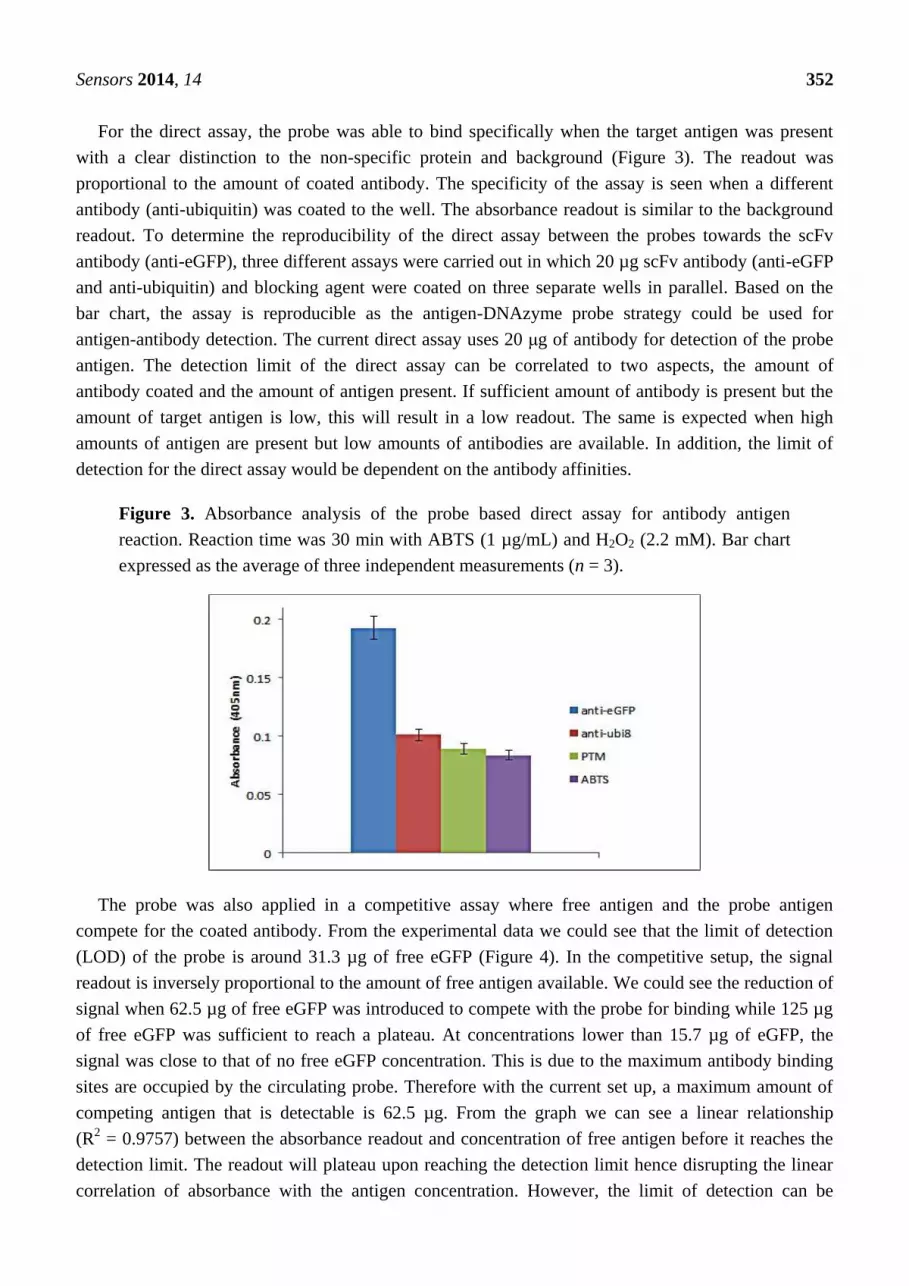
For the direct assay, the probe was able to bind specifically when the target antigen was present
with a clear distinction to the non-specific protein and background (Figure 3). The readout was
proportional to the amount of coated antibody. The specificity of the assay is seen when a different
antibody (anti-ubiquitin) was coated to the well. The absorbance readout is similar to the background
readout. To determine the reproducibility of the direct assay between the probes towards the scFv
antibody (anti-eGFP), three different assays were carried out in which 20 µg scFv antibody (anti-eGFP
and anti-ubiquitin) and blocking agent were coated on three separate wells in parallel. Based on the
bar chart, the assay is reproducible as the antigen-DNAzyme probe strategy could be used for
antigen-antibody detection. The current direct assay uses 20 μg of antibody for detection of the probe
antigen. The detection limit of the direct assay can be correlated to two aspects, the amount of
antibody coated and the amount of antigen present. If sufficient amount of antibody is present but the
amount of target antigen is low, this will result in a low readout. The same is expected when high
amounts of antigen are present but low amounts of antibodies are available. In addition, the limit of
detection for the direct assay would be dependent on the antibody affinities.
Figure 3. Absorbance analysis of the probe based direct assay for antibody antigen
reaction. Reaction time was 30 min with ABTS (1 µg/mL) and H2O2 (2.2 mM). Bar chart
expressed as the average of three independent measurements (n = 3).
The probe was also applied in a competitive assay where free antigen and the probe antigen
compete for the coated antibody. From the experimental data we could see that the limit of detection
(LOD) of the probe is around 31.3 µg of free eGFP (Figure 4). In the competitive setup, the signal
readout is inversely proportional to the amount of free antigen available. We could see the reduction of
signal when 62.5 µg of free eGFP was introduced to compete with the probe for binding while 125 µg
of free eGFP was sufficient to reach a plateau. At concentrations lower than 15.7 µg of eGFP, the
signal was close to that of no free eGFP concentration. This is due to the maximum antibody binding
sites are occupied by the circulating probe. Therefore with the current set up, a maximum amount of
competing antigen that is detectable is 62.5 µg. From the graph we can see a linear relationship
(R2 = 0.9757) between the absorbance readout and concentration of free antigen before it reaches the
detection limit. The readout will plateau upon reaching the detection limit hence disrupting the linear
correlation of absorbance with the antigen concentration. However, the limit of detection can be

Sensors 2014, 14 353
improved by introducing less competing probe into the assay. In this manner the dynamic range for
signal reduction can be increased for competition assays.
Figure 4. Competitive assay between the probe and the free eGFP against the antibody
(scFv format). Linear relationship between the absorbance readout and the concentration of
free eGFP (n = 3) where each data point represents an average of three absorbance readouts
(each error bar indicates the standard errors).
4. Conclusions
A simple and rapid direct immuno-based assay was developed using a one pot synthesis of the
daunomycin aptamer with the DNAzyme as a reporter system. The use of the biotin and streptavidin
interaction allows the antigen-DNAzyme probe system to be applied in a modular form to act as a
universal reporter system for immunoassays. The pre-assay generation of the probe eliminates the
multiple steps needed in typical ELISA system to introduce secondary antibodies. The design of the
probe allows flexibility for both direct and competitive immunoassay applications. The ability of the
daunomycin aptamer to form quadruplex structures in a range of conditions even in absence of K+ ions
is a major advantage for the use in antibody-antigen assays as antibodies and protein-protein
interactions may require other buffers. Thus DQ is our choice as it is found to be just slightly less
active than HQ. We found that the probe-based system performs better in a direct assay format in
comparison to a competitive assay. Nevertheless, we expect that with smaller molecules (haptens) the
competitive assay can be more sensitive. In conclusion, the antigen-DNAzyme probe system allows a
rapid one-step incubation system as an alternative to conventional immunoassay systems.
Acknowledgments
The work was supported by the Malaysian Ministry of Higher Education through the
Higher Institution Centre of Excellence (HICoE) Grant (Grant No. 311/CIPPM/44001005) and the
Fundamental Research Grant Scheme (Grant 203/CIPPM/6711204). Noorsharmimi Omar would
like to acknowledge financial support from the Malaysian Ministry of Education and MyBrain
Scholarship Program.

Sensors 2014, 14 354
Conflict of Interest
The authors declare no conflict of interest.
References
1. Proske, D.; Blank, M.; Buhmann, R.; Resch, A. Aptamers—Basic research, drug development,
and clinical applications. Appl. Microbiol. Biotechnol. 2005, 69, 367–374.
2. Balasubramanian, S.; Hurley, L.H.; Neidle, S. Targeting G-quadruplexes in gene promoters: A
novel anticancer strategy? Nat. Rev. Drug Discov. 2011, 10, 261–275.
3. Templin, M.F.; Stoll, D.; Schrenk, M.; Traub, P.C.; Vöhringer, C.F.; Joos, T.O. Protein
microarray technology. Drug Discov. Today 2002, 7, 815–822.
4. Neidle, S.; Read, M.A. G-quadruplexes as therapeutic targets. Biopolymers 2000, 56, 195–208.
5. White, R.R.; Sullenger, B.A.; Rusconi, C.P. Developing aptamers into therapeutics. J. Clin.
Investig. 2000, 106, 929–934.
6. Balogh, Z.; Lautner, G.; Bardóczy, V.; Komorowska, B.; Gyurcsányi, R.E.; Mészáros, T.
Selection and versatile application of virus-specific aptamers. FASEB J. 2010, 24, 4187–4195.
7. Kopylov, A.M.; Spiridonova, V.A. Combinatorial chemistry of nucleic acids: SELEX. Mol. Biol.
2000, 34, 940–954.
8. Von Hoff, D.D.; Rozencweig, M.; Slavik, M. Daunomycin: An anthracycline antibiotic effective
in acute leukemia. Adv. Pharmacol. 1978, 15, 1–50.
9. Chaires, J.B.; Fox, K.R.; Herrera, J.E.; Britt, M.; Waring, M.J. Site and sequence specificity of the
daunomycin-DNA interaction. Biochemistry 1987, 26, 8227–8236.
10. Wochner, A.; Menger, M.; Orgel, D.; Cech, B.; Rimmele, M.; Erdmann, V.A.; Glokler, J. A DNA
aptamer with high affinity and specificity for therapeutic anthracyclines. Anal. Biochem. 2008,
373, 34–42.
11. Li, C.L.; Liu, K.T.; Lin, Y.W.; Chang, H.T. Fluorescence detection of lead (II) ions through their
induced catalytic activity of DNAzymes. Anal. Chem.-Columb. 2011, 83, 225.
12. Saito, K.; Tai, H.; Hemmi, H.; Kobayashi, N.; Yamamoto, Y. Interaction between the heme and a
G-quartet in a heme–DNA complex. Inorg. Chem. 2012, 51, 8168–8176.
13. Saito, K.; Nakano, Y.; Tai, H.; Nagatomo, S.; Hemmi, H.; Mita, H.; Yamamoto, Y.
Characterization of heme coordination structure in heme-DNA complex possessing gaseous
molecule as an exogenous ligand. Nucleic Acid. Symp. Ser. 2009, 53, 241–242.
14. Zhu, X.; Cao, Y.; Liang, Z.; Li, G. Aptamer-based and DNAzyme-linked colorimetric detection
of cancer cells. Prot. Cell 2010, 1, 842–846.
15. Wang, J.; Cao, Y.; Chen, G.; Li, G. Regulation of thrombin activity with a bifunctional aptamer
and hemin: Development of a new anticoagulant and antidote pair. ChemBioChem 2009, 10,
2171–2176.
16. Wu, P.; Hwang, K.; Lan, T.; Lu, Y. A DNAzyme-gold nanoparticle probe for uranyl ion in living
cells. J. Am. Chem. Soc.2013, 135, 5254–5257.

Sensors 2014, 14 355
17. Zhu, Y.; Xu, L.; Ma, W.; Chen, W.; Yan, W.; Kuang, H.; Wang, L.; Xu, C. G-quadruplex
DNAzyme-based microcystin-LR (toxin) determination by a novel immunosensor. Biosens.
Bioelectron. 2011, 26, 4393–4398.
18. Zong, C.; Wu, J.; Xu, J.; Ju, H.; Yan, F. Multilayer hemin/G-quadruplex wrapped gold
nanoparticles as tag for ultrasensitive multiplex immunoassay by chemiluminescence imaging.
Biosens. Bioelectron. 2013, 43, 372–378.
19. Li, D.; Ying, Y.; Wu, J.; Niessner, R.; Knopp, D. Comparison of monomeric and polymeric
horseradish peroxidase as labels in competitive ELISA for small molecule detection. Microchim.
Acta 2013, 180, 711–717.
20. Li, T.; Wang, E.; Dong, S. A Grafting strategy for the design of improved G-quadruplex aptamers
and high-activity DNAzymes. PLoS One 2009, 4, e5126.
21. Lund, K.; Manzo, A.J.; Dabby, N.; Michelotti, N.; Johnson-Buck, A.; Nangreave, J.; Taylor, S.;
Pei, R.; Stojanovic, M.N.; Walter, N.G.; Winfree, E.; Yan, H. Molecular robots guided by
prescriptive landscapes. Nature 2010, 465, 206–210.
22. Li, T.; Dong, S.; Wang, E. G‐quadruplex aptamers with peroxidase‐like DNAzyme functions:
Which is the best and how does it work? Chem. Asian J. 2009, 4, 918–922.
23. Liu, W.; Zheng, B.; Cheng, S.; Fu, Y.; Li, W.; Lau, T.C.; Liang, H. G-quadruplex formation and
sequence effect on the assembly of G-rich oligonucleotides induced by Pb2+ ions. Soft Matter
2012, 8, 7107–7123.
© 2013 by the authors; licensee MDPI, Basel, Switzerland. This article is an open access article
distributed under the terms and conditions of the Creative Commons Attribution license
(http://creativecommons.org/licenses/by/3.0/).


