Embed Size (px)
Citation preview
OPEN
ORIGINAL ARTICLE
DHRS2 inhibits cell growth and motility in esophagealsquamous cell carcinomaY Zhou1,2, L Wang1, X Ban1, T Zeng1, Y Zhu1, M Li1, X-Y Guan1,3 and Y Li1
Esophageal squamous cell carcinoma (ESCC) is highly prevailing in Asia and it is ranked in the most aggressive squamous cellcarcinomas. High-frequency loss of heterozygosity occurred in chromosome 14q11.2 in many tumors including ESCC, suggestingthat one or more tumor-suppressor genes might exist within this region. In this study, we identified the tumor-suppressing role ofDHRS2 (short-chain dehydrogenase/reductase family, member 2) at 14q11.2 in ESCCs. Downregulation of DHRS2 occurred in 30.8%of primary ESCC tumor tissues vs paired non-tumorous tissues. DHRS2 downregulation was associated significantly with ESCCinvasion, lymph nodes metastasis and clinical staging (Po0.001). Survival analysis revealed that DHRS2 downregulation wassignificantly associated with worse outcome of patients with ESCC. In vitro and in vivo studies indicated that both DHRS2 variantscould suppress cell proliferation and cell motility. Moreover, we demonstrated that DHRS2 could reduce reactive oxygen speciesand decrease nicotinamide adenine dinucleotide phosphate (oxidized/reduced), increase p53 stability and decrease Rbphosphorylation; it also decreased p38 mitogen-activated protein kinase phosphorylation and matrix metalloproteinase 2. Insummary, these findings demonstrated that DHRS2 had an important part in ESCC development and progression.
Oncogene (2018) 37, 1086–1094; doi:10.1038/onc.2017.383; published online 6 November 2017
INTRODUCTIONEsophageal squamous cell carcinoma (ESCC) is ranked in the mostcommon cancers in the world, with an estimated 456 000 newcases annually.1 Esophageal cancer has two ESCC histologicaltypes: ESCC and esophageal adenocarcinoma. ESCC is a dominat-ing histological type and prevalent in certain areas, especially inthe northern China. Although advanced therapeutics in ESCC hadbeen achieved recently, the 5-year survival rate is only 15–25%because of the late diagnosis. Like most solid tumors, chromoso-mal alteration is frequently observed in ESCC. For example, loss of14q11.2 is one of the most common chromosomal changesidentified by comparative genomic hybridization and high-resolution deletion mapping in many cancers, including ESCC,2
gastrointestinal tumors,3,4 nasopharyngeal carcinoma,5
mesothelioma,6 suggesting that tumor-suppressor genes mightexist in frequently deleted regions. As a gene located in thechromosome 14q11.2,7 DHRS2 (short-chain dehydrogenase/reduc-tase family, member 2) was first acquired from a humanhepatocarcinoma complementary DNA library.8 It codes for anenzyme that is a member of the short-chain dehydrogenase/reductase (SDR) family.SDR enzyme family is characterized by some common sequence
motifs: a glycine consensus of NAD/NADP cofactor-bindingdomain, amino acids as catalytic domain and highly conservativeamino-acid sequence scattered among the sequences.9 HumanSDR enzymes function actively in signaling molecules metabolismand intermediary and xenobiotic metabolism.10–12 Through theiraffection on regulatory signals, SDR enzymes take significanteffect in controlling normal cell functions and some SDR enzymeshave been associated with some diseases of human,11,12 includingmetabolic disorders and tumors.13,14
DHRS2 was reported to bind to mouse double minute 2homolog (MDM2) and result in the weakening of MDM2-intermediated p53 degradation in osteosarcoma cell line.15 It isalso activated by c-Myb and ETV5.16,17 It was reported that DHRS2expression correlated with estrogen receptor status in breastcancer,15,18 and introduction of adenovirus harboring DHRS2could suppress renal cancer cell growth.19 However, whetherDHRS2 involved in ESCC cancer development and progressionremained unclear. In this study, expression of DHRS2 wascompared between ESCC primary tumor tissues and adjacentnon-tumorous tissues. The clinical significance of DHRS2 in ESCCpatients was explored. The tumor inhibition roles of DHRS2 inESCC were identified by functional studies. The underlyingmechanism of tumor inhibition of DHRS2 was also addressed.
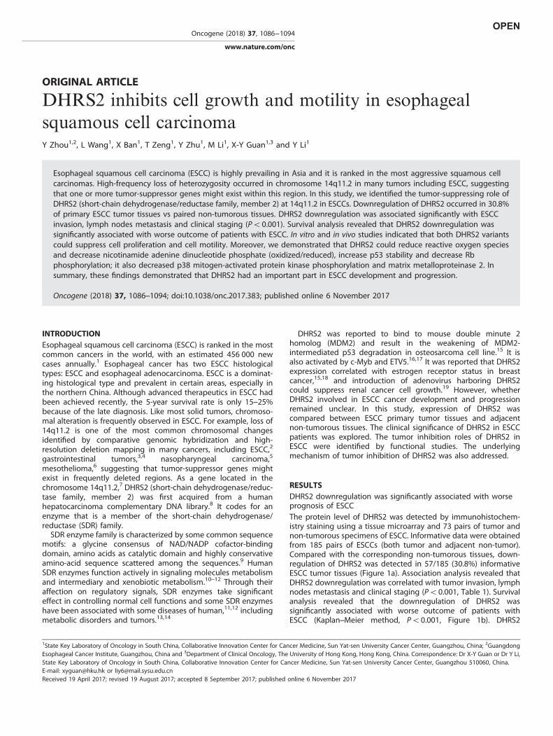
RESULTSDHRS2 downregulation was significantly associated with worseprognosis of ESCCThe protein level of DHRS2 was detected by immunohistochem-istry staining using a tissue microarray and 73 pairs of tumor andnon-tumorous specimens of ESCC. Informative data were obtainedfrom 185 pairs of ESCCs (both tumor and adjacent non-tumor).Compared with the corresponding non-tumorous tissues, down-regulation of DHRS2 was detected in 57/185 (30.8%) informativeESCC tumor tissues (Figure 1a). Association analysis revealed thatDHRS2 downregulation was correlated with tumor invasion, lymphnodes metastasis and clinical staging (Po0.001, Table 1). Survivalanalysis revealed that the downregulation of DHRS2 wassignificantly associated with worse outcome of patients withESCC (Kaplan–Meier method, Po0.001, Figure 1b). DHRS2
1State Key Laboratory of Oncology in South China, Collaborative Innovation Center for Cancer Medicine, Sun Yat-sen University Cancer Center, Guangzhou, China; 2GuangdongEsophageal Cancer Institute, Guangzhou, China and 3Department of Clinical Oncology, The University of Hong Kong, Hong Kong, China. Correspondence: Dr X-Y Guan or Dr Y Li,State Key Laboratory of Oncology in South China, Collaborative Innovation Center for Cancer Medicine, Sun Yat-sen University Cancer Center, Guangzhou 510060, China.E-mail: [email protected] or [email protected] 19 April 2017; revised 19 August 2017; accepted 8 September 2017; published online 6 November 2017
Oncogene (2018) 37, 1086–1094
www.nature.com/onc
downregulation and other clinicopathologic features (tumorinvasion, lymph nodes metastasis and staging) were analyzed byCox regression analysis. The results revealed that it was not anindependent prognosis factor for overall survival (Table 2). As the
antibody targeting the common amino-acid sequence coded byDHRS2-V1 (variant 1, NM_182908.4) and DHRS2-V2 (variant 2,NM_005794.3), we next examined the RNA level of the variants inESCC cell lines. Compared with the non-tumorous tissues pool andNE1 (an immortalized human esophageal epithelial cell line), theRNA levels of DHRS2-V1 and V2 decreased in most ESCC cell lines(Figure 1c). The DHRS2-V2 was relatively higher than DHRS2-V1 inmost ESCC cell lines (Supplementary Figure 1A).As deletion of 14q11.2 was often reported in many cancers,
fluorescence in situ hybridization (FISH) was used to examine theDNA copy number alteration of DHRS2 in ESCC cell lines andtumor tissues. The results revealed that copy number loss ofDHRS2 existed in ESCC cell lines and ESCC tumor tissues(Supplementary Figures 1B and C).
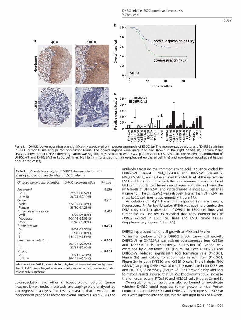
DHRS2 suppressed tumor cell growth in vitro and in vivoTo further explore whether DHRS2 affects tumor cell growth,DHRS2-V1 or DHRS2-V2 was stabled overexpressed into KYSE30and KYSE510 cells, respectively. Expression of DHRS2 wasexamined by quantitative PCR (Figure 2a). Both DHRS2-V1 andDHRS2-V2 reduced significantly foci formation rate (Po0.01,Figure 2b) and colony formation rate in soft agar (Po0.01,Figure 2c) in both KYSE30 and KYSE510 cells. Short hairpin RNA(shRNA) targeting DHRS2 was also stably transfected into KYSE180and HKESC1, respectively (Figure 2d). Cell growth assay and fociformation results showed that DHRS2 knock-down could increasethe tumorgenecity in KYSE180 and HKESC1 cells (Figures 2e and f).Xenograft formation assay was also performed to investigate
whether DHRS2 could suppress tumor growth in vivo. Vectorcontrol cells and DHRS2-V1 and DHRS2-V2 overexpressed KYSE30cells were injected into the left, middle and right flanks of 4-week-
Figure 1. DHRS2 downregulation was significantly associated with poorer prognosis of ESCC. (a) The representative pictures of DHRS2 stainingin ESCC tumor tissue and paired non-tumor tissue. The boxed regions were magnified and shown in the right panels. (b) Kaplan–Meieranalysis showed that DHRS2 downregulation was significantly associated with ESCC patients’ poorer survival. (c) The relative quantification ofDHRS2-V1 and DHRS2-V2 in ESCC cell lines, NE1 (an immortalized human esophageal epithelial cell line) and non-tumor esophageal tissuespool (three cases).
Table 1. Correlation analysis of DHRS2 downregulation withclinicopathologic characteristics of ESCC patients
Clinicopathologic characteristics DHRS2 downregulation P-value
Age (years) 0.836o60 29/92 (31.52%)4= 60 28/93 (30.11%)Gender 0.911Male 32/105 (30.48%)Female 25/80 (31.25%)Tumor cell differetiation 0.703Well 6/25 (24.00%)Moderate 40/114 (35.09%)Poor 11/46 (23.91%)Tumor invasion o0.0010–1 10/74 (13.51%)2 3/10 (30.00%)3 44/101 (43.56%)Lymph node metastasis o0.001− 30/131 (22.90%)+ 27/54 (50.00%)Staging o0.0010, I 9/74 (12.16%)II, III, IV 48/111 (43.24%)
Abbreviations: DHRS2, short-chain dehydrogenase/reductase family, mem-ber 2; ESCC, eesophageal squamous cell carcinoma. Bold values indicatestatistically significant.
DHRS2 inhibits ESCC growth and metastasisY Zhou et al
1087
Oncogene (2018) 1086 – 1094
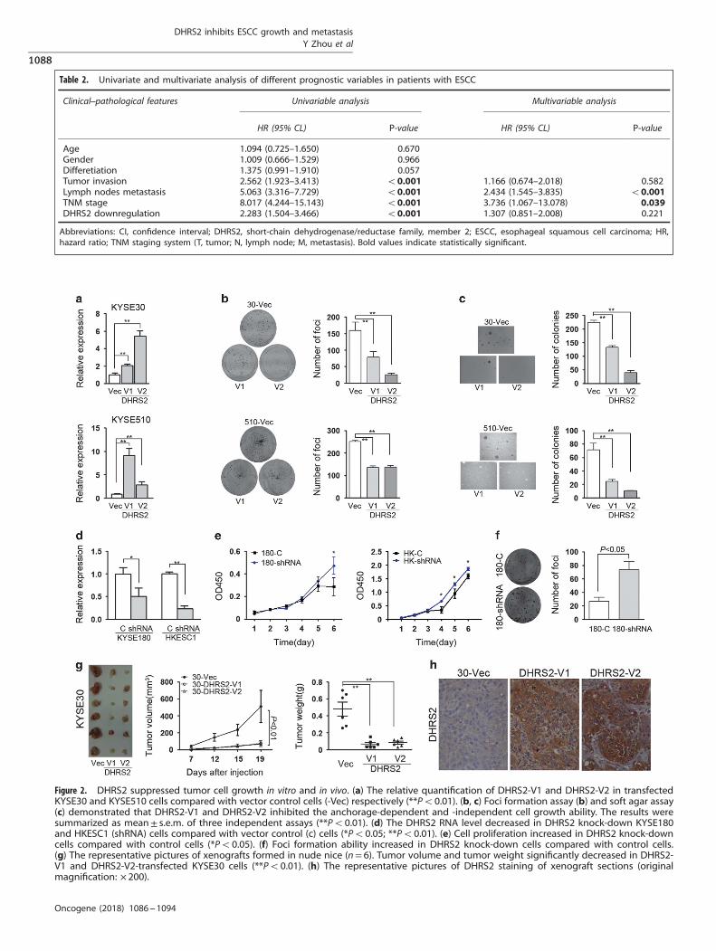
Table 2. Univariate and multivariate analysis of different prognostic variables in patients with ESCC
Clinical–pathological features Univariable analysis Multivariable analysis
HR (95% CL) P-value HR (95% CL) P-value
Age 1.094 (0.725–1.650) 0.670Gender 1.009 (0.666–1.529) 0.966Differetiation 1.375 (0.991–1.910) 0.057Tumor invasion 2.562 (1.923–3.413) o0.001 1.166 (0.674–2.018) 0.582Lymph nodes metastasis 5.063 (3.316–7.729) o0.001 2.434 (1.545–3.835) o0.001TNM stage 8.017 (4.244–15.143) o0.001 3.736 (1.067–13.078) 0.039DHRS2 downregulation 2.283 (1.504–3.466) o0.001 1.307 (0.851–2.008) 0.221
Abbreviations: CI, confidence interval; DHRS2, short-chain dehydrogenase/reductase family, member 2; ESCC, esophageal squamous cell carcinoma; HR,hazard ratio; TNM staging system (T, tumor; N, lymph node; M, metastasis). Bold values indicate statistically significant.
Figure 2. DHRS2 suppressed tumor cell growth in vitro and in vivo. (a) The relative quantification of DHRS2-V1 and DHRS2-V2 in transfectedKYSE30 and KYSE510 cells compared with vector control cells (-Vec) respectively (**Po0.01). (b, c) Foci formation assay (b) and soft agar assay(c) demonstrated that DHRS2-V1 and DHRS2-V2 inhibited the anchorage-dependent and -independent cell growth ability. The results weresummarized as mean± s.e.m. of three independent assays (**Po0.01). (d) The DHRS2 RNA level decreased in DHRS2 knock-down KYSE180and HKESC1 (shRNA) cells compared with vector control (c) cells (*Po0.05; **Po0.01). (e) Cell proliferation increased in DHRS2 knock-downcells compared with control cells (*Po0.05). (f) Foci formation ability increased in DHRS2 knock-down cells compared with control cells.(g) The representative pictures of xenografts formed in nude nice (n= 6). Tumor volume and tumor weight significantly decreased in DHRS2-V1 and DHRS2-V2-transfected KYSE30 cells (**Po0.01). (h) The representative pictures of DHRS2 staining of xenograft sections (originalmagnification: × 200).
DHRS2 inhibits ESCC growth and metastasisY Zhou et al
1088
Oncogene (2018) 1086 – 1094
old immunodeficient mice, respectively. Mice were killed 19 daysafter injection and xenografts were isolated for further analysis.The tumor volume and tumor weight of the xenografts induced byDHRS2-V1 or V2 significantly decreased compared with thexenografts induced by vector control cells (Po0.01, Figure 2g).The immunostaining results confirmed that expression of DHRS2in xenografts induced by DHRS2-overexpressed cells was muchstronger than that in the vector-transfected cells (Figure 2h).
DHRS2 inhibited cell motility in vitro and in vivoAs downregulation of DHRS2 was correlated with tumor invasionand lymph node metastasis, we further explored whether DHRS2could affect tumor cell motility. The scratch assay indicated thatDHRS2-V1 and V2-transfected KYSE30 cells obtained slowerclosure of the scratched ‘wound’ compared with vector control(Figure 3a). Cell migration experiment demonstrated that thenumber of cells migrated decreased in the DHRS2-V1 and V2-transfected KYSE30 cells (Figure 3b). Cell invasion assay demon-strated that the number of cells invaded through Matrigel alsosignificantly decreased in DHRS2-V1 and V2-transfected cells(Figure 3b). The results in DHRS2-V1 and V2 overexpressedKYSE510 cells were consistent with KYSE30 cells (Figure 3b). WhenDHRS2 was silenced in KYSE180 and HKESC1 cells, the number of
cells migrated increased in DHRS2 knock-down cells (Po0.01,Figure 3c).To determine whether DHRS2 can affect tumor metastasis
in vivo, lymph node metastasis animal models were performed innude mice. DHRS2-V1 and V2-transfected cells were subcuta-neously inoculated into the footpad of the left hind limb,respectively (n= 6 for each group). Two months later, the micewere killed, and popliteal lymph nodes were isolated and fixed. Noapparent nodule was observed in surface of lung and liver. Thepopliteal lymph nodes size significantly decreased in DHRS2-V1and V2 groups, compared with that in control cells (Figure 3d).Tumor cells infiltration decreased in lymph nodes of DHRS2-V1and V2 groups compared with that in control cells (Figure 3e).
DHRS2 affected cell cycle and induced apoptosisThe cell cycle distribution was compared by flow cytometryamong DHRS2-V1, V2 and vector-transfected cells. The resultsfound that KYSE30-DHRS2-V1 and DHRS2-V2 cells were arrested atG2/M checkpoint, exhibited as cells accumulation in G2/M phase(Figure 4a). In KYSE510 cells, no significant change was observedin cells distribution except that pre-G1 was observed in DHRS2-V1and V2-transfected cells (Figure 4a).
Figure 3. DHRS2 inhibited cell motility in vitro and in vivo. (a) Wound-healing ability decreased in DHRS2-V1 and DHRS2-V2-transfected KYSE30cells compared with vector control cells. (b) The cell migration and invasion ability decreased in DHRS2-V1 and DHRS2-V2-transfected KYSE30and KYSE510 cells compared with vector control cells. The results were summarized of three independent assays (*Po0.05; **Po0.01). (c) Thecell migration increased in DHRS2 knock-down KYSE180 and HKESC1 cells (shRNA) compared with control cells (c) (**Po0.01). (d) The size ofpopliteal lymph nodes decreased in DHRS2-V1 and DHRS2-V2 animal groups compared with vector control group. (e) The representativepictures of HE staining of popliteal lymph nodes of the DHRS2-V1, DHRS2-V2 and vector control groups (original magnification: × 200). Theboxed regions were magnified as the bottom pictures.
DHRS2 inhibits ESCC growth and metastasisY Zhou et al
1089
Oncogene (2018) 1086 – 1094
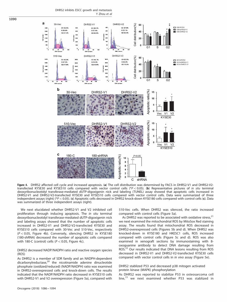
We next elucidated whether DHRS2-V1 and V2 inhibited cellproliferation through inducing apoptosis. The in situ terminaldeoxyribonucleotidyl transferase-mediated dUTP-digoxigenin nickand labeling assays showed that the number of apoptotic cellsincreased in DHRS2-V1 and DHRS2-V2-transfected KYSE30 andKYSE510 cells compared with 30-Vec and 510-Vec, respectively(Po0.05, Figure 4b). Conversely, silencing DHRS2 in KYSE180(180-shRNA) decreased the number of apoptotic cells comparedwith 180-C (control) cells (Po0.05, Figure 4c).
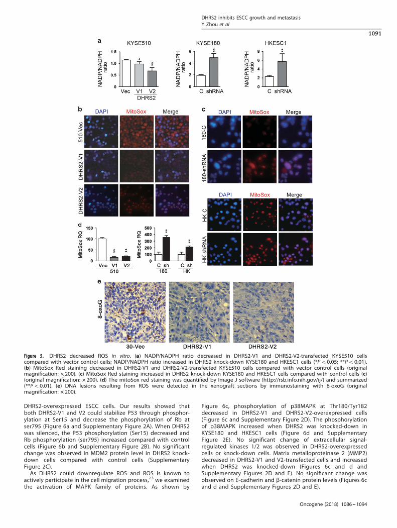
DHRS2 decreased NADP/NADPH ratio and reactive oxygen species(ROS)As DHRS2 is a member of SDR family and an NADPH-dependentdicarbonylreductase,20 the nicotinamide adenine dinucleotidephosphate (oxidized/reduced) (NADP/NADPH) ratio was examinedin DHRS2-overexpressed cells and knock-down cells. The resultsindicated that the NADP/NADPH ratio decreased in KYSE510 cellswith DHRS2-V1 and V2 overexpression (Figure 5a), compared with
510-Vec cells. When DHRS2 was silenced, the ratio increasedcompared with control cells (Figure 5a).As DHRS2 was reported to be associated with oxidative stress,21
we next examined the mitochondrial ROS by MitoSox Red stainingassay. The results found that mitochondrial ROS decreased inDHRS2-overexpressed cells (Figures 5b and d). When DHRS2 wasknocked-down in KYSE180 and HKESC1 cells, ROS increasedcompared with control cells (Figure 5c and d). ROS was alsoexamined in xenograft sections by immunostaining with 8-oxoguanine antibody to detect DNA damage resulting fromROS.22 Our results indicated that DNA lesions resulting from ROSdecreased in DHRS2-V1 and DHRS2-V2-transfected KYSE30 cellscompared with vector control cells in in vivo assay (Figure 5e).
DHRS2 stablized P53 and decreased p38 mitogen activatedprotein kinase (MAPK) phosphorylationAs DHRS2 was reported to stabilize P53 in osteosarcoma cellline,15 we next examined whether P53 was stablized in
Figure 4. DHRS2 affected cell cycle and increased apoptosis. (a) The cell distribution was determined by FACS in DHRS2-V1 and DHRS2-V2-transfected KYSE30 and KYSE510 cells compared with vector control cells (*Po0.05). (b) Representative pictures of in situ terminaldeoxyribonucleotidyl transferase-mediated dUTP-digoxigenin nick and labeling (TUNEL) assay showed that apoptotic cells increased inDHRS2-V1 and DHRS2-V2-transfected KYSE30 and KYSE510 cells compared with vector control cells. Data were summarized of threeindependent assays (right) (*Po0.05). (c) Apoptotic cells decreased in DHRS2 knock-down KYSE180 cells compared with control cells (c). Datawas summarized of three independent assays (right).
DHRS2 inhibits ESCC growth and metastasisY Zhou et al
1090
Oncogene (2018) 1086 – 1094
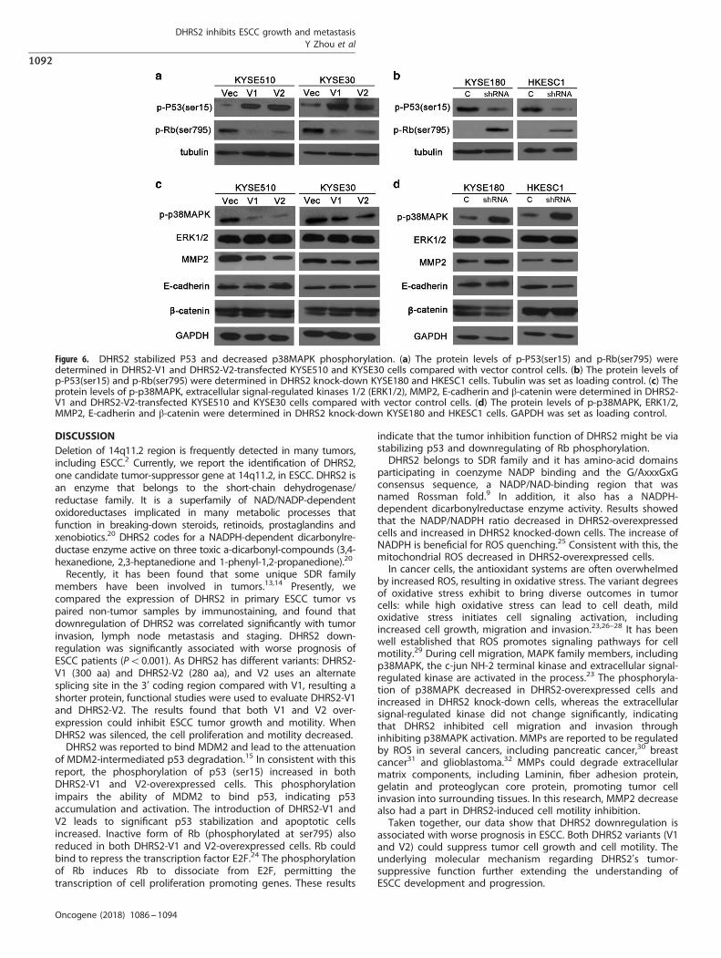
DHRS2-overexpressed ESCC cells. Our results showed thatboth DHRS2-V1 and V2 could stabilize P53 through phosphor-ylation at Ser15 and decrease the phosphorylation of Rb atser795 (Figure 6a and Supplementary Figure 2A). When DHRS2was silenced, the P53 phosphorylation (Ser15) decreased andRb phosphorylation (ser795) increased compared with controlcells (Figure 6b and Supplementary Figure 2B). No significantchange was observed in MDM2 protein level in DHRS2 knock-down cells compared with control cells (SupplementaryFigure 2C).As DHRS2 could downregulate ROS and ROS is known to
actively participate in the cell migration process,23 we examinedthe activation of MAPK family of proteins. As shown by
Figure 6c, phosphorylation of p38MAPK at Thr180/Tyr182decreased in DHRS2-V1 and DHRS2-V2-overexpressed cells(Figure 6c and Supplementary Figure 2D). The phosphorylationof p38MAPK increased when DHRS2 was knocked-down inKYSE180 and HKESC1 cells (Figure 6d and SupplementaryFigure 2E). No significant change of extracellular signal-regulated kinases 1/2 was observed in DHRS2-overexpressedcells or knock-down cells. Matrix metalloproteinase 2 (MMP2)decreased in DHRS2-V1 and V2-transfected cells and increasedwhen DHRS2 was knocked-down (Figures 6c and d andSupplementary Figures 2D and E). No significant change wasobserved on E-cadherin and β-catenin protein levels (Figures 6cand d and Supplementary Figures 2D and E).
Figure 5. DHRS2 decreased ROS in vitro. (a) NADP/NADPH ratio decreased in DHRS2-V1 and DHRS2-V2-transfected KYSE510 cellscompared with vector control cells; NADP/NADPH ratio increased in DHRS2 knock-down KYSE180 and HKESC1 cells (*Po0.05; **Po0.01).(b) MitoSox Red staining decreased in DHRS2-V1 and DHRS2-V2-transfected KYSE510 cells compared with vector control cells (originalmagnification: × 200). (c) MitoSox Red staining increased in DHRS2 knock-down KYSE180 and HKESC1 cells compared with control cells (c)(original magnification: × 200). (d) The mitoSox red staining was quantified by Image J software (http://rsb.info.nih.gov/ij/) and summarized(**Po0.01). (e) DNA lesions resulting from ROS were detected in the xenograft sections by immunostaining with 8-oxoG (originalmagnification: × 200).
DHRS2 inhibits ESCC growth and metastasisY Zhou et al
1091
Oncogene (2018) 1086 – 1094
DISCUSSIONDeletion of 14q11.2 region is frequently detected in many tumors,including ESCC.2 Currently, we report the identification of DHRS2,one candidate tumor-suppressor gene at 14q11.2, in ESCC. DHRS2 isan enzyme that belongs to the short-chain dehydrogenase/reductase family. It is a superfamily of NAD/NADP-dependentoxidoreductases implicated in many metabolic processes thatfunction in breaking-down steroids, retinoids, prostaglandins andxenobiotics.20 DHRS2 codes for a NADPH-dependent dicarbonylre-ductase enzyme active on three toxic a-dicarbonyl-compounds (3,4-hexanedione, 2,3-heptanedione and 1-phenyl-1,2-propanedione).20
Recently, it has been found that some unique SDR familymembers have been involved in tumors.13,14 Presently, wecompared the expression of DHRS2 in primary ESCC tumor vspaired non-tumor samples by immunostaining, and found thatdownregulation of DHRS2 was correlated significantly with tumorinvasion, lymph node metastasis and staging. DHRS2 down-regulation was significantly associated with worse prognosis ofESCC patients (Po0.001). As DHRS2 has different variants: DHRS2-V1 (300 aa) and DHRS2-V2 (280 aa), and V2 uses an alternatesplicing site in the 3′ coding region compared with V1, resulting ashorter protein, functional studies were used to evaluate DHRS2-V1and DHRS2-V2. The results found that both V1 and V2 over-expression could inhibit ESCC tumor growth and motility. WhenDHRS2 was silenced, the cell proliferation and motility decreased.DHRS2 was reported to bind MDM2 and lead to the attenuation
of MDM2-intermediated p53 degradation.15 In consistent with thisreport, the phosphorylation of p53 (ser15) increased in bothDHRS2-V1 and V2-overexpressed cells. This phosphorylationimpairs the ability of MDM2 to bind p53, indicating p53accumulation and activation. The introduction of DHRS2-V1 andV2 leads to significant p53 stabilization and apoptotic cellsincreased. Inactive form of Rb (phosphorylated at ser795) alsoreduced in both DHRS2-V1 and V2-overexpressed cells. Rb couldbind to repress the transcription factor E2F.24 The phosphorylationof Rb induces Rb to dissociate from E2F, permitting thetranscription of cell proliferation promoting genes. These results
indicate that the tumor inhibition function of DHRS2 might be viastabilizing p53 and downregulating of Rb phosphorylation.DHRS2 belongs to SDR family and it has amino-acid domains
participating in coenzyme NADP binding and the G/AxxxGxGconsensus sequence, a NADP/NAD-binding region that wasnamed Rossman fold.9 In addition, it also has a NADPH-dependent dicarbonylreductase enzyme activity. Results showedthat the NADP/NADPH ratio decreased in DHRS2-overexpressedcells and increased in DHRS2 knocked-down cells. The increase ofNADPH is beneficial for ROS quenching.25 Consistent with this, themitochondrial ROS decreased in DHRS2-overexpressed cells.In cancer cells, the antioxidant systems are often overwhelmed
by increased ROS, resulting in oxidative stress. The variant degreesof oxidative stress exhibit to bring diverse outcomes in tumorcells: while high oxidative stress can lead to cell death, mildoxidative stress initiates cell signaling activation, includingincreased cell growth, migration and invasion.23,26–28 It has beenwell established that ROS promotes signaling pathways for cellmotility.29 During cell migration, MAPK family members, includingp38MAPK, the c-jun NH-2 terminal kinase and extracellular signal-regulated kinase are activated in the process.23 The phosphoryla-tion of p38MAPK decreased in DHRS2-overexpressed cells andincreased in DHRS2 knock-down cells, whereas the extracellularsignal-regulated kinase did not change significantly, indicatingthat DHRS2 inhibited cell migration and invasion throughinhibiting p38MAPK activation. MMPs are reported to be regulatedby ROS in several cancers, including pancreatic cancer,30 breastcancer31 and glioblastoma.32 MMPs could degrade extracellularmatrix components, including Laminin, fiber adhesion protein,gelatin and proteoglycan core protein, promoting tumor cellinvasion into surrounding tissues. In this research, MMP2 decreasealso had a part in DHRS2-induced cell motility inhibition.Taken together, our data show that DHRS2 downregulation is
associated with worse prognosis in ESCC. Both DHRS2 variants (V1and V2) could suppress tumor cell growth and cell motility. Theunderlying molecular mechanism regarding DHRS2’s tumor-suppressive function further extending the understanding ofESCC development and progression.
Figure 6. DHRS2 stabilized P53 and decreased p38MAPK phosphorylation. (a) The protein levels of p-P53(ser15) and p-Rb(ser795) weredetermined in DHRS2-V1 and DHRS2-V2-transfected KYSE510 and KYSE30 cells compared with vector control cells. (b) The protein levels ofp-P53(ser15) and p-Rb(ser795) were determined in DHRS2 knock-down KYSE180 and HKESC1 cells. Tubulin was set as loading control. (c) Theprotein levels of p-p38MAPK, extracellular signal-regulated kinases 1/2 (ERK1/2), MMP2, E-cadherin and β-catenin were determined in DHRS2-V1 and DHRS2-V2-transfected KYSE510 and KYSE30 cells compared with vector control cells. (d) The protein levels of p-p38MAPK, ERK1/2,MMP2, E-cadherin and β-catenin were determined in DHRS2 knock-down KYSE180 and HKESC1 cells. GAPDH was set as loading control.
DHRS2 inhibits ESCC growth and metastasisY Zhou et al
1092
Oncogene (2018) 1086 – 1094
MATERIALS AND METHODSCells and tumor specimensKYSE30, KYSE140, KYSE180, KYSE410, KYSE510 and KYSE520 were acquiredfrom the German Resource Center for Biological Material(DSMZ) (Braunschweig, Germany).33 HKESC1, EC109 and EC9706, weresupplied by Dr G Srivastava (Department of Pathology, The University ofHong Kong, Hong Kong, China) and Dr G Tsao (Department of Anatomy,The University of Hong Kong), respectively.34 NE1 (an immortalizedesophageal epithelial cell line) was established in Dr G Tsao’s lab. ESCCcells were authenticated by cytogenetic methods as human origin in2009.35 The cells were tested for mycoplasma contamination recently.
Fluorescence in situ hybridizationNick translation method was adopted to label the bacterial artificialchromosome clone containing DHRS2 with Spectrum-orange-deoxyuridinetriphosphate (Vysis, Abbott Laboratories, Abbott Park, IL, USA). FISH assaywas conducted as described previously.36
Tissue microarray and immunohistochemistryIn previous report, we constructed a tissue microarray consisting of 300pairs of ESCC primary tumor tissues and corresponding non-tumoroustissues.37 Seventy-three pairs of primary tumor and non-tumor sampleswere collected at Sun Yat-sen University Cancer Center, Guangzhou, China.Patients enrolled in the research had not received follow-up radiation orchemotherapeutic treatment. Patients’ age ranged from 40 to 80 years atthe point of surgery. The research was approved by the Committees forEthical Review of Research Involving Human Subjects in Sun Yat-senUniversity Cancer Center. Signed informed consents for collecting thepatients’ specimens were acquired.In the immunohistochemistry assay, a 1:70 diluted anti-DHRS2 antibody
(PA5-25258, Thermo Fisher Scientific, Rockford, IL, USA) was applied forDHRS2 immunostaining. DHRS2 staining score was compared betweenprimary tumor and corresponding non-tumor specimens. Immunohisto-chemistry score was determined independently by two pathologistswithout knowing the clinicopathological information. A staining index (0–12) was obtained as the staining intensity (negative (0); weak (1); moderate(2); strong (3)) multiplying the proportion of positive staining (0–25% (1);25–50% (2); 50–75% (3); 75–100% (4)). DHRS2 downregulation was definedas the score of tumor tissues was less than the score of corresponding non-tumor tissues.
Quantitative PCRRNAprep Pure Cell/Bacteria Kit (TianGen Biotech, Beijing, China) was usedto extract RNA from tissues and cells. Reverse transcription wasaccomplished with FastQuant RT Kit (TianGen Biotech). Quantitative PCRwas performed on Roche 480 Fast Real-Time PCR system using SYBR GreenSupermix. The quantitative data were normalized by internal control (18Sor β-actin) and triplicate assays were performed. Dissociation curves wereanalyzed to exclude the possibility of nonspecific amplification products.dCT method was adopted to analyze the quantitative PCR data asdescribed previously.38
Cell growth assaysThe cell proliferation rate was assessed by plating cells into 96-well plateand OD450 was measured by XTT with CCK-8 (Dojindo Co., Tabaru, Japan).The anchorage-dependent assay (foci formation) and anchorage-independent assay (soft agar colony formation) were performed asdescribed.35 The assays were repeated three times independently.
Cell migration and invasion experimentsFor the scratch assay, DHRS2-V1, DHRS2-V2 or empty vector-transfected cellswere cultured in a 100 mm dish until 95% confluence and then woundedwith a pipette tip. Transwell Permeable Support (Corning, NY, USA) was usedto evaluate cell migration ability. In all, 8 ×10⁴ cells were re-suspended inmedium (no serum) in the top chamber of the insert. The cells were attractedby the medium with 10% serum that was added into the bottom well.Twenty-four hours later, penetrated cells through the filter were fixed,stained, and the number of penetrated cells was counted under microscope.Three independent assays were repeated. In the invasion experiment,BioCoat Matrigel Invasion Chamber (BD Biosciences, Bedford, MA, USA) wasused in compliance with the manufacturer’s protocol.
Animal experimentsThe research was approved by Institutional Animal Care and UseCommittee of Sun Yat-sen University Cancer Center. Animal tests werecarried out according to the principles for the Welfare of ExperimentalAnimals in Sun Yat-sen University Cancer Center. The tumorigenicity ofDHRS2-transfected cells was assessed by xenograft formation assay.Randomly selected 4-week-old female BALB/c nu/nu mice (n= 5) wereused in the study. DHRS2-V1, DHRS2-V2-overexpressed KYSE30, as well asvector control cells (2 × 10⁶) were inoculated subcutaneously into the mice.The growth of xenografts was examined two times per week. After killing,xenografts were isolated, processed and stained for hematoxylin–eosinand immunohistochemistry study.The metastasis ability of cells was evaluated by injecting tumor cells into
footpad of the mice. DHRS2-V1, DHRS2-V2 and vector-transfected KYSE30cells (8 × 10⁵) were injected into the left footpad of 4-week-oldimmunodeficient mice (female, n= 6) (randomly selected), respectively.After 2 months, animals were killed and the lungs and livers wereexamined. The popliteal lymph nodes were isolated, fixed and subjected tohematoxylin–eosin staining.
Cell cycle analysisKYSE30 and KYSE510 derivative cells (3 × 10⁶) were fixed in pre-cooled 75%ethanol, incubated with propidium iodide (Sigma-Aldrich, Saint Louis, MO,USA). Cytomics FC 500 (BECKMAN COULTER, Fullerton, CA, USA) was usedto analyze DNA content. Cell cycle analysis was performed by Modfit LT 2.0(BECKMAN COULTER). Triplicate assays were repeated independently.
In situ cell death detection experimentIn situ Cell Death Detection Kit (Roche, Mannheim, Germany) was used todetect the apoptotic cells. Cells plated on the cover slides were rinsed withphosphate-buffered saline and fixed. After blocking and permeabilization,apoptosis was detected by the terminal deoxyribonucleotidyl transferase-mediated dUTP-digoxigenin nick and labeling method. The staining signalwas transformed by Converter-POD and apoptotic cells were countedunder microscope.
Mitochondrial ROS detectionCells were plated on cover slides, stained with 5 μM MitoSOX Red(Molecular Probes, Eugene, OR, USA) for 10 min at 37 °C, rinsed, thencounterstained with 4′, 6′-diamidino-2-phenylindole. Mitochondrial ROSwas observed under fluorescence microscope (BX61, Olympus, Tokyo,Japan).
NADP/NADPH quantificationNADP/NADPH quantification was detected in DHRS2-V1 and DHRS2-V2-overexpressed and knock-down cells as the manufacturer’s protocol(Sigma-Aldrich).
Western blotting and antibodies, plasmidsWestern blotting was carried out in compliance with the standard protocol.Antibodies used were: p-P53(ser15) (#9286), p-Rb(ser795) (#9301),p-p38MAPK(Thr180/tyr182) (#9212), extracellular signal-regulated kinases1/2 (#4695), E-cadherin (#3195), β-catenin (#8481), tubulin (#2128) (CellSignaling Technology, Danvers, MA, USA), GAPDH (ap7873a) and MMP2(am1844a) (ABGENT, San Diego, CA, USA), DHRS2 (PA5-25258) (ThermoFisher Scientific), MDM2 (M4308) (Sigma-Aldrich) and 8-oxoguanine(AB20646) (Abcam, Cambridge, UK). pCDH was bought from SystemBiosciences (Palo Alto, CA, USA). pCDH-DHRS2-V1 and DHRS2-V2 wereconstructed and sequenced. pLKO.1-DHRS2-shRNA targeting both variantswas bought from Sigma-Aldrich.
Statistical analysesSPSS software package (Version 13.0; SPSS, Inc., Chicago, IL, USA) was usedin the study. We used Pearson chi-square test to examine the clinicalcorrelation between DHRS2 downregulation and clinicopathologicalcharacters. We used Kaplan–Meier analysis to generate survival curvesand log-rank test to calculate the significance. The Cox regression modelwas applied to identify independent prognostic factors. Results werereported as mean± s.e.m. Po0.05 was considered statistically significant.
DHRS2 inhibits ESCC growth and metastasisY Zhou et al
1093
Oncogene (2018) 1086 – 1094
ABBREVIATIONSDHRS2, short-chain dehydrogenase/reductase family, member 2; ESCC,esophageal squamous cell carcinoma; FISH, fluorescence in situ hybridiza-tion; MAPK, mitogen activated protein kinase; MDM2, mouse doubleminute 2 homolog.
CONFLICT OF INTERESTThe authors declare no conflict of interest.
ACKNOWLEDGEMENTSThis research was supported by the National Natural Science Foundation of China(81472255 and 81472250); Guangdong Esophageal Cancer Institute Funding(M201511); Guangdong Science and Technology Foundation (2016A020214008).
REFERENCES1 Ferlay J, Soerjomataram I, Dikshit R, Eser S, Mathers C, Rebelo M et al. Cancer
incidence and mortality worldwide: sources, methods and major patterns inGLOBOCAN 2012. Int J Cancer 2015; 136: E359–386.
2 Shi ZZ, Jiang YY, Hao JJ, Zhang Y, Zhang TT, Shang L et al. Identification ofputative target genes for amplification within 11q13.2 and 3q27.1 in esophagealsquamous cell carcinoma. Clin Transl Oncol 2014; 16: 606–615.
3 Debiec-Rychter M, Sciot R, Pauwels P, Schoenmakers E, Dal Cin P, Hagemeijer A.Molecular cytogenetic definition of three distinct chromosome arm 14q deletionintervals in gastrointestinal stromal tumors. Genes Chromosomes Cancer 2001; 32:26–32.
4 El-Rifai W, Sarlomo-Rikala M, Andersson LC, Miettinen M, Knuutila S. High-resolution deletion mapping of chromosome 14 in stromal tumors of the gas-trointestinal tract suggests two distinct tumor suppressor loci. Genes Chromo-somes Cancer 2000; 27: 387–391.
5 Shao JY, Huang XM, Yu XJ, Huang LX, Wu QL, Xia JC et al. Loss of heterozygosityand its correlation with clinical outcome and Epstein-Barr virus infection innasopharyngeal carcinoma. Anticancer Res 2001; 21: 3021–3029.
6 Bjorkqvist AM, Wolf M, Nordling S, Tammilehto L, Knuuttila A, Kere J et al. Dele-tions at 14q in malignant mesothelioma detected by microsatellite marker ana-lysis. Br J Cancer 1999; 81: 1111–1115.
7 Pellegrini S, Censini S, Guidotti S, Iacopetti P, Rocchi M, Bianchi M et al. A humanshort-chain dehydrogenase/reductase gene: structure, chromosomal localization,tissue expression and subcellular localization of its product. Biochim Biophys Acta2002; 1574: 215–222.
8 Gabrielli F, Donadel G, Bensi G, Heguy A, Melli M. A nuclear protein, synthesized ingrowth-arrested human hepatoblastoma cells, is a novel member of the short-chain alcohol dehydrogenase family. Eur J Biochem 1995; 232: 473–477.
9 Gabrielli F, Tofanelli S. Molecular and functional evolution of human DHRS2 andDHRS4 duplicated genes. Gene 2012; 511: 461–469.
10 Bray JE, Marsden BD, Oppermann U. The human short-chain dehydrogenase/reductase (SDR) superfamily: a bioinformatics summary. Chemico-Biol Interact09;178: 99–109.
11 Oppermann U. Carbonyl reductases: the complex relationships of mammaliancarbonyl- and quinone-reducing enzymes and their role in physiology. Annu RevPharmacol Toxicol 2007; 47: 293–322.
12 Wu X, Lukacik P, Kavanagh KL, Oppermann U. SDR-type human hydroxysteroiddehydrogenases involved in steroid hormone activation. Mol Cell Endocrinol 2007;265-266: 71–76.
13 Li J, Liu J, Ren Y, Yang J, Liu P. Common chromosomal fragile site gene WWOX inmetabolic disorders and tumors. Int J Biol Sci 2014; 10: 142–148.
14 Hu L, Chen HY, Han T, Yang GZ, Feng D, Qi CY et al. Downregulation of DHRS9expression in colorectal cancer tissues and its prognostic significance. Tumour Biol2016; 37: 837–845.
15 Deisenroth C, Thorner AR, Enomoto T, Perou CM, Zhang Y. Mitochondrial Hep27 isa c-Myb target gene that inhibits Mdm2 and stabilizes p53. Mol Cell Biol 2010; 30:3981–3993.
16 Rushton JJ, Davis LM, Lei W, Mo X, Leutz A, Ness SA. Distinct changes in geneexpression induced by A-Myb, B-Myb and c-Myb proteins. Oncogene 2003; 22:308–313.
17 Monge M, Colas E, Doll A, Gil-Moreno A, Castellvi J, Diaz B et al. Proteomicapproach to ETV5 during endometrial carcinoma invasion reveals a link tooxidative stress. Carcinogenesis 2009; 30: 1288–1297.
18 Wang J, Shidfar A, Ivancic D, Ranjan M, Liu L, Choi MR et al. Overexpressionof lipid metabolism genes and PBX1 in the contralateral breasts of womenwith estrogen receptor-negative breast cancer. Int J Cancer 2017; 140:2484–2497.
19 Fang L, Cheng Q, Liu W, Zhang J, Ge Y, Zhang Q et al. Selective effects of a fiberchimeric conditionally replicative adenovirus armed with hep27 gene on renalcancer cell. Cancer Biol Ther 2016; 17: 664–673.
20 Shafqat N, Shafqat J, Eissner G, Marschall HU, Tryggvason K, Eriksson U et al.Hep27, a member of the short-chain dehydrogenase/reductase family, is anNADPH-dependent dicarbonyl reductase expressed in vascular endothelial tissue.Cell Mol Life Sci 2006; 63: 1205–1213.
21 Crean D, Felice L, Taylor CT, Rabb H, Jennings P, Leonard MO. Glucose reintro-duction triggers the activation of Nrf2 during experimental ischemia reperfusion.Mol Cell Biochem 2012; 366: 231–238.
22 Wang P, Sun YC, Lu WH, Huang P, Hu Y. Selective killing of K-ras-transformedpancreatic cancer cells by targeting NAD(P)H oxidase. Chin J Cancer 2015; 34:166–176.
23 Tochhawng L, Deng S, Pervaiz S, Yap CT. Redox regulation of cancer cell migrationand invasion. Mitochondrion 2013; 13: 246–253.
24 Dyson N. The regulation of E2F by pRB-family proteins. Genes Dev 1998; 12:2245–2262.
25 Alberghina L, Gaglio D. Redox control of glutamine utilization in cancer. Cell DeathDis 2014; 5: e1561.
26 Benhar M, Engelberg D, Levitzki A. ROS, stress-activated kinases and stress sig-naling in cancer. EMBO Rep 2002; 3: 420–425.
27 Gloire G, Legrand-Poels S, Piette J. NF-kappaB activation by reactive oxygenspecies: fifteen years later. Biochem Pharmacol 2006; 72: 1493–1505.
28 Nishikawa M. Reactive oxygen species in tumor metastasis. Cancer Lett 2008; 266:53–59.
29 Hurd TR, DeGennaro M, Lehmann R. Redox regulation of cell migration andadhesion. Trends Cell Biol 2012; 22: 107–115.
30 Binker MG, Binker-Cosen AA, Richards D, Oliver B, Cosen-Binker LI. EGF promotesinvasion by PANC-1 cells through Rac1/ROS-dependent secretion and activationof MMP-2. Biochem Biophys Res Commun 2009; 379: 445–450.
31 Pelicano H, Lu W, Zhou Y, Zhang W, Chen Z, Hu Y et al. Mitochondrial dys-function and reactive oxygen species imbalance promote breast cancer cellmotility through a CXCL14-mediated mechanism. Cancer Res 2009; 69:2375–2383.
32 Chiu WT, Shen SC, Chow JM, Lin CW, Shia LT, Chen YC. Contribution ofreactive oxygen species to migration/invasion of human glioblastoma cellsU87 via ERK-dependent COX-2/PGE(2) activation. Neurobiol Dis 2010; 37:118–129.
33 Shimada Y, Imamura M, Wagata T, Yamaguchi N, Tobe T. Characterization of 21newly established esophageal cancer cell lines. Cancer 1992; 69: 277–284.
34 Wong ML, Tao Q, Fu L, Wong KY, Qiu GH, Law FB et al. Aberrant promoterhypermethylation and silencing of the critical 3p21 tumour suppressor gene,RASSF1A, in Chinese oesophageal squamous cell carcinoma. Int J Oncol 2006; 28:767–773.
35 Li Y, Chen L, Nie CJ, Zeng TT, Liu H, Mao X et al. Downregulation of RBMS3 isassociated with poor prognosis in esophageal squamous cell carcinoma. CancerRes 2011; 71: 6106–6115.
36 Guan XY, Sham JS, Tang TC, Fang Y, Huo KK, Yang JM. Isolation of a novelcandidate oncogene within a frequently amplified region at 3q26 inovarian cancer. Cancer Res 2001; 61: 3806–3809.
37 Li Y, Zhu CL, Nie CJ, Li JC, Zeng TT, Zhou J et al. Investigation of tumor sup-pressing function of CACNA2D3 in esophageal squamous cell carcinoma. PLoSOne 2013; 8: e60027.
38 Livak KJ, Schmittgen TD. Analysis of relative gene expression data usingreal-time quantitative PCR and the 2(-delta delta C(T)) method. Methods 2001; 25:402–408.
This work is licensed under a Creative Commons Attribution-NonCommercial-ShareAlike 4.0 International License. The images or
other third party material in this article are included in the article’s Creative Commonslicense, unless indicatedotherwise in the credit line; if thematerial is not included underthe Creative Commons license, users will need to obtain permission from the licenseholder to reproduce the material. To view a copy of this license, visit http://creativecommons.org/licenses/by-nc-sa/4.0/
© The Author(s) 2018
Supplementary Information accompanies this paper on the Oncogene website (http://www.nature.com/onc)
DHRS2 inhibits ESCC growth and metastasisY Zhou et al
1094
Oncogene (2018) 1086 – 1094


