Embed Size (px)
Citation preview
Eur. J. Immunol. 2014. 44: 617–626 HIGHLIGHTSDOI: 10.1002/eji.201344301 617R
eviewDiabetes and immunity to tuberculosis
Nuria Martinez and Hardy Kornfeld
Department of Medicine, University of Massachusetts Medical School, Worcester, MA, USA
The dual burden of tuberculosis (TB) and diabetes has attracted much attention in the pastdecade as diabetes prevalence has increased dramatically in countries already afflictedwith a high burden of TB. The confluence of these two major diseases presents a seri-ous threat to global public health; at the same time it also presents an opportunity tolearn more about the key elements of human immunity to TB that may be relevantto the general population. Some effects of diabetes on innate and adaptive immunitythat are potentially relevant to TB defense have been identified, but have yet to be ver-ified in humans and are unlikely to fully explain the interaction of these two diseasestates. This review provides an update on the clinical and epidemiological features ofTB in the diabetic population and relates them to recent advances in understanding themechanistic basis of TB susceptibility and other complications of diabetes. Issues thatmerit further investigation — such as geographic host and pathogen differences in thediabetes/TB interaction, the role of hyperglycemia-induced epigenetic reprogramming inimmune dysfunction, and the impact of diabetes on lung injury and fibrosis caused byTB — are highlighted in this review.
Keywords: Bacterial infections � Diabetes � Host/pathogen interaction � Infectious disease �
Tuberculosis
Introduction
An adverse effect of diabetes mellitus (DM) on the host defenseagainst tuberculosis (TB) has been recognized for over a cen-tury [1] but has only recently been investigated in detail asDM rates have increased dramatically since the 1980s in low-and middle-income countries where TB is already prevalent [2].Insulin-resistant type 2 DM comprises 90% of the global cases ofDM and a large proportion of the people afflicted with the dualTB/DM burden [1, 2]; however autoimmune type 1 DM is alsoassociated with TB susceptibility [3]. This is unsurprising sinceboth types of DM share a similar spectrum of other complica-tions that develop as a consequence of chronic hyperglycemia[4]. Diabetes increases the risk of developing TB disease aboutthreefold, as reported in a systematic review of 13 observationalstudies [5]. The range of risk estimates from individual studiesincluded in that review was notably broad; from 0.99 to 7.83 [5].
Correspondence: Dr. Hardy Kornfelde-mail: [email protected]
Divergent results have since been reported in additional studiesof DM and TB risk [6–9]. Factors contributing to this variabilityinclude almost universally retrospective study design, weak diag-nostic criteria for DM in some studies, and the likelihood that hostand microbial genetics as well as environmental factors in diversegeographic locations differentially influence TB defense. The mod-erate increase in TB risk associated with DM has a major globalpublic health impact, owing to the large and growing populationof diabetic people living in high TB burden settings [10].
The scope of the TB/DM problem in India, the country with thehighest caseload of both diseases, was highlighted in a 2011 studyby Viswanathan et al. [11]. The authors of that study performedoral glucose-tolerance tests on 827 patients newly diagnosed withTB in Chennai. In that cohort, 25.3% of TB patients were con-firmed to have DM and 24.5% had prediabetes by the WHO cri-teria [11]. A possible association of prediabetes with TB risk issurprising in light of clinical evidence linking susceptibility to poorglycemic control [6]. Prediabetes could develop as an effect ratherthan a cause of TB, or TB risk might be increased in prediabet-ics by a factor other than hyperglycemia, such as dyslipidemia.The lung is the predominant site of TB disease in the general,
C© 2014 WILEY-VCH Verlag GmbH & Co. KGaA, Weinheim www.eji-journal.eu
618 Nuria Martinez and Hardy Kornfeld Eur. J. Immunol. 2014. 44: 617–626
immunocompetent population, accounting for 70–80% of cases[12]. The study by Viswanathan [11], along with another fromIndia [13], found that DM specifically increases the risk for pul-monary TB but not extrapulmonary disease. This contrasts withincreased TB risk in the setting of HIV/AIDS or treatment withTNF inhibitors where extrapulmonary TB is disproportionatelyincreased [14, 15]. This finding suggests that the host defensemechanisms impacted by DM may be particularly relevant to TBsusceptibility in the general population.
In addition to increasing the risk to develop active TB, DMincreases TB disease severity. A systematic review by Baker et al.[16] of 33 published studies on TB outcomes reported summaryrisk ratios of 1.89 for death and 3.89 for relapse after completionof treatment in diabetic as opposed to nondiabetic TB patients.Risk estimates from individual outcomes’ studies were as variedas the literature on TB disease risk, presumably attributable tosimilar issues of study design and diverse populations among thevarious reports. A recent prospective study of TB outcomes in1262 patients in southern Mexico confirmed increased adjustedodds ratios (ORs) of greater radiographic severity (OR = 1.80),delayed sputum conversion (OR = 1.51), treatment failure (OR =2.93), TB recurrence (OR = 1.76), and TB relapse (OR = 1.83)[17] in diabetic individuals compared with nondiabetic individu-als. Recurrent TB was caused by Mycobacterium tuberculosis (Mtb)with the same genotype in 81% of cases [17], indicating a predom-inance of relapse over exogenous reinfection. Diabetes increasesthe risk that standard TB treatment will fail to sterilize infection.
The true impact of DM on TB risk and disease outcome will ulti-mately be resolved by additional prospective studies in differentgeographic locales, but the available data are sufficient to con-clude that it has a negative impact on global public health beyondthe diabetic population per se. Given their threefold increased riskfor progression from latent TB infection (LTBI) to active disease,higher rates of sputum smear positivity at diagnosis [11], andincreased risk for recurrence/relapse after TB treatment, diabeticTB patients must make a disproportionately higher contribution toTB transmission. These considerations provide a strong motivationto understand the mechanisms of TB susceptibility in DM.
Immunological mechanisms ofsusceptibility
Natural infection with Mtb occurs by inhalation of a small num-ber of bacilli that invade and replicate in resident alveolarmacrophages, which are the predominant myeloid cell type in theairspace of the healthy lung. Infected macrophages fail to restrictbacillary replication, resulting in cellular death by apoptosis ornecrosis and horizontal spread to macrophages, myeloid (mDCs),and neutrophils recruited from the periphery [18]. Bacilli are con-veyed by mDCs from the lung to thoracic nodes, where adap-tive immunity is primed [19]. Antigen-specific T cells expand andtraffic to the lung where they promote an effective antimicrobialresponse through macrophage-activity cytokines and by cytotoxicT cell targeting of Mtb-infected macrophages. This T-cell-mediated
macrophage activation limits Mtb replication and prevents TB dis-ease for the lifetime of most infected humans, but is rarely if eversterilizing. Latent infection progresses to active disease in roughly5–10% of Mtb-infected people [20]. Risk progression from latentinfection to active disease is considerably higher with comorbidCD4+ T-cell deficiency (HIV/AIDS) or loss of protective cytokinefunction (IFN-γ, IL-12, or TNF-α) through pharmacological inhi-bition, autoimmunity, or Mendelian susceptibility. Paradoxically,established TB disease in diabetic humans is characterized by anexcess of these normally protective cells and cytokines in the cir-culation, as discussed below.
Several laboratories have described increased susceptibility toTB in animal models combining TB and DM. Saiki et al. [21]used streptozotocin (STZ) to deplete insulin-producing cells andcause hyperglycemia in imprinting control region mice. Hyper-glycemic mice and untreated controls were challenged i.v. with ahigh dose of Mtb Schacht. At 3 months post infection (p.i.), >90%of hyperglycemic mice had died versus <10% of the euglycemiccontrols [21]. Yamashiro et al. [22] challenged STZ-treated ICR(imprinting control region) mice with Mtb H37Rv (105 CFU i.v.)and found an �0.5 log higher lung bacterial burden at 14 daysp.i. compared with that in untreated controls, rising to �1.5 logshigher at day 35. The IFN-γ content in lung tissues homogenateswas significantly lower in the diabetic group at 14 days p.i., aswas IFN-γ production by Mtb antigen (PPD)-stimulated spleno-cytes tested at 8 days p.i [22]. Insulin treatment starting 2 daysafter STZ administration resulted in a lower bacterial burden atday 35 [22]. Increased TB susceptibility has also been reported inrat models of type 1 and type 2 DM [23, 24], and in sucrose-fedguinea pigs [25].
Martens et al. [26] investigated TB susceptibility in STZ-treatedC57BL/6 mice that were hyperglycemic for <4 weeks (acute) or�12 weeks (chronic) before low-dose aerosol challenge with MtbErdman. Results comparing STZ-treated mice with sham-treatedcontrols are summarized as follows: (i) chronic but not acutehyperglycemia increased TB susceptibility; (ii) peak bacillary bur-den was �1.5 log higher in chronically hyperglycemic mice com-pared with sham-treated controls and acutely hyperglycemic micebut was held to a plateau value at 8 and 16 weeks p.i.; (iii) thepattern of pulmonary histopathology was similar in hyperglycemicand control healthy mice; (iv) the volume of pulmonary inflam-mation at 4 weeks p.i. was similar in hyperglycemic and controlmice but was progressively greater in the hyperglycemic group at8 and 16 weeks p.i.; (v) hyperglycemic mice had greater absolutenumbers of CD4+ and CD8+ T cells, macrophages, and neutrophilsin the lung 16 weeks p.i. but only neutrophils were proportion-ately increased compared with euglycemic controls; (vi) lung tis-sue levels of IFN-γ, IL-1β, and TNF-α were higher in STZ-treatedmice than euglycemic controls at 16 weeks p.i.; and (vii) there wasno difference in the expression of inducible nitric oxide synthase(iNOS) between groups [26]. TB susceptibility, like other diabeticcomplications, results from the cumulative effect of chronic hyper-glycemia rather than from an immediate impact of elevated bloodglucose. Preserved TB defense in acutely hyperglycemic mice indi-cates that susceptibility is not a consequence of insulin deficiency
C© 2014 WILEY-VCH Verlag GmbH & Co. KGaA, Weinheim www.eji-journal.eu
Eur. J. Immunol. 2014. 44: 617–626 HIGHLIGHTS 619
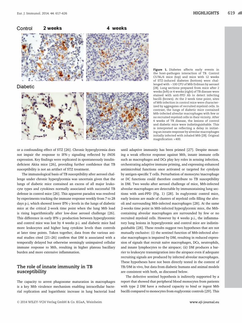
Figure 1. Diabetes affects early events inthe host–pathogen interaction of TB. ControlC57BL/6 mice (top) and mice with 12 weeksof STZ-induced diabetes (bottom) were chal-lenged with �100 CFU of Mtb Erdman by aerosol[28]. Lung sections prepared from mice after 2weeks (left) or 4 weeks (right) of TB disease werestained with anti-PPD Ab to detect infectingbacilli (brown). At the 2-week time point, sitesof Mtb infection in control mice were character-ized by aggregates of recruited myeloid cells. Incontrast, the lungs of diabetic mice containedMtb-infected alveolar macrophages with few orno recruited myeloid cells in their vicinity. After4 weeks of TB disease, the lesions of controland diabetic mice were indistinguishable. Thisis interpreted as reflecting a delay in initiat-ing an innate response by alveolar macrophagesinitially infected with inhaled Mtb [28]. Originalmagnification ×400.
or a confounding effect of STZ [26]. Chronic hyperglycemia doesnot impair the response to IFN-γ signaling reflected by iNOSexpression. Key findings were replicated in spontaneously insulin-deficient Akita mice [26], providing further confidence that TBsusceptibility is not an artifact of STZ treatment.
The immunological basis of TB susceptibility after aerosol chal-lenge under chronic hyperglycemia was uncertain given that thelungs of diabetic mice contained an excess of all major leuko-cyte types and cytokines normally associated with successful TBdefense in control mice [26]. This apparent paradox was resolvedby experiments tracking the immune response weekly from 7 to 28days p.i, which showed lower IFN-γ levels in the lungs of diabeticmice at the critical 2-week time point when the lung Mtb loadis rising logarithmically after low-dose aerosol challenge [26].This difference in early IFN-γ production between hyperglycemicand control mice was lost by 4 weeks p.i. and diabetic mice hadmore leukocytes and higher lung cytokine levels than controlsat later time points. Taken together, data from the various ani-mal studies cited [21–26] confirm that DM is associated with atemporally delayed but otherwise seemingly unimpaired cellularimmune response to Mtb, resulting in higher plateau bacillaryburden and more extensive inflammation.
The role of innate immunity in TBsusceptibility
The capacity to arrest phagosome maturation in macrophagesis a key Mtb virulence mechanism enabling intracellular bacte-rial replication and logarithmic increase in lung bacterial load
until adaptive immunity has been primed [27]. Despite mount-ing a weak effector response against Mtb, innate immune cellssuch as macrophages and DCs play key roles in sensing infection,orchestrating adaptive immune priming, and expressing enhancedantimicrobial functions once activated or targeted for cytolysisby antigen-specific T cells. Perturbation of monocyte/macrophageor DC functions could therefore contribute to TB susceptibilityin DM. Two weeks after aerosol challenge of mice, Mtb-infectedalveolar macrophages are detectable by immunostaining lung sec-tions with anti-PPD (Fig. 1) [28]. In euglycemic control mice,early lesions are made of clusters of myeloid cells filling the alve-oli and surrounding Mtb-infected macrophages [28]. At the same2-weeks time point in Mtb-infected hyperglycemic mice, the Mtb-containing alveolar macrophages are surrounded by few or norecruited myeloid cells. However by 4 weeks p.i., the inflamma-tory lung lesions in hyperglycemic and control mice are indistin-guishable [28]. These results suggest two hypotheses that are notmutually exclusive: (i) the sentinel function of Mtb-infected alve-olar macrophages is impaired by DM, resulting in reduced expres-sion of signals that recruit naıve macrophages, DCs, neutrophils,and innate lymphocytes to the airspace; (ii) DM produces a bar-rier to leukocyte transmigration into the airspace even if adequaterecruiting signals are produced by infected alveolar macrophages.These hypotheses have not been directly tested in the context ofTB/DM in vivo, but data from diabetic humans and animal modelsare consistent with both, as discussed below.
The defective sentinel hypothesis is indirectly supported by areport that showed that peripheral blood monocytes from patientswith type 2 DM have a reduced capacity to bind or ingest Mtbbacilli compared to monocytes from euglycemic controls [29]. This
C© 2014 WILEY-VCH Verlag GmbH & Co. KGaA, Weinheim www.eji-journal.eu
620 Nuria Martinez and Hardy Kornfeld Eur. J. Immunol. 2014. 44: 617–626
phenotype was associated with poor glycemic control andattributable to alterations in the complement pathway of opsoniza-tion rather than monocyte phagocytic machinery per se [29].An altered route of Mtb entry could therefore also influence thetimely activation of alveolar macrophages following inhalation ofbacilli. Alveolar macrophages from hyperglycemic rats have beenshown to produce less nitric oxide (NO) after overnight incuba-tion with Mtb than macrophages from euglycemic controls [23].That result, obtained in the absence of IFN-γ-producing lympho-cytes, implies a direct effect of the diabetic milieu on an intrinsicmacrophage response to Mtb. Data from diabetic humans with orwithout TB disease are consistent with that hypothesis. Wang et al.[30] reported that alveolar macrophages from diabetic TB patientshave a surface phenotype consistent with a reduced activationstate compared with that of macrophages from nondiabetic TBpatients or uninfected hosts. They also showed that diabetic alve-olar macrophages produce less H2O2 than cells from euglycemicsubjects in response to phorbol 12-myristate 13-acetate. However,more detailed investigation of basal and Mtb-stimulated responsesby primary alveolar macrophages is required to strengthen theargument that impaired sentinel function contributes to diabeticTB susceptibility.
There is a considerable body of literature describing adverseeffects of DM on innate immunity in contexts other thanTB. Several studies report functional changes in macrophages,including reduced phagocytic activity for SRBCs and variousmicrobes, decreased adhesion and chemotactic activity, skew-ing to an M2 phenotype, and reduced cytokine expression inresponse to diverse stimuli including lipopolysaccharide (LPS) andIFN-γ [31–35]. In contrast, other studies report enhanced mono-cyte/macrophage responses with hyperglycemia. Mo et al. [36]described increased IL-1β, CCL2/MCP-1, and TNF-α expression byalveolar macrophages from rabbits with alloxan-induced DM inresponse to particulate stimulation. Moreover, Deveraj et al. [37]reported increased basal production of IL-6 by THP-1 cells culturedin media with high (15 mmol/L) but not low (5.5 mmol/L) glu-cose content. These inconsistent results likely reflect differencesin approach including human samples versus animal models, celllines versus primary cells, acute versus chronic hyperglycemia,and the potential that DM could simultaneously depress certainmonocyte/macrophage functions while amplifying others.
Myeloid DCs convey Mtb from the site of initial macrophageinfection in the airspace to the lung-draining lymph nodes [19].Vallerskog et al. [28] showed that this process is delayed in hyper-glycemic mice but mDC migration from lung to lymph node (andcostimulatory molecule expression) in response to LPS was nodifferent in hyperglycemic mice than that in control mice. Thissuggests that DM impairs mDC trafficking to the site of infectionrather than an intrinsic effect on APC function or migration fromlung to lymph nodes. There are as yet no other published studiesof mDC function in TB/DM but the effects of DM on DCs havebeen investigated in other contexts. Musilli et al. [38] reportedincreased numbers of circulating DCs in patients with type 2 DM.Surendar et al. [39] found that circulating mDCs and plasma-cytoid DCs from diabetic subjects have increased expression of
HLA-DR and CD123, which correlates with poor glycemic control.This activation was attributed to higher plasma levels of GM-CSFin the diabetic cohort [39]. Together, these data suggest that DMis more likely to increase rather than repress mDC activation, incontrast to a repressive impact on macrophage function.
Neutrophils may play a host-protective role in TB, acceler-ating the kinetics of immune priming and Th1-cell recruitmentto the lung in Mtb-challenged mice [40, 41]. However, at latertime points in TB disease neutrophils are associated with poorlycontrolled infection and tissue injury [42]. The impact of hyper-glycemia on neutrophils in TB has not been investigated beyondthe trend for neutrophilic inflammation in mice [26] but neu-trophils have been a focus of DM research in other contexts.Key findings for neutrophils in hyperglycemic subjects includeincreased adhesion and integrin expression [43], reduced chemo-taxis [44], a phagocytic defect [45, 46], and reduced microbici-dal activity for certain bacteria and fungi as compared with neu-trophils from euglycemic controls [47–49]. Not all published stud-ies are in agreement and whether or not any of these findings relateto TB susceptibility in diabetic people remains to be seen. Onepotentially relevant observation is that glycated collagen impedesneutrophil migration compared with nonglycated collagen [50].This effect depends on the receptor for advanced glycation endproducts (RAGE), which is expressed on neutrophils and otherleukocytes [51, 52]. Lung matrix proteins have slow turnover andaccumulate glycation over time with DM [53], suggesting a mech-anism for the barrier hypothesis of delayed innate inflammationin the lungs of diabetic mice.
The role of innate lymphocytes in TB defense has not beenfully defined and might be more significant in the human hostthan in rodent models [54]. Zhang et al. [55] reported that TBpatients with DM had a higher proportion of NKT cells in periph-eral blood and bronchoalveolar lavage (BAL) fluid than nondia-betic TB patients, whereas basal peripheral blood NKT-cell countswere not different. The functional significance of increased num-bers of NKT cells, and whether it is cause or effect of diabetic TBsusceptibility, is not presently known. There are as yet no reportson NK cells in the context of TB and DM.
The role of adaptive immunity in TBsusceptibility
A timely Th1-biased adaptive immune response is the major deter-minant of outcomes in human TB and in animal models of TB[54]. With the recognition that DM increases susceptibility to TB,it was anticipated that impaired expression of adaptive immu-nity would be identified as the culprit mechanism; this hypothesiswas suggested by early data from a mouse model reporting IFN-γexpression only at 2 weeks p.i. [22]. It was therefore surpris-ing when Restrepo et al. [56] reported that PPD restimulationof whole blood in a cohort of diabetic TB patients from southernTexas and northern Mexico resulted in higher production of IFN-γ,IL-2, TNF-α, and GM-CSF than matched nondiabetic TB patients.This finding was confirmed and extended by Kumar et al. [57]
C© 2014 WILEY-VCH Verlag GmbH & Co. KGaA, Weinheim www.eji-journal.eu
Eur. J. Immunol. 2014. 44: 617–626 HIGHLIGHTS 621
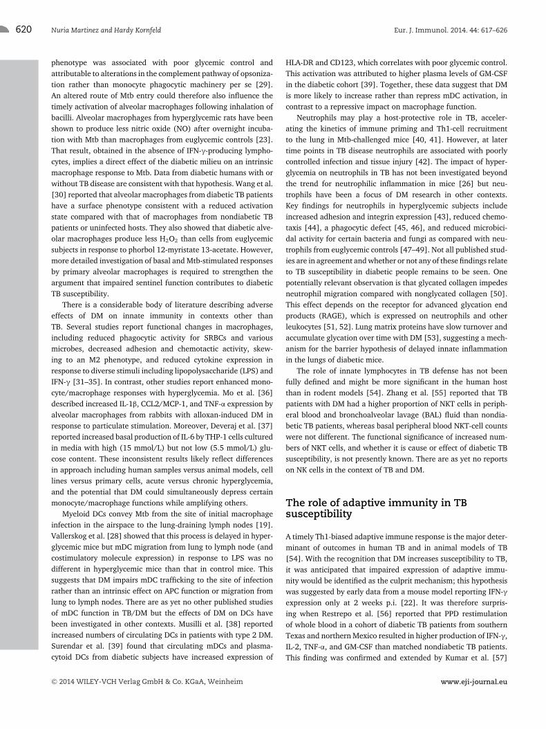
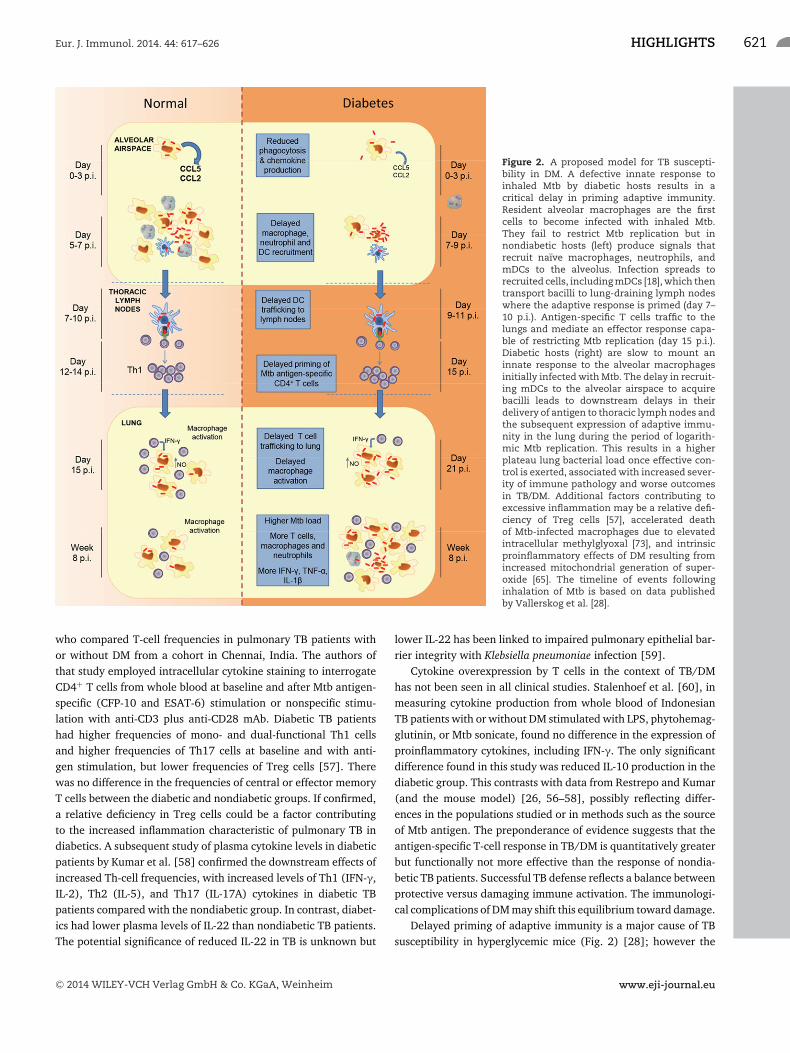
Figure 2. A proposed model for TB suscepti-bility in DM. A defective innate response toinhaled Mtb by diabetic hosts results in acritical delay in priming adaptive immunity.Resident alveolar macrophages are the firstcells to become infected with inhaled Mtb.They fail to restrict Mtb replication but innondiabetic hosts (left) produce signals thatrecruit naıve macrophages, neutrophils, andmDCs to the alveolus. Infection spreads torecruited cells, including mDCs [18], which thentransport bacilli to lung-draining lymph nodeswhere the adaptive response is primed (day 7–10 p.i.). Antigen-specific T cells traffic to thelungs and mediate an effector response capa-ble of restricting Mtb replication (day 15 p.i.).Diabetic hosts (right) are slow to mount aninnate response to the alveolar macrophagesinitially infected with Mtb. The delay in recruit-ing mDCs to the alveolar airspace to acquirebacilli leads to downstream delays in theirdelivery of antigen to thoracic lymph nodes andthe subsequent expression of adaptive immu-nity in the lung during the period of logarith-mic Mtb replication. This results in a higherplateau lung bacterial load once effective con-trol is exerted, associated with increased sever-ity of immune pathology and worse outcomesin TB/DM. Additional factors contributing toexcessive inflammation may be a relative defi-ciency of Treg cells [57], accelerated deathof Mtb-infected macrophages due to elevatedintracellular methylglyoxal [73], and intrinsicproinflammatory effects of DM resulting fromincreased mitochondrial generation of super-oxide [65]. The timeline of events followinginhalation of Mtb is based on data publishedby Vallerskog et al. [28].
who compared T-cell frequencies in pulmonary TB patients withor without DM from a cohort in Chennai, India. The authors ofthat study employed intracellular cytokine staining to interrogateCD4+ T cells from whole blood at baseline and after Mtb antigen-specific (CFP-10 and ESAT-6) stimulation or nonspecific stimu-lation with anti-CD3 plus anti-CD28 mAb. Diabetic TB patientshad higher frequencies of mono- and dual-functional Th1 cellsand higher frequencies of Th17 cells at baseline and with anti-gen stimulation, but lower frequencies of Treg cells [57]. Therewas no difference in the frequencies of central or effector memoryT cells between the diabetic and nondiabetic groups. If confirmed,a relative deficiency in Treg cells could be a factor contributingto the increased inflammation characteristic of pulmonary TB indiabetics. A subsequent study of plasma cytokine levels in diabeticpatients by Kumar et al. [58] confirmed the downstream effects ofincreased Th-cell frequencies, with increased levels of Th1 (IFN-γ,IL-2), Th2 (IL-5), and Th17 (IL-17A) cytokines in diabetic TBpatients compared with the nondiabetic group. In contrast, diabet-ics had lower plasma levels of IL-22 than nondiabetic TB patients.The potential significance of reduced IL-22 in TB is unknown but
lower IL-22 has been linked to impaired pulmonary epithelial bar-rier integrity with Klebsiella pneumoniae infection [59].
Cytokine overexpression by T cells in the context of TB/DMhas not been seen in all clinical studies. Stalenhoef et al. [60], inmeasuring cytokine production from whole blood of IndonesianTB patients with or without DM stimulated with LPS, phytohemag-glutinin, or Mtb sonicate, found no difference in the expression ofproinflammatory cytokines, including IFN-γ. The only significantdifference found in this study was reduced IL-10 production in thediabetic group. This contrasts with data from Restrepo and Kumar(and the mouse model) [26, 56–58], possibly reflecting differ-ences in the populations studied or in methods such as the sourceof Mtb antigen. The preponderance of evidence suggests that theantigen-specific T-cell response in TB/DM is quantitatively greaterbut functionally not more effective than the response of nondia-betic TB patients. Successful TB defense reflects a balance betweenprotective versus damaging immune activation. The immunologi-cal complications of DM may shift this equilibrium toward damage.
Delayed priming of adaptive immunity is a major cause of TBsusceptibility in hyperglycemic mice (Fig. 2) [28]; however the
C© 2014 WILEY-VCH Verlag GmbH & Co. KGaA, Weinheim www.eji-journal.eu
622 Nuria Martinez and Hardy Kornfeld Eur. J. Immunol. 2014. 44: 617–626
exact relationship between delayed priming and postprimary TBin adult humans is unknown. While the age of type 2 DM onsethas declined in the past decade [61], it is still likely that initialLTBI precedes DM in most cases. However, repeated exposure toMtb is common in high-burden settings where it is estimated that>70% of newly diagnosed TB cases arise from infection acquiredwithin the previous 18 months [62]. In diabetic people, progres-sion of recently acquired infection to TB disease could result fromimpaired sentinel function of alveolar macrophages regardless ofprior exposures. While this question has not yet been studieddirectly, evidence that 10–20% of LTBI patients have a peripheralblood transcriptional signature of active TB disease [63] supportsthe concept that a subgroup of people with LTBI may have clin-ically unapparent but biologically active foci of infection at highrisk for progression to clinically evident TB. It will be interestingto see if DM increases the prevalence of active TB peripheral bloodtranscriptional signatures in LTBI.
TB immune pathology
Evidence from mouse and man indicates that DM exacerbatesTB immune pathology. The pattern of histopathology in diabeticand nondiabetic mice is indistinguishable but it is quantitativelygreater in diabetic mice at 8 weeks p.i. and beyond [26]. Greaterseverity of immune pathology in human TB/DM is inferred fromthe increased mortality hazard and worse radiographic severity ofdisease. Amplified lung injury and fibrosis might result from higherbacillary load and/or result from the intrinsic proinflammatorymicroenvironment characteristic of DM [64, 65].
Nagareddy et al. [66] reported that hyperglycemia increasesneutrophil secretion of S100A6/A9, which in turn stimulatesmacrophages to secrete GM-CSF and common myeloid progen-itor cells to secrete M-CSF. These growth factors promote expan-sion of myeloid progenitors in diabetic mice, increasing numbersof circulating neutrophils and Ly6-Chi monocytes that populateatherosclerotic plaques in STZ-treated, hyperglycemic low-densitylipoprotein receptor (LDLR)−/− mice. The expansion of monocytesand neutrophils caused by hyperglycemia could be another factorcontributing to excessive inflammation in TB/DM.
Higher plateau lung bacterial load has been documented inhyperglycemic mice [22, 26] but there are presently no methodsto accurately quantify bacillary load or inflammation in humans.Higher bacillary burden is inferred from reports that TB/DMpatients are more likely to have positive sputum smear on presen-tation and delayed culture conversion with treatment [11, 17].Chest x-ray results suggest increased TB inflammation in humansbut this lacks sensitivity and is hard to quantify. Chest CT scanis superior in this regard but has not been used for most TB/DMstudies to date. The best available evidence for increased inflam-mation in human TB/DM is the increased level of proinflamma-tory cytokines in peripheral blood [58]. This association could bevalidated by future studies using more sophisticated lung imag-ing methods including CT and positron emission tomography, by
peripheral blood transcriptomics [63], or with biomarkers such asexhaled NO [67, 68].
Diabetes is not only associated with increased inflammationbut also with its persistence and impaired resolution. A compara-tive proteomic study of skin wounds in people with and withoutDM revealed a pattern of differences reflecting increased inflam-mation, impaired angiogenesis, and accelerated cell death in DM[69]. Diabetic nephropathy is one of the most common seriouscomplications of DM, culminating in irreversible fibrosis. Thisproblem has stimulated interest in antifibrotic therapies for dia-betic kidney disease [70]. If DM is shown to accelerate the damageand fibrosis that are responsible for most TB-related morbidity andmortality, diabetic individuals might be an attractive target pop-ulation to test host-directed antifibrotic or antiprotease therapieswith the goal of reducing mortality and preventing pulmonaryimpairment after TB.
Biochemical mechanisms of TBsusceptibility
It is reasonable to postulate that TB susceptibility in DM resultsfrom mechanisms related to those responsible for microvascularand macrovascular complications that are shared by type 1 andtype 2 DM. Brownlee and Giacco [4, 65] identified hyperglycemia-dependent mitochondrial overproduction of superoxide as the keyupstream event activating complication pathways that include thefollowing: increased polyol and hexomsamine flux; increased pro-tein kinase C (PKC) activation; increased formation of advancedglycation end products (AGEs); and increased expression of RAGEand its endogenous ligands. These pathways have not been exten-sively studied in hematopoietic cells but they have obvious poten-tial to disrupt immune function. Polyol pathway flux consumesNADPH, increasing oxidative stress by impeding regeneration ofthe reactive oxygen species (ROS) scavenger reduced glutathione[71]. Increased hexosamine pathway flux leads to the overpro-duction of uridine diphosphate N-acetylglucosamine, providingexcess substrate for enzymatic O-GlcNAcylation that can mod-ify the function of nuclear and cytoplasmic proteins includingtranscription factors such as NF-κB [72]. Nonenzymatic glyca-tion can similarly cause functional alterations of intracellular andextracellular proteins. Moreover, the AGE intermediate methylgly-oxal was shown to promote macrophage apoptosis following Mtbinfection [73]. Hyperglycemia-induced ROS production increasesRAGE expression and the expression of HMGB1 and S100A8/9,which are more physiologically significant as RAGE ligands thanAGE-modified proteins [74]. Finally, hyperglycemia-induced ROSinhibit GAPDH activity, thereby raising intracellular levels of triosephosphate, which is a precursor for diacylglycerol. This results inpathologically increased activation of the β and δ isoforms of PKC[75], which participate in immune regulation at many levels. Thepotential for each of these pathways to influence protective immu-nity is evident but has not been directly investigated in TB or anyother DM-related infection
C© 2014 WILEY-VCH Verlag GmbH & Co. KGaA, Weinheim www.eji-journal.eu
Eur. J. Immunol. 2014. 44: 617–626 HIGHLIGHTS 623
Epigenetic reprogramming is a more recently recognized dia-betic complication mechanism that could be a contributing factorin TB susceptibility. The term “metabolic memory” covers a rangeof phenomena in DM in which a prolonged period of tight glycemiccontrol leads to reduced vascular complications even years afterglycohemoglobin returns to a preintervention baseline [76]. Con-versely, tissue damage can progress in some diabetic people evenafter correction of hyperglycemia. In both cases, these responsesmay reflect posttranslational chromatin modification that influ-ences gene expression. Epigenetic effects of DM have not beenstudied in the context of TB but immune cells are certainly vul-nerable to this complication mechanism. Miao et al. [77] reportedthat peripheral blood T cells from type 1 DM patients have adistinct profile of chromatin histone H3 lysine 9 dimethylation.Epigenetic changes in blood monocytes from type 2 DM patientshave also been identified [78]. Diabetes is associated with ROS-mediated DNA damage [65]. One consequence of DNA damage istelomere shortening in peripheral blood monocytes, which couldaccelerate their senescence following transmigration into tissues[79].
Parsing the roles of so many candidate mechanisms in diabeticTB susceptibility will be a daunting task but selecting a treatablepathway might already be feasible. Selective blockade of individ-ual complication pathways such as polyol flux, protein glycation,and PKC activation has had modest success in animal models andhuman studies [80–82]. A more promising approach might be tar-geting the mitochondrial ROS overproduction that appears to bethe common upstream event activating all of these pathways [83].
Diabetic comorbidities
Cigarette smoking, dyslipidemia, and vitamin D deficiency actsynergistically with hyperglycemia to promote vascular and renalcomplications of DM. Smoking and vitamin D deficiency have alsobeen linked individually to human TB susceptibility [84, 85]. Themagnitude of risk imposed by either condition alone is modestbut when combined with DM could identify a subset of peoplewith LTBI having particularly high risk for progression, and/or asubset of people with TB disease at much higher risk for adverseoutcomes. The latter was shown in a cohort of Korean TB patientswhere DM or smoking independently increased 12-month mor-tality approximately twofold compared with that of nondiabeticnonsmokers while the mortality hazard was raised more than four-fold in diabetic TB patients who also smoked [86]. The mecha-nism of increased TB susceptibility in smokers is unknown, butcigarette smoke exposure was shown to inhibit the pulmonaryT-cell response to Mtb in mice [87]. The potential combined effectof DM and vitamin D deficiency on TB susceptibility has not beenstudied but the prevalence of vitamin D deficiency is increasedin type 2 diabetics and has been linked to DM pathogenesis[88].
Type 2 DM is associated with elevated total and LDL choles-terol, reduced levels of HDL cholesterol, and hypertriglyceridemia.
Hyperlipidemia has not been a focus of any clinical TB studiesperhaps since these conditions were until recently rare in highTB burden countries. Apolipoprotein E (ApoE)−/− and LDLR−/−
mice are susceptible to TB when fed high- but not low-cholesterolchow [89, 90]. In common with diabetic mice, both hypercholes-terolemic models exhibit neutrophilic pathology that is extreme inApoE−/− mice that develop massive necrotic lung lesions and Mtb-studded neutrophil extracellular traps in BAL [18, 89]. Like dia-betic mice, adaptive immune priming is delayed in Mtb-infectedApoE−/− mice but is more extreme such that the Mtb burdenreaches a lethal level and these mice die within 4–6 weeks ofaerosol challenge. The neutrophilic inflammatory response ofLDLR−/− is intermediate between diabetic and ApoE−/− mice,and LDLR−/− mice mount a timely adaptive immune responsethat controls Mtb replication. The difference between ApoE−/−
and LDLR−/− mice might reflect their different cholesterol pro-files. With equivalent elevation of total serum cholesterol, LDLcholesterol is higher in LDLR−/− mice while VLDL cholesterolis higher in ApoE−/- mice. Intercellular free cholesterol, whichis cytotoxic and could promote neutrophilic inflammation, wasnot measured in those reports. Dyslipidemia might synergize withDM to further exacerbate TB pathology in human hosts. Such amechanism, which might account for potential differences in TBsusceptibility between type 2 and type 1 diabetics, has yet to beinvestigated.
Conclusions
By increasing the risk for and severity of TB disease, DM exerts asignificant negative impact on public health, particularly in coun-tries where both conditions are prevalent. Given the complexity ofdiabetic complication mechanisms and the numerous pathwaysinvolved, it is likely that the immune response to Mtb infec-tion is affected at multiple levels. Data from mice suggest thatan impaired innate response to initial infection with a resultingdelay in the adaptive immune effector response is a key mecha-nism of susceptibility. A refined understanding of the immuno-logical and biochemical basis of TB susceptibility in DM willinform the rational development of implementation and thera-peutic strategies to mitigate the dual burden of these diseases.Such investigation can also take advantage of the perturbation inprotective immunity caused by DM to reveal critical determinantsof the host–pathogen interaction in TB relevant to the generalpopulation.
Acknowledgments: This work was supported in part by NationalInstitutes of Health grant HL081149 (to H.K.).
Conflict of interest: The authors declare no financial or commer-cial conflict of interest.
C© 2014 WILEY-VCH Verlag GmbH & Co. KGaA, Weinheim www.eji-journal.eu
624 Nuria Martinez and Hardy Kornfeld Eur. J. Immunol. 2014. 44: 617–626
References
1 Gauld, W. R. and Lyall, A., Tuberculosis as a complication of diabetes
mellitus. Br. Med. J. 1947. 1: 677–679.
2 Chen, L., Magliano, D. J. and Zimmet, P. Z. L., The worldwide epidemi-
ology of type 2 diabetes mellitus – present and future perspectives. Nat.
Rev. Endocrinol. 2011. 8: 228–236.
3 Webb, E. A., Hesseling, A. C., Schaaf, H. S., Gie, R. P., Lombard, C. J., Spi-
taels, A., Delport, S. et al., High prevalence of Mycobacterium tuberculosis
infection and disease in children and adolescents with type 1 diabetes
mellitus. Int. J. Tuberc. Lung Dis. 2009. 13: 868–874.
4 Brownlee, M., Biochemistry and molecular cell biology of diabetic com-
plications. Nature 2001. 414: 813–820.
5 Jeon, C. Y. and Murray, M. B., Diabetes mellitus increases the risk of
active tuberculosis: a systematic review of 13 observational studies. PLoS
Med. 2008. 5: e152.
6 Baker, M. A., Lin, H. H., Chang, H. Y. and Murray, M. B., The risk of
tuberculosis disease among persons with diabetes mellitus: a prospective
cohort study. Clin. Infect. Dis. 2012. 54: 818–825.
7 Leegaard, A., Riis, A., Kornum, J. B., Prahl, J. B., Thomsen, V. O., Sorensen,
H. T., Horsburgh, C. R. et al., Diabetes, glycemic control, and risk of
tuberculosis: a population-based case-control study. Diabetes Care 2011.
34: 2530–2535.
8 Chen, W., Shu, W., Wang, M., Hou, Y., Xia, Y., Xu, W., Bai, L. et al.,
Pulmonary tuberculosis incidence and risk factors in rural areas of China:
a cohort study. PLoS One 2013. 8: e58171.
9 Goldhaber-Fiebert, J. D., Jeon, C. Y., Cohen, T. and Murray, M. B., Dia-
betes mellitus and tuberculosis in countries with high tuberculosis bur-
dens: individual risks and social determinants. Int. J. Epidemiol. 2011. 40:
417–428.
10 Kapur, A. and Harries, A. D., The double burden of diabetes and
tuberculosis – public health implications. Diabetes Res. Clin. Pract.
2013. 110: 10–19.
11 Viswanathan, V., Kumpatla, S., Aravindalochanan, V., Rajan, R., Chin-
nasamy, C., Srinivasan, R., Selvam, J. M. et al., Prevalence of diabetes and
pre-diabetes and associated risk factors among tuberculosis patients in
India. PLoS One 2012. 7: e41367.
12 Hunter, R. L., Pathology of post primary tuberculosis of the lung: an
illustrated critical review. Tuberculosis 2011. 91: 497–509.
13 Gupta, S., Shenoy, V. P., Bairy, I., Srinivasa, H. and Mukhopadhyay, C.,
Diabetes mellitus and HIV as co-morbidities in tuberculosis patients of
rural south India. J. Infect. Public Health 2011. 4: 140–144.
14 Gray, J. M. and Coh, D. L., Tuberculosis and HIV coinfection. Semin. Respir.
Crit. Care Med. 2013. 34: 32–43.
15 Keane, J., Gershon, S., Wise, R. P., Mirabile-Levens, E., Kaszinca, J.,
Schwieterman, W. D., Siegel, J. N. et al., Tuberculosis associated with
infliximab, a tuberculosis factor α-neutralizing agent. N. Engl. J. Med. 2001.
345: 1098–1104.
16 Baker, M. A., Harries, A. D., Jeon, C. Y., Hart, J. E., Kapur, A., Lonnroth,
K., Ottmani, S. E. et al., The impact of diabetes on tuberculosis treatment
outcomes: a systematic review. BMC Med. 2011. 9: 81.
17 Jimenez-Corona, M. E., Cruz-Hervert, L. P., Garcia-Garcia, L., Ferreyra-
Reyes, L., Delgado-Sanchez, G., Bobadilla-Del-Valle, M., Canizales-
Quintero, S. et al., Association of diabetes and tuberculosis:
impact on treatment and post-treatment outcomes. Thorax 2013. 68:
214–220.
18 Repasy, T., Lee, J., Marino, S., Martinez, N., Kirschner, D. E., Hen-
dricks, G., Baker, S. et al., Intracellular bacillary burden reflects a
burst size for Mycobacterium tuberculosis in vivo. PLoS Pathog. 2013. 9:
e1003190.
19 Wolf, A. J., Linas, B., Trevejo-Nunez, G. J., Kincaid, E., Tamura, T.,
Takatsu, K. and Ernst, J. D., Mycobacterium tuberculosis infects den-
dritic cells with high frequency and impairs their function in vivo. J.
Immunol. 2007. 179: 2509–2519.
20 Horsburgh, R. C., Jr., Priorities for the treatment of latent tuberculosis
infection in the United States. N. Engl. J. Med. 2004. 350: 2060–2070.
21 Saiki, O., Negoro, S., Tsuyuguchi, I. and Yamamura, Y., Depressed
immunological defence mechanisms in mice with experimentally
induced diabetes. Infect. Immun. 1980. 28: 127–131.
22 Yamashiro, S., Kawakami, K., Uezu, K., Kinjo, T., Miyagi, K., Nakamura,
K. and Saito, A., Lower expression of Th1-related cytokines and inducible
nitric oxide synthase in mice with streptozotocin-induced diabetes mel-
litus infected with Mycobacterium tuberculosis. Clin. Exp. Immunol. 2005.
139: 57–64.
23 Sugawara, I. and Mizuno, S., Higher susceptibility of type 1 diabetic
rats to Mycobacterium tuberculosis infection. Tohoku J. Exp. Med. 2008. 216:
363–370.
24 Sugawara, I., Yamada, H. and Mizuno, S., Pulmonary tuberculosis in
spontaneously diabetic goto kakizaki rats. Tohoku J. Exp. Med. 2004. 204:
135–145.
25 Podell, B. K., Ackart, D. F., Kirk, N. M., Eck, S. P., Bell, C. and Basaraba, R.
J., Non-diabetic hyperglycemia exacerbates disease severity in Mycobac-
terium tuberculosis infected guinea pigs. PLoS One 2012. 7: e46824.
26 Martens, G. W., Arikan, M. C., Lee, J., Ren, F., Greiner, D., and Kornfeld,
H., Tuberculosis susceptibility of diabetic mice. Am. J. Respir. Cell Mol. Biol.
2007. 37: 518–524.
27 Armstrong, J. A. and Hart, P. D., Response of cultured macrophages to
Mycobacterium tuberculosis, with observations on fusion of lysosomes with
phagosomes. J. Exp. Med. 1971. 134: 713–740.
28 Vallerskog, T., Martens, G. W. and Kornfeld, H., Diabetic mice display
a delayed adaptive immune response to Mycobacterium tuberculosis. J.
Immunol. 2010. 184: 6275–6282.
29 Gomez, D. I., Twahirwa, M., Schlesinger, L. S. and Restrepo, B. I., Reduced
Mycobacterium tuberculosis association with monocytes from diabetes
patients that have poor glucose control. Tuberculosis. (Edinb.) 2013. 93:
192–197.
30 Wang, C. H., Yu, C. T., Lin, H. C., Liu, C. Y. and Kuo, H. P., Hypodense
alveolar macrophages in patients with diabetes mellitus and active pul-
monary tuberculosis. Tuber. Lung Dis. 1999. 79: 235–242.
31 Hand, W. L., Hand, D. L. and Vasquez, Y., Increased polymorphonuclear
leukocyte respiratory burst function in type 2 diabetes. Diabetes Res. Clin.
Pract. 2007. 76: 44–50.
32 Ferracini, M., Martins, J. O., Campos, M. R., Anger, D. B. and Jancar,
S., Impaired phagocytosis by alveolar macrophages from diabetic rats
is related to the deficient coupling of LTs to the Fc gamma R signaling
cascade. Mol. Immunol. 2010. 47: 1974–1980.
33 Lecube, A., Pachon, G., Petriz, J., Hernandez, C. and Simo, R., Phago-
cytic activity is impaired in type 2 diabetes mellitus and increases after
metabolic improvement. PLoS. One 2011. 6: e23366.
34 Liu, H. F., Zhang, H. J., Hu, Q. X., Liu, X. Y., Wang, Z. Q., Fan, J. Y., Zhan, M.
et al., Altered polarization, morphology, and impaired innate immunity
germane to resident peritoneal macrophages in mice with long-term type
2 diabetes. J. Biomed. Biotechnol. 2012. 2012: 867023.
35 Sun, C., Sun, L., Ma, H., Peng, J., Zhen, Y., Duan, K. Liu, G. et al., The phe-
notype and functional alterations of macrophages in mice with hyper-
glycemia for long term. J. Cell Physiol 2012. 227: 1670–1679.
C© 2014 WILEY-VCH Verlag GmbH & Co. KGaA, Weinheim www.eji-journal.eu
Eur. J. Immunol. 2014. 44: 617–626 HIGHLIGHTS 625
36 Mo, Y., Wan, R., Wang, J., Chien, S., Tollerud, D. J. and Zhang, Q., Diabetes
is associated with increased sensitivity of alveolar macrophages to urban
particulate matter exposure. Toxicology 2009. 262: 130–137.
37 Devaraj, S., Venugopal, S. K., Singh, U. and Jialal, I., Hyperglycemia
induces monocytic release of interleukin-6 via induction of protein
kinase c-{alpha} and -{beta}. Diabetes 2005. 54: 85–91.
38 Musilli, C., Paccosi, S., Pala, L., Gerlini, G., Ledda, F., Mugelli, A.,
Rotella, C. M. et al., Characterization of circulating and monocyte-derived
dendritic cells in obese and diabetic patients. Mol. Immunol. 2011. 49:
234–238.
39 Surendar, J., Mohan, V., Pavankumar, N., Babu, S. and Aravindhan, V.,
Increased levels of serum granulocyte-macrophage colony-stimulating
factor is associated with activated peripheral dendritic cells in type 2
diabetes subjects (CURES-99). Diabetes Technol. Ther. 2012. 14: 344–349.
40 Blomgran, R. and Ernst, J. D., Lung neutrophils facilitate activation of
naive antigen-specific CD4+ T cells during Mycobacterium tuberculosis
infection. J. Immunol. 2011. 186: 7110–7119.
41 Kang, D. D., Lin, Y., Moreno, J. R., Randall, T. D. and Khader, S. A., Pro-
filing early lung immune responses in the mouse model of tuberculosis.
PLoS. One 2011. 6: e16161.
42 Mattila, J. T., Ojo, O. O., Kepka-Lenhart, D., Marino, S., Kim, J. H., Eum,
S. Y., Via, L. E. et al., Microenvironments in tuberculous granulomas are
delineated by distinct populations of macrophage subsets and expres-
sion of nitric oxide synthase and arginase isoforms. J. Immunol. 2013.
191: 773–784.
43 Ritter, L., Davidson, L., Henry, M., Davis-Gorman, G., Morrison, H., Frye,
J. B., Cohen, Z. et al., Exaggerated neutrophil-mediated reperfusion injury
after ischemic stroke in a rodent model of type 2 diabetes. Microcirculation
2011. 18: 552–561.
44 Tater, D., Tepaut, B., Bercovici, J. P. and Youinou, P., Polymorphonu-
clear cell derangements in type I diabetes. Horm. Metab. Res. 1987. 19:
642–647.
45 Marhoffer, W., Stein, M., Schleinkofer, L. and Federlin, K., Evidence of
ex vivo and in vitro impaired neutrophil oxidative burst and phagocytic
capacity in type 1 diabetes mellitus. Diabetes Res. Clin. Pract. 1993. 19:
183–188.
46 Chanchamroen, S., Kewcharoenwong, C., Susaengrat, W., Ato, M.
and Lertmemongkolchai, G., Human polymorphonuclear neutrophil
responses to Burkholderia pseudomallei in healthy and diabetic subjects.
Infect. Immun. 2009. 77: 456–463.
47 Wykretowicz, A., Wierusz-Wysocka, B., Wysocki, J., Szczepanik, A. and
Wysocki, H., Impairment of the oxygen-dependent microbicidal mecha-
nisms of polymorphonuclear neutrophils in patients with type 2 diabetes
is not associated with increased susceptibility to infection. Diabetes Res.
Clin. Pract. 1993. 19: 195–201.
48 Boland, O. M., Blackwell, C. C., Clarke, B. F., and Ewing, D. J., Effects
of ponalrestat, an aldose reductase inhibitor, on neutrophil killing of
Escherichia coli and autonomic function in patients with diabetes melli-
tus. Diabetes 1993. 42: 336–340.
49 Ueta, E., Osaki, T., Yoneda, K. and Yamamoto, T., Prevalence of diabetes
mellitus in odontogenic infections and oral candidiasis: an analysis of
neutrophil suppression. J. Oral Pathol. Med. 1993. 22: 168–174.
50 Toure, F., Zahm, J. M., Garnotel, R., Lambert, E., Bonnet, N., Schmidt, A.
M., Vitry, F. et al., Receptor for advanced glycation end-products (RAGE)
modulates neutrophil adhesion and migration on glycoxidated extracel-
lular matrix. Biochem. J. 2008. 416: 255–261.
51 Collison, K. S., Parhar, R. S., Saleh, S. S., Meyer, B. F., Kwaasi, A. A.,
Hammami, M. M., Schmidt, A. M. et al., RAGE-mediated neutrophil dys-
function is evokded by advanced glycation end products (AGEs). J. Leukoc.
Biol. 2002. 71: 433–444,
52 Kierdorf, K. and Fritz, G., RAGE regulation and signaling in inflammation
and beyond. J. Leukoc. Biol. 2013: 94: 55–68
53 Myint, T., Hoshi, S., Ookawara, T., Miyazawa, N., Suzuki, K. and
Taniguchi, N., Immunological detection of glycated proteins in normal
and streptozotocin-induced diabetic rats using anti hexitol-lysine IgG.
Biochim. Biophys. Acta 1995. 1272: 73–79.
54 Cooper, A. M., Cell-mediated immune responses in tuberculosis. Annu.
Rev. Immunol. 2009. 27: 393–422.
55 Zhang, Q., Xiao, H. P., Cui, H. Y. and Sugawara, I., Significant increase in
natural-killer T cells in patients with tuberculosis complicated by type 2
diabetes mellitus. J. Int. Med. Res. 2011. 39: 105–111.
56 Restrepo, B. I., Fisher-Hoch, S. P., Pino, P. A., Salinas, A., Rahbar, M. H.,
Mora, F., Cortes-Penfield, N. et al., Tuberculosis in poorly controlled type
2 diabetes: altered cytokine expression in peripheral white blood cells.
Clin. Infect. Dis. 2008. 47: 634–641.
57 Kumar, N. P., Sridhar, R., Banurekha, V. V., Jawahar, M. S., Nutman,
T. B. and Babu, S., Expansion of pathogen-specific Th1 and Th17 cells in
pulmonary tuberculosis with coincident type 2 diabetes mellitus. J. Infect.
Dis. 2013. 208: 739–748.
58 Kumar, N. P., Sridhar, R., Banurekha, V. V., Jawahar, M. S., Fay, M. P.,
Nutman, T. B. and Babu, S., Type 2 diabetes mellitus coincident with
pulmonary tuberculosis is associated with heightened systemic type 1,
type 17 and other proinflammatory cytokines. Ann. Am. Thorac. Soc. 2013.
10: 441–449.
59 Aujla, S. J., Chan, Y. R., Zheng, M., Fei, M., Askew, D. J., Pociask, D.
A., Reinhart, T. A. et al., IL-22 mediates mucosal host defense against
Gram-negative bacterial pneumonia. Nat. Med. 2008. 14: 275–281.
60 Stalenhoef, J. E., Alisjahbana, B., Nelwan, E. J., van der Ven-Jongekrijg,
J., Ottenhoff, T. H. M., van der Meer, J. W., Nelwan, R. H. et al., The role
of interferon-gamma in the increased tuberculosis risk in type 2 diabetes
mellitus. Eur. J. Clin. Microbiol. Infect. Dis. 2008. 27: 97–103.
61 Mohan, V., Jaydip, R. and Deepa, R., Type 2 diabetes in Asian Indian
youth. Pediatr. Diabetes 2007. 8(Suppl 9): 28–34.
62 Dye, C., Glaziou, P., Floyd, K. and Raviglione, M., Prospects for tubercu-
losis elimination. Annu. Rev. Public Health 2013. 34: 271–286.
63 Berry, M. P., Graham, C. M., McNab, F. W., Xu, Z., Bloch, S. A., Oni,
T., Wilkinson, K. A. et al., An interferon-inducible neutrophil-driven
blood transcriptional signature in human tuberculosis. Nature 2010. 466:
973–977.
64 Nguyen, D. V., Shaw, L. C. and Grant, M. B., Inflammation in the patho-
genesis of microvascular complications in diabetes. Front Endocrinol. (Lau-
sanne) 2012. 3: 170.
65 Giacco, F. and Brownlee, M., Oxidative stress and diabetic complications.
Circ. Res. 2010. 107: 1058–1070.
66 Nagareddy, P. R., Murphy, A. J., Stirzaker, R. A., Hu, Y., Yu, S., Miller,
R. G., Ramkhelawon, B. et al., Hyperglycemia promotes myelopoiesis
and impairs the resolution of atherosclerosis. Cell Metab. 2013. 17:
695–708.
67 Birrell, M. A., McCluskie, K., Hardaker, E., Knowles, R. and Belvisi, M.
G., Utility of exhaled nitric oxide as a noninvasive biomarker of lung
inflammation in a disease model. Eur. Respir. J. 2006. 28: 1236–1244.
68 van Beek, S. C., Nhung, N. V., Sy, D. N., Sterk, P. J., Tiemersma, E. W.
and Cobelens, F. G., Measurement of exhaled nitric oxide as a potential
screening tool for pulmonary tuberculosis. Int. J. Tuberc. Lung Dis. 2011.
15: 185–192.
69 Krisp, C., Jacobsen, F., McKay, M. J., Molloy, M. P., Steinstraesser, L. and
Wolters, D. A., Proteome analysis reveals anti-angiogenic environments
in chronic wounds of diabetes mellitus type 2 patients. Proteomics. 2013.
13: 2670–2681.
C© 2014 WILEY-VCH Verlag GmbH & Co. KGaA, Weinheim www.eji-journal.eu
626 Nuria Martinez and Hardy Kornfeld Eur. J. Immunol. 2014. 44: 617–626
70 Karihaloo, A., Anti-fibrosis therapy and diabetic nephropathy. Curr. Diab.
Rep. 2012. 12: 414–422.
71 Lee, A. Y. and Chung, S. S., Contributions of polyol pathway to oxidative
stress in diabetic cataract. FASEB J. 1999. 13: 23–30.
72 Xing, D., Gong, K., Feng, W., Nozell, S. E., Chen, Y. F., Chatham, J. C. and
Oparil, S., O-GlcNAc modification of NFkappaB p65 inhibits TNF-alpha-
induced inflammatory mediator expression in rat aortic smooth muscle
cells. PLoS. One 2011. 6: e24021.
73 Rachman, H., Kim, N., Ulrichs, T., Baumann, S., Pradl, L., Nasser, E. A.,
Bild, M. et al., Critical role of methylglyoxal and AGE in mycobacteria-
induced macrophage apoptosis and activation. PLoS. One 2006. 1: e29.
74 Yao, D. and Brownlee, M., Hyperglycemia-induced reactive oxygen
species increase expression of the receptor for advanced glycation end
products (RAGE) and RAGE ligands. Diabetes 2010. 59: 249–255.
75 Koya, D. and King, G. L., Protein kinase C activation and the development
of diabetic complications. Diabetes 1998. 47: 859–866.
76 Intine, R. V. and Sarras, M. P., Jr., Metabolic memory and chronic dia-
betes complications: potential role for epigenetic mechansisms. Curr.
Diab. Rep. 2012. 12: 551–559.
77 Miao, F., Smith, D. D., Zhang, L., Min, A., Feng, W. and Natarajan, R.,
Lymphocytes from patients with type 1 diabetes display a distinct pro-
file of chromatin histone H3 lysine 9 dimethylation. Diabetes 2008. 57:
3189–3198.
78 Miao, F., Wu, X., Zhang, L., Yuan, Y. C., Riggs, A. D. and Natarajan, R.,
Genome-wide analysis of histone lysine methylation variations caused
by diabetic conditions in human monocytes. J. Biol. Chem. 2007. 282:
13854–13863.
79 Sampson, M. J., Winterbone, M. S., Hughes, J. C., Dozio, N. and Hughes,
D. A., Monocyte telomere shortening and oxidative DNA damage in type
2 diabetes. Diabetes Care 2006. 29: 283–289.
80 Sorbinil Retinopathy Trial Research Group, A randomized trial of sorbinil,
an aldose reductase inhibitor, in diabetic retinopathy. Arch. Opthalmol.
1990. 108: 1234–1244.
81 Campochirao, P. A., Reduction of diabetic macular edema by oral admin-
istration of the kinase inhibitor PKC412. Invest. Opthalmol. Vis. Sci. 2004.
45: 922–931.
82 Cameron, N. E., Gibson, T. M., Nangle, T. R. and Cotter, M. A., Inhibitors of
advance glycation end product formation and neurovascular dysfunction
in experimental diabetes. Ann. NY Acad. Sci. 2005. 1043: 784–792.
83 Folli, F., Corradi, D., Fanti, P., Davalli, A., Paez, A., Giaccari, A., Perego, C.
et al., The role of oxidative stress in the pathogenesis of type 2 dia-
betes mellitus micro- and macrovascular complications: avenues for
a mechanistic-based therapeutic approach. Curr. Diabetes Rev. 2011. 7:
313–324.
84 Jee, S. H., Golub, J. E., Jo, J., Park, I. S., Ohrr, H. and Samet, J. M., Smoking
and risk of tuberculosis incidence, mortality, and recurrence in South
Korean men and women. Am. J. Epidemiol. 2009. 170: 1478–1485.
85 Talat, N., Perry, S., Parsonnet, J., Dawood, G. and Hussain, R., Vitamin
D deficiency and tuberculosis progression. Emerg. Infect. Dis. 2010. 16:
853–855.
86 Reed, G. W., Choi, H., Lee, S. Y., Lee, M., Kim, Y., Park, H., Lee, J. et al.,
Impact of diabetes and smoking on mortality in tuberculosis. PLoS. One
2013. 8: e58044.
87 Feng, Y., Kong, Y., Barnes, P. F., Huang, F. F., Klucar, P., Wang, X.,
Samten, B. et al., Exposure to cigarette smoke inhibits the pulmonary
T-cell response to influenza virus and Mycobacterium tuberculosis. Infect.
Immun. 2011. 79: 229–237.
88 Lim, S., Kim, M. J., Choi, S. H., Shin, C. S., Park, K. S., Jang, H. C., Billings,
L. K. et al., Association of vitamin D deficiency with incidence of type 2
diabetes in high-risk Asian subjects. Am. J. Clin. Nutr. 2013. 97: 524–530.
89 Martens, G. W., Arikan, M. C., Lee, J., Ren, F., Vallerskog, T., and Korn-
feld, H., Hypercholesterolemia impairs immunity to tuberculosis. Infect.
Immun. 2008. 76: 3464–3472.
90 Martens, G. W., Vallerskog, T., and Kornfeld, H., Hypercholesterolemic
LDL receptor-deficient mice mount a neutrophilic response to tubercu-
losis despite the timely expression of protective immunity. J. Leukoc. Biol.
2012. 91: 849–857.
Abbreviations: AGE: advanced glycation end product · ApoE: apolipopro-
tein E · DM: diabetes mellitus · LDLR: low-density lipoprotein receptor
· LTBI: latent TB infection · Mtb: Mycobacterium tuberculosis · p.i.: post
infection · PKC: protein kinase C · RAGE: receptor for advanced glyca-
tion end-products · STZ: streptozotocin · TB: tuberculosis
Full correspondence: Dr. Hardy Kornfeld, Department of Medicine,University of Massachusetts Medical School, LRB-303, 55 Lake AvenueNorth, Worcester, MA 01655, USAFax: +1-508-856-7883e-mail: [email protected]
Received: 17/11/2013Revised: 8/1/2014Accepted: 15/1/2014Accepted article online: 22/1/2014
C© 2014 WILEY-VCH Verlag GmbH & Co. KGaA, Weinheim www.eji-journal.eu


